Bilastine_202189-78-4_DataSheet_MedChemExpress
碧云天细胞自噬染色检测试剂盒(MDC法)说明书

细胞自噬染色检测试剂盒(MDC 法)产品简介:碧云天生产的细胞自噬染色检测试剂盒(MDC 法),即Autophagy Staining Assay Kit with MDC ,是一种使用丹酰尸胺,也称单丹磺酰尸胺、丹酰尸胺或丹酰戊二胺(monodansylcadaverine, MDC)作为荧光探针快速便捷地检测细胞自噬的试剂盒。
自噬(autophagy)是一种在进化上高度保守的通过溶酶体吞噬并降解部分自身组分的细胞内分解代谢途径。
自噬与多种生理功能有关,在饥饿等环境条件下,细胞通过自噬降解多余或异常的细胞内组分,为细胞的生存提供能量及原材料,促进生物体的生长发育、细胞分化及对环境变化产生应答。
自噬异常与多种病理过程如肿瘤、神经退行性疾病、代谢疾病、病原体感染等都有密切关系。
由于细胞自噬在生理和病理过程中都有重要作用,自噬已经成为细胞生物学领域的一个研究热点。
MDC 是细胞自噬检测最常用的荧光探针之一。
MDC 可以通过离子捕获(ion trapping)和与膜脂的特异性结合,从而特异性标记自噬体(autophagosome),也称autophagic vacuole ,因而常用于细胞自噬的检测。
MDC 是一种嗜酸性荧光探针,很多酸性膜性结构也会被MDC 染色,因此MDC 染色时正常的细胞也会有一定的染色背景。
本产品的染色原理决定了本产品只能用于培养的细胞或者组织的细胞自噬荧光染色检测,不能用于冻存的或固定的细胞、组织或者组织切片的染色检测。
使用本产品染色后可以通过荧光显微镜拍照观察,也可以通过荧光酶标仪或流式细胞仪进行荧光检测。
荧光显微镜观察时可以使用紫外区激发光激发,发出绿色荧光。
荧光酶标仪或流式细胞仪推荐的激发波长为335nm (330-360nm 均可),发射波长为512nm (510-540nm 均可)。
本产品用于细胞自噬染色的效果参考图1。
图1. 细胞自噬染色检测试剂盒(MDC 法)的染色效果图。
Trigonox B(滴苷但羊水)产品数据表单说明书

Product Data SheetTrigonox BDi-tert-butyl peroxideTrigonox® B is a pure peroxide in liquid form.CAS number110-05-4EINECS/ELINCS No. 203-733-6TSCA statuslisted on inventory Molecular weight 146.2Active oxygen contentperoxide10.94%SpecificationsAppearance Clear liquidAssay≥ 99.0 %ApplicationsTrigonox® B (Di-tert-butyl peroxide) can be used for the market segments: polymer production, polymer crosslinking and acrylics production with their different applications/functions. For more information please check our website and/or contact us.Half-life dataThe reactivity of an organic peroxide is usually given by its half-life (t½) at various temperatures. For Trigonox® B in chlorobenzene half-life at other temperatures can be calculated by using the equations and constants mentioned below:0.1 hr at 164°C (327°F)1 hr at 141°C (286°F)10 hr at 121°C (250°F)Formula 1kd = A·e-Ea/RTFormula 2t½ = (ln2)/kdEa153.46 kJ/moleA 4.20E+15 s-1R8.3142 J/mole·KT(273.15+°C) KThermal stabilityOrganic peroxides are thermally unstable substances which may undergo self-accelerating decomposition. The lowest temperature at which self-accelerating decomposition may occur with a substance in the packaging as used for transport is the Self-Accelerating Decomposition Temperature (SADT). The SADT is determined on the basis of the Heat Accumulation Storage Test.SADT80°C (176°F)Method The Heat Accumulation Storage Test is a recognized test method for thedetermination of the SADT of organic peroxides (see Recommendations on theTransport of Dangerous Goods, Manual of Tests and Criteria - United Nations, NewYork and Geneva).StorageDue to the relatively unstable nature of organic peroxides, a loss of quality will occur over a period of time. To minimize the loss of quality, Nouryon recommends a maximum storage temperature (Ts max. ) for each organic peroxide product.Ts Max.40°C (104°F) andTs Min.-30°C (-22°F) to prevent crystallizationNote When stored according to these recommended storage conditions, Trigonox® Bwill remain within the Nouryon specifications for a period of at least 6 months afterdelivery.Packaging and transportIn North America Trigonox® B is packed in non-returnable, five gallon polyethylene containers of 30 lb net weight and steel drums of 100 or 340 lb net weight. In other regions the standard packaging is a 30-liter HDPE can (Nourytainer®) for 20 kg peroxide. Delivery in a 200 l steel drum for 150 kg peroxide is also possible in a number of countries. Both packaging and transport meet the international regulations. For the availability of other packed quantities consult your Nouryon representative. Trigonox® B is classified as Organic peroxide type E; liquid, Division 5. 2; UN 3107.Safety and handlingKeep containers tightly closed. Store and handle Trigonox® B in a dry well-ventilated place away from sources of heat or ignition and direct sunlight. Never weigh out in the storage room. Avoid contact with reducing agents (e. g. amines), acids, alkalis and heavy metal compounds (e. g. accelerators, driers and metal soaps). Please refer to the Safety Data Sheet (SDS) for detailed information on the safe storage, use and handling of Trigonox® B. This information should be thoroughly reviewed prior to acceptance of this product. The SDS is available at /sds-search.Major decomposition productsAcetone, Methane, tert-ButanolAll information concerning this product and/or suggestions for handling and use contained herein are offered in good faith and are believed to be reliable.Nouryon, however, makes no warranty as to accuracy and/or sufficiency of such information and/or suggestions, as to the product's merchantability or fitness for any particular purpose, or that any suggested use will not infringe any patent. Nouryon does not accept any liability whatsoever arising out of the use of or reliance on this information, or out of the use or the performance of the product. Nothing contained herein shall be construed as granting or extending any license under any patent. Customer must determine for himself, by preliminary tests or otherwise, the suitability of this product for his purposes.The information contained herein supersedes all previously issued information on the subject matter covered. The customer may forward, distribute, and/or photocopy this document only if unaltered and complete, including all of its headers and footers, and should refrain from any unauthorized use. Don’t copythis document to a website.Trigonox® and Nourytainer are registered trademarks of Nouryon Functional Chemicals B.V. or affiliates in one or more territories.Contact UsPolymer Specialties Americas************************Polymer Specialties Europe, Middle East, India and Africa*************************Polymer Specialties Asia Pacific************************2022-6-30© 2022Polymer crosslinking Trigonox B。
考马斯亮蓝蛋白快速染色液
贮存:
室温密封保存
使用说明 1.洗涤 电泳结束后,将凝胶剥离玻璃板, 放入烧杯或塑料盒中,加入 100ml 的蒸馏水。 煮沸或微波炉中加热至沸腾,维持 1 分钟, 弃水。 2.染色 倒入适量染色液,煮沸或微波炉中 加热至沸腾,维持 2 分钟;如果胶非常厚, 染色的时间需适当延长。
注:在微波炉中适当加热,可以大大加快染色速度。 加热时,敞开容器口,容器尽量大些 ,以免出现因
考马斯亮蓝蛋白胶快速染色液
考马斯亮蓝蛋白胶快速染色液 Commassie Blue Fast Staining Solution
包装:
Cat.No
产品名称
规格
WB021 考马斯亮蓝蛋白胶快 100ml
WB022
速染色液
50ቤተ መጻሕፍቲ ባይዱml
产品简介: ※考马斯亮蓝快速染色液(Commassie Blue Fast Staining Solution)是以考马斯亮蓝R-250 为染料配 制而成的染色液,可用于SDS-PAGE或非变性PAGE 蛋白电泳胶的快速染色,或Western转膜后PAGE胶 上残余蛋白的检测。 ※考马斯亮蓝快速染色液采取新配方,超薄胶只需 染色 10 分钟,就可以显示出清晰的蛋白电泳条带; 检测到低达 10ng的蛋白电泳条带。 ※无需对蛋白和凝胶进行固定,操作更加简单。 ※本考马斯亮蓝快速染色液是一种无毒,无刺激性 气味的高度环保型染色液。普通的常规方法需使用 剧毒的甲醇及强刺激性的乙酸,而本公司生产的考 马斯亮蓝快速染色液则采取全新配方,实现了无毒 和无刺激性气味。 ※本染色液和质谱分析兼容,即经过本染色液染色 的蛋白条带或蛋白点可以用于后续的质谱分析。
Bilastine_202189-78-4_DataSheet_MedChemExpress

Caution: Not fully tested. For research purposes only Medchemexpress LLC
m o c . s s e r p x e m e h c d e m . w w w : b e AW Sm Uo ,c 0 4. 5s 8s 0e r p Jx Ne ,m n e oh t c e cd ne i rm P @ ,o y f an Wi : l ni oa sm n iE k l i W 8 1
References: [1]. Corcostegui, R., et al., Preclinical pharmacology of bilastine, a new selective histamine H1 receptor antagonist: receptor selectivity and in vitro antihistaminic activity. Drugs R D, 2005. 6(6): p. 371-84. [2]. Jauregizar, N., et al., Pharmacokinetic-pharmacodynamic modelling of the antihistaminic (H1) effect of bilastine. Clin Pharmacokinet, 2009. 48(8): p. 543-54.
Product Data Sheet
Product Name: CAS No.: Cat. No.: MWt: Formula: Purity :
Bilastine 202189-78-4 HY-14447 463.61 C28H37N3O3 >98%
y Solubility:
β-半乳糖苷酶(β-GAL)活性检测试剂盒说明书

β-半乳糖苷酶(β-GAL)活性检测试剂盒说明书微量法注意:本产品试剂有所变动,请注意并严格按照该说明书操作。
货号:BC2585规格:100T/48S产品组成:使用前请认真核对试剂体积与瓶内体积是否一致,有疑问请及时联系索莱宝工作人员。
试剂名称规格保存条件提取液液体100 mL×1瓶2-8℃保存试剂一粉剂×2支-20℃保存试剂二液体4 mL×1瓶2-8℃保存试剂三液体15 mL×1瓶2-8℃保存标准品液体1 mL×1支2-8℃保存溶液的配制:1、试剂一:临用取1支加入1.25mL蒸馏水,充分溶解备用;用不完的试剂-20℃分装保存4周,避免反复冻融(1瓶粉剂溶解后可做100T,为了延长使用时间,此产品多给1瓶粉剂)。
2、标准品:5μmol/mL的对硝基苯酚溶液。
产品说明:β-GAL(EC 3.2.1.23)广泛存在于动物、植物、微生物和培养细胞中,能够催化β-半乳糖苷化合物中β-半乳糖苷键水解,此外还具有转半乳糖苷的作用。
β-GAL不仅可为植物的快速生长释放储存的能量,还能在正常的多糖代谢、细胞壁组分代谢以及衰老时细胞壁降解过程中催化多糖、糖蛋白以及半乳糖脂末端半乳糖残基的水解,释放自由的半乳糖。
β-GAL分解对-硝基苯-β-D-吡喃半乳糖苷生成对-硝基苯酚,后者在400nm有最大吸收峰,通过测定吸光值升高速率来计算β-GAL活性。
p-Nitrophenol(400nm)p-Nitrophenyl β-D-Galactopyranoside需自备的仪器和用品:可见分光光度计/酶标仪、低温离心机、水浴锅/恒温培养箱、超声破碎仪、可调式移液器、微量玻璃比色皿/96孔板、研钵/匀浆器、冰和蒸馏水。
操作步骤:一、样本处理(可适当调整待测样本量,具体比例可以参考文献)1、细菌或培养细胞的处理:收集细菌或细胞到离心管内,离心后弃上清;按照每500万细菌或细胞加入1mL提取液,超声波破碎细菌或细胞(冰浴,功率200W,超声3秒,间隔10秒,重复30次),15000g,4℃,离心10min,取上清,置冰上待测。
安捷伦产品目录

15
Real-Time PCR
16
Mx3000P QPCR System
17
Brilliant III Ultra-Fast SYBR Green QPCR and QRT-PCR Reagents
18
Brilliant III Ultra-Fast QPCR and QRT-PCR Reagents
Agilent / STRATAGENE
Agilent website: /genomics
Welgene | Agilent Stratagene
威健股份有限公司 | Stratagene 總代理
Table of Content
Table of Contents
/ XL1-Red Competent Cells SoloPack Gold Supercompetent Cells
/ TK Competent Cells Specialty Cells
/ Classic Cells / Fine Chemicals For Competent Cells
適用於 UNG 去汙染或 bisulphite
sequencing
適用於 TA Cloning
最高敏感性
取代傳統 Taq 的好選擇
-
2
威健股份有限公司 | Stratagene 總代理
PCR Enzyme & Instrument
Agilent SureCycler 8800
市場上領先的 cycling 速度和 sample 體積 10 ~ 100 μL 簡易快速可以選擇 96 well 和 384 well 操作盤 優秀的溫控設備讓各個 well 都能保持溫度的穩定 七吋的高解析度觸控螢幕讓操作上更為簡便 可以透過網路遠端操控儀器及監控儀器 Agilent 專業的技術支援可以幫助您應對各種 PCR 的問題
希森美康血凝仪
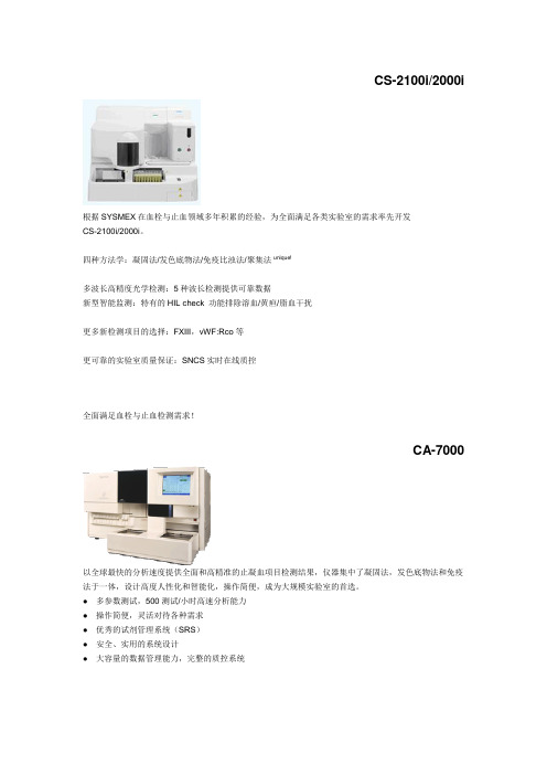
CS-2100i/2000i根据SYSMEX在血栓与止血领域多年积累的经验,为全面满足各类实验室的需求率先开发CS-2100i/2000i。
四种方法学:凝固法/发色底物法/免疫比浊法/聚集法unique!多波长高精度光学检测:5种波长检测提供可靠数据新型智能监测:特有的HIL check 功能排除溶血/黄疸/脂血干扰更多新检测项目的选择:FXIII,vWF:Rco等更可靠的实验室质量保证:SNCS实时在线质控全面满足血栓与止血检测需求!CA-7000以全球最快的分析速度提供全面和高精准的止凝血项目检测结果,仪器集中了凝固法,发色底物法和免疫法于一体,设计高度人性化和智能化,操作简便,成为大规模实验室的首选。
● 多参数测试,500测试/小时高速分析能力● 操作简便,灵活对待各种需求● 优秀的试剂管理系统(SRS)● 安全、实用的系统设计● 大容量的数据管理能力,完整的质控系统CA-1500汇集了当今血栓/止血分析仪最新的各种先进功能于一身,是市场上少见的性能/价格比极高的一台仪器,是大型教学医院,综合医院实验室的首选。
它具有快速处理能力,最快180测试/小时,集多种检测功能于一身:凝固法、发色底物法、免疫法。
具有全能随机组合能力,两种方法测定纤维蛋白质,适合常规大量和急诊使用。
● 拥有高速处理能力、随机测试功能和自动再检查功能● 三种分析方式,包括多规则监视的广泛质控文件和平行线生物分析功能● 卓越的性能可以灵活适应实验室的多样化需求CA-500系列CA-500系列包含了六款机型,设计新颖、符合经济原则,是各中小型实验室开展血栓/止血实验的最佳选择,也是半自动升级到全自动的理想机型。
小型台式仪实用可靠,具备三种检测系统即凝固法、发色底物法、免疫法的自由组合用户可根据需要选择相应机型。
CA-50设计上完全沿用了全自动CA系列的检测原理,锁定人为误差因素的设计确保它有别于其他半自动血凝仪,达到全自动仪器的准确性与重复性效果,四通道即可批量检测又可单独检测,内置质控文件,适用于小标本量实验室使用。
爱必信WB彩色凝胶试剂盒上新啦!让您的WB实验多姿多彩

爱必信WB彩色凝胶试剂盒上新啦!
让您的WB实验多姿多彩
蛋白免疫印迹实验(Western Blot)实验大家肯定都不陌生,要想做出好看的条带,从制胶开始就不容半点马虎。
WB制胶可谓实验室新手的必修课了,想想小编我在当初实验室的时候,就没少帮师兄师姐配置WB凝胶,泛黄的配方表小编还记忆犹新。
WB凝胶配
方表
说起WB凝胶配置,小编可有一肚子苦水要倒,第一个让人头疼的一点就是多种溶液都需要计算量取,每次调移液器就要调半天。
还有各种试剂的保存条件也不一样,配胶之前还需要从冰箱,室温柜子里分别拿出不同的溶液。
这还不算,最容易出问题的促凝剂APS,需要新鲜配置,4度保存不能超过一周,关键是粉末还特容易吸水,打开后需要密封保存。
小编之前就踩过一次坑,APS粉末吸水结块,称量配置后的APS含量不够,凝胶凝结不好,整个WB实验条带跑的七扭八歪。
蛋白质印迹(Western Blot)实验中产品推荐:。
药用辅料中英文对照
药用辅料中英文对照1 阿拉伯胶(Acacia)2 乙酰舒泛钾(Acesulfame Potassium)3 冰醋酸(Acetic Acid,Glacial)4 乙酰枸橼酸三丁酯(AcetyltributylCitrate)5 乙酰枸橼酸三乙酯(AcetyltriethylCitrate)6 人血白蛋白(Albumin)7 乙醇(Ethanol)8 海藻酸(Alginic Acid)9 脂肪族聚酯(Aliphatic Polyesters)10 阿力糖(Alitame)11 杏仁油(Almond Oil)12 维生素E(Alpha Tocopherol)13 氨溶液(Ammonia Solution)14 维生素C(Ascorbic Acid)15 棕榈酸维生素C酯(Ascorbyl Palmitate)16 阿司帕坦(Aspartame)17 绿坡缕石(Attapulgite)18 皂土(Bentonite)19 苯扎氯铵(Benzalkonium Chloride)20 苄索氯铵(Benzethonium Chloride)21 苯甲酸(Benzoic Acid)22 苯甲醇(Benzyl Alcohol)23 苯甲酸苄酯(Benzyl Benzoate)24 溴硝丙二醇(Bronopol)25 丁羟茴醚(Butylated Hydroxyanisole)26 丁羟甲苯(Butylated Hydroxytoluene)27 羟苯丁酯(Butylparaben)28 碳酸钙(Calcium Carbonate)29 无水磷酸氢钙(Calcium Phosphate,Dibasic Anhydrous)30 磷酸氢钙二水合物(Calcium Phosphate,Dibasic Dihydrate)31 磷酸钙(Calcium Phosphate,Tribasic)32 硬脂酸钙(Calcium Stearate)33 硫酸钙(Calcium Sulfate)34 低芥酸菜籽油(Canola Oil)35 卡波姆(Carbomer)36 二氧化碳(Carbon Dioxide)37 羧甲纤维素钙(Carboxymethylcellulose Calcium)38 羧甲纤维素钠(Carboxymethylcellulose Sodium)39 角叉菜胶(Carrageenan)40 蓖麻油(Castor Oil)41 氢化蓖麻油(Castor Oil,Hydro-genated)42 微晶纤维素(Cellulose,Microcrystalline)43 粉状纤维素(Cellulose,Powdered)44 微粉硅胶微晶纤维素(Cellulose, Silicified Microcrystalline)45 醋酸纤维素(Cellulose Acetate)46 纤维醋法酯(Cellulose Acetate Phthalate)47 角豆胶(Ceratonia)48 十八十六醇(Cetostearyl Alcohol)49 西曲溴铵(Cetrimide)50 十六醇(Cetyl Alcohol)51 壳聚糖(Chitosan)52 氯己定(Chlorhexidine)53 三氯叔丁醇(Chlorobutanol)54 氯甲酚(Chlorocresol)55 一氯二氟乙烷(Chlorodifluoroe-thane)56 氟里昂(Chlorofluorocabons)57 对氯间二甲酚(Chloroxylenol)58 胆固醇(Cholesterol)59 枸橼酸(Citric Acid Monohydrate)60 胶态二氧化硅(微粉硅胶)(Colloidal Silicon Dioxide)61 着色剂(Coloring Agents)62 玉米油(Corn Oil)63 棉籽油(Cottonseed Oil)64 甲酚(Cresol)65 交联羧甲纤维素钠(Croscarmellose Sodium)66 交联聚维酮(Crospovidone)67 环糊精(Cyclodextrins)68 环甲基硅酮(Cyclomethicone)69 苯甲地那铵(Denatonium Benzoate)70 葡萄糖结合剂(Dextrates)71 糊精(Dextrin)72 葡萄糖(Dextrose)73 邻苯二甲酸二丁酯(Dibutyl Phthalate)74 癸二酸二丁酯(Dibutyl Sebacate)75 二乙醇胺(Diethanolamine)76 邻苯二甲酸二乙酯(Diethyl Phthalate)77 二氟乙烷(Difluoroethane)78 二甲硅油(Dimethicone)79 二甲醚(Dimethyl Ether)80 邻苯二甲酸二甲酯(Dimethyl Phthalate)81 二甲亚砜(Dimethyl Sulfoxide)82 多库酯钠(Docusate Sodium)83 依地酸(乙二胺四乙酸)(Edetic Acid)84 乙酸乙酯(Ethyl Acetate)85 乙基麦芽酚(Ethyl Maltol)86 油酸乙酯(Ethyl Oleate)87 乙基香草醛(Ethyl Vanillin)88 乙基纤维素(Ethylcellulose)89 硬脂酸棕榈酸乙二醇酯(Ethylene Glycol Palmitostearate)90 羟苯乙酯(Ethylparaben)91 果糖(Fructose)92 富马酸(Fumaric Acid)93 明胶(Gelatin)94 液体葡萄糖(Glucose,Liquid)95 甘油(Glycerin)96 山萮酸甘油酯(Glyceryl Behenate)97 单油酸甘油酯(Glyceryl Monooleate)98 单硬脂酸甘油酯(Glyceryl Monostearate)99 硬脂酸棕榈酸甘油酯(Glyceryl Palmitostearate)100 四氢呋喃聚乙二醇醚(Glycofurol)101 瓜耳胶(Guar Gum)102 七氟丙烷(HFC)(Heptafluoro-propane)103 海克西定(Hexetidine)104 烷烃类(HC) (Hydrocarbons)105 盐酸(Hydrochloric Acid)106 羟乙纤维素(Hydroxyethyl Cellulose)107 羟乙甲纤维素(Hydroxyethylmethyl Cellulose)108 羟丙纤维素(Hydroxypropyl Cellulose)109 低取代羟丙纤维素(Hydroxypropyl Cellulose,Low-substituted) 110 羟丙甲纤维素(Hypromellose)111 羟丙甲纤维素酞酸酯(Hypromellose Phthalate)112 咪唑烷脲(Imidurea)113 异丙醇(Isopropyl Alcohol)114 肉豆蔻酸异丙酯(Isopropyl Myristate)115 棕榈酸异丙酯(Isopropyl Palmitate)116 白陶土(Kaolin)117 乳酸(Lactic Acid)118 拉克替醇(Lactitol)119 乳糖(Lactose)120 羊毛脂(Lanolin)121 含水羊毛脂(Lanolin,Hydrous)122 羊毛醇(Lanolin Alcohols)123 卵磷脂(Lecithin)124 硅酸镁铝(Magnesium Aluminum Silicate)125 碳酸镁(Magnesium Carbonate)126 氧化镁(Magnesium Oxide)127 硅酸镁(Magnesium Silicate)128 硬脂酸镁(Magnesium Stearate)129 三硅酸镁(Magnesium Trisilicate)130 苹果酸(Malic Acid)131 麦芽糖醇(Maltitol)132 麦芽糖醇溶液(Maltitol Solution)133 麦芽糖糊精(Maltodextrin)134 麦芽酚(Maltol)135 麦芽糖(Maltose)136 甘露醇(Mannitol)137 中链脂肪酸甘油三酯(Medium-chain Triglycerides)138 葡甲胺(Meglumine)139 薄荷脑(Menthol)140 甲基纤维素(Methylcellulose)141 羟苯甲酯(Methylparaben)142 液体石蜡(Mineral Oil)143 轻质液体石蜡(Mineral Oil,Light)144 液体石蜡羊毛醇(Mineral Oil and Lanolin Alcohols)145 单乙醇胺(Monoethanolamine)146 谷氨酸一钠(Monosodium Glutamate)147 硫代甘油(Monothioglycerol)148 氮(Nitrogen)149 一氧化二氮(Nitrous Oxide)150 油酸(Oleic Acid)151 橄榄油(Olive Oil)152 石蜡(Paraffin)153 花生油(Peanut Oil)154 凡士林(Petrolatum)155 凡士林羊毛醇(Petrolatum and Lanolin Alcohols)156 苯酚(Phenol)157 苯氧乙醇(Phenoxyethanol)158 苯乙醇(Phenylethyl Alcohol)159 醋酸苯汞(Phenylmercuric Acetate)160 硼酸苯汞(Phenylmercuric Borate)161 硝酸苯汞(Phenylmercuric Nitrate)162 磷酸(Phosphoric Acid)163 波拉克林钾(Polacrilin Potassium)164 泊洛沙姆(Poloxamer)165 葡聚糖(Polydextrose)166 聚乙二醇(Polyethylene Glycol)167 聚氧乙烯(Polyethylene Oxide)168 聚(甲基)丙烯酸树脂(Polymethacr-ylates)169 聚氧乙烯烷基醚(Polyoxyethylene Alkyl Ethers)170 聚氧乙烯蓖麻油衍生物(Polyoxyeth-ylene Castor Oil Derivatives) 171 聚山梨酯(Polyoxyethylene Sorbitan Fatty Acid Esters)172 硬脂酸聚氧乙烯酯(Polyoxyethylene Stearates)173 聚醋酸乙烯酞酸酯(Polyvinyl Acetate Phthalate)174 聚乙烯醇(Polyvinyl Alcohol)175 苯甲酸钾(Potassium Benzoate)176 碳酸氢钾(Potassium Bicarbonate)177 氯化钾(Potassium Chloride)178 枸橼酸钾(Potassium Citrate)179 氢氧化钾(Potassium Hydroxide)180 焦亚硫酸钾(Potassium Metabisulfite)181 山梨酸钾(Potassium Sorbate)182 聚维酮(Povidone)183 丙酸(Propionic Acid)184 没食子酸丙酯(Propyl Gallate)185 碳酸丙烯酯(Propylene Carbonate)186 丙二醇(Propylene Glycol)187 海藻酸丙二醇酯(Propylene Glycol Alginate)188 羟苯丙酯(Propylparaben)189 糖精(Saccharin)190 糖精钠(Saccharin Sodium)191 芝麻油(Sesame Oil)192 虫胶(Shellac)193 二氧化硅二甲硅油(Simethicone)194 海藻酸钠(Sodium Alginate)195 抗坏血酸钠(Sodium Ascorbate)196 苯甲酸钠(Sodium Benzoate)197 碳酸氢钠(Sodium Bicarbonate)198 氯化钠(Sodium Chloride)199 枸橼酸钠二水合物(Sodium Citrate Dihydrate)200 环拉酸钠(Sodium Cyclamate)201 氢氧化钠(Sodium Hydroxide)202 月桂硫酸钠(十二烷基硫酸钠)(Sodium Lauryl Sulfate)203 焦亚硫酸钠(偏亚硫酸钠)(Sodium Metabisulfite)204 磷酸氢二钠(Sodium Phosphate,Dibasic)205 磷酸二氢钠(Sodium Phosphate ,Monobasic)206 丙酸钠(Sodium Propionate)207 羧甲淀粉钠(Sodium Starch Glycolate)208 硬脂富马酸钠(Sodium Stearyl Fumarate)209 山梨酸(Sorbic Acid)210 山梨坦酯Sorbitan Esters(Sorbitan Fatty Acid Esters) 211 山梨醇(Sorbitol)212 大豆油(Soybean Oil)213 淀粉(Starch)214 预胶化淀粉(Starch,Pregelatinized)215 灭菌玉米淀粉(Starch,Sterilizable Maize)216 硬脂酸(Stearic Acid)217 硬脂醇(Stearyl Alcohol)218 羟糖氯(Sucralose)219 蔗糖(Sucrose)220 可压性蔗糖(Sugar,Compressible)221 蔗糖粉(Sugar,Confectioner’s)222 蔗糖球形颗粒(Sugar Spheres)223 硫酸(Sulfuric Acid)224 葵花籽油(Sunflower Oil)225 氢化植物油(硬脂)栓剂基质(Sup-pository Bases,Hard Fat) 226 滑石粉(Talc)227 酒石酸(Tartaric Acid)228 四氟乙烷(HFC)(Tetrafluoroe-thane)229 硫柳汞(Thimerosal)230 二氧化钛(Titanium Dioxide)231 西黄蓍胶(Tragacanth)232 海藻糖(Trehalose)233 三醋汀(Triacetin)234 枸橼酸三丁酯(Tributyl Citrate)235 三乙醇胺(Triethanolamine)236 枸橼酸三乙酯(Triethyl Citrate)237 香草醛(Vanillin)238 氢化植物油(Vegetable Oil,Hydrogenated)239 纯化水(Water)240 阴离子乳化蜡(Wax,Anionic Emulsifying)241 巴西棕榈蜡(Wax,Carnauba)242 十六醇酯蜡(Wax,Cetyl Esters)243 微晶蜡(Wax,Microcrystalline)244 非离子乳化蜡(聚西托醇乳化蜡)(Wax,Nonionic Emulsifying) 245 白蜡(Wax,White)246 黄蜡(Wax,Yellow)247 黄原酸胶(Xanthan Gum)248 木糖醇(Xylitol)796249 玉米朊(玉米蛋白)(Zein)250 硬脂酸锌(Zinc Stearate)。
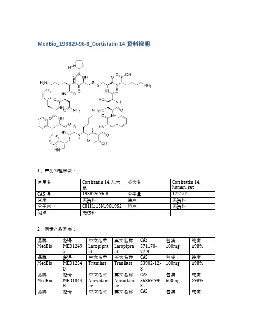
MedBio_193829-96-8_Cortistatin 14资料说明
品牌
货号
中文名称
英文名称
CAS
包装
纯度
MedBio
MED13351
Losartan Carboxylic Acid
Losartan Carboxylic Acid
124750-92-1
中文名称
英文名称
CAS
包装
纯度
MedBio
MED13366
3-chloro-5-hydroxy BA
CAS
包装
纯度
MedBio
MED12497
Laropiprant
Laropiprant
571170-77-9
100mg
≥98%
品牌
货号
中文名称
英文名称
CAS
包装
纯度
MedBio
MED12540
Tranilast
Tranilast
53902-12-8
100mg
≥98%
品牌
货号
中文名称
英文名称
CAS
包装
5mg
≥98%
品牌
货号
中文名称
英文名称
CAS
包装
纯度
MedBio
MED12697
CRF (human, rat)
CRF (human, rat)
86784-80-7
10mg
≥98%
品牌
货号
中文名称
英文名称
CAS
包装
纯度
MedBio
MED12969
2-PMDQ
2-PMDQ
139047-55-5
50mg
纯度
MedBio
MED13448
培非格司亭中英文介绍

王婕 913103860408NEULASTA(PEGFILGRASTIM)|培非格司亭注射液1.Introduction(简介)【产地英文商品名】:NEULASTA-6mg/0.6ml/Syringe【原产地英文药品名】:PEGFILGRASTIM【中文参考商品译名】:纽拉思塔-6毫克/0.6毫升/支【中文参考药品译名】:培非格司亭【生产厂家中文参考译名】:安进【生产厂家英文名】:Amgen, IncAmgen Announces Novel Drugs for Antitumor Chemotherapy Side Effects of FGT (TM) (pegfilgrastim), a drug developed by the US Food and Drug Administration (FDA), has been approved by the US Food and Drug Administration (FDA) Approval. Amphetamycin, the chief executive of Amgen, says that pemetrexedin will make it easier for healthcare workers to prevent chemotherapy-induced neutropenia and its serious complications.The third drug approved by Amgen in the past six months will significantly improve the prognosis of chemotherapy patients and is expected to enter the market in early April.BUSINESS WIRE 2002年2月1日美国加州THOUSAND OAKS消息,安进公司宣布抗肿瘤化疗副作用新药培非格司亭(TM) (pegfilgrastim)通过美国食品与药品管理局(FDA)的审批。
维格列汀与常见降糖药联合治疗2型糖尿病的研究进展
2020年12月 第17卷 第24期2型糖尿病(Type 2 Diabetes Mellitus,T2DM)以β细胞功能障碍[1-3]、较高的体质指数[4-6]、胰岛素抵抗指数[7-9]等为主要的表型特征。
在T2DM 的治疗指南中,建议将维格列汀等二肽基肽酶4抑制剂作为二线或三线药物[10]。
与其他降糖药相比,维格列汀等具有良好的药物耐受性和安全性。
那么,维格列汀与常见降糖药联合治疗T2DM的疗效与安全性如何呢?本文将综述相关研究成果。
1 维格列汀+二甲双胍联合治疗的相关研究1.1 维格列汀+二甲双胍联合用药与其他联合疗法的比较Peng等[11]以二甲双胍为基础,通过随机对照试验验证了格列美脲、吡格列酮、艾塞那肽、格列本脲、罗格列酮和维格列汀6种降血糖药物在T2DM中的不同疗效。
网络荟萃分析结果表明,这6种药物均能降低HbA1c水平。
与其他方案相比,艾塞那肽+二甲双胍联合治疗降低空腹血糖(Fasting Plasma Glucose,FPG)、空腹血浆胰岛素(Fasting Plasma Insulin,FPI)、总胆固醇(Total Cholesterol,TC)和稳态模型评估胰岛素抵抗指数(Homeostasis Model Assessment Insulin Resistance Index,HOMA-IR),且可提高高密度脂蛋白胆固醇(High-density Lipoprotein Cholesterol,HDL-C)水平;维格列汀+二甲双胍联合用药可降低FPI和低密度脂蛋白胆固醇(Low-density Lipoprotein Cholesterol,LDL-C)水平;格列本脲+二甲双胍联合用药可降低FPG水平,且提高HDL-C水平;格列美脲+二甲双胍联合用药可降低TC水平;罗格列酮+二甲双胍联合用药可降低LDL-C水平。
研究结果表明,艾塞那肽+二甲双胍和维格列汀+二甲双胍联合用药对T2DM有较好的疗效,二者均能改善胰岛素敏感性。
Incucyte
Product Information Presentation, Storage and StabilityThe Incucyte® Fabfluor-pH Antibody Labeling Reagents for antibody internalization are supplied as lyophilized solids in sufficient quantity to label 50 μg of test antibody, when used at the suggested molar ratio (1:3 of test antibody to labeling Fab). The lyophilized solid can be stored at 2-8° C for one year. Once re-hydrated, any unused reagent should be aliquoted and stored at -80° C for up to one year. Avoid repeated freeze-thaw cycles.Incucyte® Fabfluor-pH Antibody Labeling ReagentsFor Antibody Internalization AssaysAntibody Labeling Reagent Rehydrated: -80° C *Excitation and Emission maxima were determined at a pH of 4.5.Fabfluor_quick_guideBackgroundIncucyte ® Fabfluor-pH Antibody Labeling Reagents are designed for quick, easy labeling of Fc-containing test antibodies with a Fab fragment-conjugated pH-sensitive fluorophore. The pH-sensitive dye based system exploits the acidic environment of the lysosomes to quantify in-ternalization of the labeled antibody. As Fabfluor labeled antibodies reside in the neutral extracellular solution (pH 7.4), they interact with cell surface specific antigens and are internalized. Once in the lysosomes, they enter an acidic environment (pH 4.5–5.5) and a substantial in-crease in fluorescence is observed. In the absence of ex-pression of the specific antigen, no internalization occurs and the fluorescence intensity of the labeled antibodies remains low. With the Incucyte ® integrated analysis soft-ware, background fluorescence is minimized. These reagents have been validated for use with a number of different antibodies in a range of cell types. The Incucyte ® Live-Cell Analysis System enables real-time, kinetic eval -uation of antibody internalization.Recommended UseWe recommend that the Incucyte ® Fabfluor-pH Antibody Labeling Reagents are prepared at a stock concentration of 0.5 mg/mL by the addition of 100 μL of sterile water and triturated (centrifuge if solution not clear). The reagent may then be diluted directly into the labeling mixture with test antibody. Do NOT sonicate the solution.Additional InformationThe Fab antibody was purified from antisera by a combination of papain digestion and immunoaffinity chromatography using antigens coupled to agarose beads. Fc fragments and whole IgG molecules have been removed.Human Red (Cat. No. 4722) or Human Orange (Cat. No. 4812)—Based on immunoelectrophoresis and/ or ELISA, the antibody reacts with the Fc portion of human IgG heavy chain but not the Fab portion of human IgG. No antibody was detected against human IgM, IgA or against non-immunoglobulin serum proteins. The anti-body may cross-react with other immunoglobulins from other species.Mouse IgG1 (Cat. No. 4723), IgG2a (Cat. No. 4750) or IgG2b (Cat. No. 4751)—Based on antigen-binding assay and/or ELISA, the antibody reacts with the Fc portion of mouse IgG, IgG2a or IgG2b, respectively, but not the Fab portion of mouse immunoglobulins. No antibody was detected against mouse IgM or against non–immunoglobulin serum proteins. The antibody may cross-react with other mouse IgG subclasses or with immunoglobulins from other species.Rat (Cat. No. 4737)—Based on immunoelectrophoresis and/or ELISA, the antibody reacts with the Fc portion of rat IgG heavy chain but not the Fab portion of rat IgG. No antibody was detected against rat IgM, IgA or against non-immunoglobulin serum proteins. The antibody may cross-react with other immunoglobulins from other species.A.B.C.D.R e d O b j e c t A r e a (x 105 μm 2 p e r w e l l )Time (hours)A U C x 106 (0–12 h )log [α–CD71] (g/mL)Example DataFigure 1: Concentration-dependent increase in antibody internalization of Incucyte ® Fabfluor labeled-α-CD71 in HT1080 cells. α-CD71 and mouse IgG1 isotype control were labeled with Incucyte ® Mouse IgG1 Fabfluor-pH Red Antibody Labeling Reagent. HT1080 cells were treated with either Fabfluor-α-CD71 or Fabfluor-IgG1 (4 μg/mL); HD phase and red fluorescence images were captured every 30 minutes over 12 hours using a 10X magnification. (A) Images of cells treated with Fabfluor-α-CD71 display red fluorescence in the cytoplasm (images shown at 6 h). (B) Cells treated with labeled isotype control display no cellular fluorescence. (C) Time-course of Fabfluor-α-CD71 internalization with increasing concentrations of Fabfluor-α-CD71 (progressively darker symbols). Internalization has been quantified as the red object area for each time-point. (D) Concentration response curve to Fabfluor-α-CD71. Area under the curve (AUC) values have been determined from the time-course shown in panel C (0-12 hours) and are presented as the mean ± SEM, n=3 wells.CD71-FabfluorIgG-FabfluorProtocols and ProceduresMaterialsIncucyte® Fabfluor-pH Antibody Labeling ReagentTest antibody of interest containing human, mouse, or rat IgG Fc region (at known concentration)Target cells of interestTarget cell growth mediaSterile distilled water96-well flat bottom microplate (e.g. Corning Cat. No. 3595) for imaging96-well round black round bottom ULA plate (e.g. Corning Cat. No. 45913799) or amber microtube (e.g. Cole Parmer Cat. No. MCT-150-X, autoclaved) for conjugation step0.01% Poly-L-Ornithine (PLO) solution (e.g. Sigma Cat. No. P4957), optional for non-adherent cells Recommended control antibodiesIt is strongly recommended that a positive and negative control is run alongside test antibodies and cell lines. For example, CD71, which is a mouse anti-human antibody, is recommended as a positive control for the mouse Fab.Anti-CD71, clone MEM-189, IgG1 e.g. Sigma Cat. No. SAB4700520-100UGAnti-CD71, clone CYG4, IgG2a e.g. BioLegend Cat. No. 334102Isotype controls, depending on isotype being studied—Mouse IgG1, e.g. BioLegend Cat. No. 400124, Mouse IgG2a e.g. BioLegend Cat. No. 401501Preparation of Incucyte® Antibody Internalization Assay 1. Seed target cells of interest1.1 Harvest cells of interest and determine cell concentra-tion (e.g. trypan blue + hemocytometer).1.2 Prepare cell seeding stock in target cell growth mediawith a cell density to achieve 40–50% confluence be-fore the addition of labeled antibodies. The suggested starting range is 5,000–30,000 cells/well, although the seeding density will need to be optimized for each cell type.Note: For non-adherent cell types, a well coating may be required to maintain even cell distribution in the well. For a 96-well flat bottom plate, we recommend coating with 50 μL of either 0.01% Poly-L-Or-nithine (PLO) solution or 5 μg/mL fibronectin diluted in 0.1% BSA.Coat plates for 1 hour at ambient temperature, remove solution from wells and then allow the plates to dry for 30-60 minutes prior to cell addition.1.3 Using a multi-channel pipette, seed cells (50 µL perwell) into a 96-well flat bottom microplate. Lightly tapplate side to ensure even liquid distribution in well. Toensure uniform distribution of cells in each well, allowthe covered plate sit on a level surface undisturbed at room temperature in the tissue culture hood for 30minutes. After cells are settled, place the plate insidethe Incucyte® Live-Cell Analysis System to monitor cell confluence.Note: Depending on cell type, plates can be used in assay once cells have adhered to plastic and achieved normal cell morphology e.g.2-3 hours for HT1080 or 1-2 hours for non-adherent cell types. Some cell types may require overnight incubation.2. Label Test Antibody2.1 Rehydrate the Incucyte® Fabfluor-pH Antibody Label-ing Reagent with 100 µL sterile water to result in a final concentration of 0.5 mg/mL. Triturate to mix (centrifuge if solution is not clear).Note: The reagent is light sensitive and should be protected fromlight. Rehydrated reagent can be aliquoted into amber or foilwrapped tubes and stored at -80° C for up to 1 year (avoid freezing and thawing).2.2 Mix test antibody with rehydrated Incucyte® Fabfluor–pH Antibody Labeling Reagent and target cell growth media in a black round bottom microplate or ambertube to protect from light (50 µL/well).a. Add test antibody and Incucyte® Fabfluor–pH Anti-body Labeling Reagent at 2X the final concentration.We suggest optimizing the assay by starting with afinal concentration of 4 µg/mL of test antibody or theFabfluor-pH Antibody Labeling Reagent (i.e. 2Xworking concentration = 8 µg/mL).Note: A 1:3 molar ratio of test antibody to Incucyte® Fabfluor-pHAntibody Labeling Reagent is recommended. The labeling re-agent is a third of the size of a standard antibody (50 and 150KDa, respectively). Therefore, labeling equal quantities will pro-duce a 1:3 molar ratio of test antibody to labeling Fab.b. Make sufficient volume of 2X labeling solution for50 µL/well for each sample. Triturate to mix.c. Incubate at 37° C for 15 minutes protected from light.Note: If performing a range of concentrations of test antibody,e.g. concentration response-curve, it is recommended to createthe dilution series post the conjugation step to ensure consistentmolar ratio. We strongly recommend the use of both a negativeand positive control antibody in the same plate.3. Add labeled antibody to cells3.1 Remove cell plate from incubator.3.2 Using a multi-channel pipette, add 50 µL of 2X labeledantibody and control solutions to designated wells.Remove any bubbles and immediately place plate in the Incucyte® Live-Cell Analysis System and start scanning.Note: To reduce the risk of condensation formation on the lid priorto first image acquisition, maintain all reagents at 37° C prior toplate addition.4. Acquire images and analyze4.1 In the Incucyte® Software, schedule to image every15-30 minutes, depending on the speed of the specific antibody internalization.a Scan on schedule, standard. If the Incucyte® Cell-by-Cell Analysis Software Module (Cat. No. 9600-0031)is available, adherent cell-by-cell or non-adherentcell-by-cell scan types can be selected.b Channel selection: select “phase” and “red” or“phase” and "orange” (depending on reagent used).c Objective: 10X or 20X depending on cell types used,generally 10X is recommended for adherent cells,and 20X for non-adherent or smaller cells.NOTE: The optional Incucyte® Cell-by-Cell Analysis SoftwareModule enables the classification of cells into sub-populationsbased on properties including fluorescence intensity, size andshape. For further details on this analysis module and its appli-cation, please see: /cell-by-cell.4.2 To generate the metrics, user must create an AnalysisDefinition suited to the cell type, assay conditions andmagnification selected.4.3 Select images from a well containing a positiveinternalization signal and an isotype control well(negative signal) at a time point where internalizationis visible.4.4 In the Analysis Definition:Basic Analyzer:a. Set up the mask for the phase confluence measurewith fluorescence channel turned off.b. Once the phase mask is determined, turn the fluores-cence channel on: Exclude background fluorescencefrom the mask using the background subtractionfeature. The feature “Top-Hat” will subtract localbackground from brightly fluorescent objects withina given radius; this is a useful tool for analyzing ob-jects which change in fluorescence intensity overtime.i The radius chosen should reflect the size of thefluorescent object but contain enough backgroundto reliably estimate background fluorescence inthe image; 20-30 μm is often a useful startingpoint.ii The threshold chosen will ensure that objectsbelow a fluorescence threshold will not bemasked.iii Choose a threshold in which red or orange objectsare masked in the positive response image but lownumbers in the isotype control, negative responsewell. For a very sensitive measurement, for example,if interested in early responses, we suggest athreshold of 0.2.NOTE: The Adaptive feature can be used for analysis but maynot be as sensitive and may miss early responses. If interestedin rate of response, Top-Hat may be preferable.Cell-by-Cell (if available):a. Create a Cell-by-Cell mask following the softwaremanual.b. There is no need to separate phase and fluorescencemasks. The default setting of Top-Hat No Mask forthe fluorescence channel will enable backgroundsubtraction without generation of a mask. Ensurethat the Top-Hat radius is set to a value higher thanthe radius of the larger clusters to avoid excess back-ground subtraction.c. The threshold of fluorescence can be determined inCell-by-Cell Classification.Specifications subject to change without notice.© 2020. All rights reserved. Incucyte, Essen BioScience, and all names of Essen BioScience prod -ucts are registered trademarks and the property of Essen BioScience unless otherwise specified. Essen BioScience is a Sartorius Company. Publication No.: 8000-0728-A00Version 1 | 2020 | 04Sales and Service ContactsFor further contacts, visit Essen BioScience, A Sartorius Company /incucyte Sartorius Lab Instruments GmbH & Co. KGOtto-Brenner-Strasse 20 37079 Goettingen, Germany Phone +49 551 308 0North AmericaEssen BioScience Inc. 300 West Morgan Road Ann Arbor, Michigan, 48108USATelephone +1 734 769 1600E-Mail:***************************EuropeEssen BioScience Ltd.Units 2 & 3 The Quadrant Newark CloseRoyston Hertfordshire SG8 5HLUnited KingdomTelephone +44 (0) 1763 227400E-Mail:***************************APACEssen BioScience K.K.4th floor Daiwa Shinagawa North Bldg.1-8-11 Kita-Shinagawa Shinagawa-ku, Tokyo 140-0001 JapanTelephone: +81 3 6478 5202E-Mail:*************************5. Analysis GuidelinesAs the labeled antibody is internalized into the acidic environment of the lysosome, the area of fluorescence intensity inside the cells increases.This can be reported in two ways:Ways to Report Basic AnalyzerCell-by-Cell Analysis* To correct for cell proliferation, it is advisable to normalize the fluorescence area to the total cell area using User Defined Metrics.For Research Use Only. Not For Therapeutic or Diagnostic Use.LicensesFor non-commercial research use only. Not for therapeutic or in vivo applications. Other license needs contact Essen BioS cience.Fabfluor-pH Red Antibody Labeling Reagent: This product or portions thereof is manufactured under license from Carnegie Mellon University and U.S. patent numbers 7615646 and 8044203 and related patents. This product is licensed for sale only for research. It is not licensed for any other use. There is no implied license hereunder for any commercial use.Fabfluor-pH Orange Antibody Labeling Reagent: This product or portions thereof is manufactured under a license from Tokyo University and is covered by issued patents EP2098529B1, JP5636080B2, US8258171, and US9784732 and related patent applications. This product and related products are trademarks of Goryo Chemical. Any application of above mentioned technology for commercial purpose requires a separate li -cense from: Goryo Chemical, EAREE Bldg., SF Kita 8 Nishi 18-35-100, Chuo-Ku, Sapporo, 060-0008 Japan.SupportA complete suite of cell health applications is available to fit your experimental needs. Find more information at /incucyte Foradditionalproductortechnicalinformation,************************************************************/incucyte。
细胞自噬染色检测试剂盒(MDC法)
细胞自噬染色检测试剂盒(MDC 法)简介:自噬(autophagy)是细胞受到刺激后吞噬自身的细胞质或细胞器,最终将吞食物在溶酶体内降解的过程,自噬体(autophagosome)为双层膜包被的圆形或椭圆形结构,内含细胞质、长寿蛋白质和异常蛋白聚集物,损伤或多余细胞器如线粒体、粗面内质网和微体、病毒和细菌等。
单丹磺酰尸胺(Dansylcadaverine,MDC )是一种荧光色素,是嗜酸性染色剂,通常被用于检测自噬体形成的特异性标记染色剂,其检测激发滤光片波长355nm 。
阻断滤光片波长512nm 。
Leagene 细胞自噬染色检测试剂盒(MDC 法),适用于培养细胞的自噬染色,又称为MDC 染色液,可与EB 合用双染。
组成:操作步骤(仅供参考):(一)、MDC 单独染色:1、 用去离子水稀释1×Wash buffer 至1×。
2、 离心,收集细胞,用1×Wash buffer 清洗细胞1次,弃上清。
3、 加入适量的1×Wash buffer 重悬细胞,计数并调节细胞浓度至106/ml 。
4、 取适量细胞悬液至新的EP 管中,加入MDC Stain ,轻轻混匀。
5、 室温避光染色。
6、 离心收集细胞,用1×Wash buffer 清洗细胞2次,弃上清。
7、 加入Collection buffer 重悬细胞,滴加于载玻片上并加盖玻片。
8、 荧光显微镜下观察(激发滤光片波长355nm ,阻断滤光片波长512nm),计数并拍照。
(二)、与EB 双染色:1、 用去离子水稀释1×Wash buffer 至1×。
2、 离心收集细胞,用1×Wash buffer 清洗细胞1次,弃上清。
3、 加入适量的1×Wash buffer 重悬细胞,计数并调节细胞浓度至106/ml 。
4、 取适量细胞悬液至新的EP 管中,分别加入MDC Stain 和 EB 染色液,轻轻混匀。
CytoSelect 24 -Well Anoikis Assay 说明书

Product ManualCytoSelect™ 24-Well Anoikis AssayCatalog NumberCBA-080 24 assaysFOR RESEARCH USE ONLYNot for use in diagnostic proceduresIntroductionAdhesion to the extracellular matrix (ECM) is essential for survival and propagation of many adherent cells. Apoptosis that results from the loss of cell adhesion to the ECM, or inappropriate adhesion is defined as “anoikis”. Anoikis, from the Greek word for homelessness, is involved in the physiological processes of tissue renewal and cell homeostasis.A common feature of carcinoma development and growth is the ability of transformed cells to survive under “anchorage independent” or “spheroid” growth conditions. This resistance to anoikis has been shown to be involved in the loss of cell homeostasis, cancer growth, and metastasis. The inhibition of cell adhesion, spreading, and growth on the ECM is an impediment to the cellular healing process, thus making it a possible therapeutic target. Preventing anoikis and enhancing cell adhesion and spreading is a major goal in the development of cell transplantation techniques, including the therapeutic use of progenitor cells. Further studies aimed at controlling the molecular mechanisms of anoikis resistance will serve to define effective therapies for the treatment of many human malignancies.The CytoSelect™ 24-well Anoikis Assay Kit provides a colorimetric and fluorometric format to measure anchorage-independent growth and monitoring anoikis propelled cell death. The kit contains sufficient reagents for the assay of 24 samples in a Poly-Hema coated 24-well plate. Live cells are detected with MTT or Calcein AM. Cell death is detected with the Ethidium Homodimer (EthD-1). Assay PrincipleCells are cultured in poly-Hema coated plate or control plate. Cell viability is determined by MTT or Calcein AM. Anoikis propelled cell death is measured by Ethidium Homodimer (EthD-1). EthD-1 is an excellent marker for measuring dead cells. EthD-1 is a red fluorescent dye that can only penetrate damaged cell membranes. EthD-1 will fluoresce with a 40-fold enhancement upon binding ssDNA, dsDNA, RNA, oligonucleotides, and triplex DNA. Background fluorescence levels are very low because the dyes are virtually non-fluorescent before interacting with cells.Related Products1.CBA-081: CytoSelect™ 96-Well Anoikis Assay2.CBA-230: Cellular Senescence Detection Kit (SA-β-Gal Staining)3.CBA-231: 96-Well Cellular Senescence Assay (SA β-Gal Activity)4.CBA-232: Quantitative Cellular Senescence Assay (SA β-Gal)5.CBA-240: CytoSelect™ Cell Vi ability and Cytotoxicity AssayKit Components1.Anchorage Resistant Plate (Part No. 108001): One 24-well Poly-Hema coated plate.2.Calcein AM (500X) (Part No. 108002): One vial – 50 µL in DMSO.3.Ethidium Homodimer (EthD-1) (500X) (Part No. 108003): One vial – 50 µL.4.Detergent Solution (Part No. 108004): One bottle – 25.0 mL.5.MTT Solution (Part No. 113502): Three tubes – 1.0 mL each.Materials Not Supplied1.Cells for measuring anoikis2.Cell culture medium3.Inverted fluorescence/light microscope4.Fluorometer capable of reading Calcein AM (485 nm/515 nm) and EthD-1 (525 nm/590 nm)fluorescence.StorageStore the Calcein AM and Ethidium Homodimer at -20ºC. Store all other components at 4ºC.Assay Protocol1.Prepare a cell suspension containing 0.1-2.0 x 10⁶ cells/ml in culture media. Cells can be treatedwith anoikis enhancing or inhibiting reagents.2.Add 0.5 mL cell suspension to each well of the Anchorage Resistant Plate or a control 24-well cellculture plate. Culture the cells 24-72 hours at 37ºC and 5% CO2. The time and culture conditions will depend on the cell line used and may need to be adjusted by the user.3.Proceed with MTT Colorimetric or Calcein AM/EthD-1 Fluorometric detection.MTT Colorimetric Detection1.Add the 50 µL of the MTT Reagent to each well of the Anchorage Resistant Plate or control 24-well plate.2.Incubate the wells 2-4 hours or overnight at 37ºC. Monitor the cells occasionally with an invertedmicroscope for the presence of a purple precipitate.3.Add 500 µL of Detergent Solution to each well. Gently mix the solution by pipetting.4.Cover the plate to protect it from light and incubate in the dark for 2-4 hours at room temperature.5.Transfer 200 µL to a 96-well plate and measure the absorbance in each well at 570 nm in amicrotiter plate reader.Calcein AM / EthD-1 Fluorometric Detection1.Add 1 µL of Calcein AM (500X) and 1 µL of Eth-D1 (500X) to each well of the 24-wellAnchorage Resistant Plate or control plate to be detected.2.Incubate the plate 30-60 minutes at 37ºC.3.Monitor the cells microscopically for the presence of the green Calcein AM (Ex: 485 nm and Em:515 nm) or red EthD-1 (Ex: 525 nm and Em: 590 nm) fluorescence. The fluorescence can be quantitatively measured with a fluorescence microplate reader.Example of Results The following figures demonstrate typical results with the CytoSelect™ 24-well Anoikis Assay Kit. One should use the data below for reference only. This data should not be used to interpret actual results.00.20.40.60.8Control Poly-HemaO D 560 n mFigure 1. Anoikis Assay of Human Foreskin Fibroblast BJ-TERT Cells. BJ-TERT cells were seeded at 50,000 cells/well in a tissure culture control plate or a Poly-Hema coated plate. Cells were allowed to culture for 24 hours. Cell viability was determined by MTT and Calcein AM, while anoikis-like cell death was stained with EthD-1.References1.Bates RC, Buret A, van Helden DF, Horton MA, Burns GF. (1994) J Cell Biol125, 403-415.2.Frisch SM, Francis H. (1994) J Cell Biol124, 619-626.3.Frisch SM, Screaton RA. (2001) Curr Opin Cell Biol13, 555-562.4.Meredith JE, Jr Fazeli B, Schwartz MA. (1993) Mol Biol Cell 4, 953-961.5.Rak J, Mitsuhashi Y, Erdos V, Huang SN, Filmus J, Kerbel RS. (1995) J Cell Biol131, 1587-1598. Recent Product Citations1.Mao, C.G. et al. (2021). BCAR1 plays critical roles in the formation and immunoevasion ofinvasive circulating tumor cells in lung adenocarcinoma. Int J Biol Sci. 17(10):2461-2475.doi:10.7150/ijbs.61790.2.Zheng, J.L. et al. (2021). Ursolic acid induces apoptosis and anoikis in colorectal carcinoma RKOcells. BMC Complement Med Ther. 21(1):52. doi: 10.1186/s12906-021-03232-2.3.Liu, L.Q. et al. (2019). MiR-92a antagonized the facilitation effect of extracellular matrix protein 1in GC metastasis through targeting its 3'UTR region. Food Chem Toxicol. 133:110779. doi:10.1016/j.fct.2019.110779.4.Xu, J. et al. (2019). ProNGF siRNA inhibits cell proliferation and invasion of pancreatic cancercells and promotes anoikis. Biomed Pharmacother. 111:1066-1073. doi:10.1016/j.biopha.2019.01.002.5.Tan, Y. et al. (2018). Adipocytes fuel gastric cancer omental metastasis via PITPNC1-mediatedfatty acid metabolic reprogramming. Theranostics. 8(19):5452-5468. doi: 10.7150/thno.28219. 6.Hu, L. et al. (2018). G9A promotes gastric cancer metastasis by upregulating ITGB3 in a SETdomain-independent manner. Cell Death Dis. 9(3):278. doi: 10.1038/s41419-018-0322-6.7.Hu, B. et al. (2018). Herbal formula YGJDSJ inhibits anchorage-independent growth and inducesanoikis in hepatocellular carcinoma Bel-7402 cells. BMC Complement Altern Med. 18(1):17. doi:10.1186/s12906-018-2083-2.8.Chen, H.Y. et al. (2018). Integrin alpha5beta1 suppresses rBMSCs anoikis and promotes nitricoxide production. Biomed Pharmacother. 99:1-8. doi: 10.1016/j.biopha.2018.01.038.9.Fu, X.T. et al. (2018). MicroRNA-30a suppresses autophagy-mediated anoikis resistance andmetastasis in hepatocellular carcinoma. Cancer Lett. 412:108-117. doi:10.1016/j.canlet.2017.10.012.10.Yu, M. et al (2017). Interference with Tim-3 protein expression attenuates the invasion of clear cellrenal cell carcinoma and aggravates anoikis. Mol Med Rep. 15(3):1103-1108. doi:10.3892/mmr.2017.6136.11.Lu, S. et al. (2016). Expression of α-fetoprotein in gastric cancer AGS cells contributes to invasionand metastasis by influencing anoikis sensitivity. Oncol Rep.35:2984-2990.12.Lee, H.W. et al. (2013). Tpl2 kinase impacts tumor growth and metastasis of clear cell renal cellcarcinoma. Mol Cancer Res.11:1375-1386.13.Sisto, M. et al. (2009). Fibulin-6 expression and anoikis in human salivary gland epithelial cells:implications in Sjogren's syndrome. Int. Immunol.21:303-311.14.Liu, H. et al. (2008). Cysteine-rich protein 61 and connective tissue growth factor induce de-adhesion and anoikis of retinal pericytes. Endocrinology 149:1666-1677.WarrantyThese products are warranted to perform as described in their labeling and in Cell Biolabs literature when used in accordance with their instructions. THERE ARE NO WARRANTIES THAT EXTEND BEYOND THIS EXPRESSED WARRANTY AND CELL BIOLABS DISCLAIMS ANY IMPLIED WARRANTY OF MERCHANTABILITY OR WARRANTY OF FITNESS FOR PARTICULAR PURPOSE. CELL BIOLABS’ sole obligation and purchaser’s exclusive remedy for breach of this warranty shall be, at the option of CELL BIOLABS, to repair or replace the products. In no event shall CELL BIOLABS be liable for any proximate, incidental or consequential damages in connection with the products.Contact InformationCell Biolabs, Inc.7758 Arjons DriveSan Diego, CA 92126Worldwide: +1 858-271-6500USA Toll-Free: 1-888-CBL-0505E-mail: ********************©2007-2021: Cell Biolabs, Inc. - All rights reserved. No part of these works may be reproduced in any form without permissions in writing.。
黄连素抑制GRP78表达促进三阴乳腺癌细胞凋亡的研究

黄连素抑制GRP78表达促进三阴乳腺癌细胞凋亡的研究赵虹;沃立科;胡袁媛;王安妮;卢德赵【摘要】[目的]通过检测黄连素对三阴乳腺癌(triple-negative breast cancer,TNBC)细胞株MDA-MB-231细胞凋亡及葡萄糖调节蛋白78(glucose regulated protein 78,GRP78)表达的影响,探讨黄连素抗TNBC的机制.[方法]以MTT法检测不同浓度黄连素对MDA-MB-231细胞增殖的影响,筛选黄连素的处理浓度.将MDA-MB-231细胞分为对照组、40μmol·L-1黄连素组、60μmol·L-1黄连素组、免疫球蛋白重链结合蛋白诱导剂X(immunoglobulin heavy chain binding protein inducer X,BiX)组,以及BiX联合40μmol·L-1黄连素组、BiX联合60μmol·L-1黄连素组.相应药物处理24h后,应用流式细胞术检测细胞凋亡情况,并以Western blot检测细胞GRP78和凋亡相关基因Bcl-2、Bax的表达.[结果]40μmol·L-1以上浓度的黄连素可剂量依赖性抑制MDA-MB-231细胞增殖.与对照组比较,BiX组细胞凋亡率降低(P<0.05)、GRP78和Bcl-2表达增高(P<0.01,P<0.001)、Bax表达减低(P<0.05).与BiX组比较,BiX联合40、60μmol·L-1黄连素组细胞凋亡率增加(P<0.01),GRP78和Bcl-2表达减低(P<0.01,P<0.01),Bax表达增加(P<0.01).[结论]黄连素可以促进MDA-MB-231细胞凋亡,其机制可能是通过下调GRP78的表达,从而降低Bcl-2表达,并促进Bax的表达.【期刊名称】《浙江中医药大学学报》【年(卷),期】2019(043)005【总页数】6页(P407-412)【关键词】三阴乳腺癌;黄连素;葡萄糖调节蛋白78;细胞凋亡;细胞增殖;免疫球蛋白重链结合蛋白诱导剂X【作者】赵虹;沃立科;胡袁媛;王安妮;卢德赵【作者单位】浙江中医药大学附属第一医院杭州 310006;浙江中医药大学附属第一医院杭州 310006;浙江中医药大学附属第一医院杭州 310006;浙江中医药大学附属第一医院杭州 310006;浙江中医药大学生命科学学院【正文语种】中文【中图分类】R331乳腺癌的发病率逐年上升,目前已占据女性恶性肿瘤的首位,病死率仅次于肺癌[1],其中三阴乳腺癌(triple-negative breast cancer,TNBC)是一类雌激素受体(estrogen receptor,ER)、孕激素受体(progesterone receptor,PR)以及人表皮生长因子受体-2(human epidermal growth factor receptors-2,HER-2)均为阴性的乳腺癌,其发病率占乳腺癌总发病率的15%~25%。
巴曲亭

观察结果 3组患者术中止血效果对比(均值±标准差 )
组别 例数
止血时间 (s) 118.9±40.8 125.6±46.9 159.2±39.2**
切口出血量 (g) 10.0±6.0 9.6±2.8 12.5±5.4*
切口单位面积 出血(g) 0.2±0.2 0.2±0.1 0.3±0.2**
立止血组 巴曲亭组 甘露醇组
内皮细胞
纤维蛋白溶酶原
巴曲亭
t-PA 纤溶酶
纤维蛋白原
纤维蛋白肽A
纤维蛋白单体I
纤维蛋白I多聚体
纤维蛋白降解片断
被内皮网状系统吞噬
正常血管系统内巴曲亭的作用
释 放
血加 栓速 形血 成小 板
可溶性纤维蛋白Ⅱ多聚体
XⅢ
XⅢa
难溶性的纤维蛋白丝
血管内皮基底膜、胶原
局部血管不破损
血小板粘附、聚集 (白色血栓)
凝血
纤维蛋白原 FXA 释放凝血因子Ⅲ 释放 矛头蝮蛇巴曲酶
纤维蛋白肽A X Xa PF3 Va Ca2+
纤维蛋白Ⅰ多聚体 纤维蛋白肽B 凝血酶原 凝血酶 纤维蛋白单体Ⅰ 纤维蛋白Ⅱ单体 可溶性纤维蛋白Ⅱ多聚体 XⅢ (纤维蛋白稳定因子) XⅢa 难溶性的纤维蛋白丝
纤维蛋白生成阶段
凝血酶
Ca2+
巴曲亭作用机理
局部血管破损,血管内皮 下基底膜、胶原暴露 释放凝血因子Ⅲ 血小板粘附、聚集 血小板血栓 形成
加固止血栓 形成
FXA X
Xa
凝血酶原
小覆 板盖 血在 PF3 Va Ca2+ 栓血 纤维蛋白原 小 板 脱A肽 矛头蝮蛇巴曲酶 血 纤维蛋白Ⅰ多聚体 栓 及 纤维蛋白Ⅰ单体 其 附 脱B肽 近 凝血酶 巩 纤维蛋白Ⅱ单体 固 血
Birinapant (TL32711) 1260251-31-7 GlpBio

Product Data SheetProduct Name:Birinapant (TL32711)Cat. No.:GC12426Chemical PropertiesCas No.1260251-31-7Chemical Name (2S,2'S)-N,N'-((2S,2'S)-((3S,3'S,5R,5'R)-5,5'-((6,6'-difluoro-1H,1'H-[2,2'-biindole]-3,3'-diyl)bis(methylene))bis(3-hydroxypyrrolidine-5,1-diyl))bis(1-oxobutane-2,1-diyl))bis(2-(methylamino)propanamide)Canonical SMILES CCC(C(=O)N1CC(CC1CC2=C(NC3=C2C=CC(=C3)F)C4=C(C5=C(N4)C=C(C=C5)F)CC6C C(CN6C(=O)C(CC)NC(=O)C(C)NC)O)O)NC(=O)C(C)NCFormula C42H56F2N8O6M.Wt806.94Solubility ≥40.35 mg/mL in DMSO,≥46.9 mg/mL in EtOH, <2.34mg/mL in H2OStorage Store at -20°CGeneral tips For obtaining a higher solubility , please warm the tube at 37 ℃ and shake it in the ultrasonic bath for a while.Stock solution can be stored below -20℃ for several months.Shipping Condition Evaluation sample solution : ship with blue ice All other available size: ship with RT , or blue ice upon request.StructureProduct Data Sheet实验参考方法Cell experiment [1]:Cell lines SUM149 and SUM190 inflammatory breast cancer cellPreparation method Limited solubility. General tips for obtaining a higher concentration: Please warm the tube at 37 ℃ for 10 minutes and/or shake it in the ultrasonic bath for a while. Stock solution can be stored below -20℃ for several months.Reaction Conditions24 h-96 hApplications Birinapant causes a significant degradation of cIAP1 and 2, which was not enhanced by the addition of TRAIL. Birinapant is also more effective in increasing TRAIL potency than GT13402 in SUM149. In addition, Birinapant markedly decreases the viability of SUM190 cells in a dose-dependent manner.Animal experiment [2]:Animal models Melanoma tumor xenotransplantation mice Dosage form Intra-peritoneal; 30mg/kgPreparation method Dissolved in 12.5% Captisol in distilled water.Applications Compared to vehicle control, cIAP1 protein is reduced to low levels at 3h post and this effect is sustained for 24 hours in the Birinapant treated mice. Staining for activated caspase-3 in biopsies of the same tumors shows a modest increase in apoptotic cells in the Birinapant treated mice compared to vehicle control, 24h post treatment.Other notes Please test the solubility of all compounds indoor, and the actual solubility may slightly differ with the theoretical value. This is caused by an experimental system error and it is normal.References:1. Allensworth JL, Sauer SJ, Lyerly HK et al. Smac mimetic Birinapant induces apoptosis and enhances TRAIL potency in inflammatory breast cancer cells in an IAP-dependent and TNF-α-independent mechanism. Breast Cancer Res Treat. 2013 Jan;137(2):359-71.2. Krepler C, Chunduru SK, Halloran MB et al. The novel SMAC mimetic birinapant exhibits potent activity against human melanoma cells. Clin Cancer Res. 2013 Apr 1;19(7):1784-94. BackgroundBirinapant, also called TL32711, is a potent antagonist for XIAP with Kd value of 45 nM and cIAP1 with Kd value <1 nM [1].Birinapant not only binds to the isolated BIR3 domains of cIAP1, cIAP2, XIAP but the single BIR domain of ML-IAP with high affinity and degrades TRAF2-bound cIAP1 and cIAP2 rapidly accordingly inhibiting the activation of TNF-mediated NF- kB. Additionally, birinapantcan promote the formation of caspase-8: RIPK1 complex in response to TNF stimulation, which result in downstreamProduct Data Sheetcaspasesactivation [4].In the inorganic SUM149- and SUM190-derived cells, which with differential XIAP expression (SUM149 wtXIAP, SUM190 shXIAP) and other high cIAP1/2 but low XIAP binding affinity bivalent Smac mimetic GT13402, XIAP inhibition are needed for increasing TRAIL potency. Opposite, single agent efficacy of Birinapant is owing to pan-IAP antagonism. Rapid cIAP1 degradation was caused by birinapant, as well as NF-κB activation, PARP cleavage andcaspase activation. While combined withTNF-α, showing strong combination activity, the combination was more effective than individual. The response in spheroid models was conserved, whereas in vivo birinapant inhibited tumor growth without adding TNF-α in vitro to resistant cell lines. In a parental cell line, TNF-αcombined withbirinapantinhibited the growth of a melanoma cell line with acquired resistance to the same extent of BRAF inhibition [1, 2]. Drug treatment increased the mean [18F]ICMT-11 tumor uptake with a peak at 24 hours for CPA (40 mg/kg; AUC40-60: 8.04 ± 1.33 and 16.05 ± 3.35 %ID/mL × min at baseline and 24 hours, respectively) and 6 hours for birinapant (15 mg/kg; AUC40-60: 20.29 ± 0.82 and 31.07 ± 5.66 %ID/mL × min, at baseline and 6 hours, respectively). Voxel-based spatiotemporal analysis of tumor-intrinsic heterogeneity showed that [18F] ICMT-11 could detect the discrete pockets of caspase-3 activation. Caspase-3 activation that measured ex vivo associated with the increased tumor [18F] ICMT-11, and early radiotracer uptake predicted apoptosis, distinct from the glucose metabolism with [18F]fluorodeoxyglucose-PET, which depicted the continuous loss of cell viability [3].References:1.Allensworth JL, Sauer S, Lyerly HK, et al. Smac mimetic Birinapant induces apoptosis and enhances TRAIL potency in inflammatory breast cancer cells in an IAP-dependent and TNF-a-independent mechanism. Breast Cancer Research, 2013, 137:359-371.2.Krepler C, Chunduru SK, Halloran MB, et al. The novel SMAC mimetic birinapant exhibits potent activity against human melanoma cells. Clinical Cancer Research, 2013, 19 (7): 1784-1794.3.Nguyen QD, Lavdas I, Gubbins J, et al. Temporal and Spatial Evolution of Therapy-Induced Tumor Apoptosis Detected by Caspase-3–Selective Molecular Imaging. Clinical Cancer Research, 2013, 19 (14): 3914-3924.4.Benetatos CA, Mitsuuchi Y, Burns JM, et al. Birinapant (TL32711), a Bivalent SMAC Mimetic, Targets TRAF2-Associated cIAPs, Abrogates TNF-Induced NF-kB Activation, and Is Active in Patient-Derived Xenograft Models. 2014, 13(4):867-879.。
- 1、下载文档前请自行甄别文档内容的完整性,平台不提供额外的编辑、内容补充、找答案等附加服务。
- 2、"仅部分预览"的文档,不可在线预览部分如存在完整性等问题,可反馈申请退款(可完整预览的文档不适用该条件!)。
- 3、如文档侵犯您的权益,请联系客服反馈,我们会尽快为您处理(人工客服工作时间:9:00-18:30)。
Caution: Not fully tested. For research purposes only Medchemexpress LLC
m o c . s s e r p x e m e h c d e m . w w w : b e AW Sm Uo ,c 0 4. 5s 8s 0e r p Jx Ne ,m n e oh t c e cd i : l ni oa sm n iE k l i W 8 1
Product Data Sheet
Product Name: CAS No.: Cat. No.: MWt: Formula: Purity :
Bilastine 202189-78-4 HY-14447 463.61 C28H37N3O3 >98%
y Solubility:
DMSO
Mechanisms: Pathways:GPCR/G protein; Target:Histamine Receptor Biological Activity: Bilastine is a selective histamine H1 receptor antagonist used for treatment of allergic rhinoconjunctivitis and urticaria. Target: Histamine H1 Receptor Bilastine binds to histamine H1-receptors as indicated by its displacement of [3H]-pyrilamine from H1-receptors expressed in guinea-pig cerebellum and human embryonic kidney (HEK) cell lines. The studies conducted on guinea-pig smooth muscle demonstrated the capability of bilastine to antagonise H1-receptors. Bilastine is selective for histamine H1-receptors as shown in receptorbinding screening conducted to determine the binding capacity of bilastine to 30 different receptors [1]. [ ] Bilastine distribution has an apparent pp volume of distribution of 1.29 L/kg, g and has an elimination half-life of 14.5 h and plasma protein binding of 84-90% [2]. ...
References: [1]. Corcostegui, R., et al., Preclinical pharmacology of bilastine, a new selective histamine H1 receptor antagonist: receptor selectivity and in vitro antihistaminic activity. Drugs R D, 2005. 6(6): p. 371-84. [2]. Jauregizar, N., et al., Pharmacokinetic-pharmacodynamic modelling of the antihistaminic (H1) effect of bilastine. Clin Pharmacokinet, 2009. 48(8): p. 543-54.
