Actin Dynamics in Growth Cone Motility and Navigation
胸腺素β4在心脏保护和修复中的作用

胸腺素β4在心脏保护和修复中的作用汤涌【摘要】The expression of thymosin beta-4 in many kinds of tissues and cells plays an important role in cardiac development, and promotes coronary development and angiogenesis. Meanwhile, thymosin beta-4 plays another important role in tissular regeneration,remodeling,healing, and promotes angiogenesis in injured tissue,and endothelial differentiation,cardiac and endothelial cells migration,collateral development in coronary artery disease,increases survival and cardiac repair,and enhances heart function,which all suggest that thymosin beta-4 is important in cardiac protection and repair.%胸腺素β4在多种组织和细胞中都有表达,在心脏的发育过程中起着重要的作用,促进冠状血管的发生,诱导新生血管形成.胸腺素β4在损伤组织的再生、重构和愈合中发挥着重要的作用,促进了血管生成、内皮细胞的分化和生长,促进心肌细胞和内皮细胞的迁移,促进冠心病患者侧支循环的形成,增加心肌细胞的存活和修复,改善心脏功能.这些均显示了胸腺素β4在心肌保护和修复作用中的重要地位.【期刊名称】《医学综述》【年(卷),期】2013(019)003【总页数】3页(P392-394)【关键词】胸腺素β4;血管生成;心脏保护;修复【作者】汤涌【作者单位】南京市胸科医院心内科,南京,210029【正文语种】中文【中图分类】R5411966年Goldstein等[1]首先从小牛胸腺中提取出促淋巴细胞生长因子,命名为“胸腺素”。
膜突蛋白的生物学功能与血管内皮细胞损伤

膜突蛋白的生物学功能与血管内皮细胞损伤胥勇;毛华【期刊名称】《贵州医药》【年(卷),期】2013(037)001【总页数】3页(P84-86)【作者】胥勇;毛华【作者单位】贵阳市第一人民医院心内科,贵州贵阳550002【正文语种】中文【中图分类】R54膜突蛋白(moesin)为膜细胞骨架连接蛋白,是埃兹蛋白/根蛋白/膜突蛋白(ezrin/radixin/moesin)ERM家族成员之一。
moesin存在于包括血管内皮细胞在内的多种细胞中,在未被激活状态下,其不能发挥生物学功能。
各种细胞外信号刺激因子可引起moesin分子构象改变,从而激活moesin,激活的moesin 不仅在细胞的表面结构形成、细胞连接、细胞形状维持、细胞生长、迁移、有丝分裂、膜运输等方面发挥重要作用;而且可通过调控细胞信号转导通路,参与调节细胞的多种生物学功能。
在各种炎性细胞因子的作用下激活的moesin通过调节细胞间的黏附、改变细胞骨架、增加细胞通透性等方面参与血管内皮细胞损伤。
本文就moesin的生物学功能及其与血管内皮细胞损伤的关系综述如下。
1 Moesin的结构及一般生物学特征moesin由577个氨基酸残基组成,分子量约为75kDa;moesin分子主要由3部分组成:氨基端球形膜结合区、伸长的α-螺旋区、带正电的羧基末端肌动蛋白结合区。
其中,氨基端球形膜结合区在三种ERM蛋白中有很高的同源性。
研究发现,带4.1蛋白(band4.1)与ERM蛋白的氨基端氨基酸序列具有一定的同源性,因此将此区域称为FERM(for four.One protein,ezrin,radixin,moesin),该区域介导ERM蛋白与质膜蛋白及黏附分子的结合[1-3]。
最近,一项通过结晶学分析研究显示moesin的FERM区由三个机构单元组成,包含:一个完整的磷酸酪氨酸结合单元(PTB)、plekstrin homology(PH)及enabled/VASP homology 1(VHl),也就是所谓的PTB/PH/EVHl折叠;它们常出现于细胞信号传导与细胞骨架蛋白中,通过与缩氨酸配体或/和磷脂配体相结合介导蛋白与蛋白以及蛋白与细胞膜之间的相互作用[4]。
胸腺素

胸腺素β4研究的进展摘要:胸腺素β4是真核生物细胞中的一种主要的肌动蛋白调节因子,广泛分布于脊椎动物和无脊椎动物的多种组织中及有核细胞中。
尽管其分子水平的作用机制尚不明确,但胸腺素β4却与人类的许多生理及病理过程密切相关。
近年来对胸腺素β4的研究,发现其具有多重生物学功能,与组织再生、重塑、创伤愈合、维持肌动蛋白平衡、肿瘤发病与转移、细胞凋亡、炎症、血管生成、毛囊发育、角膜及心肌修复等密切相关。
随着研究的进一步深入,胸腺素β4在临床上的潜在应用价值将被开发,这对于一些疾病的诊断、治疗及预防均具有重要意义。
文中拟对近年来胸腺素β4在生物学功能上所取得的进展进行综述。
关键词胸腺素β4;肌动蛋白调节因子;伤口愈合;肿瘤转移;瘢痕疙瘩胸腺素(Thymosins)是由胸腺产生的一种淋巴生长因子,由Goldstein和White于1966年首次从胎牛胸腺蛋白提取液中发现,是一组小分子多肽,含有40多种组分[1]。
胸腺素根据等电点不同可分为α、β、γ三类,其中等电点位于5.0~7.0的为β族胸腺素(β-thymosins,Tβ)。
Tβ含40~44个氨基酸,结构高度保守,相对分子质量约为5000,广泛存在于脊椎动物和无脊椎动物中。
迄今已发现的Tβ成员有15个,人体内有3种,即Tβ4、Tβ10和Tβ15。
其中,胸腺素B4分布最广泛、含量最多,占β族胸腺素总量的70-80% [2,3]。
近年来,Tβ4的生物学功能倍受人们关注,它在许多生理和病理活动中起重要作用。
研究证明,Tβ4具有多重生物学功能,与组织再生、重塑、创伤愈合、维持肌动蛋白平衡、肿瘤发病与转移、细胞凋亡、炎症、血管生成、毛囊发育、角膜及心肌修复等密切相关。
目前,人工合成的Tβ4已大量用于实验中,以探讨其在不同生理和病理活动中的作用机制。
1 Tβ4的结构与分布Tβ4首先于1981年由Low等从胸腺中分离所得,含43个氨基酸,相对分子质量为4921(乙酰化后为4963),等电点为5.1。
Rho-GTPases参与肾小球足细胞骨架调节的研究进展
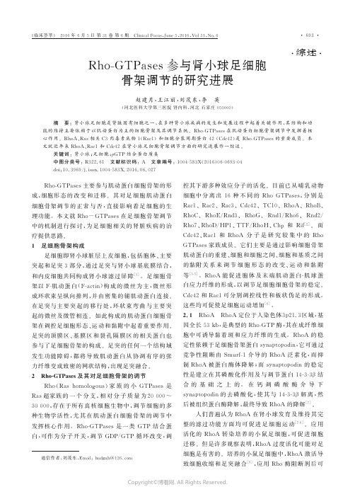
㊃综述㊃通信作者:刘茂东,E m a i l :l m d gx h @126.c o m R h o -G T P a s e s 参与肾小球足细胞骨架调节的研究进展赵建月,王江丽,刘茂东,李 英(河北医科大学第三医院肾内科,河北石家庄050000) 摘 要:肾小球足细胞是肾脏固有细胞之一,在多种肾小球疾病的发生和发展过程中起着关键作用,其结构和功能的维持主要依赖于以肌动蛋白为主的细胞骨架及其调节系统㊂R h o -G T P a s e s 在肌动蛋白细胞骨架调节中发挥着核心作用㊂R h o A ㊁R a s 相关C 3肉毒素底物1(R a c 1)和细胞分裂周期蛋白42(C d c 42)是R h o -G T P a s e s 的重要成员㊂本文就近年来R h o A ㊁R a c 1和C d c 42在肾小球足细胞骨架调节方面的研究进展作一综述㊂关键词:肾小球;足细胞;ρG T P 结合蛋白质类中图分类号:R 322.61 文献标识码:A 文章编号:1004-583X (2016)06-0693-04d o i :10.3969/j.i s s n .1004-583X.2016.06.027 R h o -G T P a s e s 主要参与肌动蛋白细胞骨架的形成,细胞形态的改变和迁移㊂其对足细胞肌动蛋白细胞骨架调节的正常与否,直接影响着足细胞的生理功能㊂本文就R h o -G T P a s e s 在足细胞骨架调节中的机制进行探讨,为足细胞相关的肾脏疾病的治疗提供思路㊂1 足细胞骨架构成足细胞即肾小球脏层上皮细胞,包括胞体㊁主要突起和足突3部分,通过足突与肾小球基底膜结合,和内皮细胞共同构成肾小球滤过屏障[1]㊂足细胞骨架以F -肌动蛋白(F -a c t i n )构成的微丝为主,微丝形成环状束呈纵向排列,并由密集的辅肌动蛋白连接㊂在足突与主要突起的移行处,环状束弯曲与主要突起的微丝及微管相连㊂如此构成的肌动蛋白细胞骨架在调控足细胞形态㊁运动和黏附中起着重要作用㊂足突的顶膜区㊁基膜区和裂孔隔膜区的相关蛋白也参与了足细胞骨架的构成㊂足突的任何一个结构域发生功能障碍,都将导致肌动蛋白从协调有序的张力纤维变成致密的网状结构,出现足突融合㊂2 R h o -G T P a s e s 及其对足细胞骨架的调节R h o (R a sh o m o l o go u s )家族的小G T P a s e s 是R a s 超家族的一个分支,相对分子质量为20000~30000,存在于所有真核细胞生物中,调节细胞的多种生物学活性,尤其在肌动蛋白细胞骨架的调节中发挥核心作用㊂R h o -G T P a s e s 是一类G T P 结合蛋白,可作为分子开关,调节G D P /G T P 循环改变,调控其下游多种效应分子的活化㊂目前已从哺乳动物细胞中分离出16种不同的R h o G T P a s e s,分别是R a c 1㊁R a c 2㊁R a c 3㊁C d c 42㊁T C 10㊁R h o A ㊁R h o B ㊁R h o C ㊁R h o E /R n d 3㊁R h o G ㊁R n d 1/R h o 6㊁R n d 2/R h o 7㊁R h o D /H P 1㊁T T F /R h o H ㊁C h p 和Ri f [2]㊂而C d c 42㊁R a c 1和R h o A 分子是研究较集中的R h o G T P a s e s 家族成员㊂它们主要是通过影响细胞骨架肌动蛋白的重建㊁细胞和细胞之间㊁细胞和基质之间的黏附关系来调节细胞形态的改变㊁运动和黏附等[3-5]㊂R h o A 能促进胞体及末端肌动蛋白-肌球蛋白应力纤维的形成,以调节足细胞细胞骨架的稳定㊂C d c 42和R a c 1可分别调控线性和板状伪足的形成㊂这些均可促使足细胞运动增加[6]㊂2.1 R h o A R h o A 定位于人染色体3p 21.3区域,基因全长53k b ,是典型的R h o -G T P 酶,其在成纤维细胞中可诱导黏着斑和应力纤维的生成㊂R h o A 的稳定性依赖于足细胞骨架蛋白s y n a p t o po d i n ,它可通过竞争性阻断由S m u r f -1介导的R h o A 泛素化,而抑制R h o A 被蛋白酶体降解;而s y n a p t o p o d i n 的稳定性是建立在其磷酸化作用及与调节蛋白14-3-3β结合的基础之上的㊂在钙调磷酸酶介导下s y n a p t o p o d i n 的去磷酸化,使其与14-3-3β解离,然后被组织蛋白酶降解,最终导致R h o A 的降解[7]㊂人们普遍认为R h o A 在肾小球发育及维持其完整的滤过功能方面均可促进足细胞运动[7-8]㊂应用活化的R h o A 转染培养的小鼠足细胞,可促进细胞迁移㊂但是许多观察表明,R h o A 过度活化可能对足细胞是有害的㊂培养的小鼠足细胞中,R h o A 激活导致细胞收缩和足突融合[9],应用R h o 酶阻断剂后可㊃396㊃‘临床荟萃“ 2016年6月5日第31卷第6期 C l i n i c a l F o c u s ,J u n e 5,2016,V o l 31,N o .6Copyright ©博看网. All Rights Reserved.促进足突的延伸,表明R h o酶的活化可能抑制足突的形成[10-11],且R h o酶阻断剂可抑制机械外力所致的肌动蛋白微管结构重组㊂在多种动物肾炎模型中,如:嘌呤霉素氨基核苷(P A N)肾病微小病变模型[9]㊁肾大部切除模型[12],足细胞中R h o A活化形式显著增多㊂应用R h o A抑制剂Y27632或f a s u d i l 后,足细胞的形态出现明显改善且实验动物的尿蛋白水平显著下降㊂其机制可能与R h o A的活性被抑制后,足细胞的特异性蛋白如n e p h r i n㊁s y n a p t o p o d i n 等的表达增加,且肌动蛋白发生重排密切相关[13-14]㊂Z h u等[15]研究发现,R h o A活化形式的减少导致足突消失和白蛋白尿,病理改变与微小病变相似㊂而当R h o A活化形式表达增加时,出现大量蛋白尿,同时伴有细胞外基质基因表达的上调,病理学变化类似于局灶节段性肾小球硬化(F S G S)㊂这表明,尽管基础水平的R h o A可能对维持足突正常结构㊁功能是必需的,但R h o A活化形式的减少或过度激活对足细胞是有害的㊂推测足细胞运动异常可能会扰乱足细胞裂孔隔膜结构并促使足突消失㊂因此,足细胞的正常生理功能需要这两种情况的相互平衡[16]㊂研究发现,R h o A的非活化形式主要表达在细胞浆,而其活化形式主要表达在细胞膜[17]㊂正常人肾组织中,R h o A主要分布在远曲小管和肾小球包曼囊上皮细胞的细胞质中;I g A肾病患者肾穿刺组织中,R h o A主要分布在近曲小管和肾小球包曼氏囊的上皮细胞,且其表达水平较正常对照组明显增加[18]㊂2.2 R a c1 R a c1定位于人染色体7p22区域,基因全长29k b,包含7个外显子,属于R h o家族蛋白中R a c亚家族成员之一㊂永生小鼠足细胞中,R a c1的活性在处于分化状态的细胞中明显增加,用活化的R a c1转染未分化小鼠足细胞,导致细胞体积增大,片状伪足的数目增加[9],其机制是表达增强的R a c1可通过调控细胞骨架蛋白重排而促进未分化足细胞的伪足形成㊂培养的小鼠肾小球上皮细胞中,n e p h r i n的过表达可通过激活磷酸肌醇-3-激酶(P I3K)的信号转导,增加R a c1的活性,抑制R h o A的活性,同时使F-a c t i n及应力纤维的分解增加,其分解将产生肌动蛋白单体,有利于肌动蛋白重排㊁足突的形成及细胞迁移㊂相反, R a c1活性的下降,可促使足突消失[19]㊂在上述实验及P A N肾炎模型中R a c1的活化形式是增加的[9],而在被动型海曼肾炎模型中足细胞R a c1的活化形式是减少的[20]㊂一些研究表明R a c1活化形式的增多可致足细胞受损㊂人类免疫缺陷病毒(H I V)1型蛋白N e f可结合R a c1的活化剂V a v2并使其激活[21],从而间接增加R a c1的活化形式㊂在培养的小鼠足细胞中,N e f在活化R a c1的同时可使R h o A失活[22],引起足细胞细胞骨架的重排,这表明N e f引起的R h o-G T P酶的活性紊乱可能参与H I V 相关肾炎的发病[23]㊂用脂多糖(L P S)诱导小鼠产生蛋白尿,发现R a c1和C d c42的活化形式均增加,且表达于足细胞的尿激酶受体(u P A R)㊁v i t r o n e c t i n㊁β3整合素参与了此过程[24]㊂但用αVβ3整合素抑制剂治疗,或在敲除u P A R的小鼠中均可发现R a c1和C d c42活性不增加[25]㊂u P A R-β3信号通路激活p130C a s-C r k复合物㊁R a c1激活剂和R a c,导致肌动蛋白细胞骨架重塑,细胞膜隆起和细胞运动增加[26]㊂u P A R的酶裂解产物可溶性u P A R(s u P A R),作为体液因子可能参与特发性F S G S的发病[6]㊂培养的小鼠足细胞经重组s u P A R作用后发现β3整合素被激活,特发性F S G S患者的血浆也被证实有此种情况[6]㊂这表明由s u P A R激活的β3整合素可能通过激活R a c1和C d c42负性调节足细胞的形态和功能㊂在敲除G D P酶解离抑制因子(G D I s)的小鼠肾脏,R a c1被激活,出现大量蛋白尿和严重的足细胞损伤,应用R a c1抑制剂后可显著降低蛋白尿水平㊂处于分化状态的足细胞内,存在一种由R h o A活化的R a c1G T P酶激活蛋白(R a c1-G A P),即A r h g a p24,其表达活性较未分化足细胞明显增加㊂A r h g a p24基因突变可增加足细胞中R a c1活化形式的表达,且其基因突变与家族性F S G S发病相关㊂因此,人们推测活化的R a c1可能参与家族型F S G S的发病[27]㊂在先天性激素抵抗型肾病综合征患者的肾小球中, A R H G D I A基因突变及R a c1㊁C d c42活化形式增多,使足细胞运动增加,足突消失出现蛋白尿;应用R a c1抑制剂后,患者的肾损伤得到部分恢复[28-29]㊂尽管这些均可表明R a c1的激活与足细胞足突消失之间存在某种联系,但目前尚缺乏直接有效的证据㊂2.3 C d c42 C d c42相对分子质量大小为25000,其编码基因定位于1p36.1㊂C d c42在细胞骨架动态变化调节中发挥着重要的作用㊂C d c42通过诱导细胞肌动蛋白聚合,对肌动蛋白细胞骨架发挥调节作用㊂有研究证实敲除足细胞中C d c42的编码基因,可抑㊃496㊃‘临床荟萃“2016年6月5日第31卷第6期 C l i n i c a l F o c u s,J u n e5,2016,V o l31,N o.6Copyright©博看网. All Rights Reserved.制肌动蛋白向n e p h r i n聚集位点的集聚,导致足突融合,引发先天性肾病综合征和肾小球硬化[30]㊂足细胞骨架蛋白s y n a p t o p o d i n可与胰岛素受体酪氨酸激酶底物(I R S p53)结合,通过阻断C d c42-I R S p53-M e n a复合物的形成,从而防止丝状伪足的形成[31]㊂应用M e n a阻滞剂可改善L P S所致蛋白尿,且s y n a p t o p o d i n减少,可引起C d c42活化形式增加,导致足细胞损伤和蛋白尿[31]㊂有研究报道,在腓骨肌萎缩神经病变和F S G S的患者中发现了I N F2基因的突变,其可引起C d c42的活化形式增加,并与突变的I N F2结合,导致C d c42在足细胞的错误定位和细胞骨架的重排[32]㊂3R h o-G T P a s e s活性的调节调控R h o-G T P a s e s活性状态的蛋白主要有3组:促进G D P向G T P转化的蛋白-鸟嘌呤核苷酸交换因子(G E F s),增加G T P a s e水解活性的蛋白-G T P酶激活蛋白(G A P s)及抑制G D P解离的蛋白-鸟嘌呤核苷酸分解抑制因子(R h o G D I s),它抑制了G E F s的催化作用,阻止G D P从G T P a s e s分离,维持G T P a s e在一个非活性状态[33],见图1㊂图1R h o-G T P a s e s活性的调节4展望R h oG T P a s e s是重要的细胞转导因子,在调控足细胞细胞骨架蛋白的排列中发挥着尤为重要作用的㊂越来越多的证据表明,不能简单的认为R h o G T P a s e s在活化抑或非活化状态下,对足细胞发挥保护抑或损伤作用㊂应当根据R h oG T P a s e s活性发生变化时的时间㊁位置,及R h oG T P a s e s其他成员的变化,进行综合且具体的分析㊂阐明R h oG T P a s e s 在细胞信号转导中的作用对于揭示复杂的信号转导机制和临床上多种疾病的发病机制具有重要的指导意义[34]㊂随着磷酸化蛋白组学㊁现代分子遗传学和肾小球疾病基因研究的发展,上述循环因子和足细胞信号转导途径有望成为今后潜在的治疗靶点㊂参考文献:[1] M u n d e l P,K r i z W.S t r u c t u r ea n df u n c t i o no f p o d o c y t e s:a nu p d a t e[J].A n a tE m b r y o l(B e r l),1995,192(5):385-397.[2] F uZ,C h e nY,Q i nF,e t a l.I n c r e a s e da c t i v i t y o fR h ok i n a s ec o n t r i b u t e st o h e m o g l o b i n-i nd u ce d e a r l y d i s r u p t i o n of t h eb l o o d-b r a i nb a r r i e r i nv i v o a f t e r t h e oc c u r r e n c e o f i n t r a c e r e b r a lh e m o r r h a g e[J].I n tJC l i nE x p P a t h o l,2014,7(11):7844-7853.[3] R i d l e y A J.R h o G T P a s e sa n dc e l lm i g r a t i o n[J].JC e l lS c i,2001,114(P t15):2713-2722.[4] E t i e n n e-M a n n e v i l l eS,H a l lA.R h o G T P a s e s i nc e l lb i o l o g y[J].N a t u r e,2002,420(6916):629-635.[5] B e v e r i d g e R D,S t a p l e sC J,P a t i lA A,e ta l.T h el e u k e m i a-a s s o c i a t e d R h o g u a n i n en u c l e o t i d ee x c h a n g ef a c t o rL A R Gi sr e q u i r e d f o r e f f i c i e n t r e p l i c a t i o n s t r e s s s i g n a l i n g[J].C e l l C y c l, 2014,13(21):3450-3459.[6] W e l s hG I,S a l e e m MA.T h e p o d o c y t ec y t o s k e l e t o n--k e y t oaf u n c t i o n i n gg l o m e r u l u si nh e a l t h a n d di s e a s e[J].N a t R e vN e p h r o l,2012,8(1):14-21.[7] F a u lC,D o n n e l l y M,M e r s c h e r-G o m e zS,e ta l.T h ea c t i nc y t o s k e l e t o no f k id ne y p o d o c y t e si s a d i r e c tt a r g e t o ft h ea n t i p r o t e i n u r i c e f f e c t o f c y c l o s p o r i n eA[J].N a tM e d,2008,14(9):931-938.[8] A s a n u m a K,Y a n a g i d a-A s a n u m a E,F a u l C,e t a l.S y n a p t o p o d i no r c h e s t r a t e sa c t i no r g a n i z a t i o na n dc e l lm o t i l i t yv i a r e g u l a t i o no fR h o As i g n a l l i n g[J].N a tC e l lB i o l,2006,8(5):485-491.[9] A t t i a sO,J i a n g R,A o u d j i tL,e t a l.R a c1c o n t r i b u t e s t o a c t i no r g a n i z a t i o n i n g l o m e r u l a r p o d o c y t e s[J].N e p h r o n E x pN e p h r o l,2010,114(3):e93-e106.[10] G a oS Y,L iC Y,C h e nJ,e ta l.R h o-R O C Ks i g n a l p a t h w a yr e g u l a t e s m i c r o t u b u l e-b a s e d p r o c e s s f o r m a t i o n o f c u l t u r e dp o d o c y t e s--i n h i b i t i o no f R O C K p r o m o t e d p r o c e s se l o n g a t i o n[J].N e p h r o nE x p N e p h r o l,2004,97(2):e49-e61. [11] K o b a y a s h i N,G a o S Y,C h e n J,e t a l.P r o c e s s f o r m a t i o n o f t h er e n a l g l o m e r u l a r p o d o c y t e:i s t h e r e c o mm o n m o l e c u l a rm a c h i n e r y f o r p r o c e s s e s o f p o d o c y t e s a n d n e u r o n s[J].A n a t S c iI n t,2004,79(1):1-10.[12] B a b e l o v aA,J a n s e nF,S a n d e rK,e ta l.A c t i v a t i o no fR a c-1a n d R h o A c o n t r ib u t e st o p o d oc y t ei n j u r y i nc h r o n i ck id ne yd i se a s e[J].P L o SO n e,2013,8(11):e80328.[13]S u z u k iH,Y a m a m o t o T,F u j i g a k iY,e ta l.C o m p a r i s o no fR O C Ka n d E G F R a c t i v a t i o n p a t h w a y si nt h e p r o g r e s s i o no fg l o m e r u l a r i n j u r i e s i nA n g I I-i n f u s e d r a t s[J].R e nF a i l,2011,33(10):1005-1012.[14]J i a n g L,D i n g J,T s a i H,e ta l.O v e r-e x p r e s s i n g t r a n s i e n t㊃596㊃‘临床荟萃“2016年6月5日第31卷第6期 C l i n i c a l F o c u s,J u n e5,2016,V o l31,N o.6Copyright©博看网. All Rights Reserved.r e c e p t o r p o t e n t i a l c a t i o n c h a n n e l6i n p o d o c y t e s i n d u c e sc y t o s k e l e t o nr e a r r a n g e m e n t t h r o u g hi n c r e a s e so f i n t r a c e l l u l a rC a2+a n dR h o Aa c t i v a t i o n[J].E x p B i o lM e d(M a y w o o d),2011,236(2):184-193.[15] Z h uL,J i a n g R,A o u d j i tL,e ta l.A c t i v a t i o n o f R h o A i np o d o c y t e s i n d u c e sf o c a ls e g m e n t a l g l o m e r u l o s c l e r o s i s[J].JA mS o cN e p h r o l,2011,22(9):1621-1630.[16] K i s t l e r A D,A l t i n t a s MM,R e i s e r J.P o d o c y t e G T P a s e sr e g u l a t ek i d n e y f i l t e r d y n a m i c s[J].K i d n e y I n t,2012,81(11): 1053-1055.[17] H a l lA.R h oG T P a s e s a n d t h e a c t i n c y t o s k e l e t o n[J].S c i e n c e,1998,279(5350):509-514.[18] M a t t i i L,S e g n a n iC,C u p i s t iA,e ta l.K i d n e y e x p r e s s i o no fR h o A,T G F-b e t a1,a n d f i b r o n e c t i n i n h u m a n I g An e p h r o p a t h y[J].N e p h r o nE x p N e p h r o l,2005,101(1):e16-e23. [19] Z h uJ,S u n N,A o u d j i t L,e ta l.N e p h r i n m e d i a t e sa c t i nr e o r g a n i z a t i o nv i a p h o s p h o i n o s i t i d e3-k i n a s e i n p o d o c y t e s[J].K i d n e y I n t,2008,73(5):556-566.[20] Z h a n g H,C y b u l s k y A V,A o u d j i t L,e ta l.R o l e o f R h o-G T P a s e si n c o m p l e m e n t-m e d i a t e d g l o m e r u l a r e p i t h e l i a lc e l li n j u r y[J].A mJP h y s i o lR e n a l P h y s i o l,2007,293(1):F148-F156.[21] F a c k l e rO T,L u o W,G e y e r M,e ta l.A c t i v a t i o no fV a vb yN e fi n d u c e s c y t o s k e l e t a l r e a r r a n g e m e n t s a n d d o w n s t r e a me f f e c t o r f u n c t i o n s[J].M o l C e l l,1999,3(6):729-739.[22] L uT C,H eJ C,W a n g Z H,e ta l.H I V-1N e fd i s r u p t st h ep o d o c y t ea c t i n c y t o s k e l e t o n b y i n t e r a c t i n g w i t h d i a p h a n o u si n t e r a c t i n gp r o t e i n[J].JB i o lC h e m,2008,283(13):8173-8182.[23]S u n a m o t o M,H u s a i nM,H e J C,e t a l.C r i t i c a l r o l e f o rN e f i nH I V-1-i n d u c e d p o d o c y t e d e d i f f e r e n t i a t i o n[J].K i d n e y I n t,2003,64(5):1695-1701.[24] N a g a s eM,F u j i t aT.R o l eo fR a c1-m i n e r a l o c o r t i c o i d-r e c e p t o rs i g n a l l i n g i nr e n a l a n dc a r d i a cd i s e a s e[J].N a tR e v N e p h r o l,2013,9(2):86-98.[25] W e iC,Möl l e r C C,A l t i n t a s MM,e ta l.M o d i f i c a t i o n o fk i d n e y b a r r i e rf u n c t i o n b y t h eu r o k i n a s er e c e p t o r[J].N a tM e d,2008,14(1):55-63.[26]S m i t h HW,M a r s h a l l C J.R e g u l a t i o n o fc e l ls i g n a l l i n g b yu P A R[J].N a tR e vM o l C e l l B i o l,2010,11(1):23-36. [27] A k i l e s hS,S u l e i m a n H,Y u H,e ta l.A r h g a p24i n a c t i v a t e sR a c1i n m o u s e p o d o c y t e s,a n da m u t a n tf o r m i sa s s o c i a t e dw i t hf a m i l i a lf o c a ls e g m e n t a l g l o m e r u l o s c l e r o s i s[J].J C l i nI n v e s t,2011,121(10):4127-4137.[28]S h i b a t aS,N a g a s e M,Y o s h i d a S,e ta l.M o d i f i c a t i o n o fm i n e r a l o c o r t i c o i d r e c e p t o r f u n c t i o n b y R a c1G T P a s e:i m p l i c a t i o n i n p r o t e i n u r i ck i d n e y d i s e a s e[J].N a t M e d,2008,14(12):1370-1376.[29] G e eH Y,S a i s a w a t P,A s h r a fS,e t a l.A R H G D I A m u t a t i o n sc a u s e n e p h r o t i c s y nd r o me v i a d ef e c t i v eR HOG T P a s e s ig n a l i n g[J].JC l i n I n v e s t,2013,123(8):3243-3253. [30]S c o t tR P,H a w l e y S P,R u s t o n J,e t a l.P o d o c y t e-s p e c i f i c l o s so fC d c42l e a d st o c o n g e n i t a ln e p h r o p a t h y[J].J A m S o cN e p h r o l,2012,23(7):1149-1154.[31] Y a n a g i d a-A s a n u m a E,A s a n u m a K,K i m K,e t a l.S y n a p t o p o d i n p r o t e c t s a g a i n s t p r o t e i n u r i a b y d i s r u p t i n g C d c42:I R S p53:M e n as i g n a l i n g c o m p l e x e si n k i d n e yp o d o c y t e s[J].A mJP a t h o l,2007,171(2):415-427.[32] B o y e r O,N e v o F,P l a i s i e r E,e ta l.I N F2m u t a t i o n si nC h a r c o t-M a r i e-T o o t hd i s e a s ew i t h g l o m e r u l o p a t h y[J].NE n g lJM e d,2011,365(25):2377-2388.[33] O l o f s s o n B.R h o g u a n i n e d i s s o c i a t i o n i n h i b i t o r s:p i v o t a lm o l e c u l e s i nc e l l u l a r s i g n a l l i n g[J].C e l l S i g n a l,1999,11(8): 545-554.[34]张雄鹰.R h o激酶抑制剂治疗急性脑梗死的疗效观察[J].临床荟萃,2013,28(4):439-440.收稿日期:2016-03-02编辑:王秋红㊃696㊃‘临床荟萃“2016年6月5日第31卷第6期 C l i n i c a l F o c u s,J u n e5,2016,V o l31,N o.6Copyright©博看网. All Rights Reserved.。
细胞骨架与细胞运动

6.微管参与细胞内信号转导。
小结:微管
化学组成及结构:由微管蛋白α、β构成的中空管状结构, γ微管蛋白位于微管组织中心。
形式:单管、二联管及三联管; 微管组装的特点:动态不稳定性; 影响微管组装的因素:微管蛋白浓度、GTP、阳离子、
pH值、温度及药物等; 细胞内微管组装:以微管组织中心为起始点;中心粒、纤
精子头端启动微丝组装,形成顶体刺突完成受精。
7.微丝参与细胞信号传递:
细胞外的某些信号分子与细胞膜上的受体结合,可触发 膜下肌动蛋白的结构变化,从而启动细胞内激酶变化的 信号转导过程。 主要参与Rho蛋白家族有关的信号转导。Rho信号通路 通过微丝调节细胞黏附和细胞运动,其活性异常往往与 细胞癌变有关。
(三)细胞骨架与遗传性疾病
人类不动纤毛综合征 、 遗传性皮肤病单纯性大疱性 表皮松解症 等。
小结
细胞骨架的定义、种类; 各类细胞骨架的化学组成、结构特点、装配特
点及影响装配的因素; 各类细胞骨架在细胞内的存在形式; 各类细胞骨架的功能,有何联系与区别?
四、细胞骨架异常与疾病
(一)细胞骨架与肿瘤
1.肿瘤细胞内细胞骨架结构的破坏和解聚 ,无序紊乱排列 造成细胞形态异常有关。 2.根据中间纤维分布具有组织特异性的特点,用作临床肿瘤 病理诊断工具 。
(二)细胞骨架与神经系统疾病
如帕金森病、 阿尔茨海默病 、肌萎缩性侧索硬化症 、 幼稚性脊柱肌肉萎缩症 等都与神经丝蛋白的异常表达与 异常修饰有关。
3.中间纤维参与细胞分化。
❖不同类型的中间纤维蛋白严格地分布在不同类型的细胞中,具有组织 细胞的特异性。 ❖发育不同阶段的细胞,会表达不同类型的中间丝蛋白,是细胞分化的 标志。(可用于鉴定干细胞、细胞分化研究、及鉴别肿瘤细胞来源)
细胞生物学-细胞骨架

29
6 形成应力纤维(stress fiber)
应力纤维是由微丝与肌球蛋白-II组装的一种不稳定性收 缩束,结构类似肌原纤维,使细胞具有抗剪切力。
30
培养的上皮细胞中的应力纤维(微丝红色、微管绿色)
31
7 参与肌肉收缩
基本结构:肌纤维是圆柱形的肌细胞(长度可达40mm, 宽为10100μm), 并且含有许多核(可多达100个核)。
性,既正极与负极之别。
微丝纤维的负染电镜照片
10
三、微丝的装配过程
微丝(F-actin)由G-actin聚合而成,单体具有极性,装配时 首尾相接。在适宜的条件下,肌动蛋白单体可自组装为纤维。 微丝的组装过程分三个步骤:即成核期、延长期、平衡期。
11
影响装配的因素
微丝的装配同样受肌动蛋白临界浓度的影响,还受一些 离子浓度的影响:在含有ATP和Mg2+, 以及很低的Na+、K+ 等阳离子的溶液中,微丝趋向于解聚成G-肌动蛋白。
32
33
骨骼肌收缩的基本结构单位——肌小节
肌小节的主要成分是肌原纤维,电镜下可见肌原纤维是由两种 类型的长纤维构成, 一种是细肌丝,直径为6nm;另一种是粗 肌丝,直径为15nm。
34
粗肌丝: 组成肌节的肌球蛋白丝。 细肌丝: 组成肌节的肌动蛋白丝。
35
粗肌丝的构成---肌球蛋白(myosin)
12
踏车现象(treadmilling)
在微丝装配时,若G-肌动蛋白分子添加到F-肌动蛋白丝 上的速率正好等于G-肌动蛋白分子从F-肌动蛋白上失去的速 率时, 微丝净长度没有改变, 这种过程称为肌动蛋白的踏车 现象.
13
永久性微丝结构
在体内, 有些微丝是永久性结构, 如肌肉中的细丝及上皮 细胞微绒毛中的轴心微丝等。有些微丝是暂时性结构, 如 胞质分裂环中的微丝。
细胞生物学中英词汇排序版

Acetylcholine乙酰胆碱ACTH促肾上腺皮质激素Actin filament肌动微丝adenine (A) /腺嘌呤,Adenosine腺苷酸Adenylyl cyclase腺苷酸环化酶adhering junction 粘着连接adhesion plaque 粘着斑Aggregation聚合aleurone grain 糊粉粒allosome, heterochromosome 异染色体Allosteric变构的amino acids /氨基酸amyloplast 造粉体analytical cytology 分析细胞学Anaphase后期annulate lamella, annulate lamellae(复)环孔片层Antenna触须apoplast 质外体Apoptosis凋亡Arginine合成酶arm ratio 臂比Arterial动脉的Asian-Pacific Organization for Cell Biology, APOCB 亚洲及太平洋地区细胞生物学aster 星体astral ray, astral fiber 星射线又称“星体丝”。
astrocenter 星心体astrosphere 星体球Autophosphorylation自磷酸化autosome 常染色体axial filament 轴丝axon 轴突Barr body 巴氏小体basal granule, basal body 基粒basal lamina 基膜又称“基板”。
basement membrane 基底膜belt desmosome 带状桥粒biogenesis 生源论又称“生源说”。
Biotin生物素Bisphosphate二磷酸blepharoplast 生毛体Broadcast spawning/散卵生殖brush border 刷状缘Bud/出芽生殖Catalyze/ 催化cell biology 细胞生物学cell coat 细胞外被cell matrix 细胞基质cell membrane 胞膜cell morphology 细胞形态学cell pathology, cytopathology 细胞病理学cell physiology, cytophysiology 细胞生理学cell sociology 细胞社会学cell theory 细胞学说cell wall 细胞壁cell 细胞cellular immunology 细胞免疫学Cellular respirProkaryotes/原核细胞cellulose/纤维素,chitin/几丁质Central plug中央栓centric fusion 着丝粒融合centric split 着丝粒分裂centriole 中心粒centrodesm 中心体连丝centromere misdivision 着丝粒错分centromere plate 着丝粒板centromere 着丝粒染色体上的一段非编码的DNA,对动粒(或动粒蛋白)有组织和整合作用。
细胞生物学单词%_%
细胞分裂与细胞周期cell division 细胞分裂cell cycle 细胞周期mitosis 有丝分裂meiosis 减数分裂amitosis 无丝分裂prophase 前期prometaphase 前中期spindle 纺锤体kinetochore microtubule 动粒微管polar microtubule 极微管metaphase 中期anaphase 后期telophase 末期cytokinesis 胞质分裂contractile ring 收缩环meiosisⅠ第一次减数分裂meiosisⅡ第二次减数分裂synapsis 联会bivalent 二价体aster 星体tetrad 四分体synaptonemal complex,SC 联会复合体recombination nodule 重组小结chiasma 交叉chiasma terminalization 交叉端化direct division 无丝分裂interphase 分裂间期mitosis,M期分裂期cyclin 细胞周期蛋白cyclin-dependent kinase,Cdk细胞周期蛋白依赖性激酶Cdk inhibitor,CKI Cdk激酶抑制物maturation promoting factor,MPF成熟促进因子checkpoint 检测点growth factor 生长因子chalone 抑素V-oncogene 癌基因proto-oncogene 原癌基因cellular oncogene,C-onc 细胞癌基因anti oncogene 抑癌基因mitotic apparatus 有丝分裂器cytostatic factor,CSF 细胞静止因子细胞的内膜系统ribosome 核糖体endoplasmic reticulum, ER 内质网RER 粗面内质网SER 滑面内质网microsome 微粒体signal hypothesis 信号假说SRP 信号肽识别颗粒SRP-R 信号肽识别颗粒受体Golgi complex 高尔基复合体lysosome 溶酶体residual body 残余小体lipofusion 脂褐质siderosome 含铁小体multivesicular 多泡体myelin figure 髓样结构peroxisome 过氧化物酶体microbody 微体细胞膜与物质的穿膜运输membrane lipid 膜脂phospholipid 磷脂cholesterol 胆固醇glycolipid 糖脂intrinsic protein 内在蛋白integrator protein 整合蛋白transmembrane protein 跨膜蛋白extrinsic protein 外在蛋白peripheral protein 周边蛋白lipid anchored protein 脂锚定蛋白lipid-linked protein 脂连接蛋白cell coat 细胞外被glycocalyx 糖萼liquid-crystal state 液晶态lateral diffusion 侧向扩散 flip-flop 翻转运动 rotation 旋转运动 flexion 弯曲运动 microdomain 微区 lipid rafts 脂筏 passive diffusion 被动扩散 membrane transport protein 膜运输蛋白 carrier protein 载体蛋白 channel protein 通道蛋白 facilitated diffusion 易化扩散 P-class ion pump P-型离子泵 symport 共运输 antiport 对向运输 aquaporin ,AQP 水孔蛋白 vesicular transport 小泡运输 endocytosis 胞吞作用 exocytosis 胞吐作用 microvillus 微绒毛 cilia 纤毛 flagella 鞭毛 ruffle 褶皱 lamellipodium 片状伪足细胞衰老与细胞死亡 cell senescence 细胞衰老 cell death 细胞死亡 Hayflick life span Hayflick 界限 senescence-associated β- galactosidase ,SA-βgal β-半乳糖苷酶 senescence associated gene 衰老相关基因 longevity gene 抗衰老基因 premature senescence 早熟性衰老 necrosis 细胞坏死 apoptosis 细胞凋亡 apoptotic bodies 凋亡小体 programmed cell death, PCD 程序性细胞死亡 phosphatidylserine, PS reactive oxygen species, ROS 活性氧类物质 permeability transition pores, PT pores 线粒体渗透转变孔 anoikis 失巢凋亡 autophagy 细胞自噬 micro-autophagy 微自噬 macroautophagy 巨自噬 chaperone-mediated autophagy,CMA 分子伴侣介导的自噬 cell shrinkage 细胞皱缩 chromatin condensation 染色质凝聚 DNA ladders DNA 梯状条带细胞生物学的研究方法resolution ,R 分辨率 light microscopy 光学显微镜 fixation 固定 embedding 包埋 section 切片 staining 染色 phase contrast microscope 相差显微镜 Fluorescence microscope 荧光显微镜 fluorescence microscope 荧光显微镜transmission electron microscope, TEM透射电子显微镜ultrastructure 超微结构submicroscopic structures 亚显微结构 Scanning electron microscope (SEM ) 扫描电子显微镜 flowcytometer, FCM 流式细胞技术 cell fractionation 细胞分级分离 sedimentation coefficient ,S 沉降系数 Differential centrifugation 差速离心 velocity sedimentation 速度沉降 isodensity centrifugation 等密度离心 column chromatography 柱层析 in vitro cell culture 体外细胞培养 primary culture 原代培养 secondary culture 继代培养 explan 外植体 primary culture 原代培养物 cell line 细胞系cell strain 细胞株cell clone 细胞克隆cell fusion 细胞融合cell hybridization 细胞杂交natural fusion 自然融合artificial induced fusion 人工诱导融合cytochemistry 细胞化学技术enzyme-cytochemistry 酶细胞化学immunocytochemistry 免疫细胞化学细胞的基本概念与分子基础procaryotic cell 原核细胞nucleoid 拟核cell wall 细胞壁mycoplasma 支原体bacteria 细菌peptidoglycan 肽聚糖plasmid 质粒mesosome 中间体eucaryotic cell 真核细胞cell membrane 质膜cytoplasm 细胞质nucleus 细胞核virus 病毒viroid 类病毒prion 朊病毒线粒体outer membrane 外膜inner memebrane 内膜intermembrane space 膜间隙matrix 基质porin 孔蛋白cristae 嵴elemetary particle 基粒intermembrane space 膜间隙Tim 内膜转位子Tom 外膜转semiautonomous organelle半自主性细胞器endosymbiosis theory 内共生起源学说non-endosymbiosis theory非内共生起源学说cellular respiration 细胞呼吸chemiosmotic hypothesis 化学渗透假说ATP synthase ATP合酶mPT 线粒体通透性改变mitochondrial disorders 线粒体疾病细胞骨架和细胞运动cytoskeleton 细胞骨架microfilament 微丝microtubule 微管intermediate filament 中间纤维actin 肌动蛋白globular actin, G-actin 球状肌动蛋白filamentous actin, F- actin纤维状肌动蛋白tonfilament 张力丝myofilament 肌丝neurofilament 神经丝nucleation phase 成核阶段elongation phase 延长阶段steady-state phase 稳定期阶段phalloidin 鬼笔环肽cytochalasin 细胞松弛素microfilament-associated protein微丝结合蛋白myosin 肌球蛋白microvilli 微绒毛stress fiber 应力纤维tubulin 微管蛋白microtubule associated protein, MAP微管结合蛋白protofilament 原纤维assembly 组装disassembly 去组装microtubule organizing center MTOC微管组织中心centrosome 中心体centriole 中心粒dynamic instability 动态不稳定性colchicine 秋水仙素taxol 紫杉酚kinesin 驱动蛋白dynein 动力蛋白intermediate filament,IF 中间纤维tetramer 四聚体cyto-keratin 胞质角蛋白skeleton 骨骼蛋白intermediate filament associated protein,IFAP 中间纤维的结合蛋白cell motility 细胞运动细胞核cell nucleus 细胞核nuclear envelope 核膜inner nuclear membrane 内核膜outer nuclear membrane 外核膜perinuclear space 核周隙nuclear pore complex, NPC 核孔复合体karyophilic protein 亲核蛋白nuclear localization signal, NLS核定位信号exportin 输出蛋白chromatin 染色质chromosome 染色体genome 基因组unique sequence 单一序列middle repetitive sequence中度重复序列highly repetitive sequence高度重复序列replication origin 复制源centromere 着丝粒telomere 端粒histone 组蛋白core histone 核小体组蛋白linker histone 连接组蛋白nonhistone 非组蛋白Euchromatin 常染色质heterochromatin 异染色质barr body 巴氏小体nucleosome 核小体satellite 棒状小体细胞分化Cell Differentiation 细胞分化totipotent nucleus 全能性细胞核cell determination 细胞决定transdetermination 转决定stability 稳定性dedifferentiation 去分化transdifferentiation 转分化cellular reprogramming 细胞重编程induced pluripotent stem cells, iPS细胞诱导多能干细胞differential expression 差异表达housekeeping protein 管家蛋白luxury protein 奢侈蛋白luxury gene 奢侈基因polyploidy 多倍体polyteny 多线体maternal effect gene, MEG 母体效应基因temporal specificity 时间特异性stage specificity 阶段特异性locus control region, LCR 基因座控制区spatial specificity 空间特异性master control gene 细胞分化主导基因combinatory control 组合调控homeobox 同源异形框histone code 组蛋白密码microRNA 微小RNA embryonic induction 诱导或胚胎诱导regeneration 再生。
细胞骨架1

微丝结合蛋白
微丝结合蛋白将微丝组织成以下三种主要形式
Parallel bundle: MF同向平行排列,主要发现于微绒毛与丝 状伪足。
Contractile bundle: MF反向平行排列,主要发现于应力纤 维和有丝分裂收缩环。
Gel-like network: 细胞皮层(cell cortex)中微丝排列形式,
8.膜结合蛋白:将肌动蛋白纤维量接在膜上,参与构成粘合带,vinculin
单体聚合蛋白
微丝解聚蛋白
核化蛋白
纤维切断蛋白
单体隐蔽蛋白
单体聚合蛋白
单体聚合蛋白
Figure 16-53 Two possible mechanisms by which an actin-monomerbinding protein could inhibit actin polymerization. It is thought that thymosin(胸腺素) inhibits actin polymerization in one of these ways.
A.
MFs are made of actin and involved in cell motility.
Using ATP, G-actin polymerizes to form MF(F-actin)
Using ATP, G-actin polymerizes to form MF(F-actin)
轻酶解肌 球蛋白
胰蛋白酶
2、 In vitro, (Polymerization) both ends of the MF grow, but the plus end faster than the minus. Because actin monomers tend to add to a filament’s plus end and leave from its minus end---- “Tread-milling”
生长素及其运输抑制剂对粗梗水蕨孢子萌发的影响

生长素及其运输抑制剂对粗梗水蕨孢子萌发的影响彭溢;刘璐;李贵生【摘要】研究了生长素IAA及其运输抑制剂NPA,TIBA,BFA对粗梗水蕨孢子萌发率的影响.抑制剂NPA,TIBA,BFA均抑制孢子萌发,但IAA减弱NPA和TIBA的抑制作用,NPA的抑制及IAA对其的拮抗在不同萌发时期均有体现;生长素及其运输抑制剂在粗梗水蕨中通过影响细胞骨架甚至囊泡运输,损害了孢子萌发中重力感应和细胞核迁移等关键事件.【期刊名称】《吉首大学学报(自然科学版)》【年(卷),期】2017(038)003【总页数】5页(P55-59)【关键词】粗梗水蕨;生长素;运输抑制剂;孢子萌发【作者】彭溢;刘璐;李贵生【作者单位】吉首大学生物资源与环境科学学院,湖南吉首416000;吉首大学生物资源与环境科学学院,湖南吉首416000;吉首大学生物资源与环境科学学院,湖南吉首416000;植物资源保护与利用湖南省高校重点实验室(吉首大学),湖南吉首416000【正文语种】中文【中图分类】Q945.3蕨类植物孢子的萌发最终导致孢子的细胞核发生一次不均等的有丝分裂,较大和较小的细胞分别发展为原丝体和假根.水蕨(Ceratopteris richardii)孢子萌发中重力影响假根生长和细胞核迁移的方向[1],后者可能发挥着“平衡石”的作用[2].水蕨假根的生长方向也受到光照方向的影响.[2]秋水仙素抑制北美球子蕨(Onoclea sensibilis)孢子细胞核的迁移和分裂,这种情况下多数孢子最后仍然是单核的单细胞,少数发展出2个细胞但仍不能形成假根.[3]甲醇则使得北美球子蕨的孢子萌发为有2个细胞但没有假根的原丝体.[4]在铁线蕨(Adiantum capillus ̄veneris)孢子萌发中,红光促进首次细胞不均等分裂,但远红光和蓝光分别可逆和不可逆地抑制这种作用;在原丝体的细胞分裂中,红光却起抑制作用而蓝光促进细胞分裂.[5]紫萁(Osmunda japonica)的孢子萌发到2个细胞核需要钙离子依赖型蛋白激酶(CDPK),这个分子一般也参与重力反应和光反应.[6]人们也研究了赤霉素、脱落酸、茉莉酸、乙烯等植物激素处理对蕨类植物孢子萌发率的影响.[7]但是,从生长素角度研究蕨类植物的孢子萌发尚未见报道.生长素是最早研究的植物激素,体内最重要的生长素是吲哚乙酸(IAA).生长素的合成、运输、失活和降解对其功能有重要影响,特别是生长素通过质膜的向外运输形成的浓度梯度决定着形态发生和向性生长.[8]萘基邻氨甲酰苯甲酸(NPA)、三碘苯甲酸(TIBA)和布雷菲尔得菌素A(BFA)通过不同的机制影响生长素的极性运输.[9-10]本研究发现,IAA,NPA,TIBA,BFA均抑制粗梗水蕨(Ceratopteris pteridoides)孢子的萌发,但IAA能减弱NPA和TIBA的抑制作用,在不同萌发时期NPA均起抑制作用且IAA对它有逆转效果.1.1 实验材料粗梗水蕨孢子,采自武汉市植物园.1.2 孢子萌发4 ℃冰箱中取干燥的已过筛(100目尼龙网)的孢子,加少量水,将此孢子悬浮液在冰箱中放置24 h.吸干孢子悬浮液中的水并加入稀释的次氯酸钠溶液(v(次氯酸钠)∶v(水)=1∶5),消毒5 min后用无菌水清洗5次,加无菌水.取50 L孢子悬浮液置于培养皿(每个培养皿约205个孢子),加入IAA和/或抑制剂,每个处理中溶剂二甲基亚砜(DMSO)的总量为0.2 mL,并且培养液的总体积为10 mL.将培养皿倾斜约30°放置,28 ℃下培养,每日光照16 h.1.3 萌发率的计算光照培养7 d后,在显微镜下观察孢子,有假根的孢子被定为已萌发[1-2].孢子萌发率(%)=×100%.除特殊说明外,各个数据均至少有3次重复.2.1 DMSO对粗梗水蕨孢子萌发率的影响因为本研究中生长素及其运输抑制剂的溶液均以DMSO为溶剂,所以先了解DMSO的使用对粗梗水蕨孢子萌发率的影响,结果如图1所示.萌发7 d后,3次重复中,作为空白对照的蒸馏水处理的萌发率分别是81%,85%,82%,而DMSO处理的萌发率分别是74%,87%,80%.2组数据间的差异无统计学意义(P>0.05),说明DMSO对粗梗水蕨孢子萌发率的影响可以忽略.2.2 IAA对粗梗水蕨孢子萌发率的影响IAA不同浓度处理的萌发率如图2所示.没有添加IAA的空白对照的孢子萌发率为(77.5±2.5)%,0.1 mol·L-1 IAA处理的孢子的萌发率为(79.5±2.5)%,1 mol·L-1处理的孢子萌发率为(78.5±2.5)%,三者之间的差异无统计学意义.但是,10 mol·L-1 IAA使得孢子萌发率急剧下降到(18±3)%,而100 mol·L-1 IAA使得孢子基本不萌发.由此可知,低浓度的IAA不影响粗梗水蕨孢子的萌发率,而高浓度IAA强烈抑制孢子的萌发.2.3 NPA对粗梗水蕨孢子萌发率的影响无重复实验结果表明:粗梗水蕨孢子萌发5 d时,空白对照和2,4,6,8,10 mol·L-1 NPA处理中的萌发率分别是72%,70%,66%,63%,58%,55%;萌发7 d时,萌发率分别是85%,82%,78%,72%,67%,63%;萌发9 d时,萌发率分别是87%,85%,79%,73%,68%,64%;萌发11 d时,萌发率分别是88%,86%,80%,74%,71%,69%.5 d和7 d之间、7 d与9 d之间萌发率的差异极具统计学意义(P<0.01),而9 d和11 d之间萌发率的差异具统计学意义(P<0.05).5 d和7 d之间萌发率的差值最大,所以本研究均在萌发7 d时统计粗梗水蕨孢子的萌发率.NPA各个处理时间中粗梗水蕨的萌发率如图3所示.由图3可知,2 mol·L-1 NPA对孢子萌发率的抑制是极其显著的(P<0.01),并且随着NPA浓度的升高抑制效果更强烈.进一步的实验结果(图4)表明:5,10 mol·L-1 NPA确实能极其显著地抑制粗梗水蕨孢子的萌发,萌发率分别是(69±1)%和(55±3)%,而空白对照的萌发率是(80.5±2.5)%;15,20 mol·L-1 NPA使得萌发率分别只有(31.5±1.5)%和(8.5±3.5)%;25 mol·L-1 NPA下孢子基本不萌发. 2.4 TIBA和BFA对粗梗水蕨孢子萌发率的影响如图5所示,在粗梗水蕨孢子萌发7 d时,空白对照和50,100,150,200 mol·L-1 TIBA处理中的萌发率分别是(80.5±2.5)%,(53±4)%,(30.5±1.5)%,(15.5±1.5)%,0,各相邻处理之间的差异极具统计学意义.如图6所示,空白对照和25,50,75,100,125 mol·L-1 BFA处理中的萌发率分别是(81.5±0.5)%,(45±2)%,(27.5±2.5)%,(4.5±0.5)%,0,各相邻处理之间的差异也极具统计学意义.2.5 IAA削弱NPA和TIBA对孢子萌发的抑制如图7所示:25 mol·L-1 NPA能完全抑制粗梗水蕨孢子萌发,在同时加入0.1 mol·L-1 IAA时的萌发率上升到(22.5±2.5)%;当加入的IAA的浓度为1 mol·L-1时,萌发率继续上升到(44±4)%;当IAA的浓度为10 mol·L-1时孢子又基本不萌发,100 mol·L-1 IAA的处理中更是如此.由此可知,不高于10 mol·L-1的IAA均能削弱NPA对粗梗水蕨孢子萌发的抑制作用,1 mol·L-1 IAA的这种拮抗作用最明显.如图8所示:200 mol·L-1 TIBA处理使得粗梗水蕨孢子基本不萌发,在同时加入0.1 mol·L-1 IAA时的萌发率上升到(1.5±0.5)%,但二者之间的差异无统计学意义;加入1 mol·L-1 IAA时的萌发率上升到(34.5±2.5)%;10 mol·L-1 IAA继续使萌发率上升到(51±1)%;100 m ol·L-1 IAA处理中孢子又基本不萌发.由此可知,不高于100 mol·L-1 的IAA均能削弱TIBA对粗梗水蕨孢子萌发的抑制作用,以10 mol·L-1 IAA的效果最好.2.6 不同萌发时期NPA的抑制作用及IAA对其的拮抗如图9所示:在孢子培养期间加入25 mol·L-1 NPA,一开始加入NPA处理的孢子不萌发,萌发6,12 h时加入的NPA也强烈地抑制孢子萌发;而在萌发24 h时加入NPA处理的萌发率可达(6.5±3.5)%,48 h时加入NPA的萌发率是(27.5±7.5)%,2种情况下萌发率的提升都是显著的.由此可知,NPA对粗梗水蕨孢子萌发的抑制作用在萌发至12,24,48 h都有一定的效果.如图10所示:一开始加入25 mol·L-1 NPA,又分别于萌发6,12,24,48 h时加入1 mol·L-1 IAA,发现萌发6 h时加入IAA的萌发率为(41±3)%,萌发12 h时加入IAA的萌发率为(33±6)%,二者之间的差异无统计学意义;萌发24 h时加入IAA的萌发率显著下降为(15±4)%,萌发48 h时加入IAA的萌发率几乎为0.由此可知,IAA对NPA的拮抗作用在萌发至6~<48 h有一定的效果.3 结论与分析IAA影响粗梗水蕨孢子的萌发率,而1 mol·L-1似乎是一个浓度临界点,低于1 mol·L-1的IAA几乎不影响萌发率,但稍高浓度时萌发率急剧下降(图2).IAA,NPA,TIBA,BFA均只抑制而不促进孢子的萌发.完全抑制孢子萌发时,按浓度从低到高依次是NPA(25 mol·L-1,图4),IAA(100 mol·L-1,图2),BFA(125 mol·L-1,图6),TIBA(200 mol·L-1,图5).但是,1,10 mol·L-1 IAA分别能显著减弱NPA和TIBA对粗梗水蕨孢子萌发的抑制(图7,8).NPA的抑制作用在粗梗水蕨孢子萌发的不同时期有不同的程度(图9),巧合的是,在这些时期IAA又不等地逆转了NPA对孢子萌发的抑制作用(图10).可见,IAA和NPA之间存在拮抗关系.TIBA和NPA促进肌动蛋白纤丝的聚合[9,11],相反,外源生长素处理使得肌动蛋白纤丝解聚[12].所以,可能存在一种整合因子,它同时与IAA和NPA结合,与IAA的结合使得肌动蛋白解聚,与NPA的结合促进聚合.[13]在粗梗水蕨孢子的萌发中,IAA和NPA单独都起着抑制作用(图2,4),可能是IAA使得肌动蛋白过度解聚但NPA使得它过度聚合,而这2种非平衡状态的肌动蛋白都不利于孢子的萌发.另一方面,在含NPA的培养液中添加IAA处理的萌发率可达40%以上(图8,10),可能是因为二者介导的肌动蛋白的过度聚合和过度解聚相互冲抵并达到了较优化的平衡点.更进一步,完全抑制孢子萌发时c(NPA)∶c(TIBA)=1∶8,接近于最大拮抗效应时二者中IAA的浓度的比(1∶10),这支持生长素及其运输抑制剂相互作用的单整合因子竞争模型[13].完全抑制孢子萌发所需TIBA的浓度高于NPA 的,可能是因为二者与整合因子的亲和力前者低于后者.[14-15]值得注意的是,在多种情况下IAA都只能恢复约50%的孢子萌发(图7,8,10).可能是因为TIBA和NPA通过间接促进肌动蛋白纤丝的聚合而封闭了整个囊泡运输[9,11],外源生长素处理中肌动蛋白纤丝解聚只影响胞吞但不影响胞吐[16].BFA抑制胞吐但不影响胞吞,另一方面,BFA的受体GNOM的胞内转运似乎借助于微管.[17]与此一致,微管抑制剂秋水仙素抑制北美球子蕨孢子的萌发.[3]可见,BFA对粗梗水蕨孢子萌发的强烈的抑制作用似乎也与细胞骨架和胞内运输有关.在水蕨孢子的萌发过程中,几乎从一开始到之后12 h地球重力决定将来假根的方位,13~18 h孢子细胞核沿着重力预决定的路线迁移,接着发生细胞分裂,20~25 h孢子开裂,45~50 h假根出现.[1-2]在这些时间点加入NPA,不仅强烈抑制粗梗水蕨的孢子萌发且相互之间差异具统计学意义(图9),说明NPA可能对萌发过程中的重要事件均有影响.IAA在萌发12 h内加入显著减弱NPA对孢子萌发的抑制,这种作用在24 h时加入明显下降,在48 h时完全消失(图10).可见,NPA 和IAA可能影响到粗梗水蕨孢子萌发中的重力响应、细胞核迁移和细胞分裂等重要事件.【相关文献】[1] EDWARDS ERIN SWINT,ROUX STANLEY J.Limited Period of Graviresponsiveness in Germinating Spores of Ceratopteris Richardii[J].Planta,1994,195:150-152.[2] EDWARDS ERIN SWINT,ROUX STANLEY J.Influence of Gravity and Light on the Developmental Polarity of Ceratopteris Richardii Fern Spores[J].Planta,1998,205(4):553-560.[3] VOGELMANN TH C,BASSEL A R,MILLER J H.Effects of Microtubule ̄Inhibitors on Nuclear Migration and Rhizoid Differentiation in Germinating Fern Spores (Onoclea sensibilis)[J].Protoplasma,1981,109(3):295-316.[4] MILLER J H,GREANY R H.Rhizoid Differentiation in Fern Spores:Experimental Manipulation[J].Science,1976,193(4 254):687-689.[5] FURUYA M,KANNO M,OKAMOTO H,et al.Control of Mitosis by Phytochrome and a Blue ̄Light Receptor in Fern Spores[J].Plant Physiology,1997,113(3):677-683.[6] KAMACHI HIROYAKI,NOGUCHI MUNENORI,INROUE HIROSHI.Possible Involvement of Ca2+ ̄Dependent Protein Kinases in Spore Gernination of the fern Osmunda Japonica[J].Journal of Plant Biology,2004,47(1):27-32.[7] SUO Jinwei,ZHAO Qi,ZHANG Zhengxiu,et al.Cytological and Proteomic Analyses of Osmunda Cinnamomea Germinating Spores Reveal Characteristics of Fern Spore Germination and Rhizoid Tip Growth[J].Molecular & Cellular Proteomics,2015,14:2 510-2 534.[8] MICHNIEWICZ MARTA,BREWER PHILIP B,FRIML JI.Polar Auxin Transport and Asymmetric Auxin Distribution[J].The Arabidopsis Book,2007,5:e0108.[9] DHONUKSHE PANKAJ,GRIGORIEV ILYA,FISCHER RAINER,et al.Auxin Transport Inhibitors Impair Vesicle Motility and Actin Cytoskeleton Dynamics in Diverse Eukaryotes[J].Proceedings of the National Academy of Sciences of the United States of America,2008,105:4 489-4 494.[10] GELDNER NIKO,FRIML JI,STIERHOF YORK ̄DIETER,et al.Auxin Transport Inhibitors Block PIN1 Cycling and Vesicle Trafficking[J].Nature,2001,413:425-428.[11] ZHU Jinsheng,BAILLY AURELIEN,ZWIEWKA MARTA,et al.TWISTED DWARF1 Mediatesthe Action of Auxin Transport Inhibitors on Actin Cytoskeleton Dynamics[J].PlantCell,2016,28:930-948.[12] HOLWEG CAROLA,SÜSSLIN CHRISTINA,NICK PETER.Capturing in Vivo Dynamics of the Actin Cytoskeleton Stimulated by Auxin or Light[J].Plant and CellPhysiology,2004,45(7):855-863.[13] ZHU Jinsheng,GEISLER MARKUS.Keeping It All Together:Auxin ̄Actin Crosstalk in Plant Development[J].Journal of Experimental Botany,2015,66(16):4 983-4 998.[14] SUSSMAN MICHAEL R,GOLDSMITH MARY HELEN M.The Action of Specific Inhibitors of Auxin Transport on Uptake of Auxin and Binding of N ̄1 ̄Baphthylphthalamic Acid toa Membrane Site in Maize Coleoptiles[J].Planta,1981(1),152:13-18.[15] THOMSON KLAUS ̄STEN,HERTEL RAINER,MÜLLER SYBILLE,et al.1 ̄N ̄Naphthylphthalamic Acid and 2,3,5 ̄Triiodobenzoic Acid:In ̄Vitro Binding to Particulate Cell Fractions and Action on Auxin Transport in CornColeoptiles[J].Planta,1973,109(4):337-352.[16] PACIOREK TOMASZ,ZAŽMALOV EVA,RUTHARDT NADIA,et al.Auxin Inhibits Endocytosis and Promotes Its Own Efflux from Cells[J].Nature,2005,435:1 251-1 256. [17] 曹文杰,李贵生.生长素输出载体PIN蛋白的质膜定位机制[J].植物学报,2016,51(2):265-273.。
Cytoskeleton
The critical concentration (Cc) is the concentration of G-actin monomers in equilibrium with actin filaments. At monomer concentrations below the Cc, no polymerization takes place. At monomer concentrations above the Cc, filaments assemble until the monomer concentration reaches Cc
Actin filaments grow faster at (+) end than at (-) end
The polarity of actin filament is also manifested by the different rates at which ATP-G-actin adds to the two ends. —— Myosin decoration experiments demonstrate unequal growth rates at the two ends of an actin filament
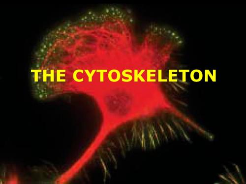
THE CYTOSKELETON
The movement of cells
In multicellular organisms, the migration of cells from one part of the body to another is critical to the development of the organism. Stationary cells may exhibit dramatic changes in their morphology. The internal movements of cells are essential elements in the growth and differentiation of cells.
细胞迁移——精选推荐

细胞迁移,与细胞移动同义,与细胞运动义近,指的是细胞在接收到迁移信号或感受到某些物质的浓度梯度后而产生的移动。
移动过程中,细胞不断重复着向前方伸出突触/伪足,然后牵拉后方胞体的循环过程。
细胞骨架和其结合蛋白,还有细胞间质是这个过程的物质基础,另外还有多种物质会对之进行精密调节。
若以移动方式与型态来比较,细胞迁移是通过胞体形变进行的定向移动,这有别于其他;如细胞靠鞭毛与纤毛的运动、或是细胞随血流而发生的位置变化,而且就移动速度来看,相比起后两者,细胞迁移要慢得多。
举例而言:成纤维细胞[注 1]的移动速度为1微米每分,若以精子的平均游动速度56.44微米/每秒,即3384微米/每分[1]来比较,两者差距约3000倍以上。
角膜细胞即使比成纤维细胞快上十倍,但是要完成从不来梅到汉堡这93公里的路程仍需要17123年[注 2]。
而且细胞用力甚轻。
成纤维细胞胞体收缩的力只有2×10⁻⁷牛顿,而角膜细胞的则是2×10⁻⁸牛顿(一牛顿约为人用手举起一鸡蛋所用的力道)[2]。
但此等“步缓力微”的细胞迁移,却是细胞觅食、伤口痊愈、胚胎发生、免疫反应、感染和癌症转移等等生理现象所涉及到的。
因此细胞迁移是目前细胞生物学研究的一个主要课题,科学家们试图通过对细胞迁移的研究,在阻止癌症转移、异体植皮等医学应用方面取得更大成果。
也因为细胞迁移独有的运动特性,成为今生物学热门研究方向。
细胞迁移的研究史1675年,显微技术的先驱人物安东尼·凡·列文虎克(Antonie van Leeuwenhoek)往英国皇家学会寄出一封信,里面描写了细菌的运动。
这封信可以说是打开了科学家对细胞迁移研究的第一页。
在往后这300多年时间,人们就一直试图去理解细胞迁移过程的细节。
而细胞迁移的关键物质—细胞骨架则要等到20世纪才被发现。
虽然1939年科学家阿尔伯特·山特吉尔吉(A. Szent-Györgyi)就已发现细胞骨架的成分—肌动蛋白和肌球蛋白,但是因为电子显微镜制作样本时需要对样品进行0到4 °C的低温固定,在这样的温度下细胞骨架会被破坏,即所谓的“解聚”。
医学生英语词汇

helicobacter pylori:幽门螺杆菌infection:感染oncogenic:致癌epigenetic:表观遗传mechanism: 机制gastric:胃carcinogenesis:致癌作用oncology:肿瘤学colonization:定植gram-negative:阴性gastritis:胃炎peptic ulcer:胃溃疡inflammation:炎症malignancy:恶性肿瘤elucidate:表明,阐明cytotoxin [,sait?0?5't?0?0ksin] -associated antigen A:细胞毒素相关抗原A epithelial [,epi'θi:li?0?5l] cell:上皮细胞pathogenicity [,p?0?3θ?0?5d?0?1i'nisiti]:致病性mucosa [mju:'k?0?5us?0?5]:粘膜multiple:多种catenin: 连环蛋白,连环素cyclooxygenase[,saikl?0?5u'?0?0ksid?0?1ineis]:环氧化酶induce:诱导methylation:甲基化modification:修饰investigate: 调查stem:干progenitor:祖shed light on: 昭示,阐明yield:产生witness:见证pathogen:病原体spiral-shaped:螺旋形noncardia adenocarcinoma ['?0?3d?0?5n?0?5u,kɑ:si'n?0?5um?0?5] :非贲门腺癌mucosal-associated lymphoid tissue lymphoma:粘膜相关淋巴组织淋巴瘤superficial:浅表individual:个人、个体present as:表现为primary:原发性lung cancer:肺癌intracellular:细胞内的cytokine:细胞因子apoptosis rate:凋亡率proliferation:增殖differentiation:分化virulence:毒力vacuolate ['v?0?3kju?0?5lit]:有空泡的,形成空泡的vacuolating sytotoxin:空泡毒素membrane:膜genome ['d?0?1i:n?0?5um] :基因组sequence:序列type IV secretion system:四型分泌系统peptidoglycan[,peptid?0?5u'ɡlaik?0?3n](PGN):肽聚糖syringe['s?0?1r?0?1nd?0?1]:注射器integrin:整合蛋白chromatin:核染色质encode:编码molecular:分子的cytoplasm:细胞质tyrosine:酪氨酸phosphorylate:磷酸化kinase:激酶glutamic acid –p rol i n e –i s oleuci n e –t y r osi n e –a l a n i n e (EPIYA) :谷氨酸、脯氨酸、异亮氨酸、酪氨酸、丙氨酸motif:基序regulate:调节morphogenesis:形态motility:运动cortactin:皮层肌动蛋白actin:肌动蛋白inactivation:灭活junction:n. 连接,接合;交叉点;接合点ezrin:埃兹蛋白scaffolding:支架,脚手架polarity:极性permeability:通透性receptor:受体phospholipase:磷脂via:通过stimulate:刺激nuclear :细胞核的vacuolization:空泡化inhibit:抑制translocate:转位target:目标range from:延伸polarity:极性cadherin:钙粘素promote:促进intestinal:肠的previous:以往mediate:介导invasiveness:侵袭critical:关键dysfunction:功能障碍aberrant:异常metabolism:代谢apoptosis:凋亡pre-incubation:预培养antagonist:拮抗剂prognosis:预后gerbil:沙鼠simultaneous:同时dysplastic:发育不良metaplasia:化生dysplasia:异型增生suppress:抑制preneoplastic lesions:癌前病变macrophage-derived:巨噬细胞源性infiltration:浸润accumulation:积累repeat:重复reveal:发现residue:残留物multimerization:多聚goblet-cell mucin(MUC2):杯状细胞粘蛋白diversity:多样化somatic:体细胞immunoglobulin:免疫球蛋白cell gap junction:细胞间隙连接ultrastructure:超微结构co-cultured:培养transmission:透射electron:电子microscope:显微镜preparation:制备fixation:固定embedding:嵌入situ:原地detect:发现rapid urease test:快速脲酶试验unit perimeter:单位周长precancerous lesions:癌前病变apical-junctional:顶部连接integrity:完整性severely:严重knockout:摧毁synapse:突触desmosome:桥粒(上皮细胞膜中的局部增厚部分)intercellular:在细胞间significantly:显著地vitro:(活)体外maintain:保持reflect:反映relation:关系eradication:根除therapy:疗法epidemiology:流行病学purchase:购买digestive:消化的isolate:分离cell line:细胞系grouping:归类、编组according to:根据diagnostic criteria:诊断标准exponential growing phase:对数生长期trypsin:胰蛋白酶inoculate:接种coverslip:盖玻片incubator:恒温箱saturation:饱和humidity:湿度suspensions:悬浮液ratio:比率fetal calf serum:胎牛血清specimen:标本glutaraldehyde:戊二醛paraformaldehyde:多聚甲醛precool:预冷store:贮存、贮备osmium acid:锇酸distill:蒸馏uranyl acetate:乙酸铀酰layer:表层dehydrated:脱水seriatim:逐一地,连续地acetone:丙酮gradient:梯度immerse:浸入epon:环氧树脂pure:纯ultrathin:超薄section:切片semi-thin section:半薄切片biopsy:活组织检查gastroscope:胃镜lead:铅uranium:铀stain:染色magnification:放大morphology:形态学statistical:统计学software:软件significant:显著criterion:标准,条件adjacent:邻近visualized:可视化internal:内部rare microvilli:罕见的微绒毛focal gap junction:局灶性间隙连接necrosis:坏死sparser:稀疏proportional:成比例的incidence:发生率crowd of genetic susceptibility:遗传易感人群decisive:决定性的indicate:表明atrophic gastritis:萎缩性胃炎intrude:侵入oxygen free radicals:氧自由基antioxidant:抗氧化剂capacity:能力concentrated in:集中在ectopic:异常aggregation:聚集attachment sites:附着点intact:完整conclusion:结论。
细胞分子生物学7(英文)

Although G-actin appears globular in the electron microscope, x-ray crystallographic analysis reveals that it is separated into two lobes by a deep cleft (Page 319). The lobes and the cleft compose the ATPase fold, the site where ATP and Mg2+ are bound. In actin, the floor of the cleft acts as a hinge that allows the lobes to flex relative to each other. When ATP or ADP is bound to G-actin, the nucleotide affects the conformation of the molecule. In fact, without a bound nucleotide, G-actin denatures very quickly.
All cell movements are a manifestation of mechanical work; they require a fuel (ATP) and proteins that convert the energy stored in ATP into motion. The cytoskeleton, a cytoplasmic system of fibers, is critical to cell motility. In the electron microscope, the cytoskeleton appears as a dense and seemingly random array of fibers. However, we now recognize that this array consists of three types of cytosolic fibers:
细胞外基质(ECM)解决方案-附参考文献
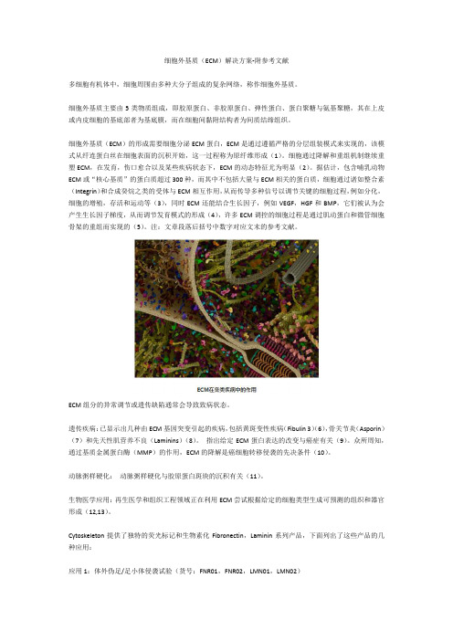
细胞外基质(ECM)解决方案-附参考文献多细胞有机体中,细胞周围由多种大分子组成的复杂网络,称作细胞外基质。
细胞外基质主要由5类物质组成,即胶原蛋白、非胶原蛋白、弹性蛋白、蛋白聚糖与氨基聚糖,其在上皮或内皮细胞的基底部者为基底膜,而在细胞间黏附结构者为间质结缔组织。
细胞外基质(ECM)的形成需要细胞分泌ECM蛋白,ECM是通过遵循严格的分层组装模式来实现的,该模式从纤连蛋白丝在细胞表面的沉积开始,这一过程称为原纤维形成(1)。
细胞通过降解和重组机制继续重塑ECM,在发育,伤口愈合以及某些疾病状态下,ECM的动态特征尤为明显(2)。
据估计,包含哺乳动物ECM或“核心基质”的蛋白质超过300种,而其中不包括大量与ECM相关的蛋白质,细胞通过诸如整合素(Integrin)和合成癸烷之类的受体与ECM相互作用,从而传导多种信号以调节关键的细胞过程,例如分化,细胞的增殖,存活和运动等(3),同时ECM还能结合生长因子,例如VEGF,HGF和BMP,它们被认为会产生生长因子梯度,从而调节发育模式的形成(4),许多ECM调控的细胞过程是通过肌动蛋白和微管细胞骨架的重组而实现的(5)。
注:文章段落后括号中数字对应文末的参考文献。
ECM组分的异常调节或遗传缺陷通常会导致致病状态。
遗传疾病:已显示出几种由ECM基因突变引起的疾病,包括黄斑变性疾病(Fibulin 3)(6),骨关节炎(Asporin)(7)和先天性肌营养不良(Laminins)(8)。
指出给定ECM蛋白表达的改变与癌症有关(9)。
众所周知,通过基质金属蛋白酶(MMP)的作用,ECM的降解是癌细胞转移侵袭的先决条件(10)。
动脉粥样硬化:动脉粥样硬化与胶原蛋白斑块的沉积有关(11)。
生物医学应用:再生医学和组织工程领域正在利用ECM尝试根据给定的细胞类型生成可预测的组织和器官形成(12,13)。
Cytoskeleton提供了独特的荧光标记和生物素化Fibronectin,Laminin系列产品,下面列出了这些产品的几种应用:应用1:体外伪足/足小体侵袭试验(货号:FNR01,FNR02,LMN01,LMN02)Cytoskeleton的荧光标记Fibronectin,Laminin可用于伪足/足小体体外侵袭试验(16)。
苏州大学 张焕相细胞本科授课大纲
主讲教师:张焕相、沈颂东、曲静细胞教学大纲一、教学目的细胞生物学是生物科学领域发展迅速、引人注目的重要学科,有关细胞生物学研究是现代生命科学的重要支柱之一,因而它已成为当今生物科学类专业本科生必修的一门主干课程。
通过细胞生物学(双语)课程的学习,要求学生达到如下目标:1 掌握细胞生物学中的基础知识和基本理论。
2 熟悉细胞生物学的基本研究手段和实验方法。
3 主动了解细胞生物学的最新研究进展和动态。
4 能将细胞生物学的基本理论融会到后续课程的学习中,从而领悟细胞生物学在整个生命科学中的重要地位。
二、课程内容Chapter 01 Introduction to Cells and Cell Biology 2学时教学内容:1. The discovery of cells2. Cell theory3. Why the cells the basic units of life?4. Basic properties of cells5. The size of cells6. Prokaryotes and eukaryotes7. Model organisims8. Cell replacement therapy9. How to learn cell biology?教学要点:1. 细胞学说的内容2. 如何理解细胞是生命体的基本单位3. 如何理解细胞的全能性4. 细胞的基本特性5. 真核细胞与原核细胞的比较6. 模式生物的意义和主要种类7. 细胞替代疗法的意义和策略2. 如何理解细胞是生命的基本单位3. 如何理解细胞的全能性4. 细胞的基本特性5. 真核细胞与原核细胞的特征比较6. 主要的模式生物及其意义7. 细胞替代疗法的意义和策略Chapter 02 Chemical Basis of Life 2学时教学内容:1. Atoms2. Chemical bonds3. Polar and nonpolar molecules4. The carbon atom5. Functional groups6. Organic molecules in cells biochemicals7. From atoms to the cells教学要点:1. 细胞的主要化学元素和化学键类型2. 细胞内分子的极性和亲水性特点Chapter 03 Membrane Structure 4学时教学内容:1. Typical plasma membranePlasma membrane,Endomembrane system,Biomembrane2. Plasma membrane structure3. Membrane lipidsPhospholipids,Three main types of membrane lipids4. Nature of the lipid bilayerMicelle,Lipid bilayer formation,Self-sealing property,Liposome,Fluidity of lipid bilayer,Lipid rafts5. Membrane proteinsIntegral proteins,Peripheral proteins,Lipid-anchored proteins6. Techniques for studying membrane proteinsFreeze-fracture replication,SDS-PAGE,2-D Gel Electrophoresis7. Membrane carbohydratesGlycolipids,Glycoproteins8. Characteristics of biomembraneMembrane fluidity and flexibility,Membrane asymmetry,Dynamic nature of membrane9. Red blood cell教学要点:1. 膜脂和膜蛋白的类型和特点2. 膜结构模型的要点3. 膜的主要特性及其意义4. 膜蛋白研究的主要技术Chapter 04 Membrane Transport and Membrane Potential 4学时教学内容:1. Movement of solutes across cell membrane2. Simple diffusionConcentration gradient and electrochemical gradient Diffusion of solutesDiffusion of waterDiffusion of ions3. Facilitated diffusion4. Active transportThree ways of driving active transportSodium-potassium pumpThree classes of ATP driven pumpsCotranport5. Membrane potentials and nerve impulsesResting potentialAction potentialPropagation of action potentialNeurotransmission教学要点:1. 被动运输与主动运输的比较和类型2. 简单扩散的影响因素3. 水通道的作用特点4. 离子通道的类型5. KcsA通道和Kv通道的结构和作用特点比较6. 易化扩散的特点和葡萄糖扩散的机制7. 钠钾泵的作用机制和意义8. 离子泵的类型和比较9. 协同运输的机制和类型,葡萄糖吸收的机制10. 静息电位和动作电位的产生原理11. 动作电位传播的机制,神经肌连接的作用机制Chapter 05 Intracellular Compartments 4学时教学内容:1. Compartmentalization of eukaryotic cellsCytoplasmic matrix(cytosol)Endomembrane systemDynamic nature of the endomembrane system2. Approaches to the study of endomembranesAutoradiographySubcellular fractionationGFPCell-free systemGenetic mutants3. Endoplasmic reticulun(ER)Structure and functions of RER and SER4. Golgi complexThe polarity of GCThe functions of GC5. LysosomesCharacteristics of lysosomesThe functions of lysosomes6. Cell nucleusNuclear envelope consists of two membranesThe nuclear laminaThe nuclear pore complex(NPC)教学要点:1. 如何理解内膜系统的动态性2. 研究内膜系统的主要方法3. 内质网的结构特点和功能4. 高尔基体的结构特点和功能5. 溶酶体的结构特点和功能6. 核膜的结构组成,核纤层和核孔复合体的特点Chapter 06 Protein Sorting 4学时教学内容:1. Road map of protein trafficProteins are imported into organelles by three mechanismsSorting signalSignal sequence2. Transport between the nucleus and cytosolNuclear localization signal(NLS)Nuclear transport receptorsImport of proteins from cytoplasm into nucleusNuclear export works like nuclear import, but in reverse3. Transport of proteins into mitochondria and chloroplastsThe protein translocators in the mitochondrial membranesTransport into matrix spaceTransport into the outer membraneTransport into the inner mitochondrial membrane and intermembrane spaceTwo signal sequences direct proteins to the thylakoid membrane in chloroplasts4. Transport of proteins into peroxisomes5. Transport of proteins from cytosol to ERCo-translational and post-translational importSignal hypothesis教学要点:1. 蛋白质运输的三种途径2. 蛋白质分选信号的类型和特点3. 核输入和核输出的意义和机制4. 蛋白质定位于线粒体不同部位的机制5. 蛋白质合成和运输的两种不同途径6. 新生肽如何输入内质网Chapter 07 Vesicular Transport 4学时教学内容:1. Coated vesiclesDifferent coats in vesicular transportClathrin-coated vesiclesCOP Ⅱ-coated vesiclesCOP Ⅰ-coated vesicles2. Rab proteins guide vesicle targeting3. SNAREs mediate membrane fusion4. EndocytosisPhagocytosisPinocytosis5. Exocytosis教学要点:1. 膜泡定向运输的决定因素2. 溶酶体酶是如何合成和运输的3. Rab蛋白在膜泡运输中的作用和机制4. SNARE在介导膜融合中的作用机制5. 微管在膜泡运输中的作用6. 如何理解高尔基体在蛋白质分选中的枢纽作用7. 胞吞作用的意义和类型,LDL摄入的机制Chapter 08 Cytoskeleton 6学时教学内容:1. MicrotubulesStructure and compositionMAPs(microtubule-associated proteins) Dynamics of MT assemblyMTOCsDrugs affecting MT assemblyMotor proteinsCilia and flagella2. MicrofilamentsStructure and compositionMF assembly and disassembly Cytochalasin and phalloidinActin-binding peoteinsMyosinsMuscle contractilityNonmuscle motility3. Intermediate filamentsStructure and compositionIF assembly and disassemblyFunctions of IF4. Nuclear matrix教学要点:1. 细胞骨架的类型和主要功能2. 微管的结构和组装特点3. MTOC的作用和中心体的结构特点4. 马达蛋白的类型和与微管的相互作用5. 纤毛和鞭毛的结构基础6. 微丝的结构和组装特点7. 不同的微丝结合蛋白和myosis与微丝的相互作用8. 肌肉收缩的微丝作用机制9. 微丝在细胞运动中的作用10. 中等纤维的结构和组装特点11. 中等纤维的不同类型和功能Chapter 09 DNA and Chromosome 4学时教学内容:1. Components of chromatinComponent of chromatin---DNAComponent of chromatin---histoneComponent of chromatin---nonhistone2. Nucleosome—structural unit of chromatinSummary of nucleosome structure3. Higher levels of chromatin structure4. Euchromatin and heterochromatin5. X-chromosome inactivationFeatures of X chromosome inactivationMechanism of X chromosome inactivation6. Structure of the mitotic chromosomeCentromere; Kinetochore; Telomere7. Giant chromosomes and Lampbrush chromosomes教学要点:1. 染色质和染色体的化学组成及关系2. 核小体的结构要点3. 染色体包装的主要模型4. 常染色质与异染色质的概念和意义5. X-染色体失活的意义、特征和机制6. 染色体的主要结构7. 巨染色体与灯刷染色体的概念和意义Chapter 10 Cell Cycle 6学时教学内容:1. Cell cycleCell cycle phasesCell cycle lengthCategories of cells in vivo based on proliferative states Synchronization of cellsMain biochemical events of cell cycle phases2. MitosisKey features during prophaseMetaphaseAnaphaseTelophaseCytokinesis3. Meiosis4. MPFDiscovery of MPFRole of MPFRegulation of MPF activity5. Diversity of cyclin-CDK complexes6. Checkpoints in cell-cycle controlG1 checkpoint (START/ Restriction point)G2 checkpoint (unreplicated-DNA checkpoint)M checkpoint(spindle-assembly checkpoint)chromosome-segregation checkpointDNA-damage checkpoint教学要点:1. 细胞根据增殖状态的分类2. 细胞周期同步化的主要方法3. 间期的意义和各时相的主要特点4. 结合纺锤体组装分析有丝分裂M期各阶段动态5. 减数分裂的特点、联会和联会复合体6. MPF的本质、功能和活性调节7. 主要的检验点控制机制Chapter 11 Cell Differentiation 4学时教学内容:1. Differentiation potency of cellsConcept and essence of cell differentiationHouse-keeping gene and Luxury geneMechanism of differential gene expressionCell totipotencyChange of totipotency during embryonic developmentSignificance of DollyTransdifferentiation2. Individual development and cell differentiationCell differentiation and cell determinationKey mechanisms of cell differentiation3. Stem cellCategories of stem cellsPatterns of stem-cell divisionEmbryonic stem cell (ES cell)Adult/tissue stem cellStem cells plasticityInduced pluripotent stem (iPS) cells4. Cancer cellBenign tumor and malignant tumorBasic properties of cancer cellsThe causes of cancerTumor-suppressor genes and oncogenesNew strategies for combating cancer教学要点:1. House-keeping gene和Luxury gene的概念及与细胞分化本质的关系2. 如何理解发育过程中的细胞全能性3. 细胞决定的机制及在分化中的意义4. 干细胞的基本特性和分类5. ES细胞的主要来源、形态和生化特征6. iPS细胞的概念和意义7. 癌细胞的主要形态和生理特征8. 原癌基因和抑癌基因的概念9. Rb和p53基因的意义和分子机制Chapter 12 Apoptosis 4学时教学内容:1. Cell aging/senescenceHayflick limitationLifespan of cells in vitro cultureCharacteristics of cell agingTheories of cell aging2. Cell apoptosis/Programmed cell deathSignificance of cell apoptosisCharacteristics of cell apoptosisTwo styles of the cell death3. Molecular mechanisms of apoptosisExtrinsic pathway of apoptosis-receptor-mediated pathwayIntrinsic pathway of apoptosis-mitochondria-mediated pathway Apoptosis is carried out by the caspase cascadeEvolutionarily conserved apoptotis pathway in C. elegans and vertebrates 教学要点:1. Hayflick界限和体外培养细胞的寿命2. 细胞衰老的特征和主要理论3. 细胞凋亡的意义和特征4. 受体介导和线粒体介导的细胞凋亡途径以及caspase级联的机制5. 线虫凋亡的基因调控机制Chapter 13 Cell Signal Transduction 6学时教学内容:1. Overview of cell signal transductionSignal transductionMain types of cell communicationReceptors2. G-protein-linked receptor and secondary messengerG-protein-linked receptorG-protein and its mechanismSecond messengercAMP signaling pathwayDouble messenger system3. Enzyme-linked receptorReceptor tyrosine kinases (RTKs)RTK-Ras signaling pathwayRTK-PIP2 pathwayRTK-PI3K pathwayJAK-STAT signaling pathway4. Intracellular receptorThe mechanism of the intracellular receptorThe role of NO as a signal molecule5. Role of calcium as an intracellular messenger6. Signals that originate from contacts between the cell surface and the substratum7. Important features of cell signaling教学要点:1. G蛋白耦联受体的概念和特征2. G蛋白的概念、特点和作用机理3. cAMP信号通路、肾上腺素升高血糖的细胞机制4. PIP2信号通路5. RTK的特点和Ras-MAPK信号途径6. 细胞内受体的特点和NO信号的作用7. 钙信使的特点和作用8. 信号转导的主要特点三、各章课时分配表。
肌动蛋白(ACTIN)和肌动蛋白丝(MF)

CytoskeletonChapter 6The terminus of growing axon from the sea hare Aplysa, MF in blue, MT in redLearning Objectives6.1 An overview6.2 M icrofilament (MF)6.3 Microtubules (MT)6.4 Intermediate filaments (IF)§6 The cytoskeleton and cell mobility6.1 Overview1.what is cytoskeletonAn important characteristic of died eukaryote cell is Brownian motion of contents (including organelles) of these cells, while the organelles and subcellular particles are orientally organised and carry out oriental motion and order metabolitic activities which are mediated by cytoskeleton.Colin SmithIF(vimentin)MTMFThe cytoskeleton is composed of three well-defined filamentousstructures1.Microtubles (MT), 22-25 nm in diameter;2.Microfilaments (MF), 7 to 9 nm in diameter;3.intermediate filaments (IF), 10 nm in diameter They are a highly dynamic group of structures capableof rapid and dramatic reorganization.In addition, many of functions of the cytoskeletonrequire a host ofaccessory proteins , which may arenot a part of the filaments themselves.2.The function of cytoskeletonThe elements that make up the cytoskeleton function ina number of interrelated activities:a)As a scaffold and structural supports within cell •dynamic shape •internal framework•positioning and organization of the variousorganelles•anchoring messenger RNAs and some proteins②As parts of the movement machinery •Swimming•Crawling and migration•Movement of materials and organelles within cells.③As signal transducors.30μm3. The study of the cytoskeletonUsing fluorescence microscopyUsing video microscopy and in vitro motility assaysUsing electron microscopic techniquesUsing molecular biology techs.Using inhibitors of cytoskeletonGFP methodDynamic changes in length of microtubuleswithin an epithelial cellVideo microscopy to follow the activities of molecular motorsLaser tweezersExpression of a mutant motor protein inhibits the dispersion of p i g m e n t granules in a pigment cell 20μmmon characteristics of proteinchemistry and structure of three typesof cytoskeletona)The constructural element proteins of MT, MFand IF are conserved in molecular evolution, butthey present in different isoforms, encoded byhighly conserved gene family.b)Cytoskeleton proteins are unsoluble in nonionicdetergent, as Triton X-100c)The element proteins of IF are filament-like butactin and tubulin are globular proteins, but theyconstruct linear, non-branching polymer ofproteins.d)Their assembling is to be regulated byCalcium ions and need using energy, in ATP or GTP form.e)Formed cytoskeleton filaments (particularly,MT, MF) are polarized filaments with (+) end and (-) end, existing as the dynamic balancebetween assembly and disassembly, whichmay change to response for their functions. f)Several associated proteins are necessary forall three kind of cytoskeleton filaments toexerting diverse functions.5. Molecular motors•Motors are machine that convert chemical or electrical energy into mechanical energy. •The molecular motors that operate in conjunction with the cytoskeleton share certain properties with this type of motor, which are mechanochemical transducers , that is, they convert chemical energy (stored inATP) to mechanical energy.three broad familiesGrouped into : myosin s, kinesin s anddynein s.The later two of them move along tracksconsisting of microtubles, whereas themyosins move along microfilament tracks.双向6.2 Structure and composition ofmicrofilament (MF )•They are solid, thinner filament structures (8 nm in diameter) composed of a double-helical polymer of the protein actin .•肌动蛋白(actin)和肌动蛋白丝(MF)Actin as monomer•The most abundantprotein in manyeukaryocytic cells, of constituting 5 % or more of the total cell proteins.•Actin has MW 43KD with 375 amino acid residues, encoded by a large, highly conserved gene family.Assembly of MF from actinG-actinF-actinAssembly conditionsAssembly in vitroIn the presence of ATP , The addition of ions-Mg 2+, K +, or Na +to a solution of G-actin will induce these actin subunits (G-actin) polymerised in a head-to-tail manner into a flexible filament (actin filament, microfilament, MF or F-actin).Tread milling (踏车行为)•G-actin binding with ATP changes itsconformation and strengthens its binding with F-actin, which leads to elongation of F-actin •When ATP hydrolyses to ADP and PPi, ADP-actin is readily loses from F-actin, which leads to shorten of F-actin•Under some conditions, plus end of F-actin elongates while minus end of F-actin shortens, which makes length of F-actin keeps stable. We called it treadmilling.+—Regulation of F-actin assembly•Free G-actin concentration•Degree of F-actin network with actin-associated proteins单体隔离封端交联单体长纤维成束膜结合切断去聚合Actin-association proteins纽蛋白(vinculin)通过结合于α-辅肌蛋白将微丝固定在质膜上,也叫黏着斑蛋白踝蛋白(talin )介导微丝连接在质膜上形成黏着斑肌球蛋白Ⅰ(mysoin Ⅰ)头部与微丝相连,尾部与质膜相连,与肌动蛋白结合蛋白引起非肌肉细胞收缩,属侧向锚定蛋白肌球蛋白Ⅱ(mysoin Ⅱ)头部与微丝接触使微丝运动,属收缩蛋白,介导细胞变形、运动和胞内物质运输α-辅肌蛋白(α-actinin )在平行的微丝正端横向连接成束,介导微丝连接到质膜上,属间隔蛋白细丝蛋白(filamin )横向连接微丝成三维网络结构或束状,也叫凝胶化蛋白原肌球蛋白(tropomyosin )与微丝平行侧向连接,对微丝有加强和稳定作用,属致稳定蛋白张力蛋白(tensin )维持微丝锚着点的张力血影蛋白(spectrin )红血细胞中与微丝相连成网,与肌球蛋白一起将微丝束连接至微绒毛膜上钙调蛋白(calmodulin )低Ca 2+下与原肌球蛋白和肌动蛋白结合,阻止肌球蛋白的结合绒毛蛋白(villin )高Ca 2+时可断解微丝,结合于断丝的端点,阻止其装配粘着带Myosin : the molecular motor foractin filamentsGenetic analysis has revealed 13 different myosins, conventional Myosin II are found in various cells,in both muscle or nonmuscle cells.All myosin share a characteristic motor (head) domain, which contains a site that binds an actin filament and a site that binds and hydrolyzes ATP to drive the myosin motor.Myosin II trypsinLMM fragment + HMM pepsinS1 S2 (with the motor activity)Myosin is enzymaticaly hydrolyzed twice into three fragments .Specific drugs to MFCytochalasin (细胞松弛素) canspecifically caps F-actin (+) and inhibits assembly of actins, which lead to disruption of MF networkPhilloidin (鬼笔环肽) can specificallybinds to F-actin and stabilize MF and promote assembly, which often is used to indicate localization of MF networkFunctions of MF•Muscular contraction •Microvillus •Stress fibre•Amoeba movement and cyclosissarcomereMuscle contraction depends on the sliding of Myosin and actin filaments•The basic contractile elements of the muscle cells is myofibril (肌原纤维)which is composed of thin and thick filaments;•The thin filaments (细肌丝) are composed of actin , tropomyosin and troponin ;•The thick filaments (粗肌丝) are composed of myosin ;•粗肌丝和细肌丝{肌节}n肌原纤维肌细胞Sliding filament modelThe shortening of the sarcomere during muscle contraction ADP+PiMicrovillus6.3 Microtubules (MT)•Microtubules (MT) are long, hollowcylinders made of the proteintubulin, with an outer diameter ofabout 25nm.There are two types oftubulins: α-tubulin and β-tubulin which formheterodimer;The heterodimer joinstogether from head to tail,which forms protofilament;MT has polars;Assembly of MTAssembly conditionsAssembly in vitro•Critical tubulin concentration (>1mg/mL)•Optimum pH 6.9•Mg 2+is beneficial and Ca 2+is harmful to the assembly •GTP supply •37ºC is beneficial to the assemblyMTOC•Microtubules typically have one end(minus end ) attached to a single microtubule-organizing center (MTOC)such as centrosome星体微管动粒微管极微管中心体动粒Function of MTInternal transport Maintain cell shapeMovement by flagellae and ciliaFormation of spindle and chromosome movement CentrosomeRegulated melanosome movements in fishpigment cellsSpecific drugs to MT •Colchicine: binds subunits and prevents their polymerization•Taxol: binds and stabilizes microtubulesKartagener Sydromesitus inversus内脏易位Respiratory infection and male infertilityMutants in a number of genes including those encoding dynein chains纤毛运动异常6.4 Intermediate Filament •Intermediate filaments are ropelike fibers of around 10nm, which are made of intermediate filament proteins•IF comes from a large and heterogeneous familyIF in Hela cell•No subunit pool•No treadmilling•No polarityProperties of MT, IF and MFMT IF MFSubunitsαβ-tubulin ActinPolarity Yes No YesEnzymeactivityGTPase None A TPaseMotor proteins KinesinsDyneinsNone MyosinsDimensions25 nm 10 nm8nm diameter Distribution All Animal AllDrugs Yes None Yes。
actin

actinActin: A Dynamic Protein Involved in Cellular FunctionsIntroductionActin is a highly conserved protein found in all eukaryotic cells and is essential for various cellular processes. It plays a crucial role in cell division, cell motility, muscle contraction, and cytoskeleton organization. This document aims to provide an in-depth understanding of actin, its structure, function, and importance in cellular functions.Structure of ActinActin is a globular protein that exists in two main forms: G-actin (monomer) and F-actin (polymer). G-actin molecules have a molecular weight of approximately 42 kilodaltons (kDa) and consist of 375 amino acids. G-actin monomers possess a specific site known as the nucleotide-binding cleft, which can bind adenosine triphosphate (ATP) or adenosine diphosphate (ADP).The filamentous form of actin, F-actin, is formed by the polymerization of G-actin monomers. The polymerization process involves the hydrolysis of ATP to ADP, resulting in the formation of a stable filament. F-actin filaments are polar structures with distinct ends: the fast-growing barbed end (plus end) and the slow-growing pointed end (minus end).Function of ActinCellular Motility:Actin is involved in various cellular motility processes, including cell crawling, cell migration, and cell contraction. The formation of lamellipodia and filopodia, which are essential for cell crawling, is driven by actin polymerization at the leading edge of the cell. Actin filaments create a force that propels the cell forward. Additionally, actin also plays a key role in muscle contraction. In muscle cells, actin and myosin proteins interact to create muscle contractions necessary for movement.Cell Division:During cell division, actin participates in several processes, including the formation of the contractile ring and cytokinesis. Actin filaments assemble at the equatorial plane of dividing cells, forming a contractile ring. The contraction of this ringfacilitates the separation of the daughter cells during cytokinesis.Cytoskeleton Organization:The cytoskeleton provides structure and support to cells, and actin is a major component of this dynamic network. Actin filaments form a mesh-like structure within cells, maintaining cell shape and facilitating intracellular transport. Additionally, actin participates in the formation of cell-cell junctions, such as adherens junctions and tight junctions, contributing to the overall stability and integrity of tissues.Importance of Actin in DiseaseActin dysfunction has been implicated in various pathological conditions. Mutations in actin genes can lead to a range of disorders, including cardiac muscle diseases, muscular dystrophy, and neurological disorders. For instance, mutations in the ACTA1 gene, which encodes actin, can cause nemaline myopathy, a neuromuscular disorder characterized by muscle weakness and hypotonia.Furthermore, actin dynamics are often dysregulated in cancer cells. Altered actin dynamics can affect cell migration,invasion, and metastasis, contributing to tumor progression. Therefore, actin and its regulators present potential targets for developing novel therapeutic approaches for cancer treatment.ConclusionActin is a versatile protein that plays a central role in a wide range of cellular processes. Its involvement in cellular motility, cell division, and cytoskeleton organization highlights its significance in maintaining normal cell function. Understanding the structure and function of actin is crucial for unraveling its role in various biological processes and exploring its potential therapeutic applications in diseases. Further research on actin and its related proteins will undoubtedly contribute to advancements in cell biology, medicine, and other scientific disciplines.。
β actin 写法 -回复

βactin 写法-回复题目:β-肌动蛋白(βactin):从结构到功能的探索引言:在细胞生物学中,肌动蛋白是一种重要的蛋白质,它在细胞的运动和结构维持中发挥着关键作用。
其中,β-肌动蛋白(βactin)是肌动蛋白家族中的一个重要成员。
本文将对β-肌动蛋白的结构、功能以及相关研究进展进行深入探讨,以期加深对这一蛋白质的了解。
一、β-肌动蛋白的结构β-肌动蛋白是一种多肽链组成的蛋白质,具有特定的二级、三级和四级结构。
其二级结构由多肽链中的氨基酸残基序列所决定,主要包括螺旋、β折叠和无规则卷曲三种形式。
β-肌动蛋白的三级结构是由多个二级结构单元通过柔性的连接区域形成的。
而四级结构则由多个三级结构单元通过各种相互作用力所稳定。
二、β-肌动蛋白的功能β-肌动蛋白在细胞的结构维持、细胞运动以及信号传导中起到了关键作用。
首先,它参与细胞的骨架结构形成,维持细胞的形态。
其次,在细胞的运动中,β-肌动蛋白作为细胞骨架的重要组成部分,与肌球蛋白相互作用,促进细胞的运动。
此外,β-肌动蛋白还参与了细胞骨架重构、胞吐和胞吸等过程,对细胞的内外环境变化有着响应能力。
最重要的是,β-肌动蛋白还参与细胞内信号传导的调控与调节,对多种细胞功能的实现起着至关重要的作用。
三、β-肌动蛋白与疾病的关联β-肌动蛋白在许多疾病中发挥着重要的作用。
近年来的研究表明,某些疾病的发病机制与β-肌动蛋白的异常功能有关。
例如,肿瘤的侵袭能力与细胞骨架的重组及β-肌动蛋白的表达水平密切相关。
此外,神经元通路的发育和心脏肌肉收缩的调节中也与β-肌动蛋白的表达和功能调控相关。
对β-肌动蛋白相关疾病的深入研究有助于阐明疾病的发病机制,并开发出更加有效的治疗策略。
结论:β-肌动蛋白作为肌动蛋白家族中的一员,在细胞的运动、结构维持和信号传导等方面发挥着关键作用。
其结构复杂多样,功能多样性强,与多种疾病的发生和发展密切相关。
对β-肌动蛋白的深入研究不仅有助于揭示细胞活动的机制,还有助于理解相关疾病的病理机制,并为相关疾病的诊断和治疗提供新的思路。
分级的方法——精选推荐

Global MapMetabolism01100 Metabolic pathways01110 Biosynthesis of secondary metabolites01120 Microbial metabolism in diverse environments Global MapMetabolismMetabolismGenetic Information Pr ocessingEnvironmental Information ProcessingCellular Pr ocessesOrganismal SystemsHuman DiseasesDrug DevelopmentMetabolismCarbohydrate metabolismEnergy metabolismLipid metabolismNucleotide metabolismAmino acid metabolismMetabolism of other amino acidsGlycan biosynthesis and metabolismMetabolism of cofactors and vitaminsMetabolism of terpenoids and polyketidesBiosynthesis of other secondary metabolites Xenobiotics biodegradation and metabolismReaction module mapsChemical structure transformation maps1 MetabolismGlobal MapMetabolismMetabolismCarbohydrate metabolismEnergy metabolismLipid metabolismNucleotide metabolismAmino acid metabolismMetabolism of other amino acidsGlycan biosynthesis and metabolismMetabolism of cofactors and vitaminsMetabolism of terpenoids and polyketidesBiosynthesis of other secondary metabolites Xenobiotics biodegradation and metabolismReaction module mapsChemical structure transformation mapsCarbohydrate metabolism00010 Glycolysis / Gluconeogenesis00020 Citrate cycle (TCA cycle)00030 Pentose phosphate pathway00040 Pentose and glucuronate interconversions00051 Fructose and mannose metabolism00052 Galactose metabolism00053 Ascorbate and aldarate metabolism00500 Starch and sucrose metabolism00520 Amino sugar and nucleotide sugar metabolism 00620 Pyruvate metabolism00630 Glyoxylate and dicarboxylate metabolism00640 Propanoate metabolism00650 Butanoate metabolism00660 C5-Branched dibasic acid metabolism00562 Inositol phosphate metabolismEnergy metabolism00190 Oxidative phosphorylation00195 Photosynthesis00196 Photosynthesis - antenna proteins00710 Carbon fixation in photosynthetic organisms 00720 Carbon fixation pathways in prokaryotes00680 Methane metabolism00910 Nitrogen metabolism00920 Sulfur metabolismLipid metabolism00061 Fatty acid biosynthesis00062 Fatty acid elongation00071 Fatty acid metabolism00072 Synthesis and degradation of ketone bodies 00073 Cutin, suberine and wax biosynthesis00100 Steroid biosynthesis00120 Primary bile acid biosynthesis00121 Secondary bile acid biosynthesis00140 Steroid hormone biosynthesis00561 Glycerolipid metabolism00564 Glycerophospholipid metabolism00565 Ether lipid metabolism00600 Sphingolipid metabolism00590 Arachidonic acid metabolism00591 Linoleic acid metabolism00592 alpha-Linolenic acid metabolism01040 Biosynthesis of unsaturated fatty acidsNucleotide metabolism00230 Purine metabolism00240 Pyrimidine metabolismAmino acid metabolism00250 Alanine, aspartate and glutamate metabolism00260 Glycine, serine and threonine metabolism00270 Cysteine and methionine metabolism00280 Valine, leucine and isoleucine degradation00290 Valine, leucine and isoleucine biosynthesis00300 Lysine biosynthesis00310 Lysine degradation00330 Arginine and proline metabolism00340 Histidine metabolism00350 Tyrosine metabolism00360 Phenylalanine metabolism00380 Tryptophan metabolism00400 Phenylalanine, tyrosine and tryptophan biosynthesis Metabolism of other amino acids00410 beta-Alanine metabolism00430 Taurine and hypotaurine metabolism00440 Phosphonate and phosphinate metabolism00450 Selenocompound metabolism00460 Cyanoamino acid metabolism00471 D-Glutamine and D-glutamate metabolism00472 D-Arginine and D-ornithine metabolism00473 D-Alanine metabolism00480 Glutathione metabolismGlycan biosynthesis and metabolism00510 N-Glycan biosynthesis00513 Various types of N-glycan biosynthesis00512 Mucin type O-Glycan biosynthesis00514 Other types of O-glycan biosynthesis00532 Glycosaminoglycan biosynthesis - chondroitin sulfate00534 Glycosaminoglycan biosynthesis - heparan sulfate00533 Glycosaminoglycan biosynthesis - keratan sulfate00531 Glycosaminoglycan degradation00563 Glycosylphosphatidylinositol(GPI)-anchor biosynthesis 00601 Glycosphingolipid biosynthesis - lacto and neolacto series 00603 Glycosphingolipid biosynthesis - globo series00604 Glycosphingolipid biosynthesis - ganglio series00550 Peptidoglycan biosynthesis00511 Other glycan degradationMetabolism of cofactors and vitamins00730 Thiamine metabolism00740 Riboflavin metabolism00750 Vitamin B6 metabolism00760 Nicotinate and nicotinamide metabolism00770 Pantothenate and CoA biosynthesis00780 Biotin metabolism00785 Lipoic acid metabolism00790 Folate biosynthesis00670 One carbon pool by folate00830 Retinol metabolism00860 Porphyrin and chlorophyll metabolism00130 Ubiquinone and other terpenoid-quinone biosynthesis Metabolism of terpenoids and polyketides00900 Terpenoid backbone biosynthesis00902 Monoterpenoid biosynthesis00909 Sesquiterpenoid and triterpenoid biosynthesis00904 Diterpenoid biosynthesis00906 Carotenoid biosynthesis00905 Brassinosteroid biosynthesis00981 Insect hormone biosynthesis00908 Zeatin biosynthesis00903 Limonene and pinene degradation00281 Geraniol degradation01052 Type I polyketide structures00522 Biosynthesis of 12-, 14- and 16-membered macrolides 01051 Biosynthesis of ansamycins01056 Biosynthesis of type II polyketide backbone01057 Biosynthesis of type II polyketide products00253 Tetracycline biosynthesis00523 Polyketide sugar unit biosynthesis01054 Nonribosomal peptide structures01053 Biosynthesis of siderophore group nonribosomal peptides 01055 Biosynthesis of vancomycin group antibioticsBiosynthesis of other secondary metabolites00940 Phenylpropanoid biosynthesis00945 Stilbenoid, diarylheptanoid and gingerol biosynthesis 00941 Flavonoid biosynthesis00944 Flavone and flavonol biosynthesis00942 Anthocyanin biosynthesis00943 Isoflavonoid biosynthesis00950 Isoquinoline alkaloid biosynthesis00960 Tropane, piperidine and pyridine alkaloid biosynthesis 01058 Acridone alkaloid biosynthesis00232 Caffeine metabolism00965 Betalain biosynthesis00966 Glucosinolate biosynthesis00402 Benzoxazinoid biosynthesis00311 Penicillin and cephalosporin biosynthesis00312 beta-Lactam resistance00521 Streptomycin biosynthesis00524 Butirosin and neomycin biosynthesis00331 Clavulanic acid biosynthesis00231 Puromycin biosynthesis00401 Novobiocin biosynthesisXenobiotics biodegradation and metabolism00362 Benzoate degradation00627 Aminobenzoate degradation00364 Fluorobenzoate degradation00625 Chloroalkane and chloroalkene degradation00361 Chlorocyclohexane and chlorobenzene degradation00623 Toluene degradation00622 Xylene degradation00633 Nitrotoluene degradation00642 Ethylbenzene degradation00643 Styrene degradation00791 Atrazine degradation00930 Caprolactam degradation00351 DDT degradation00363 Bisphenol degradation00621 Dioxin degradation00626 Naphthalene degradation00624 Polycyclic aromatic hydrocarbon degradation00984 Steroid degradation00980 Metabolism of xenobiotics by cytochrome P45000982 Drug metabolism - cytochrome P45000983 Drug metabolism - other enzymesReaction module maps01210 2-Oxocarboxylic acid metabolism01220 Degradation of aromatic compoundsChemical structure transformation maps01010 Overview of biosynthetic pathways01060 Biosynthesis of plant secondary metabolites01061 Biosynthesis of phenylpropanoids01062 Biosynthesis of terpenoids and steroids01063 Biosynthesis of alkaloids derived from shikimate pathway01064 Biosynthesis of alkaloids derived from ornithine, lysine and nicotinic acid 01065 Biosynthesis of alkaloids derived from histidine and purine01066 Biosynthesis of alkaloids derived from terpenoid and polyketide01070 Biosynthesis of plant hormonesGenetic Information Pr ocessingTranscriptionTranslationFolding, sorting and degradationReplication and repairGenetic Information Pr ocessingTranscription03020 RNA polymerase03022 Basal transcription factors03040 SpliceosomeTranslation03010 Ribosome00970 Aminoacyl-tRNA biosynthesis03013 RNA transport03015 mRNA surveillance pathway03008 Ribosome biogenesis in eukaryotesFolding, sorting and degradation03060 Protein export04141 Protein processing in endoplasmic reticulum04130 SNARE interactions in vesicular transport04120 Ubiquitin mediated proteolysis04122 Sulfur relay system03050 Proteasome03018 RNA degradationReplication and repair03030 DNA replication03410 Base excision repair03420 Nucleotide excision repair03430 Mismatch repair03440 Homologous recombination03450 Non-homologous end-joining03460 Fanconi anemia pathwayEnvironmental Information ProcessingMembrane transportSignal transductionSignaling molecules and interactionMembrane transport02010 ABC transporters02060 Phosphotransferase system (PTS)03070 Bacterial secretion systemSignal transduction02020 Two-component system04010 MAPK signaling pathway04013 MAPK signaling pathway - fly04011 MAPK signaling pathway - yeast04012 ErbB signaling pathway04310 Wnt signaling pathway04330 Notch signaling pathway04340 Hedgehog signaling pathway04350 TGF-beta signaling pathway04370 VEGF signaling pathway04630 Jak-STAT signaling pathway04064 NF-kappa B signaling pathway04066 HIF-1 signaling pathway04020 Calcium signaling pathway04070 Phosphatidylinositol signaling system04151 PI3K-Akt signaling pathway04150 mTOR signaling pathway04075 Plant hormone signal transduction Signaling molecules and interaction04080 Neuroactive ligand-receptor interaction 04060 Cytokine-cytokine receptor interaction 04512 ECM-receptor interaction04514 Cell adhesion molecules (CAMs)Cellular Pr ocessesTransport and catabolismCell motilityCell growth and deathCell communicationTransport and catabolism04144 Endocytosis04145 Phagosome04142 Lysosome04146 Peroxisome04140 Regulation of autophagyCell motility02030 Bacterial chemotaxis02040 Flagellar assembly04810 Regulation of actin cytoskeletonCell growth and death04110 Cell cycle04111 Cell cycle - yeast04112 Cell cycle - Caulobacter04113 Meiosis - yeast04114 Oocyte meiosis04210 Apoptosis04115 p53 signaling pathwayCell communication04510 Focal adhesion04520 Adherens junction04530 Tight junction04540 Gap junctionOrganismal SystemsImmune systemEndocrine systemCirculatory systemDigestive systemExcretory systemNervous systemSensory systemDevelopmentEnvironmental adaptationOrganismal SystemsImmune system04640 Hematopoietic cell lineage04610 Complement and coagulation cascades04620 Toll-like receptor signaling pathway04621 NOD-like receptor signaling pathway04622 RIG-I-like receptor signaling pathway04623 Cytosolic DNA-sensing pathway04650 Natural killer cell mediated cytotoxicity04612 Antigen processing and presentation04660 T cell receptor signaling pathway04662 B cell receptor signaling pathway04664 Fc epsilon RI signaling pathway04666 Fc gamma R-mediated phagocytosis04670 Leukocyte transendothelial migration04672 Intestinal immune network for IgA production04062 Chemokine signaling pathwayEndocrine system04910 Insulin signaling pathway04920 Adipocytokine signaling pathway03320 PPAR signaling pathway04912 GnRH signaling pathway04914 Progesterone-mediated oocyte maturation04916 Melanogenesis04614 Renin-angiotensin systemCirculatory system04260 Cardiac muscle contraction04270 Vascular smooth muscle contractionDigestive system04970 Salivary secretion04971 Gastric acid secretion04972 Pancreatic secretion04976 Bile secretion04973 Carbohydrate digestion and absorption04974 Protein digestion and absorption04975 Fat digestion and absorption04977 Vitamin digestion and absorption04978 Mineral absorptionExcretory system04962 Vasopressin-regulated water reabsorption04960 Aldosterone-regulated sodium reabsorption04961 Endocrine and other factor-regulated calcium reabsorption 04964 Proximal tubule bicarbonate reclamation04966 Collecting duct acid secretionNervous system04724 Glutamatergic synapse04727 GABAergic synapse04725 Cholinergic synapse04728 Dopaminergic synapse04726 Serotonergic synapse04720 Long-term potentiation04730 Long-term depression04723 Retrograde endocannabinoid signaling04721 Synaptic vesicle cycle04722 Neurotrophin signaling pathwaySensory system04744 Phototransduction04745 Phototransduction - fly04740 Olfactory transduction04742 Taste transductionDevelopment04320 Dorso-ventral axis formation04360 Axon guidance04380 Osteoclast differentiationEnvironmental adaptation04710 Circadian rhythm04713 Circadian entrainment04711 Circadian rhythm - fly04712 Circadian rhythm - plant04626 Plant-pathogen interactionHuman DiseasesCancersImmune diseasesNeurodegenerative diseasesSubstance dependenceCardiovascular diseasesEndocrine and metabolic diseasesInfectious diseases: BacterialInfectious diseases: ViralInfectious diseases: ParasiticHuman DiseasesCancersImmune diseasesNeurodegenerative diseasesSubstance dependenceCardiovascular diseasesEndocrine and metabolic diseasesInfectious diseases: BacterialInfectious diseases: ViralInfectious diseases: ParasiticDrug DevelopmentChronology: Antiinfectives07011 Penicillins07012 Cephalosporins - parenteral agents07013 Cephalosporins - oral agents07021 Aminoglycosides07019 Tetracyclines07020 Macrolides and ketolides07014 Quinolones07023 Rifamycins07026 Antifungal agents07044 Antivirals07053 Anti-HIV agentsChronology: Antineoplastics07040 Antineoplastics - alkylating agents07041 Antineoplastics - antimetabolic agents07042 Antineoplastics - agents from natural products 07043 Antineoplastics - hormones07045 Antineoplastics - protein kinases inhibitorsChronology: Nervous system agents07032 Hypnotics07030 Anxiolytics07033 Anticonvulsants07015 Local analgesics07039 Opioid analgesics07028 Antipsychotics07029 Phenothiazines07031 Butyrophenones07027 AntidepressantsChronology: Other drugs07016 Sulfonamide derivatives - sulfa drugs07017 Sulfonamide derivatives - diuretics07018 Sulfonamide derivatives - hypoglycemic agents07037 Antiarrhythmic drugs07038 Antiulcer drugs07046 Immunosuppressive agents07047 Osteoporosis drugs07048 Antimigraines07049 Antithrombosis agents07050 Antirheumatics - DMARDs and biological agents07051 Antidiabetics07052 Antidyslipidemic agentsTarget-based classification: G protein-coupled receptors07220 Cholinergic and anticholinergic drugs07215 alpha-Adrenergic receptor agonists/antagonists07214 beta-Adrenergic receptor agonists/antagonists07213 Dopamine receptor agonists/antagonists07212 Histamine H1 receptor antagonists07227 Histamine H2/H3 receptor agonists/antagonists07211 Serotonin receptor agonists/antagonists07228 Eicosanoid receptor agonists/antagonists07224 Opioid receptor agonists/antagonists07229 Angiotensin receptor and endothelin receptor antagonistsTarget-based classification: Nuclear receptors07225 Glucocorticoid and meneralocorticoid receptor agonists/antagonists07226 Progesterone, androgen and estrogen receptor agonists/antagonists07223 Retinoic acid receptor (RAR) and retinoid X receptor (RXR) agonists/antagonists 07222 Peroxisome proliferator-activated receptor (PPAR) agonistsTarget-based classification: Ion channels07221 Nicotinic cholinergic receptor antagonists07230 GABA-A receptor agonists/antagonists07036 Calcium channel blocking drugs07231 Sodium channel blocking drugs07232 Potassium channel blocking and opening drugs07235 N-Metyl-D-aspartic acid receptor antagonistsTarget-based classification: Transporters07233 Ion transporter inhibitors07234 Neurotransmitter transporter inhibitorsTarget-based classification: Enzymes07216 Catecholamine transferase inhibitors07219 Cyclooxygenase inhibitors07024 HMG-CoA reductase inhibitors07217 Renin-angiotensin system inhibitors07218 HIV protease inhibitorsStructure-based classification07025 Quinolines07034 Eicosanoids07035 ProstaglandinsSkeleton-based classification07110 Benzoic acid family07111 2-Aminothiazole family07112 1,2-Diphenyl substitution family07113 Furan family07114 Naphthalene family07115 Sulfonamide family07116 Butyrophenone family07117 Benzodiazepine family文- 汉语汉字编辑词条文,wen,从玄从爻。
- 1、下载文档前请自行甄别文档内容的完整性,平台不提供额外的编辑、内容补充、找答案等附加服务。
- 2、"仅部分预览"的文档,不可在线预览部分如存在完整性等问题,可反馈申请退款(可完整预览的文档不适用该条件!)。
- 3、如文档侵犯您的权益,请联系客服反馈,我们会尽快为您处理(人工客服工作时间:9:00-18:30)。
key to growth cone advance and navigation. Adhesive contacts stabilize the advancing P-domain, and coupled with the detection of guidance cues, provides the local signals that trigger growth cone turning for navigation. The constant advance and searching behavior of growth cones depends on the dynamic assembly, turnover, organization and proteinassociations of actin filaments in the P-domain (Dent et al ., 2011; Lowery and van Vactor,2009; Vitriol and Zheng, 2012; these reviews contain excellent schematic drawings).Actin Dynamics and Actin Binding ProteinsTwo aspects of actin filament dynamics are particularly significant to growth cone motility.1) The polymerization and recycling of actin filaments provides the protrusive forces for the exploratory filopodia and lamellipodia. 2) Actin filaments in the P-domain interact with the motor protein myosin II to generate traction forces that pull the growth cone forward against adhesions and steer growth cone turning. The effects of inhibitors of actin polymerization and myosin II activity indicate that these actin functions are not essential for axonalelongation (Marsh and Letourneau 1984; Turney and Bridgman 2005), which still proceeds by microtubule advance and plasma membrane expansion. However, without actindynamics, axonal elongation is slow and unresponsive to extrinsic cues. A growth cone without F-actin control is like a runaway vehicle without a driver to operate the brake,accelerator and steering wheel.Growth cones are rich in actin, with a cytoplasmic concentration up to 100 μM, which is much higher than the 0.1 μM critical concentration at which actin filaments spontaneously polymerize (Pollard et al . 2003). Yet, about 50% of growth cone actin is unpolymerized monomer, because of the abundance of actin binding proteins (ABPs) that regulate every aspect of actin dynamics and organization (Korn, 1982; Pak et al . 2008; Revenu et al . 2004).ABP functions can be grouped into various roles. Actin polymerization anddepolymerization are regulated by ABPs that nucleate actin polymerization, that bind to G-actin (globular) monomers, to barbed (+) F-actin ends, to pointed (−) F-actin ends, along F-actin, and that sever F-actin. F-actin organization is regulated by ABPs that crosslink F-actin into networks, create branched F-actin arrays, and bind F-actin into linear bundles. Actin interactions with other components are regulated by ABPs that bind F-actin to membrane proteins, microtubules, vesicles, and scaffolding proteins. F-actin-mediated mechanical force is generated by myosin motor proteins. The functions of ABPs in growth cones are discussed in the context of the motile activities of growth cone protrusion, regression and adhesion. This review does not include all ABPs identified in growth cones, but covers the roles that ABPs play in growth cone actin dynamics (See Table 1 in Dent and Gertler, 2003and Table 1 in Dent et al ., 2011 for extensive tables of ABPs in growth cones).Roles of ABPs in Filopodial and Lamellipodial ProtrusionThe force driving filopodial and lamellipodial protrusion is actin polymerization that pushes the plasma membrane forward at the growth cone leading margin, the P-domain, (Carlier and Pantaloni, 2007; Mogilner and Oster 2003; Pollard and Borisy, 2003; Yarmola andBubb, 2009). This actin polymerization requires G-actin monomers and free F-actin barbed ends, where monomers are added (Figure 2). G-actin monomer availability at the leading margin involves two actin monomer binding proteins (Kiuchi et al . 2011; Lee et al . 2013).Profilin binds ADP-G-actin released from actin filament pointed ends, speeds nucleotide exchange to ATP-G-actin, and through protein-protein interactions, profilin-ATP-G-actin concentrates at the leading edge, making ATP-G-actin readily available for polymerization.ß-thymosin binds ATP-G-actin in a non-polymerizable form, but readily releases ATP-G-actin when the free concentration drops (Kiuchi et al . 2011). This sequestration mayfacilitate maintenance of a high ATP-G-actin concentration at the leading margin (Lee et al .2013).NIH-PA Author Manuscript NIH-PA Author Manuscript NIH-PA Author ManuscriptABPs regulate the availability of free F-actin barbed ends at the leading margin. Capping proteins, like capZ, bind barbed ends and block monomer addition (Dent et al . 2011). When motility is low or when the P-domain retreats, barbed ends may be capped. Barbed end capping is inhibited by ABPs, such as ena, Vasp, and Evl (Bear et al ., 2002; Breightsprecher et al ., 2008; Dent et al, 2011). The anti-capping activity of these proteins is associated with continued polymerization of actin filaments, especially in filopodia. Two types of ABPs,Arp2/3 complex (Rotty et al . 2013) and formins (Kovar et al . 2006; Romero et al . 2004),nucleate G-actin to create new barbed ends for polymerization. The Arp2/3 complex binds at the side of an existing actin filament and nucleates a new filament that branches from the mother filament. Formins capture several actin monomers to nucleate a new filament and remain bound to the barbed end to facilitate polymerization of long filaments. Both Arp2/3and formin individually bind profilin-ATP-G-actin, enhancing G-actin availability for nucleation. Besides the nucleation of new filaments, new barbed ends are created by the severing of existing actin filaments by the closely related ABPs, actin depolymerizing factor (ADF) and cofilin, which localizes to the growth cone leading edge (Dent et al ., 2011;Marsick et al ., 2012a; Sarmiere and Bamburg, 2004;). Excess ADF/cofilin activity can promote extensive filament breakdown, however, when G-actin concentrations are high,balanced ADF/cofilin activity can create limited F-actin severing, and the increase in barbed ends will trigger accelerated actin polymerization and protrusion.Filopodial and lamellipodial shapes are determined by the relative activities of ABPs that regulate actin polymerization and filament interactions. High activities of the Arp2/3complex and barbed end capping proteins lead to the highly branched arrays of short actin filaments that are assembled in lamellipodia (Pollard and Borisy, 2003). Filopodial protrusion is associated with nucleation by formins, which remain associated with barbed ends, together with anti-capping proteins Ena, Vasp and Evl, to allow polymerization of individual filaments to be maintained during filopodial protrusion (Dent et al . 2011; Mellor,2010). However, protrusion shapes are influence by many factors, and Arp2/3-mediatedactin nucleation may also contribute to filopodial protrusion (Mellor, 2010; Yang and Svitkina, 2011). ABPs that interconnect actin filaments also influence the shapes of protrusions. In the highly branched actin networks of lamellipodia the elongated actincrosslinking protein filamin can interconnect and stabilize Arp2/3-mediated dendritic arrays.In filopodia the bundling of long actin filaments by a short F-actin crosslinker, such as fascin, stabilizes the filament core of elongating filopodia.The effective transformation of actin polymerization into protrusion involves F-actin connections to the plasma membrane. Several ABPs mediate F-actin links to membrane components. The ERM (ezrin-radixin-moesin) proteins bind F-actin to severalplasmalemmal proteins. ERM proteins are concentrated in filopodia and lamellipodia, and L1, a major neuronal adhesion molecule, is a key partner in ERM-mediated linkage of F-actin to the plasmalemma (Figure 3; Marsick et al . 2012b, Mintz et al., 2003; Sakurai et al .2008). When ERM proteins are knocked down or blocked, filopodial and lamellipodial protrusion is greatly reduced on L1 and other substrata (Marsick et al . 2012b). IRSp53 is another membrane-associated protein that localizes to the leading edge of filopodia and lamellipodia (Scita et al . 2008). The IRSp53 family includes multi-domain proteins that bind actin filaments, plasmalemmal phospholipids, and Rho GTPases, and their modulators,which regulate actin polymerization. IRSp53 and related proteins have important roles in mediating extrinsic regulation of growth cone motility.F-actin linkage to other growth cone receptors is mediated by distinct macromolecular complexes. For example, F-actin is coupled to the adhesion molecule N-cadherin by a complex of alpha- and ß-catenin (Bard et al . 2008; Giannone et al. 2009). Integrin receptors mediate growth cone adhesions to extracellular matrix molecules like laminin andNIH-PA Author ManuscriptNIH-PA Author ManuscriptNIH-PA Author Manuscriptfibronectin. Several ABPs are involved in linking F-actin to integrins. Talin plays a key role by directly connecting integrins to F-actin (Bard et al . 2008; Myers et al . 2011; Vicente-Manzanares et al . 2009). Other critical adaptors include vinculin, which binds F-actin and talin, while also binding PIP2 in the inner plasmalemmal layer, and alpha - actinin, which binds F-actin, vinculin, and also PIP2 in the plasmalemma. In addition, paxillin is a key adaptor protein in adhesions that is regulated by tyrosine phosphorylation (see below).Localized inactivation of talin and vinculin in filopodia interferes with filopodial extension (Sydor et al . 1996).Roles of ABPs in F-actin Turnover and Force Generation Turnover of actin filaments at the leading edge is equally important to growth cone motility.Most filopodia and lamellipodia are eventually withdrawn. At the side of a growth cone,protrusion ceases, as the P-domain is transformed to the central body of a growth cone (C-domain). To sustain protrusion and maintain growth cone shape, F-actin polymerized at the leading margin is recycled to release G-actin for re-polymerization at the leading margin.Actin polymerization is confined to the leading edge, but depolymerization occurs throughout the actin network, turning over the entire network within a few minutes (Van Goor et al . 2012). ABPs have roles in both promoting and inhibiting this F-actin turnover.ADF/cofilin proteins bind to the sides of actin filaments and induce filaments to break, as well as accelerating G-actin release from F-actin pointed ends. Gelsolin also severs F-actin,but its role in growth cone dynamics is minor compared to ADF/cofilin (Dent et al . 2011).As F-actin disassembles at filament pointed ends, the released monomer binds to ABPs profilin and thymosin, mentioned earlier, and returns to the leading edge by diffusion or other transport mechanisms. Tropomodulin and tropomyosin play important roles in regulating sarcomere structure and regulating actomyosin activity. Tropomodulin binds and stabilizes F-actin pointed ends, while tropomyosins are rod-shaped molecules that bind F-actin and block other ABPs, such as ADF/cofilin and myosin II, from binding F-actin.Neurons express multiple isoforms of tropomodulin and tropomyosin in growth cones(Schevzov et al . 2012). Although growth cone studies are limited, the effects of protein knockdown on neurite growth suggest that tropomodulin and tropomyosin have roles in regulating growth cone actin dynamics (Schevzov et al . 2012).Filopodia and lamellipodia are protruded and withdrawn at rates of 1-4 μm/min. This rate reflects relative differences between protrusive activities, such as actin polymerization at the plasma membrane and actin crosslinking by ABPs, and anti-protrusive activities, such as actin depolymerization and forces that move the actin network back from the leading margin (Video 2). This retrograde flow has two components. As actin polymerization pushes against the plasma membrane, membrane tension resists and pushes the actin network back. In addition, the actin network of the P-domain is engaged by myosin-II motor molecules that pull the network backwards. Thus, during protrusion, actin polymerization exceeds retrograde flow and actin depolymerization, and when filopodial and lamellipodia are withdrawn, retrograde flow and/or F-actin breakdown exceed actin polymerization.The dynamics of actin filament polymerization, depolymerization and linkage to other components of the P-domain are responsible for many aspects of growth cone motility.However, the production of mechanical forces within this actin system is another important component. Myosins are motor molecules that bind F-actin and move cargoes or exerttensions on the F-actin network. Several myosins participate in growth cone motility (Brown and Bridgman 2004). Myosin V moves vesicles towards F-actin barbed ends, and may move precursors to the plasmalemma for expansion and receptor insertion (Brown and Bridgman 2004). Myosin VI travels toward filament pointed ends and may move endocytic vesicles (Brown and Bridgman 2004; Hasson, 2003). Myosin X is a barbed end-directed motor thatNIH-PA Author ManuscriptNIH-PA Author ManuscriptNIH-PA Author Manuscriptconcentrates at filopodial tips. It may transport cargo during protrusion or otherwise promote F-actin polymerization (Kerber and Cheney 2011).The most abundant myosin in the P-domain is the barbed end-directed motor myosin II (Figure 4). Myosin II has roles that both limit and promote growth cone migration and axon guidance. It powers retrograde flow in the P-domain by pulling the actin network back from the leading edge (Lin et al . 1996). This myosin II-driven movement concentrates and warps the actin network, promoting filament breakdown and recycling of G-actin to the leading edge (Medeiros et al . 2006). These retrograde forces bend and retract filopodial and lamellipodial protrusions that do not maintain adhesion to other cells or matrices. Finally,the myosin II-powered accumulation of F-actin in the proximal P-domain blocks advance of microtubules and other components of the growing axon (Medeiros et al ., 2006; Zhou et al .,2002).The Roles of ABPs in Growth Cone Adhesion and Migration Filopodial and lamellipodial protrusion is converted to growth cone migration and axon elongation by adhesive contacts with other surfaces. The forces that drive retrograde flow of actin filaments are impeded by ABP-mediated connections between F-actin and the plasma membrane. Although these bonds may slip, they slow retrograde flow, promoting protrusion.However, more significant are F-actin connections to adhesion complexes at contact sites with other cells and extracellular matrices. These complexes involve the ABPs that mediate actin-adhesive molecule connections; ERM proteins and shootin1 (with L1), alpha- and ß-catenin (with N-cadherin) and talin, vinculin, alpha-actinin and paxillin (with integrins).These direct F-actin links to adhesive sites create a molecular “clutch” through which myosin II-derived force that pulls F-actin back from the leading edge is transduced into substratum-directed traction for growth cone advance (Figure 5; Jay 2000; Letourneau 1979,1981, 1983; Suter and Forscher 2000).Traction of individual filopodia and growth cones on substrata have been measured at >100μdynes (Bridgman et al . 2001; Heidemann et al . 1990; Koch et al . 2012). The relationship of this “clutch” to retrograde actin flow is seen in studies showing that when growth cones exert greater traction on adhesive sites, the actin retrograde flow rate is reduced (Chan and Odde 2008; Koch et al . 2012). As previously stated, the rate of filopodial protrusion can be 1-4 μm/min. Maximal axonal elongation rates have been recorded at nearly the samevelocity, up to 3 μm/min (Letourneau, observed). Presumably, in these cases adhesion and clutch activity is strong, while retrograde actin flow is negligible. The strength of the clutch (or inversely, its “slippage”) depends on how much F-actin is connected to adhesive sites,relative strengths of the links between F-actin, membrane proteins and extracellular ligands,and the force of retrograde flow. Thus, disruption of ABPs that contribute to this clutchinterferes with growth cone traction and neurite elongation. When ERM or shootin proteins are knocked down, neurite elongation on an L1 substratum is greatly reduced, and the retrograde flow rate increases (Marsick et al . 2012b; Toriyama et al . 2013). Disruption of the N-cadherin- catenin link to F-actin reduces F-actin coupling to N-cadherin, weakening the clutch (Bard et al . 2008). Growth cone migration on a N-cadherin substratum is reduced,but there is no effect on axon growth on laminin. Depletion of talin does not interfere with cell spreading on an ECM substratum. However, talin depletion reduces substratum traction together with increased retrograde actin flow (Zhang et al . 2008).This cycle of protrusion, adhesion and traction promotes axon elongation. Protrusion and adhesion of the P-domain expands cytoplasmic space for advancing the microtubulecytoskeleton. Myosin II-powered traction at adhesive sites counteracts intrinsic compressive forces and tensions that limit microtubule polymerization, microtubule advance andexpansion of the plasmalemma. When F-actin barbed ends are well anchored at adhesiveNIH-PA Author ManuscriptNIH-PA Author ManuscriptNIH-PA Author Manuscriptsites, like at a sarcomere Z-line, actin filaments and other structures associated with myosin II motors can be pulled toward the adhesive sites. Several ABPs might connect microtubules and the actin network in the P-domain. ACF7 (Drosophila Short Stop; Sanchez-Soriano et al . 2009), MAP1b (Noiges et al . 2002), CLASP1,2 (Marx et al . 2013; Svetkov et al . 2007),and drebrin (Geraldo et al . 2008; Worth et al . 2013) have all been shown to bindmicrotubules and actin filaments and have been localized in growth cones. These proteins have all been proposed to mediate microtubule-actin interactions that might direct the advance of microtubules in the growth cone P-domain (Figures 4, 6).Coordinating Actin Dynamics for Growth Cone NavigationGrowth cone navigation to synaptic targets occurs as protrusion, adhesion and traction are locally coordinated by interactions of growth cone receptors with adhesive ligands andguidance cues (Figure 7). In the following sections we discuss signaling mechanisms that act on ABPs to mediate growth cone navigation.While recent studies have begun to address the cellular and molecular details of growth cone navigation in vivo (Evans and Bashaw 2010, Quinn and Wadsworth 2008, Robles and Gomez 2006) many insights into mechanisms of growth cone navigation are known from extensive in vitro studies. When neurons are plated on a homogeneous substratum, growth cones exhibit stochastic brief turning, but generally migrate forward, pulling the trailing axon behind. However, growth cones can be directed by a chemical gradient of attractive or repulsive guidance cues released from a micropipette positioned ahead and at an angle to the orientation of axon outgrowth (Ming et al . 1999, Song et al . 1998, Zheng et al . 1994). In addition, neurons cultured on a substratum patterned with alternating stripes of adhesive and non-adhesive or repulsive molecules, growth cones migrate on the adhesive stripe,wandering from one edge of the stripe to the other, sampling, but not crossing, onto the non-adhesive or repulsive substratum (Knoll and Drescher 2004, Letourneau 1975, Snow et al .1994).Local stimulation with an attractive guidance cue promotes growth cone protrusion and adhesion in the P-domain region nearest to the guidance cue. Cytoplasmic signalingtriggered by the cue regulates ABPs to locally increase actin polymerization and/or decrease retrograde actin flow (Figure 8; Vitriol and Zheng 2012). Increased growth cone pointcontacts by filopodia located toward the attractant stabilize filopodia and lamellipodia and support further protrusion (Myers and Gomez 2011). The turn is continued and completed by engagement of the molecular clutch at adhesion sites, which allows myosin II-powered force to be directed to advance microtubules and associated organelles, completing the turn (Figures 4, 6; Suter et al . 1998).Growth cones turn away from repulsive or repellent cues by stopping protrusion and/or losing adhesion on the side of the leading edge that is closer to the negative cue. Asprotrusion continues on the side away from the repellent, the growth cone turns away from the negative cue. Video records show that the negative cues slit3 and ephrin-A2 arrest leading edge protrusion, while retrograde flow continues within the collapsing P-domain (Video 3; Marsick et al . 2012a). This suggests actin polymerization is inhibited by signaling triggered by negative cues. Repulsive signaling may further stop protrusion by increasing retrograde flow or by inducing the removal or inactivation of plasmalemmal adhesion molecules (Woo and Gomez 2006).Signal Transduction Pathways that Regulate Actin Filament DynamicsReceptor activation on growth cones through binding of adhesive ligands and soluble axon guidance cues triggers local intracellular signals that modulate actin filament dynamics to NIH-PA Author Manuscript NIH-PA Author Manuscript NIH-PA Author Manuscriptcontrol growth cone navigation (Quinn and Wadsworth 2008). In the following sections we discuss signaling mechanisms that act on ABPs to mediate growth cone navigation.The Rho family GTPases, RhoA, Rac1 and Cdc42, are key signaling intermediates activated by growth factors, adhesive ligands and guidance cue receptors (Hall and Lalli 2010). Rho GTPases are activated when GDP is exchanged for GTP by guanine nucleotide exchange factors (GEFs) and are inactivated upon hydrolysis of GTP, which is facilitated by GTPase activating proteins (GAPs) (Etienne-Manneville and Hall 2002). Many adhesion and guidance cue receptors either contain intrinsic GTPase regulatory activity or regulate messengers that act on the neuronal GEFs and GAPs (Lowery and Van Vactor 2009). Both activation and inhibition of Rho GTPases by guidance cues has been detected in pull-down assays and by measurements in fixed or live growth cones, using antibodies and biosensors (Wong et al . 2001, Yuan et al . 2003). Live imaging is particular useful, as it allows local signaling to be correlated with real time motility during stimulation with guidance cues (Myers et al . 2012). Importantly, Rho GTPases signaling is necessary downstream of many adhesion molecules and axon guidance cues, because molecular and pharmacological inhibition of these actin regulators blocks growth cone turning in vitro and proper neuronal morphogenesis in vivo (Li et al . 2002, Yuan et al. 2003).The activities of RhoA, Rac1 and Cdc42 regulate protrusion, retraction and adhesion through regulation of actin binding proteins that control actin filament polymerization,disassembly, and actomyosin contractility. In general, activities of Rac1 and Cdc42 are associated with attractive growth cone turning, and RhoA activity is associated with responses to repellent cues (Luo 2000). However, reality is more subtle, as it appears that tight spatial and temporal regulation of Rho GTPases contributes to both positive and negative turning responses to guidance cues.One of the principal targets of RhoA activity is RhoA kinase (ROCK). ROCK activates contractility by phosphorylating the regulatory myosin light chain (MLC) and by inhibitingmyosin light chain phosphatase (MLCP). This heightened myosin II activity may increase retrograde flow, thereby reducing leading edge protrusion. Several repulsive guidance cues strongly activate RhoA/ROCK signaling and actomyosin contraction (Shamah et al ., 2001;Niederost et al ., 2002; Swiercz et al ., 2002), resulting in growth cone collapse and retraction (Jalink et al ., 1994).Another target of ROCK is LIM kinase, which phosphorylates ADF/cofilin at Serine3 and inhibits ADF/cofilin severing of F-actin (Sarmiere and Bamburg 2004). Such increased F-actin stability may promote protrusion, if actin polymerization is limited relative to F-actin turnover. In fact, during chemoattraction toward BDNF, Xenopus growth cones protruded and turned toward the region of the P-domain with reduced active cofilin (higher phospho-ADF/cofilin) in response to the attractive cue (Wen et al . 2007). On the other hand, in chick embryonic neurons, three repulsive cues, slit3, ephrin-A2, and semaphorin 3A, also inhibit cofilin (increase phospho-ADF/cofilin), consistent with RhoA activation by these cues (Marsick et al . 2012a). This decreased ADF/cofilin activity in slit3- or ephrin-A2-treated growth cones is associated with a reduction in F-actin barbed ends, which are seeds for actin polymerization. Further, the combination of reduced actin turnover and increased myosin II activity in repellent-treated growth cones stimulates growth cone retraction by increased actomyosin contractility. There are two possible explanations for these discrepant results.First, cofilin may function within an optimal set-point for promoting axon outgrowth, with the basal level in a particular situation determining the effect of inhibiting cofilin activity on the rate or direction of outgrowth. Alternatively, additional simultaneous signaling activated by positive and negative cues may modulate the effects of reduced cofilin activity. In support of the set-point hypothesis, partial inhibition of cofilin using a PAK inhibitoryNIH-PA Author ManuscriptNIH-PA Author ManuscriptNIH-PA Author Manuscriptpeptide was found to stimulate axon outgrowth, while full inhibition of cofilin caused axon retraction (Santiago-Medina et al . 2013).Although growth cone collapse in response to negative cues involve RhoA, ROCK and myosin II contraction, RhoA, ROCK and myosin II also contribute to attractive growth cone turning. A modest level of ROCK activity and myosin-II-based contraction promotes integrin adhesion stabilization during protrusion (Arakawa et al . 2003, Woo and Gomez 2006) and ROCK directly phosphorylates ERM proteins, actin-membrane linkers, to promote cell-cell adhesion (Matsui et al . 1999). In addition, growth cone turning towards the attractive cue NGF is blocked by inhibiting ROCK, because of failure to suppress protrusive activity away from the NGF source (Loudon et al . 2006).The GTPases Rac1 and Cdc42 are activated by attractive cues, such as Netrin and BDNF (Briancon-Marjollet et al . 2008, Myers et al. 2012, Shekarabi et al . 2005). Rac1 and Cdc42signaling increases activities of several ABPs that promote actin filament polymerization to stimulate growth cone protrusion and turning toward attractive guidance cues. Principal targets of Rac1 and Cdc42 signaling are WAVE and N-WASP, respectively, which are activated by these GTPases resulting in Arp2/3-mediated polymerization of dendritic actin arrays (Hall and Lalli 2010). Another target of Rac1 signaling is the phosphatase Slingshot,which dephosphorylates and activates ADF/cofilin (Ng and Luo 2004). Netrin and NGF activate F-actin severing by ADF/cofilin to increase F-actin barbed ends and further stimulate actin polymerization, driving protrusion toward the source of positive cue (Marsick et al . 2010).However, the actin polymerization activities of Rac1 may also contribute to turning away from repellent cues, such as semaphorin3A (Sema3A) and ephrin-A2 (Jurney et al . 2002;Marston et al . 2003, Vastrik et al . 1999). Actin polymerization is necessary at sites of membrane endocytosis, where filopodia and lamellipodia are retracted in response torepellents, and locally increased endocytosis is sufficient to promote repulsive turning (Hines et al . 2010). Interestingly, many axon guidance cues may regulate axon outgrowth through modulation of adhesion receptor function (e.g. trafficking, ligand affinity,clustering, cytoskeleton linkage). For example, MAG, Sema3A, Sema7A, Ephrin-A1, Slit and Netrin have all been shown to regulate integrin-dependent adhesion (Hines et al . 2010,Miao et al . 2000, Pasterkamp et al . 2003, Stevens and Jacobs 2002, Woo and Gomez 2006,Yebra et al . 2003).The p21-activated kinase (PAK) is another effector downstream of Rac1 and Cdc42. At least three isoforms of PAK (PAK1-3) have been identified at distinct locations in growth cones (Santiago-Medina et al . 2013). These distinct localizations may target PAK to specific effectors. For example, in Xenopus neurons, PAK2 localizes to both paxillin-containing adhesions and to the tips of extending filopodia, suggesting a role in adhesion and actin polymerization at filopodial tips. Similar to ROCK, PAKs also activate myosin II and LIM kinase, but likely act differently in growth cone motility because of their specificlocalizations. For example, PAK2 and PAK3 bind the Rac1 GEF called PIX, which binds to paxillin at growth cone point contact adhesions to regulate adhesion formation. While PAK1does not appear to localize specifically within Xenopus growth cones, PAK1 was recently found to phosphorylate Shootin1 in hippocampal neurons. Shootin1, like ERM proteins,mediates actin linkage to the adhesion molecule L1, reducing retrograde flow by increasing clutching forces on F-actin (Toriyama et al . 2013). Other targets of Rho GTPases that likely regulate the growth cone cytoskeleton include, actin nucleating formins, ena/Vasp anti-capping factors and F-bar containing membrane curving proteins (Hall and Lalli 2010).NIH-PA Author ManuscriptNIH-PA Author ManuscriptNIH-PA Author Manuscript。
