东洋纺突变试剂盒说明书
HER2基因扩增检测试剂盒说明书
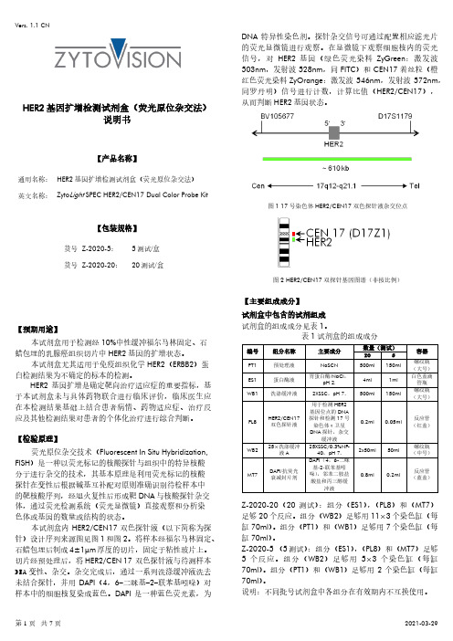
HER2基因扩增检测试剂盒(荧光原位杂交法)说明书【产品名称】通用名称:HER2基因扩增检测试剂盒(荧光原位杂交法)英文名称:Zyto Light SPEC HER2/CEN17 Dual Color Probe Kit【包装规格】货号Z-2020-5:5测试/盒货号Z-2020-20:20测试/盒【预期用途】本试剂盒用于检测经10%中性缓冲福尔马林固定、石蜡包埋的乳腺癌组织切片中HER2基因的扩增状态。
本试剂盒尤其适用于免疫组织化学HER2(ERBB2)蛋白检测结果为不确定的标本的检测。
HER2基因扩增是确定靶向治疗适应症的重要指标,基于本试剂盒未与具体药物联合进行临床评价,临床医生应在本检测结果基础上结合患者病情、药物适应症、治疗反应及其他检测结果对患者的个体化治疗进行综合判断。
【检验原理】荧光原位杂交技术(Fluorescent In Situ Hybridization, FISH)是一种以荧光标记的核酸探针与组织中的特异核酸分子进行杂交的技术,其基本原理是利用荧光标记的核酸探针在变性后根据碱基互补配对原则准确识别待检样本中的靶核酸序列,经退火复性后形成靶DNA与核酸探针杂交体,通过荧光检测系统(荧光显微镜)直接观察和分析染色体或基因的数量或结构的状态。
本试剂盒内HER2/CEN17双色探针液(以下简称为探针)设计序列来源图见图1和图2。
将样本经福尔马林固定、石蜡包埋后制成4±1μm厚度的切片,固定于粘性玻片上。
切片经预处理后,将HER2/CEN 17双色探针液与待测样本DNA变性、杂交。
杂交完成后,通过一系列洗涤缓冲液洗去未结合探针,并用DAPI(4,6-二咪基-2-联苯基吲哚)对样本中的细胞核复染成蓝色。
DAPI是一种蓝色荧光素,为DNA特异性染色剂。
探针杂交信号可通过配置相应滤光片的荧光显微镜进行观察。
在显微镜下观察细胞核内的荧光信号,对HER2基因(绿色荧光染料ZyGreen:激发波503nm,发射波528nm,同FITC)和CEN17着丝粒(橙红色荧光染料ZyOrange:激发波546nm,发射波572nm,同罗丹明)信号进行计数,计算比值(HER2/CEN17),从而判断HER2基因状态。
人类KRAS7种突变检测试剂盒(荧光PCR法)说明书-P4.2-12T-2015.10.09
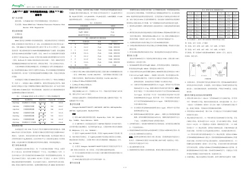
人类KRAS基因7种突变检测试剂盒(荧光PCR法)说明书【产品名称】通用名称:人类KRAS基因7种突变检测试剂盒(荧光PCR法)英文名称:Human KRAS Gene 7 Mutations Fluorescence Polymerase Chain Reaction (PCR) Diagnostic Kit【包装规格】12测试/盒【预期用途】KRAS基因是人体肿瘤中常见的致癌基因。
该基因的突变常见于多种恶性肿瘤,在肺癌患者中的突变率为15~30%,在结直肠癌患者中的突变率为20~50%。
导致KRAS处于激活状态的突变主要位于第12和13密码子上。
KRAS 基因突变一般会使肺癌患者对EGFR酪氨酸激酶抑制剂产生耐药,使结直肠癌患者对抗EGFR抗体类药物产生耐药。
但是,2010年10月的最新研究发现第13密码子上的Gly13Asp(G13D)突变亦对抗EGFR抗体类药物有治疗反应性(参见:De Roock. W. JAMA. 2010;304(16):1812-1820)。
因此,KRAS基因突变检测能提高肿瘤临床治疗的针对性,降低治疗费用,节省宝贵的治疗时间。
大部分肿瘤的突变都是体细胞突变,突变细胞往往与野生型细胞混杂在一起,因此所提取的DNA常带有大量野生型DNA,所以对体细胞突变检测需要较高的特异性,而目前广泛使用的直接测序法检测能力有限,不能完全满足临床需要。
本试剂盒用于检测人类KRAS基因的12和13密码子上7种热点体细胞突变(见表1),试剂盒以DNA为检测样本,提供突变状态的定性评估。
辅助临床医生筛选出可受益于肿瘤靶向药物的大肠癌等癌症患者。
该产品用于组织中提取DNA的KRAS基因7种突变的检测,为临床医生对大肠癌或肺癌患者选择肿瘤靶向药物治疗提供参考。
表1 人类KRAS基因的12和13密码子上7种热点体细胞突变突变名称氨基酸变化碱基变化Cosmic ID 公司命名Gly12Asp 甘氨酸到天门冬氨酸GGT>GAT 521 12-2-A Gly12Ala 甘氨酸到丙氨酸GGT>GCT 522 12-2-C Gly12Val 甘氨酸到缬氨酸GGT>GTT 520 12-2-T Gly12Ser 甘氨酸到丝氨酸GGT>AGT 517 12-1-A Gly12Arg 甘氨酸到精氨酸GGT>CGT 518 12-1-C Gly12Cys 甘氨酸到胱氨酸GGT>TGT 516 12-1-T Gly13Asp 甘氨酸到天门冬氨酸GGC>GAC 532 13-2-A 【检测原理】本试剂盒基于实时PCR平台结合了特异引物和双环探针两种技术,检测DNA样品中含有的突变基因。
Kras基因突变检测试剂盒说明书

人类K-ras基因突变检测试剂盒(PCR-熔解曲线法)说明书【产品名称】通用名:人类K-ras基因突变检测试剂盒(PCR-熔解曲线法)英文名:Diagnostic kit for Mutations of Human K-ras Gene(PCR-Melting Curve Analysis)【包装规格】20测试/盒【预期用途】K-ras基因位于12号染色体短臂上,是重要的癌基因之一,编码一种21kD 的kras蛋白,参与细胞内的信号传递,主要包括PI3K/PTEN/AKT 和RAF/MEK/ERK信号转导途径,这些转导途径是当前肿瘤靶向药物研究的热点,靶向药物通过抑制这些途径发生药理作用。
K-ras基因第12和13密码子发生突变,将导致kras蛋白变异并处于持续激活状态,使药物失效。
据中国2010版《肿瘤学临床实践指南》,在一项包含101例肺腺癌亚型细支气管肺泡癌患者的回顾性研究中,所有患者均接受厄洛替尼单药一线治疗。
K-ras突变者无一例缓解(0/18),而无K-ras突变者则有20例缓解(20/62,32%),差别有统计学意义(P <0.01)。
因此,指南建议,非小细胞肺癌和结直肠癌患者使用靶向药物前应进行K-ras基因突变状态的检测。
本试剂盒以人非小细胞肺癌、结直肠癌肿瘤组织切片提取的基因组DNA为检测样本,用于检测肿瘤组织K-ras基因第12,13密码子的12种体细胞突变(表1),提供突变状态的定性结果。
为临床肿瘤靶向药物的个体化用药提供辅助诊断依据,本品适用于进入个体化靶向治疗疗程前的患者使用。
表1 本品可检测的K-ras基因突变【检验原理】本试剂盒基于实时PCR平台,结合了特异引物、荧光探针和熔解曲线技术,定性检测DNA样品中K-ras 基因12,13密码子是否存在突变。
用一对K-ras基因特异引物,该引物可扩增12种突变型和野生型的K-ras目标序列,利用标记了FAM荧光基团和淬灭基团的双标记探针一方面抑制野生型基因的扩增,提高突变基因的检测灵敏度,另一方面在扩增后用熔解曲线法实现对扩增产物的分析,荧光探针在未杂交时因荧光基团和淬灭基团相互接近而被淬灭,不发荧光,杂交状态时,二者相互离开而产生荧光。
高效率高成功率PCR酶 KOD FX Neo (Code No.KFX-201) 说明书

2 垂询电话:021-5879 4900
【2】 PCR 实验步骤
请注意:引物条件和循环条件与原来的 KOD FX 有所不同。
(1) 引物设计
・ 请尽量使用 22ቤተ መጻሕፍቲ ባይዱ35mer(Tm 值*>63℃)的引物。 ・ GC 含量请设计为 45~60%,并请确认 GC 的位置(偏向)。GC 如靠近 3’端,则容易出现弥
Genomic DNA
Plasmid DNA cDNA λDNA
真核生物来源DNA 原核生物来源DNA
5~200 ng 0.1~100 ng 10 pg~50 ng ~200 ng (RNA相当量) 10 pg~10 ng
一般情况下模板量
50 ng 10 ng 1 ng 50 ng (RNA相当量) 1 ng
(2)PCR 反应液的配制
配制反应液前,请充分混匀各试剂。冻结的试剂请完全解冻后再使用。
Components Autoclaved, distilled water 2× PCR Buffer for KOD FX Neo 2 mM dNTPs 引物 (10μM each)
模板
KOD FX Neo (1 U/μl) Total
3
东洋纺(上海)生物科技有限公司
・ 所有液体添加以后,请用 Vortex 等充分混匀,再进行 PCR。 ・ 一般情况下,引物浓度请用 0.3μM(终浓度)。扩增 10 kb 以上的长链片段时,将引物浓度
设定为 0.15μM(终浓度)可提高扩增量。
(3) 模板
a. 使用纯化后的模板、cDNA时,添加量请参照下列数据(PCR反应液为50μl时)。
11-01
高效率・高成功率PCR酶 KOD FX Neo
R3401 Fumonisin 06-08-16 k伏马菌毒素
RIDASCREEN® FumonisinEnzymimmunoassay zur quantitativen Bestimmung vonFumonisinEnzyme immunoassay for the quantitative analysis offumonisinArt. No.: R3401In vitro TestLagerung bei 2 - 8 °CStorage at 2 - 8 °CR-Biopharm AG, Darmstadt, GermanyTel.: +49 (0) 61 51 81 02-0 / Telefax: +49 (0) 61 51 81 02-20Anschrift:R-Biopharm AGLandwehrstr. 54D-64293 Darmstadtwww.r-biopharm.deFür weitere Fragen stehen Ihnen gerne zur Verfügung:Telefon:Zentrale / Auftragsannahme (0 61 51) 81 02-0Telefax / E-Mail:Auftragsannahme (0 61 51) 81 02-20orders@r-biopharm.de Marketing (0 61 51) 81 02-40info@r-biopharm.deRIDA® und RIDASCREEN®sind eingetragene Warenzeichen der R-Biopharm AGHersteller: R-Biopharm AG, Darmstadt, DeutschlandR-Biopharm AG ist ISO 9001 zertifiziert.RIDA® and RIDASCREEN®are registered trademarks of R-Biopharm AGManufacturer: R-Biopharm AG, Darmstadt, GermanyR-Biopharm AG is ISO 9001 certified.KurzinformationRIDASCREEN® Fumonisin(Art. Nr.: R3401) ist ein kompetitiver Enzymimmuno-assay zur quantitativen Bestimmung von Fumonisin in Mais und Maisprodukten. Alle Reagenzien für die Durchführung des Enzymimmunoassays, inkl. Standards, sind im Testkit enthalten.Der Testkit ist ausreichend für 96 Bestimmungen (einschließlich Standardbe-stimmungen).Zur Auswertung benötigt man ein Mikrotiterplatten-Photometer. Probenvorbereitung: extrahieren, filtrieren und verdünnenZeitbedarf: Probenvorbereitung (für 10 Proben).........ca. 30 minTestdurchführung (Inkubationszeit)................45 min Nachweisgrenze: 25 ppbWiederfindungsrate: von künstlich kontaminierten Maisgrießproben50 und 500 µg/kg (ppb):..............................ca. 60 %mittlerer Variationskoeffizient:.......................ca. 8 % Spezifität: Die Spezifität des RIDASCREEN® Fumonisin-Testswurde durch die Bestimmung der Kreuzreaktivität zuden entsprechenden Mykotoxinen ermittelt.Fumonisin B1...................................................100 %Fumonisin B2...............................................ca. 40 %Fumonisin B3.............................................ca. 100 %1. VerwendungszweckRIDASCREEN® Fumonisin ist ein kompetitiver Enzymimmunoassay zur quantita-tiven Bestimmung von Fumonisin in Mais und Maisprodukten.2. AllgemeinesFumonisine sind karzinogene, neuro-, hepato- und pneumotoxische Stoffwechsel- produkte von Fusarium moniliforme, einer wirtspezifisch auf Mais wachsenden pathogenen Pilzart.Die zur Auslösung toxischer Wirkungen erforderlichen Dosen an Fumonisin sind starken tierartlichen Unterschieden unterworfen. Beim Pferd wirken bereits Fumonisin-Konzentrationen von ca. 5 - 10 mg/kg im Futter neurotoxisch. Bei Schweinen führt die Aufnahme von 4 - 16 mg/kg Körpergewicht zu Leberzirrhose und ab 16 mg/kg Körpergewicht zu pulmonären Ödemen. Hühnerküken und Jungmasthähnchen reagieren erst auf eine hohe Konzentration (ab 75 mg/kg) von Fumonisin im Futter. Rinder scheinen dagegen gegenüber hohen Fumonisin-Konzentrationen relativ unempfindlich zu sein.3. TestprinzipGrundlage ist die Antigen-Antikörper-Reaktion. Die Vertiefungen der Mikrotiter-streifen sind mit Fängerantikörpern gegen anti-Fumonisin-Antikörper beschichtet. Zugegeben werden Standards bzw. Probelösung, enzymmarkiertes Fumonisin (Enzymkonjugat) und anti-Fumonisin-Antikörper. Freies und enzymmarkiertes Fumonisin konkurrieren um die Fumonisin-Antikörperbindungsstellen (kompeti-tiver Enzymimmunoassay). Gleichzeitig werden auch die anti-Fumonisin-Anti-körper von den immobilisierten Fängerantikörpern gebunden. Nicht gebundenes, enzymmarkiertes Fumonisin wird anschließend in einem Waschschritt wieder entfernt. Hinzugegeben wird Substrat/Chromogen, gebundenes Enzymkonjugat wandelt das Chromogen in ein blaues Endprodukt um. Die Zugabe des Stopp-Reagenzes führt zu einem Farbumschlag von blau nach gelb. Die Messung erfolgt photometrisch bei 450 nm; die Extinktion der Lösung ist umgekehrt proportional zur Fumonisin-Konzentration in der Probe.4. PackungsinhaltMit den Reagenzien einer Packung können 96 Bestimmungen durchgeführt werden (einschließlich Standardbestimmungen). Jeder Testkit enthält:1 x Mikrotiterplatte mit 96 Kavitäten (12 Streifen à 8 Kavitäten, teilbar),b eschichtet mit Fängerantikörpern6 x Fumonisin-Standards *), je 1,3 ml0 ppm (Nullstandard), 0,025 ppm, 0,074 ppm, 0,222 ppm, 0,666 ppm, 2 ppmFumonisin in Methanol/Wasser, gebrauchsfertig1 x Konjugat (6 ml).....................................................................roter VerschlussP eroxidase-konjugiertes Fumonisingebrauchsfertig1 x Anti-Fumonisin-Antikörper (6 ml).................................schwarzer Verschlussg ebrauchsfertig1 x Substrat-/Chromogenlösung (10 ml)................................brauner Verschlussrötlich gefärbt1 x Stopp-Reagenz (14 ml)......................................................gelber Verschlusse nthält 1 N Schwefelsäure*) Die Konzentrationsangaben berücksichtigen bereits den Verdünnungsfaktor 70, der sich aus der Probenvorbereitung ergibt. So können die Fumonisin-Konzentrationen der Proben direkt aus der Standardkurve abgelesen werden.5. Zusätzlich benötigte Reagenzien – erforderliches Zubehör5.1. Geräte:− M ikrotiterplatten-Photometer (450 nm)− M esszylinder (Plastik oder Glas) 250 ml− z ur Probenvorbereitung: Filtertrichter und Auffanggefäß (300 ml) aus Glas− L abor- oder Getreidemühle− U ltra-Turrax− o ptional: Schüttler− F ilterpapier: Whatman No. 1 oder Vergleichbares− v ariable 20 µl - 200 µl und 200 - 1000 µl Mikropipetten5.2. Reagenzien:− M ethanol− 70 % Methanol: 70 ml Methanol (100 %) mit 30 ml dest. Wasser mischen− d estilliertes (oder deionisiertes) Wasser6. VorsichtsmaßnahmenDie Standards enthalten Fumonisin, besondere Vorsicht ist geboten. Hautkontakt mit dem Reagenz vermeiden (Handschuhe verwenden).Die Dekontamination von Glasgeräten und toxinhaltigen Lösungen erfolgt am zweckmäßigsten mit einer Natriumhypochlorit-Lösung (10 % (v/v)) über Nacht (Lösung mit HCl auf pH 7 einstellen).Das Stopp-Reagenz enthält 1 N Schwefelsäure (R36/38, S2-26).7. Reagenzien und ihre LagerungDie Reagenzien bei 2 - 8 °C lagern.Komponenten des Testkits auf keinen Fall einfrieren.Nicht benötigte Kavitäten zusammen mit dem Trockenmittel im Folienbeutel gut verschlossen aufbewahren und weiterhin bei 2 - 8 °C lagern.Fumonisin ist lichtempfindlich, deshalb die Fumonisin-Standards vor direkter Lichteinwirkung schützen.Die Substrat-/Chromogenlösung ist lichtempfindlich, deshalb direkte Lichteinwir-kung vermeiden.Nach Ablauf des Verfallsdatums (siehe Testkit-Außenetikett unter Expiration) kann keine Qualitätsgarantie mehr übernommen werden.Ein Austausch von Einzelreagenzien zwischen Kits verschiedener Chargennum-mern ist nicht möglich.8. Anzeichen für Reagenzienverfall− b läuliche Färbung der rötlich gefärbten Substrat-/Chromogenlösung vor Zugabe in die Kavitäten− E xtinktion kleiner 0,6 (E450 nm< 0,6) für den Nullstandard9. ProbenvorbereitungDie Proben kühl und lichtgeschützt lagern.Eine repräsentative Probe (eine unter offiziellen Probenahme-Vorschriften gezo-gene Probe) vor dem Extrahieren zerkleinern und mischen – Maisgrieß und Mais-mehl können direkt verwendet werden.− 5 g der zerkleinerten Probe einwiegen und 25 ml 70 % Methanol *) hinzufügen − d ie Probe 2 min mit einem Ultra-Turrax homogenisieren oder 3 min kräftigschütteln (manuell oder mittels Schüttler)− d en Extrakt durch einen Whatman No. 1 Papierfilter filtrieren− d en filtrierten Probenextrakt 1:14 (1+13) mit destilliertem oder deionisiertem Wasser verdünnen (z. B. 100 µl Extrakt + 1,3 ml dest. Wasser)− 50 µl des verdünnten Filtrats pro Kavität im Test einsetzen*) die Probeneinwaage kann entsprechend vergrößert werden, aber dazu muss das Volumen des Methanol/Wasser Gemisches angepasst werden, z. B. 50 g in 250 ml 70 % Methanol10. Testdurchführung10.1. TestvorbereitungenAlle Reagenzien vor Gebrauch auf Raumtemperatur (20 - 25 °C) bringen.Die Fumonisin-Standardlösungen liegen gebrauchsfertig vor. Der Verdün-nungsfaktor 70 für die Proben wurde beim Etikettieren der Standardfläschchen bereits berücksichtigt. Deshalb kann die Fumonisin-Konzentration der Proben direkt aus der Standardkurve abgelesen werden.10.2. TestdurchführungSorgfältiges Waschen ist sehr wichtig. Ein Eintrocknen der Kavitäten zwischen den Arbeitsschritten vermeiden.1. So viele Kavitäten in den Halterahmen einsetzen, wie für alle Standards undProben in Doppelbestimmung benötigt werden. Die Positionen der Standards und der Proben protokollieren.2. Je 50 µl der Standardlösung bzw. der nach Abschnitt 9. vorbereiteten Probenals Doppelbestimmung in die entsprechenden Kavitäten pipettieren.3. Je 50 µl Enzymkonjugat (roter Verschluss) in die entsprechenden Kavitätenpipettieren.4. 50 µl der anti-Fumonisin-Antikörperlösung (schwarzer Verschluss) in jedeKavität pipettieren. Vorsichtig manuell mischen und 30 min bei Raumtemperatur (20 - 25 °C) inkubieren.5. Die Kavitäten durch Ausschlagen der Flüssigkeit leeren und die Restflüs-sigkeit durch kräftiges Ausklopfen (dreimal hintereinander) auf saugfähigen Labortüchern entfernen. Die Kavitäten mit jeweils 250 µl destilliertem Wasser waschen. Diesen Waschvorgang zweimal wiederholen.6. 100 µl Substrat/Chromogen (brauner Verschluss) in jede Kavität pipettieren.Vorsichtig manuell mischen und 15 min bei Raumtemperatur (20 - 25 °C) im Dunkeln inkubieren.7. 100 µl Stopp-Reagenz (gelber Verschluss) in jede Kavität pipettieren.Vorsichtig manuell mischen und die Extinktion bei 450 nm innerhalb von10 min nach Zugabe des Stopp-Reagenzes messen.11. AuswertungFür die Auswertung ist bei R-Biopharm eine speziell für die RIDASCREEN®Enzymimmunoassays entwickelte Software, die RIDA®SOFT Win (Art. Nr. Z9999), erhältlich.Der Verlauf der Standardkurve kann dem beigefügten Analysenzertifikat entnom-men werden.Hinweis für die Berechnung ohne Software:Extinktion Standard (bzw. Probe)Extinktion Nullstandard x 100 = % ExtinktionDen Nullstandard somit gleich 100 % setzen und die Extinktionswerte in Prozent angeben. Die errechneten Werte für die Standards in einem Koordinatensystem auf halblogarithmischem Millimeterpapier gegen die Fumonisin-Konzentration [mg/kg] auftragen.Die Fumonisin-Konzentration in mg/kg kann entsprechend der Extinktion jeder Probe direkt aus der Eichkurve abgelesen werden.Diese Angaben entsprechen dem heutigen Stand unserer Kenntnisse und sollen über unsere Produkte und deren Anwendungsmöglichkeiten informieren. Sie haben somit nicht die Bedeutung, bestimmte Eigenschaften der Produkte oder deren Eignung für einen konkreten Einsatzzweck zuzusichern. R-Biopharm übernimmt keine Gewährleistung, außer für die standardisierte Qualität der Reagenzien. Defekte Produkte werden ersetzt. Darüber hinaus gehende Ansprüche für direkte oder indirekte Schäden oder Kosten aus der Nutzung der Produkte entstehen nicht.RIDASCREEN® FumonisinBrief informationRIDASCREEN® Fumonisin (Art. No.: R3401) is a competitive enzyme immuno-assay for the quantitative analysis of fumonisin in corn and corn products.All reagents required for the enzyme immunoassay - including standards - are contained in the test kit.The test kit is sufficient for 96 determinations (including standards).A microtiter plate spectrophotometer is required for quantification.Sample preparation: extraction, filtration and dilutionTime requirement: sample preparation (for 10 samples).....approx. 30 mintest implementation (incubation time).................45 min Detection limit: 25 ppbRecovery rate: in spiked semolina of corn samples50 and 500 µg/kg (ppb):...........................approx. 60 %mean coefficient of variation:.....................approx. 8 % Specificity: The specificity of the RIDASCREEN® Fumonisin testwas established by analyzing the cross-reactivity tocorresponding mycotoxins.Fumonisin B1......................................................100 %Fumonisin B2...........................................approx. 40 %Fumonisin B3.........................................approx. 100 %1. Intended useRIDASCREEN® Fumonisin is a competitive enzyme immunoassay for the quantitative analysis of fumonisin in corn and corn products.2. GeneralFumonisins are carcinogenic, neuro, hepato and pneumo toxic metabolites of Fusarium moniliforme, a mould fungi which grows hostspecific on corn.The dose of fumonisin for the release of toxic effects differs significantly depending on the animal species. A concentration of approx. 5 - 10 mg/kg fumonisin in feed induces neuro toxic effects in horses. In pigs the ingestion of 4 - 16 mg/kg body weight may result in liver cirrhosis and more than 16 mg/kg body weight may lead to pulmonary edema. Chickens tolerate higher concentrations of fumonisin in feed, up to 75 mg/kg. Cattle seem to be insensitive to high fumonisin concentrations.3. Test principleThe basis of the test is the antigen-antibody reaction. The microtiter wells are coated with capture antibodies directed against anti-fumonisin antibodies. Fumonisin standards or sample solutions, fumonisin enzyme conjugate and anti-fumonisin antibodies are added. Free fumonisin and fumonisin enzyme conjugate compete for the fumonisin antibody binding sites (competitive enzyme immuno-assay). At the same time, the anti-fumonisin antibodies are also bound by the immobilized capture antibodies. Any unbound enzyme conjugate is then removed in a washing step. Substrate/chromogen is added to the wells, bound enzyme conjugate converts the chromogen into a blue product. The addition of the stop solution leads to a color change from blue to yellow. The measurement is made photometrically at 450 nm. The absorbance is inversely proportional to the fumonisin concentration in the sample.4. Reagents providedEach kit contains sufficient materials for 96 measurements (including standard analyses). Each test kit contains:1 x Microtiter plate with 96 wells (12 strips with 8 removable wells each)coated with capture antibodies6 x Fumonisin standard solutions *), 1.3 ml each0 ppm (zero standard), 0.025 ppm, 0.074 ppm, 0.222 ppm, 0.666 ppm, 2 ppmfumonisin in methanol/water, ready to use1 x Conjugate (6 ml).................................................................................red capperoxidase conjugated fumonisinready to use1 x Anti-fumonisin antibody (6 ml).........................................................black capr eady to use1 x Substrate/chromogen (10 ml)........................................................brown capstained red1 x Stop solution (14 ml) ....................................................................yellow capcontains 1 N sulfuric acid*) The dilution factor 70 for the sample has already been considered. Therefore, the fumonisin concentrations of samples can be read directly from the standard curve.5. Materials required but not provided5.1. Equipment:− m icrotiter plate spectrophotometer (450 nm)− g raduated cylinder: (plastic or glass) 250 ml− g lassware for preparing sample extract: filter funnel and 300 ml flask− g rinder (mill)− U ltra-Turrax− o ptional: shaker− f ilter paper: Whatman No. 1 or equivalent− v ariable 20 µl - 200 µl and 200 - 1000 µl micropipettes5.2. Reagents:− m ethanol− 70 % methanol solution: prepare 70 % methanol solution by mixing 70 ml methanol (100 %) with 30 ml distilled water− d istilled (or deionized) water6. Warnings and precautions for the usersThe standard solutions contain fumonisin, particular care should be taken. Avoid contact of the reagent with the skin (use gloves).Decontamination of the glassware and toxin-content solutions is best carried out using a sodium hypochlorite (bleach) solution (10 % (v/v)) overnight (adjust solution with HCl to pH 7).The stop solution contains 1 N sulfuric acid (R36/38, S2-26).7. Storage instructionsStore the kit at 2 - 8 °C (35 - 46 °F). Do not freeze any test kit components. Return any unused microwells to their original foil bag, reseal them together with the desiccant provided and further store at 2 - 8 °C (35 - 46 °F).Fumonisin is light sensitive, therefore, avoid exposure to direct light.The substrate/chromogen solution is light sensitive, therefore, avoid exposure to direct light.No quality guarantee is accepted after the expiration date on the kit label.Do not interchange individual reagents between kits of different lot numbers.8. Indication of instability or deterioration of reagents− a ny bluish coloration of the reddish substrate/chromogen solution prior to test implementation− a value of less than 0.6 absorbance units (A450 nm< 0.6) for the zero standard9. Preparation of SamplesThe samples should be stored in a cool place, protected from light.A representative sample (according to accepted sampling techniques) should be ground and thoroughly mixed prior to proceeding with the extraction procedure – semolina of corn and corn meal could be used directly.− w eigh 5 g of ground sample into a suitable container and add 25 ml of 70 % methanol *)− b lend the sample by Ultra-Turrax for 2 minutes or shake vigorously for3 minutes (manually or with shaker)− f ilter the extract through Whatman No. 1 filter− d ilute the filtered sample extract 1:14 (1+13) with distilled or deionized water(e. g. 100 µl extract + 1.3 ml water)− u se 50 µl of the diluted filtrate per well in the test*) sample size may be increased if required, but the volume of methanol/water must be adapted accordingly, e.g.: 50 g in 250 ml of 70 % methanol10. Test implementation10.1. Preliminary commentsBring all reagents to room temperature (20 - 25 °C / 68 - 77 °F) before use.The fumonisin standards are provided ready to use. The dilution factor 70 for the sample has been considered when labeling. Therefore, the fumonisin concentration of samples can be read directly from the standard curve.10.2. Test procedureCarefully follow the recommended washing procedure. Do not allow microwells to dry between working steps.1. Insert a sufficient number of microtiter wells into the microwell holder for allstandards and samples to be run in duplicate. Record standard and sample positions.2. Add 50 µl of the standard solutions or prepared sample to separate dupli-cate wells.3. Add 50 µl of enzyme conjugate (red cap) to each well.4. Add 50 µl of anti-fumonisin antibody solution (black cap) to each well. Mixgently by shaking the plate manually and incubate for 30 min at room temperature (20 - 25 °C / 68 - 77 °F).5. Pour the liquid out of the wells and tap the microwell holder upside downvigorously (three times in a row) against absorbent paper to ensure com-plete removal of liquid from the wells. Fill all the wells with 250 µl distilled water and pour out the liquid again. Repeat the washing procedure two times.6. Add 100 µl of substrate/chromogen (brown cap) to each well. Mix gently byshaking the plate manually and incubate for 15 min at room temperature(20 - 25 °C / 68 - 77 °F) in the dark.7. Add 100 µl of stop solution (yellow cap) to each well. Mix gently by shakingthe plate manually and measure the absorbance at 450 nm. Read within10 minutes after addition of the stop solution.11. ResultsA special software, the RIDA®SOFT Win (Art. No. Z9999), is available for evaluation of the RIDASCREEN® enzyme immunoassays.The course of the standard curve is shown in the Quality Assurance Certificate enclosed in the test kit.Remark for the calculation without software:absorbance standard (or sample)absorbance zero standard x 100 = % absorbanceThe zero standard is thus made equal to 100 % and the absorbance values are quoted in percentages. The values calculated for the standards are entered in a system of coordinates on semilogarithmic graph paper against the fumonisin concentration [mg/kg].The fumonisin concentration in mg/kg corresponding to the absorbance of each sample can be read directly from the calibration curve.R-Biopharm makes no warranty of any kind, either expressed or implied, except that the materials from which its products are made are of standard quality. If any materials are defective, R-Biopharm will provide a replacement product. There is no warranty of merchantability of this product, or of the fitness of the product for any purpose. R-Biopharm shall not be liable for any damages, including special or consequential damage, or expense arising directly or indirectly from the use of this product.。
ReverTra Ace qPCR RT Kit TOYOBO说明书

1
[2] 产品内容
◆本产品包含以下几种试剂,每 10μl 反应体系可使用 200 次。 所有试剂请均保存在 -20℃条件下。
试剂名
5×RT Buffer (Reaction Buffer+MgCl2+dNTPs) Enzyme Mix (ReverTra Ace reverse transcriptase +RNase Inhibitor) Primer Mix (Random Primer+Oligo(dT) Primer) Nuclease-free Water
4
[5] 相关操作步骤
1. Total RNA 的 DNase I 处理
在通过 AGPC 法等提纯的 Total RNA 内混有基因组 DNA,可能会发生由 基因组 DNA 产生假阳性信号的情况(请参考 p3),请根据以下的方法清除基 因组 DNA< 或使用本公司去除基因组 DNA 专用试剂盒 ReverTra Ace qPCR RT Master Mix with gDNA Remover>。 (1)反应液的配制和反应 反应液组成(例) Nuclease-free Water Total RNA(<10μ g) 10×DNase I Buffer up to 10μ l Xμ l 1μ l 0.5μ l 10μ l
※ 本产品的试剂盒内没有添附 Realtime PCR 试剂。关于 Realtime PCR 产品,推荐使 用本公司的 Realtime PCR 用试剂 Realtime PCR Master Mix 系列。(详情请参考 [7] 相关产品的内容)。
◆本产品特征◆
1. 对应各种不同长度目标片段的高效率逆转录
ReverTra Ace qPCR RT Kit 采用了最适合于 Realtime PCR 用 cDNA 合 成的反应缓冲液, 和以最恰当比例混合的 Primer 混合液, 对于各种不同长度的 目标片段, 不用摸索条件就能够进行高效率的逆转录反应。
四正柏生物科技基因定点突变试剂盒说明书
基因定点突变试剂盒保存条件及效期:−20℃保存,有效期一年。
产品描述:定点突变是一种广泛应用的分析蛋白和DNA 的非常有力的工具。
普通的定点突变试剂盒通常是基于反向PCR 技术,采用高保真耐热DNA 聚合酶和互补的突变引物向目的基因引入单个碱基或多个邻近碱基的突变、缺失、或插入。
当遇到多个位点突变的需求时,利用单点突变试剂盒只能对目标位点逐一引入突变,耗时费力。
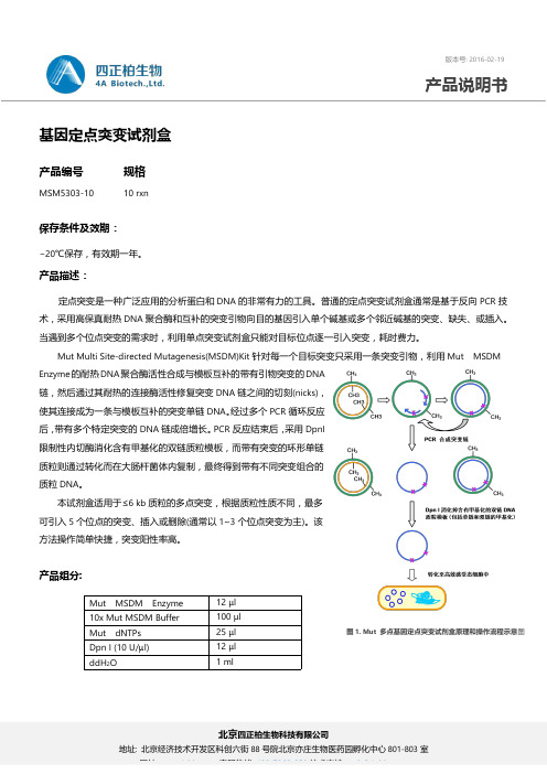
Mut Multi Site-directed Mutagenesis(MSDM)Kit 针对每一个目标突变只采用一条突变引物,利用Mut MSDMEnzyme 的耐热DNA 聚合酶活性合成与模板互补的带有引物突变的DNA 链,然后通过其耐热的连接酶活性修复突变DNA 链之间的切刻(nicks),使其连接成为一条与模板互补的突变单链DNA 。
经过多个PCR 循环反应后,带有多个特定突变的DNA 链成倍增长。
PCR 反应结束后,采用DpnI 限制性内切酶消化含有甲基化的双链质粒模板,而带有突变的环形单链质粒则通过转化而在大肠杆菌体内复制,最终得到带有不同突变组合的质粒DNA 。
本试剂盒适用于≤6kb 质粒的多点突变,根据质粒性质不同,最多可引入5个位点的突变、插入或删除(通常以1~3个位点突变为主)。
该方法操作简单快捷,突变阳性率高。
产品组分:产品编号规格MSM5303-1010rxnMut MSDM Enzyme12μl 10x MutMSDM Buffer 100μl MutdNTPs25μl Dpn I (10U/ l)12μl ddH 2O1ml版本号:2016-02-19产品说明书图1.Mut 多点基因定点突变试剂盒原理和操作流程示意图使用方法:1.模板准备:(1)请使用6kb以下的质粒作为模板;如果模板质粒过大,可将所需突变的序列亚克隆到较小的载体中,完成突变后再克隆到目的载体中。
(2)对于非甲基化的质粒(例如从大肠杆菌JM110或SCS110菌株中提取的质粒),可通过转化dam+的大肠杆菌菌株(如DH5α、TOP10、JM109、XL1-Blue等),再抽提获得甲基化的质粒作为PCR反应模板。
东洋纺 Hot Start TTx (RNA)试剂盒说明书
JAPAN CHINATOYOBO CO., LTD. TOYOBO (SHANHAI) BIOTECH CO., LTD. Tel(81)-6-6348-3888 Tel(86)-21-5879-4900www.toyobo.co.jp/e/bio F1788KHot Start TTx (RNA) KitHSTTX-111 250UStore at -20°C Contents[1] Introduction[2] Components[3] Protocol1. Standard reaction setup2. Cycling conditions[4] Example[5] Related productsCAUTIONAll reagents in this kit are intended for research purposes. Do not use for diagnosis or clinical purposes. Please observe general laboratory precautions and safety while using this kit.-TaqMan® is a registered trademark of Roche Molecular Systems, Inc.1JAPAN CHINA TOYOBO CO., LTD. TOYOBO (SHANHAI) BIOTECH CO., LTD. Tel(81)-6-6348-3888 Tel(86)-21-5879-4900www.toyobo.co.jp/e/bio ********************[1] Introduction[2] ComponentsDescriptionHot Start TTx (RNA) Kit is RT-PCR reagent based on our original polymerase, TTx DNA Polymerase. TTx DNA polymerase exhibits reverse transcriptase activity in the presence of Mn2+ ions. This system allows for 1-step real-time PCR, including reverse transcription and PCR steps.TTx DNA Polymerase has higher amplification efficiency than Taq DNA Polymerase, which is a general-purpose enzyme, and has reverse transcription activity, enabling amplification from a crude sample containing PCR inhibitors with high efficiency from both DNA and RNA.In addition, TTx DNA Polymerase has a 5 '- 3' exonuclease activity, so it can be used for real-time PCR using probe assays such as TaqMan ® assay. This enzyme contains neutralizing antibodies, thus allowing for Hot start PCR.Features- Enables highly efficient 1-enzyme 1-step RT-PCRTTx DNA Polymerase has high reverse transcription activity and enables amplification from low copies of template and enable efficient PCR even fast cycle condition.In addition, TTx DNA Polymerase is a thermostable enzyme and can reverse transcribe at high temperatures above 60°C. It is suitable for amplifying GC rich targets and targets that form higher order structures.This Kit can also be applied to DNA amplification.This kit includes the following components for 250 reactions, 20 μL total reaction volume. All reagents should be stored at -20°C.5x Buffer for rTth/ TTx (DNA/ RNA) 1 mL 2 mM dNTPs 1 mL50 mM Mn(OAc)2250 μL Hot Start TTx DNA Polymerase (4U/ μL) 62.5 μLNote:-5x Reaction Buffer for rTth/ TTx (DNA/ RNA) is 5x RT-PCR buffer not containing Mn 2+ and dNTPs. Add template DNA, primers, and attached 2 mM dNTPs, 50 mM Mn(OAc)2, Hot Start TTx DNA Polymerase, and adjust to 1x concentration with sterile water etc.-DNA Polymerase is a mixture of TTx DNA polymerase and hot start antibodies. Its concentration is 4U/ μL.-This kit doesn’t contain a passive reference dye (ROX). When using a passive reference dye to compensate fluorescence intensity and dispensing error between wells, please use the separately sold 50x ROX reference dye (Code No. ROX-101).2JAPAN CHINA TOYOBO CO., LTD. TOYOBO (SHANHAI) BIOTECH CO., LTD. Tel(81)-6-6348-3888 Tel(86)-21-5879-4900www.toyobo.co.jp/e/bio ********************[3] Protocol1. Standard reaction setupBefore preparing the mixture, all components should be completely thawed, except for the enzyme solution.Notes:-The recommended amount of primer should be 0.2-0.6 μM, and the amount of TaqMan ® probe should be 0.05-0.3 μM. If amplification efficiency is not good, performance may be improved by increasing the addition amount, but if it is added too much, it may cause non-specific reaction and detection sensitivity may be lowered.2. Cycling conditionsThe following cycle is recommended.Notes :- If sensitivity is not good, it may be improved by changing annealing/ extension temperature between 55 ~ 65°C.Components Volume Final Concentration PCR grade waterX µL 5x Buffer for rTth/ TTx (DNA/ RNA) 4 µL 1x2 mM dNTPs 4 µL 0.4 mM 50 mM Mn(OAc)2 1 µL 2.5 mM 10 µM Primer #1 0.6 µL 0.3 µM 10 µΜ Primer #20.6 µL 0.3 µM TaqMan ® Probe(10 μM)0.4 µL 0.2 µM Hot Start TTx DNA Polymerase 0.25 µL 1U Template RNA or DNA (Sample) Y µL Total reaction volume20 µLTemperature Time Predenature 1 : 90°C 30 sec. Reverse Transcription: 60°C 5 min. Predenature 2: 95°C 1 min. Denature :95°C 15 sec. Annealing/extension :60°C30 sec.40~50 cyclesJAPAN CHINA TOYOBO CO., LTD. TOYOBO (SHANHAI) BIOTECH CO., LTD. Tel(81)-6-6348-3888 Tel(86)-21-5879-4900www.toyobo.co.jp/e/bio ********************[4] Example[5] Related productsDetection of influenza virusInfluenza RNA was detected using TaqMan ® probes. As a result of adding 1 μL of diluted purified RNA to 20 μL of the reaction solution, Hot Start TTx (RNA) Kit obtained higher sensitivity than Tth DNA Polymerase-based reagent. By using TTx DNA Polymerase, efficient detection is possible even from low copies of template.Product namePackageCode No. <High efficient DNA Polymerase for PCR and RT-PCR>Hot Start TTx DNA Polymerase10,000 U 100,000 U HSTTX-129 HSTTX-159 <Reaction Buffer (containg Mg 2+) for DNA amplification>2x Buffer for rTth/ TTx (DNA)100 mL 250 mL 1,000 mL QRZ-1B1 QRZ-1B2 QRZ-1B4 <Passive reference dye>50x ROX reference dye5 mLROX-101<High efficient DNA Polymerase for PCR and RT-PCR>Hot Start rTth DNA Polymerase10,000 U HSTTH-329 < Reaction Buffer (not containg Mg 2+ and Mn 2+>5x Buffer for rTth/ Ttx (DNA/ RNA)40 mL 400 mL QRT-1B1 QRT-1B2 <Mn solution for RNA amplification>50 mM Mn (OAc)25 mLQRT-MN1<Mg solution for DNA amplification>25 mM MgCl 240 mL TAP-2S1。
TOYOBO Ligation high (Code No. LGK-100)说明书
08-03-DNA Ligation Kit-Ligation high(Code No.:LGK-100)使用说明书科研用TOYOBO CO., LTD. Life Science DepartmentOSAKA JAPAN目录[1] 前言 (1)[2] 操作程序 (1)1.使用方法 (1)2.进行高效连接 (1)[3] LIGATION HIGH的特征*1 (2)1.优良的连接反应效率 (2)2.少量液体便可进行反应 (2)3.优良的稳定性 (2)[4] 实例 (3)1.插入连接 (3)2. LINKER连接 (4)3.噬菌体连接 (5)4.通过电泳进行确认 (6)5.盐浓度的影响 (7)[5] 相关产品一览表 (8)【 注意 】本试剂盒包含的试剂均为研究用试剂,请不要当作诊断和临床试剂使用。
在本试剂盒使用过程中,请务必严格遵守实验室的各项注意事项,注意安全。
2[1] 前言DNA片段的连接反应是在遗传基因操作实验中的常用操作。
在以往的连接反应中,要根据不同的底物设置不同的条件,还要分别添加各自不同的反应组成成分,比较复杂麻烦。
另外,在进行插入连接时,要得到较高的转化效率也十分困难。
因此,东洋纺公司为了解决此类问题,开发了操作简便而高效的试剂盒。
本试剂盒的反应液中包括了所有连接反应所需要的试剂,适用于多种类型的连接反应。
另外,本说明书介绍了使用Ligation high时的标准结果。
[2] 操作程序1.使用方法·请将本品保存在-20℃以下环境中。
·在冰上使Ligation high融解。
将其放置在冰上5-10分钟便会自然融解。
·配制准备连接用的DNA溶液。
·取DNA溶液加入等量至半量的Ligation high,混匀。
·在16℃下反应30分钟。
·反应完成后,其反应液可以直接使用于转化。
2.进行高效连接·连接效率较低时,使用乙醇沉淀等提纯DNA。
东洋纺织株式会社试剂说明书
manual THUNDERBIRD Probe qPCR Mix 0910 A4250K THUNDERBIRD™ Probe qPCR MixQPS-101T 1 mL x 1QPS-101 1.67 mL x 3Store at -20°C, protected from lightContents[1] Introduction[2] Components[3] Primer/Probe design[4] Template DNA[5] Protocol1. Standard reaction set up2. Cycling conditions2-1. Real-time PCR conditions using Applied Biosystems 7900HT2-2. Real-time PCR conditions using Roche LightCycler 1.1[6] Related ProtocolcDNA synthesis[7] Troubleshooting[8] Related productsC AUTIONAll reagents in this kit are intended for research purposes only. Do not use for diagnosis or clinical purposes. Please observe general laboratory precautions and observe safety procedures while using this kit.-LightCycler™ is a trademark of Idaho Technology, Inc. and Roche Molecular Systems, Inc.-TaqMan® is a registered trademark of Roche Molecular Systems, Inc.-SYBR® is a registered trademark of Roche Molecular Systems, Inc.JAPAN CHINATOYOBO CO., LTD. TOYOBO Bio-Technology, CO., LTD.Tel(81)-6-6348-3888 Tel(86)-21-58794900.4140www.toyobo.co.jp/e/bio 1JAPAN CHINATOYOBO CO., LTD. TOYOBO Bio-Technology, CO., LTD.Tel(81)-6-6348-3888 Tel(86)-21-58794900.4140www.toyobo.co.jp/e/bio********************1[ 1 ] Introduction [ 2 ] Components DescriptionTHUNDERBIRD™ Probe qPCR Mix is a highly efficient 2x Master Mix for real-time PCR using TaqMan® probes. The master mix contains all required components, except for ROX reference dye, probe and primers (50x ROX reference dye is individually supplied with this kit). The master mix facilitates reaction setup, and improves the reproducibility of experiments.This product is an improved version of Realtime PCR Master Mix (Code No. QPK-101). In particular, reaction specificity and PCR efficiency is enhanced.Features-High specificityThe specificity for the detection of low-copy targets is improved.-Homogeneous amplificationThe dispersion of PCR efficiency between targets is reduced by a new PCR enhancer*. (*Patent pending)-Broad dynamic rangeHigh specificity and effective amplification enable the detection of a broad dynamic range.-Compatibility for various real-time cyclers.The reagent is applicable to most real-time cyclers (i.e. Block type and glass capillary type). Because the 50x ROX reference dye is individually supplied with this kit, the kit can be applied to real-time cyclers that require a passive reference dye.-Hot start PCRThe master mix contains anti-Taq DNA polymerase antibodies for hot start technology. The antibodies are easily inactivated in the first denaturation step, thereby activating the DNA polymerase.About the fluorescent probe detection systemThe TaqMan® probe system utilizes fluorescence emission from the probes. The probes hybridize to the target amplicons and then emit fluorescence upon degradation by the 5'-3' exonuclease activity of Taq DNA polymerase. This type of detection system can achieve higher specificity in real-time PCR assays than the SYBR® Green I detection system.This kit includes the following components for 40 reactions (QPS-101T) and 200 reactions (QPS-101), with 50 μl per reaction. All reagents should be stored at -20°C.<QPS-101T>THUNDERBIRD™ Probe qPCR Mix 1 ml x 150x ROX reference dye 50 μl x 1JAPAN CHINATOYOBO CO., LTD. TOYOBO Bio-Technology, CO., LTD.Tel(81)-6-6348-3888 Tel(86)-21-58794900.4140www.toyobo.co.jp/e/bio********************2[ 3 ] Primer/Probedesign <QPS-101>THUNDERBIRD™ Probe qPCR Mix 1.67 ml x 350x ROX reference dye 250 μl x 1Notes:-THUNDERBIRD™ Probe qPCR Mix can be stored, protected from light, at 2-8°C for up to 3 months. For longer storage, this reagent should be kept at -20°C and protected from light. No negative effect was detected by 10 freeze-thaw cycles of THUNDERBRID™ Probe qPCR Mix. This reagent does not contain the ROX reference dye.-50x ROX reference dye can be stored, protected from light, at 2-8°C or -20°C. For real-time cyclers that require a passive reference dye, this reagent must be added to the reaction mixture at a concentration of 1x or 0.1x. The master mix solution with the ROX reference dye can be stored, protected from light, at 2-8°C for up to 3 months. For longer storage, this reagent should be kept at -20°C and protected from light. The pre-mixed reagents can be prepared according to the following ratios. [5] Table 1 shows the optimal concentration of the ROX dye.1x solutionTHUNDERBIRD™ Probe qPCR Mix : 50x ROX reference dye = 1.67 ml : 66.8 μl THUNDERBIRD™ Probe qPCR Mix : 50x ROX reference dye = 1 ml : 40 μl0.1x solutionTHUNDERBIRD™ Probe qPCR Mix : 50x ROX reference dye = 1.67 ml : 6.7 μl THUNDERBIRD™ Probe qPCR Mix : 50x ROX reference dye = 1 ml : 4 μlFor real-time cyclers that do not require a passive reference dye, THUNDERBIRD™ Probe qPCR Mix without the ROX reference dye can be used.1. Primer conditionsHighly sensitive and quantitative data depend on primer design. The primer should be designed according to the following suggestions;-Primer length: 20-30 mer-GC content of primer: 40-60%-Target length: ≤ 200 bp (optimally, 80-150 bp)-Melting temperature (Tm) of primers: 60-65°C-Purification grade of primers: Cartridge (OPC) grade or HPLC gradeNotes:-Longer targets (>200 bp) reduce efficiency and specificity of amplification.-Tm of the primers can be flexible, because the Tm value depends on the calculation formula.JAPAN CHINATOYOBO CO., LTD. TOYOBO Bio-Technology, CO., LTD.Tel(81)-6-6348-3888 Tel(86)-21-58794900.4140www.toyobo.co.jp/e/bio********************3[ 4 ] Template DNA 2. Fluorescent probeThe probes should be designed according to the guidelines of each probe system. Because insufficiently purified probes may inhibit the reaction, HPLC-grade probes should be used.The following DNA samples can be used as templates.1. cDNANon-purified cDNA, generated by reverse transcription reactions, can be used directly for real-time PCR using THUNDERBIRD™ Probe qPCR Mix. Up to 10% of the volume of a cDNA solution can be used for a real-time PCR reaction. However, excess volume of the cDNA may inhibit the PCR. Up to 20% (v/v) of the cDNA solution from ReverTra Ace® qPCR RT Kit (Code No. FSQ-101) can be used for real-time PCR (see [6]).2. Genomic DNA, Viral RNAGenomic DNA and viral RNA can be used at up to 200 ng in 50 μl reactions.3. Plasmid DNAAlthough super-coiled plasmids can be used, linearized plasmid DNA produces more accurate assays. The copy number of the plasmid DNA can be calculated by the following formula.Copy number of 1μg of plasmid DNA = 9.1 x 10¹¹ / Size of plasmid DNA (kb)JAPAN CHINATOYOBO CO., LTD. TOYOBO Bio-Technology, CO., LTD.Tel(81)-6-6348-3888 Tel(86)-21-58794900.4140www.toyobo.co.jp/e/bio********************4[ 5 ] Protocol1. Reaction mixture setupReaction volume FinalReagent50µl20µlConcentrationDWXµlXµlTHUNDERBIRD™ Probe qPCR Mix 25 µl 10 μl 1xForward Primer 15 pmol 6 pmol 0.3 μM*1Reverse Primer 15 pmol 6 pmol 0.3 μM*1TaqMan® Probe 10 pmol 4 pmol 0.2 μM*150x ROX reference dye 1μl / 0.1 μl 0.4μl / 0.04μl 1x / 0.1x*2DNA solution Y µl Y µlTotal50µl20µlNotes:*1 Primer / probe concentration should be determined according to the manufacturer’sinstructions.Higher primer concentration tends to improve the amplification efficiency, and lowerprimer concentration tends to reduce the non-specific amplification. The primerconcentration should be set between 0.2-0.6 μM.*2 50x ROX reference dye must be added when using real-time cyclers that require apassive reference dye, according to Table 1. Table 1 shows the optimum concentrationof the ROX reference dye. This dye is not necessary for real-time cyclers that do notrequire a passive reference dye.Table 1 Recommended ROX dye concentrationReal-time cycler Optimal dye concentration(dilution ratio)Applied Biosystems 7000, 7300, 7700, 7900HT etc. 1x (50:1)Applied Biosystems 7500, 7500Fast,Stratagene cyclers (Optional) etc.0.1x (500:1)Roche’ cyclers, Bio-Rad cyclers, BioFlux cyclers etc. Not requiredNotes:The ROX dye in Realtime PCR Master Mix (Code No. QPK-101) corresponds to 1xconcentration.JAPAN CHINA TOYOBO CO., LTD. TOYOBO Bio-Technology, CO., LTD.Tel(81)-6-6348-3888 Tel(86)-21-58794900.4140 www.toyobo.co.jp/e/bio********************52. PCR cycling conditionsThe following table shows the recommended thermal conditions using primers designed according to the recommended primer and probe conditions described in [3]. Almost all targets can also be amplified using the ongoing conditions with other real-time PCR reagents.*1Due to the anti-Taq antibody hot start PCR system, the pre-denaturation can be completed within 60 sec. The pre-denaturation time should be determined according to the recommendations of each real-time cycler. If the optimal pre-denaturation time cannot be determined, the time should be set at 60 sec.Table 2 The recommended pre-denaturation time for each real-time cycler Real-time cycler Pre-denaturation time High speed cycler (e.g. Applied Biosystems 7500Fast) 20 sec Capillary cycler (e.g. Roche LightCycler™ 1.x, 2.0) 30 secGeneral real-time cyclers (Applied Biosystems 7700, 7500,7900HT (normal block), Stratagene cyclers, BioFlux cyclers 60 sec *2The following table shows the optimal denaturation times for each real-time cycler. If the optimal denaturation time cannot be determined, the time should be set at 15 sec.Table 3 The recommended denaturation time for each real-time cycler Real-time cycler denaturation time High speed cycler (e.g. Applied Biosystems 7500Fast) 3 sec Capillary cycler (e.g. Roche LightCycler™ 1.x, 2.0) 5 secGeneral real-time cyclers (Applied Biosystems 7700, 7500,7900HT (normal block), Stratagene cyclers, BioFlux cyclers 15 sec *3Insufficient amplification may be improved by decreasing the extension temperature, and non-specific amplification (e.g. abnormal shapes of the amplification curve at low template concentrations) may be reduced by increasing the extension temperature. The extension temperature should be set at 56-64°C. *4If the target size is smaller than 300 bp, the extension time can be set at 30 sec on almost all real-time cyclers. Instability of the amplification curve or variation of data from each well may be improved by setting the extension time at 45-60 sec. Some real-time cyclers or software need over 30 sec for the extension step. In these cases, the time should be set according to each instruction manual (e.g. Applied Biosystems 7000/73000: ≥ 31 sec; Applied Biosystems 7500: ≥ 35 sec.).<3-step cycle> Temperature Time Ramp Pre-denaturation:95°C 20-60 sec *1Maximum Denaturation:95°C 1-15 sec *2 MaximumExtension: 60°C *3 30-60 sec *4 Maximum (data collection should be set at the extension step)JAPAN CHINA TOYOBO CO., LTD. TOYOBO Bio-Technology, CO., LTD.Tel(81)-6-6348-3888 Tel(86)-21-58794900.4140 www.toyobo.co.jp/e/bio********************62-1. Real-time PCR conditions using Applied Biosystems 7900HT(Normal block type, software version 2.2.2)The following is an example of a TaqMan ® assay using Applied Biosystems 7900HT.(1) The cycling parameters should be set according to the following “Thermal CyclerProtocol” window under the “Instrument” tab.Notes:- Appropriate sample volumes should be set. - ≥ 45 sec is necessary for the extension step.(2) Click the “Data collection” tab.(3) Insert the PCR plate(4) Start the programJAPAN CHINATOYOBO CO., LTD. TOYOBO Bio-Technology, CO., LTD.Tel(81)-6-6348-3888 Tel(86)-21-58794900.4140www.toyobo.co.jp/e/bio********************72-2. Real-time PCR conditions using Roche LightCycler 1.1(Software version 3.5)The following is an example of a TaqMan® probe assay using Roche LightCycler 1.1. (1)The cycling parameters should be set according to the following window. Analysisand Acquisition mode of the initial denaturation step must be set at “None”. (2)The cycling parameters should be set according to the following window. Analysismode of the PCR step must be set at “Quantification”. Acquisition modes of 95°C and 60°C must be set at “None” and “Single”, respectively.JAPAN CHINATOYOBO CO., LTD. TOYOBO Bio-Technology, CO., LTD.Tel(81)-6-6348-3888 Tel(86)-21-58794900.4140www.toyobo.co.jp/e/bio********************8(3)The cycling parameters should be set according to the following window. Analysisand Acquisition mode of the cooling step must be set at “Non”.(4) Insert the capillaries in the carousel, and start the cycling program.JAPAN CHINATOYOBO CO., LTD. TOYOBO Bio-Technology, CO., LTD.Tel(81)-6-6348-3888 Tel(86)-21-58794900.4140www.toyobo.co.jp/e/bio********************9[ 6 ] Related Protocol1. cDNA synthesiscDNA synthesized by various cDNA synthesis reagents can be used withTHUNDERBIRD™ Probe qPCR Mix. However, cDNA synthesized by a reagentspecialized for real-time PCR can increase sensitivity.ReverTra Ace® qPCR RT Kit (Code No. FSQ-101) is a cDNA synthesis kit suitable forreal-time PCR. Here, the protocol with ReverTra Ace® qPCR RT Kit is described.However, for the detailed protocol, please refer to the instruction manual of the kit.(1) Denaturation of RNAIncubate the RNA solution at 65°C for 5 min and then chill on ice.Notes:-This step can be omitted. But this step may increase the efficiency of the reversetranscription of RNA, which forms secondary structures.-Do not add 5x RT Buffer and/or enzyme solution at this step.(2) Preparation of the reaction solutionReagent V olume (amount)Nuclease-freeWaterXµl5x RT Buffer 2 µlPrimerMix 0.5µlEnzymeMix 0.5µlRNAsolution 0.5pg-1 µgTotal10µl(3) Reverse transcription reaction-Incubate at 37°C for 15 min. <Reverse transcription>-Heat at 98°C for 2 min. <Inactivation of the reverse transcriptase>-Store at 4°C or -20°C.**This solution can be used in the real-time PCR reaction directly or after dilution.Notes:The above temperature conditions are optimized for ReverTra Ace® qPCR RT Kit.JAPAN CHINATOYOBO CO., LTD. TOYOBO Bio-Technology, CO., LTD.Tel(81)-6-6348-3888 Tel(86)-21-58794900.4140www.toyobo.co.jp/e/bio********************10[ 7 ] TroubleshootingSymptom Cause SolutionLoss of linearity in the high cDNA/DNA concentration region. Inhibition by the components in thecDNA/DNA solution.-DNA: The DNA sample may contain PCRinhibitors. The DNA samples should berepurified.-cDNA: The components in the cDNA synthesisreagent may inhibit the PCR reaction. The cDNAsample should be used after dilution.The template DNA is insufficient. When the DNA/cDNA copy number is lower than10 copies per reaction, the linearity of the reactiontends to be lost. The template concentrationshould be increased.Adsorption of the DNA to the tubewall.The diluted DNA templates tend to be absorbedonto the tube wall. Dilution should be performedjust prior to experiments.Lost of linearity or lower signal in the lowDNA/cDNAconcentration region. Competition with primer dimerformation. In the probe assay, primer dimers are not detected. However, dimer formation may reduce the amplification efficiency of the target, especially for reactions at low template concentration. The reaction conditions should be optimized or the primer sequences should be changed.Loss of linearity of the amplification carves. Competition with non-specificamplification.In the probe assay, non-specific amplification isnot detected. However, non-specific amplificationmay reduce the amplification efficiency of thetarget. The reaction conditions should beoptimized or the primer sequences should bechanged.Inappropriate cycling conditions. Optimize the cycling conditions according to [5]. Degradation of the primers. Fresh primer solution should be prepared.The PCR efficiency is lower than 90% (slope:<-3.6) The calculation of the PCRefficiency is inappropriate. The Ct value on the linear region should be used to calculate PCR efficiency.The PCR efficiency is higher than 110%. The calculation of the PCRefficiency is inappropriate.The Ct value on the linear region should be usedto calculate PCR efficiency.Poor purification of the templateDNA.Low-purity DNA may contain PCR inhibitors.Re-purify the DNA samples.Absorption of the template DNA tothe tube wall.Diluted DNA templates tend to be absorbed ontothe tube wall. Dilution of the templateDNA/cDNA should be performed just prior toexperiments.Plasmid DNA or PCR product isused as a template.In general, plasmid DNA or PCR product is usedat low concentration. Diluted DNA templates tendto be absorbed onto the tube wall. Dilution of thetemplate DNA/cDNA should be performed justprior to experiments. Dilution with a carriernucleic acid solution (Yeast RNA) is alsoeffective in improving linearity.Inappropriate thermal conditions. Optimize the thermal conditions according to [5].Reproducibility is notgood.Low purity of the primers or probes.Different lots of primers or probes may showdifferent results. When the lot is changed, priortesting of the primer or probe should beperformed.JAPAN CHINATOYOBO CO., LTD. TOYOBO Bio-Technology, CO., LTD.Tel(81)-6-6348-3888 Tel(86)-21-58794900.4140www.toyobo.co.jp/e/bio********************11Symptom Cause SolutionContamination or carry over of the PCR products. Change the contaminated reagent.Amplification from the non-template control(NTC). Inappropriate settings offluorescence measurement, such asin the case of multiplex PCR. In multiplex experiments, inappropriate setting of fluorescence measurement may cause the detection of noise by the cross talk of fluorescent dyes. Settings should be reconfirmed.Excessive amount of ROX reference dye. Excessive amount of ROX reference dye may cause low signal. 50x ROX reference dye should be used according to [5] Table 1.Inappropriate settings of fluorescence measurement. Settings should be confirmed according to the instruction manual of each detector.Low purity of fluorescent probes. Low purity of the probe may increase the baseline. HPLC grade probes should be used.Excessive intensity of the quencher Dye. Certain quenchers (e.g. TAMRA) may cause a higher baseline because of its fluorescence. Use of a non-fluorescent quencher may improve the high baseline.Degradation of the probe. Store the probes according to the manufacture’srecommendations.Insufficient fluorescence measurement time. Certain detection systems require a longer time to detect the fluorescent signal. Longer extension (measurement) time (45-60 sec) may improve the unstable signal.Low amplificationcurve signal /Unstable amplificationcurve signal.Insufficient reaction volume. Low reaction volume may cause an unstablesignal. Increase the reaction volume.JAPAN CHINATOYOBO CO., LTD. TOYOBO Bio-Technology, CO., LTD.Tel(81)-6-6348-3888 Tel(86)-21-58794900.4140www.toyobo.co.jp/e/bio********************12[ 8 ] Related productsProduct name Package Code No.High efficiency real-time PCR master mix THUNDERBIRD™ SYBR® qPCR Mix 200reactionsQPS-201High efficiency cDNA synthesis kit for real-time PCR ReverTra Ace® qPCR RT Kit 200reactionsFSQ-101One-step Real-time PCR master mix for probe assayRNA-direct™ Realtime PCR Master Mix 0.5 mL x 20.5 mL x 5QRT-101TQRT-101One-step Real-time PCR master mix for SYBR® Green assayRNA-direct™ SYBR® Realtime PCR Master Mix 0.5 mL x 20.5 mL x 5QRT-201TQRT-201JAPAN CHINA TOYOBO CO., LTD. TOYOBO Bio-Technology, CO., LTD.Tel(81)-6-6348-3888 Tel(86)-21-58794900.4140 www.toyobo.co.jp/e/bio********************NOTICE TO PURCHASER: LIMITED LICENSEA license to perform the patented 5’ Nuclease Process for research is obtained by the purchase of (i) both Authorized 5' Nuclease Core Kit and Licensed Probe, (ii) a Licensed 5’ Nuclease Kit, or (iii) license rights from Applied Biosystems.This product is an Authorized 5’ Nuclease Core Kit. Use of this product is covered by one or more of the following US patents and corresponding patent claims outside the US: 5,079,352, 5,789,224, 5,618,711, 6,127,155, 5,677,152, 5,773,258, 5,407,800, 5,322,770, 5,310,652, 5,210,015, 5,487,972, and claims outside the US corresponding to US Patent No. 4,889,818. The purchase of this product includes a limited, non-transferable immunity from suit under the foregoing patent claims for using only this amount of product for the purchaser’s own internal research. Separate purchase of a Licensed Probe would convey rights under the applicable claims of US Patents Nos. 5,538,848, 5,723,591, 5,876,930, 6,030,787, 6,258,569, 5,804,375 (claims 1-12 only), and 6,214,979, and corresponding claims outside the United States. No right under any other patent claim and no right to perform commercial services of any kind, including without limitation reporting the results of purchaser's activities for a fee or other commercial consideration, is conveyed expressly, by implication, or by estoppel. This product is for research use only. Diagnostic uses under Roche patents require a separate license from Roche. Further information on purchasing licenses may be obtained from the Director of Licensing, Applied Biosystems, 850 Lincoln Centre Drive, Foster City, California 94404, USA.。
东洋纺
(3) 去除基因组DNA反应(DNase反应)
请在冰上配制如下反应液。
4x DN Master Mix (已添加gDNA Remover)
RNA template Nuclease-free Water Total
2 μl 0.5pg~0.5μg
8 μl
将反应液轻轻地搅拌均匀后,在37°C条件下温育5分钟。
本试剂盒中添附了具有强力 DNA 分解活性的 gDNA Remover,通过该组分 将混入模板 RNA 的 gDNA 分解后,无需纯化即可对 RNA 进行逆转录,可简便 地配制出不含 gDNA 的 cDNA。此外,所有用于反应的试剂已预混,可非常容易 地配制反应液。
本产品不含 Realtime PCR 试剂,进行 Realtime PCR 实验时,推荐我司高 性能 Realtime PCR 试剂 THUNDERBIRD qPCR Mix 或 Realtime PCR Master Mix 系列(请参考[7]相关产品)。
使用本产品,可以把Total RNA直接作为模板使用。从组织、培养细胞等得 到Total RNA中,作为表达分析对象的mRNA的含量通常为1-2%。除了进行检测 表达量极低的目的基因之外,通常情况下都可以明显地检测到作为模板的Total RNA。
3
东洋纺(上海)生物科技有限公司
3. 可对 RNA 的整个区域进行均一的逆转录反应 采用了最适用于 Realtime PCR 用 cDNA 合成的反应 buffer,和按最佳比例
混合的 Primer mix(Oligo dT 及 Random Primer),可对 RNA 的整个区域进行 均一、高效率的逆转录反应。
4. 与 Realtime PCR 试剂的高适应性 采用了对 Realtime PCR 的反应体系影响最小的组分,即便向 PCR 反应液
东洋纺 Can Get Signal immunostain 免疫染色说明书

JAPAN CHINA TOYOBO CO., LTD. TOYOBO (SHANHAI ) BIOTECH, CO., LTD. Tel(81)-6-6348-3888 Tel(86)-21-58794900.4140 www.toyobo.co.jp/e/bioF0992KCan Get Signal TMimmunostainImmunoreaction Enhancer SolutionNKB-401 5 mL x 2 NKB-501 20 mL NKB-601 20 mLStore at 4°CContents[1] Introduction [2] Components [3] Protocol1. Materials required2. Protocol for paraffin-embedded sections3. Protocol for paraffin-embedded sections using secondary antibody reagents previously optimized or for polymer complex method4. Protocol for frozen sections5. Protocol for cultured cells [4] Reagent [5]TroubleshootingC AUTIONAll reagents in this kit are intended for research purposes. Do not use for diagnostic or clinical purposes. Please observe general laboratory precaution and utilize safety while using this kit.JAPAN CHINA TOYOBO CO., LTD. TOYOBO (SHANHAI) BIOTECH, CO., LTD. Tel(81)-6-6348-3888 Tel(86)-21-58794900.4140 www.toyobo.co.jp/e/bio ********************1[ 1 ] Introduction[ 2 ] ComponentsDescriptionCan Get Signal TM immunostain is a reaction solution that contains an accelerator for antigen-antibody reactions, which improves sensitivity, specificity, and S/N of immunohistochemistry (IHC) and immunocytochemistry.Features-Can Get Signal TM immunostain improves sensitivity, specificity, and S/N of IHC.-This system can be applied to various detection systems (e.g., chromogenic, chemiluminescence, or fluorescence).-This system can be used with ABC or polymer complex methods.-Can Get Signal TM immunostain consists of Solution A and B, which exhibit various properties for improving results. These reagents can be used independently. -Reagents can be used directly without dilution. <Ready-to-use type> Fig. 1 Flow chart of ABC staining with IHCNotes-Can Get Signal TM immunostain cannot be used as a blocking reagent. Blocking and detection steps should be performed using conventional methods.-This reagent is not applicable to the avidin-biotin reaction in ABC methodThis kit includes the following components. All reagents should be stored at 4°C and protected from light.Reagent NameCode No.NKB-401 NKB-501 NKB-601Solution A 5 mL 20 mL - Solution B 5 mL-20 mLNotesCan Get Signal TM immunostain contains Solution A and B. These solutions exhibit various acceleration effects, which are antigen/antibody-dependent, and can be used independently. Both solutions should be examined prior to use.JAPAN CHINA TOYOBO CO., LTD. TOYOBO (SHANHAI) BIOTECH, CO., LTD. Tel(81)-6-6348-3888 Tel(86)-21-58794900.4140 www.toyobo.co.jp/e/bio ********************2[ 3 ] ProtocolIHC is a method for detection of proteins located in tissue sections, which is accomplished through the use of antibodies that recognize target proteins. The antibody-antigen interaction is visualized by 1) chromogen detection, where an enzyme conjugated to an antibody cleaves a substrate to produce colored precipitate at the protein location, or 2) fluorescent detection, where a fluorophore is conjugated to an antibody and can be visualized using fluorescence microscopy.The following is a protocol for IHC in fixed tissue (typically neutral buffered formalin), which is embedded in paraffin prior to sectioning, using the ABC method with HRP-conjugated antibodies. If secondary antibodies have been previously optimized, or for the polymer complex method (e.g. ENVISION+, Dako), refer to [3] 3.1. Materials required(1) Equipment: - Hellendahl jar - Slide - Coverslip- Mounting medium - Marker pen(2) Reagents and consumables: -Ethanol -Xylene -PBS*-Endogenous peroxidase blocking buffer* -Blocking reagent* -Chromogen substrate2. Protocol for paraffin-embedded sections(1) Place slides in rack and perform the following wash steps: -Xylene: three times for 3 minutes each wash -Ethanol: three times for 3 minutes each wash -90% Ethanol: 3 minutes -80% Ethanol: 3 minutes -70% Ethanol: 3 minutes-Distilled water: 5 minutes-2 hoursNotesFormalin-fixed tissue sections often require an antigen retrieval step prior to IHC staining. During formalin fixation, methylene bridges between proteins are formed and antigenic sites become masked. Several antigen retrieval methods are effective for breaking the methylene bridges and exposing antigenic sites to allow antibodies to bind. Heat-mediated (or heat-induced) or enzymatic antigen retrieval method is generally sufficient.*See [7] ReagentJAPAN CHINA TOYOBO CO., LTD. TOYOBO (SHANHAI) BIOTECH, CO., LTD. Tel(81)-6-6348-3888 Tel(86)-21-58794900.4140 www.toyobo.co.jp/e/bio ********************3(2) Optional : Incubate in endogenous peroxidase blocking buffer for 30 minutes at room temperature in the dark.NotesSome cells or tissues contain endogenous peroxidase. Endogenous peroxidase activity, which may cause high background, can be significantly reduced by pre-treating cells or tissues with hydrogen peroxide prior to incubation with HRP-conjugated antibodies.(3) Wash in distilled water for 5 minutes, and two times in PBS for 5 minutes.(4) Add 100 μL blocking reagent and incubate at room temperature for 1 hour.(5) Dilute primary antibodies in Can Get Signal TM Immunostain Solution A or B to an appropriate concentration.NotesCan Get Signal TM Immunostain Solution A and B exhibit different acceleration effects, depending on antigens and antibodies. These solutions can be used independently; however, both solutions should be examined previously.(6) After removing blocking reagent, add 100 μL diluted primary antibody solution and incubate at room temperature for 1 hour.NotesThis reaction can be performed at 4°C overnight.(7) Wash 3 times in PBS for 5 minutes.(8) Dilute secondary antibodies in Can Get Signal TM Immunostain Solution A or B to an appropriate concentration.Notes-Can Get Signal TM Immunostain Solution A and B exhibit different acceleration effects, depending on antigens and antibodies. These solutions can be used independently; however, both solutions should be examined previously.-Optimal antibody concentrations tend to be lower in this method than conventional methods. Therefore, antibody concentrations should be optimized based on lower concentrations.(9) Subsequent to removal of primary antibody solution, add 100 μL diluted secondary antibody solution, and incubate at room temperature for 1 hour.(10) Wash 3 times in PBS for 5 minutes.JAPAN CHINA TOYOBO CO., LTD. TOYOBO (SHANHAI) BIOTECH, CO., LTD. Tel(81)-6-6348-3888 Tel(86)-21-58794900.4140 www.toyobo.co.jp/e/bio ********************4(11) After removing residual PBS, add 100 μL avidin-biotin complex solution andincubate at room temperature for 30 minutes.NotesThe avidin-biotin complex solution should be used within 30 minutes after preparation.(12) Wash 3 times in PBS for 5 minutes.(13) After removing residual PBS, add 200 μL substrate solution and incubate at room temperature for an appropriate time.(14) Rinse in distilled water to terminate the reaction.(15) Optional : Counterstain.(16) Mount coverslip with aqueous mounting medium or glycerol.3. Protocol for paraffin-embedded sections using previously optimized secondary antibody concentrations or the polymer complex method(1) Perform step (1)-(7) in [3] 2.(2) After removing residual PBS, add 100 μL secondary antibody solution and incubate at room temperature for 30 minutes.(3) Perform step (10)-(16) in [3] 2.4. Protocol for frozen sections(1) Wash the section 3 times in PBS for 10 minutes.(2) Fix with the pre-cooled fixative (e.g., acetone) for 5-10 minutes at room temperature.(3) Wash in PBS for 10 minutes.(4) Perform (2)-(16) in [3] 2.NOTESThe use of paraffin-embedded sections with previously optimized secondary antibody concentrations, or the polymer complex method ([3] 3.), can be applied to this protocol.JAPAN CHINA TOYOBO CO., LTD. TOYOBO (SHANHAI) BIOTECH, CO., LTD. Tel(81)-6-6348-3888 Tel(86)-21-58794900.4140 www.toyobo.co.jp/e/bio ********************5[ 4 ] Reagents5. Protocol for cultured cells(1) Fix cells according to a general protocol.(2) Optional : Permeabilize the cells using an appropriate detergent solution.(3) Perform (2)-(16) in [3] 2.1. 10X PBS(-) (10X PBS) (500 mL)5.75 g Na 2HP04•7H 20 1.0 g KH 2PO 4 40.0 g NaCl 1.0 g KClAdjust volume to 500 mL2. Endogenous peroxidase blocking solution (200 mL)194 mL methanol 6 mL 10% H 2O 23. Blocking solution (10 mL)10 mL 1X PBS(-) 150 μL normal serumNotesAnimal species should be same between normal serum and secondary antibodies.JAPAN CHINA TOYOBO CO., LTD. TOYOBO (SHANHAI) BIOTECH, CO., LTD. Tel(81)-6-6348-3888 Tel(86)-21-58794900.4140 www.toyobo.co.jp/e/bio ********************6Symptom CauseSolutionHigh background/ Non-specific signalExcessive primary antibodyIn this method, optimal concentrations tend to belower than conventional methods. Therefore, antibody optimization should be based on the lower concentrations.Excessive secondary antibody-In this method, the optimal concentrations for secondary antibodies tend to be lower than conventional methods. Therefore, antibody optimization should be based on lower concentrations.-Previously optimized secondary antibodies can be diluted with this reagent. Insufficient blocking -Prolong blocking time.-Change the blocking reagent. Insufficient washing Increase wash steps or time.Endogenous peroxidase -Prolong treatment time with endogenous peroxidase blocking buffer.-Increase H 2O 2 concentration of endogenous peroxidase blocking buffer up to 3%. Excessive exposure time (Fluorescent stain)Decrease exposure time.Weak signalInsufficient primary antibody Increase concentration of primary antibodies. Excessive blocking - Optimize blocking time. - Change the blocking reagent Excessive washing Decrease wash steps or time.Lack of antigenicity-Tissue fixation method might be inappropriate. Change fixation method.-Antigen retrieval might be effective.Masking of antigenicity Formalin-fixed tissue sections often require antigen retrieval prior to IHC staining. Antigen retrieval method is inappropriateOptimize antigen retrieval conditions. Excessive exposure time (Fluorescent stain)Decrease exposure time. Excessive excitation light bleaches fluorescence.[ 5 ] Troubleshooting。
GENMED藻类细胞甲磺酸乙脂突变诱导试剂盒 产品说明书(中文版)

GENMED SCIENTIFICS INC. U.S.A GMS15048 v.A GENMED藻类细胞甲磺酸乙脂突变诱导试剂盒产品说明书(中文版)主要用途GENMED藻类细胞甲磺酸乙脂突变诱导试剂是一种旨在使用烷化剂甲磺酸乙酯饱和性诱导藻类细胞发生变异而产生基因功能缺失或获得的突变株,成为细胞遗传标记和生理代谢特征筛选的权威而经典的技术方法。
该技术由大师级科学家精心研制、成功实验证明的。
其适用于各种藻类细胞包括蓝藻、微藻等的诱导突变。
产品严格无菌,即到即用,性能稳定,突变频率高。
技术背景诱导突变(mutagenesis),包括物理(放射性)、化学、插入方法等,作为一种有效手段,用来确定和分析生物体的基因功能,即通过基因突变所产生的表型变异和生理反应。
甲磺酸乙酯(ethyl methanesulfonate;methanesulfonic acid ethyl ether;ethylmesylate;EMS)是一种化学诱变致畸的烷化剂(alkylating agent)。
通过导致错配和碱基变异,即C/G替换成T/A,产生同一基因上或基因组上随机多样性突变位点,以此发现基因的功能缺失或功能获得,以及特定氨基酸在蛋白质中的功能所在(正向筛选)。
同时进一步分析其单核苷酸多态性与基因功能的关系(逆向筛选),从而了解藻类生长和代谢的生物学机制。
产品内容GENMED诱导液A(Reagent A)毫升GENMED诱导液B(Reagent B)毫升GENMED中和液(Reagent C)毫升产品说明书1份保存方式保存在4℃冰箱里,有效保证6月用户自备藻类细胞特定培养液:用于培养藻类细胞50毫升无菌玻璃管:用于藻类细胞突变诱导的容器摇床:用于细胞孵育台式离心机:用于收集细胞超声仪:用于分离细胞琼脂平板培养基:用于观察藻类细胞群落实验步骤(一)诱导1.准备2个无菌的50毫升无菌玻璃管:1管为对照管,另1管为诱导管2.分别加入50毫升用户自备的藻类细胞特定培养液3.分别接种用户待测的藻类细胞(蓝藻、金藻等)4.放进摇床(速度为100RPM),25℃温度条件,在直接光照(强度5000 lx)下,孵育细胞,直至细胞生长到指数生长期,达2 X 10⁷细胞/毫升5.放进台式离心机离心30分钟,速度为3500g6.分别小心抽去上清液7.分别加入50毫升用户自备的新鲜培养液,混匀细胞颗粒群8.放进台式离心机离心30分钟,速度为3500g9.分别小心抽去上清液10.分别加入12.5毫升用户自备的新鲜培养液,混匀细胞颗粒群11.每次移取2.5毫升细胞悬液到超声处理管里,进行超声处理,功率20%,时间15秒12.超声完成,转移到2个新的对应编号的50毫升无菌玻璃管13.放进台式离心机离心30分钟,速度为3500g14.分别小心抽去上清液15.分别加入50毫升用户自备的新鲜培养液,混匀细胞颗粒群16.放进台式离心机离心30分钟,速度为3500g17.分别小心抽去上清液18.分别加入19毫升用户自备的新鲜培养液,混匀细胞颗粒群19.加入 毫升GENMED对照液(reagent A)到1号管,混匀20.加入 毫升GENMED诱导液(reagent B)到2号管,混匀21.同时放进摇床(速度为100RPM),25℃温度条件,在直接光照(强度5000 lx)下,孵育90分钟 22.放进台式离心机离心30分钟,速度为3500g23.分别小心抽去上清液(二)筛选24.加入 毫升GENMED中和液(Reagent C)到2号管,混匀细胞颗粒群25.放进台式离心机离心30分钟,速度为3500g26.小心抽去上清液27.分别加入20毫升用户自备的新鲜培养液到1号和2号管,混匀细胞颗粒群28.放进台式离心机离心30分钟,速度为3500g29.分别小心抽去上清液30.分别加入20毫升用户自备的新鲜培养液,混匀细胞颗粒群31.同时放进摇床(速度为100RPM),48℃温度条件,在直接光照(强度5000 lx)下,孵育30分钟 32.放进台式离心机离心30分钟,速度为3500g33.分别小心抽去上清液34.分别加入3毫升用户自备的新鲜培养液,混匀细胞颗粒群35.同时放进摇床(速度为100RPM),25℃温度条件,在直接光照(强度5000 lx)下,孵育10小时 36.放进台式离心机离心30分钟,速度为3500g37.分别小心抽去上清液38.分别加入1毫升用户自备的新鲜培养液,混匀细胞颗粒群39.筛选处理如下:(1) 分别移取300微升细胞悬液在用户自备的氯霉素(30倍浓度)或5-氟胞嘧啶(40倍浓度)琼脂平板培养基上铺板培养:25℃温度条件,在直接光照(强度5000 lx)下,孵育48小时,或直至群落出现――表明诱导成功(比较对照组)(2) 分别移取300微升细胞悬液在用户自备的特殊琼脂平板培养基上铺板培养:25℃温度条件,在直接光照(强度5000 lx)下,孵育48小时,或直至群落出现――观察特异表型变异(比较对照组)(3) 分别移取300微升细胞悬液在用户自备的一般琼脂平板培养基上铺板培养:25℃温度条件,在直接光照(强度5000 lx)下,孵育48小时,或直至群落出现――观察色泽白化变化(比较对照组)(4) 分别移取50微升细胞悬液在载玻片上(或进行染色),置于显微镜下——观察鞭毛变化(比较对照组)注意事项1.本产品为10次操作规格2.操作时,须戴手套;严格注意操作安全3.操作时,须无菌操作4.甲磺酸乙酯为有毒挥发性致畸剂,因此所有接触过的器皿和用品都要使用0.1 M硫代硫酸钠(Sodium Thiosulfate ;Na2S2O3)冲洗或中和。
人类EGFR基因突变荧光PCR检测试剂盒说明书

人类EGFR基因突变荧光PCR检测试剂盒说明书【产品名称】通用名称:人类EGFR基因突变荧光PCR检测试剂盒英文名称:Shuwen®Human EGFR Gene Mutation Detection Kit for Real-Time PCR【包装规格】7测试/盒【预期用途】EGFR是一种细胞膜表面的糖蛋白受体,具有酪氨酸激酶(Tyrosine Kinase,TK)活性,是原癌基因c-erbB-1(HER-1)的表达产物。
EGFR的主要信号转导途径有:PI3K-PDK通路,RAS-RAF-MEK-ERK-MAPK通路,PLC-γ通路,JAK-STA T通路。
通过这些途径,将胞外信号转化为胞内信号,从而有效应对外界的信号刺激,调节细胞的生长、增殖、分化,抑制细胞的凋亡。
EGFR异常调节通过多种机制促进细胞恶性转化,包括受体的过度表达、突变、生长因子-受体自分泌环的活化以及特定的磷酸酶失活,其中涉及肿瘤发生和进展的机制中最常见的是EGFR的基因突变和过度表达。
EGFR基因位于7号染色体短臂7pl2-14区,由28个外显子组成。
其突变主要发生在EGFR酪氨酸激酶的A TP结合位点的编码区(第18-20外显子),研究表明,EGFR酪氨酸激酶抑制剂(例如吉非替尼、厄洛替尼和埃可替尼等)疗效与EGFR基因的突变有密切的相关性。
目前已经报道大约有30种突变与吉非替尼的药物反应相关,主要是19外显子上的缺失突变和21外显子上的L858R的点突变。
外显子19上747-750位氨基酸的大约20种缺失约占所有突变的45%,其中以两种delE746-A750(2235_2249del15和2236_2250del15)最为常见,占到外显子19缺失总数的75%;外显子21上L858R的点突变占所有突变的45%左右;外显子18的3种点突变(G719X)约占5%;外显子20的突变占1%左右。
另外研究发现外显子20上的T790M突变与酪氨酸激酶抑制剂药物的耐药性相关。
TAKARA公司pMD19-T说明书

40.2
0
* 效率是指白色菌落中的目的 DNA Insert 片段的连入效率。
■一般DNA片段的克隆实验
1)在微量离心管中配制下列 DNA 溶液,全量为 5 μl。
pMDTM19-T Vector*1
1 μl
Insert DNA*3
0.1 pmol~0.3 pmol
dH2O
up to 5 μl
-2-
*1 pMDTM19-T Vector的使用量 取 0.5 μl实验也可得到满意的结果。实际操作时,可按实验需要确定T载体的使用量。pMDTM19-T Vector 1 μl(50 ng)的摩尔数约为 0.03 pmol。
*2 Control Insert Control Insert 为 500 ห้องสมุดไป่ตู้p 的 3′末端带有 A 碱基的 PCR 产物,Control Insert 1 μl(50 ng)的摩 尔数约为 0.15 pmol。
●使用注意
1. Solution I 请于冰中融解。 2. 克隆时使用的 Insert DNA 片段(PCR 产物)建议进行切胶回收纯化,否则 PCR 产物中的短片段
DNA(甚至是电泳也无法确认的非特异性小片段)、残存引物等杂质都会影响 TA 克隆效率。 3. 按照本实验操作进行连接后,直接进行转化时的连接液不要超过 20 μl。当要转化的 DNA 量较大或
注) ① 室温(25℃)也能正常进行连接反应,但反应效率稍微降低。 ② 5 分钟也能正常进行连接反应,但反应效率稍微降低。
4)全量(10 μl)加入至 100 μl JM109 感受态细胞中,冰中放置 30 分钟。 5)42℃加热 45 秒钟后,再在冰中放置 1 分钟。 6)加入 890 μl SOC 培养基,37℃振荡培养 60 分钟。 7)在含有 X-Gal、IPTG、Amp 的 L-琼脂平板培养基上培养,形成单菌落。计数白色、蓝色菌落。 8)挑选白色菌落,使用 PCR 法确认载体中插入片段的长度大小。
东洋纺PVDF封阻剂Can Get SignalTM说明书
JAPAN CHINA TOYOBO CO., LTD. TOYOBO (SHANGHAI) BIOTECH, CO., LTD. Tel(81)-6-6348-3888 Tel (+86)-21-58794900 www.toyobo.co.jp/e/bio ********************TM 2004 F1029KPVDF Blocking Reagentfor Can Get Signal TMNYPBR01 500 mLStore at 4 °C.Contents[1] Introduction [2] Components [3] Protocol[4] Application data [5] Related productsC AUTIONAll reagents in this kit are intended for research purposes. Do not use for diagnosis or clinical purposes. Please observe general laboratory precautions and follow safety guidelines while using this kit.JAPAN CHINA TOYOBO CO., LTD. TOYOBO (SHANGHAI) BIOTECH, CO., LTD. Tel(81)-6-6348-3888 Tel (+86)-21-58794900 www.toyobo.co.jp/e/bio ********************1[ 1 ] Introduction[ 2 ] Components[ 3 ] ProtocolDescriptionPVDF Blocking Reagent for Can Get Signal TM is a high performance blocking reagentoptimized for Western blot. The reagent consists of a synthesized polymer, with no protein components. The reagent can be used efficiently with Can Get Signal TM Immunoreaction Enhancer Solution.Features– PVDF Blocking Reagent for Can Get Signal TM has been optimized for use together with Can Get Signal TM Immunoreaction Enhancer Solution (Code No. NKB-101) for Western blot analysis.– The reagent is suitable for detection of phosphorylated proteins, because it does not contain any protein components.– The reagent minimizes the masking effects of low signal intensities, whereas conventional blocking reagents (e.g ., non-fat milk and gelatin) can mask Western blot protein signals.This reagent should be stored at 4°C.PVDF Blocking Reagent for Can Get Signal TM 500 mLNotes:- The reagent contains 0.1% sodium azide.PDVF Blocking Reagent should be used directly to block non-specific protein binding on Western blots.The standard protocol is as follows:(1) Wash transferred membranes in Wash Buffer (e.g ., TBS-T: TBS/0.1% Tween 20) for5 min. while shaking.(2) Replace Wash Buffer with PVDF Blocking Reagent for Can Get Signal TM .(3) Incubate at RT-37°C for 1 hr, or at 4°C overnight.(4) Rinse the membrane in Wash Buffer.(5) Wash the membrane in Wash Buffer for 15 min, and twice for 5 min. per wash whileshaking.(6) Proceed to the primary antibody reaction step.JAPAN CHINA TOYOBO CO., LTD. TOYOBO (SHANGHAI) BIOTECH, CO., LTD. Tel(81)-6-6348-3888 Tel (+86)-21-58794900 www.toyobo.co.jp/e/bio ********************2[ 4 ] Application dataNotes:- PVDF Blocking Reagent for Can Get Signal TM can be applied to PVDF (polyvinylidene difluoride) and nitrocellulose membranes used for Western blot analysis. The reagent works more efficiently on PVDF membranes.- This reagent has been optimized for Western blot analysis with Can Get Signal TM Immunoreaction Enhancer Solution. Although this reagent can be used for conventional Western blot analysis, blocking efficiency may be decreased.- Because this reagent contains 0.1% sodium azide, residual sodium azide may inhibit HRP activity. Therefore, the washing step should not be skipped after the blocking step.Example 1<Assay conditions>SDS-PAGE: 8-16% polyacrylamide gel, 15 mA × 90 min. Transfer: 0.8 mA/cm 2 at RT for 60 min. (semi-dry method) Blocking: RT for 60 min.Sample: HeLa cell lysates 2 × 10⁴ cells/well (1/1), 4n dilution (1/4, 1/16)Primary antibody: rabbit anti-ERK2 (C-14) antibody (0.1 ng/μL) in Can Get Signal TMSolution 1Secondary antibody: HRP-conjugated anti-rabbit IgG antibody (0.02 ng/μL) in Can GetSignal TM Solution 2Detection reagent: ECL Plus (GE Healthcare)<Result>PVDF Blocking Reagent for Can Get Signal TM successfully increased protein signal intensity and reduced non-specific background staining.JAPAN CHINA TOYOBO CO., LTD. TOYOBO (SHANGHAI) BIOTECH, CO., LTD. Tel(81)-6-6348-3888 Tel (+86)-21-58794900 www.toyobo.co.jp/e/bio ********************3[ 5 ] Related productsExample 2<Assay conditions>SDS-PAGE: 8-16% polyacrylamide gel, 15 mA × 90 min. Transfer: 0.8 mA/cm2 at RT for 60 min. (semi-dry method) Blocking: RT for 60 min.Sample: HeLa cell lysates 2 × 10⁴ cells/well (1/1), 4n dilution (1/4, 1/16) Cells were stimulated with EGF.Primary antibody: mouse anti-p-ERK2 (E-14) monoclonal antibody (0.2 ng/μL) in Can GetSignal TM Solution 1Secondary antibody: HRP-conjugated anti-mouse IgG antibody (0.01 ng/μL) in Can GetSignal TM Solution 2Detection reagent: ECL Plus (GE Healthcare)<Result>The distinct bands (p-ERK1: 44 kDa, p-ERK2: 42 kDa) were successfully detected with minimal background staining.<Manufacturer>Product namePackageCode No.Can Get Signal TMSolution 1 for primary antibody Solution 2 for secondary antibody 250 mL each 50 mL each NKB-101 NKB-101T Can Get Signal TMSolution 1 for primary antibody 250 mL NKB-201 Can Get Signal TMSolution 2 for secondary antibody250 mLNKB-3011/11/41/161/11/41/16Simulation by EGF (500 ng/ml, 5 min) (-)(+)。
东洋纺反转录试剂盒
本产品为科研用试剂。请勿作为诊断、临床试剂使用。此外,对于本产品的 有害性调查还不十分全面,因此,在使用过程中,请严格遵守实验室作业的一般 注意事项,适当使用保护用品,安全操作。
组分中的5×RT Buffer在溶解时,可能会出现白色沉淀现象,但不影响其品 质。此时,请使用振荡器等仪器使其混合均匀,等完全溶解后再使用。
G3PDH mRNA
Primer F 559
intron
Size(bases)
1634
1237
1110 Primer R
90 129 90 92 k-193 104
图 2. Control Primer F,R 的 location
[Positive Control RNA]
本 试 剂 盒 作 为 Positive Control 用 RNA , 添 附 有 Human G3PDH (Glyceraldehyde 3-Phosphate Dehydrogenase)遗传基因的 in vitro 转录产物。 (在 3’末端添加了 22mer 的 Poly(A)tail)(请参照图 3)。
G3PDH 基因是在各种哺乳类组织上表达的“管家基因”。其 mRNA 表达水 平不受一部分细胞因子和含有 Tumor-promoting Phorbol Esters 等的诱导物质 的影响,并且,在几乎所有组织上都是恒定的。因此,使用从各种组织中抽提出 来的 RNA 样品的时候,可以作为最合适的对照而使用。
cDNA
5’
Synthesis of the Second Strand cDN A
DN A Polym erase
Am plification
PCR
图1. RT-PCR法的原理
广东环凯生物科技有限公司白斑综合征病毒(WSSV)核酸检测试剂盒(PCR荧光探针法)说明书
白斑综合征病毒(WSSV)核酸检测试剂盒(PCR荧光探针法)使用说明书(使用前请详细阅读本说明书)【产品名称】通用名称:白斑综合征病毒(WSSV)核酸检测试剂盒(PCR荧光探针法)英文名称:PCR Detection Kit for White Spot Syndrome Virus (Fluorescent Probe Assay) 【包装规格】48测试/盒【产品编号】FZ001AF2【产品简介】本试剂盒适用于水产样本中白斑综合征病毒(WSSV)的检测。
【检测原理】基于实时荧光PCR技术,针对WSSV特异性基因设计引物和荧光探针并优化反应所需试剂组分,加入待检样品核酸即可进行扩增反应。
在扩增过程中,荧光探针与目的基因片段结合,可被Taq酶分解并产生荧光信号,此时荧光定量PCR仪可识别该荧光信号,同时根据其强弱变化绘制出相应的实时扩增曲线,进而判定WSSV是否检出。
【产品组分】组分名称规格×数量裂解液 1 mL×2管阳性对照200 μL×1管阴性对照200 μL×1管预混液 1 mL×1管说明书1份【储存条件与保质期】-20℃储存,有效期为9个月,避免反复冻融。
【灵敏度】最低检验限:10-100 Copies/Test【所需其他材料和适用仪器】荧光定量PCR仪(具有能够检测FAM标记的荧光通道)、高速离心机、移液器、移液枪头及离心管等。
【使用指南】1.样品前处理取50 mg左右的组织样品(对虾鳃、胃及各期虾苗幼体)于玻璃匀浆器中匀浆,匀浆后置于1.5 mL离心管中。
2.DNA提取1)对上述处理好的样品加入50 μL裂解液,100℃恒温处理10分钟,13000 rpm离心5分钟,上清即含待检测样品DNA。
如不立刻开展检测请将上清液转移至0.2mL无菌离心管中,-20℃保存。
2)DNA的提取也可以采用商品化的水生动物病毒核酸提取试剂盒进行提取,请严格按照试剂盒说明书进行操作。
ATBFUNGUS3产品说明书.docx
ATB TM FUNGUS 3酵母菌样真菌敏感性检测概要和解释在与微量稀释(根据 EUCAST(1,6,8) 以及NCCI.S(2,6) 建议的参考方法)相似的条件下,在半固体培养基中,ATB FUNGUS 3 试测定念珠菌属Candida 和新型隐球菌Cryptococcus neoformans 对于抗真菌剂的敏感性。
原理ATB FUNGUS 3 试条包括16 对杯状凹cupules 。
第一对不含任何抗真菌剂,用作阳性生长对照。
另外的 15 对包含不同稀释度的 5 种抗真菌剂,用于测定最小抑菌浓度( MIC )和(或)区分临床敏感性。
将准备好的待测酵母样真菌的悬浮液转移到培养基中,并接种到试条上。
孵育后,我们可以通过肉眼判读,或者应用 ATB 仪器或 mini API( 自动判读软件稍后提供 ) 判读杯状凹中液体的生长情况,获得MIC (两性霉素B[AMB] ,氟康唑[FCA] ,伊曲康唑[ITR] ,伏立康唑 [VRC] 5 氟胞嘧啶 [5FC] )),和 /或分为敏感,中介或耐药。
试剂包装( 25 份药敏性测验)- 25 个独立包装的ATB FUNGUS 3试条,包括干燥剂-25 个孵育盖-25 安瓿 ATB F3 培养基-25 张结果记录单- 1 份说明书成份试条ATB FUNGUS 3 试条的成份在这份说明书最后的《质量控制》表格中有简要说明培养基 MediumATB F2 培酵母氮基质 Yeast Nitrogen Base 6.7g 养基葡萄糖 Glucose 6.5g天冬酰胺 Asparagine 1.5g磷酸氢二钠 Disodium phosphate 2.5g三钠柠檬酸盐 trisodium citrate 5.5g琼脂 agar 1.5g软化水 demineralized water1000mlPH: 6.5 -6.8注意: ATB F2 培养基的颜色在不同的安瓿中会稍有不同,但这并不会影响产品的性能。
- 1、下载文档前请自行甄别文档内容的完整性,平台不提供额外的编辑、内容补充、找答案等附加服务。
- 2、"仅部分预览"的文档,不可在线预览部分如存在完整性等问题,可反馈申请退款(可完整预览的文档不适用该条件!)。
- 3、如文档侵犯您的权益,请联系客服反馈,我们会尽快为您处理(人工客服工作时间:9:00-18:30)。
[4] 注意事项
(1)模板用的质粒 DNA
在本试剂盒中,作为模板的质粒DNA必须在转化前用Dpn I酶切,以提高 突变子的得率。Dpn I可专一性识别Gm6ATC序列,只能作用于甲基化或半甲基化 的DNA,因此,作为模板的质粒DNA必须经甲基化处理。甲基化可甲基化酶作 用而产生,常用的大肠杆菌菌株JM109或DH5a都含有甲基化酶,经这些菌株扩 增产生的质粒都是甲基化质粒。如果由JM110或SCS110等甲基化缺失的菌株产生 的质粒,则不会被甲基化,必须通过甲基化酶另外处理或再用JM109等菌株扩增, 才能作为模板使用。
CH3
CH3 Template(B)ຫໍສະໝຸດ CH3CH3 CH3
CH3 Template
CH3 CH3
CCHH33
Inverse PCR (5-10cycle) <0.5~2hr>
CH3 CH3
CCHH33
Dpn I 消化模板 DNA <37℃,1hr>
(C)
PCR 产物自身环化 磷酸化/连接同步进行
<16℃,1hr>
(20 回用* 1)
25μl 125μl 125μl 50μl 50μl 250μl
10μl 10μl 10μl
*1) 本试剂盒可进行 20 个反应,其中包括 5 个阳性对照反应。 <其它需要的试剂>
z ·LB 琼脂培养基; z ·50mg/ml 氨苄青霉素或 20mg/ml 卡那霉素; z ·4% X-Gal 和 100mM IPTG;(需要蓝白斑筛选时) z ·感受态细胞。
3. 操作简单*
使用本试剂盒不需使用磷酸化引物。PCR产物自身环化时,磷酸化反应和连 接反应同时进行,包括转化在内一共只需简单的三个步骤。(图1)
*专利申请中
1
东洋纺(上海)生物科技有限公司
[2] 本试剂盒反向 PCR 突变的流程
(A)
CH3 CH3
(1)相关培养基和试剂的组成 ............................................................................. 10 (2)相关产品 ......................................................................................................... 11
[5] 操作说明................................................................................................................ 7
(1)反向PCR ........................................................................................................... 7 (2)用DPN I对模板质粒DNA进行消化 ................................................................. 8 (3)PCR产物自身环化 ........................................................................................... 8 (4)转化 ................................................................................................................... 8 (5)突变体的确认 ................................................................................................... 9
(1)模板用的质粒DNA .......................................................................................... 3 (2)PCR引物的设计和要求 ................................................................................... 4 (3)PCR条件 ........................................................................................................... 5 (4)PCR过程中的第二位点突变(目标突变以外的突变) ............................... 5 (5)对照质粒 ........................................................................................................... 5
【 注意 】 本产品为研究用试剂。请勿作为诊断、临床试剂用。 在使用本产品时,请严格遵守实验室的一般注意事项,安全操作。
NOTICE TO PURCHASER: LIMITED LICENSE Use of this product is covered by one or more of the following US patents and corresponding patent claims outside the US: 5,079,352, 5,789,224, 5,618,711, 6,127,155 and claims outside the US corresponding to US Patent No. 4,889,818. The purchase of this product includes a limited, non-transferable immunity from suit under the foregoing patent claims for using only this amount of product for the purchaser’s own internal research. No right under any other patent claim (such as the patented 5’ Nuclease Process claims in US Patents Nos. 5,210,015 and 5,487,972), no right to perform any patented method, and no right to perform commercial services of any kind, including without limitation reporting the results of purchaser's activities for a fee or other commercial consideration, is conveyed expressly, by implication, or by estoppel. This product is for research use only. Diagnostic uses under Roche patents require a separate license from Roche. Further information on purchasing licenses may be obtained by contacting the Director of Licensing, Applied Biosystems, 850 Lincoln Centre Drive, Foster City, California 94404, USA.
[6] 疑难解答............................................................................................................. 10
[7] 参考资料............................................................................................................. 10
本产品特征:
1. 突变域大 由于采用反向PCR法,不但可以实现几个碱基的替换、插入或缺失,还能进
行几十个碱基的插入(Tag导入)和数百碱基的缺失。而且,也可以用任一种氨 基酸替换特定部位的氨基酸进行饱和突变(Saturation Mutagenesis)
2. 突变的真实性
可以得到高达95%的突变体。而且,由于采用KOD –Plus-,并通过优化条件 将PCR循环数降至最低,PCR过程中的第二位点突变(目标突变以外的突变)的 可能性极小。对于10kb以上的质粒也可成功进行突变。
[2] 本试剂盒反向PCR突变的流程............................................................................. 2
[3] 制品内容............................................................................................................... 2
(D)
Mutant
Transform E.coli
图 1.本试剂盒用反向 PCR 进行突变的流程
(A)质粒 DNA 模板进行甲基化,在引物上导入突变位点,进行反向 PCR。 (B)利用 Dpn I 内切酶对甲基化的质粒 DNA 模板降解。 (C)T4 多聚磷酸激酶和连接酶同时对 PCR 产物磷酸化反应和连接,自身环化。 (D)最后 PCR 产物形成环状质粒,进行转化。
