Hematoma growth and outcomes in intracerebral hemorrhage the INTERACT1 study
肿瘤相关巨噬细胞外泌体调控KRAS信号通路影响胰腺癌细胞糖酵解功能
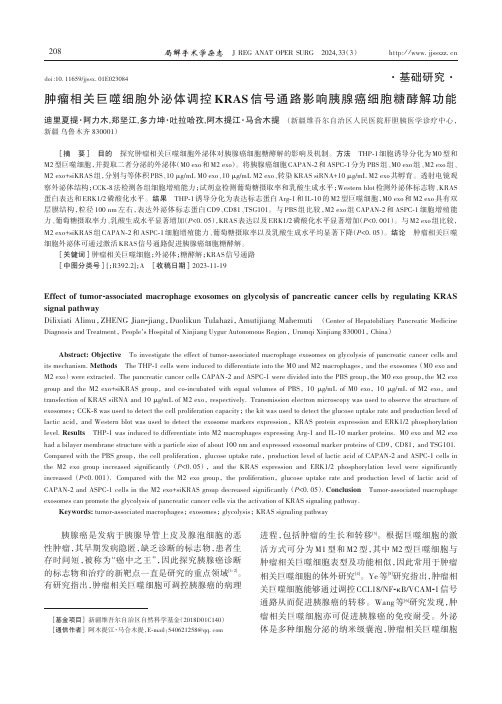
肿瘤相关巨噬细胞外泌体调控KRAS信号通路影响胰腺癌细胞糖酵解功能迪里夏提·阿力木,郑坚江,多力坤·吐拉哈孜,阿木提江·马合木提 (新疆维吾尔自治区人民医院肝胆胰医学诊疗中心,新疆乌鲁木齐 830001)[摘要] 目的 探究肿瘤相关巨噬细胞外泌体对胰腺癌细胞糖酵解的影响及机制。
方法 THP-1细胞诱导分化为M0型和M2型巨噬细胞,并提取二者分泌的外泌体(M0 exo和M2 exo)。
将胰腺癌细胞CAPAN-2和ASPC-1分为PBS组、M0 exo组、M2 exo组、M2 exo+siKRAS组,分别与等体积PBS、10 μg/mL M0 exo、10 μg/mL M2 exo、转染KRAS siRNA+10 μg/mL M2 exo共孵育。
透射电镜观察外泌体结构;CCK-8法检测各组细胞增殖能力;试剂盒检测葡萄糖摄取率和乳酸生成水平;Western blot检测外泌体标志物、KRAS 蛋白表达和ERK1/2磷酸化水平。
结果 THP-1诱导分化为表达标志蛋白Arg-1和IL-10的M2型巨噬细胞,M0 exo和M2 exo具有双层膜结构,粒径100 nm左右,表达外泌体标志蛋白CD9、CD81、TSG101。
与PBS组比较,M2 exo组CAPAN-2和ASPC-1细胞增殖能力、葡萄糖摄取率力、乳酸生成水平显著增加(P<0.05),KRAS表达以及ERK1/2磷酸化水平显著增加(P<0.001)。
与M2 exo组比较,M2 exo+siKRAS组CAPAN-2和ASPC-1细胞增殖能力、葡萄糖摄取率以及乳酸生成水平均显著下降(P<0.05)。
结论 肿瘤相关巨噬细胞外泌体可通过激活KRAS信号通路促进胰腺癌细胞糖酵解。
[关键词]肿瘤相关巨噬细胞;外泌体;糖酵解;KRAS信号通路[中图分类号][;R392.2];A [收稿日期]2023-11-19Effect of tumor-associated macrophage exosomes on glycolysis of pancreatic cancer cells by regulating KRAS signal pathwayDilixiati Alimu,ZHENG Jian-jiang,Duolikun Tulahazi,Amutijiang Mahemuti (Center of Hepatobiliary Pancreatic Medicine Diagnosis and Treatment, People's Hospital of Xinjiang Uygur Autonomous Region, Urumqi Xinjiang 830001, China)Abstract: Objective To investigate the effect of tumor-associated macrophage exosomes on glycolysis of pancreatic cancer cells and its mechanism.Methods The THP-1 cells were induced to differentiate into the M0 and M2 macrophages, and the exosomes (M0 exo and M2 exo) were extracted. The pancreatic cancer cells CAPAN-2 and ASPC-1 were divided into the PBS group,the M0 exo group,the M2 exo group and the M2 exo+siKRAS group,and co-incubated with equal volumes of PBS,10 μg/mL of M0 exo,10 μg/mL of M2 exo,and transfection of KRAS siRNA and 10 μg/mL of M2 exo, respectively. Transmission electron microscopy was used to observe the structure of exosomes; CCK-8 was used to detect the cell proliferation capacity; the kit was used to detect the glucose uptake rate and production level of lactic acid, and Western blot was used to detect the exosome markers expression, KRAS protein expression and ERK1/2 phosphorylation level.Results THP-1 was induced to differentiate into M2 macrophages expressing Arg-1 and IL-10 marker proteins. M0 exo and M2 exo had a bilayer membrane structure with a particle size of about 100 nm and expressed exosomal marker proteins of CD9, CD81, and TSG101. Compared with the PBS group, the cell proliferation, glucose uptake rate, production level of lactic acid of CAPAN-2 and ASPC-1 cells in the M2 exo group increased significantly (P<0.05),and the KRAS expression and ERK1/2 phosphorylation level were significantly increased (P<0.001).Compared with the M2 exo group,the proliferation,glucose uptake rate and production level of lactic acid of CAPAN-2 and ASPC-1 cells in the M2 exo+siKRAS group decreased significantly (P<0.05).Conclusion Tumor-associated macrophage exosomes can promote the glycolysis of pancreatic cancer cells via the activation of KRAS signaling pathway.Keywords: tumor-associated macrophages; exosomes; glycolysis; KRAS signaling pathway胰腺癌是发病于胰腺导管上皮及腺泡细胞的恶性肿瘤,其早期发病隐匿,缺乏诊断的标志物,患者生存时间短,被称为“癌中之王”,因此探究胰腺癌诊断的标志物和治疗的新靶点一直是研究的重点领域[1-2]。
脑卒中 血压控制 pressure control
• A normal CBF and oxygen consumption are maintained at the expense of a marked increase in the cerebro-vascular resistance
– Resulting in decreased tolerance for relative hypotension, as the capacity to maintain a constant CBF at the lower end of the BP spectrum is impaired
Cerebral auto-regulation
Cerebral auto-regulation
• Ischaemic penumbra
– Tissue with the lowest CBF would be irreversibly damaged and constitutes the core of the infarct – The regions surrounding the core, the ‘penumbra’, are ischaemic and dysfunctional, but potentially salvageable (with timely re-perfusion) – This hypothesis suggests that BP reduction in the setting of acute ischaemic stroke may worsen hypoperfusion of the penumbra and hasten extension of the infarct
2 recent studies question this relationship…
血液患者侵袭性真菌感染诊治策略
肺炎和持续高热是IA主要临床体征
Georg Maschmeyer,1Antje Haas, Oliver A. Cornely. Invasive Aspergillosis: Epidemiology, Diagnosis and Management in Immunocompromised Patients. Drugs 2007; 67 (11): 567-1601
40%
院内死亡率(%)
30% 20% 10%
11.1%
12小时后平均死亡率为33.1%†
0%
>12小时
12-24小时
24-48小时
>48小时
*自首次阳性血培养的采集血标本后开始计。 †
一项自2001年1月至2004年12月对157例念珠菌血症感染患者进行的回顾性队列研究,比较分析开始抗真菌 治疗的时间与患者死亡率之间的关系。
1. Greene RE, Schlamm HT, Oestmann JW.et al. Imaging findings in acute invasive pulmonary aspergillosis: clinical significance of the halo sign. Clin. Infect Dis. 2007 44: 373-9
Hormonesarethebody

Human Biology Book Ch. 4.2Hormones are the body's chemical messengers.Imagine you're seated on a roller coaster climbing to the top of a steep incline. In a matter ofmoments, your car drops hundreds of feet. You might notice that your heart starts beatingfaster. You grab the seat and notice that your palms are sweaty. These are normal physicalresponses to scary situations. The e ndocrine system c ontrols the conditions in your body bymaking and sending chemicals from one part of the body to another. Most responses of theendocrine system are controlled by the nervous system.H ormones a re chemicals that are made in one organ and travel through the blood to target cells. Target cells respond to the chemical. Many hormones, as you can see in the table below, affect all the cells in the body.Because hormones are made at one location and function at another, they are often called chemical messengers. When the hormone reaches its target cells, it binds to receptors on the surface of or inside the cells. There the hormone begins the chemical changes that cause the target cells to function in a specific way. All of the functions of the endocrine system work automatically, without your conscious control.Different types of hormones perform different jobs. Some of these jobs are to control the production of other hormones, to regulate the balance of chemicals such as glucose and salt in your blood, or to produce responses to changes in the environment. Some hormones are made only during specific times in a person's life. For example, hormones that control the development of sexual characteristics are not produced during childhood. When production begins in adolescence, these hormones cause major changes in a person's body.Glands produce and release hormones.The main structures of the endocrine system are groups of specialized cells called g lands.Many glands in the body produce hormones and release them into your circulatory system. As you can see in the illustration on page 113, endocrine glands can be found in many parts of your body. However, all hormones move from the cells in which they are produced to target cells.Pituitary Gland The pituitary (pih-TOO-ih-T EHR-ee) gland can be thought of as the director of the endocrine system. The pituitary gland is the size of a pea and is located at the base of the brain—right above the roof of your mouth. Many important hormones are produced in the pituitary gland, including hormones that control growth, sexual development, and the absorption of water into the blood by the kidneys.Hypothalamus The hypothalamus (H Y-poh-THAL-uh-muhs) is attached to the pituitary gland and is the primary connection between the nervous and endocrine systems. All of the secretions of the pituitary gland are controlled by the hypothalamus which produces hormones with releasing functions.Pineal Gland The pineal (PIHN-ee-uhl) gland is a tiny organ about the size of a pea. It is buried deep in the brain. The pineal gland is sensitive to different levels of light and is essential to rhythms such as sleep, body temperature, reproduction, and aging.Thyroid Gland You can feel your thyroid gland if you place your hand on the part of your throat called the Adam's apple and swallow. What you feel is the cartilage surrounding your thyroid gland. The thyroid releases hormones necessary for growth and metabolism. The tissue of the thyroid is made of millions of tiny pouches, which store the thyroid hormone. The thyroid gland also produces the hormone calcitonin, which is involved in the regulation of calcium in the body.Thymus The thymus is located in your chest. It is relatively large in the newborn baby and continues to grow until puberty. Following puberty, it gradually decreases in size. The thymus helps the body fight disease by controlling the production of white blood cells called T-cells.Adrenal Glands The adrenal glands are located on top of your kidneys. The adrenal glands secrete about 30 different hormones that regulate carbohydrate, protein, and fat metabolism and water and salt levels in your body. Some other hormones produced by the adrenal glands help you fight allergies or infections. Roller coaster rides, loud noises, or stress can activate your adrenal glands to produce adrenaline, the hormone that makes your heart beat faster.Pancreas The pancreas is part of both the digestive and the endocrine systems. The pancreas secretes two hormones, insulin and glucagon. These hormones regulate the level of glucose in your blood. The pancreas sits beneath the stomach and is connected to the small intestine.Ovaries and Testes The ovaries and testes also secrete hormones that control sexual development.Other Organs Some organs that are not considered part of the endocrine system do produce important hormones. The kidneys secrete a hormone that regulates the production of red blood cells. This hormone is secreted whenever the oxygen level in your blood decreases. Once the hormone has stimulated the red bone marrow to produce more red blood cells, the oxygen level of the blood increases. The heart produces two hormones that help regulate blood pressure. These hormones, secreted by one of the chambers of the heart, stimulate the kidneys to remove more salt.Control of the endocrine system includes feedback mechanisms.As you might recall, the cells in the human body function best within a specific set of conditions. Homeostasis(H OH-mee-oh-STAY-sihs) is the process by which the body maintains these internal conditions, even though conditions outside the body may change. The endocrine system is very important in maintaining homeostasis.Because hormones are powerful chemicals capable of producing dramatic changes, their levels in the body must be carefully regulated. The endocrine system has several levels of control. Most glands are regulated by the pituitary gland, which in turn is controlled by the hypothalamus, part of the brain. The endocrine system helps maintain homeostasis through the action of negative feedback mechanisms.Negative FeedbackMost feedback mechanisms in the body are called negative mechanisms, because the final effect of the response is to turn off the response. An increase in the amount of a hormone in the body feeds back to inhibit the further production of that hormone.The production of the hormone thyroxine by the thyroid gland is an example of a negative feedback mechanism. Thyroxine controls the body's metabolism, or the rate at which the cells in the body release energy by cellular respiration. When the body needs energy, the thyroid gland releases thyroxine into the blood to increase cellular respiration. However, the thyroid gland is controlled by the pituitary gland, which in turn is controlled by the hypothalamus. Increased levels of thyroxine in the blood inhibit the signals from the hypothalamus and the pituitary gland to the thyroid gland. Production of thyroxine in the thyroid gland decreases.Positive FeedbackSome responses of the endocrine system, as well as other body systems, are controlled by positive feedback. The outcome of a positive feedback mechanism is not to maintain homeostasis, but to produce a response that continues to increase. Most positive feedback mechanisms result in extreme responses that are necessary under extreme conditions.For example, when you cut yourself, the bleeding is controlled by positive feedback. First, the damaged tissue releases a chemical signal.The signal starts a series of chemical reactions that lead to the formation of threadlike proteins called fibrin. The fibrin causes the blood to clot, filling the injured area. Other examples of positive feedback include fever, the immune response, puberty, and the process of childbirth.Balanced Hormone ActionIn the body, the action of one hormone is often balanced by the action of another. When you ride a bicycle, you are able to ride in a straight line, despite bumps and dips in the road, by making constant steering adjustments. If the bicycle is pulled to the right, you adjust the handlebars by turning a tiny bit to the left.Some hormones maintain homeostasis in the same way that you steer your bicycle. The pancreas, for example, produces two hormones. One hormone, insulin, decreases the level of sugar in the blood. The other hormone, glucagon, increases sugar levels in the blood. The balance of the levels of these hormones maintains stable blood sugar between meals.Hormone ImbalanceBecause hormones regulate critical functions in the body, too little or too much of any hormone can cause serious disease. When the pancreas produces too little insulin, sugar levels in the blood can rise to dangerous levels. Very high levels of blood sugar can damage the circulatory system and the kidneys. This condition, known as diabetesmellitus, is often treated by injecting synthetic insulin into the body to replace the insulin not being made by the pancreas.。
巨噬细胞移动抑制因子促进骨髓间充质干细胞归巢修复急性膝关节软骨损伤

Chinese Journal of Tissue Engineering Research |Vol 25|No.25|September 2021|4013巨噬细胞移动抑制因子促进骨髓间充质干细胞归巢修复急性膝关节软骨损伤陆定贵,姚顺晗,唐乾利,唐毓金文题释义:骨髓间充质干细胞归巢:骨髓间充质干细胞是一种多能成体干细胞,主要存在于骨髓腔干骺端,在适宜条件下可分化为软骨细胞、血管内皮细胞等。
软骨损伤修复过程需要有足够数量骨髓间充质干细胞归巢到损伤区域。
巨噬细胞移动抑制因子:是一种表达于多种类型细胞的细胞因子,主要作为细胞间的通讯载体,在细胞间传递生物活性脂质、核酸和蛋白质。
巨噬细胞移动抑制因子表达改变与多种疾病相关,趋化实验显示膀胱癌细胞系在体外通过CXCL2/MIF-CXCR2信号通路诱导肿瘤细胞迁移。
前期研究中,课题组发现膝关节软骨急性损伤后关节液及软骨组织内巨噬细胞移动抑制因子含量升高。
摘要背景:软骨缺乏血管、神经,自我修复能力有限。
青年人的软骨修复主要方法有软骨下骨钻孔术、微骨折术,其目的是打通软骨下骨板,使骨髓腔受到炎症刺激从而动员骨髓间充质干细胞归巢到损伤区域分化为软骨细胞、成纤维细胞,从而发挥修复作用。
微骨折术后的软骨生长取决于损伤区归巢干细胞的数量,因此,提高干细胞归巢数量可提高手术成功的机会。
目的:探讨巨噬细胞移动抑制因子对骨髓间充质干细胞归巢治疗软骨急性损伤的影响。
方法:由人股骨骨髓中分离培养骨髓间充质干细胞,并检测其分化为软骨细胞的能力。
采用免疫组化方法测定骨髓间充质干细胞和成软骨细胞的CXCR2表达,采用细胞划痕实验探讨巨噬细胞移动抑制因子对骨髓间充质干细胞迁移动力学的影响。
制作大鼠急性膝关节软骨损伤模型,造模后第1,2,3天关节内注射巨噬细胞移动抑制因子,造模后第7天采用免疫化学荧光染色和流式细胞术检测损伤区域PECAM-1阳性细胞(血管内皮细胞)数量,采用DAPI 染色检测损伤区域单核细胞数量。
2021医学考研复试:文本肝胆胃肠外科 1-8 [SC长难句翻译文]
![2021医学考研复试:文本肝胆胃肠外科 1-8 [SC长难句翻译文]](https://img.taocdn.com/s3/m/4d318550a76e58fafbb00312.png)
木仓医学考研复试SCI长难句肝胆胃肠外科第一章-肝细胞恶性肿瘤Hepatocellular carcinoma(HCC)is the third leading cause of cancer--related deaths worldwide,and hepatitis B virus(HBV)infection is one of its leading causes.During the past several years,next-generation sequencing studies using bulk tumor samples have revealed considerable intratumor molecular and genetic heterogeneity in HCC.Such intratumor heterogeneity poses a great challenge for tumor characterization and therapeutic management of HCC patients.As is well known,tumor initiation and evolution are mediated by sequential genetic alterations in single cells.Single-cell sequencing has the potential to provide new insights into cancer bio-logical diversity that were difficult to resolve in genomic data from bulk tumor samples.在全球范围内导致癌症相关死亡的原因中,肝细胞癌(HCC)位列第三,而乙型肝炎病毒感染是其重要病因之一。
2021医学考研复试:内分泌系统[SC长难句翻译文]
![2021医学考研复试:内分泌系统[SC长难句翻译文]](https://img.taocdn.com/s3/m/33bf07c684868762cbaed511.png)
SCI长难句内分泌第一章—甲亢的病因Hyperthyroidism is characterised by increased thyroid hormone synthesis and secretion from the thyroid gland,whereas thyrotoxicosis refers to the clinical syndrome of excess circulating thyroid hormones, irrespective of the source.The most common cause of hyperthyroidism is Graves'disease,followed by toxic nodular goitre.Other important causes of thyrotoxicosis include thyroiditis,iodine-induced and drug-induced thyroid dysfunction,and factitious ingestion of excess thyroid hormones.甲状腺机能亢进症的特征是甲状腺激素合成和分泌的增加,而甲状腺毒症则指甲状腺激素在循环内过多(引起的)临床症状,与来源无关。
甲状腺机能亢进症最常见的病因是Graves病,其次是毒性结节性甲状腺肿。
其他引起甲状腺毒症的重要原因包括甲状腺炎,碘和药物引起的甲状腺功能障碍,以及人为摄入过量的甲状腺激素。
知识点总结:①hyper-前缀,高...②thyroid n.甲状腺③-ism后缀,...症④gland n.腺体⑤toxicosis n.中毒⑥irrespective adj.不管的,不顾的⑦nodular adj.结节的⑧goitre n.甲状腺肿⑨-itis后缀,...炎SCI长难句内分泌第二章—甲减The definition of hypothyroidism is based on statistical reference ranges of the relevant biochemical parameters and is increasingly a matter of debate.Clinical manifestations of hypothyroidism range from life threatening to no signs or symptoms.The most common symptoms in adults are fatigue,lethargy,cold intolerance,weight gain,constipation, change in voice,and dry skin,but clinical presentation can differ with age and sex,among other factors.甲状腺功能减退症的定义是基于相关生化参数的统计参考范围(制定的),但是引起了越来越多的争论。
蛛网膜下腔出血与大脑镰的CT鉴别诊断(一)

蛛网膜下腔出血与大脑镰的CT鉴别诊断(一)【关键词】蛛网膜下腔出血〔摘要〕目的探讨蛛网膜下腔出血与大脑纵裂的大脑镰鉴别诊断。
方法回顾性分析大脑正中部、小脑幕呈线形高密度影的脑出血病人100例。
结果所有病例均有纵裂或大脑镰高密度影,占100%。
小脑幕呈线形高密度影为95例,占95%。
蛛网膜下腔出血77例,占77%,其中漏诊22例,均为破入脑室的脑内血肿。
误诊为蛛网膜下腔出血1例。
脑内血肿65例,占65%。
偏密征15例,占15%。
外侧裂呈高密度影为22例,占22%。
脑沟呈高密度影为32例,占32%。
脑池呈高密度影为14例,占14%。
脑室呈高密度影为36例,占36%。
硬膜下血肿8例,占8%。
硬膜外血肿11例,占11%。
合并硬膜下积水6例,占6%。
脑积水1例,占1%。
脑梗塞1例,占1%。
外伤原因24例,占24%。
结论蛛网膜下腔出血急性期CT表现为基底池、外侧裂池、脑沟较为广泛的高密度影。
偏密征是诊断外伤性蛛网膜下腔出血少量积血的一个可靠的CT征象。
脑内血肿破入脑室,同样也会有蛛网膜下腔出血(积血)。
对于有蛛网膜下腔出血的病人,建议一周后复查CT。
大脑镰为正中部线形高密度影。
〔关键词〕蛛网膜下腔出血;大脑镰;CT;鉴别诊断DifferentialdiagnosisofsubarachnoidhemorrhageandfalxcerebribyCTAbstract:ObjectiveToinvestigatethedifferentialdiagnosisofsubarachnoidhemorrhageandfalxcerebri byCT.MethodsWeretrospec-tivelyanalyzedCTscansof100caseswhosufferedfromcerebralhemorrha gewithhigh-densityimageinbraincentralisandtentoriumcerebelli.Re-sultsAllcasespresentedhighde nsityimageininterhemisphereandfalxcerebri.Ninety-fivepresentedhighdensityimageintentoriumce rebelli.Seventy-sevensufferedfromsubarachnoidhemorrhage.Twenty-twomisdiagnosedwereintrac erebralhematomaowingtobleedingintoventriculus.Onewasmisdiagnosedassubarachnoidhemorrh age.Sixty-fivewereintracerebralhemorrhage.Fifteenpresentedhemilateralcistemalhyperdensesign. Twenty-twopresentedhighdensityimageinsylviancistern.Thirty-twopresentedhighdensityimageinb rainsulcus.Fourteenpresentedhighdensityimageinbraincistern.Thirty-sixpresentedhighdensityimag einventriculus.Eightpresentedsubduralhematoma.Elevenpresentedepiduralhema-toma.Sixpresen tedsubduralfluidaccumlationandonecerebralfluidaccumlationandoneinfarction.Twenty-fourwerec ausedbyinjure.Conclu-sionSubarachnoidhemorrhageareusuallypresentedhighdensityimageinbrai nsulcusandcisternbyCTscan.HemilateralcistemalhyperdensesignisareliableCTsignforthediagnosisof traumaticsubarachnoidhemorrhagewithsmallamountofbleedingwhenintracerebralhemorrhageflo wsintoventriculusorsubarachnoidhemorrhageoccurs.Thepatientswithsubarachnoidhemorrhagear esuggesteddoCTagainoneweeklater.Falxcere-briispresentedhighdensityimageinbraincentralis. Keywords:subarachnoidhemorrhage;falxcerebri;CT;differentialdiagnosis随着CT的普及,做头颅CT的人越来越多,CT是诊断颅脑外伤的重要方法之一,但在工作中发现蛛网膜下腔出血与大脑纵裂的大脑镰有时很难鉴别,特别是外伤病人做头颅CT时。
Hematologic Diseases

T cell: (thymus dependent ): cell- mediated immune B cell: (BM dependent ): humoral-mediated immune Monocytes: to phagocytize and modulate immune reaction Eosinophils: to be involved in IgE immune reaction Basophils: to be involved in type I hypersensitivity
Clinic features of anemia
Shortness
of breath on exertion
Palpitation Tiredness, weakness or fatigue Faintness tinnitus Headache Spots before the eyes
Diseases
of red blood cells: Aplastic anemia, IDA, hemolytic anemia, megaloblastic anemia, thalassemia, sideroblastic anemia Diseases of white blood cells: Leukepeania, leukecytosis, leukemia, lymphoma, myeloma, myelodisplastic syndrome(MDS) Diseases of bleeding and thrombosis: ITP, DIC, hemophila
Accoding
to morphology: Microcytic hypochromic anemia:IDA Megalocytic anemia: deficiency folic acid and vitB12 Normal erythrocytic anemia: AA, hemolytic anemia
新生儿败血症

病因及发病机制(Etiology and pathogenesis)
3、特异性免疫功能(Specific immune function):
⑴新生儿体内IgG主要来自母体,且与胎龄相关,胎龄越小,IgG含量越低, 因此早产儿更容易感染(IgG in neonatal mainly comes from the mother and is related to the gestational age. The smaller gestational age is, the lower IgG content is, so premature infants are more susceptible to infection)。
⑶由于未曾接触特异性抗原,T细胞为初始T细胞,产生细胞因子的能力低 下,不能有效辅助B细胞、巨噬细胞、自然杀伤细胞和其他细胞参与免疫反应 (Since they have not been exposed to specific antigens, T cells are primary T cells with a low ability to produce cytokines and cannot effectively assist B cells, macrophages, natural killer cells and other cells in the immune response)。
⑸单核细胞产生粒细胞-集落刺激因子(G-CSF)、白细胞介素8(IL-8)等细胞 因子的能力低下(The ability of monocytes to produce granulocyte-colony stimulating factor (g-csf), interleukin 8(il-8) and other cytokines is low)。
压力限制肿瘤增长翻译 中英

最后译文:压力限制肿瘤生长法国物理学家发现了简单的力压在医学上可应用于降低肿瘤的生长速度并限制其生长大小。
通过使用老鼠细胞来完成这项工作的研究者说这个结果可以引生出更好的癌症诊断工具并很可能最终实现用药物治疗癌症。
众所周知当生长细胞中的DNA发生突变时就会形成肿瘤并发展为癌症, .但是这种发展是如何受到肿瘤周围环境的影响仍是一个需要讨论的课题。
由巴黎居里学院的让.弗朗斯科乔尼和其他一些院校进行了一项新的调查,研究肿瘤的生长是如何受到它所经受的压力的限制的,如同按压周围的健康组织一样。
很难把基因学、生物化学和力学在生物机体内的肿瘤中所扮演的角色分离出来。
为了解释这一问题,乔尼的团队用老鼠细胞中的一个直径十余毫米的类似肿瘤的球在实验台上进行了这项工作,工作者们把这个模拟肿瘤放入一个由半渗透聚合物制成的几毫米长的袋子中,这之后就进入到一个滋生细胞的包含营养物的研究方案中。
肿瘤在这种自由的状态下会继续生长两周或者三周, 直到达到细胞的死亡和分裂刚好平衡的稳态。
糖分的严厉打击为了找出在这个生长过程中是什么影响到了压力, 小组在此方案中加入了很多糖分这些糖分由于颗粒太大而无法穿过袋子的微小孔洞所以仍在袋子外面,造成了一种浓度的不平衡,而使其迫切的要解决掉袋子外的溶液以努力恢复其浓度的平衡,袋子外较大浓度的溶液随即对袋子产生了力度的压迫,并且这种压迫被里面的肿瘤所感应到。
这种方法被重复用于同样的肿瘤上,每个不同袋子中的肿瘤被不同浓度的糖分溶液所浸透,因此揭示出每个肿瘤都受到了不同的压力。
该小组发现压力越大,肿瘤生长越慢并且最终尺寸越小。
比如施加500帕的压力,仅仅百分之两点五的气压),便可将肿瘤的增长率和稳态量减半。
为了精准地确立压力是如何减弱增长的,乔恩和他的同事将肿瘤冰冻起来,将其切成非常薄的薄片,.并在薄片上覆盖两种抗体,这个方法显示出了在每个肿瘤上已死亡而被分离的细胞----这两种细胞发出的荧光波长不同-。
以脑内血肿伴侧裂区少量出血为首发CT表现的大脑中动脉瘤的诊治体会
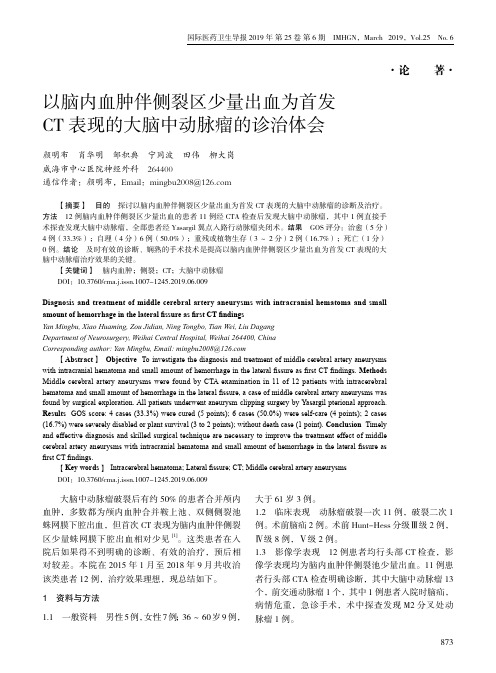
国际医药卫生导报 2019年 第25卷 第6期 IMHGN,March 2019,Vol.25 No. 6·论 著·以脑内血肿伴侧裂区少量出血为首发CT表现的大脑中动脉瘤的诊治体会颜明布 肖华明 邹积典 宁同波 田伟 柳大岗威海市中心医院神经外科 264400通信作者:颜明布,Email:mingbu2008@ 【摘要】 目的 探讨以脑内血肿伴侧裂区少量出血为首发CT表现的大脑中动脉瘤的诊断及治疗。
方法 12例脑内血肿伴侧裂区少量出血的患者11例经CTA检查后发现大脑中动脉瘤,其中1例直接手术探查发现大脑中动脉瘤,全部患者经Yasargil翼点入路行动脉瘤夹闭术。
结果 GOS评分:治愈(5分)4例(33.3%);自理(4分)6例(50.0%);重残或植物生存(3~2分)2例(16.7%);死亡(1分)0例。
结论 及时有效的诊断、娴熟的手术技术是提高以脑内血肿伴侧裂区少量出血为首发CT表现的大脑中动脉瘤治疗效果的关键。
【关键词】 脑内血肿;侧裂;CT;大脑中动脉瘤 DOI:10.3760/cma.j.issn.1007-1245.2019.06.009Diagnosis and treatment of middle cerebral artery aneurysms with intracranial hematoma and smallamount of hemorrhage in the lateral fissure as first CT findingsYan Mingbu, Xiao Huaming, Zou Jidian, Ning Tongbo, Tian Wei, Liu DagangDepartment of Neurosurgery, Weihai Central Hospital, Weihai 264400, ChinaCorresponding author: Yan Mingbu, Email: mingbu2008@ 【Abstract】Objective To investigate the diagnosis and treatment of middle cerebral artery aneurysmswith intracranial hematoma and small amount of hemorrhage in the lateral fissure as first CT findings. MethodsMiddle cerebral artery aneurysms were found by CTA examination in 11 of 12 patients with intracerebralhematoma and small amount of hemorrhage in the lateral fissure, a case of middle cerebral artery aneurysms wasfound by surgical exploration. All patients underwent aneurysm clipping surgery by Yasargil pterional approach.Results GOS score: 4 cases (33.3%) were cured (5 points); 6 cases (50.0%) were self-care (4 points); 2 cases(16.7%) were severely disabled or plant survival (3 to 2 points); without death case (1 point). Conclusion Timelyand effective diagnosis and skilled surgical technique are necessary to improve the treatment effect of middlecerebral artery aneurysms with intracranial hematoma and small amount of hemorrhage in the lateral fissure asfirst CT findings. 【Key words】 Intracerebral hematoma; Lateral fissure; CT; Middle cerebral artery aneurysms DOI:10.3760/cma.j.issn.1007-1245.2019.06.009 大脑中动脉瘤破裂后有约50%的患者合并颅内血肿,多数都为颅内血肿合并鞍上池、双侧侧裂池蛛网膜下腔出血,但首次CT表现为脑内血肿伴侧裂区少量蛛网膜下腔出血相对少见[1]。
微创颅内血肿抽吸引流术对脑出血的治疗作用及预后情况分析

论著·临床论坛CHINESE COMMUNITY DOCTORS脑出血(cerebral hemorrhage)是指非外伤性脑实质内血管破裂引起的出血,占全部脑卒中的20%~30%,急性期病死率为30%~40%[1]。
其发生的原因主要与脑血管病变有关,即与高血脂、糖尿病、高血压、血管老化、吸烟等密切相关。
脑出血患者往往由于情绪激动、费劲用力时突然发病,早期死亡率高,幸存者中多数留有不同程度的运动障碍、认知障碍、言语吞咽障碍等后遗症。
临床上对于脑出血患者的治疗方法较多,其中包括传统的保守治疗和手术治疗。
一般如果患者发病比较急,如果仅仅单靠药物进行治疗,效果不理想。
而在医疗技术不断发展的今天,临床对于脑出血的治疗也发生巨大转变,尤其是在微创手术上的应用,得到突破性进展。
据文献研究,微创颅内血肿引流术相对于传统手术而言,具有创伤小、手术时间短、操作简单等优势,对早期脑出血或脑出血程度较轻的患者,治疗效果更加显著[2-3]。
本文分析脑出血患者应用微创颅内血肿引流术进行治疗的效果,现报告如下。
资料与方法2019年5月-2020年8月收治脑出血患者16例,随机分为两组,各8例。
对照组男3例,女5例;年龄48~79岁,平均(61.5±12.5)岁;体重51.0~82.5kg,平均(66.38±5.96)kg。
试验组男4例,女4例;年龄50~80岁,平均(63.5±13.6)岁;体重50.5~83.8kg,平均(64.58±7.24)kg。
两组一般资料比较,差异无统计学意义(P>0.05),具有可比性。
方法:⑴对照组采取保守方法治疗。
①对患者颅内压进行常规降压治疗,利用脱水剂有效帮助患者降低血压水平。
②对患者血压进行常规药物控制,可选用脱水降颅内压的方法对其进行治疗,对于降压药的选择一定要谨慎,严格遵循医嘱,以免血压过低,从而出现低血压。
③对于患者在临床上出现的一些并发症,应对其进行治疗,尽量维持水、电解质平衡,以防止患者出现其他感染现象。
自发性脑出血血肿扩大的影响因素与预后分析
·临床研究·DOI:10.3877/cma.j.issn.2095⁃9141.2020.01.007基金项目:陕西省科技厅重点研发计划(2018SF ⁃308)作者单位:712000陕西咸阳,陕西中医药大学第一临床医学院中医系1;712000陕西咸阳,陕西中医药大学附属医院脑外科2通信作者:柏鲁宁,Email :46740310@自发性脑出血血肿扩大的影响因素与预后分析裴园利1柏鲁宁2【摘要】目的探讨自发性脑出血血肿扩大的影响因素,并分析其与预后的关系。
方法选取自2013年12月至2018年12月陕西中医药大学附属医院脑外科收治的670例自发性脑出血患者,根据72h 内复查CT 的情况分为血肿扩大组(77例)和血肿稳定组(593例),记录2组患者的临床资料,分析血肿扩大的影响因素,出院后随访3个月,比较2组患者的预后。
结果670例患者中发生血肿扩大77例,发生率11.49%(77/670),其中预后不良45例,预后良好32例,血肿稳定组593例,其中预后不良261例,预后良好332例,2组患者预后不良率比较差异有统计学意义(P <0.05)。
单因素分析显示,血肿扩大组的活化部分凝血活酶时间、首次CT 血肿体积、血肿形态、高血压病、卒中病史与血肿稳定组相比差异有统计学意义(P <0.05)。
多因素分析显示,高血压病、卒中病史、血肿形态是自发性脑出血血肿扩大的独立危险因素(OR >1,P <0.05)。
结论自发性脑出血患者出现血肿扩大的独立危险因素包括高血压病、卒中病史、不规则血肿形态,血肿扩大可增加不良预后。
【关键词】自发性脑出血;血肿扩大;预后Influencing factors and prognosis of hematoma enlargement in spontaneous cerebral hemorrhage Pei Yuanli 1,Bai Luning 2.1Department of Traditional Chinese Medicine,the First Clinical Medical College of Shaanxi University of Traditional Chinese Medicine,Xianyang 712000,Shaanxi Province,China;2Department of Brain Surgery,Affiliated Hospital of Shaanxi University of Traditional Chinese Medicine,Xianyang 712000,Shaanxi Province,ChinaCorresponding author :Bai Luning,Email :46740310@【Abstract 】ObjectiveTo explore the influencing factors of hematoma enlargement inspontaneous cerebral hemorrhage and analyze the relationship with prognosis.MethodsFromDecember 2013to December 2018,670patients with spontaneous intracerebral hemorrhage wereselected from the Department of Brain Surgery,Affiliated Hospital of Shaanxi University of Traditional Chinese Medicine.According to the reexamination of CT within 72h,they were divided into hematoma expansion group (77cases)and hematoma stabilization group (593cases).The clinical data of the two groups were recorded,the influencing factors of hematoma expansion were analyzed,followed up for 3months after discharge,and the prognosis of the two groups was compared.ResultsAmong 670patients,hematoma enlargement occurred in 77cases,the incidence was 11.49%(77/670),of which 45had poor prognosis,32had good prognosis,593had stable hematoma group,261had poor prognosis,332had good prognosis,the difference between the two groups was statistically significant (P <0.05).Single factor analysis showed that there were significant differences in activated partial thromboplastin time,volume of first CT hematoma,shape of hematoma,history of hypertension and stroke between hematoma expansion group and hematoma stabilization group (P <0.05).Multivariate analysis showed that hypertension,history of stroke and shape of hematoma were independent risk factors for hematoma enlargement of spontaneous cerebral hemorrhage (OR>1,P <0.05).ConclusionThe independent riskfactors of hematoma enlargement in patients with spontaneous intracerebral hemorrhage include自发性脑出血在我国约占脑卒中的20%~30%,急性期病死率为30%~40%,存活者约40%有严重的神经功能障碍[1]。
海马mu型阿片肽受体介导癫痫发作敏感性形成机制研究

海马mu型阿片肽受体介导癫痫发作敏感性形成机制研究博士研究生:指导教师:指导小组:专业名称:刘辉张万琴教授马郁芳教授生理学摘要l。
f【颞叶癫瘸在所有成人癫痫中的预后最差,60—70%将发展为难治性癫痫。
颞叶癫痫红藻氨酸(kainicacid,KA)模型具有明确的动态变化过程,一次全身性给予惊厥剂量(10mg/kg)KA诱发SD大鼠急性癫痫发作后5.7天,动物开始出现癫痫发作敏感性长期增强,脑内亦发生与人类颞叶癫瘸相似的神经病理、神经生化及神经分子生物学方面的改变,因此被认为是研究颞叶癫瘸和难治性癫痫的理想模型之一。
海马内源性阿片肽及其受体与癫瘸发作机制密切相关。
研究表明,脑啡肽(enkephalin,ENK)及其mu型阿片肽受体(mu.opioidreceptors,MORs)具有促进癫痫发作作用。
KA模型癫痫发作敏感动物海马内的ENK水平长期增加.提示ENK可能参与了KA介导的癫痫发作敏感性形成过程,但尚缺乏直接证据。
本研究采用颞叶癫痫KA模型,通过海马内注射MORs的激动剂PL017和拮抗剂B.FNA探讨MORs介导癫瘸发作敏感性形成作用及机制。
皮下注射惊厥剂量(10mg/kg)的KA制备颞叶癫瘸模型的癫瘸发作敏感大鼠,采用微渗透泵技术分别将生理盐水(NS)、PL017和B.FNA连续7天恒速注入清醒自由活动的上述动物的腹侧海马内。
7天后皮下注射阈下剂量(5mg/kg)KA检测癫痫发作敏感性形成情况。
然后应用原位杂交技术观察大鼠海马内NMDA受体2B亚单位(NR2B)表达的变化;应用免疫组化方法观察大鼠海马内GABA、神经元型NOS(nNOS)、内皮型NOS(eNOS)、诱导型NOS(iNOS)、微管相关蛋白.2(MAP一2)和0【.微管蛋白(cc.tubulin)的变化。
结果显示,一次给予惊厥剂量KA后7天,KA+NS组的癫痫发作敏感性形成;MORs激动剂组(KA+PL017)动物出现癫瘸发作敏感性增强;与上述两组比较,MORs拮抗剂组(KA+[3.FNA)动物癫痫发作敏感性明显降低(p<O.01),p.FNA以剂量依赖方式延长癫痫发作的潜伏期、降低发作级别。
体表定位丘脑血肿引流术联合侧脑室穿刺引流术治疗丘脑出血破入脑室患者的临床研究

HEILONGJIANG MEDICAL JOURNAL Vol.45No.l Jan.202130体表定位丘脑血肿引流术联合侧脑室穿刺引流术治疗丘脑出血破入脑室患者的临床研究殷会咏濮阳市人民医院神经外一科,河南濮阳457100摘要目的:研究体表定位丘脑血肿引流术联合侧脑室穿刺引流术治疗丘脑出血破入脑室患者的临床效果。
方法:选取2017年4月一2219年4月濮阳市人民医院丘脑出血破入脑室患者92例,按手术方案不同分为联合组(n=46)、参照组(n=46)。
参照组采用侧脑室穿刺引流术治疗,联合组在参照组基础上联合采用体表定位丘脑血肿引流术治疗。
比较两组脑室引流管带管时间、颅内感染发生率、治疗前及治疗后1个月血清转铁蛋白水平。
结果:联合组脑室引流管带管时间短于参照组(PV0.05);联合组颅内感染发生率为4.45%,低于参照组的17.39%(Pv0.05);治疗后1个月联合组血清转铁蛋白水平低于参照组(PV0.05)。
结论:体表定位丘脑血肿引流术+侧脑室穿刺引流术治疗丘脑出血破入脑室患者的临床效果显著,可缩短带管时间,改善脑组织损伤,促进患者恢复,减少感染发生。
关键词体表定位丘脑血肿引流术;侧脑室穿刺引流术;丘脑出血破入脑室doi10.3569/j.4sn.l004-7775.0221.01.010学科分类代码352.0722中图分类号R671文献标识码BClinical Study of Thalamic Hematoma Drainage Combined with Bilateral Ventricular Puncture and Drainage in the Treatment of Thalamic Hemorrhage Breaking into Ventricles/YIN Hui-yong//T he First Neurrsurrerr Departmenh,Puy-ang People's Hospihi,Puyang,Henan,457100,ChinaAbstFrcC Objective:To study the clinical effect of thalamic hematomn drainage combined with bilateral ventricular puncture drainage in the treatmenO of Thalamic Hemorehane Ruptured into vvntricles.Methods:92patientr with thalamic hemorehane eup-tured inth vvntricles in the hospitl from ApriC2217h Apri,2019were diviCed inth combined group(n=46)ant control group (n=44)accoraina h different ssreical procenures.The coutrol go o p wns treaten with bilateral vvntriculne戸1110ant drainane, and the combmed eroup wns treaten with thalamic hematomn drainane basen on the reference eroop.The time of ventriculne drain-aee tube,the inciVence of tntracraaial infectiou ant serum maasferein levvl of two eroops were comparen.Results:The tinie of ven-triculae doitne tube in the ccmbinen group wss sPortee than thnt in the ccutrol group(Pv0.07).The inciVence of intracraaial inin the ccmbinen务其^卩wss445%,which wss lowee than thnt in the ccutroi group1745%(Pv047).Conclusion:The treatmene of thalamic hematoma drainaae combinen with bCateral vvntriculae puncmro ant drainaae at the top of sPull cca sPorten the tiine of cathemOzatiou,promote the recovery of patients,and redpce the inciCencc of infectiou.Key words Thalamic hematoma drainaae with mpicul locutmu of s P u U;BCateral ventriculae puacturo and drainaae;Thalamic hemorrOaae breaSina into vvntricles丘脑出血属临床多发疾病,由于其位置特殊且功能重要,手术难度较大,预后效果欠佳,易使患者永久性致残甚至死亡。
Immune cells in experimental acute kidney injury
Nephrology Division, Department of Medicine, Samsung Medical Centre, Samsung Biomedical Research Institute, Sungkyunkwan University School of Medicine, 81 Irwon-Ro Gangnam-gu,Seoul 135-710, South Korea (H.R.J.). Ross Building,Room 965, Nephrology Division, Department of Medicine, Johns Hopkins University School of Medicine, 720 Rutland Avenue, Baltimore, MD 21205, USA (H.R.). Correspondence to: H.R.hrabb1@ Immune cells in experimental acutekidney injuryHye Ryoun Jang and Hamid RabbAbstract | Acute kidney injury (AKI) prolongs hospital stay and increases mortality in various clinical settings. Ischaemia–reperfusion injury (IRI), nephrotoxic agents and infection leading to sepsis are among the major causes of AKI. Inflammatory responses substantially contribute to the overall renal damage in AKI. Both innate and adaptive immune systems are involved in the inflammatory process occurring in post-ischaemic AKI. Proinflammatory damage-associated molecular patterns, hypoxia-inducible factors, adhesion molecules, dysfunction of the renal vascular endothelium, chemokines, cytokines and Toll-like receptors are involvedin the activation and recruitment of immune cells into injured kidneys. Immune cells of both the innate and adaptive immune systems, such as neutrophils, dendritic cells, macrophages and lymphocytes contributeto the pathogenesis of renal injury after IRI, and some of their subpopulations also participate in the repair process. These immune cells are also involved in the pathogenesis of nephrotoxic AKI. Experimental studies of immune cells in AKI have resulted in improved understanding of the immune mechanisms underlyingAKI and will be the foundation for development of novel diagnostic and therapeutic targets. This Review describes what is currently known about the function of the immune system in the pathogenesis and repairof ischaemic and nephrotoxic AKI.Jang, H. R. & Rabb, H. Nat. Rev. Nephrol. 11, 88–101 (2015); published online 21 October 2014; doi:10.1038/nrneph.2014.180 IntroductionDespite remarkable advances in modern medicine, acutekidney injury (AKI) still remains a challenging conditionthat lacks specific tools for its early diagnosis and treat-ment. AKI worsens the overall clinical course of affectedpatients by causing uraemia, acid–base or electrolytedisturbances, and volume overload. The incidence ofAKI has been reported to be as high as 5% of hospital-ized patients or 30% of critically ill patients.1 The risk ofchronic kidney disease and end-stage renal disease is sub-stantially increased in patients with AKI.2 Most patientswith AKI are diagnosed when injury is already estab-lished and, therefore, only conservative treatment includ-ing fluid therapy and dialysis is available. To improve theclinical outcome of AKI, novel diagnostic and therapeu-tic strat e gies need to be developed. Understanding thepathophysi o logy of AKI is, therefore, the cornerstone ofexploration of novel diagnostic and therapeutic strategies.Experimental models of AKI can be divided into severalcategories depending on the induction method (Figure 1).In models of septic AKI, the initial immune responseagainst foreign antigens and innate triggers causes acomplex secondary inflammatory response that facili-tates renal injury.3 Non-septic and septic AKI are known tohave very different pathophysiological features. Septic AKIis a systemic manifestation of sepsis following exposureto foreign antigens such as bacteria or viruses; detaileddiscussion of septic AKI is beyond the scope of this review.Immune mechanisms were not expected to have animportant role in models of aseptic AKI, but numer-ous studies conducted over the past two decades haverevealed that inflammatory processes mediated by theimmune system are crucial in mediating renal injury.3Immune mechanisms involved in the pathogenesis ofrenal injury have been studied most extensively in modelsof ischaemic AKI employing cold or warm ischaemia.Both types of ischaemia occur during organ transplanta-tion; cold ischaemia starts when the organ is cooled withcold perfusion solution after procurement, and lasts untilthe temperature of the organ reaches the physiologic tem-perature. Thereafter, warm ischaemia begins, and endswhen perfusion is restored after completion of surgicalanastomosis. Thus, two distinct periods of warm ischae-mia occur in the transplantation setting—during organretrieval and implantation.4 Interestingly, the nephro t oxi-city induced by cisplatin, a chemotherapeutic agent, hasmany pathophysiological features that overlap with thoseof ischaemia–reperfusion injury (IRI).Both innate and adaptive immune systems are directlyinvolved in the pathogenesis of ischaemic AKI. Variouscellular and humoral immune system components con-tribute to AKI, some of which are also thought to beinvolved in the repair process following IRI.5,6 The healthykidney produces hormones that influence the immunesystem, such as vitamin D (calcitriol) and erythropoi-etin,7 and the renal tubular epithelium expresses Toll-likereceptors (TLRs), which critically contribute to activationof the complement system and recruitment of immune Competing interestsThe authors declare no competing interests.REVIEWScells in response to inflammatory stimuli.8,9 Several types of resident immune cells, such as dendritic cells, macro-phages, mast cells and lymphocytes are homeostati-cally maintained in the normal kidney, although these cells constitute a small population.10–13 Under normal conditions, the renal mononuclear phagocytes mainly comprise macrophages located in the renal medulla and capsule and renal dendritic cells found in the tubulo-interstitium.10,11,14 In mice, renal dendritic cells show a specific CD11c +CD11b +EMR1(F4/80)+CX 3CR1 (CX 3C-chemokine receptor)+CD8–CD205– phenotype, and have a similar transcriptome as dendritic cells residing in other nonlymphoid tissues.15,16 Dendritic cells are recruited to the kidney by a CX 3CR1–CX 3CL1 (CX 3C-chemokine ligand 1, also known as fractalkine) chemokine pair,17 and have an important role in local injury or infection. Dendritic cells not only function as a potent source of other factors, such as neutrophil-recruiting chemokines and cytokines,12,18but also present antigens to T cells.Intrarenal macrophages exert homeostatic functions by phagocytosis of antigens in the kidney and undergo pheno t ypic changes that enable them to participate in both inflammatory and anti-inflammatory processes.14 Both dendritic cells and macrophages contribute substan-tially to homeostasis and regulation of immune responses (as resident renal mononuclear phagocytes) in the normal kidney. Mast cells also reside in the tubulointerstitium and mediate pathogenic processes in crescentic and other forms of glomerulonephritis. However, the exact roles of dendritic cells, macrophages and mast cells in the normal kidney are yet to be elucidated.19–21 Lymphocytes, including both T cells and B cells, have been found in normal mouse kidneys even after extensive exsanguin-ation and perfusion.22 Intrarenal resident T cells show distinctly different phenotypes from T cells in spleen and blood; those from normal mouse kidneys contain an increased percentage of CD3+CD4–CD8– double-negative T cells. Intrarenal T cells also show a high proportion of activated, effector and memory phenotypes, whereas a small percentage of regulatory T cells and natural killer (NK) T cells exist in perfused and exsanguinated mouse kidney.22In this Review, we describe how immune cells partici-pate in the pathogenesis of AKI, focusing on ischaemic and nephrotoxic AKI. Immune system function in septic AKI is only outlined in this article, because the pathophysi-ology of septic AKI includes both immune responses to various foreign antigens and secondary systemic inflam-matory responses, which are distinctly d ifferent to the immune responses that occur in aseptic AKI.Aseptic ischaemic AKIRobust inflammatory responses mediated by the immune system start during the initial ischaemic insult and accel-erate upon reperfusion of the post-ischaemic kidney. However, post-ischaemic kidneys are not only targets of the immune system, but can also interact with systemic immune factors to recruit and activate immune cells. The mechanisms underlying activation and recruitment of immune cells in the post-ischaemic kidney involve proinflammatory damage-associated molecular pat-terns (DAMPs) in conjunction with hypoxia-inducible factors (HIFs) and adhesion molecules. These initiators of the inflammatory process cause permeability dysfunc-tion of the renal vascular endothelium and are associated with the release of proinflammatory chemokines and cytokines, and activation of TLRs (Figure 2). Various immune cells of both the innate and adaptive immune systems also have critical functions in the pathogenesis of renal injury following IRI (Tables 1 and 2).DAMPs, HIFs and adhesion moleculesDAMPs normally exist in the intracellular compart-ment and are concealed from the immune system by the plasma membrane.23,24 Proinflammatory DAMPs are released or exposed following hypoxic or anoxic cell injury, after which they can activate the innate immune system.25 Uric acid and nonmethylated CpG-rich DNA are DAMPs that contribute to the inflammation inducedFigure 1 | Experimental models of AKI. Models of AKI can be broadly categorized according to whether foreign antigens are involved (aseptic or septic AKI). Each category can be subdivided according to the method used to induce AKI. Ischaemic AKI is induced by ischaemia–reperfusion injury and by the type of ischaemia (warm or cold). Nephrotoxic AKI is induced by nephrotoxic agents, such as cisplatin. Abbreviation: AKI, acute kidney injury.REVIEWSby cell death.26,27 Among several DAMPs with intrinsic proinflammatory activity, IL-1α28 might have an impor-tant role in the recruitment of neutrophils in the post-ischaemic kidney. The induction of heat shock protein (HSP)27, one of the DAMPs in renal tubular cells, attenuated necrosis in vitro.27 However, in vivo, systemic up-regulation of HSP27 worsened renal injury by exacer-bating inflammation in post-ischaemic kidneys,29 which suggests that HSP27 recruits circulating immune cells. Overall intrarenal inflammatory processes, including the recruitment of immune cells, can also be triggered by the recognition of altered or injured cell structures and decreased expression of anti-inflammatory factors on injured cells.30Intra-renal activation of HIFs occurs in tubular, inter-stitial and endothelial cells following IRI. Upregulation of HIF-1α occurs within 1 h and is sustained up to 7 days, and induces the infiltration of macrophages fol-lowing IRI.31 Cobalt, an inhibitor of HIF-1α degrada-tion, showed renoprotective effects in post-ischaemic kidneys of rats, which was attributed to attenuation of macrophage infiltration.32 Preconditioning treatment resulting in activation of HIF improved both short-term and l ong-term renal outcomes after IRI in rats.33 Upregulation of adhesion molecules also substantially contributes to recruitment of immune cells into the post-ischaemic kidney. The expression of intercellular adhesion molecule-1 (ICAM-1, also known as CD54) is augmented within 1 h after IRI,32 and anti-ICAM-1 anti-bodies have renoprotective effects in normal mice, but not in neutrophil-depleted mice.34 Subsequent studies found that other adhesion molecules (P-selectin and E-selectin) affect the infiltration of immune cells and have important roles in the pathogenesis of renal IRI.35Renal vascular dysfunctionMechanical interruption of renal vascular endothelial integrity caused by IRI, and the consequent increase in vascular permeability, is another factor that facilitates infiltration of immune cells into the post-ischaemic kidney.36,37 Endothelial cell dysfunction is thought to con-tribute to the failure of blood to reperfuse an ischaemic area after removal of any physical obstruction (termed the ‘no-reflow’ phenomenon) in post-ischaemic kidneys. One study found that endothelial cell transfer attenu-ated renal injury in a rat model of renal IRI.36 Increased micro v ascular permeability after IRI was also attenuated in mice deficient in CD3+ T cells, suggesting that mol-ecules such as sphingosine-1-phosphate (S1P, a major regulator of both immune system and vascular function) and immune system components such as T cells are also mediators of increased vascular permeability after IRI.38Figure 2 | Major effector cells of both innate and adaptive immune systems contribute to the establishment of renal injury in ischaemic AKI. An immune response is initiated in post-ischaemic kidneys by resident immune cells and is potentiated by a rapid influx of immune cells through the disrupted endothelium. TLRs, adhesion molecules and DAMPs released from dying cells facilitate the recruitment and activation of various immune cells including neutrophils, macrophages, dendritic cells, NK cells, T cells and B cells during the early injury phase. Activation of the complement system and increased production of proinflammatory cytokines and chemokines are important promoters of leucocyte infiltration into the post-ischaemic kidney. Major effector cells of the innate immune system, such as macrophages, dendritic cells and NK cells are involved in the pathogenesis of renal injury after IRI. T cells, the major effector cells of the adaptive immune system, also substantially contribute to the development of renal injury from the early to late injury phase. Plasma cells seem to participate in the tubular damage process during the late injury phase. Abbreviations: AKI, acute kidney injury; DAMPs, damage-associated molecular patterns; HIF, hypoxia-inducible factor; IRI, ischaemia–reperfusion injury; NK, natural killer;TLR, Toll-like receptor; TREG cell, regulatory T cell. Modified with permission from Elsevier © Jang, H. R. & Rabb, H. Theinnate immune response in ischemic acute kidney injury. Clin. Immunol. 130, 41–50 (2009).Cytokines and chemokinesCytokines and chemokines are crucial mediators that regulate the infiltration of immune cells into post- ischaemic kidneys. Cytokine production is facilitated in the post-ischaemic kidney through interaction between cytokines and the transcriptional response induced directly by hypoxia. Intrarenal activation of transcrip-tion factors such as nuclear factor κB (NF-κB), heat shock factor protein 1 and HIF-1α occurs after IRI39,40 and stimulates the synthesis of a cascade of proinflam-matory cytokines, such as IL-1, IL-6 and tumour necro-sis factor (TNF).35,41,42 Splenectomy attenuated renal IRI by decreasing systemic production of inflammatory cytokines, including TNF, in rats.43 Chemokines are also direct mediators of chemotaxis and activation of immune cells: specifically, they guide neutrophils and proinflammatory (M1) macrophages to the injury site.44,45 Previous studies showed that IL-8 (also known as C-X-C motif chemokine ligand 8, or CXCL8) induced neutrophil recruitment into the post-ischaemic kidney.46,47 The aug-mented expression of CXC receptor 3 (CXCR3) follow-ing IRI orchestrates recruitment of T helper type 1 (TH1) cells into the post-ischaemic kidney because this receptor is predominantly expressed on activated TH1 cells.48 The infiltration and activation of macrophages following IRI are enhanced by C-C motif chemokine2 (also known as monocyte chemo a ttractant protein 1, or MCP-1) via C-C chemokine receptor type 2 (CCR2) signalling49 and C-X3-C motif chemokine receptor 1 (CX3CR1, also known as fractalkine receptor) signalling, which regulates the infiltration and phenotype change of macrophages, and affects renal interstitial fibrosis.50REVIEWSTLRsTLR expression on renal tubular epithelial cells is an important contributor to the recruitment and activa-tion of immune cells, especially effector cells of the innate immune system. TLR2 and TLR4 are expressed on normal renal tubular epithelial cells and their expres-sion further increases after IRI.8,9,51 DAMPs such as histones or high-mobility-group protein B1 released from necrotic tubules activate TLRs on dendritic cells or macrophages and inflammasomes in the cytosol to trigger the secretion of proinflammatory cytokines and chemokines in the post-ischaemic kidney.51–55NeutrophilsNeutrophils are important effector cells of the innate immune system that phagocytose pathogens and par-ticles, generate reactive oxygen and nitrogen species, and release antimicrobial peptides. Neutrophil infil-tration has been detected in post-ischaemic mouse kidneys 56,57 and in biopsy samples from patients with early AKI.58,59 Neutrophils were, therefore, expected to have an i mportant role in the pathogenesis of renal injury f ollowing IRI.IL-17 produced by neutrophils regulates IFN-γ-mediated neutrophil migration into the post-ischaemic kidney,60 and warm ischaemia promotes neutrophil trafficking into the post-ischaemic kidney in mice.61 However, the precise role and kinetics of neutrophil trafficking into the post-ischaemic kidney after IRI remain controversial, despite many studies focusing on the role of neutrophils in renal IRI. In one study, renal injury was attenuated by inhibition of neutrophil infiltration or activity in rats,34 whereas others failed to find a protective effect of neutrophil blockade or deple-tion.62,63 Many factors that affect neutrophil infiltration or activation, including neutrophil elastase, tissue type plasminogen activator, hepatocyte growth factor and CD44 expression contribute to renal injury following IRI.64–67 Treatments that target several adhesion mol-ecules involved in migration of neutrophils (as well as other leucocytes), such as selectins, ICAM-1, and CD11a–CD18 (integrin αL β2, also known as lympho-cyte function-associated antigen-1, LFA-1), exert partial protection in the post-ischaemic kidney in rodents.34,57,63 A phase I clinical trial of ICAM-1-blocking antibodies showed a reduced rate of delayed graft function fol-lowing kidney transplantation in the treated group.68 However, a randomized controlled trial of anti-ICAM-1 monoclonal antibody in recipients of cadaveric renal transplants failed to show a reduction in the rate of delayed graft function or acute rejection.69 Blockade of platelet-activating factor (PAF), which facilitates neutrophil adherence to the endothelium, also had a protective effect in a rat model of cold IRI.70Despite conflicting results reported thus far, neutro-phils are likely to participate in the induction of renal injury, by obstructing the renal microvasculature and secreting oxygen free radicals and proteases. It is likely that neutrophils have a much less important role in renal IRI than they do in cardiac or skeletal muscle IRI.59,71,72MacrophagesMacrophages were expected to have an important function in immune-mediated renal injury because these cells function as both effector cells and antigen- presenting cells, thereby connecting the innate and adaptive immune systems. Activated macrophages exert potent phagocytic activity and release several impor-tant cytokines, such as IL-1, IL-6, IL-8, IL-12 and TNF. Although the resident macrophages in normal kidneys are few, their number markedly increases in post- ischaemic kidneys (especially in the outer medulla), soon after IRI.73 Monocytes adhere to the vasa recta 2 h after reperfusion, and most macrophage recruitment occurs around post-capillary venules in the outer medulla.74 IRI facilitated endothelial damage and modifications of heparin sulphate proteoglycans in the microvascular basement membrane, which promoted their binding to L-selectin, as well as induction of MCP-1. These changes induced the early influx of monocytes and macrophages into the post-ischaemic kidney.75Macrophage influx upon reperfusion of the post-ischaemic kidney seems to facilitate the inflammatory cascade through secretion of cytokines, recruitment of neutrophils and induction of apoptosis, which contribute to the establishment of renal injury. Systemic depletion of monocytes and macrophages using liposomal clodronate attenuated early renal injury in a mouse model of renal IRI.76 Although IL-18 was suggested as a key mediator ofmacrophage influx in the pathogenesis of IRI, a study of liposomal clodronate treatment in wild-type and caspase- 1-knockout mice revealed that macrophages are not the source of the injurious IL-18 in ischaemic AKI.77 Although macrophages do have a role in injury occurring in the early phase of IRI,76,78 the augmented production of haem oxygenase 1 by infiltrated macrophages has been associated with the protective effects of statins in AKI.79Macrophages are also suspected to have a role in renal repair following IRI (Figure 3). In one study, post- ischaemic kidneys of mice with knockout of osteopontin (a macrophage chemoattractant) had fewer infiltrating macrophages and less fibrosis than did post-ischaemic kidneys of wild-type mice.80 A few reports show that macrophages influence the development of renal fibro-sis during the recovery phase of IRI, which supports the concept that macrophages have an adverse effect on the repair of post-ischaemic kidneys.81,82 However, macrophage-specific deletion of transforming growth factor (TGF)-β1 did not halt the process of renal fibrosis following severe IRI.83 Colony-stimulating factor-1 pro-motes renal repair and attenuates interstitial fibrosis by inducing the expression of insulin-like growth factor-1 and anti-inflammatory genes in macrophages.84 One well-designed study showed that macrophages promote the renal repair process by switching from a proinflamma-tory M1 phenotype characterized by expression of indu-cible nitric oxide synthase, to an anti-inflammatory (M2)Figure 3 | Immune modulation during the repair phase of ischaemic AKI is a key factor in determining the outcome of AKI. T REG cells, B cells and macrophages have substantial roles in determining whether repair results in tubular regeneration or atrophy and interstitial fibrosis. T REG cells and M2 macrophages have important roles in tubular regeneration, whereas B cells enhance tubular atrophy and suppress tubular regeneration. Humoral factors, such as proinflammatory or anti-inflammatory cytokines and chemokines, also change the intrarenal microenvironment and affect phenotype switching of macrophages. The exact mechanisms by which these immune processes regulate tubular atrophy or regeneration are not yet known. Abbreviations: AKI, acute kidney injury; IL-10, interleukin-10; TGF-β, transforming growth factor β; T REG cells, regulatory T cells. Modified with permission from Elsevier © Jang, H. R. & Rabb, H. The innate immune response in ischemic acute kidney injury. Clin. Immunol . 130, 41–50 (2009).REVIEWSphenotype characterized by expression of arginase-1 and the mannose receptor.85 This report suggests that macrophages have a complex role in both IRI-induced i nflammation and the subsequent repair process. Switching of macrophages to an anti-inflammatory M2 phenotype seems to be induced by changes in the intrarenal microenvironment as well as by the phago-cytic uptake of apoptotic neutrophils by macrophages during the injury phase in post-ischaemic kidneys.45,86 In mouse models of renal IRI and selective proximal tubule injury induced by diphtheria toxin, the increased number of M2 phenotype macrophages resulted mainly from in situ proliferation of resident renal macrophages. Furthermore, genetic or pharmacological inhibition of macrophage colony-stimulating factor 1 (CSF-1) signal-ling blocked intrarenal proliferation of macrophages and dendritic cells, reduced M2 polarization, and inhibited renal recovery.87 Treatment of macrophages with netrin 1 suppressed the inflammatory response by inducing conversion to an M2 phenotype, which protected the kidney against subsequent IRI.88 IL-1 receptor-associated kinases (IRAKs) are involved in the IL-1 receptor–TLR–Myd88-dependent activation of NF-κB and are impor-tant regulators of macrophage phenotype polarization.89 IRAK-M selectively inhibits IRAK-4–mediated phos-phorylation of TNF receptor–associated factor 6, which is an essential step in this signalling pathway in mono-cytes and macrophages.90 IRAK-M induction during the recovery phase after renal IRI facilitates renal recovery by suppressing M1-macrophage-dependent renal inflam-mation, whereas IRAK-M inhibition (achieved by loss-of-function mutations or transient exposure to bacterial DNA) halted the repair process and induced persistent macrophage-related renal inflammation.91Renal dendritic cellsThe basic function of dendritic cells is the presentation of antigens to T cells; thus, they act as messengers between the innate and adaptive immune systems. The results of several studies show that dendritic cells participate in ischaemic AKI. In a rat model of transplant-induced IRI, recipient leucocytes that expressed MHC class II antigens were trafficked into the transplanted kidney despite no signs of acute rejection, and some of them were identified as dendritic cells.92 The number of renal dendritic cells and their expression of MHC class II antigens increased after IRI.9 A subsequent study revealed that the population of resident dendritic cells predominantly consists of TNF-secreting cells in the early phase of AKI following IRI.93 Furthermore, binding of dendritic cells to the endo t helium and their migration seem to be facilitated during the initial inflammatory response following IRI.94 Trafficking of immature myeloid dendritic cells into the transplanted kidneys is also increased following IRI, resulting in an increased ratio of myeloid to plasmacytoid dendritic cells that might predispose to delayed graft function and acute rejection.95 In a study of syngeneic kidney transplantation from wild-type rats to transgenic rats expressing green fluorescent protein, cold IRI was associated with loss of graft-specific dendritic cells and progressive recruitment of host dendritic cells and T cells.96 Contrary to previous studies, this report suggested that renal resident dendritic cells might have protective regulatory functions in the post-ischaemic kidney.LymphocytesLymphocytes are key cells of the adaptive immune system. Lymphocytes were not expected to contribute to post-ischaemic AKI, given the traditional concept that lymphocytes respond to alloantigens or self-antigens in a delayed fashion. However, many studies performed during the past decade have revealed the substantial role of a diverse subset of lymphocytes in post-ischaemic and nephrotoxic AKI.Natural killer cellsNK cells are a class of large, granular, cytotoxic lym-phocytes that lack T-cell and B-cell receptors. They kill infected cells directly and produce a variety of cytokines, including IFN-γ and TNF. NK cells were expected to have a role in inducing renal injury following IRI because in other organs, they secrete cytokines that facilitate the inflammatory process and activate macrophages and neutrophils.97,98 So far, few reports exist on the role of NK cells in AKI. NK cells were reported to contribute directly to renal injury following IRI by killing tubular epithelial cells, and in the same report, depletion of NK cells attenuated renal injury after IRI both functionally and structurally.99 A subsequent study by the same team reported that osteopontin expressed on renal tubular epithelial cells can directly activate NK cells to mediate apoptosis of tubular epithelial cells, and can also regulate chemotaxis of NK cells to the tubular epithelium.100CD4+ and CD8+ T cellsSeveral research teams have reported that T cells, particu-larly CD4+ T cells, contribute both directly and indirectly to the establishment of renal injury in the early phase of IRI.74,101–105 T-cell-targeted medications such as tacroli-mus and mycophenolate mofetil substantially attenuated early renal injury following IRI.106,107 Blockade of the T-cell CD28–B7 co-stimulatory pathway with CTLA-4–Ig (a recombinant fusion protein containing CTLA-4, a structural homologue of CD28, fused to an IgG1heavy chain), also substantially reduced early renal injury after cold IRI.108 Furthermore, CTLA-4–Ig treatment on the day of cold IRI and during the first week after cold IRI decreased proteinuria in uninephrectomized rats (a model of chronic, progressive proteinuria).109 Direct evidence of the pathophysiological role of T cells in ischaemic AKI was demonstrated in a mouse model of warm IRI.101 In this study, CD4,CD8 double-knockout mice were largely protected from early renal injury, and their T cells showed a twofold increase in adherence to renal tubular epithelial cells in vitro after hypoxia and reoxygenation. Another T-cell-knockout mouse strain, athymic Foxn1nu/nu mice, was also protected from IRI. Adoptive transfer of T cells into these mice restored renal injury following IRI, demonstrating that T-cell deficiency conferred renal protection from IRI.102 CD4-knockoutREVIEWS。
医学英语教程生物医学unit11

Cancer PathogenesisCancer CellIn adult humans some 3 to 4 million cells complete the normal life-sustaining process of cell division every second,largely without mistake,and guided by the genetic code.The central question in cell biology has been this:How are the process and rate of cell reproduction maintained so precisely in normal cells?The question in cancer research,based on the increased knowledge of normal cell physiology,follows:How and why this normal,precise regulation of celll reproduction and function lost in cancer cells,and why do cancer cells then remain mutant and malformed,functionally immature,imperfect,and incapable of normal cell life?癌症的发病机制癌细胞在成人体内,每秒有3到4万的细胞去完成正常的维持生命的细胞分裂过程,基本上没什么差错,并且由遗传密码指导。
细胞生物学的中心问题是:在正常细胞内细胞的增殖过程和速率为何能维持得如此精准无比?基于逐渐增多的有关正常细胞生理学知识,这个问题在癌症研究上延续为:如何以及为什么这个正常、精确的细胞增殖和功能规律对癌细胞不适用?为什么癌细胞随后保持变异和畸形,功能上不成熟、不完善,无正常细胞的生命能力?Principles of Cancer PathogenesisCells arise only from preexisting cells by division and carry the genetic pattern of the preexisting cells.As this knowledge is applied to disease and to problems in cancer paphogenesis,several key principles emerge.癌症发病原理细胞只能通过细胞分裂从先前存在的细胞中产生并携带先前细胞的遗传模式。
- 1、下载文档前请自行甄别文档内容的完整性,平台不提供额外的编辑、内容补充、找答案等附加服务。
- 2、"仅部分预览"的文档,不可在线预览部分如存在完整性等问题,可反馈申请退款(可完整预览的文档不适用该条件!)。
- 3、如文档侵犯您的权益,请联系客服反馈,我们会尽快为您处理(人工客服工作时间:9:00-18:30)。
DOI 10.1212/WNL.0b013e318260cbba; Published online before print June 27, 2012;2012;79;314Neurology Candice Delcourt, Yining Huang, Hisatomi Arima, et al.The INTERACT1 studyHematoma growth and outcomes in intracerebral hemorrhage :January 3, 2013This information is current as of/content/79/4/314.full.html located on the World Wide Web at:The online version of this article, along with updated information and services, isrights reserved. Print ISSN: 0028-3878. Online ISSN: 1526-632X.All since 1951, it is now a weekly with 48 issues per year. Copyright © 2012 by AAN Enterprises, Inc. ® is the official journal of the American Academy of Neurology. Published continuously NeurologyHematoma growth and outcomes in intracerebral hemorrhageThe INTERACT1studyCandice Delcourt,MD Yining Huang,MD Hisatomi Arima,MD,PhDJohn Chalmers,PhD,FRACPStephen M.Davis,MD,FRACPEmma L.Heeley,PhD Jiguang Wang,MD Mark W.Parsons,PhD,FRACPGuorong Liu,MD Craig S.Anderson,PhD,FRACPFor the INTERACT1InvestigatorsABSTRACTObjective:Uncertainty exists over the size of potential beneficial effects of medical treatmentstargeting hematoma growth in intracerebral hemorrhage (ICH).We report associations of hema-toma growth parameters on clinical outcomes in the pilot phase,Intensive Blood Pressure Reduc-tion in Acute Cerebral Hemorrhage Trial (INTERACT1)( NCT00226096).Methods:In randomized patients with both baseline and 24-hour brain CT (n ϭ335),associationsbetween measures of absolute and relative hematoma growth and 90-day poor outcomes of death and dependency (modified Rankin Scale score 3–5)were assessed in logistic regression models,with data reported as odds ratios (OR)and 95%confidence intervals (CI).Results:A total of 10.7mL (1SD)increase in hematoma volume over 24hours was stronglyassociated with poor outcome (adjusted OR 1.72,95%CI 1.19–2.49;p ϭ0.004).An associa-tion was also evident for relative growth (adjusted OR 1.67,95%1.22–2.27;p ϭ0.001for 1SD increase).The analyses were adjusted for age,sex,achieved systolic blood pressure,elevated NIH Stroke Scale score (Ն14),hematoma location,baseline hematoma volume,intraventricular extension,antithrombotic therapy,baseline glucose,time from ICH to baseline CT scan,and time from baseline to repeat CT scan.A 1mL increase in hematoma growth was associated with a 5%(95%CI 2%–9%)higher risk of death or dependency.Conclusion:Medical treatments,such as rapid intensive blood pressure lowering,could achieveϳ2–4mL absolute attenuation of hematoma growth.There is hope that this could translate into modest but still clinically worthwhile (ϳ10%–20%better chance)outcome from ICH.Neurology ®2012;79:314–319GLOSSARYBP ؍blood pressure;CI ϭconfidence interval;FAST ϭFactor VII for Acute hemorrhagic Stroke Trial;GCS ϭGlasgow Coma Scale;ICH ϭintracerebral hemorrhage;INTERACT1ϭIntensive Blood Pressure Reduction in Acute Cerebral Hemorrhage Trial;IVH ϭintraventricular hemorrhage;mRS ϭmodified Rankin Scale;NIHSS ϭNIH Stroke Scale;OR ϭodds ratio;rFVIIa ϭrecombinant activated factor VIIa.Although intracerebral hemorrhage (ICH)affects over 1million people in the world each year,1most of whom either die or are left seriously disabled,there is still no routinely available medical therapy that has been proven to improve outcome.Various factors that have been shown to influence outcome in ICH,including age,initial hematoma volume,hematoma growth,neurologic deficit,intraventricular extension,and infratentorial location.2–4Among these factors,hematoma growth is the major focus of therapeutic attention as it is the only modifiable factor which occurs in most patients.5–8However,there is uncertainty over the clinical significance of any potential effect of medical treatments that target hematoma growth in ICH.There has only been one detailed analysis of the relationship between hematoma growth and outcome,4a pooling of 115patients from the recombinant activated factor VIIa (rFVIIa)trials (patients treated with placebo,enrolled in 1of 3trials investigating the safety,From The George Institute for Global Health (C.D.,H.A.,J.C.,E.L.H.,C.S.A.),Royal Prince Alfred Hospital,University of Sydney,Australia;Department of Neurology (Y.H.),Peking University First Hospital,China;Department of Neurology (S.M.D.),Royal Melbourne Hospital,University of Melbourne,Australia;Shanghai Institute for Hypertension (J.G.W.),China;Department of Neurology (M.W.P.),John HunterHospital,Hunter Medical Research Institute,Newcastle,Australia;and Department of Neurology (Y.L.),Baotou Central Hospital,Baotou,China.Coinvestigators are listed on the Neurology ®Web site at .Study funding :Supported by the National Health and Medical Research Council of Australia (Program Grant 358395).Go to for full disclosures.Disclosures deemed relevant by the authors,if any,are provided at the end of this article.Editorial,page 298Supplemental data at Supplemental DataCorrespondence &reprint requests to Dr.Anderson:canderson@.audosage,and proof-of-concept of rFVIIa)and 103untreated patients from a population-based study in greater Cincinnati/northern Kentucky.Although this study demonstrated an independent prognostic effect of hema-toma growth on both mortality and func-tional outcome after acute ICH,the subsequent pivotal Factor VII for Acute hem-orrhagic Stroke Trial(FAST)9failed to show a clear improvement in clinical outcomes de-spite there being an attenuation of hematoma expansion of approximately4–5mL.Finally, the pilot phase of the Intensive Blood Pressure Reduction in Acute Cerebral Hemorrhage Trial(INTERACT1)showed beneficial effects on hematoma growth(ϳ2mL)but not on any clinical outcome.10Our aim was to quantify as-sociations of hematoma growth parameters ac-cording to achieved blood pressure(BP)levels between1and24hours postrandomization on death and functional outcomes among all par-ticipants in INTERACT1.METHODS Standard protocol approvals,registra-tions,and patient consents.The study protocol was ap-proved by appropriate ethics committees at each site and registered with (NCT00226096).Written in-formed consent was obtained from each patient or their legal surrogate in situations in which they were unable to do so. Study design.The design of INTERACT1has been described in detail elsewhere.10,11Briefly,404patients were recruited from a network of hospital sites in China,South Korea,and Australia during2005and2007.Eligible patients were agedՆ18years with CT-confirmed spontaneous ICH and elevated systolic BP (2measurements ofՆ150mm Hg andՅ220mm Hg recorded Ն2minutes apart)with the capacity to start randomly assigned BP-lowering treatment within6hours of ICH in a suitably mon-itored environment.Patients on antithrombotic agents(i.e.,an-tiplatelets or anticoagulation)were eligible for the trial. Exclusion criteria were a clear indication for,or contraindication to,intensive BP lowering;ICH secondary to a structural cerebral abnormality or thrombolysis;recent ischemic stroke;deep coma (Glasgow Coma Scale score[GCS]3–5);significant prestroke disability or medical illness;and early planned neurosurgical in-tervention.Patients were randomly assigned to receive either an early intensive BP-lowering treatment strategy(140mm Hg sys-tolic goal)or the recommended standard of BP lowering at the time(Յ180mm Hg systolic).Assessments included use of the NIH Stroke Scale(NIHSS)on enrollment,at24and72hours, and at7days,and the modified Rankin Scale(mRS,dependency defined as scores3–5)at28and90days after randomization. CT analyses.Cerebral CT was performed using standardized techniques at baseline and24Ϯ3hours later.For each patient, uncompressed digital images were supplied to a central labora-tory in DICOM format on a CD-ROM identified only by the patient’s unique study number.Hematoma volume and location were determined independently by2trained neurologists blind to clinical data,treatment,and date and sequence of CT using computer-assisted multislice planimetric and voxel threshold techniques in MIStar version3.2(Apollo Medical Imaging Technology,Melbourne,Australia).Statistical analysis.We excluded69patients without a24-hour CT or were missing90-day outcome data.Hematoma growth was treated both as a categorical variable,using categories commonly used in the literature,and as a continuous variable,to evaluate the impact of modest incremental changes in volume on outcomes.Baseline differences between the included and ex-cluded patients were assessed using the2test for categorical variables and the Wilcoxon test for continuous variables.Average achieved systolic BP levels in individual patients from1to24 hours postrandomization were used.Absolute and relative hema-toma growth at24hours was categorized into4“clinically mean-ingful”groups based on what has been used elsewhere5and on the distribution of the data:for absolute change,this was as“no growth,”“minimal change”(Յ5mL),“moderate change”(5.1–12.5mL),and“massive change”(Ͼ12.5mL);and for relative change,“no growth,”“minimal change”(Յ33%),“moderate change”(34%–50%),and“massive change”(Ͼ50%)was used. Logistic regression models were used to estimate odds ratios (OR)and95%confidence intervals(CI)for a1SD increase in growth(10.7mL for absolute increase).The analyses were ad-justed for age,sex,achieved systolic BP during the first24hours, elevated NIHSS score(Ն14),hematoma location,baseline he-matoma volume,intraventricular hemorrhage(IVH),anti-thrombotic therapy,baseline glucose,time from ICH to baseline CT scan,and time from baseline to repeat CT scan.Associations and95%CI for subgroups were estimated by treating the OR as “floating absolute risks,”12and the testing for trends was under-taken using median values in each group.Estimates of associa-tions for hematoma growth as a continuous variable required values to be log-transformed to remove skewness by the addition of the value1.1,and to eliminate negative values which may have arisen because of genuine shrinkage in hematoma or minor between-group sequential CT measurement error.Associations of hematoma growth on death or dependency by3different hematoma locations were compared by adding an interaction term to the statistical model.All analyses were performed using SAS version9.2(SAS Institute).RESULTS Among the404patients included in INTERACT1,there were346(86%)with sequential CTs(baseline and24hours)and335(83%)with clinical outcome data.The table shows that the base-line characteristics were broadly similar between in-cluded and excluded patients:mean age62years, most male with ICH in a deep location(basal ganglia or thalamus),and about one-fifth with IVH;and pa-tients had similar levels of neurologic severity(me-dian NIHSS and median GCS scores).Of the69 excluded patients,none died but6had some form of neurosurgical intervention before the24-hour CT scan.Overall,60%of patients had evidence of hema-toma growth,and it was clinically significant (Ͼ33%)in24%of patients.Figure1shows that strong associations were evident between relative and absolute hematoma growth and death or dependencyat90days(pϭ0.0008andϽ0.0001,respectively). With absolute and proportional growth as continu-ous measures,the respective OR(per1SD increase)at90days for the key outcomes were as follows:for death or dependency,1.72(95%CI1.19–2.49)and 1.67(95%CI1.22–2.27);death1.24(95%CI 0.90–1.72)and1.26(95%CI0.86–1.83);and for dependency alone,1.10(95%CI0.88–1.42)and 1.40(95%CI1.06–1.85)(figure2).A1SD abso-lute increase in hematoma volume corresponded to an increase of10.7mL,which equated to a1mL hematoma growth being associated with a5%(95% CI2%–9%)higher chance of being dead or depen-dent at90days.Conversely,any intervention that could reduce hematoma growth by2–4mL could equate to a10%–20%reduction in the risk of death or dependency at90days.In INTERACT1,a1.7 and a3.4mL absolute difference in hematoma growth for patients randomized within6and4 hours,respectively,would be expected to translate into at least8.2%and15.8%relative reductions in the chances of a poor outcome.Hematoma location had no independent influence on effects of hema-toma growth on outcomes(figure3).These results did not change with2further adjusted analysis:the first related to the exclusion of3participants who were taking warfarin at the time of entry in the study and the other related to the inclusion of new-onset IVH observed among59patients(23%of256pa-tients without IVH at baseline)on the repeat24-hour CT scan as a covariate into the statistical model. DISCUSSION These data from the INTERACT1 study reaffirm the importance of hematoma growth as a key determinant of death and dependency in ICH.There was a near continuous(linear)associa-tion between hematoma growth and outcome,with every1mL of hematoma growth estimated to be associated with5%increase in the odds of death and dependency.This association took account of other influencing factors such as age,sex,BP,clinical sever-ity,baseline volume,location of hematoma,and presence of IVH.However,potentially more relevant to clinical practice is the observation that growth in hematoma volumes greater than5mL is clearly clin-ically significant.These analyses were able to confirm the pooled data from the preliminary rFVIIa trials and observa-tional Cincinnati study,where a1mL increase in hematoma volume was associated with a7%relative increase in the risk of death or worsening of disability (1-point increase on the mRS)at90days.4In the2 major rFVIIa trials,treatment with rFVIIa at80g per kg resulted in a3.8mL reduction in hematoma growth compared to placebo,which equated to a 50%overall relative reduction in hematoma growth. However,there were discordant clinical outcomes between the2clinical trials13,14;the second pivotalAbbreviations:BPϭblood pressure;GCSϭGlasgow Coma Scale;NIHSSϭNIH Stroke Scale.a Data are n(%),mean(SD),or median(interquartile range).b Scores range from0(normal)to42(coma with quadriplegia).c Scores range from3(deep coma)to15(normal).study did not confirm the earlier finding of a signifi-cant reduction in death and disability with the hemo-static therapy.Various explanations for this unexpected result have included an imbalance in the randomized allocation of groups,an increase in arte-rial thromboembolic events from rFVIIa offsetting beneficial effects,the inclusion of very elderly pa-tients at high risk of non-neurologic causes of death,and the play of chance resulting in better outcomes for the placebo group.13,14In INTERACT1,a 10–14mm Hg difference in systolic BP between randomized groups in the first 24hours of treatment for patients included within 6hours is expected to translate into at least an 8.5%relative reduction in poor outcome in ICH,which could not be detected reliably in a study with a sam-ple size of only 400.However,secondary analysis of this study indicated that an earlier (Ͻ4hours)initia-tion of treatment to lower BP could be expected to produce greater effects on ICH growth (15.8%rela-tive reduction in poor outcomes).Although the cur-rent analysis has not identified any significant interaction between time to treatment and efficacy,the power to show such an interaction was limited by the small sample size,and observational studies show that most hematoma growth occurs soon after stroke onset.5Thus,intensive BP lowering appears to have an ability to attenuate hematoma growth at between 2mL (0–6hours)and 4mL (0–4hours),which could result in between 10%and 20%increased chances of survival free of disability in ICH.We recognize the potential for selection bias to influence our data as these analyses pertain to a clini-cal trial population in which patients with more se-vere ICH (e.g.,low GCS)or requiring early decompressive surgery were excluded.Moreover,pa-tients with missing data are likely to have had more severe deficits,died early,and by inference had larger hematoma volumes than on average in the analysis cohort.Also,baseline ICH volume in INTERACT1was relatively small (9vs about 22mL in the rFVIIa trials).Thus,a bias toward smaller hematomas which are less likely to increase in size and have a better prognosis in these analyzes may have overestimated the efficacy of BP lowering and for such treatment to be not directly extrapolated to all cases of ICH.An-other possible source of bias is that INTERACT1included predominantly Chinese participants with a higher proportion of deep ICH than Western popu-lations and who may have a different response to BP-lowering treatment.However,our analysis does not suggest any difference in the relationship between he-matoma growth and death and dependency between Chinese and Western patients.Finally,the analysis did not include several biochemical (e.g.,cholesterol)and hematologic (e.g.,platelet and coagulation)pa-Odds ratios (OR)(95%confidence interval [CI])are adjusted for age,sex,achieved systolic blood pressure during the first 24hours,elevated NIH Stroke Scale score (Ն14),blood glucose,time from onset to the first CT,time from the first to the second CT,hematoma location,baseline hematoma volume,intraventricular extension,and antithrombotic therapy.Solid boxes represent estimates of OR on death or dependency.Centers of boxes are placed at the estimates of OR;areas of the boxes are proportional to the reciprocal of the variance of the estimates.Vertical lines represent 95%CI.rameters known to predict hematoma growth be-cause they were not routinely measured in such a pragmatic clinical trial.This study not only reinforces the strength of associ-ation between hematoma growth and clinical outcome in ICH,but it also confirms the assumptions underly-ing the sample size calculations underlying the ongoing second(main phase)INTERACT2study,11originally powered on the basis of the pooled analysis of the rFVIIa trials and Cincinnati study.4The finding thatProportional increase in hematoma volume was log-transformed to remove skewness after adding the value1.1to eliminate negative values.Odds ratios (OR)represent a difference of a SD(10.7mL for absolute increase and0.42for log-transformed proportional increase).Odds ratios and p values were controlled for age,sex,achieved systolic blood pressure during the first24hours,elevated NIH Stroke Scale score(Ն14),blood glucose,time from onset to the first CT,time from the first to the second CT,hematoma location,baseline hematoma volume,intraventricular extension,and antithrombotic therapy. Solid boxes represent estimates of OR on outcomes.Centers of boxes are placed at the estimates of OR;areas of the boxes are proportional to the reciprocal of the variance of the estimates.Horizontal lines represent95%confidence intervals(95%CI).Odds ratios(OR)and p values were controlled for age,sex,achieved systolic blood pressure during the first24hours,elevated NIH Stroke Scale score (Ն14),blood glucose,time from onset to the first CT,time from the first to the second CT,baseline hematoma volume,intraventricular extension,and antithrombotic therapy.Diamonds represent the estimates and95%confidence intervals(95%CI)of overall effects.Other conventions as for figure2. p Homogeneity tested the consistency of effects across different locations of hematoma.BP lowering significantly attenuates hematoma growth suggests a direct biological benefit of this treatment strategy,which is now being tested for clinical benefit in larger,adequately powered trials.11,15AUTHOR CONTRIBUTIONSC.D.,H.A.,J.C.,E.L.H.,and C.A.contributed to the concept and ratio-nale for the study.H.A.and E.L.H.contributed to data analysis.C.D., H.A.,E.L.H.,and C.A.contributed to the interpretation of the results. All authors participated in the drafting and approval of the final manu-script and take responsibility for the content and interpretation of this article.DISCLOSUREC.Delcourt and Y.Huang report no disclosures.H.Arima holds a Future Fellowship from the Australian Research Council.J.Chalmers holds re-search grants from Servier as chief investigator for ADVANCE-ON,ad-ministered through the University of Sydney.S.Davis,E.Heeley,and J. Wang report no disclosures.M.Parsons holds a Future Fellowship from the Australian Research Council.G.Liu reports no disclosures.C.Ander-son holds a Senior Principal Research Fellowship from the National Health and Medical Research Council of Australia.Go to for full disclosures.Received September7,2011.Accepted in final form December8,2011.REFERENCES1.Qureshi AI,Tuhrim S,Broderick J,Batjer H,Hondo H,Hanley D.Spontaneous intracerebral haemorrhage.N Engl J Med2001;344:1450–1460.2.Hemphill JC,Bonovich D,Besmertis L,Manley G,John-ston SC.The ICH score:a simple,reliable grading scale for intracerebral haemorrhage.Stroke2001;32:891–897. 3.Broderick J,Brott TG,Duldner J,Tomsick T,Huster G.Volume of intracerebral hemorrhage:a powerful and easy-to-use predictor of30-day mortality.Stroke1993;24:987–993.4.Davis S,Hennerici M,Brun N,Diringer M,Mayer S,Begtrup M.Hematoma growth is a determinant of mortal-ity and poor outcome after intracerebral hemorrhage.Neu-rology2006;66:1175–1181.5.Broderick J,Diringer M,Hill M,et al.Determinants ofintracerebral hemorrhage growth:an exploratory analysis.Stroke2007;38:1072–1075.6.Ohwaki K,Yano E,Nagashima H,Hirata M,Nakagomi T,Tamura A.Blood pressure management in acute intracere-bral hemorrhage:relationship between elevated blood pressure and hematoma enlargement.Stroke2004;35: 1364–1367.7.Fujii Y,Takeuchi S,Sasaki O,Minakawa T,Tanaka R.Multivariate analysis of predictors of hematoma enlarge-ment in spontaneous intracerebral hemorrhage.Stroke 1998;29:1160–1166.8.Kazui S,Minematsu K,Yamamoto H,Sawada T,Yamagu-chi T.Predisposing factors to enlargement of spontaneous intracerebral hematoma.Stroke1997;28:2370–2375. 9.Mayer S,Brun N,Begtrup K,et al.Efficacy and safety ofrecombinant activated factor VII for acute intracerebral hemorrhage.N Engl J Med2008;358:2127–2137.10.Anderson CS,Huang Y,Wang JG,et al.Intensive bloodpressure reduction in acute cerebral haemorrhage trial (INTERACT):a randomised pilot ncet Neurol 2008;7:391–399.11.Delcourt C,Huang Y,Wang J,et al.The second(main)phase of an open,randomised,multicentre study to investi-gate the effectiveness of an intensive blood pressure reduction in acute cerebral haemorrhage trial(INTERACT2).Int J Stroke2010;5:110–116.12.Easton D,Peto J,Babiker A.Floating absolute risk:analternative to relative risk in survival and case-control anal-ysis avoiding an arbitrary reference group.Stat Med1991;10:1025–1035.13.Mayer S,Brun NC,Begtrup K,et al.Recombinant acti-vated factor VII for acute intracerebral hemorrhage.N Engl J Med2005;352:777–785.14.Mayer S,Davis S,Skolnick B,et al.Can a subset of intra-cerebral hemorrhage patients benefit from hemostatic ther-apy with recombinant activated factor VII?Stroke2009;40:833–840.15.Qureshi AI,Palesch YY.Antihypertensive Treatment ofAcute Cerebral Hemorrhage(ATACH)II:design,meth-ods,and rationale.Neurocrit Care Epub2011May28.Refresh Your Annual Meeting Experience with New2012AAN On Demand●More than600hours of cutting-edge educational content and breakthrough scientific research ●Online access within24hours of end of program●Mobile streaming for most iPad®,iPhone®,and Android®devices●USB Flash Drive offers convenient offline access(shipped after the Annual Meeting)●Enhanced browser,search,and improved interface for better overall experienceGet a great value with special pricing on AAN On Demand and the Syllabi on CD.Learn more at /view/ondemand2.DOI 10.1212/WNL.0b013e318260cbba; Published online before print June 27, 2012;2012;79;314Neurology Candice Delcourt, Yining Huang, Hisatomi Arima, et al.studyHematoma growth and outcomes in intracerebral hemorrhage : The INTERACT1January 3, 2013This information is current as ofServices Updated Information &/content/79/4/314.full.html including high resolution figures, can be found at:Supplementary Material3e318260cbba.DC3.html /content/suppl/2013/01/03/WNL.0b013e318260cbba.DC2.html /content/suppl/2012/06/28/WNL.0b013e318260cbba.DC1.html /content/suppl/2012/06/28/WNL.0b01Supplementary material can be found at:References /content/79/4/314.full.html#ref-list-1This article cites 14 articles, 8 of which can be accessed free at:Citationsls /content/79/4/314.full.html#related-ur This article has been cited by 2 HighWire-hosted articles:Subspecialty Collections/cgi/collection/outcome_research Outcome researchage /cgi/collection/intracerebral_hemorrh Intracerebral hemorrhagezed_controlled_consort_agreement /cgi/collection/clinical_trials_randomi agreement)Clinical trials Randomized controlled (CONSORT following collection(s):This article, along with others on similar topics, appears in the Permissions & Licensing/misc/about.xhtml#permissions tables) or in its entirety can be found online at:Information about reproducing this article in parts (figures,Reprints/misc/addir.xhtml#reprintsus Information about ordering reprints can be found online:。
