环境微生物学(英文)
微生物生态学复习资料

微生物生态学一.生态学概念(ecology):研究生物有机体与其周围环境(生物环境与非生物环境)之间相互关系的一门科学。
生物环境(biotic environment)包括微生物、动物和植物;非生物环境(abiotic environment)包括非生命物质,如土壤、岩石、水、空气、温度、光和PH等。
生态学又称环境生物学environment biology。
微生物生态学(microbial ecology):研究微生物有机体(细菌、真菌、病毒、放线菌、单细胞藻类及原生动物)与其周围生物环境(生物环境和非生物环境)之间相互作用及其作用规律的一门科学。
又称环境微生物学。
二.土著微生物(Autochthonous microorganism):指在一个给定的生境中那些能生存、生长和进行活跃代谢的微生物,并且这些微生物能与来自其他群落的微生物进行有效的竞争。
土著微生物一般包括:G+球菌类、色杆菌、芽孢杆菌、节杆菌、分支杆菌、放线菌、青霉、曲霉等。
外来微生物(Allochthonous microorganism):指来自于其他生态系统的微生物,所以这些微生物不能在这一生境中长期生活下去。
群落(Community):指一定区域里,各种群体(Population)相互松散结合的一种结构单位。
生态系统:生态系统就是在一定的时间和空间内,生物和非生物的成分之间,通过不断的物质循环和能量流动而相互作用、相互依存的统一体,构成一个生态学的功能复合体。
生态系统=生物群落+无机环境。
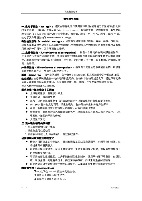
影响土壤中微生物分布的因素●土壤颗粒性质腐殖质》砂土●土壤水分游动微生物●氧气上层好氧微生物多(穴居动物活动可以给微生物好氧生长提供条件)●pH pH对营养物质的利用,微生物吸附,胞外酶的产生和分泌产生影响●温度蓝细菌能抗变化范围很大的温度;耐寒的藻类(雪藻)●营养状况有机物对自养细菌有抑制作用(刍溪藻喜欢在营养丰富的鸟粪中)(土壤颗粒中细菌的不均匀分布)●人类生产活动三.淡水微生物的共同特征:1 能在低营养物浓度下生长2 微生物是可以游动的3 表面积和体积比大(柄细菌),有效吸收营养。
周群英《环境工程微生物学》(第3版)课后习题(第八章 微生物在环境物质循环中的作用)【圣才出品】

第八章微生物在环境物质循环中的作用1.自然界中碳素如何循环?答:自然界中的碳素循环以二氧化碳为中心。
二氧化碳被植物、藻类利用进行光合作用,合成植物性碳;动物摄食植物就将植物性碳转化为动物性碳;动物和人呼吸放出二氧化碳,有机碳化合物被厌氧微生物和好氧微生物分解所产生的二氧化碳均返回大气。
而后,二氧化碳再一次被植物利用进入循环。
2.详述纤维素的好氧和厌氧分解过程。
有哪些微生物和酶参与?答:(1)纤维素的好氧分解①在有氧条件下,纤维素被纤维素酶水解为纤维二糖,纤维二糖在纤维二糖酶的作用下分解为葡萄糖,葡萄糖进一步分解为水和二氧化碳。
②参与好氧分解的微生物和酶有黏细菌、镰状纤维菌和纤维弧菌等,纤维素酶、纤维二糖酶、氧化酶、脱氢酶、脱羧酶、细胞色素氧化酶。
(2)纤维素的厌氧分解①纤维素厌氧发酵产生葡萄糖之后有两种发酵途径,一是丙酮丁醇发酵,产物为丙酮、丁醇、乙酸、二氧化碳和氢气,二是丁酸发酵,产物为丁酸、乙酸、二氧化碳和氢气。
②参与厌氧分解的微生物有产纤维二糖梭菌、无芽孢厌氧分解菌及热解纤维梭菌等。
3.详述淀粉的好氧分解和厌氧分解过程。
有哪些微生物和酶参与?答:(1)淀粉的好氧分解①淀粉在糊精酶的作用下分解为糊精,糊精在麦芽糖酶的作用下水解为麦芽糖,麦芽糖在葡萄糖苷酶的作用下水解为葡萄糖,葡萄糖进一步水解为水和二氧化碳。
②参与淀粉的好氧分解的微生物有枯草芽孢杆菌、根霉、曲霉等。
③参与纤维素的好氧分解的酶有1,4-糊精酶、麦芽糖酶、氧化酶、葡萄糖苷酶、脱氢酶、脱羧酶、细胞色素氧化酶。
(2)淀粉的厌氧分解①淀粉降解为葡萄糖后,厌氧条件下,葡萄糖被酵母菌发酵为乙醇和二氧化碳。
②参与淀粉的厌氧分解的微生物有根霉、曲霉、酵母菌。
③参与淀粉的厌氧分解的酶有淀粉-1,6-糊精酶。
(3)专性厌氧菌对淀粉的分解①淀粉经厌氧发酵后水解为葡萄糖,之后有两种发酵途径:a.丙酮丁醇发酵,产物为丙酮、丁醇、乙酸、二氧化碳和氢气;b.丁酸发酵,产物为丁酸、乙酸、二氧化碳和氢气。
微生物学

微生物学(Microbiology)微生物(microorganism):通常是指一切肉眼看不见或看不清,必须借助于显微镜才能看到的一大类形态微小、结构简单的较为低等的微小生物的总称。
约在17世纪,林奈(Linnaeus,1707~1778)提出生物可以划分为:植物动物18世纪,由于显微镜制造上的发展,人类发现在自然界中还存在许多肉眼看不清的微小生物。
1866年,海格尔(E.H.Haeckle)提出将生物分为植物界、动物界和原生生物界,原生生物界是由低等生物组成(微生物)。
生物植物界动物界原生生物界20世纪40年代,依靠电子显微镜,人类发现所有生物的细胞核可区分为二类,既真核和原核。
在原生生物界中,有真核的生物,也有原核的生物,因此海格尔提出的原生生物界实际上包括了在进化上相差很远的生物种类。
1969年,R.H.Whittaker 提出了将生物分为五界:生物植物界动物界原生生物界真菌界细菌界生物六界生物植物界动物界原生生物界真菌界细菌界病毒界根据生物六界学说,微生物分属原核生物界、原生生物界、真菌界和病毒界。
原生生物界包括单细胞藻类和原生动物。
1970年,Woese 和Wolfe 在对代表性细菌类群的16S rRNA碱基序列进行比较研究后发现:产甲烷细菌(methanogens)与其他细菌(或称为真细菌,eubacteria)有明显的区别,进一步的研究又发现极端嗜盐细菌(extreme halophiles)和嗜热酸细菌(thermo-acidophiles)的16S rRNA谱也与产甲烷细菌相似。
这三类细菌在厌氧、高温和强酸的条件下生活,与地球上生命出现初期的环境相似,因此将它们命名为古菌(archaea)。
根据上述研究结果,1977年,Woese提出了著名的三原界(域,domain)学说。
该学说认为,在生物进化的早期,各种生物存在一个共同祖先,由这一共同祖先分3条路线进化,形成了三个原界,既古菌原界、真细菌原界和真核原界。
课后习题答案《环境工程微生物学》第三版周群英

课后习题答案《环境工程微生物学》第三版周群英绪论1、何谓原核微生物?它包含什么微生物?答:原核微生物的核很原始,发育不全,只有DNA链高度折叠形成的一个核区,没有核膜,核质裸露,与细胞质没有明显界限,叫拟核或者似核。
原核微生物没有细胞器,只有由细胞质膜内陷形成的不规则的泡沫体系,如间体核光合作用层片及其他内折。
也不进行有丝分裂。
原核微生物包含古菌(即古细菌)、真细菌、放线菌、蓝细菌、粘细菌、立克次氏体、支原体、衣原体与螺旋体。
2、何谓真核微生物?它包含什么微生物?答:真核微生物由发育完好的细胞核,核内由核仁核染色质。
由核膜将细胞核与细胞质分开,使两者由明显的界限。
有高度分化的细胞器,如线粒体、中心体、高尔基体、内质网、溶酶体与叶绿体等。
进行有丝分裂。
真核微生物包含除蓝藻以外的藻类、酵母菌、霉菌、原生动物、微型后生动物等。
3、微生物是如何分类的?答:各类微生物按其客观存在的生物属性(如个体形态及大小、染色反应、菌落特征、细胞结构、生理生化反应、与氧的关系、血清学反应等)及它们的亲缘关系,由次序地分门别类排列成一个系统,从大到小,按界、门、纲、目、科、属、种等分类。
种是分类的最小单位,“株”不是分类单位。
4、生物的分界共有几种分法,他们是如何划分的?答:1969年魏泰克提出生物五界分类系统,后被Margulis修改成为普遍同意的五界分类系统:原核生物界(包含细菌、放线菌、蓝绿细菌)、原生生物界(包含蓝藻以外的藻类及原生动物)、真菌界(包含酵母菌与霉菌)、动物界与植物界。
我国王大教授提出六界:病毒界、原核生物界、真核生物界、真菌界、动物界与植物界。
5、微生物是如何命名的?举例说明。
答:微生物的命名是使用生物学中的二名法,即用两个拉丁字命名一个微生物的种。
这个种的名称是由一个属名与一个种名构成,属名与种名都用斜体字表示,属名在前,用拉丁文名词表示,第一个字母大写。
种名在后,用拉丁文的形容词表示,第一个字母小写。
《环境工程实验Ⅰ》(环境工程微生物)教学大纲

《环境工程实验Ⅰ》(环境工程微生物)教学大纲一、基本信息1二、教学目标及任务环境工程微生物学实验是一门操作技能较强的课程。
通过一系列的实验操作,强化学生无菌操作的理念,使学生掌握微生物实验的基本方法。
通过观察不同的实验材料,进行各项实验操作、分析实验结果、完成实验作业等环节,配合课堂教学,以验证和巩固课堂讲授的微生物学的理论知识。
本课程支撑环境工程专业毕业要求1、2、4和6。
三、学时分配教学课时分配四、实验内容及教学要求实验一:光学显微镜的使用及细菌形态的观察实验目的:学会光学显微镜的使用方法,观察细菌的形态。
本实验教学要求:了解光学显微镜的构造,理解光学显微镜成像原理,掌握显微镜的操作和保养方法。
观察细菌的个体形态,学会生物图的绘制。
本实验重点、难点:显微镜的操作和保养方法,油镜观察细菌的形态,生物图的绘制。
实验二:细菌的简单染色和革兰氏染色实验目的:学会细菌染色的操作技术。
本实验教学要求:了解细菌的图片及染色在微生物实验中的重要性,理解革兰氏染色的原理,掌握细菌染色的操作技术。
本实验重点、难点:掌握微生物的一般染色和革兰氏染色方法。
实验三:真菌的形态观察实验目的:学会用显微镜观察真菌的个体形态本实验教学要求:进一步熟悉和掌握显微镜的操作方法,掌握制作水浸片的方法,观察真菌(酵母、霉菌)的个体形态。
本实验重点、难点:学习制作水浸片的方法,观察真菌的个体形态实验四:微生物细胞的计数和测量实验目的:学会用血球计数板对微生物计数本实验教学要求:了解血球计数板的结构,理解血球计数板计数计微生物的原理和掌握计数方法。
学习测微技术,测量细胞(酵母菌)的大小。
本实验重点、难点:测微技术原理,测量细胞(酵母菌)的大小实验五:培养基的配制,器皿的包扎,灭菌方法实验目的:学习培养基的配制及灭菌方法本实验教学要求:了解配制培养基的过程,理解配制培养基的原理,掌握配制方法。
学会各种各种器皿的包扎,灭菌技术。
本实验重点、难点:培养基配制的原理及方法。
国外微生物学基础实验(一 )-英文版

A transmission electron micrograph of Escherichia coli (E.coli).
Scanning Electron Microscope (SEM):
3-D image: Scanning Electron Micrograph
• Use alcohol swab to clean stage and lens paper to clean lenses. • Shortest lens (the one with the red band) should be facing down toward stage. • Use course focus to position stage as low as it can go.
Welcome to Lab!
Getting Ready for Today’s Lab • Stash backpacks in cubbies on right side of room. • • Choose your seat. Put on your “lab coat” and set goggles, a „dirty‟ copy of lab exercise/report #1 and your lab card on bench next to you. (The other copy of the lab exercise and report should be in your backpack. This is your „clean‟
You will not be able to clearly see individual bacteria with this lens. Just get the image in focus as much as possible.
2022年微生物学《环境微生物》题库试卷

2022年微生物学《环境微生物》题库试卷1、大部分微生物( )。
2、噬菌体是一种感染( )的病毒。
3、G+菌由溶菌酶处理后所得到的缺壁细胞是( )。
4、下列微生物中,( )属于革兰氏阴性菌5、酵母菌适宜的生长pH值为( )。
6、( )不是细菌细胞质中含有的成分。
7、下列孢子中属于霉菌有性孢子的是( )。
8、蓝细菌属于( )型的微生物。
9、所有微生物的世代时间( )。
10、嗜碱微生物是指是指那些能够( )生长的微生物。
11、下列关于水体富营养化的叙述,错误的是( )12、关于微生物在水体中的分布情况,表述正确的是( )。
13、关于微生物降解有机物说法正确的是( )14、关于污泥指数(SVI,正确的叙述是( )15、地衣是( )关系的最典型例子。
16、出芽繁殖的过程( )。
17、鞭毛是细菌细胞的特殊结构,它( )。
18、干燥可以( )。
19、生活用水通常用氯气和漂白粉消毒,原理是氯气和漂白粉( )。
20、微生物生长的良好环境是( )。
21、微生物分批培养时,在衰亡期( )。
22、天然环境中微生物生长的最好培养基是( )23、下列有关微生物共同特点的论述,不正确的是( )24、可引起海洋“赤潮”的微生物是( )25、能引起人类疾病的致病微生物是( )26、下列有关绿藻的描述,不正确的是( )27、关于A2/O工艺,正确的说法是( )28、在富营养水体中形成水华的微生物是( )29、活性污泥法与厌氧消化法相比,两者处理废水的剩余污泥量( )。
30、产甲烷菌属于( )。
31、可见光( )。
32、实验室常用的培养细菌的培养基是( )。
33、微生物细胞氧化葡萄糖获得的能量主要以( )形式被细胞利用。
34、下列孢子中属于霉菌无性孢子的是( )。
35、决定病毒感染专一性的物质基础是( )。
36、各种微生物具有一些共同点,下列选项中描述错误的是( )37、下列有关好氧生物膜的描述,正确的是( )38、下列有关铁循环的描述,错误的是( )39、能引起海湾“赤潮”和湖泊“水华”现象的原核生物是( )40、下列有关细菌细胞质膜的描述错误的是( )。
环境微生物学6-(补充)系统发育树

测序结果分析•获得基因序列将所测得的DNA序列,利用Ribosomal Database ProjectII软件Classifier对分离的菌株进行分类,在GenBank上注册得到注册编号,通过Blast检索,与GenBank中的已知菌株的序列进行同源性分析,确定与鉴定菌株同源性程度最高的序列。
•全序列菌种鉴定给定结果中已经确定菌株种类Dear Dr. Li:We have received the following 9 sequence submissions from you:BankIt1464287 ,BankIt1464297 , BankIt1464298 , BankIt1464299 , BankIt1464300 ,BankIt1464301,BankIt1464302 , BankIt1464303 ,BankIt1464304Please provide the following information about your sequence submissions:[1] Are these sequences from:a) pure culture: a culture that contains only one microbial species orb) enrichment culture: use of selective culture media to enrich for a set of microorganisms with aparticular phenotypic property, resulting in a partially purified, mixed culture. Please do not choose this option for purified strains orc) bulk environmental DNA: PCR-amplified directly from source/host DNA using:i) universal primers orii) species-specific primers[2] You have not provided valid organism names. You have simply listed the isolation source. Pleaseprovide more detailed organism name if possible. For example are these from uncultured bacterium, uncultured fungus, etc.[3] Provide unique names (such as clone, isolate, strain, or laborator designation) that we can use todistinguish the separate sequence submissions. For a more detailed explanation, see below.[4] Provide additional details describing the environmental conditions and geographic location wherethese sequences or organisms were isolated. Please provide this as a spreadsheet or tab-delimited table:If you submitted using BankIt:bankitno. sequenceID identifier environmentbankit123456 Seq1 abc1 soil ,bankit123457 Seq2 def2 ocean waterIf you submitted using sequin:SeqID identifier environmentAB1 abc1 soil, CD2 def2 ocean waterFor your reference, please find your preliminary flatfiles below with the information we currently have. Sincerely,Linda Frisse, PhD基因序号的获得Dear Dr. LindaThank you for your letter. I'll give you more information about the 9 sequence submissions.[1] All the sequences are from enrichment culture.[2] From your letter, I can't understand clearly what is organism names. So I as I understand providesall names. If you have any problem you can ask me.The organism names about the bacteriais following:BankIt1464287 Staphylococcus. YA1, BankIt1464297 Pseudomonas. YA6BankIt1464298 Aeromonas. SB9, BankIt1464299 Sphingobacterium. BB11BankIt1464300 Aeromonas. TB13, BankIt1464301 Staphylococcus. JB17BankIt1464302 Comamonas. CB22, BankIt1464303 Arthrobacter. JB18BankIt1464304 Galactomyces geotrichum. SE3[3] unique names are all strain.[4] All enviroment informations are shown in following tablebankitno. name environmentBankIt1464287 YA1 Sludge from Songjiang sewage treatment plant aeration anaerobic zone BankIt1464297 YA6 Sludge from Songjiang sewage treatment plant aeration anaerobic zone BankIt1464298 SB9 Activated sludge from Songjiang sewage treatment plant second pond BankIt1464299 BB11 Soil from cabbage fields in SongjiangBankIt1464300 TB13 Soil from Songjiang University city’s refectoryBankIt1464301 JB17 Soil from gas station beside the university subway stationBankIt1464302 CB22 Songjiang sewage treatment plant effluentBankIt1464303 JB18 Soil from gas station beside the university subway stationBankIt1464304 SE3 Activated sludge from Songjiang sewage treatment plant second pondSincerely,Shan Li Dear GenBank Submitter:Thank you for your direct submission of sequence data to GenBank. Wehave provided GenBank accession numbers for your nucleotide sequences:BankIt1464287 BankIt1464287 JN226389, BankIt1464297 BankIt1464297 JN226390 BankIt1464298 BankIt1464298 JN226391, BankIt1464299 BankIt1464299 JN226392 BankIt1464300 BankIt1464300 JN226393, BankIt1464301 BankIt1464301 JN226394 BankIt1464302 BankIt1464302 JN226395, BankIt1464303 BankIt1464303 JN226396 BankIt1464304 BankIt1464304 JN226397We strongly recommend that these GenBank accession numbers appear inany publication that reports or discusses these data, as they give the community unique labels with which they may retrieve your data from ouron-line servers.Sincerely,Linda Frisse, PhDThe GenBank Direct Submission StaffBethesda, Maryland USAFile——load Sequence Trees——Bootstrap N-J TreeOptions古菌的系统发育树(Madigan et al.,2000)系统发育进化树(Phylogenetic trees)•系统学分类描述了不同生物之间的相关关系,通过系统学分类分析可以帮助人们了解所有生物的进化历史过程。
- 1、下载文档前请自行甄别文档内容的完整性,平台不提供额外的编辑、内容补充、找答案等附加服务。
- 2、"仅部分预览"的文档,不可在线预览部分如存在完整性等问题,可反馈申请退款(可完整预览的文档不适用该条件!)。
- 3、如文档侵犯您的权益,请联系客服反馈,我们会尽快为您处理(人工客服工作时间:9:00-18:30)。
• Pick up a fistful of garden soil and you're holding hundreds if not thousands of different kinds of microbe in your hand. A single teaspoon of that soil contains over 1,000,000,000 bacteria, about 120,000 fungi and 25,000 algae.
• Antony van Leeuwenhoek (1632-1723) and his microscope.
• Louis Pasteur (1822-1895) working in his laboratory.
• Robert ch (18431910)
Examining a specimen in his laboratory.
called the Galapagos Vent.
• Some scientists even believe there is the possibility bacteria may have once lived on Mars. This photograph taken through a microscope shows what some scientists believe may be the fossils of tiny bacteria in a rock that formed on Mars about 4.5 billion years ago. The rock crash-landed on Earth as a meteorite thousands of years ago.
• Bacteria consist of only a single cell, but don't let their small size and seeming simplicity fool you. They're an amazingly complex and fascinating group of creatures. Bacteria have been found that can live in temperatures above the boiling point and in cold that would freeze your blood. They "eat" everything from sugar and starch to sunlight, sulfur and iron. There's even a species of bacteria—Deinococcus radiodurans—that can withstand blasts of radiation 1,000 times greater than would kill a human being.
• Two fundamentally different types of cells exist. Procaryotic cells have a much simpler morphology than eucaryotic cells and lack a true membrane-delimited nucleus. All bacteria are procaryotic. In contrast, eucaryotic have a membraneenclosed nucleus; they are more complex morphologically and are usually larger than prokaryotes. Algae, fungi, protozoa, higher plants, and animals are eucaryotic. Procaryotic and eucaryotic cells differ in many other ways as well.
pollution break up.
• We're using bacteria, like those pictured here, as one of the tools to clean up oil spills, like the Exxon Valdez mess. These bacteria chow on the oil, turning it into carbon dioxide and other harmless by-products.
• Other heat-loving microbes live in volcanic cracks miles under the ocean surface where there is no light and the water is a brew of poisonous arsenic, sulfur and other nasty chemicals. The little blobs shown in this photo are bacteria that live on mussel shells around a volcanic vent
• Diatoms have hard shells made up in part by silica, or glass. When diatoms die, these shells sink to the bottom. They are mined and used to make products we use everyday. For example, diatom shells are the grit in your toothpaste and the stuff that makes painted stripes on the road shiny.
• This is a cowpea virus, which infects certain bean plants including snap peas, pinto beans, green beans and, of course, cowpeas.
• This image was created by a computer.
• This is protozoan called paramecium (pair-ah-me-see-um). It looks hairy because it's covered by long, thin projections called cilia (silly-uh). The cilia beat in a regular, continuous pattern, moving the paramecium through its freshwater home and sweeping food into the mouth-like opening you can see on its side.
Early Bacterial Exposure May Extend Fly Life
Exposure to bacteria during the first week of adult life increased Drosophila lifespan, but exposure late in life decreased longevity, new research reports. All animals develop in at least partially sterile environments and are colonized by bacteria soon after birth. In model organisms such as zebrafish, paramecia, and various mammals, scientists have noted that microorganisms can have a profound effect on the animal’s health, especially on digestion, conditioning the immune system, and longevity. Seymour Benzer from the California Institute of Technology and colleagues investigated a possible connection between the microscopic fauna on Drosophila and longevity by growing the fruit flies in sterile environments, then periodically exposing them to non-sterile conditions. The researchers report that flies grown only under sterile conditions had a 30% decrease in mean lifespan. Further, Benzer’s team discovered a window of time during which bacterial exposure is beneficial: exposure to bacteria during the first 4-7 days of adult life produced the full life-extending effect, while exposure after 7 days had no beneficial effect. Alternatively, the researchers found that transferring adult flies from normal conditions to a sterile environment, effectively removing bacteria late in life, showed a 10% increase in longevity. Additional experiments with mutant fruit flies provided evidence that microscopic fauna interact with the host on a genetic level. Benzer and his team indicate that model organisms such as Drosophila may give important clues to understanding longevity in humans and other animals.
