检测TNF的 elisa试剂盒说明书.
RD TNF-α ELISA试剂盒说明书
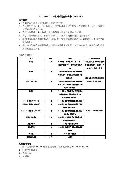
RD TNF-αELISA检测试剂盒说明书(MTA00B)技术提示1.当混合或冲悬蛋白质溶液时,避免产生气泡。
2.为了避免交叉污染,每个标准品,样品以及相关试剂加完后要更换枪头。
此外,每种试剂要有单独的储液槽。
3.为了达到最佳效果,将试剂和样品等滴加到每个孔的中心位置。
4.为了保证准确的结果,在孵育步骤中,有必要对酶标板进行适当地密封。
5.底物溶液在加入到酶标板之前应为无色。
要保持底物溶液避光。
底物溶液应由无色渐渐变为蓝色。
6.终止液应与底物溶液相同的加样顺序加到酶标板孔里。
加入终止液后,酶标孔中的颜色应由蓝色变为黄色。
其他配套物品1.酶标仪能够在450 nm处测量吸光度,校正波长设为540 nm或570 nm。
2.移液枪和移液器3.去离子水4.试剂瓶5.用于稀释标准品的EP管注意事项1.终止液为酸性溶液,注意防护。
2.本试剂盒中的某些成分含有防腐剂,可能会引起皮肤过敏反应。
避免吸入。
3.显色剂B可能引起皮肤、眼睛和呼吸道刺激。
避免吸入。
4.佩戴好口罩和手套,实验服。
实验完成后要仔细洗手。
5.关于样本,细胞培养上清收集后存放在-20℃以下。
避免反复冻融。
试剂准备:1.使用前将所有试剂放于室温。
2.小鼠TNF-αControl:使用1mL去离子水冲悬对照,充分混匀。
3.洗涤液:如果浓缩液中已形成晶体,加热至室温,轻轻搅拌,直到晶体完全溶解。
可配制满足一个酶标板的洗涤液。
如:将20毫升洗涤液浓缩液加到480毫升去离子水或蒸馏水中,配制500毫升洗涤液。
4.底物溶液:底物显色试剂A和B应在使用15分钟内按等体积混合。
避光放置。
每个酶标孔需要100 μL混匀后的AB混合物。
5.小鼠TNF-α标准品:参照瓶上的冲悬体积进行冲悬。
用去离子水或蒸馏水冲悬小鼠TNF-α标准。
原液浓度为7000 pg/mL。
轻轻混匀,室温放置至少5分钟,然后再进行稀释。
先在1号管中加入900ul Calibrator稀释液(RD5K),2-7号加入200ul Calibrator稀释液。
Human TNF-a ELISA Kit 操作手册说明书
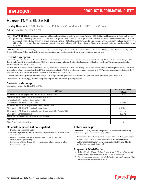
Human TNF‑a ELISA KitCatalog Number KHC3011 (96 tests), KHC3012 (2 × 96 tests), and KHC3011C (5 × 96 tests)Pub. No. MAN0003931 Rev.5.0 (32)CAUTION! This kit contains materials with small quantities of sodium azide and Proclin™ 300. Sodium azide reacts with lead and copper plumbing to form explosive metal azides. Upon disposal, flush drains with a large volume of water to prevent azide accumulation. In case of contact, rinse affected area with plenty of water. Proclin™ 300 is toxic, corrosive, and a skin irritant. Avoid ingestion and contact with eyes, skin and mucous membranes. In case of contact, rinse affected area with plenty of water. Observe all federal, state, and local regulations for disposal.Note: For safety and biohazard guidelines, see the “Safety” appendix in the ELISA Technical Guide (Pub. no. MAN0006706). Read the Safety Data Sheets (SDSs) and follow the handling instructions. Wear appropriate protective eyewear, clothing, and gloves.Product descriptionThe Invitrogen™ Human TNF-a ELISA Kit is a solid-phase sandwich Enzyme-Linked Immunosorbent Assay (ELISA). This assay is designed to detect and quantify the level of human TNF-a in human serum, plasma, buffered solutions, or cell culture medium. The assay recognizes both natural and recombinant human TNF-a.Human tumor necrosis factor alpha (Hu TNF-a), also called cachectin, is a 157 AA non-glycosylated polypeptide cytokine produced by activated macrophages. Lipopolysaccharide (LPS) is a potent stimulus for TNF-a production in macrophages, and TNF-a is an important mediator of the in vivo effects of LPS. The biological activities of TNF-a may be classified as:- Immunomodulating and proinflammatory: TNF-a regulates the production of antibodies by B cells and stimulates cytotoxic T cells.- Metabolic: TNF-a strongly inhibits lipoprotein lipase and adipocyte gene expression.Contents and storageUpon receipt, store the kit at 2°C to 8°C.Materials required but not supplied•Distilled or deionized water•Microtiter plate reader with software capable of measurement at or near 450 nm•Plate washer–automated or manual (squirt bottle, manifolddispenser, or equivalent)•Calibrated adjustable precision pipettes and glass or plastic tubes for diluting solutions Before you beginIMPORTANT! Reagents are lot-specific. Do not mix or interchange different reagent lots from various kit lots.•Review the Procedural guidelines and Plate washing directions in the ELISA Technical Guide available at .•Allow reagents to reach room temperature before use. Mix toredissolve any precipitated salts.Prepare 1X Wash Buffer1.Dilute 16 mL of Wash Buffer Concentrate (25X) with 384 mL ofdeionized or distilled water. Label as 1X Wash Buffer.2.Store the concentrate and 1X Wash Buffer in the refrigerator. Usethe diluted buffer within 25 days.Sample preparation guidelines•Refer to the ELISA Technical Guide at for detailed sample preparation procedures.•Collect samples in pyrogen/endotoxin-free tubes.•Freeze samples after collection if samples will not be tested immediately. Avoid multiple freeze-thaw cycles of frozen samples. Thaw completely and mix well (do not vortex) prior to analysis.•Avoid the use of hemolyzed or lipemic sera. If large amounts of particulate matter are present in the sample, centrifuge or filter sample prior to analysis.Pre-dilute samplesSample concentrations should be within the range of the standard curve. Because conditions may vary, each investigator should determine the optimal dilution for each application.Perform sample dilutions with Standard Diluent Buffer (serum/plasma) or with the corresponding cell culture medium (cell culture supernatant). Dilute standardsNote: Use glass or plastic tubes for diluting standards.Note: This assay has been calibrated against the International Standard preparation (87/650) for Hu TNF-a (NIBSC, Hertforshire, UK, EN6 3QG). One microgram equals 40,000 International Units.1.Reconstitute Hu TNF-a Standard to 2000 pg/mL with Standard Dilution Buffer. Refer to the standard vial label for instructions. Swirl or mixgently and allow the contents to sit for 10 minutes to ensure complete reconstitution. Label as 2000 pg/mL human TNF-a. Use the standard within 1 hour of reconstitution.2.Add 300 µL Reconstituted Standard to one tube containing 300 µL Standard Diluent Buffer and mix. Label as 1000 pg/mL human TNF-a.3.Add 300 µL Standard Diluent Buffer to each of 7 tubes labeled as follows: 500, 250, 125, 62.5, 31.2, 15.6, and 0 pg/mL human TNF-a.4.Make serial dilutions of the standard as shown in the following dilution diagram. Mix thoroughly between steps.5.Discard all remaining reconstituted and diluted standards after completing assay. Return the Standard Diluent Buffer to the refrigerator.DiluentVolumeStd5Std4Std3Std2Std1Std6Std7Std0300 μL250 pg/mL1000 pg/mL500 pg/mL15.6 pg/mL31.2 pg/mL62.5 pg/mL125 pg/mL2000 pg/mL300 μL300 μL300 μL300 μL300 μL300 μL300 μL300 μL0 pg/mLPrepare 1X Streptavidin‑HRP solutionNote: Prepare 1X Streptavidin-HRP within 15 minutes of usage.The Streptavidin-HRP (100X) is in 50% glycerol, which is viscous. To ensure accurate dilution:1.For each 8-well strip used in the assay, pipet 10 µL Streptavidin-HRP (100X) solution, wipe the pipette tip with clean absorbent paper toremove any excess solution, and dispense the solution into a tube containing 1 mL of Streptavidin-HRP Diluent. Mix thoroughly.2.Return the unused Streptavidin-HRP (100X) solution to the refrigerator.Perform ELISA (Total assay time: 4 hours)IMPORTANT! Perform a standard curve with each assay.•Allow all components to reach room temperature before use. Mix all liquid reagents prior to use.•Determine the number of 8-well strips required for the assay. Insert the strips in the frames for use. Re-bag any unused strips and frames, andstore at 2°C to 8°C for future use.a.Add 50 µL of Incubation Buffer to wells for serum or plasma samples, standards, or controls; or 50 µLof Standard Diluent Buffer to the wells for cell culture samples. Leave the wells for chromogen blanksempty.b.Add 100 µL of standards, controls, or samples (see “Pre-dilute samples” on page 2) to the appropriatewells. Leave the wells for chromogen blanks empty.c.Tap the side of the plate to mix. Cover the plate with a plate cover and incubate for 2 hours at roomtemperature.d.Thoroughly aspirate the solution and wash wells 4 times with 1X Wash Buffer.1Bind antigenCulture media +Standard DiluentBufferStandards/Serum/Plasma/Controls +Incubation Buffera.Add 100 µL Hu TNF-a Biotin Conjugate solution into each well except the chromogen blanks.2Add Biotin ConjugateAdd Streptavidin‑HRPAdd Stabilized ChromogenAdd Stop Solution1.Read the absorbance at 450 nm. Read the plate within 2 hours after adding the Stop Solution.e curve-fitting software to generate the standard curve. A four parameter algorithm provides the best standard curve fit. Optimally, thebackground absorbance may be subtracted from all data points, including standards, unknowns and controls, prior to plotting.3.Read the concentrations for unknown samples and controls from the standard curve. Multiply value(s) obtained for sample(s) by theappropriate factor to correct for the sample dilution.Note: Dilute samples producing signals greater than the upper limit of the standard curve in Standard Diluent Buffer (serum/plasma) or with the corresponding cell culture medium (cell culture supernatant) and reanalyze. Multiply the concentration by the appropriate dilution factor. Performance characteristicsStandard curve exampleThe following data were obtained for the various standards over therange of 0−1000 pg/mL human TNF-a.Inter-assay precisionSamples were assayed 18 times in multiple assays to determineprecision between assays.Intra-assay precisionSamples of known human TNF-a concentration were assayed inreplicates of 16 to determine precision within an assay.Expected valuesHuman PBMCs or whole blood were stimulated from 4 to 72 hours with lipopolysaccharide (LPS), phytohaemagglutinin (PHA), or ionomycin and phorbol myristate acetate (PMA) then evaluated for the presence of human TNF-a in this assay. Whole blood samples were pre-diluted 10-fold for the assay.Linearity of dilutionHuman serum containing 806 pg/mL of measured human TNF-a was serially diluted in Standard Diluent Buffer over the range of the assay. Linear regression analysis of samples versus the expected concentration yielded a correlation coefficient of 0.99.RecoveryThe average recovery of Human TNF-a ELISA Kit added to a variety of samples is listed in the following table.SensitivityThe analytical sensitivity of ths assay is 1.7 pg/mL human TNF-a. This was determined by adding two standard deviations to the mean O.D. obtained when the zero standard was assayed 20 times. SpecificityBuffered solutions of The following panel of substances were assayed at 50 ng/mL and were found to have no cross-reactivity: human IL-2, IL-4, IL-6, IL-7, IL-8, IL-10, IFN-a, IFN-b, IFN-g, GM-CSF, OSM,MIP-1a, MIP-1b, LIF, MCP-1, G-CSF, TGF-b, RANTES; swine TNF-a; rat TNF-a; mouse TNF-a.Limited product warrantyLife Technologies Corporation and/or its affiliate(s) warrant their products as set forth in the Life Technologies' General Terms and Conditions of Sale at /us/en/home/global/terms-and-conditions.html. If you have any questions, please contact Life Technologies at /support.Product label explanation of symbols and warningsManufacturer's address: Bender MedSystems GmbH | Campus Vienna Biocenter 2 | 1030 Vienna, AustriaThe information in this guide is subject to change without notice.DISCLAIMERTO THE EXTENT ALLOWED BY LAW, THERMO FISHER SCIENTIFIC INC. AND/OR ITS AFFILIATE(S) WILL NOT BE LIABLE FOR SPECIAL, INCIDENTAL, INDIRECT, PUNITIVE, MULTIPLE, OR CONSEQUENTIAL DAMAGES IN CONNECTION WITH OR ARISING FROM THIS DOCUMENT, INCLUDING YOUR USE OF IT.Important Licensing Information: These products may be covered by one or more Limited Use Label Licenses. By use of these products, you accept the terms and conditions of all applicable Limited Use Label Licenses.©2019 Thermo Fisher Scientific Inc. All rights reserved. All trademarks are the property of Thermo Fisher Scientific and its subsidiaries unless otherwise specified./support | /askaquestion。
TNF-a说明书

人(human)肿瘤坏死因子a(TNF-a)说明书本试剂盒仅供研究使用标本:血清或者血浆一、试剂组成精密度微孔板96孔 (Microtitration Strips) 1块 2~8℃干燥保存酶标偶合液 (Conjugate ) 1瓶 12.0毫升 2~8℃冷藏保存标准品 (Standard) 5瓶各1.0毫升 2~8℃冷藏保存呈色剂A (Substrate A) 1瓶 6.0毫升 2~8℃避光冷藏保存呈色剂B (Substrate B) 1瓶 6.0毫升 2~8℃避光冷藏保存终止液 (Stopping Solution) 1瓶 6.0毫升室温保存20倍浓缩洗涤液 (Rinsing Buffer x 20) 1瓶 60.0毫升 2~8℃冷藏保存5倍浓缩样品稀释液 (Diluent x 5) 1瓶 15.0ml 2~8℃冷藏保存英文说明书,中文说明书各一份室温保存二、注意事项1. 此试剂为体外检测试剂,效期内使用,试剂应视为传染物,不同总批号的试剂不能混用。
2.使用前应将盒内各试剂取出。
室温放置至少30分钟,3.浓缩洗涤液出现结晶后,请于37℃孵育15分钟。
4.浓缩样品稀释液出现结晶后,请于37℃孵育15分钟5.若24小时内进行实验,标本可存放于2~8℃。
不需及时实验,标本-20℃保存,避免反复冻融。
6.在反复清洗微孔板,并扣干微孔中的残余液体,否则将降低精确度,造成吸光度偏离的假像。
7.加样完毕后,应注意轻微摇动微孔反应条,以便使孔中的液体充分混匀。
8.试剂盒保存于2~8℃,请勿冷冻,有效期请见盒内标示。
三、实验前准备1.使用前应将盒内各试剂取出,室温放置至少30分钟。
2.准备各种实验仪器及材料,如微量移液器,吸头,医用蒸馏水等3.浓缩洗液与医用蒸馏水1︰19倍稀释后成为应用洗涤液4.浓缩样品稀释液与医用蒸馏水1︰4倍稀释成应用样品稀释液5.用应用样品稀释液来稀释样品,按照1:100的体积比来稀释样品如10μl的样品加入到1ml的应用样品稀释液中,充分混匀待用。
肿瘤坏死因子TNFα

人肿瘤坏死因子α(TNF-α)酶联免疫分析(ELISA)试剂盒使用说明书本试剂仅供研究使用目的:本试剂盒用于测定人血清,细胞上清及相关液体样本中肿瘤坏死因子α(TNF-α)的含量。
实验原理:本试剂盒应用双抗体夹心法测定标本中人肿瘤坏死因子α(TNF-α)水平。
用纯化的人肿瘤坏死因子α(TNF-α)抗体包被微孔板,制成固相抗体,往包被单抗的微孔中依次加入肿瘤坏死因子α,再与HRP标记的肿瘤坏死因子α(TNF-α)抗体结合,形成抗体-抗原-酶标抗体复合物,经过彻底洗涤后加底物TMB显色。
TMB在HRP酶的催化下转化成蓝色,并在酸的作用下转化成最终的黄色。
颜色的深浅和样品中的人肿瘤坏死因子α呈正相关。
用酶标仪在450nm波长下测定吸光度(OD值),通过标准曲线计算样品中人肿瘤坏死因子α(TNF-α)浓度。
试剂盒组成:试剂盒组成48孔配置96孔配置保存说明书1份1份封板膜2片(48)2片(96)密封袋1个1个酶标包被板1×481×962-8℃保存标准品:450pg/ml0.5ml×1瓶0.5ml×1瓶2-8℃保存标准品稀释液 1.5ml×1瓶 1.5ml×1瓶2-8℃保存酶标试剂3ml×1瓶6ml×1瓶2-8℃保存样品稀释液3ml×1瓶6ml×1瓶2-8℃保存显色剂A液3ml×1瓶6ml×1瓶2-8℃保存显色剂B液3ml×1瓶6ml×1瓶2-8℃保存终止液3ml×1瓶6ml×1瓶2-8℃保存浓缩洗涤液(20ml×20倍)×1瓶(20ml×30倍)×1瓶2-8℃保存样本处理及要求:1.血清:室温血液自然凝固10-20分钟,离心20分钟左右(2000-3000转/分)。
仔细收集上清,保存过程中如出现沉淀,应再次离心。
碧云天Mouse TNF-α ELISA Kit (小鼠TNF-α酶联免疫吸附检测试剂盒)说明书
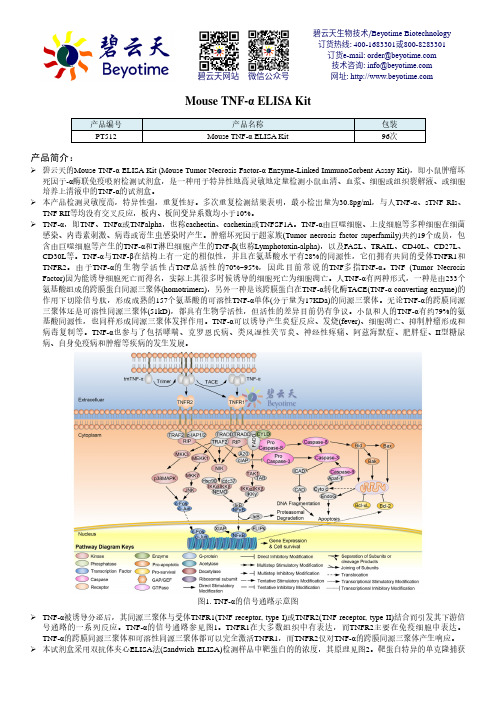
Mouse TNF-α ELISA Kit产品编号 产品名称包装 PT512Mouse TNF-α ELISA Kit96次产品简介:碧云天的Mouse TNF-α ELISA Kit (Mouse Tumor Necrosis Factor-α Enzyme-Linked ImmunoSorbent Assay Kit),即小鼠肿瘤坏死因子-α酶联免疫吸附检测试剂盒,是一种用于特异性地高灵敏地定量检测小鼠血清、血浆、细胞或组织裂解液、或细胞培养上清液中的TNF-α的试剂盒。
本产品检测灵敏度高,特异性强,重复性好。
多次重复检测结果表明,最小检出量为30.8pg/ml ,与人TNF-α、sTNF RIs 、TNF RII 等均没有交叉反应,板内、板间变异系数均小于10%。
TNF-α,即TNF 、TNF α或TNFalpha ,也称cachectin 、cachexin 或TNFSF1A 。
TNF-α由巨噬细胞、上皮细胞等多种细胞在细菌感染、内毒素刺激、病毒或寄生虫感染时产生。
肿瘤坏死因子超家族(Tumor necrosis factor superfamily)共约19个成员,包含由巨噬细胞等产生的TNF-α和T 淋巴细胞产生的TNF-β(也称Lymphotoxin-alpha),以及FASL 、TRAIL 、CD40L 、CD27L 、CD30L 等。
TNF-α与TNF-β在结构上有一定的相似性,并且在氨基酸水平有28%的同源性,它们拥有共同的受体TNFR1和TNFR2。
由于TNF-α的生物学活性占TNF 总活性的70%~95%,因此目前常说的TNF 多指TNF-α。
TNF (Tumor Necrosis Factor)因为能诱导细胞死亡而得名,实际上其很多时候诱导的细胞死亡为细胞凋亡。
人TNF-α有两种形式,一种是由233个氨基酸组成的跨膜蛋白同源三聚体(homotrimers),另外一种是该跨膜蛋白在TNF-α转化酶TACE(TNF-α converting enzyme)的作用下切除信号肽,形成成熟的157个氨基酸的可溶性TNF-α单体(分子量为17KDa)的同源三聚体。
TNF-a说明书

人(human)肿瘤坏死因子a(TNF-a)说明书本试剂盒仅供研究使用标本:血清或者血浆一、试剂组成精密度微孔板96孔 (Microtitration Strips) 1块 2~8℃干燥保存酶标偶合液 (Conjugate ) 1瓶 12.0毫升 2~8℃冷藏保存标准品 (Standard) 5瓶各1.0毫升 2~8℃冷藏保存呈色剂A (Substrate A) 1瓶 6.0毫升 2~8℃避光冷藏保存呈色剂B (Substrate B) 1瓶 6.0毫升 2~8℃避光冷藏保存终止液 (Stopping Solution) 1瓶 6.0毫升室温保存20倍浓缩洗涤液 (Rinsing Buffer x 20) 1瓶 60.0毫升 2~8℃冷藏保存5倍浓缩样品稀释液 (Diluent x 5) 1瓶 15.0ml 2~8℃冷藏保存英文说明书,中文说明书各一份室温保存二、注意事项1. 此试剂为体外检测试剂,效期内使用,试剂应视为传染物,不同总批号的试剂不能混用。
2.使用前应将盒内各试剂取出。
室温放置至少30分钟,3.浓缩洗涤液出现结晶后,请于37℃孵育15分钟。
4.浓缩样品稀释液出现结晶后,请于37℃孵育15分钟5.若24小时内进行实验,标本可存放于2~8℃。
不需及时实验,标本-20℃保存,避免反复冻融。
6.在反复清洗微孔板,并扣干微孔中的残余液体,否则将降低精确度,造成吸光度偏离的假像。
7.加样完毕后,应注意轻微摇动微孔反应条,以便使孔中的液体充分混匀。
8.试剂盒保存于2~8℃,请勿冷冻,有效期请见盒内标示。
三、实验前准备1.使用前应将盒内各试剂取出,室温放置至少30分钟。
2.准备各种实验仪器及材料,如微量移液器,吸头,医用蒸馏水等3.浓缩洗液与医用蒸馏水1︰19倍稀释后成为应用洗涤液4.浓缩样品稀释液与医用蒸馏水1︰4倍稀释成应用样品稀释液5.用应用样品稀释液来稀释样品,按照1:100的体积比来稀释样品如10μl的样品加入到1ml的应用样品稀释液中,充分混匀待用。
四正柏生物 人类TNF-α ELISA试剂盒说明书
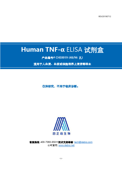
REV20190712仅供研究,不用于临床诊断。
客服热线: 400-7060-959﹡技术支持邮箱: **************公司官网: 目录简介 ......................................................................................................................................................................... - 3 -检测原理 ................................................................................................................................................................. - 3 -试剂盒组分 ............................................................................................................................................................. - 4 -储存条件 ................................................................................................................................................................. - 5 -其他实验材料 ......................................................................................................................................................... - 5 -注意事项 ................................................................................................................................................................. - 5 -样本收集处理及保存方法 ..................................................................................................................................... - 6 -试剂准备 ................................................................................................................................................................. - 6 -操作步骤 ................................................................................................................................................................. - 7 -操作流程图 ............................................................................................................................................................. - 8 -操作要点提示 ......................................................................................................................................................... - 8 -结果判断 ................................................................................................................................................................. - 9 -结果重复性 ........................................................................................................................................................... - 10 -灵敏度 ................................................................................................................................................................... - 10 -特异性 ................................................................................................................................................................... - 10 -参考文献 ............................................................................................................................................................... - 10 -该产品由北京四正柏生物科技有限公司研制。
人肿瘤坏死因子α(TNF-α)说明书
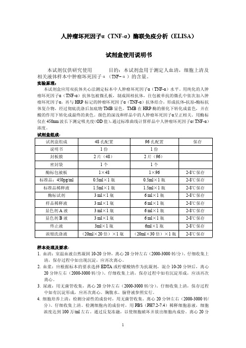
人肿瘤坏死因子α(TNF-α)酶联免疫分析(ELISA)试剂盒使用说明书本试剂仅供研究使用目的:本试剂盒用于测定人血清,细胞上清及相关液体样本中肿瘤坏死因子α(TNF-α)的含量。
实验原理:本试剂盒应用双抗体夹心法测定标本中人肿瘤坏死因子α(TNF-α)水平。
用纯化的人肿瘤坏死因子α(TNF-α)抗体包被微孔板,制成固相抗体,往包被单抗的微孔中依次加入肿瘤坏死因子α,再与HRP标记的肿瘤坏死因子α(TNF-α)抗体结合,形成抗体-抗原-酶标抗体复合物,经过彻底洗涤后加底物TMB显色。
TMB在HRP酶的催化下转化成蓝色,并在酸的作用下转化成最终的黄色。
颜色的深浅和样品中的人肿瘤坏死因子α呈正相关。
用酶标仪在450nm波长下测定吸光度(OD值),通过标准曲线计算样品中人肿瘤坏死因子α(TNF-α)浓度。
试剂盒组成:试剂盒组成48孔配置96孔配置保存说明书1份1份封板膜2片(48)2片(96)密封袋1个1个酶标包被板1×481×962-8℃保存标准品:450pg/ml0.5ml×1瓶0.5ml×1瓶2-8℃保存标准品稀释液 1.5ml×1瓶 1.5ml×1瓶2-8℃保存酶标试剂3ml×1瓶6ml×1瓶2-8℃保存样品稀释液3ml×1瓶6ml×1瓶2-8℃保存显色剂A液3ml×1瓶6ml×1瓶2-8℃保存显色剂B液3ml×1瓶6ml×1瓶2-8℃保存终止液3ml×1瓶6ml×1瓶2-8℃保存浓缩洗涤液(20ml×20倍)×1瓶(20ml×30倍)×1瓶2-8℃保存样本处理及要求:1.血清:室温血液自然凝固10-20分钟,离心20分钟左右(2000-3000转/分)。
仔细收集上清,保存过程中如出现沉淀,应再次离心。
小鼠 TNF-α elisa试剂盒说明书
以标准物的浓度为横坐标(对数坐标),O
D 值为纵坐标(普通坐标),在半对数坐标纸上
绘出标准曲线,根据样品的OD 值由标准曲线查出相应的浓度;再乘以稀释倍
数;或用标准
物的浓度与OD 值计算出标准曲线的直线回归方程式,将样品的OD 值代入方程式,计算出
2. 血浆:可用EDTA 或肝素作为抗凝剂,标本采集后30 分钟内于2 - 8° C 1000 x g 离心15
分钟,或将标本放于-20℃或-80℃保存,但应避免反复冻融。
3. 细胞培养物上清或其它生物标本:请1000 x g0℃或-80℃保存,但应避免反复冻融。
注:标本溶血会影响最后检测结果,因此溶血标本不宜进行此项检测。
标本的稀释原则:
首先通过文献检索的方式了解待测样本的大致含量,确定适当的稀释倍数。只有稀释至标准
曲线的范围内,检测的结果才是准确的。稀释的过程中,应做好详细的记录。最后计算浓度
时,稀释了“N”倍,标本的浓度应再乘以“N”。
标准品的稀释原则:2 瓶,每瓶临用前以样品稀释液稀释至1ml,盖好后静置10 分钟以上,
本试剂盒仅供研究使用
检测范围:31.2 pg/ml - 2000 pg/ml
最低检测限:7.8 pg/ml
特异性:本试剂盒可同时检测天然或重组的小鼠TNF-α,且与其他相关蛋白无交叉反应。
有效期:6 个月
准品、生物素化的抗TNF-α抗体、HRP 标记的亲和素,经过彻底洗涤后用底物TMB 显色。
TMB 在过氧化物酶的催化下转化成蓝色,并在酸的作用下转化成最终的黄色。颜色的深浅
和样品中的TNF-α呈正相关。用酶标仪在450nm 波长下测定吸光度(OD 值),计算样品浓
TNF检测
2. 实验内容
数据处理
标准品
浓度(Y) OD值-1 160 80 40 20 10 0(空白)
1.774 1.133
样品
OD值-2
1.892 1.051
均值
1.833 1.092
-空白(X)
1.828 1.087
编号 1-1 1-2 1-3 1-4 1-5 ….
OD值-1
OD值-2
均值
-空白(X)
TNF-α
酶标抗体
酶底物:TMB
4
2. 实验内容
实验目的 检测感染性休克兔血样中的TNF-α含量
实验材料
检测品: 兔血清
试剂:兔TNF-α ELISA 检测试剂盒 仪器:酶标仪
96孔板:固化TNF- α抗体 标准品储备液:Standard (TNF- α) 标准品稀释液:Standard diluent 样本稀释液:Sample diluent 浓缩洗涤液:Wash solution 酶标试剂:HRP Conjugate reagent 显色剂A:Chromogen Solution A 显色剂B:Chromogen Solution B 终止液:Stop Solution
2. TNF-α在感染性休克发生中的作用。
3. 内毒素休克时器官系统功能变化的特点和发生 机制。 4. 感染性休克时微循环变化的特点和发生机制。 5. 分析丹参、去甲肾上腺素、多巴胺、山莨菪碱 在抢救休克中的应用及作用机理。
作业:实验报告(四种药物均要讨论),6月5日(周五)提交,生理楼241 讨论课:6月16日(周二)上午8点,机能实验室2 6月12日(周五)晚上12点之前提交PPT 文件名:综合实验-姓名-组别
150 l
BD TNF ELISA 说明书 555268

The NIBSC/WHO Reference Standard 88/532 (recombinant mouse TNF) was evaluated in this set. The conversion factor for NIBSC material is as follows: 1 mg NIBSC 88/532 mouse TNF= 0.61 mg BD OptEIA™ mouse TNF
Plot the standard curve on log-log graph paper, with TNF concentration on the x-axis and absorbance on the y-axis. Draw the best fit curve through the standard points.
2. “Typical Standard Curve” and 20-plate yield were obtained in the BD Biosciences Pharmingen laboratory, using the recommended procedure and manual plate washing.
Troubleshooting
Biotinylated Anti-Mouse TNF monoclonal antibody Standards
Poor Precision Possible Source • Inadequate washing/ aspiration of wells • Inadequate mixing of reagents • Imprecise/ inaccurate pipetting • Incomplete sealing of plate
ELISA 检测试剂盒 说明书
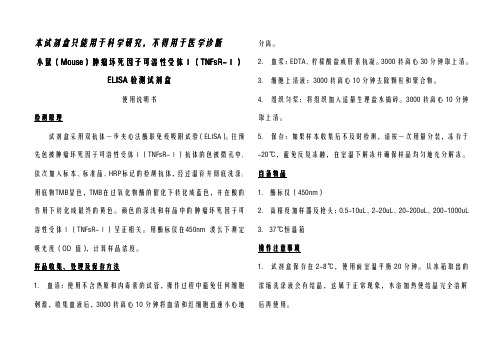
本试剂盒只能用于科学研究,不得用于医学诊断小鼠小鼠((MouseMouse))肿瘤坏死因子可溶性受体肿瘤坏死因子可溶性受体ⅠⅠ(TNFsR-TNFsR-ⅠⅠ)ELISA检测试剂盒使用说明书检测原理试剂盒采用双抗体一步夹心法酶联免疫吸附试验(ELISA)。
往预先包被肿瘤坏死因子可溶性受体Ⅰ(TNFsR-Ⅰ)抗体的包被微孔中,依次加入标本、标准品、HRP标记的检测抗体,经过温育并彻底洗涤。
用底物TMB显色,TMB在过氧化物酶的催化下转化成蓝色,并在酸的作用下转化成最终的黄色。
颜色的深浅和样品中的肿瘤坏死因子可溶性受体Ⅰ(TNFsR-Ⅰ)呈正相关。
用酶标仪在450nm波长下测定吸光度(OD值),计算样品浓度。
样品收集、处理及保存方法1.血清:使用不含热原和内毒素的试管,操作过程中避免任何细胞刺激,收集血液后,3000转离心10分钟将血清和红细胞迅速小心地分离。
2.血浆:EDTA、柠檬酸盐或肝素抗凝。
3000转离心30分钟取上清。
3.细胞上清液:3000转离心10分钟去除颗粒和聚合物。
4.组织匀浆:将组织加入适量生理盐水捣碎。
3000转离心10分钟取上清。
5.保存:如果样本收集后不及时检测,请按一次用量分装,冻存于-20℃,避免反复冻融,在室温下解冻并确保样品均匀地充分解冻。
自备物品1.酶标仪(450nm)2.高精度加样器及枪头:0.5-10uL、2-20uL、20-200uL、200-1000uL3.37℃恒温箱操作注意事项1.试剂盒保存在2-8℃,使用前室温平衡20分钟。
从冰箱取出的浓缩洗涤液会有结晶,这属于正常现象,水浴加热使结晶完全溶解后再使用。
2.实验中不用的板条应立即放回自封袋中,密封(低温干燥)保存。
3.浓度为0的S0号标准品即可视为阴性对照或者空白;按照说明书操作时样本已经稀释5倍,最终结果乘以5才是样本实际浓度。
4.严格按照说明书中标明的时间、加液量及顺序进行温育操作。
5.所有液体组分使用前充分摇匀。
ELISA检测试剂盒使用指南
ELISA检测试剂盒使用指南ELISA(酶联免疫吸附试验)是一种常用的免疫学实验方法,用于检测病原体、抗体或其他分子的存在和浓度。
它具有高灵敏度、高特异性、易操作和较低成本的优势,被广泛应用于生物医学研究、临床诊断和免疫学领域。
本篇文章将介绍ELISA检测试剂盒的使用指南,包括实验准备、试剂的使用步骤和结果解读。
一、实验准备1.阅读检测项目的说明书:在使用试剂盒前,仔细阅读说明书,了解试剂的使用方法、灵敏度和特异性等关键信息。
2.样本准备:根据实验要求,准备样本。
如果需要检测血清中的抗体水平,可以采集血液样本,离心分离血清;如果需要检测细胞培养上清中的分子,将上清收集,并使其清晰,避免细胞或杂质的污染。
3.样本预处理:根据实验需求,对样本进行必要的预处理。
例如,可以通过加热、稀释、酶解等方式来处理样本,以改变样本组分和浓度。
4.准备品质控制样品:制备阳性和阴性控制样品,用于评估试剂盒和实验操作的稳定性和准确性。
5.实验器材准备:准备所需实验器材,如酶标板、移液器、微孔板洗涤仪等。
确保器材的干净和完整,并按照说明书要求对其进行预处理。
二、试剂使用步骤1.试剂准备:根据说明书要求,将试剂从冰箱中取出,并在室温下放置一段时间使其恢复到室温。
确保试剂瓶盖紧闭,并避免暴露在直接阳光下。
2.实验操作:按照说明书的要求,将试剂加入到酶标板中,并根据实验设计进行标准曲线的设置。
标准曲线用于测量未知样品的数量,并计算出浓度。
3.孵育:根据试剂盒的要求,将酶标板放入孵育箱中进行孵育。
孵育温度和时间应根据实验要求进行调整。
4.洗涤:使用洗涤缓冲液对酶标板上的不特异性结合物进行洗涤。
洗涤过程应准确控制洗涤孔板次数和洗涤液的体积。
5.补液:在洗涤完成后,加入辣根过氧化物酶标况稀释液,促进酶标物与特异性结合物的反应。
6.孵育:根据试剂盒的要求,将酶标板放入孵育箱中进行二次孵育。
7.反应停止:根据试剂盒的要求,加入相应的停止液,停止酶反应。
小鼠tnf-a ELISA kit说明书

钟。
3
7. 洗板4次。 8. 加入显色剂100ul/孔,避光37℃孵箱孵育10--20分钟。 9. 加入终止液100ul/孔,混匀后即刻测量OD450值(5分钟内)。
结果判断: 1. 每个标准品和标本的OD值应减去零孔的OD值。 2. 手工绘制标准曲线。以标准品浓度作横坐标,OD值作纵坐标,以平滑线连接各标准品的坐标
4℃ 4℃ 4℃ 4℃ 4℃(避光) 4℃ 4℃ 4℃(避光) 4℃ 4℃
*:[96/48 Tests]
所需物品(不提供,但可协助购买) : 1. 酶标仪(450nm) 2. 高精度加液器及吸头:0.5-10,2-20,20-200,200-1000ul 3. 37℃温箱, 双蒸水或去离子水,坐标纸。
4
参考文献: Vet Immunol Immunopathol. 1995 Aug;47(3-4):187-201. Immunobiology. 1995 Jul;193(2-4):186-92. Annu Rev Med. 1994;45:491-503. Int J Cardiol. 1993 Dec 31;42(3):231-8. J Periodontol. 1993 May;64(5 Suppl):445-9.
点。通过标本的OD值可在标准曲线上查出其浓度。 3. 若标本OD值高于标准曲线上限,应适当稀释后重测,计算浓度时应乘以稀释倍数
OD
2.5 2
1.5 1
0.5 0
0
m TNF-a
500
1000
pg/ml
1500
注意:本图仅供参考,应以同次试验标准品所绘标准曲线计算标本含量。
人淋巴细胞因子ELISA试剂盒说明书

人淋巴细胞因子ELISA试剂盒说明书
人淋巴细胞因子ELISA定量检测试剂盒等ELISA试剂盒检测类型:双抗体夹心法测抗原、双抗原夹心法测抗体、间接法测抗体、竞争法测抗体、竞争法测抗原、捕获包被法测抗体、ABS-ELISA法。
我司人淋巴细胞因子ELISA定量检测试剂盒等ELISA试剂盒,灵敏性高,高效性,原装进口抗体,吸附均匀、吸附性好,回收利用率高、可靠性强,无效包退包换,公司提供免费代测服务,各种种属ELISA 试剂盒产品齐全,质量可靠。
详情可来电咨询销售人员或咨询我司在线客服。
人淋巴细胞因子ELISA定量检测试剂盒操作步骤:
1、加样:分别设空白孔(空白对照孔不加样品及酶标试剂,其余各步操作相同)、标准孔、待测样品孔。
在酶标包被板上标准品准确加样50µl,待测样品孔中先加样品稀释液40µl,然后再加待测样品10µl (样品最终稀释度为5倍)。
加样将样品加于酶标板孔底部,尽量不触及孔壁,轻轻晃动混匀。
2、温育:用封板膜封板后置 37℃温育30分钟。
3、配液:将30倍浓缩洗涤液用蒸馏水30倍稀释后备用
4、洗涤:小心揭掉封板膜,弃去液体,甩干,每孔加满洗涤液,静置30秒后弃去,如此
重复5次,拍干。
5、加酶:每孔加入酶标试剂50µl,空白孔除外。
6、显色:每孔先加入显色剂A50µl,再加入显色剂B50µl,轻轻震荡混匀,37℃避光显色
10 分钟.
7、终止:每孔加终止液50µl,终止反应(此时蓝色立转黄色)。
8、测定:以空白空调零,450nm波长依序测量各孔的吸光度(OD值)。
测定应在加终止液后15分钟以内进行。
TNF-α试剂盒说明书
1000 mL
500 mL 500 mL 500 mL 500 mL 500 mL 500 mL 250 125 62.5 31.3 15.6 7.8 pg/mL
Stock Solution
500
Health Hazard Warnings
1. Reagents that contain preservatives may be harmful if ingested, inhaled or absorbed through the skin. Refer to the MSDS online for details (/msds). 2. Substrate Solution A and Substrate Solution B are harmful if ingested. Additionally, avoid skin, eye or clothing contact. 3. To reduce the likelihood of blood-borne transmission of infectious agents, handle all serum and/or plasma in accordance with NCCLS regulations.
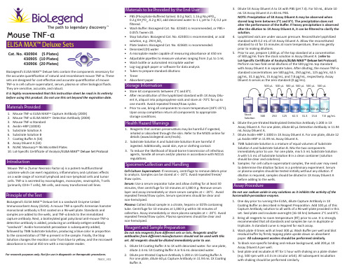
3. Dilute 5X Assay Diluent A to 1X with PBS (pH 7.4). For 50 mL, dilute 10 mL 5X Assay Diluent A in 40 mL PBS. NOTE: Precipitation of 5X Assay Diluent A may be observed when stored long term between 2°C and 8°C. The precipitation does not alter the performance of the Buffer. If heavy precipitation is observed after the dilution to 1X Assay Diluent A, it can be filtered to clarify the solution. 4. Lyophilized vials are under vacuum pressure. Reconstitute lyophilized standard with 0.2 mL of 1X Assay Diluent A. Allow the reconstituted standard to sit for 15 minutes at room temperature, then mix gently prior to making dilutions. 5. Prior to use, prepare 1,000 μL of the top standard at a concentration of 500 pg/mL from the stock solution in Assay Diluent A (refer to Lot-Specific Certificate of Analysis/ELISA MAX™ Deluxe Set Protocol). Perform six two-fold serial dilutions of the 500 pg/mL top standard with Assay Diluent A in separate tubes. After diluting, the mouse TNF-α standard concentrations are 500 pg/mL, 250 pg/mL, 125 pg/mL, 62.5 pg/mL, 31.3 pg/mL, 15.6 pg/mL, and 7.8 pg/mL, respectively. Assay Diluent A serves as the zero standard (0 pg/mL).
ELISA检测试剂盒使用指南
ELISA检测试剂盒使用指南ELISA检测试剂盒使用指南1.简介:1.1 本文档旨在提供ELISA检测试剂盒的详细使用指南,以帮助用户正确、高效地进行ELISA检测。
1.2 ELISA(酶联免疫吸附试验)是一种常用的免疫测定方法,用于检测体液中特定抗原或抗体的存在与浓度。
1.3 本文档将涵盖ELISA检测试剂盒的准备、样本处理、试剂操作、结果读取等详细步骤。
2.准备:2.1 确保实验室环境整洁,无污染。
2.2 检查所需试剂、仪器和设备是否完整,并检查有效期和储存条件。
2.3 根据实验需要,准备所需样本和质控品,并储存于适当的条件下。
3.样本处理:3.1 根据实验目的选择合适的样本类型,如血清、血浆、尿液等。
3.2 根据试剂盒说明书的要求,对样本进行适当的稀释和预处理。
3.3 严格控制操作条件,避免样本污染和交叉污染。
4.试剂操作:4.1 根据试剂盒说明书,将所需试剂从冰箱中取出,并在室温下适当恢复。
4.2 严格按照说明书的要求,对试剂进行稀释和混合。
4.3 操作过程中注意洁净实验台面,避免试剂交叉污染。
5.检测步骤:5.1 将适量的稀释后的样本、质控品和标准品分别装入试管中。
5.2 添加适量的酶标记抗体或底物,并充分混匀。
5.3 按照试剂盒说明书的要求,进行孵育和洗涤步骤。
5.4 添加底物并进行反应,控制反应时间。
5.5 停止反应,并使用酶标仪测量吸光度。
6.结果读取:6.1 根据试剂盒说明书,根据吸光度值和标准曲线,计算样本中目标物质的浓度。
6.2 将测量结果记录,并进行统计和分析。
6.3 评估实验结果的可靠性,并进行结果的解释和报告。
7.注意事项:7.1 严格按照试剂盒说明书的要求进行操作,避免任何操作步骤的误差和误操作。
7.2 注意实验室安全,避免接触有害化学物质,正确使用防护设备。
7.3 在实验过程中遵循生物安全操作规范,避免潜在的感染风险。
此外,本文档涉及附件,请在附件部分查看相关详细信息。
- 1、下载文档前请自行甄别文档内容的完整性,平台不提供额外的编辑、内容补充、找答案等附加服务。
- 2、"仅部分预览"的文档,不可在线预览部分如存在完整性等问题,可反馈申请退款(可完整预览的文档不适用该条件!)。
- 3、如文档侵犯您的权益,请联系客服反馈,我们会尽快为您处理(人工客服工作时间:9:00-18:30)。
QuantikineÒRat TNF-a ImmunoassayCatalog Number RTA00SRTA00PRTA00For the quantitative determination of rat tumor necrosis factor alpha (TNF-a concentrations in cell culture supernates, rat serum, and plasma.This package insert must be read in its entirety before using this product.FOR RESEARCH USE ONLY.NOT FOR USE IN DIAGNOSTIC PROCEDURES.TABLE OF CONTENTSContents PageINTRODUCTION2 PRINCIPLE OF THEASSAY. . . . . . . . . . . . . . . . . . . . . . . . . . . . . . . . . .3 LIMITATIONS OF THE PROCEDURE3 PRECAUTION. . . . . . . . . . . . . . . . . . . . . . . . . . . . . . . . . . . . . . . . . .3 TECHNICAL HINTS3 MATERIALSPROVIDED. . . . . . . . . . . . . . . . . . . . . . . . . . . . . . . . . . . .4 STORAGE5 OTHER SUPPLIES REQUIRED. . . . . . . . . . . . . . . . . . . . . . . . . . . . . . . . .5 SAMPLE COLLECTION AND STORAGE6 SAMPLEPREPARATION. . . . . . . . . . . . . . . . . . . . . . . . . . . . . . . . . . . .6 REAGENT PREPARATION7 ASSAYPROCEDURE. . . . . . . . . . . . . . . . . . . . . . . . . . . . . . . . . . . . . .8 PROCEDURESUMMARY AND CHECKLIST9 CALCULATION OFRESULTS. . . . . . . . . . . . . . . . . . . . . . . . . . . . . . . . .10 TYPICAL DATA10 PRECISION. . . . . . . . . . . . . . . . . . . . . . . . . . . . . . . . . . . . . . . . . .11 RECOVERY11 LINEARITY. . . . . . . . . . . . . . . . . . . . . . . . . . . . . . . . . . . . . . . . . . .12 SENSITIVITY12 CALIBRATION. . . . . . . . . . . . . . . . . . . . . . . . . . . . . . . . . . . . . . . . .13 SAMPLE VALUES13 SPECIFICITY. . . . . . . . . . . . . . . . . . . . . . . . . . . . . . . . . . . . . . . . .13 REFERENCES14 PLATE LAYOUT. . . . . . . . . . . . . . . . . . . . . . . . . . . . . . . . . . . . . . . .15MANUFACTURED AND DISTRIBUTED BY:R&D Systems, Inc.TELEPHONE:(800 343-7475614 McKinley Place NE(612 379-2956Minneapolis, MN 55413FAX:(612 656-4400United States of America E-MAIL:info@DISTRIBUTED BY:R&D Systems Europe, Ltd.19 Barton Lane TELEPHONE:+44 (01235 529449Abingdon Science Park FAX:+44 (01235 533420Abingdon,OX143NB E-MAIL:info@United KingdomR&D Systems China Co. Ltd.24A1 Hua Min Empire Plaza TELEPHONE:+86 (21 52380373726 West Yan An Road FAX:+86 (21 52371001Shanghai PRC 200050E-MAIL:info@INTRODUCTIONTumor necrosis factor alpha (TNF-a, also known as cachectin; and tumor necrosis factor beta (TNF-b, also known as lymphotoxin, are two closely related proteins (approximately 34% amino acid sequence identity that bind to the same cell surface receptors and show many common biological functions. TNF-a and -b play critical roles in normal host resistance to infection and to the growth of malignant tumors, serving as immunostimulants and as mediators of the inflammatory response. Over-production of TNFs, however, has been implicated as playing a role in a number of pathological conditions, including cachexia, septic shock, and autoimmune disorders. TNF-a is produced by activated macrophages and other cell types including T and B cells, NK cells, LAK cells, astrocytes, endothelial cells, smooth muscle cells and some tumor cells (1 - 4.Rat TNF-a cDNA encodes a 235 amino acid (aa residue type II membrane protein (5. The 156 aa residue soluble TNF-a is released from the C-terminus of themembrane-anchored TNF-a by TNF-a-converting enzyme (TACE, a matrix metalloprotease (6, 7. The membrane-anchored form of TNF-a has been shown to have lytic activity and may also play an important role in intercellular communication (8. The biologically active TNF-a has been shown to exist as a trimer (9, 10.Two distinct TNF receptors, referred to as type I (or type B or p55 and type II (or type A or p75, that specifically bind TNF-a and TNF-b with equal affinity have been identified (11, 12. The two TNF receptors transduce signals independently of one another. The amino acid sequence of the extracellular domains of the two receptors are homologous and both receptors are members of the TNF receptor family which also include the NGF receptor, fas antigen, CD27, CD30, and CD40. The intracellular domains of the two receptors are apparently unrelated, suggesting that the two receptorsemploy different signal transduction pathways. Soluble forms of both types of receptors have been found in human serum and urine (13 - 15. These soluble receptors are capable of neutralizing the biological activities of the TNFs and may serve to modulate the activities of TNF.The Quantikine Rat TNF-a Immunoassay is a 4.5 hour solid phase ELISA designed to measure rat TNF-a levels in cell culture supernates, serum, and plasma. It containsE. coli-expressed recombinant rat TNF-a and antibodies raised against the recombinant factor. This immunoassay has been shown to quantitate the recombinant rat TNF-a accurately. Results obtained using natural rat TNF-a showed dose response curves that were parallel to the standard curves obtained using the recombinant kit standards. These results indicate that the Quantikine Rat TNF-a Immunoassay kit can be used to determine relative mass values for natural rat TNF-a.PRINCIPLE OF THE ASSAYThis assay employs the quantitative sandwich enzyme immunoassay technique. A monoclonal antibody specific for rat TNF-a has been pre-coated onto a microplate. Standards, Control, and samples are pipetted into the wells and any rat TNF-a present is bound by the immobilized antibody. After washing away any unbound substances, an enzyme-linked polyclonal antibody specific for rat TNF-a is added to the wells. Following a wash to remove any unbound antibody-enzyme reagent, a substrate solution is added to the wells. The enzyme reaction yields a blue product that turns yellow when the Stop Solution is added. The intensity of the color measured is in proportion to the amount of rat TNF-a bound in the initial step. The sample values are then read off the standard curve.LIMITATIONS OF THE PROCEDURE·FOR RESEARCH USE ONLY. NOT FOR USE IN DIAGNOSTIC PROCEDURES.·The kit should not be used beyond the expiration date on the kit label.·Do not mix or substitute reagents with those from other lots or sources.·If samples generate values higher than the highest standard, further dilute the samples with Calibrator Diluent and repeat the assay.·Any variation in operator, pipetting technique, washing technique, incubation time ortemperature, and kit age can cause variation in binding.·This assay is designed to eliminate interference by soluble receptors, binding proteins, and other factors present in biological samples. Until all factors have been tested in the Quantikine Immunoassay, the possibility of interference cannot be excluded.PRECAUTIONThe Stop Solution provided with this kit is an acid solution. Wear eye, hand, face, and clothing protection when using this material.TECHNICAL HINTS·When mixing or reconstituting protein solutions, always avoid foaming.·To avoid cross-contamination, change pipette tips between additions of each standard level, between sample additions, and between reagent additions. Also, use separatereservoirs for each reagent.·When using an automated plate washer, adding a 30 second soak period following the addition of wash buffer, and/or rotating the plate 180 degrees between wash steps may improve assay precision.·For best results, pipette reagents and samples into the center of each well.·It is recommended that the samples be pipetted within 15 minutes.·To ensure accurate results, proper adhesion of plate sealers during incubation steps is necessary.·Substrate Solution should remain colorless until added to the plate. Keep SubstrateSolution protected from light. Substrate Solution should change from colorless togradations of blue.·Stop Solution should be added to the plate in the same order as the Substrate Solution.The color developed in the wells will turn from blue to yellow upon addition of the Stop Solution.MATERIALS PROVIDEDDescription Part #Cat. #RTA00Cat. #SRTA00Rat TNF-a Microplates- 96 well polystyrene microplates(12 strips of 8 wells coated with a monoclonal antibody specificfor rat TNF-a.890682 2 plates 6 platesRat TNF-a Conjugate- 23 mL/vial of a polyclonal antibodyagainst rat TNF-a conjugated to horseradish peroxidase withpreservatives.892668 1 vial 3 vialsRat TNF-a Standard- 1.6 ng/vial of recombinant rat TNF-a in abuffered protein base with preservatives; lyophilized.890684 3 vials9 vials Rat TNF-a Control- Recombinant rat TNF-a in a bufferedprotein base with preservatives; lyophilized. The concentrationrange of rat TNF-a after reconstitution is shown on the vial label.The assay value of the Control should be within the rangespecified on the label.890685 3 vials9 vialsAssay Diluent RD1-41- 12.5 mL/vial of a buffered protein basewith preservatives.895514 1 vial 3 vials Calibrator Diluent RD5-17- 21 mL/vial of a buffered proteinbase with preservatives.895512 2 vials 6 vials Wash Buffer Concentrate- 50 mL/vial of a 25-fold concentratedsolution of a buffered surfactant with preservative.895024 1 vial 3 vials Color Reagent A- 12.5 mL/vial of stabilized hydrogen peroxide.895000 1 vial 3 vials Color Reagent B- 12.5 mL/vial of stabilized chromogen(tetramethylbenzidine.895001 1 vial 3 vials Stop Solution- 23 mL/vial of a diluted hydrochloric acid solution.895174 1 vial 3 vials Plate Covers- Adhesive strips.___8 strips24 strips RTA00 contains sufficient materials to run ELISAs on two 96 well plates.SRTA00 (SixPak contains sufficient materials to run ELISAs on six 96 well plates.This kit is also available in a PharmPak (R&D Systems, Catalog # PRTA00. PharmPaks contain sufficient materials to run ELISAs on 50 microplates. Specific vial counts of each component may vary. Please refer to the literature accompanying your order for specific vial counts.*Provided this is within the expiration date of the kit.OTHER SUPPLIES REQUIRED·Microplate reader capable of measuring absorbance at 450 nm, with the correction wavelength set at 540 nm or 570 nm.·Pipettes and pipette tips.·Deionized or distilled water.·Squirt bottle, manifold dispenser, or automated microplate washer.·100 mL and 1000 mL graduated cylinders.·Polypropylene test tubes for dilution.SAMPLE COLLECTION AND STORAGECell Culture Supernates- Remove particulates by centrifugation and assay immediately or aliquot and store samples at£-20° C. Avoid repeated freeze-thaw cycles.Serum- Allow blood samples to clot for 2 hours at room temperature before centrifuging for 20 minutes at 1000 x g. Remove serum and assay immediately or aliquot and store samples at£-20° C. Avoid repeated freeze-thaw cycles.Plasma- Collect plasma using EDTA or heparin as an anticoagulant. Centrifuge for20 minutes at 1000 x g within 30 minutes of collection. Assay immediately or aliquot and store samples at£-20° C. Avoid repeated freeze-thaw cycles.Note:Grossly hemolyzed or lipemic samples may not be suitable for measurement of rat TNF-a with this assay.SAMPLE PREPARATIONRat serum and plasma samples require a 2-fold dilution into Calibrator Diluent RD5-17 prior to assay. A suggested 2-fold dilution is 75m L sample + 75m L Calibrator Diluent RD5-17. Mix well.Rat cell culture supernate samples require a 3-fold dilution into Calibrator Diluent RD5-17 prior to assay. A suggested 3-fold dilution is 50m L sample + 100m L CalibratorDiluent RD5-17. Mix well.REAGENT PREPARATIONBring all reagents to room temperature before use.Rat TNF-a Kit Control- Reconstitute the Kit Control with 1.0 mL deionized or distilled water. Assay the Control undiluted.Wash Buffer- If crystals have formed in the concentrate, warm to room temperature and mix gently until the crystals have completely dissolved. To prepare enough Wash Buffer for one plate, add 25 mL Wash Buffer Concentrate into deionized or distilled water to prepare 625 mL of Wash Buffer.Substrate Solution- Color Reagents A and B should be mixed together in equal volumes within 15 minutes of use. Protect from light. 100m L of the resultant mixture is required per well. Rat TNF-a Standard- Reconstitute the rat TNF-a Standard with 2.0 mL of CalibratorDiluent RD5-17. Do not substitute other diluents. This reconstitution produces a stock solution of 800 pg/mL. Allow the standard to sit for a minimum of 5 minutes with gentle mixing prior to making dilutions.Use polypropylene tubes.Pipette 200m L of Calibrator Diluent RD5-17 into each tube. Use the stock solution to produce a dilution series (below. Mix each tubethoroughly before the next transfer. The undiluted rat TNF-a Standard serves as the high standard (800 pg/mL. Calibrator Diluent RD5-17 serves as the zero standard (0 pg/mL.ASSAY PROCEDUREBring all reagents and samples to room temperature before use. It isrecommended that all samples, standards, and control be assayed in duplicate.1.Prepare reagents, working standards, control, and samples as directed in the previoussections.2.Remove excess microplate strips from the plate frame, return them to the foil pouchcontaining the desiccant pack, and reseal.3.Add 50m L of Assay Diluent RD1-41 to each well.4.Add 50m L of Standard, Control, or sample* to each well. Mix by gently tapping the plateframe for 1 minute. Cover with the adhesive strip provided. Incubate for 2 hours at room temperature. A plate layout is provided to record standards and samples assayed.5.Aspirate each well and wash, repeating the process four times for a total of five washes.Wash by filling each well with Wash Buffer (400m L using a squirt bottle, manifolddispenser, or autowasher. Complete removal of liquid at each step is essential to good performance. After the last wash, remove any remaining Wash Buffer by aspirating or by inverting the plate and blotting it against clean paper towels.6.Add 100m L of Rat TNF-a Conjugate to each well. Cover with a new adhesive strip.Incubate for 2 hours at room temperature.7.Repeat the aspiration/wash as in step 5.8.Add 100m L of Substrate Solution to each well. Incubate for 30 minutes at roomtemperature.Protect from light.9.Add 100m L of Stop Solution to each well. Gently tap the plate to ensure thorough mixing.10.Determine the optical density of each well within 30 minutes, using a microplate readerset to 450 nm. If wavelength correction is available, set to 540 nm or 570 nm. Ifwavelength correction is not available, subtract readings at 540 nm or 570 nm from the readings at 450 nm. This subtraction will correct for optical imperfections in the plate.Readings made directly at 450 nm without correction may be higher and less accurate. *Samples require dilution as directed in the Sample Preparation section.PROCEDURE SUMMARY AND CHECKLISTCALCULATION OF RESULTSAverage the duplicate readings for each standard, control, and sample and subtract the average zero standard optical density.Create a standard curve by reducing the data using computer software capable ofgenerating a four parameter logistic (4-PL curve-fit. As an alternative, construct a standard curve by plotting the mean absorbance for each standard on the y-axis against theconcentration on the x-axis and draw a best fit curve through the points on the graph. The data may be linearized by plotting the log of the rat TNF-a concentrations versus the log of the O.D. and the best fit line can be determined by regression analysis. This procedure will produce an adequate but less precise fit of the data.Because samples have been diluted, the concentration read from the standard curve must be multiplied by the dilution factor.TYPICAL DATAThis standard curve is provided for demonstration only. A standard curve should be generated for each set of samples assayed.(pg/mL012.52550100200400800O.D.0.0340.0340.0850.0800.1280.1270.2140.2160.3830.3 720.6920.6981.2181.2222.0231.988Average 0.0340.0820.1280.2150.3780.6951.2202.006Corrected___0.0480.0940.1810.3440.6611.1861.972PRECISIONIntra-assay Precision(Precision within an assayThree samples of known concentration were tested twenty times on one plate to assess intra-assay precision.Inter-assay Precision(Precision between assaysThree samples of known concentration were tested in twenty assays to assess inter-assay precision.Intra-assay Precision Inter-assay precisionSample123123n202020202020Mean (pg/mL6523259363246656Standard3.3 5.112.5 6.123.657.6deviationCV (% 5.1 2.2 2.19.79.68.8RECOVERYThe recovery of rat TNF-a spiked to three levels throughout the range of the assay in various matrices was evaluated.*Samples were diluted as directed in the Sample Preparation section.LINEARITYTo assess the linearity of the assay, samples spiked with various concentrations of rat TNF-a were diluted with Calibrator Diluent RD5-17 and then assayed. Results from typical sample dilutions are shown.*Samples were diluted prior to assay, as directed in the Sample Preparationsection.SENSITIVITYThe minimum detectable dose (MDD of rat TNF-a is typically less than 5 pg/mL.The MDD was determined by adding two standard deviations to the mean optical density value of 20 zero standard replicates and calculating the corresponding concentration.CALIBRATIONThis immunoassay is calibrated against a highly purified E. coli -expressed recombinant rat TNF-a produced at R&D Systems. The recombinant N-methionyl form of rat TNF-a contains 157 amino acid residues and has a predicted molecular mass of 17 kDa.Based on total amino acid analysis, the absorbance of a 1 mg/mL solution of the E. coli-expressed recombinant rat TNF-a at 280 nm was determined to be 1.33 A.U.SAMPLE VALUESSerum/Plasma - Forty individual rat serum samples and thirteen individual rat plasma samples were evaluated for detectable levels of rat TNF-a in this assay. All samples measured less than the lowest rat TNF-a Standard, 12.5 pg/mL.Cell Culture Supernates - Rat splenocytes (1 x 10⁷cells/mL were cultured for 2 days in DMEM supplemented with 10% fetal calf serum and stimulated with 5m g/mL Conconavalin A.The cell culture supernate was removed, assayed for levels of rat TNF-a and measured 900 pg/mL.SPECIFICITYThis assay recognizes both recombinant and natural rat TNF-a . The factors listed below were prepared at 50 ng/mL in Calibrator Diluent RD5-17 and assayed for cross-reactivity.Preparations of the following factors prepared at 50 ng/mL in a mid-range rat TNF-a control were assayed for interference. No significant cross-reactivity or interference was observed.Some cross-reactivity was observed with the following:Recombinant rat:CINC-1GDNF IFN-g IL-1b IL-2IL-4b -NGF PDGF-BBRecombinant mouse:TNF sRI TNF sRIIRecombinant human:TNF-a TNF-b TNF sRI TNF sRIIREFERENCES1. Vilcek J. and T.H. Lee (1991 J. Biol. Chem.266:7313.2. Ware, C.F.et al.(1996 J. Cellular Biochemistry60:47.3.Tumor Necrosis Factor: Structure, Function and Mechanism of Action,Aggarwal,B.B. andJ. Vilcek eds. (1991 Marcel Dekker, Inc., New York.4. Beutler, B. and A. Cerami (1989 Annu. Rev. Immunol.7:625.5. Kwon, J.et al.(1993 Gene132:227.6. Gearing, A.J.H.et al.(1994 Nature370:555.7. Maskos, K.et al.(1998 Biochem.95:3408.8. Perez, C.et al.(1990 Cell63:251.9. Jones, E.Y.et al.(1989 Nature338:225.10. Eck, M.J. and S.R. Sprang (1989 J. Biol. Chem.264:17595.11. Tartaglia, L.A. and D.V. Goeddel (1992 Immunol. Today13:151.12. Aggarwal, B. and S. Reddy (1994 in Guidebook to Cytokines and their Receptors, N.A. Nicolaed., Oxford University Press, New York, p. 110.13. Seckinger, P.et al.(1989 J. Biol. Chem.264:11966.14. Olsson, K.et al.(1989 Eur. J. Haematol.42:270.15. Engelmann, H.et al.(1990 J. Biol. Chem.265:1531.PLATE LAYOUT Use this plate layout to record standards and samples assayed. © 2010 R&D Systems, Inc. 11.98 751180.3 15 6/10。
