OCT检测眼底视网膜黄斑部的病变分析
眼底病OCT解读

正常视乳头OCT
不同的扫描方位OCT图像示:水平方位(0°)RNFL较薄,垂直方位(90 ° ) RNFL较厚。
正常视网膜神经纤维层OCT图像
视乳头周围环形断层扫描
视乳头神经纤维层厚度上方(S)和下方(I)较厚,鼻侧(N)和颞侧(T) 较薄。 ★ 对诊断青光眼特别重要
叁
异常视网膜OCT影像
一.组织形态异常: 1.视网膜整体轮廓异常 2.视网膜内部结构异常 a.正常结构消失 b.异常结构出现
*经典性CNV和隐匿性CNV是造影上的用语;type1 CNV , type2 CNV一般是病理用语。
type1 CNV
type2 CNV
湿性AMD--CNV
OCT显示色素上皮隆起,连续性破坏,典型CNV表现为中高反射信号 并突破了色素上皮长到感觉神经层下,周围视网膜积液。
湿性AMD--CNV造影
AMD--AREDS标准
晚期AMD:同一只眼具有以下一个或几个特点(在缺少其它原因的情况下)
累及黄斑中心凹的色素上皮和脉络膜毛细血管层的地图样萎缩。
有下列表现的新生血管性黄斑病变:
脉络膜新生血管 视网膜神经上皮或RPE浆液性和/或出血性脱离 视网膜硬性渗出(由任何来源的慢性渗漏所导致的继发现象) 视网膜下和RPE下纤维血管性增殖 盘状瘢痕
IR SLO
Retro Mode
早期黄斑部水肿呈圆形或椭圆形,范围1~2个PD,略隆起并颜色发暗,边缘 清晰,与健康视网膜的交界处常有一反光轮或反光弧。中央凹反射模糊,在 水肿边缘区域的血管呈痉挛性弯曲。
CSC--病程
在包含上述特征同时,当水肿发生在3~4周后黄斑区常有黄白色渗出小点 或细碎的渗出物,少数病例伴有暗红色出血点。一般1~3个月后转为恢复 阶段,水肿和渗出物逐渐吸收。
oct黄斑裂孔描述
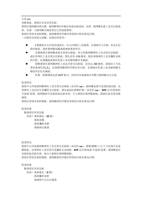
正常oct:双眼视盘、黄斑区未见异常反射。
黄斑区视网膜结构完整,视网膜神经纤维层表面反射连续、光滑,视网膜色素上皮层反射连续、光滑,与脉络膜毛细血管层之间连接紧密。
黄斑区厚度及容积测量、视网膜神经纤维层厚度的分析结果见后图。
(右眼屈光间质欠清晰,结果仅供参考)⏹I期黄斑中心凹变形或消失,中心凹神经上皮脱离,呈现暗区与空腔,未见全层组织缺损,或伴视网膜前膜或玻璃体黄斑牵引。
⏹Ⅱ期黄斑区视网膜表面光带部分缺损,伴小的视网膜神经上皮光带全层缺损,或仅有神经上皮光带全层缺损,裂孔直径<350微米,裂孔周缘神经上皮有囊样水肿的空腔。
孔缘翘起处相应色素上皮及脉络膜反光减弱。
⏹Ⅲ期黄斑区视网膜神经上皮反光带全层缺损,直径达500微米,缺损区上可见带状黄绿色带(盖),孔缘视网膜神经纤维层有空腔,孔缘相应色素上皮及脉络膜毛细血管层反光减弱。
⏹Ⅳ期Ⅲ期黄斑孔的OCT特点,同时伴有玻璃体后界膜与视网膜完全分离。
检查所见黄斑中心凹处视网膜神经上皮光带全层缺损(直径约um),视网膜表面可见线状强反射,孔周神经上皮层间可见囊样无反射腔,裂孔底部向两侧扩散(直径约um),RPE层光带连续/不连续/变薄。
视网膜前可见线状低反射光带,不与黄斑区视网膜相贴,缺损区前光带局限增厚。
黄斑区厚度及容积测量、视网膜神经纤维层厚度的分析结果见后图。
检查结果眼黄斑区异常反射性质?黄斑裂孔(III期)黄斑前膜黄斑囊样水肿玻璃体后脱离检查所见黄斑中心凹处视网膜神经上皮光带全层缺损(直径约um),鼻侧/颞侧/上方/下方孔缘可见盖膜相连,孔周神经上皮层间可见囊样无反射腔,RPE层光带连续/不连续/变薄。
视网膜前可见线状低反射光带,部分与黄斑区视网膜相贴。
黄斑区厚度及容积测量、视网膜神经纤维层厚度的分析结果见后图。
检查结果眼黄斑区异常反射性质?黄斑裂孔(II期)黄斑囊样水肿玻璃体不完全后脱离检查所见黄斑中心凹处内层视网膜神经上皮光带缺损(直径约um),外层神经上皮光带完整(厚约um),RPE层光带连续/不连续/变薄。
黄斑裂孔黄斑裂孔分期在OCT中的表现

手术治疗
手术并发症:出血、炎症、视网膜脱离等
术后恢复:定期复查,注意眼部卫生
手术目的:封闭裂孔,减轻或消除玻璃体对黄斑的牵拉
手术方法:玻璃体切除联合内界膜剥除术
预后评估
视力恢复情况:黄斑裂孔患者手术后视力恢复情况因个体差异而异,需根据患者病情和手术效果进行评估
并发症发生情况:黄斑裂孔手术后可能发生一些并发症,如视网膜脱落、黄斑水肿等,需密切观察并及时处理
视网膜前出现玻璃体后脱离
视网膜下出现新生血管
视力下降至0.1以下
晚期黄斑裂孔
视网膜色素上皮层出现囊样改变
视盘黄斑纤维束受损
视网膜内界膜消失
视网膜神经上皮层完全断裂
04
OCT在黄斑裂孔诊断中的应用
OCT的基本原理
OCT技术原理:基于光学相干理论,通过测量光在生物组织中的相干散射来获取组织结构信息
定量测量:OCT可以定量测量视网膜各层的厚度和结构变化,为黄斑裂孔的诊断提供更准确的数据支持。
早期诊断:OCT能够发现早期黄斑裂孔,有助于及时采取治疗措施,保护视力。
随访观察:通过定期随访观察,OCT可以评估治疗效果,为调整治疗方案提供依据。
OCT在黄斑裂孔分期中的应用
OCT在黄斑裂孔早期诊断中的应用
视网膜内界膜的破坏和视网膜色素上皮层和脉络膜的损害与年龄、遗传、环境等因素有关
黄斑裂孔的临床表现
视力下降:黄斑裂孔会导致中心视力的明显下降,患者难以看清近距离的物体。
视野缺损:黄斑裂孔会导致视野中心出现暗点,影响患者的视野范围。
视觉扭曲:黄斑裂孔患者可能会出现视物变形、扭曲的现象,影响视觉质量。
闪光感:部分黄斑裂孔患者可能会出现眼前闪光的现象,这是由于视网膜受到牵拉所致。
OCT在黄斑裂洞诊断

FFA:无透见荧光点 普通眼底:裂洞大小不等,洞底
无黄色点,周边无晕 裂洞周边有水肿,范围大、出血、
网脱 治疗原则
萎缩性黄斑裂洞
FFA黄斑处透见荧光,分不出裂洞所在 直接检眼镜检查:黄斑未见明显裂洞 OCT:可见裂洞的神经上皮缺损 OCT:黄斑神经上皮菲薄
继发性黄斑裂洞
病理性近视黄斑劈裂 a 无裂洞 b 全层洞 c 外洞 d 内洞
囊样变性
普通眼底 OCT:未见神经上皮缺损,神经上皮变
薄 OCT:在神经上皮内囊样无反射
视网膜囊样变性
1. 定义 2.眼底彩像 3.OCT特征:和劈裂一样,只是比劈裂更局 限呈球形,液体来源视网膜血管和脉络膜 血管,不是玻璃体。
先天性黄斑色素上皮缺损
眼底:红色斑,有中心反光,双眼 对称
OCT:色素上皮部分缺损、毛细血 管存在
假性黄斑裂洞
眼底:红色斑,膜样组织,血管弯曲 OCT:神经上皮增厚,存在陡峭的凹
洞,中心凹神经上皮厚薄正常,无明 显裂洞。
板层裂洞伴神经 上皮脱离
术前
术后
玻璃体和中心凹周边紧密粘连 钝挫伤时是外力的间接作用点 黄斑中心凹是高耗氧处
临床诊断黄斑裂洞主要依据
症状是视力突然下降,伴视物变形 视力:≤0.1(中心视力) 视野:绝对中心暗点 注视性质:旁中心注视 注视直线带有缺损 裂隙灯显微镜检查:光带移位 病灶底部可见黄色斑点,洞缘存在灰白色晕 FFA:病灶处呈透见荧光点
正常黄斑OCT影像
NFL表示神经纤维层,
ILM表示内界膜,
GCL表示神经节细胞层, IPL表示内丛状层,
OPL表示外丛状层,
IS/OS表示内外感光层结联,
oct检查报告

oct检查报告近期,我作为一位患者经历了一次 OCT(光学相干断层扫描)的检查,因为我的视力出现了一些问题,经过眼科医生的建议,我接受了这项检查。
在本文中,我将分享我的 OCT 检查报告,以及一些相关的知识,希望能对有类似问题的读者有所帮助。
OCT 检查是一种无创检测人眼视网膜和视神经的方法,它使用光学相干断层扫描技术,通过红外线光束扫描可以解剖眼底结构。
这项检查是一种相对简便、无创、准确、可重复的非手术诊断方法,可以帮助医生全面了解患者的视力健康状况。
我接受的 OCT 检查主要是为了检测我眼底的状况,以查找视网膜裂孔、视网膜色素变性、黄斑变性等可能的问题。
当我拿到检查报告时,我发现它分为五个部分:视网膜相关参数、视网膜单点分析、视神经头相关参数、视神经单点分析和附注。
这篇文章将依次介绍这些部分,并解释每个部分的意义。
第一部分是视网膜相关参数,它主要包括中心凹深度(CDA)、中心凹厚度(CT)和固有偏差(IPT)。
中心凹深度是指从视网膜表面到中央凹底部的距离,而中心凹厚度是指中央凹底部到视网膜表面的距离。
这两个参数一起描述中央凹的深度和宽度,是评价黄斑病变的重要指标。
固有偏差是指与平均值相比,个体数据的离散程度。
它可以反映测量数据的稳定性,越小表示测量的准确性越高。
在我的检查报告中,中心凹深度为1052µm,中心凹厚度为144µm,固有偏差为7.96,这些数值处于正常范围内。
第二部分是视网膜单点分析,它包括了视网膜厚度图、视网膜血管密度图和视网膜神经纤维层厚度图。
这些图形反映了视网膜的各个区域的厚度和血管密度,并且可以帮助诊断人员判断是否存在视网膜病变。
在我的检查报告中,这些图形显示我的视网膜整体厚度、血管密度和神经纤维层厚度都处于正常范围内。
这说明我的眼睛没有明显的问题,但是也需要继续注意保护视力健康。
第三部分是视神经头相关参数,它包括视神经头反射图和视神经头相关参数表。
视神经头反射图是截取眼底静止图像时看到的视神经径线的反射像,它可以帮助医生评估患者的视神经是否正常,以及是否存在视神经盘萎缩、视神经乳头水肿等问题。
眼科实验报告OCT

眼科实验报告OCT
OCT眼科实验报告
光学相干断层扫描(OCT)是一种非侵入性的眼科检测技术,可以提供高分辨率的眼部图像,帮助医生诊断和治疗眼部疾病。
在本次实验中,我们使用OCT 技术对患有不同眼部疾病的患者进行了检测,取得了一些有意义的结果。
首先,我们对患有青光眼的患者进行了OCT检测。
通过OCT扫描,我们观察到了患者眼球内部的视网膜和视神经头的变化。
这些变化包括视网膜厚度的增加和视神经头的形态改变,这些都是青光眼的典型表现。
通过OCT技术,我们可以及时发现青光眼患者的病情变化,指导医生制定更有效的治疗方案。
其次,我们对患有黄斑变性的患者进行了OCT检测。
黄斑变性是一种老年性眼部疾病,常常导致中央视力丧失。
通过OCT扫描,我们观察到了患者黄斑区域的变化,包括黄斑厚度的减少和黄斑下区域的异常增生。
这些变化可以帮助医生及时干预治疗,延缓病情恶化。
最后,我们对正常人群进行了OCT检测作为对照组。
通过OCT扫描,我们观察到了正常人眼球内部结构的清晰和完整,没有明显的异常变化。
这进一步验证了OCT技术在眼科检测中的可靠性和准确性。
综上所述,OCT技术在眼科领域有着广泛的应用前景。
通过OCT技术,我们可以及时发现眼部疾病的变化,指导医生制定更有效的治疗方案。
随着技术的不断进步,相信OCT技术将为眼科医生提供更多更准确的诊断信息,为患者带来更好的治疗效果。
oct黄斑裂孔描述
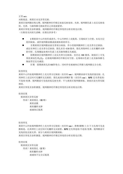
正常oct:双眼视盘、黄斑区未见异常反射。
黄斑区视网膜结构完整,视网膜神经纤维层表面反射连续、光滑,视网膜色素上皮层反射连续、光滑,与脉络膜毛细血管层之间连接紧密。
黄斑区厚度及容积测量、视网膜神经纤维层厚度的分析结果见后图。
(右眼屈光间质欠清晰,结果仅供参考)⏹I期黄斑中心凹变形或消失,中心凹神经上皮脱离,呈现暗区与空腔,未见全层组织缺损,或伴视网膜前膜或玻璃体黄斑牵引。
⏹Ⅱ期黄斑区视网膜表面光带部分缺损,伴小的视网膜神经上皮光带全层缺损,或仅有神经上皮光带全层缺损,裂孔直径<350微米,裂孔周缘神经上皮有囊样水肿的空腔。
孔缘翘起处相应色素上皮及脉络膜反光减弱。
⏹Ⅲ期黄斑区视网膜神经上皮反光带全层缺损,直径达500微米,缺损区上可见带状黄绿色带(盖),孔缘视网膜神经纤维层有空腔,孔缘相应色素上皮及脉络膜毛细血管层反光减弱。
⏹Ⅳ期Ⅲ期黄斑孔的OCT特点,同时伴有玻璃体后界膜与视网膜完全分离。
检查所见黄斑中心凹处视网膜神经上皮光带全层缺损(直径约um),视网膜表面可见线状强反射,孔周神经上皮层间可见囊样无反射腔,裂孔底部向两侧扩散(直径约um),RPE层光带连续/不连续/变薄。
视网膜前可见线状低反射光带,不与黄斑区视网膜相贴,缺损区前光带局限增厚。
黄斑区厚度及容积测量、视网膜神经纤维层厚度的分析结果见后图。
检查结果眼黄斑区异常反射性质?黄斑裂孔(III期)黄斑前膜黄斑囊样水肿玻璃体后脱离检查所见黄斑中心凹处视网膜神经上皮光带全层缺损(直径约um),鼻侧/颞侧/上方/下方孔缘可见盖膜相连,孔周神经上皮层间可见囊样无反射腔,RPE层光带连续/不连续/变薄。
视网膜前可见线状低反射光带,部分与黄斑区视网膜相贴。
黄斑区厚度及容积测量、视网膜神经纤维层厚度的分析结果见后图。
检查结果眼黄斑区异常反射性质?黄斑裂孔(II期)黄斑囊样水肿玻璃体不完全后脱离检查所见黄斑中心凹处内层视网膜神经上皮光带缺损(直径约um),外层神经上皮光带完整(厚约um),RPE层光带连续/不连续/变薄。
眼科oct检查报告解读

眼科oct检查报告解读
眼科OCT(光学相干断层扫描)是一种先进的眼科检查技术,通过使
用红外光线来获取眼部结构的高分辨率图像。
这种非侵入性的检查可以检
测和诊断多种眼科疾病,如黄斑变性、青光眼、视网膜血管阻塞等。
下面
是一份常见的OCT检查报告的解读。
首先,报告中会提到眼睛的结构,如视网膜、黄斑、视神经盘等。
这
些结构在正常情况下应当具有正常的形态和厚度。
接下来,报告中可能会提到一些数字,比如视网膜厚度和黄斑中心凹
厚度。
这些数字是与正常参考范围进行比较的,从而评估眼睛的健康状况。
如果视网膜或黄斑厚度超出正常范围,可能表明存在异常情况,如黄斑变
性或视网膜水肿等。
报告还可能包含一些图像,如眼底照片、视网膜红外照片以及水平断
层扫描图像等。
这些图像可以帮助医生更好地评估眼球的结构和异常情况。
例如,水平断层扫描图像可以显示不同层次结构的细节,从而评估视网膜
和黄斑的状态。
在解读报告时,医生还会注意报告中的其他细节,如眼底出血、视网
膜脱落、视网膜下脱离等病变。
这些病变常常是一些严重眼科疾病的表现,需要进一步的治疗和随访。
需要注意的是,OCT检查只是一种辅助的诊断工具,诊断结果应该结
合医生的临床经验和其他检查结果来进行综合判断。
因此,在报告解读时,建议患者与医生进行面对面的沟通,共同讨论检查结果以及可能的治疗方案。
总之,眼科OCT检查报告的解读需要医生的专业知识和经验。
患者应该在专业医生的指导下进行诊断和治疗,并密切关注视力变化和眼部症状的出现,及时就医。
眼底病OCT解读

干性AMD--玻璃膜疣
SLO
SLO中黄斑区呈色素紊乱
FFA
F-10 Retro mode
Retro Mode中玻璃膜疣呈小颗粒状,并有立体感
OCT
动静脉期黄斑区呈散在、边界清晰的点状高荧光。 此图为硬性玻璃膜疣。
色素上皮呈波浪性隆起,并突破椭圆体带,隆
起的外界膜已进入外核层
27
干性AMD--地图状萎缩
正常视网膜OCT影像
6
最新国际OCT分层命名共识
7
组织分层
8
反射信号
9
正常视乳头黄斑OCT
黄斑特点:
(1)中心凹视网膜最薄,只有一层外核层,缺少神经纤维层、神
经节细胞层、内丛状层、内核层、外丛状层。
(2)外核层在中心凹处最厚。
(3)外核层的神经细胞中,只有视锥细胞,无视杆细胞。
(4)此处的视锥细胞形态与其他部位不一样,较细长。
异常新生血管从脉络膜生长到视网膜中
22
AMD--AREDS标准
无AMD:无或者仅有很小的玻璃膜疣 (直径<63um)。
早期AMD:同时存在多个小的玻璃膜 疣和少量中等大小的玻璃膜疣(直径 为63-124um),或有色素上皮异常。
23
AMD--AREDS标准
中期AMD(AREDS分类3):广泛存在中等大小的玻璃膜疣,至少有一个大 的玻璃膜疣(直径≥125um),或有未涉及黄斑中心凹的地图样萎缩。
传统分类: 干性AMD, 湿性AMD
英国指南讨论的3个标准,各有优劣 ➢Wisconsin Age related Maculopathy Grading Scheme ( WARMGS) ➢Simplified version Early age related maculopathy ARM international classification system ➢AREDS age related eye disease study 美国指南推荐标准: AREDS
oct检查报告

oct检查报告患者信息:姓名:XXX 性别:男年龄:XX岁检查日期:XXXX年XX月XX日一、检查目的Oct(光相干断层扫描)是一种高分辨率的眼科检查技术,用于评估眼底组织的结构和病变情况。
本次检查的目的是详细评估患者的眼底情况,了解可能存在的病变并提供诊断参考。
二、检查结果1. 衰老性黄斑变性:根据Oct图像显示,患者双眼黄斑区结构略显不规则,呈现出典型的衰老性黄斑变性表现,包括黄斑变性区域的增厚、囊样水肿、色素上皮脱落等特征。
建议患者加强眼部保健,定期复查,并遵循医生的治疗建议。
2. 黄斑裂孔:在右眼的Oct图像中,观察到下视盘区域存在一处黄斑裂孔,裂孔的边缘光滑清晰,大小约为XXXXum。
鉴于该病变的存在,建议患者密切关注视力变化,遵循医生的治疗方案,并及时进行治疗。
3. 视网膜脱离:左眼Oct图像显示,视网膜脱离的迹象明显,视网膜分层出现异常,形成明显的囊样空隙。
视网膜脱离的原因可能与眼球外伤、结膜手术等因素有关。
患者需及时咨询专科医生,制定进一步治疗方案。
4. 视网膜血管病变:双眼Oct图像中观察到视网膜血管存在异常变化,表现为黄斑区域附近的视网膜血管充血、扩张和弯曲。
这种病变可能与血管疾病、高血压等因素有关,建议患者积极控制血压,定期随访,并配合医生治疗。
三、医生建议1. 药物治疗:针对衰老性黄斑变性和黄斑裂孔,患者可口服抗氧化剂、维生素类药物,以帮助减缓病情进展,维护眼部健康。
2. 手术治疗:对于视网膜脱离的患者,如病情较重,医生可能会建议采取手术治疗,如激光光凝、玻璃体手术等。
3. 注意事项:- 增加户外活动时间,避免长时间疲劳看电子屏幕。
- 忌烟、忌酒,保持健康的生活习惯。
- 定期复查,密切关注视力变化和病情发展。
四、关于Oct检查Oct检查是一种非侵入性、非接触性的眼科检查方法,通过扫描眼底组织结构,获取高分辨率的图像资料,对多种眼底疾病进行早期诊断和跟踪观察,为医生制定治疗方案提供重要参考。
黄斑变性症状检查和治疗

黄斑变性症状检查和治疗黄斑变性(Age-related Macular Degeneration,简称AMD)是一种常见且逐渐进展的视网膜疾病,主要影响中央视觉的成熟人群。
黄斑变性是导致老年人失明的首要原因之一,严重影响了患者的生活质量。
因此,及早进行黄斑变性的病情检查和有效的治疗至关重要。
本文将介绍黄斑变性症状的检查方法和常用的治疗手段。
一、症状检查1. 影像学检查:OCT光学相干层析扫描(Optical Coherence Tomography,简称OCT)是一种非侵入性的检查方法,通过影像学技术扫描黄斑区域,并能够显示出黄斑上皮层和视网膜的结构。
OCT可以检测黄斑区域是否出现病变,并提供详细的图像资料,有助于医生做出准确的诊断。
2. 视力检查对于患者来说,视力下降是黄斑变性的主要症状之一。
医生通常会使用视力表进行视力检查,通过测量患者的远视力和近视力来了解黄斑变性的影响程度。
此外,还可以进行视野检查,以评估患者的中央视野是否受损。
3. 荧光素绿染色荧光素绿染色(Fluorescein angiography,简称FA)是一种常用的检查方法,可直观地观察和评估黄斑区域血管的异常情况。
医生会将荧光素绿注射入患者的静脉,然后使用专用相机拍摄黄斑区域的血管显影情况,以帮助确定病变的范围和类型。
二、治疗方法1. 抗血管内皮生长因子(anti-VEGF)治疗抗血管内皮生长因子治疗是目前黄斑变性的常用方法之一。
抗血管内皮生长因子药物可以抑制新生血管的生长,减少渗漏和出血。
常用的抗血管内皮生长因子药物包括阿瓦斯汀(Avastin)、卢卡斯汀(Lucentis)和甲磺酸雷珠单抗(Eylea)等。
这些药物通常通过注射到眼内进行治疗,治疗过程需要经过多次注射。
2. 光动力疗法光动力疗法是一种通过激光光源和光敏剂联合作用,抑制异常新生血管生长并保护黄斑区域的治疗方法。
在治疗中,医生会将光敏剂注射到患者的静脉,然后利用特定波长的激光照射黄斑区域,以达到治疗的效果。
眼部oct检查报告解读
眼部 OCT(Optical Coherence Tomography)检查是一种无创的眼科检查方法,可以用来评估眼球内部各个结构的形态和厚度,帮助医生确定眼部疾病的诊断及治疗方案。
以下是 OCT 检查报告的一些常见术语及其解释:
1. 视网膜厚度:指视网膜的厚度,正常情况下,视网膜中央区域的厚度应该在 200-250 微米之间。
2. 视杯盘比(C/D值):指视神经盘的中央凹陷部分(杯形区域)与整个视神经盘直径之间的比例。
正常情况下,这个比值应该在 0.3-0.5 之间。
3. 黄斑区域厚度:指黄斑区域的厚度,黄斑是视网膜的中央区域,对高清晰度视力起着关键作用。
4. 眼底前节:指眼球前部的透明液体,包括前房和后房。
5. 视网膜神经纤维层(RNFL):指视网膜内部最靠近玻璃体的一层,它包含了视网膜神经元的轴突。
RNFL 的损伤可能与青光眼、多发性硬化等疾病有关。
6. 玻璃体:指填充在眼球后部的一种透明胶状物质,它对保持眼球形态和光的折射起着重要作用。
需要注意的是,以上术语仅为 OCT 检查报告中较常见的一些术语,具体的解释和诊断需要结合个人情况进行分析。
如果您对自己的 OCT 检查报告有任何疑问和不理解的地方,建议及时咨询眼科医生。
OCT结果解读

OCT结果解读
梁平县人民医院眼科 邓宗勇
.
1
OCT
1.正常视乳头黄斑束黄斑OCT图像
左图:眼底图,白箭为OCT扫描方向,
右图:OCT图,左侧凹陷为黄斑中心凹,右侧为视乳 头凹陷
.
2
OCT
注:在OCT断层图像中,红色高反射为视网膜后界,对应 于视网膜色素上皮(RPE)和脉络膜毛细血管层,此后界 在筛板水平与脉络膜循环一起终止于视乳头边缘。在RPE 与脉络膜毛细血管层的上方显示最弱反射的一层暗区代表 视网膜光感受器外节,视网膜表层与此层之间为中等反射, 对应于视网膜内、外颗粒层和内、外丛状层。在视网膜的 内侧缘显示另一高散射区域,红色的反射层为视网膜神经 纤维层(RNFL),在图像中可见如正常组织解剖一样, RNFL厚度从黄斑至视乳头是逐渐增加的。
33
.
12
OCT
4.异常黄斑OCT图像
视网膜厚度改变
A厚度增加
B厚度减少
.
13
OCT
视网膜反射性改变
A发射增强
B反射减弱
.
14
OCT
血液、血浆、和渗出的鉴别
.
15
OCT
.
16
OCT
.
17
OCT
视网膜形态学改变
A视网膜神经上皮层和色素上皮层脱离
.
18
OCT
.
19
OCT
.
20
OCT
中心凹凹陷改变
.
6
OCT
OCT正常视网膜各层图像
哥白尼OCT(可能是第四代)
.
7
.
8
.
9
.
10
OCT
3.正常视网膜神经纤维层OCT图像
oct检查报告

oct检查报告患者信息:姓名:XXX性别:XXX年龄:XXX检查日期:XXX前言:光学相干断层扫描(OCT)是一种无创检查方法,通过对眼部进行断层扫描,可以提供详细的眼部结构图像。
本报告将就患者的OCT检查结果进行分析和解读。
1. 双眼视网膜层检查结果:根据OCT图像显示,患者双眼视网膜层结构正常,显示出清晰的各层次分界线。
具体如下:1.1 黄斑区:在黄斑区,OCT图像显示患者双眼的视网膜层结构完整,黄斑中央凹反射光强度正常。
黄斑中央凹正常,无明显异常变化。
1.2 视网膜神经纤维层:患者双眼的视网膜神经纤维层显示规整,厚度均匀一致。
未观察到明显的视网膜神经纤维层损伤,无明显的神经纤维层缺损。
1.3 视网膜杯盘比例:患者双眼的视盘呈现正常的结构和形态,杯盘比例(C/D比)正常。
未观察到明显的视盘改变。
2. 双眼视网膜血管检查结果:通过OCT图像的分析,患者双眼的视网膜血管显示正常,无明显的血管异常。
3. 双眼视网膜厚度检查结果:根据OCT图像,患者双眼的视网膜厚度显示正常范围内,并且呈现对称分布,无明显的厚度异常。
4. 结论:经过对患者的OCT检查结果分析和解读,患者双眼的视网膜层结构正常,视网膜神经纤维层、视网膜杯盘比例、视网膜血管以及视网膜厚度等各项指标均无明显异常。
备注:本报告仅基于患者的OCT检查结果,结合其他临床资料综合判断,最终诊断和治疗方案请咨询医生。
总结:OCT检查是一种安全、无创的眼部检查方法,通过对眼部组织进行断层扫描,可以提供详细的结构信息。
患者的OCT检查结果表明视网膜层结构正常,各项指标符合正常范围。
这为进一步的眼部健康评估和疾病诊断提供了重要的依据。
附注:本文摘自专业医学资料,仅作为参考信息,具体情况请咨询专业医生。
黄斑病变OTC影像报告

黄斑病变OTC影像报告摘要本报告将介绍黄斑病变的OTC(光学相干断层扫描)影像报告。
黄斑病变是一种常见的视网膜疾病,OTC是一种非侵入性的影像检测方法,可用于评估黄斑病变的病变类型和严重程度。
本报告将通过OTC影像来描述黄斑病变的特征和诊断结果。
引言黄斑病变是一种发生在黄斑区的疾病,黄斑是眼睛视网膜的中心区域,负责清晰视力的产生。
黄斑病变是导致视力减退的主要原因之一。
OTC是一种无创的检测方法,通过测量反射光的干涉模式来生成图像,能够高分辨率地观察黄斑区的细微结构和病变。
方法OTC影像的获取是通过使用OTC设备进行扫描来实现的。
患者在病床上保持静止,并将头部放置在适当的位置。
操作员使用OTC探头对眼睛进行扫描,其中探头会发射一束光线并记录反射光的干涉模式。
扫描过程通常需要几分钟,在此期间,患者需要尽量保持不动。
结果以下是OTC影像报告的重要部分:图像一:黄斑中央凹影像该影像显示了黄斑中央凹的结构。
黄斑中央凹是黄斑区域的中央部分,是视力最敏感的区域。
在该影像中,黄斑中央凹区域呈现出明亮的反射,并且没有明显的异常结构或异常反射。
这表明黄斑中央凹区域没有明显的病变。
图像二:黄斑病变该影像显示了黄斑区域的病变特征。
在黄斑区域的上方可以观察到一些异常反射和形状不规则的区域。
这些结构有可能是黄斑病变的病变部分。
根据其形态和位置,可能是黄斑区域的黄斑变性。
图像三:黄斑区域断层图像该影像是黄斑区域的断层图像,可以更清晰地观察黄斑区域的结构和层次。
在该影像中,可以观察到视网膜色素上皮层、视锥细胞层和神经纤维层等结构。
这些结构的完整性对于保持视力非常重要。
讨论通过OTC影像,我们能够观察和评估黄斑区域的结构和病变。
黄斑病变是一种常见的视网膜疾病,可导致视力问题。
OTC影像可以帮助医生确定病变的类型和严重程度,进而制定治疗计划。
然而,OTC影像并不能提供关于病变的具体原因和治疗方法的信息。
结论黄斑病变OTC影像报告对评估黄斑病变的病变类型和严重程度具有重要意义。
黄斑 oct 解读
黄斑oct 解读
黄斑OCT检查,是指黄斑的光学相干断层成像检查,特点是非创伤方式,操作简单。
OCT检查利用光的干涉原理,能够观察到黄斑区视网膜结构,通过结构变化推测疾病的可能性,或者疾病的严重程度,可以判断黄斑水肿、视网膜出血等情况。
OCT检查主要是对黄斑区,或者是视盘区进行定性和定量的分析检查,黄斑区检查可能会更多一些。
定性检查是分析病变所处位置、形状、牵涉结构。
定量检查主要是量化视网膜,具体厚度是多少,体积是多大。
通过黄斑区OCT检查,可以诊断出黄斑相关的一些疾病,比如黄斑前膜、黄斑水肿等。
如果黄斑受到影响,视力会明显下降,视物会变形,视觉质量也会受到影响。
如果有黄斑水肿,还有黄斑出血的情况下,高度怀疑可能是有系统性疾病,比如糖尿病视网膜病变、高血压视网膜病变,应尽快进行干预和治疗。
OCT结果解读概述

OCT结果解读概述OCT(Optical Coherence Tomography)是一种无损、无创的成像技术,可用于观察和评估眼部疾病。
它利用光学干涉技术,生成高分辨率的视网膜断层图像,从而帮助医生诊断和监测疾病。
本文将对OCT结果的解读进行概述。
OCT技术通过测量不同层次的组织反射时间来创建断层图像。
这些图像显示了视网膜不同层次的结构特征,从而提供了详细的解剖信息。
通过OCT,医生可以观察到视网膜的神经纤维层、色素上皮层、黄斑等部位,并评估其结构和状态。
根据OCT结果,医生可以对多种眼部疾病进行诊断与监测,如黄斑变性、青光眼、视网膜色素变性等。
在OCT图像中,主要的解剖结构包括视杯、视盘、视网膜神经纤维层、色素上皮层和黄斑区域。
视杯代表视神经头部,正常情况下,其形状通常为圆或椭圆,正常大小约为盘径的0.25-0.4倍。
因此,如果视杯呈现异常扩大或缩小的情况,可能是视神经头的疾病迹象。
视盘是其中的一部分,也是神经纤维层的起点。
视盘的外缘被称为视杯边缘或视盘边缘。
视网膜神经纤维层也是OCT图像的重要结构之一、神经纤维层厚度的改变可以反映神经退化的程度,如青光眼导致的视神经头损伤等。
通过测量视网膜神经纤维层的厚度,可以了解到视神经的病程进展情况。
色素上皮层是视网膜下层,起到维护视细胞的功能。
OCT可以观察色素上皮的形态和厚度,以评估其健康状态。
色素上皮的损伤可能导致视网膜疾病和黄斑变性的发生。
黄斑区域是视网膜中最关键的区域之一,它包括黄斑颞侧核、黄斑大脑皮质和视翳等。
OCT可以提供详细的黄斑结构图像,可以评估黄斑的厚度、形态和组织结构。
黄斑功能恶化可能导致视力减退、中心视野缺损等临床表现。
除了以上提到的解剖结构,OCT还可以测量视网膜层的厚度和结构特征,比如内、外节间隙、外核区和外位棘状神经节等。
这些指标可以用于评估视网膜疾病的严重程度和进程。
总的来说,OCT是一种非常有用的眼科成像技术,可以提供高分辨率、详细的视网膜断层图像。
oct检查报告怎么看

oct检查报告怎么看OCT(光相干断层扫描)是一种非侵入性的眼科检查技术,它使用光学原理来生成高分辨率的眼底图像。
这种检查可以帮助眼科医生准确诊断和治疗多种视网膜疾病,如黄斑变性、青光眼等。
本文将简要介绍OCT检查报告的解读方法,以帮助您更好地了解个人眼健康情况。
一、基本信息在OCT检查报告的第一部分,通常会包含受检者的基本信息,如姓名、年龄、性别等。
这些信息有助于医生更好地识别患者并记录检查历史。
二、检查日期和技师检查日期记录了您接受OCT检查的具体时间,而技师栏显示了为您进行检查的专业人员。
这些信息对于了解检查过程和参考以后检查结果的变化非常重要。
三、图像解读OCT检查结果的核心部分是图像,一般有水平扫描和垂直扫描两个方向的断层图像。
这些图像显示了不同部位的眼底结构,如视网膜、黄斑区等,通过分析这些结构的形态和密度,医生可以评估眼部健康情况。
1. 视网膜厚度OCT检查可以提供详细的视网膜层厚度数据,通常以图像和数字两种方式呈现。
医生会关注视网膜中心区域的厚度,因为这是检测黄斑变性等疾病的重要指标。
2. 黄斑区评估OCT图像中的黄斑区是眼睛最关键的部位之一,它对清晰视觉起着重要作用。
医生会检查黄斑区的形态,包括中心凹的深度和完整性等。
任何异常的变化都可能与视力下降相关。
3. 前房和角膜OCT检查也可以提供前房和角膜的图像,这有助于评估青光眼等眼压相关疾病。
医生会关注前房深度、角膜曲率等参数,并与正常范围进行对比。
四、诊断结果在OCT检查报告的最后部分,医生通常会给出诊断结果和相应的建议。
这些结果是基于医生对OCT图像的解读和对比分析得出的。
如果发现了眼部异常,医生可能会进一步建议您进行其他检查或治疗。
需要注意的是,虽然本文介绍了OCT检查报告的一般内容和解读方法,但具体报告的格式可能因医院和设备的不同有所差异。
因此,建议您仔细阅读和咨询医生,以便更准确地理解您自己的OCT检查报告。
综上所述,通过仔细阅读OCT检查报告中的基本信息、图像解读以及诊断结果,您可以更好地了解您的眼部健康情况。
糖尿病视网膜病变患者黄班厚度OCT测量定量分析
动异常,最终引起足底压力异常,反复局部高压造成足底软组织损伤。
在此基础上的感染促进了糖尿病足的发生和发展[1]。
可见糖尿病患者足底压力的变化是造成糖尿病足病的中间环节[2,3]。
发达国家对足底压力的研究起步较早,目前已经积累了较多的参数值。
目前国际上在该领域的工作重心为多学科交叉研究,根据足底压力研究结果找出预测因子,预防足病[4-6]。
国内对足底压力的研究仍处于模仿国外早期研究的探索阶段。
对于糖尿病足底压力的研究更是没有得到应有的重视。
本研究是个小样本量的探索。
考虑到行走过程中与较多因素相关,本研究采用统一斜率(0度)及速率(1kg /h )的行走速度,保证所有受试者为统一速度的匀速直线平地运动。
二组患者在一般资料上情况类似,但神经病变组患者的眼底视网膜病变例数大于无神经病变组,提示微血管病变和神经病变的相关性。
两组患者足底压力参数比较(表2)提示神经病变患者行走过程中与地面接触时间更长,接触面积更大,足底应力时间积分明显延长,相对应的足底离地的时间缩短,所以神经病变患者平时走路更慢,步幅更小。
本研究未发现糖尿病神经病变者足底压力峰值增高,这与部分国外文献相悖[7],但与国内中山大学严励等人的研究结果相一致。
可能与以下几方面原因相关:①本文纳入的神经病变程度较轻,未引起足底峰值压力的变化;②样本量太小;③中国人群足底压力特点有别于欧美人群。
为突出糖尿病合并周围神经病变患者的足底压力特点,本文对足底应力值进行了区域间比值。
发现神经病变患者足底应力向外侧足倾斜,特别是足弓部位及前足外侧部位。
在小样本的研究中,足底峰值压力绝对值没有显著差异的情况下(表3),区域应力比值已有显著差异(表4)。
特别是外侧与内侧足应力比值差异明显,较能体现周围神经病变患者早期的足底压力分布异常。
参考文献:[1] Wrobel JS ,Najafi B.Diabetic foot biomechanics and gaitdysfunction [J ].Diabetes Sci Technol ,2010,4(4):833-845.[2] Caselli A ,Pham H ,Giurini JM ,et al.The forefoot -to -rearfoot plantar pressure ratio is increased in severe diabetic neuropathy and can predict foot ulceration [J ].Diabetes Care ,2002,25(6):1066-1071.[3] Mueller MJ ,Zou D ,Bohnert KL ,et al.Plantar stresses onthe neuropathic foot during barefoot walking [J ].Phys Ther ,2008,88(11):1375-1384.[4] Acharya UR ,Tong J ,Subbhuraam puter-based i⁃dentification of type 2diabetic subjects with and without neu⁃ropathy using dynamic planter pressure and principal compo⁃nent analysis [J ].Med Syst ,2011,27.[5] Bus SA ,Haspels R ,Busch-Westbroek TE.Evaluation andoptimization of therapeutic footwear for neuropathic diabeticfoot patients using in-shoe plantar pressure analysis [J ].Di⁃abetes Care ,2011,34(7):1595-1600.[6] Cavanagh PR ,Bus SA.Off-loading the diabetic foot for ul⁃cer prevention and healing [J ].Plast Reconstr Surg ,2011,127(Suppl 1):248S-256S.[7] Yavuz M ,Tajaddini A ,Botek G ,et al.Temporal character⁃istics of plantar shear distribution :relevance to diabetic pa⁃tients [J ].Biomech ,2008,41(3):556-559.文章编号:1006-6233(2013)05-0738-03糖尿病视网膜病变患者黄斑厚度OCT 测量定量分析张季瑾(天津市大港油田总医院眼科, 天津 大港区 300280)摘 要院目的:探究与分析糖尿病视网膜病变患者黄斑厚度OCT 。
- 1、下载文档前请自行甄别文档内容的完整性,平台不提供额外的编辑、内容补充、找答案等附加服务。
- 2、"仅部分预览"的文档,不可在线预览部分如存在完整性等问题,可反馈申请退款(可完整预览的文档不适用该条件!)。
- 3、如文档侵犯您的权益,请联系客服反馈,我们会尽快为您处理(人工客服工作时间:9:00-18:30)。
Hans Journal of Ophthalmology 眼科学, 2015, 4, 38-41Published Online June 2015 in Hans. /journal/hjo/10.12677/hjo.2015.42007Analyzing the Application of OCT inExamining Retinal Macular DiseasesBo Ding, Guiyuan Guo, Fang Gao, Hewen Wu, Guobing ZhongPhysical Examination Center, Hangzhou Sanatorium of Nanjing Military Command, Hangzhou ZhejiangEmail: 1647514415@Received: May 29th, 2015; accepted: Jun. 26th, 2015; published: Jun. 29th, 2015Copyright © 2015 by authors and Hans Publishers Inc.This work is licensed under the Creative Commons Attribution International License (CC BY)./licenses/by/4.0/AbstractPurpose: Discussing the application of OCT in examining retinal macular diseases. Method: Col-lecting data from people who were willing to have OCT examination in retinal macula, during their health examination in the hospital. Among 9202 volunteers, 5926 are male, 3276 are female. Their age is between 23 - 72 (47.5 ± 24.5), vison acuity 0.05 - 1.5. Analysing the severity and reasons of retinal macular diseases among health examination population, 9202 volunteers were examined, 1928 of whom were diagnosed of macular diseases, the positive rate was 21%. Among them, 733 volunteers (7.97%) were diagnosed of posterior vitreous detachment, 465 volunteers (5.05%) macular pigment disorder, 262 volunteers (2.85%) vitreous wart, 199 volunteers (2.16%) macu-lar epiretinal membrane, 114 volunteers (1.24%) pigment epithelium detachment, 96 volunteers(1.04%) macular hole, 29 volunteers (0.32%) central serous chorioretinopathy (CSC), 17 volun-teers (0.18%) retinoschisis, 13 volunteers (0.14%) central serous infiltration. Conclusion: OCT examination has significant effect on observing layer structure of retinal macula as well as its le-sion distribution. Besides, the shape, area and boundary of lesions can be observed as well, which plays a vital role in diagnosing and determining the treatment plan timely.KeywordsOptical Coherence Tomography, Test, Retina, Macular, Pathological ChangesOCT检测眼底视网膜黄斑部的病变分析丁波,过贵元,高方,吴和文,钟国兵南京军区杭州疗养院体检中心,浙江杭州Email: 1647514415@收稿日期:2015年5月29日;录用日期:2015年6月26日;发布日期:2015年6月29日摘要目的:探讨在体检中应用OCT检测眼底视网膜黄斑部的病变情况。
方法:收集在本院健康体检中,自愿应用OCT在眼底视网膜黄斑部检查9202名,其中男性5926名,女性3276名,年龄23~72 (47.5 ± 24.5)岁。
视力0.05~1.5。
分析在体检人群中发生视网膜黄斑部的病变情况及原因。
结果OCT体检9202人,视网膜黄斑区病灶1928人,检出率21%。
其中:玻璃体后脱离733人7.97%,黄斑色素紊乱465人5.05%,玻璃体疣262人2.85%,黄斑前膜199人2.16%,色素上皮脱离114人1.24%,黄斑裂孔96人1.04%,中浆29人0.32%,视网膜劈裂17人0.18%,中渗13人0.14%。
结论:采用OCT检测能够清楚直观了解眼底黄斑区视网膜各层细胞组织结构和病变分布,清楚黄斑区各种病变的形态大小、边界,对患者确定早期治疗方案及预后具有重要的指导价值。
关键词OCT,检测,视网膜,黄斑部,病变分析1. 引言眼底视网膜黄斑区位于眼球后极部,负责视觉和色觉的视锥细胞就分布于该区域,是视力最敏感区。
因此任何累及黄斑部的病变都会引起中心视力的明显下降、视物色暗、变形等。
光学相干断层成像(OCT)技术是指对眼透光组织做断层成像,是光学诊断近十年来一种新型非接触属于无创光学影像诊断技术,通过扫描,观察分析不同组织分布构成位置,得到二维或三维立体构成图[1]。
我院眼科从2012年10月~2015年4月,应用OCT体检9202人,发现视网膜黄斑区病变1928人,现报告如下:2. 资料与方法2.1. 一般资料2012年10月~2015年4月收集在本院健康体检中,自愿应用OCT在眼底视网膜黄斑部检查9202名,其中男性5926名,女性3276名,年龄23~72 (47.5 ± 24.5)岁。
视力0.05~1.5。
纳入标准:无眼部活动性炎症者,无影响OCT检查结果的屈光间质浑浊者。
排除标准:眼球震颤、上睑下垂,斜视等不能固视者。
2.2. 仪器与设备电子视力表,裂隙灯显微镜,佳能眼底照相机,莫廷-眼科光学相干断层扫描仪(OCT)等。
2.3. 方法电子视力表检测视力(裸眼和矫正视力),裂隙灯显微镜检测屈光间质,眼科光学相干断层扫描仪(OCT)在黄斑中心凹采用镜头内注视点方法对受检者行水平及垂直径线扫描,对清晰度稳定的扫描成像进行观察分析[2]。
对眼底黄斑进行高清六线扫描,能快速准确地反映并记录眼底情况。
用SPSS17.0统计软件包χ±)表示。
进行统计处理;计数统计资料以百分比表示,计量资料以均数±标准差(s3. 结果应用OCT体检9202人,发现视网膜黄斑区病变1928人,检出率21%。
见表1。
Table 1. OCT detection of macular lesions表1. OCT检测眼底黄斑部病变情况黄斑部病变检出人数检出率玻璃体后脱离733 7.97%黄斑色素紊乱465 5.05%玻璃膜疣262 2.85%黄斑前膜199 2.16%色素上皮脱离114 1.24%黄斑裂孔96 1.04%中浆29 0.32%视网膜劈裂17 0.18%中渗13 0.14%4. 讨论光学相干断层成像(Optical coherence tomography, OCT)是20世纪90年代发展起来的一种新型的生物医学成像技术,它可以对生物组织内部的微观结构进行高分辨率横断面层析成像。
OCT拥有高分辨率成像系统,获得5 μm高分辨率的图像,可清晰显示初期病变[3]。
眼底视网膜黄斑区位于眼球后极部,负责视觉和色觉的视锥细胞就分布于该区域,是视力中心。
黄斑中心凹处薄而无血管,许多眼科疾病和全身性疾病都可造成视网膜黄斑区的病变而引起中心视力的明显下降、视物色暗、变形等。
因此,眼底视网膜黄斑部病变造成的后果非常严重,一旦遭到破坏,视力便会永久性的受到损害。
本研究应用OCT体检9202人,视网膜黄斑区病灶1928人,检出率21%。
其中:玻璃体后脱离733人7.97%,黄斑色素紊乱465人5.05%,玻璃体疣262人2.85%,黄斑前膜199人2.16%,色素上皮脱离114人1.24%,黄斑裂孔96人1.04%,中浆29人0.32%,视网膜劈裂17人0.18%,中渗13人0.14%。
研究数据显示,用OCT对眼底黄斑进行高清六线扫描,能快速准确地反映眼底情况;尤其是对眼底黄斑区病变的检测具有分辨率高、精准度强等特点。
目前,在眼科眼底病检测中,OCT认可度非常高,已经成为眼科常规检查项目,尤其是眼底黄斑部疾病诊断金标准。
随着年龄增长,玻璃体的组织学也发生变化,其原因是透明质酸逐渐耗竭溶解,胶原的稳定性被破坏,玻璃体内部分胶原网状结构塌陷,产生液化池,形成玻璃体液化腔,继之后玻璃体腔液体的玻璃体通过皮层孔进入玻璃体后腔,逐渐导致玻璃体后脱离。
本研究显示玻璃体后脱离占检出人群的38%,玻璃体后脱离可导致老年特发性黄斑裂孔形成。
检测中发现黄斑区玻璃体疣、色素紊乱、色素上皮脱离占检出人群的43.6%,可能与遗传因素、黄斑长期慢性光损伤、代谢及营养因素等有关,其后果形成年龄相关性黄变性,视力呈进行性损害。
研究还发现黄斑前膜、黄斑裂孔、中浆、中渗和视网膜劈裂占检出人群的18.4%,其发生与各种眼及全身疾病有关。
本研究提示采用OCT检查能够清楚直观了解眼底黄斑区视网膜各层细胞组织结构和病变分布,清楚黄斑区各种病变的形态大小、边界,对患者确定早期治疗方案及预后具有重要指导价值。
眼是人体十分重要的感觉器官,能接受外部的光刺激,并将光冲动传送到大脑中枢而引起视觉。
