实验五 空肠绒毛
胃 小肠的实验报告
胃小肠的实验报告一、实验目的本实验旨在探究胃和小肠在消化过程中的功能和作用。
二、实验原理胃和小肠是人体消化系统中重要的器官。
胃负责储存和分解进食物,小肠则吸收和消化食物中的养分。
消化食物初步从口腔开始,经过食道直至胃中。
胃是一个呈扁球形的器官,位于胸腔和腹腔之间的上腹部。
在胃的内壁上有许多褶皱,形成了许多称为胃腺的小突起。
这些胃腺分泌胃液,包含盐酸和胃蛋白酶。
胃蛋白酶有能力分解蛋白质。
消化食物在胃内停留一段时间,经过蠕动作用和胃液的分泌,被分解并形成称为胃糜的液体。
胃糜会通过幽门进入小肠进一步消化和吸收。
小肠位于胃的下方,是一个长约6米的细长器官。
主要分为三部分:十二指肠、空肠和回肠。
十二指肠是小肠的起始段,它连接到胃幽门的附近。
空肠是小肠的主要吸收区,其内壁上有许多绒毛,能增加吸收表面积。
回肠则是将未消化和未吸收的物质传递到结肠。
小肠的内壁有许多微细绒毛,绒毛上分布着小肠腺,这些腺体分泌消化酶,用于分解食物中的碳水化合物、脂肪和蛋白质。
通过小肠腺分泌的胰液和肠液,以及空肠绒毛的吸收作用,食物中的养分被吸收进入血液循环系统,供给全身各个细胞使用。
三、实验步骤1. 准备实验材料:一只新鲜动物的胃和小肠。
2. 将动物的胃和小肠取出,洗净并剪成适当大小的碎片。
3. 分别将胃和小肠放入两个容器中。
4. 向每个容器中加入适量的消化液,模拟胃液和肠液。
5. 将容器密封,并置于恒温水浴中。
6. 观察胃和小肠在消化液的作用下的变化。
可以使用显微镜观察细胞的形态和结构的改变。
7. 在一定时间后,取出胃和小肠,进行观察和记录。
四、实验结果经过一段时间后,胃中的食物在胃液的分解作用下被分解成胃糜。
小肠中的食物经过肠液和胰液的分泌作用,被分解成最终比较简单的物质如葡萄糖、脂肪酸和氨基酸,从而可以被绒毛上的小肠腺吸收。
五、实验分析和讨论通过本实验的观察和结果分析,我们可以得出以下结论:1. 胃在消化过程中起着重要的作用,通过胃液的分泌和蠕动作用,食物被分解成胃糜。
制作小肠绒毛结构模型

较深 , 怎么处理 ? ” 在思考 解答 问题 的基础 上 , 引导学生
・
3 7・
性; 引导学生确定 合理 的实验方法 , 体会 选择合理 的实
验方法是实验 成功 的前提条件 。在确定 了实验材 料和
分 析结果 , 得 出结论 : 植 物 在光 合作 用 中产 生 了淀粉 ;
毛 的实验 , 学生不能完 全 了解 绒毛结构 , 更 难理解其 吸
收功能。为此 , 笔者制作 了小肠 绒毛结构模型 。在课 堂 教学 中运用此模 型 , 来 展示小肠 绒毛指 状突起 、 绒 毛壁
单层上皮细胞、 内部的毛细血管及毛细淋 巴管等细微结
构, 取得 了良好 的效果。现将 制作方法简要介绍如下 :
以“ 设计 实验探究 : 光合作用 的过程 中是否需要二
生 的氧气的来 源” , 由学生 完成探 究实验 。首先 由学生 提 出问题 、 做出假设 , 之后 教师 引 导学 生 “ 设 计 实验 ” 。 在实验设计 过程 中 , 给 出几 种实 验材 料 ( 天竺葵 、 小球 藻、 豌豆) 让学生进行选择 , 并体会 选择实验材料的重要
探究方法 的及 时运用 , 可让学 生掌握科学 探究 的方所 在  ̄ I I UL ? ” 回忆
恩格尔曼 的实 验及 结论 , 明确 叶绿体 是 绿色植 物进 行 光合作用 的场所 ; 由学生归 纳总结光合 作用 的原 料 、 产
物、 场所及 条件 , 教 师 引导 学 生 书 写光 合 作 用 的反 应 式, 并给 出光合 作用的概 念。 3 . 2 . 2 设计探 究 实验 , 运 用探 究 方法 利 用光 合作用
光合作用需 要光 。教 师重 点分 析实 验 中 的对照 实验 :
空肠实验报告小结
一、实验目的1. 了解空肠的结构特点;2. 掌握空肠的生理功能;3. 熟悉空肠实验操作方法;4. 分析空肠在消化吸收过程中的作用。
二、实验原理空肠是人体消化系统中的一部分,位于胃和回肠之间,主要功能是消化和吸收营养物质。
空肠内壁有大量绒毛,绒毛内有毛细血管和毛细淋巴管,有利于营养物质的吸收。
本实验通过观察空肠的结构和功能,了解其在消化吸收过程中的作用。
三、实验材料与仪器1. 实验材料:新鲜猪空肠、生理盐水、盐酸、硫酸铜、碘液、蒸馏水等;2. 实验仪器:解剖显微镜、解剖剪、镊子、手术刀、玻璃棒、滴管、烧杯、显微镜等。
四、实验步骤1. 准备实验材料:取新鲜猪空肠,用生理盐水清洗干净,剪成适当大小;2. 观察空肠外观:观察空肠的长度、颜色、质地等,了解其基本结构;3. 观察空肠绒毛:用解剖显微镜观察空肠绒毛的形态、长度和密度;4. 观察空肠壁:观察空肠壁的厚度、肌肉层、黏膜层等;5. 消化实验:将空肠置于含有盐酸的烧杯中,观察消化过程;6. 吸收实验:将空肠置于含有硫酸铜的烧杯中,观察吸收过程;7. 结果分析:根据实验观察结果,分析空肠在消化吸收过程中的作用。
五、实验结果与分析1. 空肠外观:实验观察到猪空肠呈粉红色,质地柔软,长度约为20-30cm;2. 空肠绒毛:实验观察到空肠绒毛呈指状,长度约为0.5-1.0mm,密度较高;3. 空肠壁:实验观察到空肠壁由黏膜层、肌层和浆膜层组成,黏膜层有丰富的绒毛;4. 消化实验:实验观察到盐酸对空肠有一定的消化作用,使空肠变得松弛;5. 吸收实验:实验观察到硫酸铜对空肠有吸收作用,使空肠内的营养物质进入血液。
六、实验结论1. 空肠是人体消化系统中重要的器官,具有消化和吸收营养物质的功能;2. 空肠绒毛是其消化吸收功能的重要结构,有利于营养物质的吸收;3. 空肠壁的结构有利于消化和吸收过程的进行;4. 盐酸和硫酸铜对空肠的消化和吸收有促进作用。
七、实验讨论1. 空肠在消化吸收过程中的作用:空肠是人体消化系统中最重要的器官之一,其主要功能是消化和吸收营养物质。
小肠实验报告单
实验名称:小肠消化吸收功能实验实验日期:____年__月__日实验地点:____实验室实验目的:1. 观察小肠的结构特点,了解其消化和吸收功能。
2. 学习使用显微镜观察小肠绒毛结构。
3. 掌握小肠消化吸收实验的操作方法。
实验材料:1. 小肠标本2. 显微镜3. 载玻片4. 滴管5. 生理盐水6. 0.9%NaCl溶液7. 纤维素、淀粉、蛋白质等消化酶8. 记录纸实验步骤:1. 将小肠标本置于生理盐水中,清洗干净。
2. 将小肠横切面展平,用刀片沿小肠绒毛方向切开,观察小肠绒毛结构。
3. 将小肠绒毛部分用镊子夹起,置于载玻片上,滴加生理盐水。
4. 使用显微镜观察小肠绒毛结构,记录观察结果。
5. 将小肠标本置于消化酶溶液中,观察消化酶对小肠的影响。
6. 将消化后的小肠绒毛部分再次置于载玻片上,滴加生理盐水。
7. 使用显微镜观察消化后的小肠绒毛结构,记录观察结果。
8. 将消化后的小肠绒毛部分置于0.9%NaCl溶液中,观察吸收情况。
9. 使用显微镜观察消化后的小肠绒毛结构,记录观察结果。
实验结果:1. 小肠绒毛结构观察结果:- 观察到小肠绒毛表面有许多细小的突起,称为微绒毛。
- 小肠绒毛之间有绒毛间细胞,细胞间有紧密连接,形成单层上皮细胞。
- 小肠绒毛内有毛细血管和毛细淋巴管,有利于营养物质的吸收。
2. 消化酶对小肠的影响:- 消化酶能分解食物中的蛋白质、淀粉、纤维素等,使其变为小分子物质。
- 观察到消化后的小肠绒毛表面出现大量消化酶,绒毛间细胞间隙增大。
3. 消化后小肠绒毛吸收情况:- 消化后的小肠绒毛部分在0.9%NaCl溶液中,观察到绒毛间细胞间隙增大,有利于营养物质的吸收。
- 消化后的小肠绒毛部分在显微镜下观察到,绒毛表面出现大量吸收小泡。
实验结论:1. 小肠具有消化和吸收功能,绒毛结构有利于营养物质的吸收。
2. 消化酶能分解食物中的营养物质,为小肠吸收提供条件。
3. 小肠绒毛间细胞间隙增大,有利于营养物质的吸收。
组胚切片报告
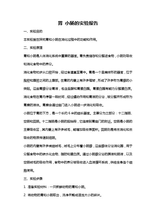
组织学与胚胎学 实验报告指定主题:实验一 、上皮组织 基本组织1报告主题:假复层纤毛柱状上皮 主题属性:指定 标本号:27# 染色:HE 染色教学要求:掌握假复层纤毛柱状上皮的光镜结构特征光镜下可见假复层纤毛柱状上皮的各种上皮细胞界限不清,细胞核染蓝紫色,高矮不一,其中柱状细胞最多,游离面有大量纤毛,柱状细胞之前常夹有杯状细胞,其底部狭窄,顶部膨大,细胞核深染。
杯状细胞 Goblet cell结缔组织 Connective Tissue纤毛 Cilium所有细胞的基底膜均附着在基膜上,即使细胞核高低不齐排列,形似复层,实为单层。
Then visible pseudostratified ciliated columnar epithelium of epithelial cell line is not clear, the nucleus dyed purple blue, height is differ, the columnar cells most, free surface has a lot of ciliated columnar cells, before often clip has goblet cell, its bottom is narrow, the top swollen, with hyperchromatic nuclei. All the cells of the basement membrane are attached on the basement membrane, whose nucleus are irregular, and the shape see as compound layer, and it is a single.实验二 结缔组织 基本组织2报告主题:骨单位 主题属性:指定 标本号:6# 材料:骨切片染色:Schmorl 氏法块染教学要求:掌握骨组织的结构图2骨单位(骨切片,Schmorl 氏法块染,40×100) Figure 2 osteon (osseous smear,Schmorl stained,40×100)骨单位位于内外环骨板之间,数量最多,中央为圆心的中央管,中央管与穿通管相通,数层同心圆排列的骨板围绕中央管。
小肠上皮绒毛实验报告(3篇)
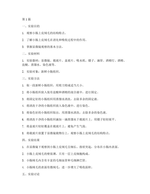
第1篇一、实验目的1. 观察小肠上皮绒毛的结构特点。
2. 了解小肠上皮绒毛在消化和吸收过程中的作用。
3. 掌握显微镜观察的基本方法。
二、实验材料1. 实验器材:显微镜、载玻片、盖玻片、吸水纸、镊子、滴管、酒精灯、酒精、盐酸、蒸馏水、染色液等。
2. 实验对象:新鲜小肠组织。
三、实验方法1. 取一段新鲜小肠组织,用剪刀剪成适当大小。
2. 将小肠组织放入装有盐酸和酒精的混合液中,进行固定。
3. 将固定好的小肠组织用蒸馏水清洗,去除多余的固定液。
4. 将清洗干净的小肠组织放入染色液中,进行染色。
5. 将染色好的小肠组织取出,用蒸馏水清洗,去除多余的染色液。
6. 将清洗干净的小肠组织滴加一滴蒸馏水于载玻片上,用镊子轻轻展平。
7. 将盖玻片轻轻覆盖在载玻片上,避免产生气泡。
8. 将载玻片放置于显微镜载物台上,观察小肠上皮绒毛的结构特点。
四、实验结果1. 在显微镜下观察到小肠上皮绒毛呈细长、指状突起,分布在小肠内表面。
2. 小肠上皮绒毛的壁很薄,只有一层上皮细胞构成。
3. 小肠绒毛内含有丰富的毛细血管和毛细淋巴管。
4. 小肠绒毛的表面有微绒毛,进一步增大了吸收面积。
五、实验讨论1. 小肠上皮绒毛的结构特点有利于消化和吸收。
由于绒毛壁很薄,只有一层上皮细胞构成,有利于营养物质的快速通过。
2. 小肠绒毛内含有丰富的毛细血管和毛细淋巴管,有利于营养物质的吸收和运输。
3. 小肠绒毛的表面有微绒毛,进一步增大了吸收面积,有利于提高消化吸收效率。
六、实验结论1. 小肠上皮绒毛是消化和吸收的主要场所。
2. 小肠上皮绒毛的结构特点有利于消化和吸收营养。
3. 通过观察小肠上皮绒毛,我们可以了解消化系统的结构和功能。
七、实验注意事项1. 实验过程中注意无菌操作,避免污染。
2. 操作显微镜时,保持载玻片和显微镜载物台平稳,避免产生气泡。
3. 观察时,注意调整显微镜的焦距,以获得清晰的图像。
4. 实验过程中,注意观察小肠上皮绒毛的结构特点,并做好记录。
第二节《消化和吸收》知识背景观察小肠绒毛(人教版初一下)
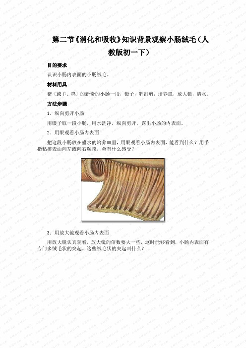
第二节《消化和吸收》知识背景观察小肠绒毛(人
教版初一下)
目的要求
认识小肠内表面的小肠绒毛。
材料用具
猪〔或羊、鸡〕的新奇的小肠一段,镊子,解剖剪,培养皿,放大镜,清水。
方法步骤
1.纵向剪开小肠
用镊子取一段小肠,用水洗净,纵向剪开,露出小肠的内表面。
2.用眼观看小肠内表面
把这段小肠放在盛水的培养皿里,用眼观看小肠内表面,能看到什么?用手指粘摸表面向左或向右触摸,会有什么感受?
3.用放大镜观看小肠内表面
用放大镜认真观看,放大镜的倍数要大一些,这时能够看到,小肠内表面有专门多绒毛状的突起。
这些绒毛状的突起叫什么?
摸索:小肠内表面有许多绒毛状的突起,这种结构关于营养物质的吸取有什么意义?。
观察小肠绒毛
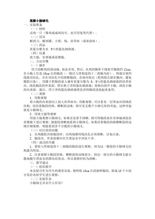
观察小肠绒毛一、实验准备(一)材料活鸡一只(雏鸡或成鸡均可,也可用兔等代替)。
(二)用具解剖刀,解剖镊,大剪,线,培养皿(或表面皿)。
(三)药品质量分数为0.9%的氯化钠溶液。
(四)仪器放大镜,实体镜或显微镜。
二、方法步骤(一)操作用刀切断鸡的颈动脉,放血至死。
然后,从鸡的胸骨下缘剪开腹壁约12cm,在小肠上任选10cm长的肠段(一般以与胃相连的十二指肠为好),两端分别用线做双结扎,并在双结扎中间剪断肠段,从体内取出(把两线头留在腹内,避免腹腔污染),用镊子把肠段放入盛有质量分数为0.9%的氯化钠溶液的培养皿内,清洗肠段的外表面,然后换干净的氯化钠溶液;再纵向剖开小肠,清洗小肠的内表面。
最后,用干净的氯化钠溶液将洗净的肠段浸泡起来备用。
(二)观察1.用眼观察将小肠的内表面向上放人培养皿内,用眼观察,可以看见一层带血丝的绒状结构,而沿着肠段的纵、横断面边缘,则可见无数个小细毛状的突起,这种突起就是小肠绒毛。
2.用放大镜等观察用放大镜观察小肠绒毛,如果还觉着不清晰,则可将肠段放在实体镜或低倍显微镜下进行观察,就能较清晰地看到小肠绒毛。
如果沿着肠段的纵横断面的边缘仔细观察,则能看到各个分散的小肠绒毛。
(三)应注意的问题1.从鸡腹腔内取肠段时,应两端都用线结扎后再剪断,以免污染。
2.肠段内、外表面都应在生理盐水中冲洗干净。
(四)成功的关键1.要取与胃相连的十二指肠的肠段进行观察,因为这一肠段的小肠绒毛结构最为明显。
2.注意观察小肠段的纵、横断面的边缘部分,因这一部分的小肠绒毛能分散地随生理盐水的漂动而晃动,所以观察时较为清晰。
三、教学建议(一)组织教学本实验可作为学生的课堂实验。
教师取10cm长的新鲜肠段,剪成10个小段分发给各组学生进行观察。
(二)实验作业小肠绒毛具有什么作用?。
空肠绒毛实验报告(3篇)
第1篇一、实验目的1. 了解空肠绒毛的结构和功能;2. 掌握观察空肠绒毛的方法;3. 分析空肠绒毛在消化吸收中的作用。
二、实验原理空肠绒毛是肠道上皮的一种特殊结构,由柱状上皮细胞组成,具有吸收营养物质的作用。
通过观察空肠绒毛的结构和功能,可以了解其在消化吸收过程中的重要作用。
三、实验材料1. 新鲜空肠组织;2. 稀盐酸;3. 生理盐水;4. 滴管;5. 显微镜;6. 粘膜切片。
四、实验步骤1. 取新鲜空肠组织,用稀盐酸进行固定;2. 将固定后的空肠组织进行切片,厚度约为5μm;3. 将切片放入生理盐水中浸泡,去除多余水分;4. 将切片置于显微镜载玻片上,用滴管滴加适量生理盐水;5. 使用显微镜观察空肠绒毛的结构和功能;6. 记录观察结果。
五、实验结果与分析1. 空肠绒毛的结构在显微镜下,我们可以观察到空肠绒毛呈指状突起,表面有密集的微绒毛。
绒毛间的柱状上皮细胞排列整齐,细胞核位于细胞中央。
绒毛基部有丰富的毛细血管和淋巴管,有利于营养物质的吸收。
2. 空肠绒毛的功能(1)增大吸收面积:空肠绒毛的指状突起和密集的微绒毛使肠道表面积增大,有利于营养物质的吸收。
(2)加速营养物质吸收:绒毛内的柱状上皮细胞具有丰富的线粒体,为营养物质吸收提供能量。
同时,绒毛内的酶类物质可以分解营养物质,使其更容易被吸收。
(3)促进淋巴液形成:绒毛基部丰富的毛细淋巴管可以将吸收的营养物质转化为淋巴液,有利于营养物质的运输。
3. 空肠绒毛在消化吸收中的作用(1)空肠绒毛可以吸收大部分的营养物质,如葡萄糖、氨基酸、脂肪酸等,为机体提供能量和营养物质。
(2)空肠绒毛可以吸收水、无机盐和维生素等,维持体内水盐平衡和电解质平衡。
(3)空肠绒毛可以吸收药物和毒素,降低其对人体的影响。
六、实验结论通过本次实验,我们成功观察了空肠绒毛的结构和功能,了解了其在消化吸收过程中的重要作用。
空肠绒毛具有增大吸收面积、加速营养物质吸收和促进淋巴液形成等功能,为机体提供能量和营养物质,维持体内水盐平衡和电解质平衡,具有重要的生理意义。
小肠组织观察实验报告(3篇)

第1篇一、实验目的1. 了解小肠的结构特点。
2. 掌握小肠组织的观察方法。
3. 分析小肠组织在消化吸收过程中的功能特点。
二、实验原理小肠是消化系统中最重要的器官之一,其主要功能是消化和吸收营养物质。
小肠壁由黏膜层、黏膜下层、肌层和外膜层组成。
黏膜层具有皱襞和小肠绒毛,增加了消化吸收面积;黏膜下层含有丰富的血管和淋巴管,有利于营养物质的吸收;肌层由平滑肌组成,具有收缩和舒张功能,有助于食物的推进;外膜层则起到保护作用。
三、实验材料与仪器1. 实验材料:新鲜猪小肠、解剖剪、镊子、放大镜、培养皿、清水、盐酸、氢氧化钠、盐酸溶液、氢氧化钠溶液、肾上腺素、乙酰胆碱等。
2. 实验仪器:显微镜、解剖镜、电子天平、温度计、pH计、微机生物信号采集处理系统等。
四、实验步骤1. 准备实验材料:取一段新鲜猪小肠,用水清洗干净,用解剖剪剪成约2cm长的小段,置于培养皿中备用。
2. 观察小肠外表面:用解剖镜观察小肠外表面,记录其颜色、质地等特征。
3. 观察小肠内表面:用解剖剪纵向剪开小肠,露出内表面。
将小肠放在盛有清水的培养皿中,沿剪口翻开。
用手指抚摸内表面,观察是否有滑腻感觉。
4. 观察小肠绒毛:用放大镜仔细观察小肠内表面,观察皱襞和小肠绒毛的分布情况,记录绒毛的形状、大小和数量。
5. 观察小肠绒毛中的毛细血管和毛细淋巴管:用显微镜观察小肠绒毛横切面,观察绒毛壁、毛细血管和毛细淋巴管的分布情况。
6. 观察小肠平滑肌:用显微镜观察小肠横切面,观察肌层和平滑肌的分布情况。
7. 实验验证:(1)观察盐酸对小肠平滑肌的影响:将盐酸溶液滴加在小肠横切面上,观察平滑肌的收缩和舒张情况。
(2)观察氢氧化钠对小肠平滑肌的影响:将氢氧化钠溶液滴加在小肠横切面上,观察平滑肌的收缩和舒张情况。
(3)观察肾上腺素对小肠平滑肌的影响:将肾上腺素滴加在小肠横切面上,观察平滑肌的收缩和舒张情况。
(4)观察乙酰胆碱对小肠平滑肌的影响:将乙酰胆碱滴加在小肠横切面上,观察平滑肌的收缩和舒张情况。
组织胚胎学实验报告
组织学与胚胎学实验报告专业临床五年制班级 2012级8班姓名路海燕学号 201250426电子信箱完成日期 2013.5.24实验一上皮组织报告主题:假复层纤毛柱状上皮主题属性:指定标本号:27#染色:HE材料:豚鼠气管横切片教学要求:掌握假复层纤毛柱状上皮的侧面形态结构杯状细胞柱状细胞纤毛基膜假复层纤毛柱状上皮(豚鼠气管切片,HE染色,40×10)假复层纤毛柱状上皮由柱形细胞、梭形细胞、锥体形细胞和杯状细胞组成。
光镜下杯状细胞最多,游离面有大量纤毛。
所有细胞基底面均附着在基膜上,细胞高矮不一,核的位置高低不齐地排列在不同水平面上,垂直切面观形似复层,实为单层。
此种上皮以保护功能为主。
实验二软骨和骨报告主题:骨单位主题属性:指定标本号:6#染色:Schmorl式染色材料:脱钙骨切片教学要求:掌握光镜下骨组织的结构特点骨陷窝骨单位骨板中央管骨小管骨单位(骨切片,Schmorl氏法块染,40×10)骨单位是长骨起支持作用的主要结构单位,光镜下可见骨单位中央为圆形的中央管,多层骨板围绕中央管呈同心圆排列,骨单位间还有一些不规则的间骨板,骨板之间或骨板中可见可见棕黄色的小腔,为骨陷窝。
骨陷窝之间有细小的骨小管相连通,最内层的骨小管开口于中央管。
实验三肌组织报告主题:心肌主题属性:指定标本号:12#染色:HE染色材料:人心肌切片教学要求:掌握人心肌纤维纵断面和横断面的结构特点闰盘心肌(人心肌切片,HE染色,40×10)心肌分布于心壁和邻近心脏的大血管壁上,光镜下可见心肌纤维走向无一定规则,心肌纤维连接处为闰盘,闰盘染色深。
心肌纤维也呈明暗相间的周期性横纹。
实验四报告主题:多极神经元主题属性:指定标本号:9#染色:HE染色材料:小牛脊髓及脊神经节横切片教学要求:①掌握脊髓前角多极神经元(运动神经元)的形态与结构。
②掌握脊神经节假单极神经元(感觉神经元)的形态与结构。
③掌握有髓神经纤维纵、横断面的结构。
小肠绒毛的实验步骤

小肠绒毛的实验步骤
实验目的:
认识小肠内表面的小肠绒毛。
实验材料:
猪的新鲜的小肠一段、镊子、解剖剪、培养皿、放大镜、清水
实验过程:
一.讲解实验步骤:
1.纵向剪开小肠。
实验台上的培养皿中放有一小段新鲜的猪的小肠。
用镊子取出,将小肠用清水冲洗干净。
用解剖剪将小肠纵向剖开,露出小肠的内表面。
2.用眼观察小肠的内表面。
将剪开的小肠放在盛有清水的培养皿里,内表面向上,用眼观察,能看到小肠内表面并不光滑,毛绒绒的。
用手指在粘膜表面向左或向右抚摸,有粗糙的感觉。
3.用放大镜仔细观察小肠的内表面。
用放大镜仔细观察可以看到:小肠的内表面有许多绒毛状的突起—小肠绒毛
二.学生实验:
提问:小肠绒毛对于完成小肠的功能有什么意义?
(答:增大了消化和吸收的面积。
)
三.整理实验台:将观察过的小肠放入废物杯中,实验器材洗净,放回原处。
小肠解剖观察实验报告(3篇)
第1篇一、实验目的1. 了解小肠的结构特点。
2. 观察小肠内表面皱襞和小肠绒毛。
3. 掌握小肠解剖观察的实验方法。
二、实验原理小肠是人体消化系统的重要组成部分,具有消化和吸收食物的功能。
小肠内表面有皱襞和小肠绒毛,增大了消化吸收的面积,有利于营养物质的吸收。
三、实验材料1. 猪的新鲜小肠一段2. 解剖剪3. 镊子4. 放大镜5. 培养皿6. 清水四、实验步骤1. 将猪的新鲜小肠用解剖剪纵向剪开,露出小肠内表面。
2. 将小肠放在盛有清水的培养皿中,观察小肠的外形。
3. 用手指抚摸小肠内表面,感受其滑腻的感觉。
4. 用放大镜仔细观察小肠内表面,观察皱襞和小肠绒毛的存在。
5. 记录观察到的结构特点。
五、实验结果与分析1. 小肠外形呈管状,两端封闭,中间开口。
2. 小肠内表面有环形皱襞,皱襞表面有许多绒毛状突起,即为小肠绒毛。
3. 皱襞和小肠绒毛的存在增大了小肠的表面积,有利于营养物质的吸收。
六、实验结论通过本次实验,我们观察到了小肠的结构特点,了解到小肠内表面皱襞和小肠绒毛的存在及其作用。
小肠的皱襞和小肠绒毛增大了消化吸收的面积,有利于营养物质的吸收,体现了结构与功能相适应的生物学观点。
七、实验心得1. 通过本次实验,我对小肠的结构和功能有了更深入的了解。
2. 实验过程中,我学会了如何使用解剖剪、镊子、放大镜等实验器材,提高了自己的实验操作能力。
3. 在实验过程中,我明白了观察细节的重要性,只有仔细观察,才能发现生物体的结构与功能之间的关系。
八、实验注意事项1. 实验过程中,注意安全,避免使用解剖剪等尖锐物品划伤自己。
2. 实验过程中,保持实验台整洁,避免污染实验器材。
3. 实验结束后,将实验器材洗净,放回原处。
九、实验拓展1. 研究不同动物的小肠结构特点及其功能差异。
2. 探讨小肠疾病对消化吸收功能的影响。
3. 研究小肠黏膜的再生机制及其应用。
第2篇实验名称:小肠解剖观察实验实验时间:2023年10月25日实验地点:生物实验室实验目的:1. 观察小肠的结构,了解小肠的形态特点。
实验报告空肠实验分析
实验报告空肠实验分析空肠实验是一种常用的实验方法,在医学和生物领域中被广泛应用。
本实验主要通过对动物的空肠进行实验,以研究空肠的结构、功能和生理过程。
第一部分:实验目的和背景实验的目的是通过观察和分析空肠的结构、功能和生理过程,进一步了解空肠的作用和机制。
空肠是人体消化吸收的重要器官之一,长度约为3-5米。
它是由许多细小的绒毛组成的,这些绒毛上存在着上皮细胞。
空肠上皮细胞对营养物质和水分的吸收起着重要作用。
此外,空肠还有许多特殊的结构,如皱褶、肠梗阻和巨大的周期运动等。
这些结构和功能与空肠的吸收功能密切相关。
第二部分:实验方法和步骤实验前,我们需要准备好实验所需的材料和设备,如动物样本(小鼠或大鼠)、显微镜、取样器等。
具体实验步骤如下:1. 麻醉动物:将动物用适当的方式麻醉,如给小鼠或大鼠注射麻醉剂。
2. 打开动物腹腔:在麻醉后,使用手术刀或剪刀打开动物的腹腔,暴露出空肠。
3. 取样:使用取样器沿着空肠的长度,取出几个不同位置的组织样本。
4. 准备显微镜片:将取样的组织样本放在显微镜片上,用草酸水溶液进行固定。
5. 显微镜观察:将显微镜片放在显微镜下,使用适当的放大倍数观察样本,记录空肠的结构和组织细胞的特征。
6. 数据分析:根据观察结果,分析空肠的结构特点、绒毛密度和绒毛高度等指标。
第三部分:实验结果和分析通过显微镜观察,我们发现空肠呈现出细长的管状结构。
绒毛在空肠上皮细胞表面呈现出类似指状的形态,增加了其表面积,方便吸收。
绒毛密度和绒毛高度的变化也表明不同区域的营养吸收能力不同。
此外,我们还发现空肠上还存在着一些特殊结构,如皱褶和肠梗阻。
这些结构的存在增加了空肠的表面积,并进一步提高了吸收能力。
根据实验结果,我们可以得出以下结论:1. 空肠是一种重要的消化吸收器官,通过绒毛的存在增加了其表面积,方便吸收。
2. 空肠绒毛密度和绒毛高度的变化表明不同区域的吸收能力不同。
3. 空肠的皱褶和肠梗阻结构增加了其表面积,进一步提高了吸收能力。
画空肠的实验报告
一、实验目的1. 熟悉空肠的解剖结构;2. 掌握空肠的画法;3. 培养学生的动手能力和观察力。
二、实验原理空肠是消化系统的重要组成部分,具有消化和吸收营养物质的功能。
本实验通过观察空肠的解剖结构,了解其形态、位置和功能,并通过绘画技巧将其表现出来。
三、实验材料1. 实验动物尸体;2. 解剖器械;3. 绘画工具(铅笔、橡皮、彩色铅笔等);4. 解剖图谱。
四、实验步骤1. 观察空肠的整体结构:首先观察空肠的整体形态,了解其位置、长度和直径等特征。
2. 解剖空肠:使用解剖器械将空肠从动物体内取出,并观察其内部结构。
3. 绘制空肠轮廓:在纸上用铅笔轻轻勾勒出空肠的轮廓,注意保持其形状和大小。
4. 细节描绘:用彩色铅笔填充空肠的内部结构,如血管、淋巴管等。
同时,描绘空肠与周围器官的连接关系。
5. 完善作品:检查绘画作品,修正不足之处,确保作品符合实验要求。
五、实验结果与分析1. 空肠的位置:空肠位于小肠的中间部分,呈螺旋状排列,前端与十二指肠相连,后端与回肠相连。
2. 空肠的形态:空肠壁较厚,内壁有环形皱襞,皱襞上有绒毛,有利于营养物质的吸收。
3. 空肠的功能:空肠是消化系统中重要的吸收器官,主要吸收水分、葡萄糖、氨基酸、维生素等营养物质。
4. 绘画作品分析:通过绘画作品,可以看出空肠的整体结构、内部结构和与周围器官的连接关系,达到了实验目的。
六、实验总结1. 本实验通过观察空肠的解剖结构,掌握了空肠的位置、形态和功能。
2. 通过绘画技巧,将空肠的表现形式固定下来,提高了学生的动手能力和观察力。
3. 实验过程中,应注意解剖器械的使用,避免损伤实验材料。
4. 绘画过程中,要注意保持作品的整体美观,突出空肠的特点。
5. 本次实验取得了圆满成功,达到了预期目的。
空肠绒毛
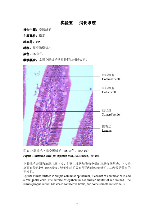
实验五 消化系统
报告主题:空肠绒毛
主题属性:指定
标本号:19#
材料:猫空肠横切片
染色:HE 染色
教学要求:掌握空肠绒毛结构特征与判断依据。
图5 小肠绒毛(猫空肠绒毛,HE 染色,40×10)
Figure 1 intestine villi (cat jejunum villi, HE stained, 40×10)
空肠绒毛表面为单层柱状上皮,主要由柱状细胞和少量的杯状细胞组成。
上皮游离面有染色较红的纹状缘。
绒毛中轴的固有层为细密结缔组织,其内常见散在的平滑肌。
Jejunal villous surface is simple columnar epithelium, it consist of columnar cells and a few goblet cells. The surface of epithelium has striated border of red stained. The lamina propria in villi has dense connective tissue, and some smooth muscle cells.。
教具小肠绒毛结构模型制作说明讲解学习
教具小肠绒毛结构模型制作说明
仅供学习与交流,如有侵权请联系网站删除 谢谢2
教具小肠绒毛结构模型制作说明
制作人:
1.用两张A4纸分别沿纵向对折并用小刀割开,将其中两小张用胶水对粘在一起,其面积就接近一张A4纸,将这一张白纸折成扇子状,作为小肠的内层皱襞。
另以一张A4的一半作为小肠的外表面。
2.用适量一段毛线充当小肠绒毛贯穿于纸的褶皱部分。
3.这样就形成内表面积和外表面积的大小对比了。
特点:通过材料使用前和使用后的对比,可以直观的看出小肠的特殊结构──绒毛和皱襞使消化和吸收的面积增加。
这样就可以给学生一个直观的感性认识,解决教学上的重点和难点。
仅供学习与交流,如有侵权请联系网站删除谢谢3。
- 1、下载文档前请自行甄别文档内容的完整性,平台不提供额外的编辑、内容补充、找答案等附加服务。
- 2、"仅部分预览"的文档,不可在线预览部分如存在完整性等问题,可反馈申请退款(可完整预览的文档不适用该条件!)。
- 3、如文档侵犯您的权益,请联系客服反馈,我们会尽快为您处理(人工客服工作时间:9:00-18:30)。
实验五 空肠绒毛 报告主题:空肠绒毛
主题属性:定选
标本号:19#
材料:猫空肠
染色:HE 染色
教学要求:
掌握空肠绒毛的结构
图五 猫空肠绒毛(猫空肠,HE 染色,40×10)
Fig five cat jejunum villus (cat jejunum, stained with HE, 40 × 10) 上皮和固有层共同向肠腔突出形成小肠绒毛,绒毛的表面为单层柱状上皮细胞,主要有柱状细胞和少量的杯状细胞,上皮游离面有染色较红色的纹状缘,绒毛的中轴的固有层为细密结缔组织,其内常可见散在纵行的平滑肌,丰富毛细血管,1~2条中央乳糜管及大量的淋巴细胞The epithelium and lamina propria is common to the intestinal cavity with prominent formation of intestinal villi, villous surface of columnar epithelial cells, mainly with columnar cells and a small number of goblet 纹状缘Striated border
杯状细胞Goblet cell 柱状细胞Columnar cell
平滑肌细胞Smooth muscle cells
淋巴细胞Lymphocyte
cells, epithelial free surface stained more red striated border, villus axis of the lamina propria is a dense connective tissue, which often can be seen scattered in longitudinal smooth, rich in capillaries, 1~2 central lacteal and large numbers of lymphocytes。