蔡司VISU200p高级眼科手术显微镜
ZEISS OPMI LUMERA 700 眼科手术微观电子显微镜说明书

ZEISS OPMI LUMERA 700 Seeing to succeedPart of the ZEISS Cataract Suite O C T an d m a r ke r l e s si n o neSeeing to succeed. ZEISS OPMI LUMERA 7002What drives a surgeon? A commitment to preservingand restoring patients’ sight – to saving vision.We share your dedication.One example is with the OPMI LUMERA® 700 from ZEISS, an operating microscope ideally suited for every ophthalmic surgery speciality. Experience markerless IOL alignment and integrated intraoperative OCT* imaging – all in one device. ZEISS OPMI LUMERA 700 – our commitmentto helping you see to succeed.3*ZEISS RESCAN 7004With ZEISS CALLISTO eye markerless alignment,manual marking steps can be skipped altogether foran efficient and precise* toric IOL alignmentto reduce residual astigmatism.For cataract surgeries, ZEISS OPMI LUMERA 700,with its well-known patented SCI illumination,ZEISS optics and CALLISTO eye ® from ZEISS, provides thebest anterior views and precise* assistance functions.Seeing to succeed in cataract surgeryPrecise* and efficient** markerless toric IOL alignmentI save 6 minutes per patient and improve alignment precision by 40% compared to manual marking.Wolfgang Mayer, MD, Augenklinik der Universität München, Germany »»Part of theZEISS Cataract SuiteConnecting theCataract Workflow* V IROS research team of Prof. Findl: Clinical data of Dr. Varsits "Deviation between the postoperative (at the end of surgery in the operating room) and aimed IOL axes was 0.52 degrees± 0.56 (SD)" published in J Cataract Refract Surg 2019; 45:1234–1238 and Clinical data of Dr. Hirnschall presented at ESCRS 2013.** C linical data of Dr. Mayer: "Toric IOL implantation was significantly faster using digital marking" published in J Cataract Refract Surg 2017; 43:1281–1286.5Cataract assistance functions for every step of the surgeryThe assistance functions of ZEISS CALLISTO eye are completely surgeon-controlled – with either the foot control panel or handgrips.CALLISTO eye informationOPMI LUMERA 700 parametersEfficient markerless IOL alignmentStarting with a biometry referenceimage from the IOLMaster ® from ZEISS,data is transferred smoothly to ZEISSCALLISTO eye. This data is used to createoverlays in the eyepiece. Save time,increase efficiency and reduce residualastigmatism when you:• skip manual preoperative marking• skip manual data transfer• skip manual intraoperative marking Efficient surgery setup The image quality check supports you to optimize light intensity, magnification and centration of the microscope to efficiently set up the reference axis. The well-proven* eye tracking automatically compensates for eye movements and supports the use of the assistance functions.» C ALLISTO eye enabled easy and exacttoric IOL alignment in all cases.«Prof. Findl,VIROS, Hanusch Hospital, Vienna, AustriaLRI Perform limbal relaxing incisionsZ ALIGN ®Perform toric IOL centration on the visualaxis provided by the ZEISS IOLMaster andperform rotational alignment Incision Position incisions, optionally on the steep axis; add opposite clear cornea incision and paracentesesRhexis Precisely* size and shape capsulorhexisand align the IOL on the visual axisprovided by the ZEISS IOLMasterSeeing to succeed in glaucoma surgery Improved visualizationAs minimally invasive glaucoma surgery (MIGS) and canaloplasty procedures evolve, intraoperative OCT plays an increasingly important role in difficult to see spaces. The integrated intraoperative OCT* images of the ZEISS OPMI LUMERA 700 aids the visualization of the device placement.6*ZEISS RESCAN 700More information to supportyour decisions during surgery Integrated intraoperative OCT*allows visualization of orientationand placement of the MIGS implant. Intraoperative OCT* images enable physicians to visualize detailed structures in the natural physiological shape.Stay focused on thearea of interestSave time by maintaining the selected intraoperative OCT* scan locationwith the new automatic XY tracker. In addition to the proven Z tracker, the XY tracker compensates for movements of the eye or the microscope.Protect the retinaShield the retina from excessive light exposure with the integrated retina protection filter.Flexible perspective fora better viewTilt the microscope head as needed to better observe the iridocorneal angle.Intraoperative OCT gives me better control in modern glaucoma surgery through visualization of MIGS and canaloplasty.Hagen Thieme, MD, Otto-von-Guericke-Universität Magdeburg, GermanyVerify the position and function of innovative glaucoma drainage devices (e.g. stents)*ZEISS RESCAN 70078Seeing to succeed in cornea surgeryReduce graft manipulationA presentation of Dr. Alain Saad, et al, clinical data wasshown at AAO 2015 comparing severe corneal edema casesof 13 eyes supported by intraoperative OCT* (ZEISS) and 15without. The conclusion showed that there was reduced cellloss using intraoperative OCT*. Literature has shown thatZEISS intraoperative OCT* can provide valuable anatomicalinformation and help with surgical decision making**.The integrated intraoperative OCT* of the ZEISS OPMI LUMERA 700 visualizes the actual physiological shape of the cornea intwo different scan views. Switch between views with a touch of the finger or tap of the foot to make your decisions faster.Make faster decisions with two scan depths and a realistic view.Quickly change between high-resolution OCT scans (2.9 mm scan depth in tissue) and large overview images (5.8 mm scan depth in tissue) to visualize and assess graft orientation.Observe the natural physiological shape of the cornea with distortion-free intraoperative OCT* images. See how intuitive OCT image navigation is duringsurgery.OCT* image of graft orientation during DMEK surgery in the ocularSee the graft orientation without manipulation in DMEK surgery with intraoperative OCT*ZEISS RESCAN 700DMEK: save time with easygraft monitoringMonitor the graft orientation andassess the interface with the patient´scornea. Verify proper graft positioning as well as visualize fluid interface and graft adherence.DALK: secure big-bubble procedure OCT* imaging helps the surgeon during DALK to assess the dissection depthin order to reduce perforation risk and9** C linical data of Cost B, Goshe JM, Srivastava S, Ehlers JP published in Am J Ophthalmol. 2015 Sep; Intraoperative optical coherence tomography-assisted descemet membrane endothelial keratoplasty in the DISCOVER study.***Not available in combination with intraoperative OCT.I reduced graft manipulation time by 4.2 minutes during DMEK.*Alain A. Saad, MD, Fondation Rothschild, Paris, France»»potentially improve the reproducibilityof the big-bubble procedure.Full integration forincreased efficiencyThe integrated slit illuminator***provides four slit widths with left-rightslit movement to simplify observation ofthe cornea and anterior chamber –withoutthe hassle of fitting extra accessories.*ZEISS RESCAN 70010Seeing to succeed in retina surgery Make more informed decisionsSuperb OCT images forinformed decisionsIntegrated intraoperative OCT* addsa real-time third dimension tovisualization capabilities for viewingtransparent structures of the eyeduring surgery.Monitor the surgical progress and makedecisions accordingly. The superb clarityof the intraoperative OCT* images canprovide unexpected insights, allowingstrategy adjustments during surgery.With innovative technologies such as integrated intraoperative OCT* and the non-contact fundus viewing system RESIGHT ® 700 from ZEISS, the ZEISS OPMI LUMERA 700 gives new meaning to “insight” when performing retina surgery procedures.Intraoperative OCT* revealed undetected macular holes after peeling in 10% of highly myopic eyes.Ramin Tadayoni, MD, PhD, University of Paris VII, Sorbonne Paris Cité, Paris, France »»*ZEISS RESCAN 70011Keep your focusThe new automatic XY tracker, in addition to the proven Z tracker, compensates for movements of the eye or the microscope, saving time by maintaining the selected intraoperative OCT* scan plete your surgery with confidenceVerify that all necessary membrane residue has been completely removed following ILM peelings with OCT* imaging. Detect macular holes that might easily be overlooked and monitor vitreomacular traction.128D wide-field lensFor peripheral visualization and a clear overview during vitrectomy60D macular lensFor high magnification of the maculaSee the retina in more detail The proven non-contact retina visualization system ZEISS RESIGHT 700provides a clear, detailed view of the retina. Varioscope optics from ZEISS enable surgeons to stay fully focused on the area of interest. Switch magnification quickly with the two aspheric lenses. It is also possible to use a direct or indirect contact glass.With the ZEISS RESIGHT 700, the surgical microscope automatically adjusts the camera settings, Invertertube E settings, lighting and speed of motion to the preset values for retina surgery.*ZEISS RESCAN 70012Seeing to succeed in teaching Share your knowledge• Integrated intraoperative OCT* that provides a clearer image of what is happening during surgery• Integrated assistant scope with independent magnification, which can be linked to the main microscope for teaching purposes • ZEISS CALLISTO eye cockpit to observe and share informationZEISS OPMI LUMERA 700 features excellent tools for enhancing the learning experience. Students need to see every detail to have a clear understanding of the surgical process. Whether during surgery, viewing through the assistant scope orreviewing post-surgery, it is important to provide images with excellent contrast, color, and high resolution.The optical performance from ZEISS enables students to see deep into the ophthalmic world using:More documentation – faster Video documentation is important for recordkeeping and for teaching. Simply insert a USB device to document the cockpit view, assistance functions and intraoperative OCT* images in HD quality. ZEISS CALLISTO eye, together with a data management system such as FORUM ® from ZEISS, records themicroscope live image on both the internal hard drive and the external USB drive simultaneously to avoid time-consuming video exports.13All details are available for you and your students The ZEISS CALLISTO eye cockpit provides even more information for surgery and teaching. Both the doctor and the student can now view data in the eyepiece, from all connected devices, shown on the ZEISS CALLISTO eye screen or from recorded video.Your students can clearly follow the surgery to unblock the Schlemm's canal.Technical dataOPMI LUMERA 700 from ZEISSZEISS OPMI LUMERA 700Surgical microscope Motorized zoom system with apochromatic lens, zoom ratio 1:6Magnification factor = 0.4 x – 2.4 xFocusing: electric / motorized, focus range: 70 mmObjective lens: f = 200 mm (optionally also f = 175 mm or f = 225 mm with support ring)Binocular tube: Invertertube E (optionally also Invertertube, 180° swivel tube,f = 170 mm, inclined tube, f = 170 mm)Wide-angle eyepiece 10 x (optionally also 12.5 x)Light source SCI: Coaxial and full-field illuminationFiber-optic illumination Superlux® Eye:• Xenon short arc reflector lamp with HaMode filter• Backup lamp in lamp housing, can be slid into position manuallyLED fiber-optic illumination:• Near-daylight color temperature• 50,000 hour lifetime at 50% light intensity• HaMode filter• 25% gray filterFor all light sources:• Blue blocking filter• Optional: Fluorescence filterIntegrated slit illuminator Slit widths: 0.2 mm, 2 mm, 3 mm, 4 mm Slit height: 12 mm14XY coupling Travel range: max. 61 mm x 61 mmAutomatic centering at the touch of a buttonVideo monitor22" LCD displayResolution: 1,680 x 1,050Stand Maximum permissible weight load of the spring arm:When the surgical microscope is attached to the arm (without tube, eyepiece or objective lens)and the XY coupling is also attached, a maximum of 9 kg of additional accessories can beattached to the spring armZEISS intraoperative OCTOCT engine SD (spectral domain) OCTWavelength 840 nmScanning speed 27,000 A-scans per secondScan parameters A-scan depth: 2.9 and 5.8 mm in tissueAxial resolution: 5.5 µm in tissueScan length adjustable 3–16 mmScan rotation adjustable 360°Scan modes for live and capture acquisitionLive: • 1-line Capture: • 1-line• 5-lines • 5-lines• cross hair • cubeZEISS RESIGHT familyMechanical data Focus range with LH175 lens holder: 31 mm (position of intermediate image)Focus range with LH200 lens holder: 38 mm (position of intermediate image)Rotation angle of lens revolver and holder: 0°–360°Lenses included60D, 128DWeight ZEISS RESIGHT 500 (manual): 0.45 kgZEISS RESIGHT 700 (motorized): 0.50 kgZEISS CALLISTO eye panel PCTouch screen Projected Capacitive Touch (PCT) with anti-reflective coating, scratch-proofProcessor Intel® Core i5 6442EQ 1.9 GHzHard drive SSD for operating system, SATA HDD 1 TB for dataDisplay Integrated 24" color flat screen with high luminosity and wide viewing angleVideo signals PAL 576i50; NTSC 480i60; 1080i50; 1080i60Only possible with camera models from Carl Zeiss Meditec AGPorts 1 × CAN-Bus, 2 × 1 Gigabit Ethernet, 5 × USB 3.0, 1 × potential equalizationVideo input 1 × Y/C, 1 × HD-SDIVideo output 2 × HDMIConnectivity Integrated RJ45 10/100Base-T Ethernet port for connection to ZEISS OPMI LUMERA 700 and hospitalnetworkWeight ca. 10 kgZEISS CALLISTO eye softwareVersion 3.7, 3.615The statements of the healthcare professional giving this presentation reflect only his personal opinions and experiences and do not necessarily reflect the opinions of any institution with whom they are affiliated.The healthcare professional giving this presentation may have a contractual relationship with Carl Zeiss Meditec, AG., and may have received financial compensation.OPMI LUMERA 700 RESIGHT 700 CALLISTO eye Panel PC RESCAN 700 CALLISTO eye Software 0297Carl Zeiss Meditec AG Goeschwitzer Strasse 51–52 07745 JenaGermany/lumera /med/contactsSUR.12PrintedintheUnitedStates.CZ-II/22UnitedStatesedition:Onlyforsaleinselectedcountries.Thecontentsofthebrochuremaydifferfromthecurrentstatusofapprovaloftheproductorserviceofferinginyourcountry.Pleasecontactourregionalrepresentativeformoreinformation.Subjecttochangeindesignandscopeofdeliveryandduetoongoingtechnicaldevelopment.OPMILUMERA,RESIGHT,CALLISTOeye,RESCAN,andZALIGNareeithertrademarksorregisteredtrademarksofCarlZeissMeditecAGorothercompaniesoftheZEISSGroupinGermanyand/orothercountries.©CarlZeissMeditec,Inc.,22.Allrightsreserved.Carl Zeiss Meditec, Inc.5160 Hacienda DriveDublin, CA 94568USA/us/med。
蔡司显微镜产品及附件汇总

蔡司显微镜产品及附件产品型号:Pro 580ES技术参数:像素:580万像素像素大小:4.65μ(H) x 4.65μ(V)靶面:彩色冷CCD采集分辨率:2776x2074 1392x1040 640x480 灵敏度:0.05lux最大曝光时间:64 minutes色彩深度:24 or 48-bit RGB白平衡:自动白平衡手动曝光:自动曝光、手动曝光快门速度:1/10000 sec. to 60 sec采集速度:最大15fps文件格式:BMP, JPEG, TIFF, DIB, and FlashPix产品名称:研究级正立智能数字万能材料显微镜Axio Imager A2m技术参数:1、物镜倍数:5X 10X 20X 50X 100X 可选1.25X 2.5X 150X2、目镜倍数:10X 16X 25X3、视场数:23、254、放大倍数:12.5X-6000X(选配)5、物镜转盘:6-7孔6、观察功能:明场★高级暗场★圆偏光微分干涉荧光7、光源: 12V100W卤素灯,智能化光路管理器,光强自动可调8、可扩展性:可配图像分析系统(数码相机、摄像头、图像分析软件)9、可配显微硬度附件10、可配透射光附件注:带★为蔡司公司独有专利技术,未向任何公司转让。
全新一代智能型正立式显微镜Axio Imager A2m是成功的Axio Imager A1m的全新升级产品,在Axio Imager A1m卓越的产品性能基础上,对光路设计尤其是照明系统进行了全新的升级,将光学系统的优化发挥到了极致,展现给您无微不至的细节和最锐利的显微图像。
Axio Imager A2m 的诞生给显微镜提出了全新的标准,将蔡司的显微技术又一次推向了巅峰。
凭借Axio Imager ,蔡司再次向世界证明了其全球技术先锋的领导地位!性能描述:高精度新标准ICCS物镜--让更多细节呈现在您眼前卡尔·蔡司专为 Axio Imager 重新设计并系统地扩充了高性能物镜系列。
ZEISS ARTEVO 800数字眼科显微镜说明书
25% better
light transmission 3
Dramatically reduced light intensity Provide your patients with more comfort during surgery. The DigitalOptics of the ZEISS ARTEVO 800 offers the choice to operate at reduced light intensity – while an uncompromised level of brightness is maintained.
Jean-François Korobelnik, MD CHU de Bordeaux, France
4
25% higher
resolution 1
Excellent resolution and natural colors Experience natural colors when you view the surgical field. The optical system is optimized to produce remarkably clear images.
Khalid Al-Sabti, MD, FRCS Kuwait Specialized Eye Center, Kuwait
ZEISS ARTEVO 800
Cloud Connectivity allows surgeons to access essential information no matter where they are – and when they want. So whether it’s in the OR during surgery, in the office reviewing a procedure, or in the classroom with students, Cloud Connectivity ensures the data is within easy reach and improves your workflow.
神经外科手术显微镜参数

神经外科手术显微镜参数一、神经外科手术显微镜技术参数要求及基本配置1主镜系统:1.1 主镜系统:双人四目,适用于显微神经外科手术。
1.2 全复消色差光学系统,高分辨率,大景深,保证最佳对比度和清晰度。
*1.3为满足临床科研及教学需求,徕卡公司应提供带有融合光学设计的显微镜技术的机型。
蔡司公司应提供带有Deep View景深增强功能的显微镜技术的机型。
其他厂家应提供最高端技术。
请提供进口医疗器械注册证、产品注册登记表及各类必要证件(书),全套设备必须为全新,以CFDA注册证为准。
1.4 机器人式一键自动平衡系统。
1.5无级连续变倍,变倍比1:6或6:1,也可手动式调节。
*1.6放大倍率(10×目镜下):最小放大倍率≤1.4×,最大放大倍率≥12×,电动/手动连续可调。
(附产品彩页证明)1.7目镜屈光补偿:最大值≥+5D 最小值≤-5D。
1.8单一连续可调物镜工作距离:最小工作距离≤225 mm,最大工作距离≥490mm。
1.9自动照明光栅:随放大倍数增加而自动缩小照明光圈,保护病人暴露的脑组织,并适宜深部、狭窄腔隙手术需要。
1.10自动光强度保护功能:随焦距的减少光强度自动锁定在安全线以内,光亮度和光强度成正比关系,防止灼伤病人组织。
2助手镜2.1 配多通道分光器2.2 配置双目助手镜,倾角可调3手柄3.1多功能手柄:左、右各1个。
3.2手柄可编程,电动连续调焦、电动连续变倍、释放电磁锁、调整光亮度、影像、X/Y 平移、荧光开关。
4光源4.1氙灯照明 :主光源与备用光源均为≥300W,4.2主备用光源具备一键切换功能。
4.3触摸屏可现实先等使用时间。
开放式结构,便于未来进行软件开发。
4.4双光路照明技术,辅助光源照明,减少深部手术时的阴影,保证在锁孔手术中对深部区域的照明更清晰。
5支架5.1六关节电磁锁落地式支架,可做二级解锁。
5.2原厂内置高清数码影像工作站系统,摄像头内置于显微镜光学头,无外置摄像适配器:可完成影像记录,储存,刻录,传输,配置内置式原厂高清影像工作。
ZEISS OPMI pico 诊断微镜说明书

/OPMI-picoZEISS OPMI picoSeeing without compromise.S 100 / O P M I p i c o23ZEISS OPMI picoSimple, compact, provenOPMI ® pico from ZEISS, the most frequently sold ZEISS surgical microscope worldwide, is the trusted choice among doctors. It makes details and fine structures clearly visible. The system enables you to better visualize regions of interest and consistently provide your patients with high-quality examinations and treatments.ZEISS OPMI pico’s cost-effective LED illumination meets your standards and needs. The complete integration of the light source makes it easier to clean the system.Restorative dentistry Quickly detect enamel and dentine fractures as well as approximal caries. High-precision views enable accurate assessment of crown edges, preparation levels, and veneers.EndodonticsVisualize fine anatomical structures and details of root canals and isthmuses for a clear view right down to the apex.Implantology Conduct high-precision examinations and implant treatments quickly and confidently. Reliably detect important anatomical structures.Periodontics Benefit of support for soft-tissue evaluation and management to assist healing, low scarring, and improved cosmetic outcomes.The compact and easy-to-use ZEISS OPMI pico offers solid support for even the most demanding applications – whether in restorative dentistry, endodontics, implantology, or periodontics:Ergonomic seating enables users to conveniently maintain an upright working posture.ZEISS OPMI pico also features camera options to facilitate patient consultation and documentation, depending on thespecific needs in your practice.4Better viewLong-lasting LEDFor color rendition and light that strongly resembles natural daylight. It is a very cost-effective and virtually maintenance-free light source. The low heat output requires minimal ventilation and generates little noise.Full overview of fine detailsZEISS OPMI pico delivers high-quality images at every magnification level.ZEISS OPMI pico enables you to visualize high-contrast, true-color images – the key to improving the quality of examination and treatment. With ZEISS OPMI pico, optics and illumination go hand in hand.View the whole mouth The Varioskop ® 100 objective lens allows you to adjust the focal length over a range of 200 mm to 300 mm to focus on the whole oral cavity in the vertical axis – without moving themicroscope.5While treating your patients you can sit comfortably in an upright, ergonomically correct position for a more relaxed working day.Comfortable working conditions and improved visionOver 75% of dentists found that using a dental microscope had a positive effect on their neck and back pain.5 Working in a comfortable posture allows you to fully focus on your patient and their procedure, and increases your productivity.6A dental microscope can offer other ergonomic benefits, such as improved vision and reduced eye fatigue, common in dental professionals and corresponding to increasing age.7–9Better ergonomicsAdjust the ZEISS OPMI pico to your demands and not vice versa Reduce or increase the distance for the required treatment field as needed with the patented multi-link design of Foldable Tube f170 / f 260. With its long reach, this highly flexible system easily accommodates the needs of the user and different positions of the patient.6Better integrationBenefits of an integrated full HD camera • Complete function integration • Co-observation and documentation • Simplified cleaning of the housing • Images and videos recorded onto a shared network drive or a USB device• Capture of full HD images during recording or from a recorded videoBenefits of live viewing and streamings • Video livestream into the network• Smart Recording: retroactively record video that occurred in the previous 30 secondsComplete integration of technology and design in the suspension arm for a well-balanced architectureFunctional elements such as the video control console, HD video camera, cables, light sources, and light guides are completely integrated into the stand to avoid workspace clutter.Better educationReal-life pictures are very convincing. ZEISS OPMI pico can accommodate a full HD video camera with recording and streaming, allowing you to present patients with high-definition material to explain procedures.A highly compact instrument with a small footprint, ZEISS OPMI pico fits seamlessly into virtually any practice workflow environment. Simply plug in the cable, switch on the power, and the ZEISS OPMI pico isready for use.7Basic configurationPackage optionsAdd-onsSuspension system optionsFloor standCeiling mountWall mount (wallplate)Workplace integrationFurther space-saving suspension system options are available for the Centro carrier system and specified treatment units.Carl Zeiss Meditec AG Goeschwitzer Strasse 51–52 07745 JenaGermany/opmi-pico /med/contacts en-INT_3_1_12IPrintedinGermany.CZ-II/223Internationaledition:Onlyforsaleinselectedcountries.Thecontentsofthebrochuremaydifferfromthecurrentstatusofapprovaloftheproductorserviceofferinginyourcountry.Pleasecontactourregionalrepresentativesformoreinformation.Subjecttochangesindesignandscopeofdeliveryandduetoongoingtechnicaldevelopment.OPMI,VarioskopandVisionGuardareeithertrademarksorregisteredtrademarksofCarlZeissMeditecAGorothercompaniesoftheZEISSGroupinGermanyand/orothercountries.©CarlZeissMeditecAG,223.Allrightsreserved.Application images courtesy of:1 Dr. Claudia Cia Worschech, Sao Paulo, Brazil2 Dr. José Aranguren Cangas, Madrid, Spain3 Dr. Behnam Shakibaie, Tehran, Iran4 Dr. Rino Burkhardt, Zurich, Switzerland5Z augg B et al. Influence of magnification tools on the recognition of artificaila preparation and restoration defects (in German).Schweiz Monatsschr Zahnmed 2004;114:890-896. [Abstract]6L inger W. Advantages for patients under the dental microscope.Available from: https:///blog/dental-microscope7P errin P, et al. Visual acuities of dentists in their respective clinical conditions. Clin Oral Investig 2014;19:2055-2058.8E ichenberger M, et al. Visual acuity of dentists under simulated clinical conditions. Clin Oral Investig 2013;17:725-729.9Y adav VS, et al. Periodontal microsurgery: Reaching new heights of precision.J Indian Soc Periodontol 2018;22(1):5-11.。
蔡司 kinevo 900 手术显微镜使用说明书
/us/kinevoZEISS KINEVO 900Mastering the complex.• Surgeon-Controlled Robotics • Digital Hybrid Visualization • ZEISS QEVO – The Micro-Inspection ToolZEISS KINEVO 900The Robotic Visualization SystemJust like you, we love challenging the status quo.The result? Over 100 innovations to perfect the already acclaimed surgical visualization platform. KINEVO® 900 from ZEISS is designed to deliver more functionalities than any surgical microscope today. ZEISS KINEVO 900 combines digital and optical visualization modalities, offers a unique Micro-Inspection Tool and will impress you with its Surgeon-Controlled Robotics. All to enable you to gain greater certainty in a virtually disruption-free workflow.Designed to meet real needs. To make a real difference!A lot more. And, a lot less too.When treating complex vascular conditions, you typically work at high magnification. Even the slightest vibrations can cause disruptions. And constant manual repositioning to better visualize structures or precisely approach deep-seated lesions can become extremely tedious. Not anymore! ZEISS KINEVO 900 delivers a lot more positioning precision with a lot less effort.PointLockSurgeon-Controlled Robotics adds a complete new level of ease to precise positioning. Imagine being able to focus and move around a structure to visualize the targeted anatomy – reducing any manual hassle. In addition, PointLock enables you to do a KeyHole movement to observe a larger area inside a cavity – a particular benefit in areas with narrow access. Simply put:Focus. Activate. Swivel.Active vibration dampingYou know the problems that can be created by the tiniest vibrations. The active damping provided by ZEISS KINEVO 900 minimizes collateral system vibrations, ensuring rock-solid stability. Enabling you to completely, and steadily, focus on what matters most: your treatment.Focus Activate Swivel5When you need it. Where you need it. The new navigation interface of ZEISS KINEVO 900 is designed to work in concert with your navigation device. When you require precise repositioning to reexamine previously visualized structures or when you need to align with a pre-mapped trajectory, making use of all six axes, the Robotic Visualization System® delivers precise positioning at the push of a button. Putting you exactly where you need to be – when you need to be there. PositionMemoryWhen working on a tumor case, you may already have identified regions of concern where you want to protect the functional structure. After storing these in PositionMemory, you can come back and visualize them at the exact same magnification, working distance and focus – without losing time for manual repositioning. In a nutshell:Save. Move. Recall.Image-guided surgeryMinimize time-consuming efforts in approaching challenging neurosurgical pathologies. Combine the Surgeon-Controlled Robotics of ZEISS KINEVO 900 with navigation interface to approach deep-seated pathologies in cranial surgery, brain stem or skull base tumor removals – right when you need it.Save Move RecallImage with Brainlab Microscope Navigation Software7New dimensions. Freedom of choice. Working through oculars at extreme angles can sometimes be a pain in the neck. Literally. With no way out, you might have to contend with uncomfortable working positions causing fatigue. Now, relief and revolutionary dimensions in visualization arein sight.The Digital Hybrid Visualization with integrated 4K technology of ZEISS KINEVO 900 welcomes you to a world of heads-up ocular-free surgery, giving you freedom of movement. And freedom of choice to use an optical setup, depending on the application need.Fully integrated 4K camera technologyDuring lateral lumbar or thoracic spine and posterior fossa approaches,ZEISS KINEVO 900’s integrated 4K visualization can be essential. It providesyou with multimodal visualization capabilities – the flexibility to decouple fromthe classic optical approach and to work with outstanding 4K picture qualityand clarity. Even when magnifying tiny details.What’s more… your assistant surgeon, OR staff and residents also benefit from the 4K visual clarity of ZEISS KINEVO 900. They share the same high-resolution, digital image to follow the procedure with comparable fidelity. Delivering indispensable education and training.9Critical challenge. Vital solution.Your challenge: When working from an external perspectiveof a surgical microscope, your visualization of the anatomy is limited to a straight line of sight – missing critical information behind tissue or corners. Efficient and effortless access to this comprehensive information is essential for treatment.Our solution: QEVO® from ZEISSThe unique, proprietary Micro-Inspection Tool from ZEISS complements intraoperative microsurgical visualization, enabling you to discover unexplored areas during the surgical intervention without additional footprint. You can look around corners and eliminate blind spots. And most importantly, you can gain greater insights – for better clinical decisions.To support your surgical workflow, ZEISS QEVO is engineered with an angled design – keeping your hands out of the lineof sight during insertion in the surgical field. And, it allowsfor an easy fit between the ZEISS KINEVO 900 and the situs, eliminating the need to reposition the head of the device. Greater insights, on demand.ZEISS QEVO enables you to inspect the perforator or examine the distal neck of the aneurysm to ensure the clip blades are fully extended.11Ease of use. Peace of mind.Surgical certainty is your imperative. Enabling you to achieve it is ours. That’s why, in the development of the Micro-Inspection Tool, we placed a high priority on its ease of use.ZEISS QEVO is truly integrated. You don’t have to plan for an additional device during surgery. Just plug it into your ZEISS KINEVO 900 for a seamless surgical workflow and to easily switch back and forth between views.ZEISS QEVO is fully autoclavable.So there’s no need forany additional draping. This is another attribute that makes ZEISS QEVO an indispensable tool – always available during surgery. On demand.ZEISS QEVO. Innovation in action.ZEISS QEVO in clinical use during a left mini-pterional approach for clipping an aneurysm.1314For the fluorescence distribution: The Intensity Map enables you to conveniently identify relative fluorescence levels reached during the INFRARED 800 observation period.For the speed of the flow: The Speed Map indicates how fast the fluorescence intensity increased during the observation period – indicating the speed of the blood flow.For the indicative time: The Delay Map (or Summary Map) provides quick information about the time when the fluorescent signal appeared for each image point in the map.1 P ZEISS BLUE 4001ZEISS YELLOW 5601Visualization of fluorescence-stained structures using BLUE 400 during surgery.Visualization of fluorescence-stained structures using YELLOW 560.For a complete picture: The Diagram Function outlines assessment of fluorescence intensity variation over time and fast access to the key indicators for further analysis.BeforeFor no compromises:After 15Setting new benchmarks. Shaping a new future. When we envisioned the all-new Robotic Visualization System,we conceived a design that can deliver so much more withoutlosing its familiarity. With ZEISS KINEVO 900, we continue tolive our vision of supporting you in becoming one with yourvisualization system – of delivering purposeful innovations.ones that matter the most for you.The Robotic Visualization System: The first of its kind.Surgeon-Controlled RoboticsDelivering precise positioning with a lotless effort – with motors in all axes.ZEISS QEVO – The Micro-Inspection ToolComplementing intraoperative microsurgicalvisualization to discover unexplored areasduring surgical intervention. Gain greaterinsight. On demand.16Digital Hybrid VisualizationProviding an opportunity for ocular-free surgery, with the freedom to use a traditional optical setup – depending on the application need.Integrated Intraoperative Fluorescence – The Power of Four.The redesigned intraoperative fluorescence technologies from ZEISS offer you the Power of Four – so you always have the tools you need.17Digital connectivity. Transforming OR’s.ZEISS ConnectZEISS Observe Neurosurgery, in particular, is a technologically intensivesurgical discipline. This has pushed us toward the edge oftransformation: to develop leading digital technologiesenabling you to expand the boundaries of surgical care –to the next level.ZEISS KINEVO 900 offers full digital connectivity.Manage surgical data wherever you are: ZEISS Connect Appenables you to access your surgical data from your iOS device,and also delivers dedicated functionalities for efficient work-flows.Take teaching to new heights: ZEISS Observe App enablesyou to virtually broadcast your procedure in the OR. Yourstudents can follow the live surgery directly on mobile screensor immerse themselves in a rich VR Experience.Gain value with new digital services: ZEISS Smart Servicesenables faster support for you and your team with remoteconnectivity. Benefit from the increased system availabilitypowered by a secure connection to your ZEISS KINEVO 900.18Connecting simplicity and innovation.ZEISS SMARTDRAPEYour visualization needs are paramount to us. And, soare the needs of your team. That’s why we gave a specialfocus to the OR preparation process in the developmentof ZEISS KINEVO 900.Being an integral part of the optical path, the SMARTDRAPEwith VisionGuard® from ZEISS is designed together withZEISS KINEVO 900 so you and your team can have thebenefits of a vivid view, and effective patient protection.At the same time – the new innovations make the drapingprocess simply simple!• Innovative folding: to eliminate guesswork and complexity.• Intuitive attachment: for an effortless and simple self-locking mechanism.• Integrated RFID chip: for easy activation of AutoDrape®.Designed for ZEISS KINEVO 900.Support whenever you need it.ZEISS OPTIMEIf you rely on high system availability, consider our ZEISSOPTIME service agreements, which are designed to ensurethe readiness of our medical equipment when you need it.ZEISS OPTIME service agreements for ZEISS KINEVO 900now come with connectivity for ZEISS Smart Services.19Technical DataKINEVO ®900 from ZEISS Technical DataRated Voltage 100 V – 240 VCurrent Consumption Max. 1.350 VARated Frequency 50 Hz – 60 HzElectrical Standard Complying with IEC 60601-1:2005/A1:2012Protection class I, degree of protection IP20Class 2 laser product as perIEC 60825-1:2007, IEC 60825-1:2014Weight Weight max. 395 kgWeight of system incl. transport container: T r a n s p or t d i r e c t io n 850 m mma x.c a .1760m m c a . 530 - 1635 m m-25° / +135°A x i s 4±45°A x i s 5-28° / +20°A x i s 3-A x i s M o n i t o r R o t a t e : ±125°T i l t :-20° / +5° (±3°) 360° c o n t i n u os A x i s1 25° / +225°A x i s 6T o le r a n c M in .M i n .M in .20Cable length: 5 mQEVO ® from ZEISS and QEVO ECUTechnical Data Direction of View 45° upwards Shaft Diameter 3.6 mm Shaft Length 120.0 ± 1.0 mm Total Diameter 13.0 mmField of View 100° +5°/-10° (ISO 8600-3:2019-08)Illumination20 – 35 lumen LED Weight (without cable)250 g Sterilization AutoclavableImage Resolution 1920 x 1080 pixel full HD Length of Cable5000 mmOperation Temperature +10 to +40 °C (500/1000 s intermittent use)QEVO ECU Dimensions Length = 265.0 ± 1 mm, height = 59.3 ± 1 mm and depth = 212.2 ± 1 mm Weight2.2 kgOperating Voltage 24V (+/- 10%) ADC Video OutputDVI-D full HD21Technical DataOptions VideoStereo video camera 3D HD, fully integrated, 2 x 3-chip HD, 1080p incl. 2nd HD 3D monitor 4K video camera, fully integrated 3-chip 4K, 2160p Stereo video camera 4K 3D, fully integrated, 2 x 3-chip 4K, 2160p, incl. 2nd HD 3D monitor Integrated HD video recording, withSmartRecording, low-Resolution recording, editing and streaming 2nd system monitor HD 2DAttachment for consumer (SLR) photo camera External 55" 4K 3D video monitor, with mobile cartIntraoperative FluorescenceBLUE 400INFRARED 800INFRARED 800 Compact INFRARED 800 with FLOW 800YELLOW 560Connectivity / Data Manage-mentDICOM module for image and video data transfer from / to PACS. Patient management by modality worklist management.Shared Network Data storage WLAN option, with WiFi Hotspot Navigation Interface Standard Navigation Interface ExtendedAccessoriesZEISS QEVO and QEVO ECU12.5x magnetic wide field eyepieces with integrated eyecups Stereo co-observation tubeFoldable Tube f170 / f260, including the PROMAG function for additional 50 % magnification and integrated rotate functionTiltable binocular tube, swivel range 180°, focal length f = 170 mm14-function, wired foot control panel 14-function, wireless foot control panel 2-function foot switch Mouth switch3-step magnification changerStandard Configuration Apochromatic OpticsMotorized focus; Varioskop ® with working distance 200 – 625 mmMotorized zoom; zoom ratio 1:6, magnification factor y = 0.4x – 2.4x10x magnetic wide field eyepieces with integrated eyecupsAutoFokus with 2 visible laser dots, automatic mode with magnetic brakesIllumination2 x 300 W Xenon, with automatic lamp exchange Automatic Iris Control for adjusting the illumination to the field of view Individual light threshold settingFocus Light Link: working distance controlled light intensityManual adjustment of diameter of field of illuminationAdditional illumination beam to brighten up shadows, motorizedSystem OperationMultifunctional programmable handgrips Magnetic clutches for all system axes Central user interface with full-screen video XY robotic movement in 6 axes (variable speed)Active dampingManual and motorized PointLock function with variable speedPositionMemory (with variable speed)Motorized XY lateral movement with variable speedMultiVision System (HD), with shutter controlSystem SetupAutoBalanceAutoDrape – air evacuation system 1Park Position Drape PositionVideoIntegrated 3-chip Full HD video camera, 1080p 24" HD video touchscreen on extendable arm, 16:9 aspect ratioIntegrated still image capturing both on HDD and USB-mediaConnectivity / Data Manage-ment Video-in for external HD video sources Remote diagnosis via internet / VPN Sterile DrapeZEISS SMARTDRAPE1Available with ZEISS SMARTDRAPE only.22Your needs. Our packages.Select a ZEISS KINEVO 900 built to fit your typical clinical use-cases. ZEISS KINEVO 900 comes with pre-defined packages giving you a head start in planning the most suitable configuration for your specific needs.Interested in digital visualization? Check out the digital package. That’s our commitment to cover you for tomorrow while keeping your present needs into focus.always included always included as INFRARED 800 only optional23e n -U S _30_010_0099I P r i n t e d i n t h e U n i t e d S t a t e s . C Z -V I I I /2021 I n t e r n a t i o n a l e d i t i o n : o n l yf o r s a l e i n s e l e c t e d c o u n t r i e s .T h e c o n t e n t s o f t h e b r o c h u r e m a y d i f f e r f r o m t h e c u r r e n t s t a t u s o f a p p r o v a l o f t h e p r o d u c t o r s e r v i c e o f f e r i ng i n y o u r c o u n t r y . P l e a s e c o n t a c t o u r r e g i o n a l r e p r e s e n t a t i v e s f o r m o r e i n f o r m a t i o n . S u b j e c t t o ch a n g e si n d e s i g n a n d s c o p e o f d e l i v e r y a n d d u e t o o n g o i n g t e c h n i c a l d e v e l o p m e n t . R o b o t i c V i s u a l i z a t i o n S y s t e m , K I N E V O , Q E V O , F L O W , A u t o D r a p e , V a r i o s k o p , S M A R T D R A P E a n d V i s i o n G u a r d a r e e i t h e r t r a d e m a r k s o r r e g i s t e r e d t r a d e m a r k s o f C a r l Z e i s s M e d i t e c A G o r o t h e r c o m p a n i e s o f t h e Z E I S S G r o u p i n G e r m a n y a n d /o r o t h e r c o u n t r i e s .© C a r l Z e i s s M e d i t e c A G , 2021. A l l r i g h t s r e s e r v e d .View onto cerebellum and lower cranial nerves. Image courtesy of Dr. Robert F. Spetzler, Barrow Neurological Institute, Phoenix, Arizona, USA. (Cover page) Front temporal area for STA-MCA bypass procedure. Image courtesy of Dr. Peter Nakaji, Barrow Neurological Institute, Phoenix, Arizona, USA (Cover page) Aneurysm clipping using ICG and overlay. Image courtesy of Prof. Dr. Andreas Raabe, Inselspital, University Hospital of Bern, Switzerland (Page 2 and 3) View onto optic nerve and internal carotid artery. Image courtesy of Dr. Peter Nakaji, Barrow Neurological Institute, Phoenix, Arizona, USA (Page 4)Image-guided surgery. Image courtesy of BrainLab AG (Page 6 and 7)View onto spinal cord dura. Image courtesy of Dr. Robert F. Spetzler, Barrow Neurological Institute, Phoenix, Arizona, USA (Page 8 and 9)Small view of the cerebellum through the Retrosigmoid Approach. Image courtesy of Dr. Peter Nakaji, Barrow Neurological Institute, Phoenix, Arizona, USA (Page 10)Left mini-pterional approach for clipping an aneurysm. Image courtesy of Dr. Peter Nakaji, Barrow Neurological Institute, Phoenix, Arizona, USA (Page 11 and 13)View onto corpus callosum and septum pellucidum. Image courtesy of Dr. Peter Nakaji, Barrow Neurological Institute, Phoenix, Arizona, USA (Page 12)Transnasal transspenoidal for re-exploration and excision of recurrent pituitary Macroadenoma with possible abdominal fat. Image courtesy of Dr. William White, Barrow Neurological Institute, Phoenix, Arizona, USA (Page 13)Hemmorrhage from right temporal AVM. Image courtesy of Dr. Gary K. Steinberg, MD PhD, Stanford University (Page 14)Right temporal Craniotomy for AVM. Image courtesy of Dr. Robert F. Spetzler, Barrow Neurological Institute, Phoenix, Arizona, USA (Page 15)Glioma surgery using BLUE 400. Image courtesy of Prof. Dr. Walter Stummer, University Clinic, Münster, Germany (Page 15)Left-temporal craniotomy for tumor resection with YELLOW 560. Image Courtesy of Dr. Peter Nakaji, Barrow Neurological Institute, Phoenix, Arizona, USA. (Page 15)Carl Zeiss Meditec AG Goeschwitzer Strasse 51–5207745 Jena Germany/med /kinevoCarl Zeiss Meditec USA, Inc.5300 Central Parkway Dublin, CA 94568USA/med/us。
眼底相机

国产:康华瑞明、锐视福达、方舟FundusVue(科林仪器)、DFE-53系列眼底照相机(重庆厚鼎科技)、激光共聚焦眼底造影仪(苏州微清)、六六视觉(YZ50A系列)、新眼光(TNF505)、东光。
国外:xx(VIUCAM200、VISUCAM)、佳能、尼德克(AFC-330)、眼镜蛇(意大利CSO COBRA)-科以康、欧堡(OPTOS)、拓普康、KOWA-高视远望。
xx华瑞明操作:1、双圆点对位2、自动聚焦3、自动曝光4、拍摄(轻松快速获得高质量/稳定的图像)图像:1、顶尖光学性能(进口物镜,工艺严密)2、10秒快速成像免散瞳设计(最小瞳孔直径低至3.3mm)单张图像可达53度范围全面的固视系统(可选单点、五点、九点内固视)高达180度眼底拍摄(有倾角和转角可拍摄周边)锐视xx完美的造影动态极好的周边大范围的拼接创新的LED光源,色纯。
高效。
FDC-50A:彩色、黑白普通眼底照相(COL、B/W)、免药物散瞳眼底照相眼底荧光血管造影照相(FFA)、吲哚菁绿脉络膜造影照相(ICGA)FDC-50B:彩色、黑白普通眼底照相(COL、B/W)、免药物散瞳眼底照相眼底荧光血管造影照相(FFA)FDC-50C:彩色、黑白普通眼底照相(COL、B/W)、眼底荧光血管造影照相(FFA)FDC-50D:彩色、黑白、无赤光眼底照相、免药物散瞳眼底照相(特别适用于门诊体检及糖尿病、心血管等疾病的筛查)FDC-50E:台式/手持两用,彩色、黑白、无赤光眼底照相,免药物散瞳眼底照相(1、适用于门诊体检及糖尿病、心血管等疾病的筛查)(2、适用于行动不便的病人,怀抱婴儿及动物实验)(3、便于出诊携带)方舟FundusVue—科林仪器免散瞳眼底相机采用500万像素高清CCD进行拍摄,包括外眼、眼底45°彩色照相数码无赤光最新LED技术与高敏感CCD相结合,确保只需低闪光就可得到完美图像外观简洁、操作方便能快速获取高质量图像专业图像软件,从启动到获取高质量图像只需两分钟厚鼎科技DFE-53系列眼底照相机1、先进的光学系统,保证最清晰的眼底影像2、专业的黑白、彩色成像系统,实现眼底彩色照相和荧光黑白照相功能3、轻松实现免散瞳及散瞳照相,满足不同临床需求激光共聚焦眼底造影仪(xx微清)激光共焦扫描技术:全球领先技术免散瞳xx成像:实时观察、动态捕捉4种(FFA/ICGA/IR/RF)成像模式:xx呈现眼底细节有效最小瞳孔直径低至2.5mm:适用于更广泛的人群弱光下xx成像:拍摄便捷,体验舒适进口眼底相机xx(TOPCON)TRC-NW400(全自动免散瞳眼底相机)1、真实色彩眼底图像采用专利技术的高质量传感器2、操作简单全自动眼底照相清晰直观的彩色触控屏舒适的低闪光强度3、节省空间360度可旋转触屏控制面板,允许贴墙摆放紧凑、现代化设计4、符合现代化标准可与DICOM系统无缝连接(接入医院PACS和EMR系统)TRC-NW8F plus(散瞳/免散瞳一体机)(彩照、无赤光、FFA、自发荧光)1、多功能眼底相机2、自动对焦,自动拍摄3、自动识别小瞳孔功能4、操作简便5、独有的spaide自发荧光滤光片(spaide自发荧光滤光片仅需要较低曝光强度便可拍出对比度更高的照片)6、连续拍摄功能7、低曝光强度8、节省空间设计9、自动拼图功能(可以拼接出范围超过100度的眼底图片,边缘自动平滑)TRC-NW8(免散瞳眼底相机)(糖尿病视网膜早期病变筛查)操作:自动对焦、自动闪光、自动拍摄图像质量:1、真实的光学无赤光图像2、自动探测小瞳孔3、图像传输速度快,传输质量高4、闪光强度低5、美学性设计6、固视标(固视标模式被设定为DCF,D是视盘成像模式,C是视盘和黄斑成像模式,F是黄斑成像模式)TRC-NW300(免散瞳眼底相机)内置800万超高像素医学专用CCD1、自动聚焦、自动曝光、自动拍照2、一体化设计3、自动小光圈探测(自动探测小瞳孔)4、直接打印输出5、成像系统功能(带有中心、鼻侧及颞侧固视标,设定DCF模式)TRC-50DX1、自发荧光(无需注射造影剂)2、劈裂线聚焦(更好,更容易获得清晰地图像)3、闪光强度可控可调节(21档可调)4、简洁的设计5、外标固视灯(红、绿两种颜色)6、触摸操作控制面板佳能CX-1(数码眼底照相机,散瞳/免散瞳)1、散瞳与免散瞳一体型2、外观流畅且体型小巧3、操作简单4、五种拍摄模式(彩色、荧光造影、无赤光、钴蓝光、自发荧光)5、散瞳与免散瞳兼备的自发荧光拍摄6、先进的立体拍摄系统7、xx的佳能EOS照相机成像技术CR-1MarkⅡ1、低闪光强度2、高品质视网膜成像3、45°拍摄视角4、2倍数字放大5、便捷的对位和调焦(劈裂线)6、安全、稳固的患者定位7、符合人体工学的操控8、并且带有控制软件,兼容DICOM网络接口,可连接医院的PACS和HIS 系统佳能CR-2AF(免散瞳数字眼底相机)(彩色、数码无赤光、数码钴蓝光)1、丰富的自动拍摄功能①操作更加简便缩短了拍摄时间(眼前/眼底观察自动切换)②自动对焦③自动拍摄④自动曝光校正(自动设置合适的观察亮度和拍摄光亮度)2、CR-2AF新功能①增强对比度(图像突出功能,与以前的眼底图像相比,血管更加容易识别)②白内障的图像处理(针对不清晰的白内障等图像提升了视神经乳头和血管的可视性)③睑板腺IR拍摄(搭载睑板腺红外拍摄模式功能,可以确认睑板腺的状态)3、丰富的基本功能1、机身小巧2、操作性能提升(多功能操作手柄)3、监控器校准4、搭载EOS专用数字照相机5、xx补偿切换欧堡(optos)(超广角激光扫描检眼镜)专为眼科全新设计的欧堡超广角激光扫描检眼镜200Tx。
蔡司手术显微镜课件

具备瞳距记忆功能,方便医生在不 同手术中快速切换。
照明系统
高亮度LED照明
采用高亮度LED照明系统,提供 均匀、稳定的照明。
可调照明角度
支持可调照明角度调节,满足不 同手术需求。
长寿命LED
采用长寿命LED,减少更换频率 和维护成本。
03
蔡司手术显微镜操作说 明
开箱检查
确保所有配件齐全
打开包装后,仔细检查显微镜及其配件,确保没有缺失。
检查外观是否有损坏
检查显微镜的外观是否完好,没有明显的刮痕或损坏。
核对规格和型号
确认显微镜的规格和型号与订购的一致,确保符合使用需求。
安装调试
按照说明书安装
01
参照显微镜的安装说明书,正确安装显微镜的各个部分。
调整显微镜高度和倾斜度
02
根据使用者的身高和使用习惯,适当调整显微镜的高度和倾斜
度。
连接电源和控制系统
03
确保显微镜正确连接电源,并按照要求连接控制系统,如电脑
或平板等。
操作步骤
开机准备
确认电源连接稳定,检 查控制系统是否正常工
作。
调整焦距
根据需要观察的物体, 调整显微镜的焦距,使
图像清晰。
观察操作
通过控制系统控制显微 镜的移动和旋转,以便 观察不同角度的物体。
记录数据
如需记录观察结果,可 通过控制系统将图像导
光学系统
01
02
Hale Waihona Puke 03光学设计采用复消色差光学设计, 确保图像清晰度和对比度 。
透镜材料
采用高质量的透镜材料, 确保图像稳定性和色彩还 原。
防尘防雾
具备防尘防雾功能,保证 在各种手术环境下都能获 得清晰的视野。
ZEISS VISUMAX 800 眼科仪器说明书

/visumax800ZEISS VISUMAX 800Right on target.Faster. Robotic. Connected.Fast performance.Open more opportunities with confidence.1 VISUMAX 800: CE available for flap cut andSMILE. CE certification for further applications, e.g. ICR, KP and CIRCLE expected in 2022.2 Data on file, narrow spot and track spacing.3 Data on file, myopia with optical zone 6.5 mm.4 Data on file, flap diameter ≤ 8.0 mm,spot distance 4.5 µm, track distance 2.0 µm.VISUMAX ® 800 from ZEISS 1 lets you create a comfortable patient experience and gain confidence in your workflow. It represents a new evolution of ZEISS femtosecond lasers, offering reduced laser time in comparison to its predecessors while making tissue separation with SMILE ® pro from ZEISS easier than before 2.Increased laser frequency and faster cut speeds.Create the lenticule in less than 10 seconds 3 and complete a flap cut in approximately 5 seconds 4. This performance is driven by a faster laser pulse repetition rate of 2 MHz and an innovative scanner system. All this ensures a very short overall suction time.Shorter suction time and reduced stress.The faster cutting speed leads to a shorter suction time, which reduces the probability of a potential suction loss. This can increase peace of mind and reduce stress for you and your patients during the laser treatment.Rainer Wiltfang, MD, Smile Eyes Augenklinik, Munich Airport, Munich, GermanyThe biggest advantage, I think, is the laser time – 10 seconds 3. It’s unbelievable. For us, the surgeons, and also for the patients. By the time I start talking, it’s done. Brilliant.“Smart robotic assistance.Get in position to take control.Digitally connected workflow.Take advantage of integrated efficiency.Tracking distances positioning made easy.Ultrasound sensors assist in actuating the robotic arms. Integrated top-view, side-view and therapy cameras allow you to intelligently observe the surgical environment between device and patient. You are able to observe the patient with ease and ergonomic comfort while docking the cornea correctly.CentraLign systemfor easy centration with little effort.The CentraLign ® assistant system is a computer-controlled function for easy centration. It uses pupil center and vertex position, giving you full control of centration already during the docking phase. There is no need to shift the cutting pattern after docking.Scan the QR code to watch the simulation.OcuLign systemfor easy cyclotorsion alignment.ZEISS VISUMAX 800 features the intuitive OcuLign ® pattern rotation. The sophisticated yet easy-to-use system automatically re-calculates the treatment pattern and helps to counter cyclotorsion that may occur.Scan the QR code to watch the simulation.The VISUMAX 800 connects seamlessly with different products and solutions from ZEISS. The reliable integration can help you achieve faster procedures and more efficiency in a streamlined workflow while assisting you in reducing sources of error.ZEISS Refractive WorkplaceEasily manage, evaluate, store and transfer data.VISUMAX 800 connects to the powerful Refractive Workplace software 5, which runs on the FORUM ® data management solution from ZEISS. Before and after laser vision correction treatments, all relevant surgical procedure documentation, including all videos, is assigned to each respective patient, and stored after confirmation in ZEISS FORUM. You can streamline your workflow and carry out patient data management and treatment planning from anywhere in your clinic. Additionally, you get support in reducing the amount of administrative tasks performed inthe OR.Easy patient access with comfort and control.From the start of a procedure, ZEISS VISUMAX 8001 makes the surgical experience comfortable and reassuring for everyone in the OR. While patients comfortably lie on the surgical bed, you can access patients without difficulty. And as soon as the patient is situated properly, the arm required can be brought into working position – either the laser arm or the OPMI arm for manual surgery.Intelligent robotic assistant systems, such as cyclotorsion and centration aid, can help in enhancing control during your procedures. In every way, the device has been designed to deliver state-of-the-art performance that adds efficiency and ergonomics.5 CE certification Refractive Workplace planned in 2022.Streamline your refractive workflow.The new ZEISS Corneal Refractive Workflow offers a digital connected infrastructure and improves efficiency with the right solution for any eligible patient.ZEISS Refractive WorkplaceRefractive Workplace 5 with connectivity to FORUM is the treatment planning software for our refractive lasers. It offers comprehensive remote planning outside the OR with the option to align integrated diagnostic data. The remote planning station supports an efficient workflow and comfortable treatment strategy.ZEISS Patient Communication Materials Helping you to interact with patients at each step of the patient journey for better understanding of treatment options and expectation management.ZEISS Practice DevelopmentOur ZEISS Practice Development consultants will provide you with an unbiased evaluation of the patient experience in your clinic and collaborate with you to define improvements tailored to your specific needs.ZEISS VISUMAX 800 – MEL 90 combinationThe digital connection of the VISUMAX 800 femtosecond laser 1 and MEL 90 excimer laser 6 to FORUM gives you the freedom for easy remote treatment planning via ZEISS Refractive Workplace. The combination allows you to perform a broad range of laser vision corrections: Lenticule Extraction with ZEISS SMILE pro, Femto-LASIK, PRK/LASEK, and ZEISS PRESBYOND up to 500 Hz repetition rate.ZEISS VISULYZE 7The software VISULYZE ® from ZEISS provides a clear overview of your clinical results and generates nomograms customized to each user with a simple and intuitive interface. The comprehensive software collects, stores and statistically analyzes your own clinical data in a standardized way – helping you to examine and verify refractive outcomes.6 C E certification for MEL 90 and connectivity toFORUM expected in 2022.7 C E certification for VISULYZE in progress.Building on an already groundbreaking system.Additional highlights and accessories of our new evolutionfemtosecond laser.SMILE profor Lenticule Extraction.With SMILE® pro, the ZEISS VISUMAX 800 offers Lenticule Extraction1 for patients with myopia andastigmatism. During the procedure a lenticule is created inside the cornea. The lenticule is subsequentlyextracted through a small incision, thereby achieving the desired vision correction.Flap cutfor Femto-LASIK.For treatments such as Femto-LASIK, the ZEISS VISUMAX 800 delivers highly precise flaps1. Everysurgeon has the opportunity to use their preferred flap configuration. This creates the possibility ofwidely adjustable flap diameter, thickness, hinge position and side cut angle. Thanks to the high cuttingprecision, the flap lift is easy to perform, facilitated by the high cutting speed and optimized parametersettings.Tunnel incisionsfor ICR.The femtosecond laser technology of the ZEISS VISUMAX 800 is also ideally suited to create incisions1 inpreparation for intracorneal ring (ICR) implantations. It provides the possibility of creating corneal tunnelsquickly, precisely and with a high degree of flexibility. When defining tunnel parameters, it even performsinclined cutting geometries and full circle tunnels as well as tunnel segments of 90 to 270°. This allowsthe implantation requirements of various kinds of ring segments to be met.Keratoplasty optionfor corneal transplant.With the Keratoplasty option1, the ZEISS VISUMAX 800 covers several corneal transplant procedures.It enables smooth lamellar and circular incisions for Penetrating Keratoplasty (PKP) and Anterior LamellarKeratoplasty (ALK). High-precision cutting quality and a rapid incision speed enable the efficientpreparation of precision corneal grafts and recipient corneas.The practical Keratoplasty adapter provides a robust and sterile work surface for preparing corneal graftsand the specially designed curved contact glass (type KP) prevents unnecessary compression of thecorneal tissue.CIRCLEfor retreatment.In rare cases when a retreatment may be necessary, the software option CIRCLE from ZEISS1 convertsthe initial cap created with SMILE or SMILE pro into a flap. The actual retreatment is then performed asa LASIK procedure, e.g. with the ZEISS MEL 90.Interactive touchscreensa smart unit.While smart robotic features continuously monitor the patient’s position and makeautomatic adjustments, the interactive touchscreen and intuitive software assist thesurgeon and the assistant at every step throughout the procedure.Surgical microscope and digital video camerafor brilliant visual control.The integrated high-quality ZEISS surgical microscope ensures precise and completevisual control during every manual surgical manipulation. It offers 5 magnificationsteps and it additionally includes a digital video camera for recording surgicalprocedures.Integrated slit projectorfor reassurance right on the spot.As a universal workstation for corneal refractive surgery, the system features anintegrated slit illumination with two different slit widths for immediate checking –without the patient needing to be moved.Operating tableGenius Eye ZThe headrest of the operating table “Genius Eye Z” can be adjusted in threedimensions to account for anatomic specifics of your patients. You can also re-adjust the patient position thanks to the automatic length adjustment of the headshell. Legroom and easy access for the surgeon provide comfortable workingergonomics. The Keratoplasty adapter perfectly fits to the operating table.Surgeon’s chairBalance SupremeThe arm rests of the surgeon’s chair “Balance Supreme” can be swivelled forward andbackward in addition to the height adjustment and rotation adjustment. Also, back rest aswell as height and inclination of the sitting surface can be adjusted for an ergonomic positionduring the surgery.Technical DataLaser type Femtosecond laserAvailable treatment options Flap, SMILE® pro, CIRCLE, ICR, Keratoplasty1 Digital assistant systems Centration aid CentraLign®Cyclotorsion adjustment OcuLign®import of VISULYZE® user nomograms Optical dataMaximum laser repetition2 MHzf requencyWavelength1043 nmSurgical microscopeMagnification0.7×Factors for magnification change0.4 / 0.6 / 1.0 / 1.6 / 2.5Eyepiece magnification12.5× (10×)Filters blue, barrier filter (yellow)Slit ilumination slit width ≤ 0.3 mm / 0.7 mmslit height 11.0 mmAmbient conditions for operationTemperature+18 °C to +25 °CHumidity30 % to 70 %Device mass520 kgMinimum floor load 2.5 kN/m2Footprint standalone L × W: 1,710 mm × 925 mme n -I N T _34_010_0027I P r i n t e d i n G e r m a n y . C Z -X /2021 I n t e r n a t i o n a l e d i t i o n : O n l yf o r s a l e i n s e l e c t e d c o u n t r i e s .T h e c o n t e n t s o f t h e b r o c h u r e m a y d i f f e r f r o m t h e c u r r e n t s t a t u s o f a p p r o v a l o f t h e p r o d u c t o r s e r v i c e o f f e r i ng i n y o u r c o u n t r y . P l e a s e c o n t a c t o u r r e g i o n a l r e p r e s e n t a t i v e s f o r m o r e i n f o r m a t i o n . S u b j e c t t o ch a n g e si n d e s i g n a n d s c o p e o f d e l i v e r y a n d d u e t o o n g o i n g t e c h n i c a l d e v e l o p m e n t . V I S U M A X , S M I L E , C I R C L E , C e n t r a L i g n , O c u L i g n , M E L , P R E S B Y O N D , R e f r a c t i v e W o r k p l a c e , F O R U M a n d V I S U L Y Z E a r e e i t h e r t r a d e m a r k s o r r e g i s t e r e d t r a d e m a r k s o f C a r l Z e i s s M e d i t e c A G o r o t h e r c o m p a n i e s o f t h e Z E I S S G r o u p i n G e r m a n y a n d /o r o t h e r c o u n t r i e s . © C a r l Z e i s s M e d i t e c A G , 2021. A l l r i g h t s r e s e r v e d .Carl Zeiss Meditec AG Goeschwitzer Strasse 51–5207745 Jena Germany//med/contacts0297VISUMAX 8001MEL 906FORUM。
蔡司 Axiovert 5 智能倒置细胞培养显微镜说明书

质臻至简蔡司Axiovert 5用于细胞培养和研究的智能显微镜/axiovert20 µm HeLa Kyoto细胞,物镜:LD Plan-Neofluar 63×。
双通道荧光成像:细胞核为蓝色,微管蛋白为红色。
正在为您的实验室寻找一款功能强大的显微镜?想要一款成像时间短、图像质量优的显微镜?这很有必要!拥有一款高质易用的显微镜,对于需要在实验室进行长时间工作的您来说显得十分重要。
智能的倒置细胞培养显微镜蔡司Axiovert 5是您明智的选择:您仅需专注于样品和工作流,按下拍照按钮,即可获得用于数据记录的清晰图像。
该设备将透射光配备的各种观察方式与多通道荧光相结合,用于研究您的细胞或组织培养。
不仅如此,当实验室空间紧张时,您甚至可以将该智能显微镜作为单机使用,将图像保存在U盘上,而无需使用额外的计算机或软件。
用于细胞培养和研究的智能显微镜› 简介› 优势› 应用› 系统› 技术参数›售后服务更简单、更智能、更高度集成使用智能显微技术,让工作更智能蔡司Axiovert 5显微镜十分智能,且成像快速、结果出众。
您只需专注于样品,按下按钮,即可保存细胞或组织培养的清晰图像。
这款智能显微镜还会自动为您调整透射光以及多通道荧光图像的设置及参数。
自动叠加的多通道荧光图像包含标尺信息,该信息将自动保存在图像文件的元数据中。
轻松自如,享受您的日常工作Axiovert 5让您不用再时时刻刻焦急地等待实验结果。
其设计符合人体工程学,功能巧妙,可为您全天候的工作提供支持。
您只需专注于样品本身,使用单手便能完成包括拍照、移动载物台、调焦和控制亮度在内的各种主要操作。
光强管理功能可在所有放大倍率下提供统一的亮度,让您无需在更换物镜时手动调节灯泡亮度。
为了进一步提高细胞分析流程的速度和数据可靠性,您可以选择使用Labscope 中的AI 细胞融合度和AI 细胞计数分析功能,实时获得可重复的结果。
放眼未来,选择一款立足前沿的活细胞 显微镜从常规细胞培养到研究,Axiovert 5可无缝融入您的实验室和工作流。
ZEISS VISUSCREEN 100 500 和 VISUPHOR 500 眼科设备说明书

VISUSCREEN 100/500 and VISUPHOR 500 from ZEISS Easy subjective refraction withacuity charts and digital phoropterConfidently findingthe best correctionZEISS modern vision testing at its bestSkillful determination of best-corrected visualacuity can be time-consuming, requiring operatorexperience and various fine adjustments. It iswell worth the effort when the result is perfectvision correction, a superior viewing experienceand highly satisfied patients – as with the digitalphoropter VISUPHOR® 500 and the acuity chartsystems VISUSCREEN 100 and VISUSCREEN 500from ZEISS. Combining comfort and conveniencewith speed and efficiency, they offer refractionspecialists a sophisticated, modular solution.Made to assist your expertiseSimplicity best describes the handling experienceof the ZEISS subjective refraction system. Its clear,uncluttered user interface puts the control elementsyou need directly at the center of focus. The systemis operated via touch screen, both with an optionalmedical grade panel PC or an iPad. Thanks to itsWLAN capabilities, cables are kept to a minimum,allowing you to completely concentrate on therefraction procedure.• S elect patients from a daily worklist or simplystart the refraction• U pload presets from ZEISS autorefractors andlensmeters or access input from a patientdatabase• C hoose from a wide range of optotypes andpreconfigure your favorite test workflowHighlights at a glanceSilent operationFast and smooth lens exchanges support efficient vision optimization and final prescription in real time.Simple touch screen controlThe clearly structured user interface makes working with the ZEISS VISUSCREEN/VISUPHOR very easy. With the fully integrated GUI, both the phoropter and acuity screen can be controlled as one subjective refraction unit. Even single-handedly, with the optional, medical grade panel PC or an iPad.Smart workflowThe ZEISS VISUPHOR 500 enables preconfigured workflows. Thereby, settings for each test areop t imally adjusted, fully automatically, making standard refraction tests simple and fast. Settings and workflows can also be individually configured in the freestyle mode.Choice of acuity chart systemsThe ZEISS VISUPHOR 500 digital phoropter is an accessory for the ZEISS VISUSCREEN 100/500 LCD acuity chart system. It is available either with red/ green separation or with the renowned Polatest®technology from ZEISS. The acuity chart system features over 20 tests for accurately assessing monocular and binocular vision, and can also be operated as stand-alone device if you prefer to work with a trial lens set or a manual phoropter.Headrest controlPatient contact with the headrest is continuously monitored and displayed on the screen.Key benefits for you• F lexible: Subjective refraction systems from ZEISS are modular and expandable to suit your needs.• S imple: Acuity charts and phoropter allow easy, intuitive operation via touch-screen interface.• E fficient: Preconfigured workflows let you get right to work, saving you time preparing refraction tests.• C onnected: You can upload presets from the autorefractor VISUREF® 100 and the digital lensmeter VISULENS® 500 from ZEISS; or directly connect to selected data management systems such as FORUM®from ZEISS.E N _33_022_0011I I I P r i n t e d i n G e r m a n y . C Z -X I /2016 I n t e r n a t i o n a l e d i t i o n : o n l y f o r s a l e i n s e l e c t e d c o u n t r i e s .T h e c o n t e n t s o f t h e b r o c h u r e m a y d i f f e r f r o m t h e c u r r e n t s t a t u s o f a p p r o v a l o f t h e p r o d u c t o r s e r v i c e o f f e r i n g i n y o u r c o u n t r y . P l e a s e c o n t a c t o u r r e g i o n a l r e p r e s e n t a t i v e s f o r m o r e i n f o r m a t i o n . S u b j e c t t o c h a n g e s i n d e s i g n a n d s c o p e o f d e l i v e r y a n d d u e t o o n g o i n g t e c h n i c a l d e v e l o p m e n t . V I S U P H O R , V I S U R EF , V I S U L E N S , P o l a t e s t a n d F O R U M a r e e i t h e r t r a d e m a r k s o r r e g i s t e r e d t r a d e m a r k s o f C a r l Z e i s s M e d i t e c AG o r o t h e r c o m p a n i e s o f t h e Z E I S S G r o u p i n G e r m a n y a n d /o r o t h e r c o u n t r i e s . i P a d a n d i P a d A i r a r e t r a d e m a r k s o f A p p l e I n c ., r e g i s t e r e d i n t h e U .S . a n d o t h e r c o u n t r i e s . i O S i s a t r a d e m a r k o f C i s c o T e c h n o l o g y I n c ., r e g i s t e r e d i n t h e U .S . a n d o t h e r c o u n t r i e s .© C a r l Z e i s s M e d i t e c A G , 2016. A l l r i g h t s r e s e r v e d.Left eye: 10 ∆ BI (up to 5 ∆ complement)Physical data VISUSCREEN 100/500 VISUPHOR 500Line voltage 100 – 240 V AC ± 10%, 50…60 Hz 100 – 120 / 200 – 240 V AC ± 10%, 50…60 Hz Power consumption 50 VA 145 VADimensions (W x H x D)594 x 594 x 110 mm361 x 280 x 108 mm Weight 16 kg5 kgInterfaces1x USB / WLAN 2x RS232 / Bluetooth System requirementsiPad 3, 4 or Air with iOS 7 or lateriPad 3, 4 or Air with iOS 7 or laterZEISS VISUSCREEN 100/500 − advanced LCD acuity chart systems for fast, accurate monocular andbinocular vision assessmentZEISS VISUPHOR 500 −a digital phoropter that streamlines examination workflows and also con-tributes to the positive professional image of your eye care facility *Only valid for VISUSCREEN 500Carl Zeiss Meditec AG Goeschwitzer Strasse 51–5207745 Jena Germany//med/contactsCarl Zeiss Vision GmbH Turnstrasse 2773430 Aalen Germany/visionVISUPHOR 500VISUSCREEN 100VISUSCREEN 500。
眼科医院蔡司手术显微镜和门诊检查设备维修及保养项目拟采用单一招投标书范本

关于蔡司手术显微镜和门诊检查设备维修服务拟采用单一来源采购方式的公示(公开招标限额标准以下项目).采购人名称:天津市眼科医院.采购项目名称:蔡司手术显微镜和门诊检查设备等维修服务.采购项目内容:(包括货物的品牌、型号、规格、种类、参数;服务的技术水平、需求等)④蔡司手术显微镜和门诊检查等设备技术参数⑤采购设备维修服务具备的特点:其维修技术水平必须具备专业性、精准性。
⑥售后服务:该公司拥有丰富经验的资深工程师,均受过产品原厂技术维修及保养方面的专业培训。
负责仪器的安装、调试、维修及技术培训等工作。
对售出产品进行跟踪随访,并用专用的工具、原厂配件等对设备进行维护、保养及维修,确保仪器正常运转。
公司还配备专职技术支持人员,负责产品技术咨询及维修培训,对售出产品实行现场维修保养培训。
响应时间小时,并按具体情况在小时内到位。
.采购项目说明:(包括项目建设背景、目的、意义等)我院此次购置的维修服务合同是:负责蔡司手术显微镜和门诊检查等设备的售后服务相关工作。
包括:设备定期保养、维修及配件供应(含部分指定耗材)等,使设备正常运转,从而保障检查结果及手术治疗效果。
.采用单一来源采购方式的原因及相关说明:)蔡司手术显微镜和门诊检查等设备,属于非常高精尖的设备。
设备的工作状态,直接影响着病人的检查结果和手术的术后效果;)工程师需要经过德国原厂反复、定期的技术培训,取得相关资质证书,并需配备昂贵的专业校准工具,方可有资格做相关的工作。
就维修技术而言,未经过专业设备培训的人员或机构是不具备相应能力进行相关服务的。
)由于这些设备都是直接用于手术或者是为患者的疾病诊断带来直接的诊断依据,所以,每次保养或维修后,工程师都会根据原厂要求并提供的功能及安全表格进行详细验证、核对,从而保障设备的正常运转。
)截止目前,设备制造商蔡司公司只针对公司内部工程师给以培训并授权相关售后服务工作。
卡尔蔡司(上海)管理有限公司作为德国蔡司公司在中国的分公司,被授权负责中国大陆地区相关设备的售后服务工作。
蔡司手术显微镜课件

反射镜
将光线反射到物镜,照亮手术区域。
目镜
医生观察手术区域的部件,通常配有屈光 度调节。骤
01 开启显微镜
首先打开电源开关,确保 显微镜正常启动。
03 调整焦距
根据需要,通过旋转调焦
手轮来调整焦距,使观察
02 宽广的视野
蔡司手术显微镜具有较大的视野范围,能够让医 生更好地掌握手术部位的全面情况,有利于手术 的顺利进行。
03 稳定的图像
蔡司手术显微镜采用了先进的防震和防抖技术, 能够保证图像的稳定性和清晰度,避免医生在手 术过程中出现视觉疲劳。
蔡司手术显微镜的应用领域
眼科手术
蔡司手术显微镜在眼 科手术中广泛应用, 如白内障、青光眼等 手术。
蔡司手术显微镜的发展趋势
05
与未来展望
显微镜的技术创新与升级
数字智能化
通过引入人工智能和机器学习技术, 实现显微镜的自动对焦、图像增强和 实时分析等功能,提高手术效率和准 确性。
高清超分辨
模块化设计
采用模块化设计理念,使显微镜能够 根据不同的手术需求进行快速组装和 调整,提高手术的灵活性和适应性。
存放环境
保持显微镜存放环境的干 燥、通风良好,避免阳光 直射和高温环境。
专业维护
定期进行专业维护和保养 ,以确保显微镜的性能和 寿命。
蔡司手术显微镜的临床应用
04
案例
显微镜在眼科手术中的应用
总结词
精细操作、高清晰度、提高手术效率
详细描述
蔡司手术显微镜在眼科手术中发挥着重要作用,提供了高清晰度的视野和精细操作能力,使医 生能够更准确地识别和分离组织,提高手术效率,减少并发症。
蔡司Pentero手术显微镜故障维修三例
蔡司Pentero手术显微镜故障维修三例邹渤;梁明琴【摘要】手术显微镜在医院外科精细手术中有着至关重要的作用,德国蔡司Pentero显微镜为神经外科和眼科手术提供了优异的光学品质,操作灵活便利.本文总结了Pentero显微镜3例典型故障,第一例为步进马达损坏导致自动调整平衡失败,第二例为电源模块的UPS部分损坏导致黑屏现象,第三例为由于拉伸弹簧断裂引起不能倒像.维修过程中采用逐步分析法和替代法来解决这些故障,总结出对该型号机器日常保养的方向和方法,为同行提供一定的借鉴.【期刊名称】《中国医疗设备》【年(卷),期】2019(034)005【总页数】3页(P173-174,178)【关键词】手术显微镜;自动平衡;步进马达;倒像【作者】邹渤;梁明琴【作者单位】烟台毓璜顶医院医学工程处,山东烟台 264002;烟台毓璜顶医院医学工程处,山东烟台 264002【正文语种】中文【中图分类】TH742.9引言手术显微镜是外科行精细手术不可缺少的设备,所谓是看得清才可以行的远[1]。
脑外科各种脑瘤的切除,眼科晶状体的超声粉碎及置换等等,只有首先看清楚了,手术时对每一个步骤的掌控和对病灶的处理才可以游刃有余[2-3]。
德国蔡司Pentero手术系列显微镜,在我院神经外科手术和眼科手术中广泛应用。
在日常使用中,由于设备受使用年限,保养不到位等客观因素,可能会出现自动平衡失败、无法倒像等故障,下面通过分析解决我们碰到的3例典型故障,希望给同行以后维修工作有所帮助。
1 设备基本原理和结构我院神经外科及眼科专用的德国蔡司Pentero显微镜拥有高质量的图像和优异的分辨率和色保真度[4-5],主要由照明系统、光学系统、支架系统、控制系统、手术录像工作站等组成,其中光学系统分为主刀镜和助手镜两部分,临床医生根据手术需要,可以将助手镜由左边转移到右边,该机具有自动调节平衡功能,通过内置电脑进行控制[6-8]。
该系列显微镜采用高分辨率、高清晰度光学系统及显像系统,对病灶组织放大,对微小部位的手术操作导航[9]。
ZEISS VISUCAM 524 224 24-兆像素传感器筛查眼科设备说明书
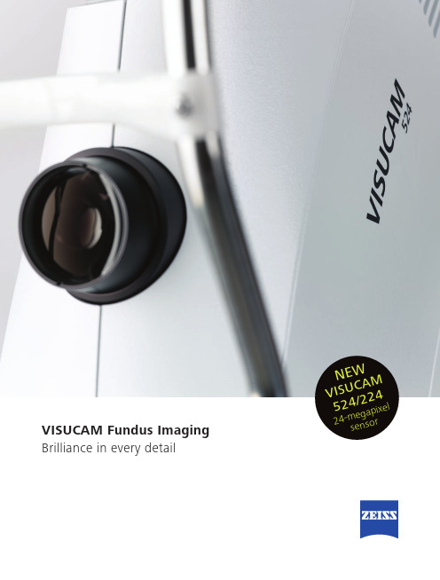
VISUCAM Fundus Imaging Brilliance in every detailN E WV I S U CA M 524/224 24-m eg a p i x els e n s orVISUCAM Fundus ImagingExcellent clarity, ultra-high resolution, legendary ZEISS optics.The new ZEISS VISUCAM fundus camera with a 24-megapixel sensor produces brilliant, detail-rich images to effectively aid in diagnosing and monitoring a broad range of eye diseases – from glaucoma and diabetic retinopathy to AMD.Greater diagnostic insight – High-resolution fundus imagingVersatility –Fully-featured camera with a full spectrum of imaging modes* Enhanced practice performance –Simple design, user friendly, full integration with clinic workflowSetting a new standard for resolutionDetails define your decisionsUltra-high resolution and excellent clarity promote efficient navigation from full-image overview to magnification of the smallest detail, allowing precise visualization within a particular area of interest.Fundus autofluorescence (FAF)FAF, included on both VISUCAM models, is an important non-invasive tool for the diagnosis and monitoring of dry AMD, including geographic atrophy.More than a pretty pictureVISUCAM is a complete system with numerous on-board image capture modes – fundus autofluorescence, non-mydriatic Color, Red-free, Red, Blue – and visualization functionality that provide powerful diagnostic insights for optimal patient care.Advanced features such as fluorescein angiography and indocyanine green angiography* further extend its diagnostic applications. * Available only on VISUCAM 524Color VISUCAM Fundus Camera with24-megapixel SensorStereo image pairRed-free// U LTRA-HIGH RESOLUTIONMADE BY ZEISSBest-in-class images from a 24-megapixel sensorAvailable in two modelsVISUCAM 224 with FAF is a fully featured non-mydriatic and mydriatic color camera.VISUCAM 524 adds fluorescein angiography with an optional ICGA mode for doctorswho perform their own dye-based angiography.Red FAF FA ICGAAnterior segmentFundus camera systemField angle 45° and 30°Capture modesColor, red-free, blue, red and fundus autofluorescence images, stereo pairs and images of the anterior segmentVISUCAM 524 only fluorescein angiography VISUCAM 524 only optional: ICG angiographyFiltersOptical filters for capture modes: Filters for green and blue pictures, filters for fundus autofluorescence images, UV/IR barrier filters Compensation for ametropia +35 D … -35 D, continuousCapture sequence from 1.5 seconds (depends on flash energy)Pupil diameter≥ 4.0 mm≥ 3.3 mm (30° small pupil mode)Working distance 40 mm (patient’s eye – front lens)Capture sensor CCD 24-megapixelsMonitor 23” TFT (1920 x 1080), connected via medical power supplyFixation targetsExternal and internal; four sizes of internal fixation target including a circle (for AMD patients). Attention mode for internal fixation target; various programmed sequences or freely positionable as combination with stereo mode too Flash energy Xenon flash lamp, 24 flash levels (max 80 Ws)DatabasePatient information and images with field angle, FA time, R/L recognition and date of visit are storedComputer / AccessoriesOperating system Windows Embedded Standard 7Hard drive Storage of approx. 80,000 images possible (present size of HDD: 420 GB) Interfaces USB ports and network connectors, DVI portExport/import Supported image formats: DICOM-OP and VL, BMP , TIFF, JPEG Patient list, DICOM MWL, DICOM storage Instrument table Asymmetric, suitable for wheelchairAccessoriesNetwork printer, USB memory stick, monitor bracket, sliding keyboard shelf for instrument table, VISUPAC archiving and image analysis system, Network isolatorDimensionsBasic device 410 mm x 480 mm x 735 mm (W 16.14 x D 18.90 x H 28.94 inches)Monitor544 mm x 45 mm x 329 mm (W 21.4 x D 1.8 x H 12.9 inches) (depends on model)Weight (basic device)27.5 kg (60.7 lbs)Rated voltage 100 … 240 V ±10% (self-adjusting)Frequency50 / 60 HzPower consumption340 VA maximum (basic device); 60 VA maximum (monitor)Technical dataE N _31_022_0024I / U S _31_022_0024I P r i n t e d i n G e r m a n y C Z -07/2016T h e c o n t e n t s o f t h i s b r o c h u r e m a y d i f f e r f r o m t h e c u r r e n t s t a t u s o f a p p r o v a l o f t h e p r o d u c t o r s e r v i c e o f f e r i n g i n y o u r c o u n t r y . P l e a s e c o n t a c t o u r r e g i o n a l r e p r e s e n t a t i v e s f o r m o r e i n f o r m a t i o n . S u b j e c t t o c h a n g e i n d e s i g n a n d s c o p e o f d e l i v e r y a n d d u e t o o n g o i n g t e c h n i c a l d e v e l o p m e n t . V I S U C A M i s e i t h e r a t r a d e m a r k o r r e g i s t e r e d t r a d e m a r k o f C a r l Z e i s s M e d i t e c , I n c . i n t h e U n i t e d S t a t e s a n d /o r o t h e r c o u n t r i e s . © C a r l Z e i s s M e d i t e c , I n c . 2016 A l l c o p y r i g h t s r e s e r v e d .Carl Zeiss Meditec AG Goeschwitzer Str. 51-5207745 Jena Germany/med0297VISUCAM NM/FAVISUCAM 524/224。
ZEISS OPMI Lumera i 眼科手术镜系列说明书
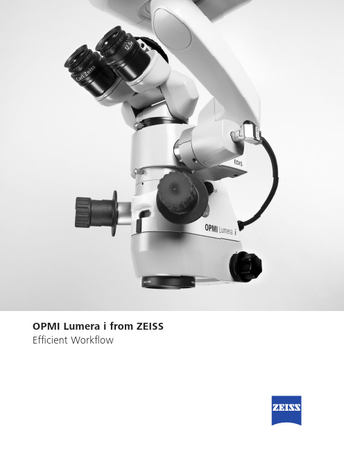
OPMI Lumera i from ZEISS Efficient WorkflowZEISS OPMI Lumera iShedding light on the details for better decision making The OPMI LUMERA® family from ZEISS represents excellence in optics andillumination. The ZEISS OPMI Lumera i with ZEISS high-quality visualizationtechnology, including Stereo Coaxial Illumination (SCI), will make a differencein how well you see the details during cataract and retina surgery.• See the most minute structures during surgery• Identify the details of the retina• See overlays of the assistance functions in the eyepiece• Manage depth of field with a push of a button• View structures in the eye in natural colors2Instant Red Reflex brightly illuminates the eye – due to Stereo Coaxial Illumination (SCI) even with mature cataracts.External Data Injection System (EDIS) allows you to see overlays of the assistance functions in the eyepiece of your ZEISS Lumera i.RESIGHT ® from ZEISS provides a clear, detailed view of the retina.Deep View depth of field management system allows you to choose between maximum depth of field or optimum light transmission.Unmatched ZEISS optics for exceptional clarity, contrast and light.The 1Chip HD camera system provides excellent visualization of natural color renditions and crisp anatomical details.3Z ALIGN ® – toric assistant 4> VClearly recognize different structuresof the anterior segment with SCI> A ligned just rightPrecise1,2,3, markerless alignmentof toric IOLs with Z ALIGNZEISS CALLISTO eyePrecise1,2,3 premiumIOL surgery made easy5Retina surgeryZEISS OPMI Lumera i and ZEISS RESIGHT fundus viewing systemsallow you to clearly recognize the details of the retina.4 2nd-generation optics with clearly better overall optical quality and clearly better sharpness, depth and detail recognition for use with the 60D lens – 2013 internal clinical customer survey with international key opinion leader surgeons67Carl Zeiss Meditec AG Goeschwitzer Strasse 51–5207745 Jena Germany/medOPMI Lumera i RESIGHT 500EDISCALLISTO eyeE N _32_010_0106I I I P r i n t e d i n G e r m a n y . C Z -X I I /2018 T h e c o n t e n t s o f t h e b r o c h u r e m a y d i f f e r f r o m t h e c u r r e n t s t a t u s o f a p p r o v a l o f t h e p r o d u c t o r s e r v i c e o f f e r i n g i n y o u r c o u n t r y . P l e a s e c o n t a c t o u r r e g i o n a l r e p r e s e n t a t i v e s f o r m o r e i n f o r m a t i o n . S u b j e c t t o c h a n g e s i n d e s i g n a n d s c o p e o f d e l i v e r y a n d d u e t o o n g o i n g t e c h n i c a l d e v e l o p m e n t . O P M I L U M E R A , R E S I G H T , C A L L I S T O e y e , S u p e r l u x , I n v e r t e r t u b e , Z A L I G N a n d K T R A C K a r e e i t h e r t r a d e m a r k s o r r e g i s t e r e d t r a d e m a r k s o f C a r l Z e i s s M e d i t e c A G . © C a r l Z e i s s M e d i t e c A G , 2018. A l l r i g h t s r e s e r v e d .0297。
VISU200 plus 操作指南
VISU 200 plus/S88 操作指南1.准备工作:将手术显微镜推到手术位置,踩下脚踏板,显微镜的轮子将被锁住(注:这时请不要推动显微镜)。
轻轻踩下脚踏板可以把轮子从锁住状态释放出来。
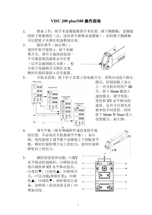
2.限位调节(如右图):逆时针松开旋钮1,按下电磁锁开关,调节主镜的高低到一个可能需要的最低安全位置(以不会碰到病人为准),松开按下电磁锁开关锁住支架,顺时针旋转旋钮1直至旋紧。
3.开机及设置:按下位于支架上的电源开关,系统自动进入默认模式,控制面板上显示上一次关机时的用户ID号。
按下Mode键进入速度模式,调节变焦、变倍和XY水平移动的速度。
总共可以预先存储9组不同设置,同时按下Mode和Store进入设置模式,如左图:4.调节平衡(除非增减附件或改变助手镜的位置,不必每次开机都调节平衡):如右图,使用旋钮2调节整个显微镜上下的配重平衡,顺时针旋转增大向上的拉力,逆时针旋转降低向上的拉力。
5.脚控的设置和功能:⑴XY水平移动控制摇杆;⑵降低光亮度⑶调焦和XY水平移动复位;⑷变倍▼;⑸变倍▲;⑹照明开关;⑺无功能(预留位置);⑻调焦▲;⑼调焦▼;⑽控制其它设备,如照相(需该设备支持)⑾增加亮度。
6.显微镜各部位示意图:如下图,⑴自动复位按钮,调焦和XY 水平移动系统都回到中心位置;⑵瞳距调节旋钮;⑶目镜;⑷屈光补偿旋钮;⑸电磁锁手柄开关;⑹倍率显示;⑺助手镜变倍调节;⑻助手镜焦距微调旋钮;⑼调节主镜倾斜角度旋钮;⑽调焦范围及位置指示;⑾助手镜倾角调节旋钮;⑿手动变倍调节;⒀照明方式选择旋钮;⒁+6°光照明;⒂光纤接口;⒃助手镜锁紧装置;⒄DeepView智能景深增强系统。
7.开始使用:a)调节双目镜筒旋钮1(如右图),使两个目镜下的图象合二为一。
b)目镜的调节:1. 调节眼杯的高低:当术者佩戴眼镜使用手术显微镜时,需要将眼杯完全旋入目镜;反之,可以根据用户的使用习惯将眼杯旋出,旋出的程度根据术者的感觉来调节。
- 1、下载文档前请自行甄别文档内容的完整性,平台不提供额外的编辑、内容补充、找答案等附加服务。
- 2、"仅部分预览"的文档,不可在线预览部分如存在完整性等问题,可反馈申请退款(可完整预览的文档不适用该条件!)。
- 3、如文档侵犯您的权益,请联系客服反馈,我们会尽快为您处理(人工客服工作时间:9:00-18:30)。
消色差
复消色差
当光线通过光学系统时会发生棱镜效应
,
传统的显微镜采用消色差光学 (achromatic) 来纠正色差,即使可见光谱中二个不同波长 的颜色光聚焦在一个平面上,但其余的波长 光并不同焦从而导致了色差和图象对比度下 降 复消色差光学采用多点波长纠正,从而彻底 纠正了色差并大大提高了对比度(尤其在图 象边缘)和清晰度。
•
内置裂隙照明光应用: – Anterior vitrectomy – Dense cataract capsulorhexis – Lens extractions
水平,可移动
垂直
2.5 or 5 mm width
2.5 mm width
支架面板(卤素灯配置时)
• • • • 主电源 后备电源 功能设定键 大屏幕液晶参数显示
Байду номын сангаас
蔡司手术显微镜中用来避免病人光损伤的设计
• 1.显微镜的通光量大大提高,用较低的亮度即可在目镜 中看到明亮的图象(亮度0.3,最大1.5) • 2.不可见的UV紫外光滤光片 • 3.GG475滤光片滤掉所有波长475nm以下的光线。 • 4.专利的视网膜保护装置 • 5.冷光源:其反射曲面使红外光不能聚焦从而滤掉红外 光。
S88支架系统
• 1.支架无重式移动,各关节电磁锁功能。手术中如需 紧急移开显微镜可瞬间完成.左右手柄可分别控制XY平 面和Z轴运动以方便术前消毒及显微镜粗调 • 2.全防水脚控可控制光源亮度、开关、平移、调焦、 变倍。 • 3.XY平移速度根据倍数大小自动调整。 • 4.调焦、XY平移、放大倍数自动回原功能。. • 5.多种记忆功能(亮度,变倍速度,调焦速度,XY移动速 度,焦平面). • 6.采用大直径轮子方便移动,四轮均可转向. • 7.手柄可控制IMAGEBOX(选件)的单张取像.
• 新的REDFLEX设计使红光反射大大增强
• 智能景深调节系统 Depth of Field Management System • ► 打开该开关时: • 加大景深Optimum depth of field at all magnification levels • ► 关闭该开关时: • 保持最大的通光量 和分辨率
卤素灯灯箱(双电源,四灯泡)
• 主电源灯箱(手术中 灯泡坏,备用灯泡会 自动切换) • 后备电源灯箱 • 滤光片选择
支架说明
• 1.推拉扶手 • 2.导线和脚控悬挂处 • 3.导向轮(大脚轮以 方便显微镜在不同手 术室之间移动)
支架电磁锁
• 支架关节有电磁锁, 只要按下电磁锁开关 ,显微镜体可在无重 状态下移动 • 电磁锁开关
四光路结构,副手 主刀镜不分光 助手镜 助手的左右眼均 有好的红反射
主镜
M841助手镜的一个眼无红光反射
白光照明
• 氙灯照明优点:
– – – – 白色,自然光 摄像效果好 对比度好,尤其是白/红 图象清晰度高
卤素灯
氙灯
对眼睛光损伤的对比
卤素灯 氙灯
光亮度设定在1.0时的对比 (equivalent phototoxicity hazard potential of 500 mW/cm² *sr, weighted according to hazard function)
蔡司VISU200p高级眼科手术显微镜
• Carl zeiss shanghai
光学系统
• 1.最优的光学性能:独有的复消色差(apochromatic)和T*镀膜技术保证了 最优的清晰度和对比度,光路设计能保证医生长时间手术眼睛不疲劳,具 有极好的景深和立体感。
• 2.采用12V、100W冷光源照明,新设计的光路提高透光性能50%以利于用眼 内光纤照明的眼底手术。(是目前透光性最好的手术显微镜,符合新的ISO 光损伤标准),采用螺纹防折光纤. • 1:6无级变倍。最好的机械工艺(保证长的使用寿命),目镜和物镜最新采 用超级防反光及超硬镀膜(防止擦镜时对光学面的损伤)。