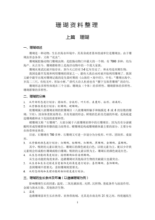
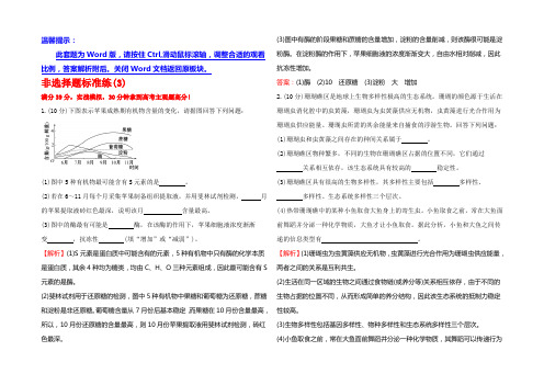
珊瑚与虫黄藻
珊瑚资料整理

珊瑚资料整理上篇珊瑚一、珊瑚综述珊瑚是一种动物,生长在海水环境中,其真身或者基本组成单位是珊瑚虫。
由于珊瑚虫形态各异,又称“花虫”。
珊瑚属腔肠动物门珊瑚虫纲,是腔肠动物门中最大的一个纲,有7000多种,均为海产。
从古至今,珊瑚都称得上是海洋动物中的一个庞大家族。
珊瑚从寒武纪就开始存在,到今天已经有5-6亿年历史了,和水母是同期生物。
我国是最早发现和利用珊瑚的国家之一,据传大禹治水时就开始利用珊瑚了。
我国文献中最早出现对珊瑚记载的是先秦时期的《山海经·海中经》,中有:“珊瑚出海中,岁高二三尺,有枝无叶,形如小树。
”唐代大诗人杜甫也有“腰下宝珠青珊瑚”的诗句。
珊瑚形态多样性体现在三个方面:珊瑚虫(个体)的多样性、珊瑚群体的多样性、珊瑚群聚的多样性。
二、珊瑚的分类1.从外部形态进行划分:团块形、分枝形、叶片形、表覆形、柱形、游离形。
2.从骨骼性质进行划分:软珊瑚、硬珊瑚。
软珊瑚属八放珊瑚亚纲的软珊瑚目(八放珊瑚即触手和隔膜是6或6的倍数的珊瑚,下同),因身体柔软而得名,具有美丽的形态、鲜艳的色彩及亮丽的外观,是海底建造珊瑚礁和水下花园的重要种类。
硬珊瑚又称“石珊瑚”,大部分属于六放珊瑚亚纲中的石珊瑚目,因为具有分泌碳酸钙形成坚硬群体骨骼的能力而得名。
硬珊瑚是构成珊瑚礁体最主要的部分,主要分布在热带和亚热带。
目前,石珊瑚有700多种。
石珊瑚又可进一步划分为分枝形、叶形、团块形、桌面形。
3.从骨骼的颜色进行划分:白珊瑚、粉珊瑚、红珊瑚、黑珊瑚、金珊瑚、蓝珊瑚。
其中,吸附的元素以镁为主,珊瑚石的颜色就是白色;以镁元素为主,配以少许铁元素则会形成粉红珊瑚或粉白珊瑚;吸附的元素以铁为主,珊瑚石的颜色就是红色。
4.从生态功能的角度划分:造礁珊瑚和非造礁珊瑚。
从生态功能的角度来讲,造礁珊瑚对其他海洋生物的贡献最大也最突出。
5.从生存和生长过程是否需要阳光的角度进行划分:喜阴珊瑚、喜阳珊瑚。
喜阴珊瑚不需要光,喜阳珊瑚则需要光。
藻类对珊瑚的间接影响:藻类诱发的、微生物介导的珊瑚的大量死亡

藻类对珊瑚的间接影响:藻类诱发的、微生物介导的珊瑚的大量死亡珊瑚覆盖率的下降往往与藻类丰富度的增加相关联。
过去不清楚是藻类直接或间接造成了珊瑚的死亡,还是藻类仅附着到已死亡的珊瑚表面。
本研究证明藻类通过释放可溶的化合物提高微生物活性而间接导致珊瑚死亡。
当珊瑚和藻类被一起放置在容器中,之间用一个0.02μm的过滤器分离,珊瑚死亡率达100%。
而加入广谱抗生素氨必西林,珊瑚的死亡则完全被避免。
生理测试的结果显示珊瑚受到的胁迫随着附近的藻类而增强,呈互补模式。
我们的研究结果表明,由于人类活动的影响日益增加以及珊瑚礁上的藻类变得更丰富,由此可能会创建一个正反馈环路:海藻释放的化合物增强活珊瑚礁表面微生物的活性,造成珊瑚大量死亡,并进一步促进藻类的生长。
以前的研究表明,藻类的很多种或属可以对珊瑚产生负面影响(McCook et al.2001; Jompa&McCook2003a,b;Nugues et al.2004)。
潜在的机制包括化感作用、窒息、遮光、磨损、过度生长(见McCook et al.,2001的综述)以及带有可能致病的微生物(Nugues et al. 2004)。
然而,关于藻类对珊瑚的健康的间接影响的研究较少,特别是藻类分泌物对珊瑚-微生物的相互作用间接影响。
现在已证明,在无机养分(氮和磷)的大部分升幅并不直接杀死珊瑚,而有机碳负荷的增加则造成了珊瑚的死亡(Kuntz et al.2005;Kline et al.出版中)。
有机碳负荷造成的珊瑚死亡是可以预防或添加抗生素来延缓(Mitchell&Chet1975),并与微生物活性的增加相关,表明微生物介导了珊瑚的死亡。
这项研究的目标是明确地确认是否存在着藻类诱发的、微生物介导的珊瑚大量死亡的机制。
通过结合围隔试验和野外调查,我们明确地验证了:(a)当珊瑚被放置在藻类附近而不直接接触时,珊瑚的健康水平是否会下降及/或珊瑚的大量死亡是否会被诱发;(b)这些效应是否是由微生物介导的;(c)这些效应是否在各种珊瑚-藻物种组合中普遍存在;及(d)这些模式是否可以在野外直接观察到。
高考题中的海底珊瑚和珊瑚礁

高考题中的海底珊瑚和珊瑚礁珊瑚礁由造礁珊瑚的石灰质遗骸和石灰质藻类堆积而成的一种石灰岩礁石。
造礁珊瑚主要分布于热带地区,水深一般是小于25米的浅水。
下图是与火山活动有关的某种珊瑚礁的形成,读图完成问题。
珊瑚礁形成过程中,海平面A.先升后降B.先降后升C.持续升高D.持续降低我们先从火山岛说起火山岛火山岛是指海底(主要是指海底)火山的不断喷发,火山活动喷发的岩浆冷却,不断堆积,越来越高,最终高出水面而形成的岛屿。
火山岛的形成,需要有火山喷发条件,所以多形成在海底多火山分布的区域,所以在海底板块的生长边界附近和环太平洋区域分布较广。
火山岛由于是火山喷发而成,往往由于火山的持续喷发,岛屿越来越高,火山岛的地形往往比较崎岖,和大陆岛相比,火山面积要小得多。
火山岛有单个的,也有群岛式的,世界上著名的火山岛群有阿留申群岛、夏威夷群岛等。
夏威夷群岛的形成过程珊瑚是植物吗?珊瑚是地球上最古老的海洋生物,将近五亿年前就出现了。
珊瑚是动物,是由无数微小的珊瑚虫聚集形成。
珊瑚虫是一种腔肠动物,它个头很小,往往只有几毫米,体态玲珑,色泽美丽,十分娇气,珊瑚虫繁茂生长,建造珊瑚礁。
由于珊瑚的向光性及捕食的需要,它向上、向四周"生长",终而形成树枝状的结构。
有趣的是,在很长一段时间它类似植物的特征迷惑了人类,让人们以为它是植物。
珊瑚的栖息环境是怎样的?水深100-200米的平静而清澈的岩礁、平台、斜坡和崖面、凹缝中。
珊瑚的分布范围分布在温度高于20℃的赤道及其附近的热带、亚热带地区。
简述有利于珊瑚生长的条件(1)纬度低,位于热带地区,海水水温较高(全年水温保持在22-28摄氏度的水域);(2)位于大陆架,属于浅水环境;(3)水域环境稳定,如退潮时不能长时间暴露在水面之上。
珊瑚与虫黄藻共生,它通过光合作用为珊瑚提供营养。
全球气候变暖,海平面上升会引起虫黄藻的死亡,造成珊瑚白化。
(4)风浪小,有利于珊瑚附着在基底岩石上生长;(5)光照充足,有利于藻类生长,食物(营养供给)丰富;(6)水质洁净、透明度高,有利于藻类光合作用。
珊瑚的亲密伴侣——虫黄藻

龙源期刊网 珊瑚的亲密伴侣——虫黄藻作者:马晓惠来源:《百科探秘·海底世界》2012年第10期就像永远面朝阳光、积极向上的向日葵一样,珊瑚也有向光生长的特性,这难免让人联想起靠光合作用生长的植物。
因此,即便你已经知道珊瑚是动物了,眼前这情景也会不由得令你疑惑:难道是弄错了吗?其实,珊瑚喜欢沐浴在阳光中,是因为在它体内有一种叫“虫黄藻”的共生藻。
这是一种单细胞藻类,个头儿非常小,只有借助显微镜才能发现。
生物学家估计,每立方毫米的珊瑚组织内就有30000个虫黄藻。
虫黄藻需要阳光来进行光合作用,为了满足它“吃”阳光的胃口,珊瑚才有了这种向阳生长的特性。
19世纪40年代,日本生物学家在珊瑚虫体内观察到很多褐色小球。
这些褐色小球能在有光条件下进行光合作用,并能自身进行分裂繁殖。
这是虫黄藻第一次进入人类视线。
由于缺乏令人信服的证据,它的存在一直饱受质疑。
19世纪50年代末,生物学家终于成功将虫黄藻从珊瑚虫体内分离出来了,至此它的‘江湖地位’才得以确立。
虫黄藻与珊瑚虫之间是一种互利共生的关系。
虫黄藻进行光合作用的原料是珊瑚虫的代谢废物,合成的氧气和有机物则奉献给珊瑚虫;珊瑚虫也不会白白享用,除了负责给虫黄藻提供原料外,还为它提供了居所和保护。
有了虫黄藻后,珊瑚虫的生存压力大大减小,平时只需捕捉些“零食”作加餐即可,而赖以生存的主食就仰仗于虫黄藻的慷慨奉献了。
除了能给珊瑚虫提供主食外,虫黄藻还能促成它们骨骼的生长。
当然啦,并非所有的珊瑚都能与虫黄藻一起打拼,比如大部分造礁珊瑚都有虫黄藻共生相伴,而非造礁珊瑚则没有那么幸运了。
但是,万一虫黄藻跑掉或死掉了,与它相依存的造礁珊瑚就会得病,即出现白化现象。
在白化初期,如果虫黄藻能及时回到珊瑚体内,生病的珊瑚仍能起死回生;但要是持续白化时间太长,珊瑚就会因为严重缺乏营养而死亡。
虫黄藻对珊瑚的依赖则相对较弱,因为它们除了能与珊瑚共生之外,还能在某些原生动物和无脊椎动物体内存在,比如珊瑚的“近亲”海葵等。
气候变暖珊瑚

气候变暖珊瑚1、为什么全球变暖会导致珊瑚白化?变暖了珊瑚体内的共生藻类不应该繁殖得更好吗?其实全球变暖并不是珊瑚白知化的直接原因。
珊瑚是由珊瑚虫产生的分泌物,珊瑚的主要化学组成部分是碳酸钙,由于大气中的CO2浓度增加,道造成CO2与珊瑚中的碳酸钙反应,生成碳酸氢钙,因此被溶内解,珊瑚便失去原有的绚丽颜色。
生物良好地生活都是经过千万年的共同进化的结果,共生藻适应了珊瑚中的生活容,生活环境的变化使共生藻难以适应现在的生活。
2、珊瑚礁因水温上升会消失,那靠近南极或北极地方的珊瑚礁还会消失吗?会。
首先,南北极的温度极低,大多是冰川所在,靠近南北极珊瑚礁世界上珊瑚礁多见于南北纬30°之间的海域中,尤以太平洋中、西部为多。
珊瑚礁虽然大部分分布在热带海域,但实际上它只适合生存在水温28度以下的海域,异常的温度,30度、31度也会使珊瑚大量白化死亡,所以如今的全球变暖导致水温也不断上升,会加快珊瑚礁的消亡。
尽管南北极所处地区温度较低,但是全球变暖所带来的影响是涉及到全世界的,南北极的水平面也受到了下降趋势,还记得前几年因全球变暖导致北极的北极熊“无家可归”的图像吗?那个时候的全球变暖就已经导致了整个南北极的冰山快速融化,而增加整个海平面的上升。
礁珊瑚对水温、盐度、水深和光照等条件都都有着比较严格的要求:珊瑚生长的水温约为20~30°C左右,23~27°C是造礁珊瑚生长发育的最佳水温,最佳水温上限可达29°C。
热带海区,这一最佳水温出现在冬季和春季,因而许多学者认为冬季珊瑚生长最快。
海南岛和西沙群岛水温平均为25~27°C,属珊瑚生长最佳水温范围,但海南岛的季节变化大,水温不稳定,对珊瑚生长有抑制作用。
海南岛和台湾岛的珊瑚礁被称为“高纬度珊瑚礁”。
礁珊瑚生长在盐度为27~40%的海水中,最佳盐度范围是34~36%。
南海盐度为34,属最佳盐度范围,而一般认为造礁珊瑚生长的水深范围是 0~50米,最佳水深为20米以浅。
高考地理专题训练:珊瑚、珊瑚礁(附参考答案)

高考地理专题训练:珊瑚、珊瑚礁(附参考答案)1.阅读图文材料,完成下列问题。
珊瑚礁白化是由于珊瑚失去体内共生的虫黄藻或共生的虫黄藻失去体内色素而导致五彩缤纷的珊瑚礁变白的生态现象。
1998年和2002年曾两度发生过严重的珊瑚白化事件。
到了2014年,由于全球温度上升了0.9℃,珊瑚白化现象又一次大规模出现。
2015年,由于白化,澳大利亚大堡礁浅水区67%的珊瑚不幸死亡。
据材料结合所学知识分析珊瑚礁白化产生的根本原因及应对措施。
参考答案:根本原因:化石燃料的燃烧,森林的破坏,温室气体增加,全球气候变暖;措施:减少化石燃料的燃烧;优化能源消费结构,开发利用新能源;植树造林;加强宣传教育,树立环保意识;发展清洁燃料技术;回收利用CO2和CH4等温室气体;加强国际合作;制定法律法规,实现达标排放2.(2019·广东高三期末)阅读材料,回答相关问题。
涠洲岛位于广西北部湾中部地区,属亚热带季风气候。
岛屿北部、东部海岸主要为基岩岩滩,西部海岸以海蚀崖为主,该岛是全球珊瑚礁分布的北缘,珊瑚主要生活在热带海域,其生长条件较苛刻,最适宜温度为25℃—27℃之间,对水环境要求高。
随着气候变化,全球的珊瑚礁正在不断减少。
目前科学家正在涠洲岛海域进行人工繁育珊瑚多项实验,主要采用固定式苗圃和悬浮式苗圃进行海底珊瑚种植,由于海况复杂,工作人员在海底种植珊瑚面临许多困难和问题。
下图为涠洲岛等深线及珊瑚礁的分布图(阴影区为现代珊瑚覆盖度>5%)(1)推测涠洲岛西部和南部地区珊瑚分布较少的原因。
(2)分析气候变暖对涠洲岛珊瑚的影响。
(3)简述在涠洲岛海底种植珊瑚可能面临的困难和问题。
(4)指出涠洲岛珊瑚礁的可持续开发利用方向。
参考答案:(1)西部风浪大,侵蚀强,水较深,形成侵蚀海岸;南部为海湾,人类活动密集,水体污染较严重,海底以淤泥沉积物为主,不利珊瑚生长。
(2)气候变暖导致海水温度升高,影响珊瑚生存,导致珊瑚总量减少;使热带的珊瑚类型增加,成为热带珊瑚的避难所;珊瑚分布范围向北扩大。
2022版高考生物二轮复习 非选择题标准练(3) Word版含答案

温馨提示:此套题为Word版,请按住Ctrl,滑动鼠标滚轴,调整合适的观看比例,答案解析附后。
关闭Word文档返回原板块。
非选择题标准练(3)满分39分,实战模拟,30分钟拿到高考主观题高分!1.(10分)下图表示苹果成熟期有机物含量的变化,请据图回答下列问题:(1)图中5种有机物最可能含有S元素的是。
(2)若在6~11月每个月采集苹果制备组织提取液,并用斐林试剂检测,月的苹果提取液砖红色最深,说明该月含量最高。
(3)图中的酶最有可能是酶,在该酶的作用下,苹果细胞液浓度渐渐变,抗冻性(填“增加”或“减弱”)。
【解析】(1)S元素是蛋白质中可能含有的元素,5种有机物中只有酶的化学本质是蛋白质,其余4种均为糖类,均由C、H、O三种元素组成,因此最可能含有S 元素的是酶。
(2)斐林试剂用于还原糖的检测,图中5种有机物中果糖和葡萄糖为还原糖,蔗糖和淀粉是非还原糖。
葡萄糖含量从7月份后基本稳定,而果糖在10月份含量最高,所以,10月份还原糖的含量最高,则10月份苹果提取液用斐林试剂检测,砖红色最深。
(3)图中有酶的阶段果糖和蔗糖的含量增加,淀粉的含量削减,则该酶很可能是淀粉酶。
在淀粉酶的作用下,苹果细胞液的浓度渐渐变大,自由水相对削减,因此抗冻性增加。
答案:(1)酶(2)10 还原糖(3)淀粉大增加2.(10分)珊瑚礁区是地球上生物多样性极高的生态系统。
珊瑚的颜色源于生活在珊瑚虫消化腔中的虫黄藻,珊瑚虫为虫黄藻供应无机物,虫黄藻进行光合作用为珊瑚虫供应能量。
珊瑚虫所需的其余能量来自捕食的浮游生物。
回答下列问题:(1)珊瑚虫和虫黄藻之间存在的种间关系属于。
(2)珊瑚礁区物种繁多,不同的生物在珊瑚礁区占据的位置不同,它们通过关系相互依存,该生态系统具有较高的稳定性。
(3)珊瑚礁区具有很高的生物多样性,其多样性主要包括多样性、多样性、生态系统多样性三个层次。
(4)热带珊瑚礁中的某种小鱼取食大鱼身上的寄生虫。
小鱼取食之前,常在大鱼面前舞蹈并分泌一种化学物质,大鱼才让小鱼取食。
温度胁迫对珊瑚共生虫黄藻超微结构及相关基因表达的影响

2.利用透射电子显微镜观察了热应激过程中虫黄藻细胞的形态与结构变化,当温 度从 28℃缓慢升至 32℃24h 时,虫黄藻细胞形态结构正常,表明此时并未对虫黄藻 产生应激作用。到 32℃72h 时,受热应激的影响,虫黄藻的细胞膜开始出现变化,32℃ 168 h 时,虫黄藻的细胞损伤程度进一步加大,绝大多数虫黄藻开始表现出细胞凋亡 的迹象,此时,珊瑚开始出现白化,表明虫黄藻热应激敏感性高于珊瑚组织。此外, 还观察到极少数虫黄藻发生细胞坏死现象。
4.By in vitro culture of zooxanthellae,we found that the algae which isolated from Galaxea astreata can survive in ASP-8A medium for 13 days,and 9 days in f/2 medium. And when we improve the concentration of Fe3+ to 1.8*10-5M in ASP-8A medium,the zooxanthellae can alive for 15days.The results showed that Fe3+ play an important role in zooxanthellae cultivation.
上海市浦东新区2024届高考二模考试生物试卷(附答案)

上海市浦东新区2024届高考二模考试生物试卷(本试卷满分100分。
考试时间为60分钟)请将所有答案写在答题纸上,否则不给分一、海洋中的“热带雨林”——珊瑚礁生态系统1. 珊瑚礁常位于营养较贫乏的水域,却有极高的生物多样性价值,如可为海洋生物提供食物和栖息地。
珊瑚礁由珊瑚虫的骨骼聚结形成,其绚丽多彩的颜色是因珊瑚虫体内生活的虫黄藻所致。
虫黄藻适合生长的温度范围是:20℃~28℃,盐度范围是27%~40%,超出这些范围会使虫黄藻逃离珊瑚虫。
图1表示虫黄藻和珊瑚虫,图2表示珊瑚礁生态系统的部分生物的关系,黑色箭头表示捕食。
(1)据图2分析,该生态系统中生产者有___(编号选填)。
①浮游动物②珊瑚虫③虫黄藻④其他藻类⑤核果螺(2)下列生物学事实能体现群落结构的是___。
A. 不同藻类分布在海洋不同深度B. 虫黄藻生活在珊瑚虫体内C. 珊瑚礁呈现五彩缤纷的颜色D. 鱼类可以是第二营养级312//(4)通过测定动物组织15N 比值可对珊瑚礁生态系统能量流动进行研究,15N 比值随着营养级的升高而增大。
研究发现,随着季节更替大法螺的15N 比值一年四季会发生显著变化,根据已有知识推测可能的原因是,随着季节更替___(多选)。
A. 大法螺的食物来源和种类发生改变B. 珊瑚礁生态系统的物质循环发生改变C. 珊瑚礁生态系统的信息传递发生改变D. 温度的变化直接导致15N 比值发生改变(5)假设珊瑚礁生态系统中只存在“藻类→食藻鱼类→食肉鱼类”营养结构,流经这三个营养级的总能量分别为a ,b ,c ,下列表达式正确的是___(编号选填)。
①a+b=c ②a<b<c ③a>b+c ④a>b>c ⑤a=b+c2. 某海岛北侧是游客聚集地,水体浑浊度显著高于南侧。
科研人员在该岛的南北两侧分别对两种优势珊瑚(杯型珊瑚、滨珊瑚)采样,并检测它们体内虫黄藻的相关指标。
图1是计数板上一个中方格的视野。
珊瑚:海洋中最美丽的“树枝”

珊瑚:海洋中最美丽的“树枝”作者:***来源:《百科探秘·海底世界》2020年第11期珊瑚:是植物还是动物?在清澈的热带浅海区,生长着许多五彩斑斓的珊瑚。
每一株珊瑚都是由无数只珊瑚虫组成的。
珊瑚虫通过分泌碳酸钙,将自己固定在由祖辈共同构建的骨骼上。
因此,它们一旦选定栖息地就无法再挪窝儿。
这些珊瑚虫不但通过碳酸钙的外壳黏合成一个整体,还共用一个胃。
珊瑚的外表长得像树枝,不仅像植物一样固定不动,还能进行光合作用。
珊瑚虫的身体呈圆筒状,口的四周长有许多触手。
这些触手的数目不是6或6的倍数,就是8或8的倍数。
这些珊瑚虫不仅能够主动捕猎,身体结构也更具有动物的特征。
那么,由珊瑚虫组成的珊瑚到底是植物还是动物呢?古希腊学者亚里士多德曾把珊瑚称为“zoophyla”,即一种介于动物和植物之间的特殊物种。
科学的精神是求真,不可能让珊瑚无法被归类。
千百年来,科学家一直孜孜不倦地探求如何给珊瑚分类,并由此引发了诸多争论。
直到18世纪,珊瑚才正式被归入动物界,这场长达二十多个世纪的争论才落下帷幕。
白化不等于死亡全球气候变暖引起海水温度升高,造成海水酸化,这会对珊瑚生长造成严重影响。
现在,几乎全世界的珊瑚都在遭受白化的威胁。
许多人都认为珊瑚白化就是死了,其实这种说法并不准确。
说到珊瑚白化,就得先说说珊瑚的好朋友虫黄藻。
还在幼年时,虫黄藻就住进了珊瑚虫体内。
虫黄藻能“捕捉”阳光进行光合作用。
光合作用的产物不仅能养活它们自己,还能给珊瑚虫“交房租”呢。
珊瑚虫这位“房东”也很勤劳,除了“收房租”外,还会主动用自己的触手捕捉浮游生物。
健康珊瑚的颜色来自体内的虫黄藻。
一旦虫黄藻离开,珊瑚就会原形毕露,露出透明的珊瑚虫以及碳酸钙骨骼。
珊瑚的骨骼大多是白色的,只有少数有颜色。
虫黄藻很娇气,海水温度升高或海水酸化都会导致它们“离家出走”。
珊瑚白化只是“病危通知单”,并没有宣告珊瑚已经死亡。
只要环境能在短时间内恢复正常,“离家出走”的虫黄藻就会回家,白化的珊瑚也能恢复健康。
2020届高三一轮复习地理小专题之珊瑚、珊瑚礁

2020届高三一轮复习地理小专题之珊瑚、珊瑚礁典型例题一:阅读图文材料,完成下列问题。
珊瑚礁白化是由于珊瑚失去体内共生的虫黄藻或共生的虫黄藻失去体内色素而导致五彩缤纷的珊瑚礁变白的生态现象。
1998年和2002年曾两度发生过严重的珊瑚白化事件。
到了2014年,由于全球温度上升了0.9℃,珊瑚白化现象又一次大规模出现。
2015年,由于白化,澳大利亚大堡礁浅水区67%的珊瑚不幸死亡。
据材料结合所学知识分析珊瑚礁白化产生的根本原因及应对措施。
参考答案:根本原因:化石燃料的燃烧,森林的破坏,温室气体增加,全球气候变暖;措施:减少化石燃料的燃烧;优化能源消费结构,开发利用新能源;植树造林;加强宣传教育,树立环保意识;发展清洁燃料技术;回收利用CO2和CH4等温室气体;加强国际合作;制定法律法规,实现达标排放典型例题二:阅读材料,回答相关问题。
涠洲岛位于广西北部湾中部地区,属亚热带季风气候。
岛屿北部、东部海岸主要为基岩岩滩,西部海岸以海蚀崖为主,该岛是全球珊瑚礁分布的北缘,珊瑚主要生活在热带海域,其生长条件较苛刻,最适宜温度为25℃—27℃之间,对水环境要求高。
随着气候变化,全球的珊瑚礁正在不断减少。
目前科学家正在涠洲岛海域进行人工繁育珊瑚多项实验,主要采用固定式苗圃和悬浮式苗圃进行海底珊瑚种植,由于海况复杂,工作人员在海底种植珊瑚面临许多困难和问题。
下图为涠洲岛等深线及珊瑚礁的分布图(阴影区为现代珊瑚覆盖度>5%)(1)推测涠洲岛西部和南部地区珊瑚分布较少的原因。
(2)分析气候变暖对涠洲岛珊瑚的影响。
(3)简述在涠洲岛海底种植珊瑚可能面临的困难和问题。
(4)指出涠洲岛珊瑚礁的可持续开发利用方向。
参考答案:(1)西部风浪大,侵蚀强,水较深,形成侵蚀海岸;南部为海湾,人类活动密集,水体污染较严重,海底以淤泥沉积物为主,不利珊瑚生长。
(2)气候变暖导致海水温度升高,影响珊瑚生存,导致珊瑚总量减少;使热带的珊瑚类型增加,成为热带珊瑚的避难所;珊瑚分布范围向北扩大。
气候变化对珊瑚礁的危害分析

气候变化对珊瑚礁的危害分析珊瑚礁被誉为“海洋的雨林”,是地球上最为丰富和多样化的生态系统之一。
它们不仅承载着丰富的海洋生物,还在人类社会中发挥着重要的生态、经济和文化作用。
然而,随着全球气候变化的加剧,珊瑚礁正面临着前所未有的威胁。
本文将分析气候变化对珊瑚礁的多重危害,包括海洋温度升高、海洋酸化以及海平面上升等因素,探讨这些因素如何影响珊瑚礁生态系统,并提出相应的保护建议。
一、海洋温度升高1.1 温度对珊瑚生长的影响珊瑚是共生生物,其与藻类(称为虫黄藻)进行共生,藻类为珊瑚提供养分,珊瑚则为藻类提供庇护和二氧化碳。
理想的水温模式对珊瑚生长至关重要。
根据研究,珊瑚最适宜的生长温度在23℃到29℃之间。
然而,由于全球变暖,海洋表面温度在不断上升。
当水温超过设定阈值时,珊瑚就会发生“白化”现象。
此时,虫黄藻会从珊瑚体内被排出,使得原本色彩斑斓的珊瑚变得苍白。
如果海水温度持续异常升高,珊瑚将无法再获得足够的营养,最终导致死亡。
在过去的几十年中,全球范围内经历了多次大规模的珊瑚白化事件,其中1998年和2010年的事件尤为严重。
1.2 白化后的生态后果珊瑚白化不仅影响了单个个体,还对整个生态系统造成了深远影响。
白化后的珊瑚对各种疾病更加敏感,从而提高了腐烂和死亡率。
此外,由于珊瑚群落减少,相关海洋动物如鱼类、墨鱼等失去了栖息地。
同时,许多依赖于健康珊瑚礁生存的物种,包括一些濒危物种,也因此受到了威胁。
二、海洋酸化2.1 酸化现象及其成因随着大气中二氧化碳浓度不断增加,大量二氧化碳溶解在海洋中形成碳酸,使海水酸度逐渐升高。
根据科学家的估计,自工业革命以来,全球海洋的平均pH值已下降约0.1,这意味着海洋酸性增强了约30%。
这一变化会影响到海洋中许多生物,包括那些依赖碳酸钙构建外壳或骨骼的生物,而这些正是构成珊瑚礁基础的重要元素。
2.2 对珊瑚建造及生长的威胁酸化会直接影响到许多种类的珊瑚生成其骨骼所需的钙质结构。
珊瑚礁海岸的特征及其相关分析

珊瑚礁海岸的特征及其相关分析珊瑚礁海岸的特征及其相关分析生物海岸是一种较为特殊的海岸类型,在潮间带或者潮下浅水区生长有相当规模的底栖生物群落,其生物过程通常对海滨沉积物质的供应(主要为碳酸盐)、保持、稳定及较小程度上对侵蚀起重要作用[1],还对海岸动力、沉积和地貌过程产生显著影响或者成为海岸发育的主导因素。
典型的热带生物海岸是珊瑚珊瑚礁海岸和红树林海岸。
珊瑚礁海岸的特点是再大潮低潮线一下的潮下浅海区生长一种与营光合作用的单细胞虫黄藻共生因而能高效分泌碳酸钙骨骼的属腔肠动物门或刺胞动物门的造礁石珊瑚群落,它的原地碳酸盐骨骼堆积和各种生物碎屑填充胶结,形成具有抗浪性的海底隆起地貌结构单元。
生物海岸由于其与生物相关的特殊性,在海岸带的研究中具有重要地位。
海岸带位于海陆交互地带,人类活动频繁,社会经济发展迅速,因在地球系统功能中扮演关键的角色而受到全球变化研究的重视。
生物海岸的特殊生物栖息环境往往成为对维持海岸生物多样性和资源生产力有特别价值的各种生物活动高度集中地海岸生态关键区[2],成为对海岸经济社会可持续发展有重要意义的生态环境资源。
1 珊瑚礁的基本概念及其影响因素1.1 珊瑚礁的性质及形成珊瑚礁是由造礁珊瑚构成的。
珊瑚一般是指体型呈辐射对称、有石灰质外骨骼或皮层中含有大量的骨针、底栖固着生活的海洋腔肠动物。
珊瑚按照其形态特征可分为两类:造礁珊瑚和非造礁珊瑚。
造礁珊瑚由于有单细胞的虫黄藻与其共生,钙化生长速度快,所以能造礁。
造礁珊瑚具有分泌碳酸钙形成外骨骼的功能,它们世代交替生长,最终生长到低潮线,从而形成具有抗浪功能的海底隆起,即珊瑚礁。
珊瑚礁是造礁珊瑚死亡之后,其骨骼和外壳聚集在一起形成的沉积建造,是一种较为特殊的岩土介质类型。
1.2 珊瑚礁的类别根据珊瑚礁和岸线的关系,可划分为岸礁、堡礁和环礁。
1.2.1 岸礁沿大陆或者岛屿边缘生长发育,也可成为裙礁或者边缘礁。
岸礁是由生长在1大陆或岛屿周围浅海海底的珊瑚和其他钙质有机物构成,这种礁体的表面与低潮潮位的高度相差不多,粗糙而不平坦,外缘向海洋倾斜。
珊瑚体内通常都有大量共生色彩斑斓

我们看到的色彩斑斓的海底珊瑚是因为珊瑚体内通常都有大量共生的什么?
海底珊瑚是因为珊瑚体内通常都有大量共生的虫黄藻。
虫黄藻,这种藻类可以通过光合作用为珊瑚提供能量。
因为虫黄藻的存在,珊瑚的颜色才五颜六色。
珊瑚主要能量来源就是体内共生的虫黄藻,它们可以为珊瑚提供90%以上的能量。
作为回报,珊瑚为虫黄藻提供保护、居所、营养(主要是含有氮和磷的废料)和恒定供应光合作用所需的二氧化碳。
虫黄藻和珊瑚的关系:
虫黄藻不仅供给珊瑚虫O2和营养物,而且与珊瑚虫的石灰质骨骼的形成密切相关。
有人作过这样的实验:将虫黄藻从珊瑚虫的身体内全部分离出来,并在人工条件下供给珊瑚虫O2,结果珊瑚虫虽然能活下来,但其骨骼得不到正常的发育。
其原因是珊瑚虫在代谢过程中排出的大量碳酸气(CO2)妨
碍着骨骼的增长,虫黄藻却有迅速解除CO2的功能,从而促进珊瑚骨骼的形成。
- 1、下载文档前请自行甄别文档内容的完整性,平台不提供额外的编辑、内容补充、找答案等附加服务。
- 2、"仅部分预览"的文档,不可在线预览部分如存在完整性等问题,可反馈申请退款(可完整预览的文档不适用该条件!)。
- 3、如文档侵犯您的权益,请联系客服反馈,我们会尽快为您处理(人工客服工作时间:9:00-18:30)。
Coral Reef Science
பைடு நூலகம்
Fig. 1 Multiple scales of symbiotic system among coral-algal symbionts, coral reefs and humans
Fig. 8 A photo showing differential bleaching susceptibility of adjacent colonies of Porites randalli in the Ofu back reef in American Samoa. Bleaching is commonly patchy across a reef and bleached and unbleached colonies are often observed in close proximity. Whether this variability is caused by adaptation or acclimatization of the host or Symbiodinium remains to be determined
Fig. 2 Degradation of the coral reef symbiotic system by global and local human stressors
Fig. 3 Coral-reef scenes in the mid-1970s. As quoted in Jackson (2014), Sylvia Earle noted in 1972 that “. . .tropical reefs, notable for their dazzling profusion of animal life, are almost devoid of conspicuous plants”. Bruno et al. (2014) found that in natural undisturbed baseline conditions, benthic algae is patchily distributed and can occupy up to 10–30% of the substratum, averaging 22%. Clockwise from upper left: Ucubsui Reef, San Blas Islands, Caribbean Panama; Islas Secas, Pacific Panama; Arekabesan Island, Palau; Aunu’u Island, American Samoa; also Aunu’u. These are not a random selection of photographs, but were selected to show how easy it was to be impressed with the prevalence of animal tissue in the 1970s. Odum and Odum (1955) argued that many, if not most, of the algae were endolithic and out of sight
Fig. 4 A Caribbean boring sponge (Cliona cf caribbea) covering and eroding several square meters of reef substrate, San Blas, Panama, 3 m depth (30 June 1993). Arrows denote perimeter of sponge patch
Fig. 5 Reproductive modes of scleractinian corals. Upper: spawning coral releasing egg– sperm bundles. Lower: brooding coral releasing planula larva (Illustration by Dwi Haryanti)
Fig. 6 Types of zooxanthellae observed in coral tissue. (a, b) Healthy cells with a spherical shape and expanded chloroplast. (c, d) Shrunken, darkly colored cells with reduced sizes and partially fragmented chloroplasts. (e) Bleached cells with pale and colorless chloroplasts. (f) Three categories of zooxanthellae. (g) White light micrograph and (h) fluorescence image of healthy and shrunken cells (shrunken cells are indicated by arrowheads)
Fig. 9 Molecular analyses have had a profound impact on our understanding of evolutionary relationships of many coral reef organisms, including corals. Kitahara et al. (2010, tree above from Fig. 1), in a recent comprehensive phylogenetic analysis of scleractinian corals, showed that deep-sea azooxanthellate species are basal to the group and most recognized shallow-water zooxanthellate families are polyphyletic. In some cases, Atlantic and Pacific species once thought to belong to the same genus, are now placed in different families, such as Homophyllia (formerly Scolymia) australis from the Pacific (Lobophylliidae, left image) and Scolymia cubensis from the Atlantic (Mussidae, right image) (Photos courtesy of JEN Veron)
Fig. 7 New concept of coral bleaching revealed from counting and observation of zooxanthellae
Fig. 8 Highthroughput RNA sequencing is increasingly used as a tool to understand the physiological response of corals to stressors. Libro et al. (2013, above from Figs. 1 and 2) investigated the immune response of Acropora cervicornis to White-Band Disease (upper photo) by comparing RNAseq profiles of healthy and infected tissues. Their approach identified a series up- and downregulated genes that repre- sent 4 % of the coral transcriptome (plotted in red below)
