植物体内的亚细胞组分分离
亚细胞结构分离的技术

亚细胞结构分离的技术分离亚细胞组分的第一步是制备组织匀浆或细胞匀浆。
匀浆(Homogenization)是在低温条件下,将组织或细胞放在匀浆器中加入等渗匀浆介质(即0.25moL/L蔗糖一0.003mol/L氯化钙溶液)研磨,使细胞被机械地研碎成为各种亚细胞组分和包含物的混合物。
分离亚细胞组分的第二步是分级分离。
它通过由低速到高速离心技术,使非均一混合体中的颗粒按其大小轻重分批沉降到离心管的不同部位,再分部收集,即可得到各种亚细胞组分。
由于样品中各种大小和密度不同的颗粒在离心开始时是均匀分布于整个离心管中,故每级分离得到的第一次沉淀必然不是纯的最重的颗粒,须经反复悬浮和离心加以纯化。
分离亚细胞组分主要离心技术是差速离心和密度梯度离心。
差速离心(differential centrifugation)是在密度均一的介质中由低速到高速逐级离心,用于分离不同大小的细胞和细胞器。
在差速离心中细胞器沉降的顺序依次为:细胞核、线粒体、溶酶体与过氧化物酶体、内质网与高尔基体、最后为核糖体。
由于各种细胞器在大小和密度上相互重叠,一般重复2~3次效果会好一些。
通过差速离心可将细胞器初步分离,但常需进一步通过密度梯离心再行分离纯化。
密度梯度离心(density gradient centrifugation)是用一定的介质在离心管内形成一连续或不连续的密度梯度,将细胞混悬液或匀浆置于介质的顶部,通过重力或离心力场的作用使细胞分层、分离。
这类分离又可分为速度沉降和等密度沉降两种。
密度梯度离心常用的介质为氯化铯,蔗糖和多聚蔗糖。
分离活细胞的介质要求:能产生密度梯度,且密度高时,粘度不高; pH中性或易调为中性;浓度大时渗透压不大;对细胞无毒。
①速度沉降(velocity sedimentation)主要用于分离密度相近而大小不等的细胞或细胞器。
这种方法所采用的介质密度较低,介质的最大密度应小于被分离生物颗粒的最小密度。
水稻亚细胞提取实验步骤

水稻亚细胞提取实验步骤
1、采集水稻新鲜样品,用去离子水清洗3次,分开根和茎秆两部分;
2、准确称取植株鲜样0.5000 g,将样品剪成约2 mm的小段,在预冷的研钵下加入液氮研磨,研磨成粉末后放入4℃下保存,后续所有的操作步骤都在4℃进行;
3、采用分级离心法分离细胞不同组分,亚细胞各组分提取剂为:250 mmol/L 蔗糖、50 mmol/L tris-HCl、1.0 mmol/L二硫赤藓糖醇、5.0 mmol/L抗坏血酸、1.0% (质量分数)Polyclar AT PVPP,pH 为7.5;
4、样品和提取剂以1∶5的比例充分混合,用240 μmol/L尼龙膜过滤,尼龙膜上残骸作为细胞壁组分;
5、上清液在5000 r/min离心20 min后,沉淀为细胞器组分;
6、上清液继续在65000 r/min离心3h,所得上清液为可溶性组分;
7、将各组分移入三角瓶中,在电热板上蒸发至5 mL左右,加入10 mL硝酸和3 mL高氯酸,消煮至澄清透明后定容,过滤(0.45 μm)后放入4℃冰箱保存,待测。
亚细胞的分离概述

目前,免疫荧光显微术已广泛应用于研究胚胎发育、病理组 织、癌症及正常细胞表型,植物、真菌、细菌、病毒、亚细 胞定位,以及抗原的辅助定位等。 该技术的关键是抗体的特异性、标本的制备、自身荧光、显 微镜的使用及操作者。 抗体特异性取决于免疫时用的抗原的纯度,使用亲和纯化的 多克隆抗体或单克隆抗体可以获得满意的结果。自身荧光通 常限制细胞和组织中荧光抗体探针的可检测性。
(2)对反应产物的检测结果模棱两可:
如果对过氧化物酶反应产物判断不清,可以在 DAB 底物溶液中加入镍或钴等金属盐,会使反应更为敏 感,此时反应产物呈紫色或黑色。
首先用水配制8% NiCl2贮存液,再按每0.5 ml DAB 溶液(0.5 mg/m1)加入4 µ L 8% NiCl2贮存液进行孵 育,最后加3 µ L 2% H202使反应进行。
通过光谱辨别(spectrum discrimination)可以将自身荧光 的影响减至最低。光谱辨别包括选择适当的探针和滤光器以使 探针的荧光信号相对于自身荧光之比达到最大。
哺乳动物细胞中自身荧光物质主要是黄素辅酶(FDA和 FMN,吸收光波长为450 nm,发射光波长为515 nm)和还原型 吡啶核苷酸(NADH,吸收光波长340 nm,发射光波长为460 nm)。
2. 阻断滤片:在光路中吸收那些经过标本后未被标本转化, 吸收而射到视野的激发光,起保护观察者眼睛的作用。激 发与阻断滤片配合使用的范围。
3. 吸热滤片:由于光源都含有一定量的红光而产生热量, 此类滤片具有吸收热量的作用。
(三)荧光显微镜 荧光显微镜依照光路照射方向分为透射式和落射式两种。大部分荧 光显微镜兼有透射和落射两种功能。通过改变光路中的反光镜可以进 行透射光或落射光荧光观察。
亚细胞分离实验报告

亚细胞分离实验报告亚细胞分离是一种重要的实验方法,用于研究细胞内各种亚细胞结构和功能。
本篇实验报告将详细介绍亚细胞分离实验的步骤、目的以及实验结果的分析。
一、实验目的本次实验的主要目的是分离细胞的亚细胞结构,进一步研究细胞内各个组成部分的功能和分布情况。
通过亚细胞分离,可以获得纯净的亚细胞结构,并进一步研究其功能和相互作用。
二、实验步骤1. 细胞培养:选取适当的细胞系进行培养,依据实验需求选择能够分离的亚细胞结构。
2. 细胞裂解:使用适当的细胞裂解缓冲液,将培养的细胞裂解,使细胞组分溶于溶液中。
3. 离心分离:将裂解后的细胞悬液进行离心,以分离出不同密度的细胞组分。
4. 收集上层液体:从离心后的上层液体中,将含有目标亚细胞结构的液体收集下来。
5. 重复离心:为了获得更纯的亚细胞结构,可以重复进行离心分离步骤,收集不同密度的上层液体。
6. 分析与鉴定:使用适当的分析方法、显微镜观察等手段,对所分离的亚细胞结构进行分析鉴定。
三、实验结果分析通过以上步骤,我们成功地进行了亚细胞分离实验。
下面我们将对实验结果进行分析。
1. 分离的亚细胞结构:根据实验设计和步骤,我们成功地分离出了目标亚细胞结构。
比如,如果我们选择了线粒体作为目标,那么我们得到的上层液体中就会富集有线粒体。
2. 亚细胞结构纯度:通过鉴定和观察分离后的亚细胞结构,我们可以评估其纯度。
纯度越高,说明我们得到的亚细胞结构越纯净,可以对其进行更准确的分析。
3. 亚细胞结构功能:通过对分离的亚细胞结构进行功能研究,我们可以进一步了解其在细胞内的功能和作用。
比如,我们可以通过酶活性分析了解线粒体的能量产生情况,进一步揭示其在细胞代谢过程中的作用。
四、实验应用与展望亚细胞分离实验是现代生物学研究中常用的方法之一。
通过对亚细胞结构的研究,可以揭示它们在细胞功能和疾病发展中的作用,为相关的医学研究提供依据。
此外,亚细胞分离还可以用于研究细胞分化、发育和细胞信号转导等领域。
叶绿体的分离、纯化和鉴定.pdf

(2)细胞破碎时,不必过细。用普通的家用食品 料理机匀浆2 min同样可以达到很好的效果。此 外,过滤时不要用力挤压,以避免对叶绿体被膜 的破碎。在用细胞器分离缓冲液悬浮叶绿体粗提 物时应轻缓,在冰上轻轻晃动使叶绿体分散开来。
心机配平), 轻轻吸取上清液。 5. 在2.0ml离心管内依次加入50%蔗糖溶液0.9ml和15%蔗糖溶液 0.5ml(注意15%蔗糖溶液要缓缓沿离心管壁注入,不能搅动 50%蔗糖液面)。 6. 小心地沿离心管壁加入0.4ml上清液。 7. 离心8000r/min, 20 min。 8. 取出离心管,可见叶绿体在密度梯度 液中间形成带。
冲液 ph=7.4) 50%蔗糖溶液,15%蔗糖溶液,0.01%吖啶橙
Байду номын сангаас
实验步骤
1.选取菠菜叶片(选择嫩绿色的新鲜叶片),洗净擦干后去除 叶梗和粗脉,撕成小碎块(剪碎更宜碾磨),称2~3g放于玻 璃匀浆器中
2.加入预冷(放在冰上预冷)到0℃匀浆介质10ml,在冰上用研 钵研磨。
3.捣碎液用尼龙网过滤于50ml烧杯中。 4.将滤液平分到2个离心管中(2ml),1000r/min下离心1min(离
(4)加样时一定要小心,防止破坏梯度。为此,应采用宽口 吸头吸取叶绿体粗提物悬浮液,释放时,将枪头贴于管壁 接近15%蔗糖浓度处缓慢释放。
(5)离心时,为防止速度快速上升和快速下降对浓度梯度层 的破坏,离心机的加速一定要缓慢,而下降时也要缓慢停 下。此外,为防止高速离心过程中温度上升可能造成的由 于颗粒扩散而引起的梯度破坏,离心前一定要让离心机在 0℃或更低的温度下充分预冷【1】。
9.用吸管对准叶绿体那一层吸取一滴叶绿体悬液滴于载片上, 加盖片观察: 1)在普通光镜下观察;
植物蛋白质的亚细胞定位研究进展

植物蛋白质的亚细胞定位研究进展一、本文概述植物蛋白质在细胞中的亚细胞定位对于理解其生物功能及在植物生命活动中的作用至关重要。
近年来,随着生物技术的飞速发展,尤其是分子生物学、遗传学和蛋白质组学等领域的突破,植物蛋白质亚细胞定位的研究取得了显著进展。
本文旨在综述当前植物蛋白质亚细胞定位的研究现状,探讨其方法和技术,分析面临的挑战,并展望未来的发展趋势。
文章首先简要介绍了植物蛋白质亚细胞定位的基本概念和研究意义,随后综述了目前常用的定位方法和技术,包括生物信息学预测、荧光标记、免疫电镜等。
接着,文章重点分析了近年来在植物蛋白质亚细胞定位研究方面取得的重要成果,包括新发现的定位模式、定位机制以及定位与功能关系的研究等。
文章对当前研究中存在的问题和挑战进行了讨论,并提出了未来研究的方向和建议。
通过本文的综述,希望能够为植物蛋白质亚细胞定位领域的研究者提供有价值的参考和启示。
二、植物蛋白质亚细胞定位方法随着分子生物学和生物技术的快速发展,植物蛋白质的亚细胞定位研究取得了显著的进步。
亚细胞定位是理解蛋白质功能的关键环节,它有助于我们揭示蛋白质在细胞内的确切位置,从而推测其可能参与的生物过程。
目前,植物蛋白质亚细胞定位的方法主要包括生物信息学预测、荧光标记显微观察以及细胞分馏技术等。
生物信息学预测:这是一种基于计算机算法的方法,通过分析蛋白质的氨基酸序列,预测其可能的亚细胞定位。
这种方法具有快速、高效的特点,可以在蛋白质表达之前提供初步的定位信息。
目前,已有多个在线工具和数据库可供使用,如TargetP、WoLF PSORT等。
荧光标记显微观察:这种方法通过将荧光基团与特定的蛋白质标记结合,然后利用显微镜观察荧光信号在细胞内的分布,从而确定蛋白质的亚细胞位置。
常用的荧光标记技术包括绿色荧光蛋白(GFP)标记、免疫荧光标记等。
这种方法直观、准确,是目前研究蛋白质亚细胞定位的主要手段之一。
细胞分馏技术:这是一种基于生物化学原理的方法,通过利用不同细胞组分在物理和化学性质上的差异,将细胞内的各种组分进行分离和纯化,从而得到特定的亚细胞组分。
植物细胞分离实验报告
一、实验目的1. 熟悉植物细胞分离实验的基本原理和方法。
2. 掌握植物细胞器分离纯化的操作技能。
3. 了解不同细胞器在分离过程中的沉降特性。
二、实验原理植物细胞分离实验是细胞生物学和分子生物学研究的重要技术之一。
通过采用差速离心和密度梯度离心等方法,可以将植物细胞中的各种细胞器分离纯化,从而研究其结构和功能。
本实验采用差速离心法分离植物细胞中的线粒体和叶绿体。
三、实验用品1. 植物材料:洋葱鳞片叶或菠菜叶片。
2. 实验试剂:生理盐水、蔗糖、盐酸、缓冲液、龙胆紫染液。
3. 实验仪器:离心机、匀浆器、显微镜、载玻片、盖玻片、滴管、镊子、培养皿、铅笔。
四、实验步骤1. 细胞破碎:将植物材料放入匀浆器中,加入适量生理盐水,高速匀浆,破碎细胞。
2. 差速离心:将匀浆液转移至离心管中,以3000r/min的速度离心10分钟,分离出细胞碎片。
3. 叶绿体分离:将离心后的上清液转移至新的离心管中,以5000r/min的速度离心10分钟,分离出叶绿体。
4. 线粒体分离:将离心后的沉淀物重新悬浮于生理盐水中,以12000r/min的速度离心15分钟,分离出线粒体。
5. 染色:将分离出的线粒体和叶绿体分别加入适量龙胆紫染液,染色5分钟。
6. 观察:将染色后的线粒体和叶绿体分别滴在载玻片上,盖上盖玻片,用显微镜观察。
五、实验结果1. 叶绿体:呈绿色,呈球形或椭球形,细胞器较大,直径约2-4微米。
2. 线粒体:呈蓝色,呈杆状或圆形,细胞器较小,直径约0.5-1微米。
六、实验讨论1. 在差速离心过程中,不同细胞器的沉降速度不同,因此可以通过调整离心速度和时间,实现细胞器的分离纯化。
2. 本实验中,叶绿体和线粒体的分离效果较好,说明差速离心法适用于植物细胞器的分离。
3. 实验过程中,要注意控制匀浆时间和离心速度,以免影响细胞器的结构和功能。
七、实验结论本实验成功分离出植物细胞中的叶绿体和线粒体,并通过显微镜观察了其形态特征。
亚细胞组分分离鉴定

持离心机的平稳,以免产生振动。 • 放置离心管时需注意配平:加液应称量平衡;
试管必须对称放入 • 加液体积应小于离心管体积2/3 • 若运行时有离心试管破裂,会引起较大振动应
立即停机处理
亚细胞组分的分离及观察鉴定
实验目的
只用于分离密度和大小悬殊的细胞,更 多用于分离细胞器
密度梯度离心法
• 用一定的介质在离心管内形成一连续或不连续 的密度梯度,将细胞混悬液或匀浆置于介质的 顶部,通过重力或离心力场的作用使细胞分层、 分离
• 分为速度区带离心和等密度离心两种 可分离各种细胞、病毒、染色体、脂蛋 白、DNA和RNA等生物样品
1. 家兔处死,取肝组织。(已完成) 2. 取大约2g大小的肝组织,匀浆,过滤
3. 离心,得上清 1 4. 沉淀加蔗糖混匀,离心,得上清 2
实验步骤
5.沉淀加蔗糖混匀,离心,得沉淀(细胞核) 6.上清 1 & 2 混匀,离心,得到的沉淀再加蔗糖
混匀离心,得沉淀(线粒体) 7.染色
实验报告
姓名:班次:组别:实验室:日期: 一、实验题目 二、实验目的 三、实验原理 四、实验结果
常用的亚细胞组分分离方法 (P14)
1. 差速(分级)离心法 速度区带离心法 2. 密度梯度离心法 3. 超速离心法 等密度离心法
差速(分级)离心法
• 交替使用低速和高速离心,用不同强度的离心 力使具有不同质量的物质分级分离的方法
• 特点:介质密度均一,速度逐渐提高,样品按 大小先后沉淀
• 细胞器沉降的顺序:核、线粒体、溶酶体与过 氧化物酶体、内质网与高基体、最后为核蛋白 体
超速离心
细胞亚组分的分离方法

细胞亚组分的分离可以通过多种方法实现,这些方法包括:
1.密度梯度离心:通过使用不同的介质,如Ficoll或Percoll,制成密度梯度,使细胞按密度分离。
2.免疫分离:使用特定的抗体和细胞表面抗原反应,通过免疫磁珠或流式细胞术等方法分离特定的细胞群体。
3.细胞筛选:使用具有选择性吸附特性的材料或细胞,如细胞层或亲和层析介质,分离特定大小的细胞或细胞内成分。
4.细胞撕碎:使用机械方法或酶消化技术将细胞破坏,通过离心或过滤去除碎片和细胞内容物。
5.微粒体动态分析:利用细胞内微粒体(如微颗粒、细胞骨架组件)的运动特性进行细胞分组和分析。
6.分子生物学方法:使用基因标记和分子探针,通过分子杂交或PCR等方法,选择性分离具有特定基因表达的细胞。
7.激光捕获显微切割(LCM):使用激光显微切割技术选择性去除组织中的特定细胞或细胞群。
亚细胞分离
亚细胞分离亚细胞分离是一种生物学实验技术,用于研究细胞内不同亚细胞组分的功能和组成。
通过亚细胞分离,可以分离出细胞的不同组分,如细胞核、线粒体、内质网等,从而深入研究它们的结构和功能。
亚细胞分离的首要目标是分离细胞核。
细胞核是细胞内的重要组成部分,负责存储细胞的遗传信息。
细胞核内含有DNA和RNA等核酸,以及与基因表达相关的蛋白质。
分离细胞核的方法有多种,常用的方法包括机械破碎、渗透脆化和离心等。
其中,离心是最常用的方法之一。
通过离心,可以使细胞核沉淀到离心管的底部,与其他细胞组分分离开来。
除了细胞核,线粒体也是亚细胞分离中常被关注的对象。
线粒体是细胞内的能量中心,负责细胞内的氧化还原反应和能量转化。
线粒体内含有线粒体DNA、线粒体RNA和多种线粒体蛋白质。
线粒体分离的方法与细胞核类似,也可以通过离心来实现。
离心速度和时间的调整可以根据线粒体的大小和密度来确定,以使线粒体能够分离出来并沉淀到离心管的底部。
另外一个常被研究的亚细胞组分是内质网。
内质网是细胞内的重要细胞器,负责合成和修饰蛋白质。
内质网内含有许多蛋白质和膜结构。
分离内质网的方法可以通过亲和纯化、离心和差速离心等实验技术来实现。
通过这些方法,可以将内质网与其他细胞组分分离开来,从而研究其结构和功能。
亚细胞分离技术的发展为研究细胞内亚细胞组分的功能和相互关系提供了有力的手段。
研究人员可以通过分离细胞核、线粒体和内质网等亚细胞组分,深入研究它们的结构和功能,并揭示其在细胞生物学中的作用。
亚细胞分离的技术不断创新和改进,为细胞生物学领域的研究提供了强大的支持。
亚细胞分离是一种重要的生物学实验技术,通过分离细胞核、线粒体、内质网等亚细胞组分,可以深入研究它们的结构和功能。
亚细胞分离技术的发展为细胞生物学研究提供了有力的工具,促进了我们对细胞内亚细胞组分的理解和认识。
随着技术的不断发展,相信亚细胞分离技术在细胞生物学研究中的应用将会更加广泛和深入。
Zn在续断菊中的亚细胞分布及化学形态分析.
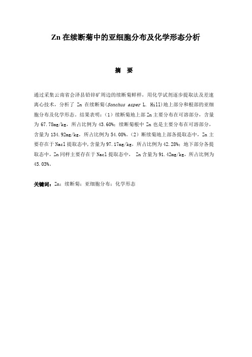
Zn在续断菊中的亚细胞分布及化学形态分析摘要通过采集云南省会泽县铅锌矿周边的续断菊鲜样,用化学试剂逐步提取法及差速离心技术,分析了Zn在续断菊(Sonchus asper L. Hill)地上部分和根部的亚细胞分布及化学形态。
结果表明:(1)续断菊地上部Zn主要分布在可溶部分,含量为67.78mg/kg,所占比例为43.60%;续断菊根中Zn也是主要分布在可溶部分,含量为134.92mg/kg,所占比例为54.08%。
(2)断续菊地上部各提取态中,Zn主要存在于Nacl提取态中,含量为97.17mg/kg,所占比例为42.28%;地下部分各提取态中,Zn同样主要存在于Nacl提取态中, Zn含量为91.42mg/kg,所占比例为45.03%。
关键词:Zn;续断菊;亚细胞分布;化学形态Subcellular Distribution and Chemical Forms of Zn inSonchus asper (L.) Hill.AbstractDifferential centrifugation and sequential chemical extraction were carried out to study the subcellular distribution and chemical form of Zn in the leaves and roots of Sonchus asper(L.) Hill. which was collected from the Pb and Zn mining area of Huize. The result showed that: (1) Zn in the leaves of Sonchus asper(L.) Hill. were mainly distributed in soluble fraction, and its content was 67.78mg/kg, the percentage of Zn contents in soluble fraction reached 43.60%. Zn in the roots of Sonchus asper (L.) Hill. were also mainly distributed in soluble fraction, and its content was 134.92mg/kg, the percentage of Zn contents in soluble fraction reached 54.08%. (2) Zn in the leaves of Sonchus asper (L.) Hill. mainly existed in Nacl-extractable, the content was 97.17mg/kg, the percentage reached 42.28%. In the roots, Zn also mainly existed in Nacl-extractable, the content was 91. 42mg/kg, the percentage reached 45.03%.Key words:Zn;Sonchus asper (L.) Hill.;chemical forms;subcellular distributionZn在续断菊中的亚细胞分布及化学形态分析1前言在全球环境和食品安全问题日趋受到关注的今天,土壤重金属污染的植被修复已成为世界科学研究和技术开发的前沿。
硒对水稻镉含量及其在亚细胞中的分布的影响

硒对水稻镉含量及其在亚细胞中的分布的影响李虹颖;唐杉;王允青;刘英;郭熙盛【摘要】通过元素含量测定与亚细胞分离的方法,分析水稻镉在不同器官、组织之间的差异分布特征,从微观水平上阐释硒增强水稻镉耐受能力的机理,阐明硒降低稻米镉累积量的作用机理。
研究结果显示,(1)随着硒浓度的增加,植株各营养器官干物重均增加。
在2、4、8和16 mg·kg-1 Cd质量分数处理时,1.2 mg·kg-1 Se处理的糙米干物质量比Se 空白处理分别增加了6.81%、7.73%、14.24%和49.62%。
(2)当土壤镉质量分数在2~16 mg·kg-1时,水稻各营养器官和糙米、精米中镉含量随土壤镉浓度的增高而显著增加。
未施硒时,4、8和16 mg·kg-1 Cd处理糙米中镉质量分数分别为0.23、0.37和0.57 mg·kg-1,均超过我国国家食品安全标准中稻米镉的限量(0.20 mg·kg-1)。
(3)相同镉浓度下,随着硒浓度的增加,水稻各营养器官和糙米、精米的镉含量和镉积累量均显著下降,4、8 mg·kg-1 Cd处理组中,糙米的镉含量均低于0.20 mg·kg-1,且1.2 mg·kg-1 Se处理优于0.4和0.8 mg·kg-1 Se处理。
(4)镉在水稻各器官中的分配比例为:根系>茎叶>糙米>精米。
随着硒浓度的增加,镉在精米中的分配比例下降。
结论:硒能够通过调整镉在水稻不同器官中的分配比例,降低稻米中的镉含量;而硒元素对镉毒害的抑制作用,可能是通过细胞对镉的区室化分隔而实现的。
%This paper illustrated the effects of Se application on Cd accumulation and distribution in rice organs and subcellular to explore countermeasures for rice in microcosmic level. The study clarified the mechanism of Cd accumulation reduction in rice. The results were:(1) The dry matter weights of different organs are increased significantly with the increase of Se concentration under same Cd concentration. Under theconcentrations of 2(Cd1)、4(Cd2)、8 (Cd3)and 16(Cd4) mg·kg-1 Cd, the dry matter weights of grain with 1.2 mg·kg-1 Se treatment are increased by 6.81%, 7.73%, 14.24%and 49.62%respectively compared to Se0. (2) When the added Cd concentration in soil is increased from 2 to 16 mg·kg-1, the Cd contents in different organs of rice,brown rice and polished rice are increased significantly.The Cd contents in brown rice are 0.23, 0.37 and 0.57 mg·kg-1 under the Cd2, Cd3 and Cd4 treatments without Se application, higher than the national food safety standards of China for Cd content (0.20 mg·kg-1). (3) The Cd contents and Cd accumulation amounts in different organs of rice and brown rice, polished rice are decreased significantly with the increase of Se concentration under the same Cd concentration, and larger declines of the Cd content are observed under 0.4 and 0.8 mg·kg-1 Se treatment, lower than the national food safety standards of China for Cd content (0.20 mg·kg-1). (4) Sequence of the Cd distribution in rice organs is roots > stems and leave > brown ric > polished rice. The Cd distribution portion in roots is enhanced with the increase of Se concentration as Cd distribution in other organs is declined at the same time. The Cd distribution portion in rice husk is increased with the increase of Se concentration, meanwhile, the Cd distribution portion in polished rice is decreased. Conclusions:Se application could reduce Cd concentration in rice plants, brown rice and polished rice, and reduce Cd transportation into polished rice. Se application shows the most significant inhibition effects to Cd poison by separating Cd in different cell areas.【期刊名称】《生态环境学报》【年(卷),期】2016(025)002【总页数】7页(P320-326)【关键词】硒;镉;糙米;精米;镉含量;镉分配【作者】李虹颖;唐杉;王允青;刘英;郭熙盛【作者单位】安徽省农业科学院土壤肥料研究所,安徽合肥 230031;安徽省农业科学院土壤肥料研究所,安徽合肥 230031;安徽省农业科学院土壤肥料研究所,安徽合肥 230031;安徽省农业科学院土壤肥料研究所,安徽合肥 230031;安徽省农业科学院土壤肥料研究所,安徽合肥 230031【正文语种】中文【中图分类】X53引用格式:李虹颖,唐杉,王允青,刘英,郭熙盛.硒对水稻镉含量及其在亚细胞中的分布的影响[J].生态环境学报,2016, 25(2):320-326.LI Hongying,TANG Shan,WANG Yunqing,LIU Ying,GUO Xisheng.Mechanism of Se on Cd Content and Subcell Distribution in Rice[J].Ecology and Environmental Sciences,2016,25(2):320-326.我国是世界上最大的水稻生产和消费国,稻米安全是我国粮食安全的重要保障.然而,随着我国现代工农业的迅猛发展,重金属通过多种途径进入农业生态环境中,造成农田土壤重金属污染.对生物体而言,Cd是典型的有害重金属,极易在土壤中残留、富集,并可通过食物链的传递,对人体健康构成严重的威胁.如何控制稻米Cd累积量,是保证稻米安全亟需解决的问题.研究表明,施用硒肥可以提高水稻对Cd胁迫环境的抵抗能力,降低Cd在稻米中的积累量.梁程等(2012)报道,Se提高了水稻幼苗中GSH和PCs的含量,促进水稻体内Cd和PC的络合作用,从而减缓Cd对水稻幼苗的毒害.庞晓辰等(2014)发现,在Cd 处理浓度为5.0 μmol.L-1条件下,外源Se(Ⅳ)会显著降低水稻对Cd的吸收和转运.于淑慧等(2013)的研究表明,Se能降低Cd的转移系数,抑制Cd从水稻根部向地上部的转移,降低地上部Cd的累积量;而植物总是尽量避免重金属元素损伤其相对重要的组织、细胞和细胞器,从而表现出选择性分布.土壤中的重金属通过根系的吸收途径进入植物体内,一部分贮存在根系,另一部分通过输导组织向植物的各个器官进行转运分配.因此,Cd在不同亚细胞成分之间的分配差异反应了水稻对Cd的耐受机制,而Cd在不同器官之间的分配差异则会影响稻米中Cd的累积量.本研究通过分离水稻的亚细胞组分并测定镉在不同细胞组分中的含量,分析在硒、镉互作条件下,水稻Cd的亚细胞分布特征,从微观水平上揭示Se增强水稻Cd耐受能力的机理;并通过Cd在不同器官、组织之间的差异分布,阐明Se降低稻米Cd累积量的作用机理.1.1 试验材料供试水稻品种为早籼788.试验地土壤基本理化性状为:有机质含量25.40 g.kg-1,全氮2.33 g.kg-1,碱解氮107.1 mg.kg-1,全磷0.33 g.kg-1,速效磷7.21 mg.kg-1,速效钾107.8 mg.kg-1,pH 5.32;全镉含量0.042 mg.kg-1,全硒0.03 mg.kg-1.氮肥为尿素,磷肥用磷酸氢二铵,钾肥为硫酸钾,用分析纯亚硒酸钠、分析纯氯化镉处理土壤.1.2 试验设计试验于2015年4─7月在安徽省农业科学院水稻栽培大棚盆栽试验场进行.采用盆栽方法,每盆栽水稻3穴,每穴3株.设5个土壤镉浓度,分别为0、2、4、8、16 mg.kg-1,用Cd0、Cd1、Cd2、Cd3、Cd4表示.每个镉浓度下设0、0.4、0.8、1.2 mg.kg-14个土壤硒水平,用Se0、Se1、Se2、Se3表示.共20个处理,每处理4次重复.试验用塑料桶直径30 cm,高50 cm,装土15 kg.氮肥按基肥∶返青肥∶分蘖肥∶穗肥=4∶2.5∶1.5∶2施用,磷肥作基肥一次性施入,钾肥按基肥∶分蘖肥∶穗肥=4∶2∶4施用.移栽前,先用水泡土3 d,然后将肥料施入土中混匀,同时将亚硒酸钠和氯化镉配成溶液均匀施入.1.3 测定项目与方法1.3.1 样品制备将成熟期的水稻植株整盆收获,依次用自来水和蒸馏水清洗,将根、茎、叶、籽粒分开,105℃下杀青30 min,80℃下烘干至恒质量.砻谷机除去稻壳,将稻壳收集起来;然后用精米机将糙米处理为精米,将精米和糠皮分别收集并粉碎备用.1.3.2 镉含量的测定称取1 g样品,置于25 mL三角瓶中,加入10 mL体积比4∶1的优级纯HNO3-HClO4混合酸,冷消化过夜.第2天,将三角瓶置于砂浴上,在165~175℃下消化,直至溶液变为无色透明,稍冷却后,分别加两次约1 mL的蒸馏水排酸,蒸发浓缩消化液至2 mL左右,用5%的HCl转移并定容至10 mL比色管中,用AA7003原子吸收光谱仪测定镉含量.1.3.3 镉的亚细胞分离称取新鲜的水稻根系和地上部分各0.2 g,分别置于研钵中,加入预冷的提取缓冲液充分研磨成匀浆液.提取缓冲液的组成为:250 mmol.L-1蔗糖, 50 mmol.L-1Tris-HCl(pH值7.5)和1 mmol.L-1的二硫赤鲜醇,实验操作过程均在4℃下进行.将匀浆液在3000 r.min-1条件下离心15 min,沉淀即为细胞壁组分;继续将上清液在15000 r.min-1条件下离心30 min,沉淀为细胞器组分,上清液为胞液组分;测定Cd 全量,并计算得出回收率为92%~101%,之后将各组分用HNO3-H2O2法消化后,用原子吸收仪(AAS ZEEnit700)测定样品Cd含量.1.4 数据处理将实验数据进行平均值和标准差统计分析,统计检验采用SPSS for Windows 11.5软件,显著性差异水平为P≤0.05.2.1 硒-镉互作对水稻生物量的影响如表1所示,在相同的土壤Cd浓度下,随着施Se浓度的增加,植株各营养器官干物重和总干物重均增加,但增加幅度不同.Cd0浓度时,Se3处理的糙米干物重比Se0处理增加了5.53%,而在Cd1、Cd2、Cd3和Cd4浓度时,Se3处理的糙米干物重比Se0处理分别增加了6.81%、7.73%、14.24%和49.62%.在相同硒浓度下,随着土壤镉浓度的增加,植株各器官干物重和总干物重均下降(表1).在Se0浓度下,Cd1、Cd2、Cd3、Cd4处理比Cd0处理糙米的干物重分别减少了2.90%、4.01%、10.73%和39.24%.在Se3浓度下,Cd1、Cd2、Cd3、Cd4处理比Cd0处理的糙米干物重分别减少了1.81%、2.04%、3.45%和13.81%.由此说明,施硒能有效抑制由镉引起的水稻减产.2.2 硒对水稻镉含量和积累量的降低作用从表2可以看出,水稻各器官中镉含量的大小顺序为根系>茎叶>糙米.在相同的土壤硒浓度下,随着土壤中镉浓度的增加,各营养器官中镉含量也随之增加.Se0处理时,各营养器官的镉含量在不同镉处理间差异极显著;其中,Cd4浓度下各营养器官的镉含量最高,根系、茎叶、糙米中镉含量高达8.54、3.02和0.57 mg.kg-1,分别是Cd0处理的78、43和57倍.相同土壤镉浓度下,各营养器官的镉含量随着硒浓度的增加而减少.表2显示,未施硒(Se0)时,Cd2、Cd3和Cd4处理糙米中镉含量分别为0.23、0.37和0.57 mg.kg-1,均超过我国国家食品安全标准(GB2762─2012)中大米镉限量0.20 mg.kg-1,分别是Cd0处理的23、37和57倍.在Se3处理条件下, Cd1、Cd2和Cd3处理的糙米镉含量均低于我国国家食品安全规定的大米镉限量,分别是Cd0处理的9、14和19倍;而在Cd4组中,Se3处理条件下的糙米镉含量为0.21 mg.kg-1,仍然高于0.20 mg.kg-1.相同镉浓度时,随着硒浓度的增加,糙米的镉含量均明显下降.在Cd1、Cd2、Cd3和Cd4组中,Se3处理的糙米镉含量分别比Se0下降了47.06%、39.13%、48.65%和63.16%.由表3可知,土壤镉浓度和硒浓度分别对水稻各器官镉积累量的影响极显著.土壤镉浓度与土壤硒浓度的交互作用对根系、茎叶、糙米镉积累量均有极显著影响.水稻各器官镉积累量的大小顺序为根系>茎叶>糙米.相同外源镉浓度时,随着土壤硒浓度的增加,水稻地上部分各器官镉积累量均下降.在Cd4处理条件下,随着土壤硒浓度的增加,水稻根系镉积累量的最大下降幅度是21.47%.在外源添加镉浓度为4mg.kg-1时,水稻茎叶镉积累量下降幅度最大,Se3处理比Se0处理的镉积累量下降了24.75%.在外源添加镉浓度为16 mg.kg-1时,随着土壤硒浓度的增加,糙米镉积累量下降幅度最大,Se3处理比Se0处理的糙米镉积累量下降了44.63%.在不同的外源镉处理浓度下,随着土壤硒浓度的增加,水稻各器官镉分配比例的变化趋势也不相同.根系中镉的分配比例最高,并且随着土壤硒浓度的增加而增加.其中,在外源添加镉浓度为16 mg.kg-1时,水稻根系中镉分配比例最高,Se0、Se1、Se2和Se3处理的水稻根系镉分配比例分别为63.82%、65.03%、66.41%和66.29%.糙米中镉的分配比例最低,并随着土壤硒浓度的增加而降低.在外源添加镉浓度为4 mg.kg-1时,Se3处理比Se0处理的糙米镉分配比例下降幅度最小,为34.42%;在外源添加镉浓度为16 mg.kg-1时,Se3处理比Se0处理的糙米镉分配比例下降幅度最大,为44.63%.2.3 硒对水稻镉分布特征的影响由表4可知,土壤镉浓度、土壤硒浓度、土壤镉-硒交互作用均对稻谷各部位镉积累量有极显著的影响.土壤镉添加浓度为0 mg.kg-1时,稻谷不同部位镉积累量随着土壤硒浓度的增加而无显著变化.在Cd1、Cd2、Cd3和Cd4等土壤镉浓度时,随着外源硒浓度的增加,稻谷各部位镉积累量均下降.在Cd4条件下,Se3处理比Se0处理稻壳镉积累量下降了36.79%.在Cd3土壤镉浓度时,糠皮镉积累量下降幅度最大,Se3处理比Se0处理的糠皮镉积累量减少45.00%.在Cd1、Cd2、Cd3和Cd4处理组中,Se3处理比Se0处理的精米镉积累量分别下降了56.06%、46.43%、53.91%和54.78%.在相同镉浓度下,随着硒浓度的增加,稻壳中镉的分配比例增加,精米镉的分配比例下降,糠皮中镉的分配比例变化不大.在Cd1处理时,稻壳镉分配比例增幅最大,Se3处理比Se0处理的稻壳镉分配比例增加了11.42%.在Cd1处理组中,精米镉的分配比例下降幅度最大,Se3处理比Se0处理的精米镉分配比例下降了12.16%.从表5可知,施硒可以显著降低水稻根系各亚细胞组分中的镉含量.在相同镉浓度下,随着硒浓度的增加,水稻根系各亚细胞组分中的镉含量下降.从各亚细胞组分分配率上看,根系组织中的镉主要分布在细胞壁上;且在同一镉浓度下,随着土壤硒处理浓度的增加,细胞壁上的镉分配率呈上升趋势,而镉在细胞器上的分配率最低.在Cd1、Cd2、Cd3和Cd4组中,Se3处理比Se0处理的细胞器镉分配率分别下降了3.89%、3.73%、2.74%和2.90%.从表5可以看出,水稻茎叶各亚细胞组分中的镉含量随着外源镉浓度的升高而增加,硒显著降低了茎叶各亚细胞组分中的镉含量.在Cd1组中,茎叶细胞器中镉含量下降幅度最大,Se3处理比Se0处理的细胞器镉含量下降了58.82%;在Cd3组中,茎叶细胞器中镉含量下降幅度最小,Se3处理比Se0处理的细胞器镉含量下降了48.39%.从各亚细胞组分分配率上看,随着硒处理浓度的增加,细胞壁上的镉分配率呈上升趋势.在Cd1、Cd2、Cd3和Cd4组中,Se3处理比Se0处理的细胞壁镉分配率分别增长了12.17%、10.76%、11.00%和12.35%.3.1 硒对镉胁迫下水稻干物重的影响研究结果表明,在水稻全生育期中,100 μmol.L-1Cd处理显著降低水稻单株株高、结实率和千粒重,并影响产量(林莉,2011).在本试验条件下,随着土壤镉浓度的增加,水稻各器官干物重均下降,表明镉胁迫能抑制水稻的生长发育,与林莉的观点一致.杨洋等(2015)的研究结果表明:在镉污染的农田中施用硒素可以明显提高水稻幼苗茎基宽、根冠比及生物量.本试验结果显示,在相同镉胁迫浓度时,随着土壤硒浓度的增加,水稻各营养器官干物重均增加,Se3处理对干物重的影响优于Se1和Se2处理,表明施用硒元素可以明显减小镉胁迫对水稻生物量的降低幅度.这一结论说明施硒能促进镉污染土壤上水稻的生长,实现水稻增产.3.2 硒对镉胁迫下水稻镉含量的影响研究表明,随着土壤中镉浓度的升高,水稻各器官镉含量随之增加(黄冬芬,2008).王凯荣等(1996)的水培试验结果表明,当培养液镉浓度升至0.01 mg.kg-1时,糙米中的镉富集系数超过500.在镉胁迫条件下,水稻根系的镉含量要高于茎叶和稻米,即根>茎叶>稻米(赵雄等,2009).曾翔(2006)指出,水稻成熟后,各器官镉含量的大小顺序为:根系>茎鞘>叶片>糙米.上述结论与本文结论相一致,在本试验条件下,当土壤镉浓度在2~16 mg.kg-1范围内时,水稻营养器官和糙米中的镉含量随土壤镉浓度的增高而显著增加;而本试验测得的水稻各器官中镉含量顺序为根系>茎叶>糙米>精米. 对57个水稻品种籽粒中镉含量的研究证明,稻谷中硒含量与镉含量呈负相关关系(李正文等, 2003).方勇(2010)通过在水稻叶面喷硒的试验表明,当叶面喷硒量超过750 g.hm-2时,稻米中镉含量显著下降.叶面施硒可以有效地降低精米中的镉含量(李佳,2010).施硒稻米中镉含量比未施硒的下降29.8%(谭周磁等,2000).本文的试验结果与上述研究结论基本一致,即在所有土壤镉浓度下,随着外施硒浓度的增加,糙米、精米中镉含量均明显下降,尤其当土壤镉浓度为16 mg.kg-1时,精米镉含量在水稻施用硒素后下降最明显,且Se3处理对降低精米中镉含量的效果优于Se1和Se2处理.3.3 硒对镉胁迫下水稻镉积累和分配的影响水稻生长在镉污染环境中,植株长势虽未受到明显伤害,但土壤中的镉会在稻米中积累,并通过食物链的传递对人体健康造成极大的威胁.水稻镉积累量与环境镉含量具有一致性,但水稻不同器官中镉的积累量存在很大差异.研究表明,当土壤镉浓度为5 mg.kg-1时,水稻体内镉的分配规律为茎叶>根系>颖壳>籽粒(刘侯俊等,2011).曾翔(2006)针对水稻基因型差异对镉积累分配的影响作用,对130个水稻品种的镉积累分配规律进行了研究.结果表明:镉在水稻中分配规律表现为根系>茎叶>籽粒.本试验结果表明,在所有镉处理组中,成熟期水稻各器官镉的分配顺序均为根系>茎叶>糙米>精米,与上述研究结果相近.刘春梅等(2015)的研究结果表明,施硒可显著降低精米中镉的分配比例,尤其在土壤镉加入量为2 mg.kg-1条件下,施硒效果最显著,以施硒量为0.07 mg.kg-1的处理效果最好,稻米中的镉含量下降明显,继而可以保证稻米的食用安全性.江巧君等(2013)的研究结果表明,施用有机肥抑制了镉向精米输送的效应,降低了精米中的镉分配比例,从而显著降低了精米中的镉含量.本文研究了镉在稻谷中分配特征,结果表明:随着土壤镉浓度的增加,稻壳中镉分配比例逐渐上升,精米中镉分配比例逐渐降低.说明除了根系能阻滞土壤中镉向地上部运转外,稻壳也能阻滞镉向精米中的运输.在相同镉浓度下,随着土壤硒浓度的增加,稻壳中的镉分配比例相应增加,而精米中的镉分配比例却不断下降,这说明:施硒能增加镉在稻壳中的分配,减少镉向精米中运转,有助于提高精米的食用安全性,这一结论与刘春梅等的研究结论相一致.土壤镉浓度在0~16 mg.kg-1时,水稻各器官镉含量随着土壤镉浓度的增加而增加,镉含量顺序为根系>茎叶>糙米>精米.施硒影响植株和籽粒中的镉含量及其分配比例.施用硒肥可以降低水稻各器官和稻米(糙米和精米)中的镉含量:硒促进水稻根系镉积累量的下降,并提高了镉在根系中的分配比例;同时,硒降低了稻米中镉的积累量和分配比例.随着土壤硒浓度的增加,水稻各器官亚细胞组分中的镉含量降低;根系和茎叶中细胞壁上的镉分配率呈上升趋势,细胞器中的镉分配率呈下降趋势.上述结果说明,硒肥能够通过调整镉在水稻不同器官中的分配比例,降低稻米中的镉含量;而硒元素对镉毒害的抑制作用,可能是通过细胞对镉的区室化分隔实现的.因此,施硒能降低稻米中的镉含量和积累量,是保证稻米食用安全性的一项简单易行的农艺措施. SHANG Q M,LI P L,GAO L H.1997.Selenium Uptake and Inversion by Hydroponic Lettuce[J].Acta Horticulturae Sinica,24(3):255-258.曾翔.2006.水稻镉积累和耐性机理及其品种间差异研究[D].长沙:湖南农业大学:55. 方勇.2010.外源硒在水稻籽中的生物强化和化学形态研究[D].南京:南京农业大学:18.黄冬芬.2008.水稻对土壤重金属镉的响应及其调控[D].扬州:扬州大学:36-39.江巧君,周琴,韩亮亮,等.2013.有机肥对镉胁迫下不同基因型水稻镉吸收和分配的影响[J].农业环境科学学报,32(1):9-14.李佳.2010.硒对寒地水稻产量和稻米安全品质的影响[D].哈尔滨:东北农业大学:36. 李正文,张艳玲,潘根兴,等.2003.不同水稻品种籽粒Cd、Cu和Se的含量差异及其人类膳食摄取风险[J].环境科学,24(3):112-115.梁程,林匡飞,张雯,等.2012.不同浓度硫处理下硒镉交互胁迫对水稻幼苗的生理特性影响[J].农业环境科学学报,31(5):857-866.林莉.2011.硒缓解水稻镉毒害的机理研究[D].杭州:浙江大学:16-18.刘春梅,罗盛国,刘元英.2015.硒对镉胁迫下寒地水稻镉含量与分配的影响[J].植物营养与肥料学报,21(1):190-199.刘侯俊,梁吉哲,韩晓日,等.2011.东北地区不同水稻品种对Cd的累积特性研究[J].农业环境科学学报,30(2):220-227.庞晓辰,王辉,吴泽嬴,等.2014.硒对水稻镉毒性的影响及其机制的研究[J].农业环境科学学报,33(9):1679-1685.谭周磁,陈嘉勤,薛海霞.2000.硒(Se)对降低水稻重金属Pb、Cd、Cr污染的研究[J].湖南师范大学自然科学学报,23(3):80-83.王凯荣,龚惠群.1996.两种基因型水稻对环境镉吸收与再分配差异性比较研究[J].农业环境保护,15(4):145-149,176.杨洋,李楠,葛鑫,等.2015.硫硒配施对镉胁迫下水稻幼苗生长及其吸收积累镉的影响[J].北方水稻,45(2):7-9.于淑慧,周鑫斌,王文华,等.2013.硒对水稻幼苗吸收镉的影响[J].西南大学学报(自然科学版),35(9):17-22.赵雄,李福燕,张冬明,等.2009.水稻土镉污染与水稻镉含量相关性研究[J].农业环境科学学报,28(11):2236-2240.。
01 叶绿体的分离及荧光观察

山东大学实验报告2018年3月12日________________________________________ _________________________姓名:丁志康系年级:2016级组别:1-1 同组者:科目:细胞生物学实验题目:叶绿体的分离及荧光观察学号:201600140055一、目的要求1、通过植物细胞叶绿体的分离,了解细胞器分离的一般原理和方法。
2、观察叶绿体的自发荧光和次生荧光,并熟悉荧光显微镜的使用方法。
二、实验原理真核细胞由细胞膜、细胞核和细胞质组成。
细胞质中含有若干细胞器和细胞骨架,这些结构被称为亚细胞组分。
对于细胞的结构和功能的研究,是细胞生物学的基本课题。
其重要的研究手段之一是分离、纯化各种亚细胞组分,然后观察他们的结构,对它们的功能进行生化分析。
1、分离:分离亚细胞组分的主要方法由差速离心和密度梯度离心两种,本次试验中使用差速离心法。
差速离心法是在密度均一的介质中有低速到高速的逐渐离心,用于分离不同大小的物体。
离心速度逐渐提高,样品会按先大后小的顺序沉淀。
在差速离心中细胞器沉降的顺序为:细胞核、线粒体与叶绿体、溶酶体与过氧化物酶体、内质网与高尔基体,最后为核糖核蛋白复合体。
由于各细胞器在大小和密度上可能相互重叠,一般重复差速离心2-3次,分离效果会好一些。
差速离心只用于分离密度和大小相差悬殊的细胞或细胞器,并且得到的产物纯度很低。
若对纯度的要求较高,则需要用密度梯度离心来分离、纯化。
2、叶绿体:叶绿体是植物细胞所特有的能量转换细胞器,光合作用就是在叶绿体中进行的。
由于具有这一重要功能,所以它一直是细胞生物学、遗传学和分子生物学的重要研究对象。
叶绿体是植物细胞中较大的一种细胞器,利用低速离心即可分离集中进行各种研究。
3、荧光显微技术:荧光显微术是利用荧光显微镜对可发荧光的物质进行观测的一种技术。
某些物质在一定短波长的光(如紫外光)的照射下吸收光能进入激发态,从激发态回到基态时,就能在极短的时间内放射出比照射光波长更长的光(如可见光),这种光就称为荧光。
亚细胞的分离

(6)用PBS+1%NGS洗3次,每次10 min。
(7)加入一抗,室温孵育1 h。
(8)用PBS+1%NGS洗3次,每次10 min。 ( 9 )用 1:50 稀释的过氧化物酶偶联的二抗(用亲和层析纯 化)室温孵育1 h,反应在潮湿仓里进行。 (10)用PBS洗3次,每次10 min。 (11)用0.05 mol/L Tris-HCl(pH 7.6)洗2次,每次l0 min。
DAPI 与 A-T 碱基结合后,其荧光比处于溶液中的自 由状态或与 G-C 碱基结合的荧光强约 20 倍。 DAPI 可 用于显示支原体污染及线粒体,但最常用于显示细 胞核和染色体。缺点是,在多色成像中,用 360 nm 的光激发时, DAPI 发射的光太亮,从而掩盖了其他 的荧光信号,可改用405 nm的光作为激发光。 长波长的光不仅能很好地激发 DNA,还可减少对细 胞的损害,而且也减少对其他荧光染料的漂白作用。 由于DAPI有足够的明亮度,故仍是标准的DNA荧光 染料。
(三)方法 1.过氧化物酶偶联抗体进行蛋白质的定位 (1)在35 mm的培养皿或6孔细胞培养板的一个孔中 放入一张盖玻片(22 mm×25 mm),在盖玻片上培 养细胞。当细胞在盖玻片上铺满50-70%是最理想的 状态,但具体情况应视实验研究决定。 悬浮培养的细胞可通过细胞离心涂片器(Cytospin, Shandon Lipshaw)或用多聚L赖氨酸包被盖玻片来 使其黏附在盖玻片上。
2. 抗体标记
抗体与抗原的结合方法:直接法和间接法。
1. 直接法是将带有标记的抗体与抗原反应,显示出抗原存在
的部位。
2. 间接法是在抗体抗原初级反应的基础上,再用带标记的次
级抗体同初级抗体反应,从而使初级反应得到放大,显示 增强。
实验六 菠菜叶绿体分离与荧光观察 (1)
菠菜叶片叶绿体分离与荧光观察
实验目的
1、了解细胞器分离的一般原理,
掌握基本分离方法。 2、观察叶绿体自发荧光与间接荧
光
实验原理
细胞器的分离:
先匀浆→差速离心。
将组织匀浆后在等渗介质中差速离心, 是分离细胞器的常用方法。
实验原理
用低渗、超声破碎或研磨等匀浆方法可使细胞质 膜破损,形成细胞核、线粒体、叶绿体、内质 网、高尔基体、溶酶体等细胞器和细胞组分组 成的混合匀浆,再通过差速离心,可将各种亚 细胞组分和各种颗粒分开。
实验原理
差速离心:即利用不同的离心速度所产生的不同离心力, 分离各种亚细胞组分和各种颗粒。 在一定的离心场中,同一时间内,密度、大小不同的颗粒 其沉降速度不同,依次增加离心力和离心时间,密度及大 小不同的颗粒会先后分批沉降在离心管底部,分批收集即 可获得各种亚细胞组分。 颗粒在离心场中,其沉降速度取决于其大小、形状和密度,
注意事项:
实验步骤
二、叶绿体的间接荧光观察
取叶绿体悬液1滴于载玻片上, 滴加1滴0.01%吖啶橙,染色10min, 加盖片,荧光显微镜观察叶绿体 的间接荧光。 注意事项:
思考题
P110 第3题:
叶绿体分离实验的原理是什么? 在分离叶绿体时应注意什么问题?ຫໍສະໝຸດ 也与离心力及介质黏度有关。
Differential centrifugation 沉降顺序:核——线粒体——溶酶 体与过氧化物酶体——内质网与高 尔基体——核蛋白体。
High speed
Low speed
实验原理
自发荧光:细胞内含有的某些天然物质,
经短波光照射后所发出的光。
诱发荧光:物体经荧光染料染色后,再 经短波光照射所发出的荧光。
亚细胞分级分离的基本方法和原理

亚细胞分级分离的基本方法和原理亚细胞分级分离,听起来是不是有点高大上?其实啊,它就像我们生活中的一种“分拣”过程,想象一下,咱们在超市买菜的时候,总是要把好的和不好的分开对吧?在生物科学的世界里,亚细胞分级分离也是在做类似的事情,目的是为了把细胞里的小伙伴们一一分开,找出它们各自的“特色”。
今天,就让我们轻松聊聊这个神奇的过程,绝对不会让你打瞌睡哦!1. 什么是亚细胞分级分离?亚细胞分级分离,顾名思义,就是把细胞里的成分进行分类。
细胞就像一个热闹的市场,里面有核糖体、线粒体、内质网等各种“摊位”,每个“摊位”都有自己的特产。
可是,想要研究这些特产,我们必须先把它们分开。
想象一下,若是在市场里你想找新鲜的水果,可是摊位全都混在一起,那得多麻烦呀!所以,亚细胞分级分离就派上用场了。
1.1 基本原理要分离这些细胞成分,科学家们可不是随便来一招。
其实,亚细胞分级分离主要依赖一些物理和化学原理,比如离心、过滤和沉淀。
你可以把这个过程想象成一场“体育竞技”,不同的细胞成分根据自身的特性(比如大小、密度等)进行“比赛”。
有的跑得快,有的慢;有的沉得下去,有的飘得上来。
通过精心设计的实验,科学家们就能把这些小家伙们一一“请”出来。
1.2 常见方法接下来,我们来聊聊几种常用的分离方法,嘿,这可是科学家们的“拿手好戏”!离心法:这可是个明星方法哦!通过高速旋转,利用离心力让不同密度的细胞成分分层,就像是沙滩上建的沙堡,重的在底下,轻的在上面。
这招可谓是“势不可挡”!过滤法:简单直接,利用滤网把细胞混合物中的小家伙筛选出来。
就像你在筛米一样,把米和杂质分开,干干净净!沉淀法:把细胞混合物静置一段时间,让沉重的成分慢慢沉底,轻的部分则漂在上面。
这就像在河里捞鱼,先把沉底的鱼儿抓上来,轻松又高效!2. 过程中的挑战当然,这个过程也不是一帆风顺的,挑战可是多得很。
比如,细胞成分有时候会“叛变”,也就是在分离的时候发生变性,导致结果不理想。
简要说明细胞组分的分级分离原理及其意义。

简要说明细胞组分的分级分离原理及其意义。
细胞组分的分级分离原理及其意义:
一、分级分离原理
1、离心分离
把细胞组分,如核糖体、细胞质等,悬浮在生化介质中,用中央偏心
的方法将悬浮体快速分级,分为悬浮层和沉淀层,从而脱除低分子的
悬浮物,获得纯的细胞组分。
2、逆流洗涤法
介质会具有不同的物质等离子强度、温度等,将原悬浮液放置在此种
不同介质下,形成微孔隙,有效运载溶液,将微粒通过微孔小于亚细
胞级分子得到分离。
3、冷凝分离
冷凝作用可有效地将微粒形态,质量,大小不同而可悬液的细胞组分,按分子量和形状进行有效分离。
二、分级分离的意义
1、节约成本
由于分级分离的方法节省了消耗的流体,可以有效的节约经济成本。
2、提高效率
分级分离能以高速度进行解离,可以大大减少操作时间,从而提高效率。
3、保护过滤目标物
使用分级分离可以减少操作时间和免疫试剂,可以有效地预防细胞组分的损伤,从而保护需要分离的目标物质。
4、增强产品的纯度
使用分级分离技术可以使分离的细胞组分具有更高的纯度,并可以便捷的获得细胞质、线粒体、核糖体等细胞组分的复合物。
RBA
2.亲和力要高:
亲和力高的标记配基在浓度较低时便 可达到饱和,因此用量可节省。此外, 这种配基与受体形成的复合物往往解离 较慢,有利于结合和游离配基的分离。 不少拮抗剂的亲和力比激动剂高,因此 用得较多。
3.特异性要高:
首先是与其它种类的受体交叉反应要尽量少, 否则需要采取措施预先除去标本中有交叉反应 的受体。
质量作用定律 (Mass action law): 受体和配基的结合反应服从可逆反应的质量
作用定律,可用下式表示:
k1, v1 [R]+[L] ---- [RL]
k2, v2
上式中,[R]和[L] 为游离受体和游离配基的浓度,[RL]为复合物浓度, v1和v2为结合速率和解离速率,k1和k2为结合速率常数 (association rate constant) 和解离速率常数 (dissociation rate constant)。
如果在放射配基与受体结合后将受体 周围多余的放射配基移去(例如淋洗或透 析),则放射配基与受体的复合物会逐步 解离,亦即这种结合是可逆反应。
如果向反应系统中加入大量非标记配 基,使其浓度远高于放射配基,则将取 代大部分原已结合的放射配基,使放射 性复合物减少, 也说明配基与受体的结合 是可逆的。
4、受体分布的组织专一性: 在正常的生物体内,受体与配基的结合是在
刮下组织切片测定放射性活度,也可用 放射自显影或免疫组化染色方法研究受 体的分布。
3、完整的细胞:
可以是血细胞、肝或脾细胞、肿瘤的腹 水型细胞、传代或原代培养细胞等的悬 液。
其主要优点是:
①可以同时观察配基与受体结合特性和细 胞的生物效应;
②受体处于接近正常的环境中,得到的结 果更能反映受体的生理特性;
- 1、下载文档前请自行甄别文档内容的完整性,平台不提供额外的编辑、内容补充、找答案等附加服务。
- 2、"仅部分预览"的文档,不可在线预览部分如存在完整性等问题,可反馈申请退款(可完整预览的文档不适用该条件!)。
- 3、如文档侵犯您的权益,请联系客服反馈,我们会尽快为您处理(人工客服工作时间:9:00-18:30)。
植物体内的亚细胞组分分离
步骤:准确称取植株鲜样0.5000 g,加入20 mL提取液(0.25 mmol/L蔗糖+50 m mol/L Tris HCl缓冲液(pH 7.5)),研磨匀浆,用尼龙纱布过滤,滤渣为细胞壁部分;滤液在600 r/min下离心10min,沉淀为细胞核部分;上清液在2000 r/min下离心15 min,沉淀为叶绿体部分;上清液在10000 r/min下离心20 min,沉淀为线粒体部分;上清液为含核糖体的可溶部分,每组两次离心,全部操作在4下进行。
植物亚细胞组分的分离
步骤:准确称取鲜样0.5000 g,加入20 mL提取液[0.25mol·L-1蔗糖+50 mmol·L-1 Tris-HCl缓冲液(pH7.5)+1mmol·L-1二硫赤鲜糖醇,研磨匀浆,匀浆液在冷冻离心机300×g下离心30 s,沉淀为细胞壁组分;上清液在2000×g下离心15 min,沉淀为细胞核和叶绿体组分;上清液在10000×g下离心20 min,沉淀为线粒体组分;上清液为含核糖体的可溶组分。
全部操作在4℃下进行。
Fractionation of Leaves and Roots.Leaves and roots were homogenized using a mortar and pestle in a medium containing
0.25 M sucrose,50 mM Tris-HCl(pH 7.5),and 1 mm
dithioerythritol.All steps were performed at 4 C.The resulting brei was strained through eight layers of cheesecloth,and liquid was expressed from the residue.The residue was washed twice with the grinding medium.The pooled washes,together with the first filtrate,were centrifuged at 300g for 30 s.The resulting pellet combined with the residue of the cheesecloth filtration contained mainly cell walls and cell wall debris and was designated as cell wall fraction(I).The supernatant of the first centrifugation step was then centrifuged at 20,000g for 45 min to sediment cell organelles.The pellet was taken as organelle fraction(II).The resultant supernatant solution referred to as soluble fraction(III)was employed in subsequent characterization studies as described.Fractions I and II were analyzed for their Cd content.The supernatant,fraction III,was further fractionated by gel filtration ,using Sephadex G-100,G-25,and G-10(Pharmacia Fine Chemicals, Uppsala,Sweden).An aliquot of fraction III was applied to a column(100 x 2.6 cm)of G-100 using 50 mm Tris-HCl(pH 7.5) and 0.1 mM dithioerythritol as eluting buffer.Fractions containing Cd were pooled and chromatographed on calibrated G-25(roots) or G-10(leaves)columns(70 x 2.3
cm).Fractions were collected on an LKB Ultrarac fraction collector.The effluents were monitored at 280 nm with a Gilford spectrophotometer 2400 S.The activities of the glutamate dehydrogenase and malate dehydrogenase were determined according to references 9 and 23.。