医学影像学英文课件:Bone and joint X ray appearance
医学影像学英文课件

医学影像学英文课件Medical Imaging Course Slides1. Introduction to Medical ImagingMedical imaging is a broad field that encompasses various techniques used to visualize the internalstructures and functions of the human body. These techniques play a crucial role in the diagnosis, treatment, and monitoring of various medical conditions. The most commonly used medical imaging modalities include X-ray, computed tomography (CT), magnetic resonance imaging (MRI), ultrasound, and nuclear imaging.医学影像学是一个广泛的领域,包括各种用于可视化人体内部结构和功能的技术。
这些技术在诊断、治疗和监测各种医疗状况中发挥着关键作用。
最常用的医学影像模态包括X射线、计算机断层扫描(CT)、磁共振成像(MRI)、超声波和核医学成像。
2. X-ray ImagingX-ray imaging is one of the oldest and most widely used medical imaging techniques. It utilizes high-energy electromagnetic radiation to create images of the body's internal structures. X-rays are able to pass through thebody, and the degree of absorption by different tissues is used to create the image. This technique is particularly useful for visualizing bones, joints, and the chest cavity.X射线成像是最古老和最广泛使用的医学影像技术之一。
骨科影像学PPT课件

关节脱位
92
关节脱位
93
2019/12/5
.
94
边状、骨膜三角。 临床:多见于炎症、肿瘤、外伤等,骨膜增生提
示病变存在,但无法确定病变性质,须结合 其它征象。
58
骨膜增生
59
骨膜增生 骨膜三角(Codman’s 三角):多见于恶性
肿瘤,也见于急性炎症。
60
骨骼基本病变
骨质坏死(necrosis of bone)
定义:骨组织血供中断,代谢停止。坏死的骨质 称为死骨(sequestrum)。
17
膝关节空气造影
18
检查方法
血管造影:主要用于良恶性肿瘤 的鉴别。恶性骨肿瘤常有特征性 征象。
19
下 肢 血 管 M RA
20
头颈部MRA
21
检查方法
CT 常用于脊柱、骨盆、头颅五官。 CT 较少用于四肢骨骼,因其为横断
扫描。多排螺旋CT重建功能现广泛 应用于骨关节外伤。
22
腰椎间盘突出
46
骨骼基本病变-骨质疏松
47
MRI全脊柱成像—骨质疏松
48
骨骼基本病变
骨质软化(osteomalacia)
定 义:一定单位体积内骨组织有机成分正常, 矿物质含量不足。大量类骨质堆积得不到 钙化。
X线表现:骨质密度减低,显影模糊,皮质变 薄,小梁减少。承重骨骼各种变形,X形 腿、O形腿。此外,还可见假骨折线。
浸润性生长,边缘模 膨胀性生长,边界清楚,
糊,形态不规则,骨 形态规则或不规则,骨皮
皮质破坏中断,多伴骨 质膨大,保持连续性,较
膜增生。
少骨膜增生。
53
54
55
骨骼基本病变
【英文课件】Bones and Joints -北京协和医院学习

Swelling
Space narrowing
n Destruction
Degeneration
n Bony Ankylosis :Septic arthritis, Ankylosing spondylitis AS
n Fibrotic ankylosis: Tuberculosis, Rheumatoid arthritis
n Periosteum; n Cortical bone; n Trabecular bone; n Medullary space
Variation
n Epiphyseal plate residue
n Maldevelopment line
n Bone spot
Joint
n Joint space n Bony articular
CT
n Complex bone structure/fracture n Window width
MR
n Soft tissue lesions(fat,muscle, ligment, etc.)
n Calcification
Osteoporosis (骨质疏松)
n 骨组织中的有机和无机成分都减少,但比例正常。 皮质薄;哈氏管扩大;小梁少,间隙大。
n 骨密度降低; n 骨松质骨小梁变细减少,间隙增宽,骨皮质出现分
层和变薄。 n 脊椎中,纵行小梁明显,椎体变扁,上下缘内凹,
椎间隙增宽,梭形。 n 压缩骨折时呈楔状。压缩骨折(双凹、楔形变、只
有前后都短才是压缩骨折) n 全身性:老年性、代谢性(甲旁亢、绝经期)、废
用性、药物性(激素)等 n 局限性:肢体废用性、炎症、血管神经功能障碍等
英文影像学PPT

杨绛先生:翻译的技巧
要把西方语文翻成通顺的汉语,就得翻个大跟头才颠倒得过来 汉语和西方语言同样是从第一句开始,一句接一句,一段接一段 ,直到结尾;不同主要在句子内部的结构。西方语言多复句,可 以很长;汉文多单句,往往很短。即使原文是简短的单句,译文 也不能死挨着原文一字字的次序来翻,这已是常识了。所以翻译 得把原文的句子作为单位,一句挨一句翻。
Plain scan of lumbar vertebra CT was performed. 腰椎平扫 Plain scan of lumbar vertebra CT reveals that…..腰椎平扫显示(主 谓) Be displayed/Be showed :显示的
The mediastinum and heart shadow are normal.Bilateral diaphragms are smooth. The ribs and clavicles are normal too.肋骨、锁骨未见异常(肯定与否定交换) Mediastinum (ˌmi:dɪæs'taɪnəm ):纵膈 diaphragm['daɪəfræm] : 隔膜 hemi+diaphragm : 偏侧膈,半隔 Hemi: pref.表示“半; 偏侧;单侧 例:The left hemidiaphragm is blurred.左膈面不清楚
检查部位检查部位examinationpositionexaminationpositionlateralfilmlateralfilm33影像所见影像所见findingsfindings如影像所见如影像所见应将图像内显示的异常变化按病变的主次及左右上下应将图像内显示的异常变化按病变的主次及左右上下upsublowsupersub前后前后anteriorposterioranteriorposterior内外顺序内外顺序alignmentalignment进行描述进行描述记录病变的范围记录病变的范围thelesionlesionrangerange大大小形态轮廓小形态轮廓contourcontour内在结构内在结构intraluminalcerebellarvenousintraluminalcerebellarvenous及其与周围组织的关系或增强及其与周围组织的关系或增强后表现并后表现并描述正常结构
骨肌系统影像英文 PPT
1: What are X-rays 2: Different types of radiologic tests 3: Examples of clinical scenarios & radiologic investigations
Wilhelm Conrad Roentgen, 1895
How is an X-ray Image obtained
•The X-rays pass through the body and are detected on photographic film placed behind the patient
X-ray cassette 暗盒 (with film)
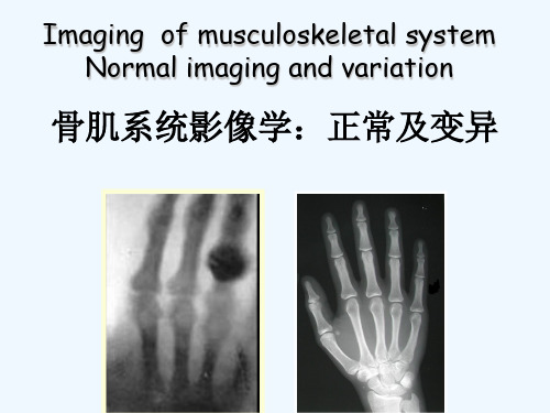
Imaging of musculoskeletal system Normal imaging and variation
骨肌系统影像学:正常及变异
Imaging of musculoskeletal system imaging modalities
• Whn (uses and dangers) – Types of imaging modalities
- Ovaries, testis 软巢 睾丸
- Thyroid 甲状腺 - Fetus 胎儿 - GI tract 胃肠道
• Radiation damage is Dose dependent • Factors to reduce dose
• Reduce unnecessary studies • Use non-radiation studies
(between 10-2 and 10 nm) •X-rays are generated by using a high voltage current to accelerate electrons within evacuated glass tubes, towards a tungsten target
骨骼肌肉系统影像诊断 ppt课件
(五)四肢关节
人体关节分三类:①不动关节;②微动关节;③能动关节; 能动关节各结构影像特点: 1) 关节骨端:
骨性关节面:线样密质骨 关节软骨:X线和CT不能区分,MRI呈等T1等T2信号,压脂
呈高信号
2)关节间隙 : X线表现为两个骨性关节面间的透亮间隙,关节间隙与解剖关 节间隙不同,X线所见关节间隙包括关节软骨及其真正关节 腔和少量滑液 CT表现为关节骨端间的低密度间隙 MRI表现可区分软骨和滑液
骨干
小儿长骨CT、MRI特点
✓ 骨皮质为高密度或低信号线带影 ✓ 骨髓腔为软组织密度(信号)或脂肪密
度(信号) ✓ 干骺端骨松质为骨小梁交错呈网状,网
格间为骨髓组织,MRI可见骨小梁,信 号低于骨髓腔,临时钙化带CT呈高密度 而MRI呈低信号 ✓ 骺及骺板为软骨,CT呈软组织密度,骨 化中心呈高密度,MRI呈中等信号,骨 化中心信号降低
脊柱由脊椎和其间的椎 间盘所组成
1)颈7、胸12、腰5、 骶5、尾4共33节,由 椎间盘及韧带、椎间 上下关节形成
2)上小下大,四个弯曲
3)脊椎 ( 除颈1-2和骶尾) 椎体:长方体;外四周为皮 质,内为松质骨 附件:椎弓及椎弓根、棘突 、横突 椎间隙:半透明带状间隙 椎间孔:神经出入通道
胸腰椎正侧位片
骨的结构
骨质按结构分为: 1)、密质骨:致密骨(哈氏系统),如骨皮质 2)、松质骨:骨小梁及其间的骨髓 3)、骨髓腔:富含骨髓等结缔组织
(二)骨的发育
✓ 骨化:膜化骨-结缔组织膜上 形成骨化中心而发育成骨,颅 盖诸骨和面骨;软骨内化骨- 软骨原基上形成骨化中心而发 育成骨,躯干骨、四肢骨、颅 底骨与筛骨等
(一)骨肌系统X线检查
X线检查的价值与限度 价值:
最新[PPT]MedicalImaginginGeneral医学影像学总论讲学课件
![最新[PPT]MedicalImaginginGeneral医学影像学总论讲学课件](https://img.taocdn.com/s3/m/19c16728ce2f0066f43322b6.png)
Physical foundations
Radiofrequency (RF) pulse 射频脉冲 RF sequence 射频序列 Repetition time (TR) 重复时间 Echo time (TE) 回波时间 T1-weighted imaging T1加权像 T2-weighted imagingT2加权像 PD-weighted imaging质子加权像
Fundamentals of X-Ray Imaging X线诊断的基础
X-ray characteristics Densities and thickness of human body
High density Medium density Low density
Methods of Examination 检查方法
Protection from X-Ray X线防护
Should not be overlooked Unnecessary fear should not be
bourn in mind
II. X-Ray Computed Tomography CT
Hounsfield 1969 Clinical application 1972 Nobel prize 1979
X-Ray Diagnosis and Analysis X线诊断和分析
Thorough observation Exact analysis
Site and distribution, number, size, shape, density, outline, etc. The essence of lesions diseases / signs Correlated with the clinical data
医学影像学的英文
医学影像学的英文Medical Imaging in EnglishMedical imaging refers to the techniques and processes used to create images of the human body for clinical purposes. It plays a crucial role in the diagnosis and treatment of various medical conditions. In this article, we will explore the field of medical imaging and discuss its importance in the healthcare industry.One of the most common modalities used in medical imaging is X-ray imaging. X-rays are a form of electromagnetic radiation that can pass through the body, allowing healthcare professionals to visualize internal structures such as bones and organs. X-ray images are particularly useful in the diagnosis of fractures, lung conditions, and gastrointestinal issues.Another important modality in medical imaging is computed tomography (CT) scanning. CT scans use a combination of X-rays and computer technology to create cross-sectional images of the body. This allows for detailed examination of internal organs and tissues, making it an invaluable tool in the detection of tumors, infections, and other medical abnormalities.Magnetic resonance imaging (MRI) is another widely used modality in medical imaging. MRI uses magnetic fields and radio waves to generate detailed images of the body's internal structures. MRI is particularly useful in imaging soft tissues such as the brain, spinal cord, and muscles. It is often used to diagnose neurological disorders, joint injuries, and tumors.Ultrasound imaging, also known as sonography, uses sound waves to create images of the body's internal structures. Ultrasound is commonly used to visualize the fetus during pregnancy, as well as to assess the health of organs such as the heart, liver, and kidneys. Ultrasound is a safe and non-invasive imaging modality that is widely used in medical practice.Nuclear medicine is a specialized branch of medical imaging that uses radioactive substances to visualize and diagnose medical conditions. Techniques such as positron emission tomography (PET) and single-photon emission computed tomography (SPECT) are commonly used in nuclear medicine to detect diseases such as cancer, heart conditions, and neurological disorders.In addition to these modalities, medical imaging also encompasses a variety of other techniques such as fluoroscopy, mammography, and angiography. Each modality has its own strengths and limitations, and healthcare professionals must choose the most appropriate imaging technique based on the patient's condition and the clinical question at hand.The field of medical imaging is constantly evolving, with new technologies and techniques being developed to improve diagnostic accuracy and patient outcomes. Artificial intelligence (AI) and machine learning are increasingly being used in medical imaging to analyze images, detect abnormalities, and assist in diagnosis. These advances hold great promise for the future of medical imaging and have the potential to revolutionize healthcare delivery.In conclusion, medical imaging is an essential component of modern healthcare, providing healthcare professionals with valuable information todiagnose and treat medical conditions. From X-rays to MRI to nuclear medicine, the field of medical imaging offers a wide range of modalities to visualize the human body and provide insights into the underlying pathology. As technology continues to advance, the role of medical imaging in healthcare will only continue to grow in importance.。
医学影像学专业英语X-RAY IMAGING
X-RAY IMAGING
DIGITAL SUBTRACTION IMAGING
Digital subtraction imaging (DSI) is a process whereby a computer removes unwanted information from a radiographic image. It is particularly useful for angiography, referred to as DSA.
X-RAY IMAGING
After an X-ray exposure is made the films are processed in a darkroom or more commonly in free-standing daylight processors. The resulting image is commonly known as an ‘X-ray’. The common terms ‘chest X-ray’ and ‘abdomen Xray’ are widely accepted and commonly abbreviated to CXR and AXR, respectively. More correct terms for an X-ray image are ‘radiograph’ or ‘plain film’.
X-RAY IMAGING
Consolidated lung lying against the heart border will therefore obscure that border. A good example is consolidation or collapse of the right middle lobe causing loss of definition of the right heart boder. These comments apply to all radiographically visible anatomical interfaces in the body.
医学影像学教学新20165骨关节课件
囊状破坏
片状破坏
骨质破坏
虫 蚀 状
骨质破坏
结构消失密度减低
左肱骨近端干骺端骨破坏
骨破坏
CT
?
平片与CT/ MRI对比
X线片
CT片
MRI
骨质软化与骨质疏松的区别
1 骨结构模糊 2 骨外形改变 3 假骨折线
骨质软化
?
?
骨质疏松
教材:《医学影像学》第6版,吴恩惠主编, 人卫出版社
CT
骨质疏松
正常
?
(osteomalacia)
1、定义 骨的一定单位体积内有机成分正常而矿物质减少。
2、常见于以下疾病:小儿 —— 佝偻病;成人— — 骨软化症或其它代谢性疾病。
3、X线表现: (1)密度减低,皮质变薄。 (2)骨结构模糊 (3)骨外形改变; (4)假骨折线;
密度减低 结构模糊
骨关节检查方法
1 X线平片 (Radiography): 常规位置:正侧位
必要时加照斜位、切线位、 轴位、透视点片、对比片
2 CT
平扫 增强 CTA
3 MRI
平扫 增强 MRA
一
检查方法
常
规
平 正位
片
ቤተ መጻሕፍቲ ባይዱ
侧位
腰椎X线平片
正位
侧位
斜位
检查方法
2、 CT检查 :可避开解剖重叠,显示横断解剖的
空间关系;密度分辨力高,能确定病灶的部位、范围、 形态、结构;可做平扫(软组织/骨窗)与增强扫描,还
膝关节
(冠状位) (矢状位)
检查方法选择原则 与首选方法
原则:由简到繁;由无创到有创;由经济
到昂贵。
首选:X线平片(正侧位),CT、MRI应