EDC-NHS法偶联乳胶微球
EDC-NHS
EDC-NHS引言Thermo Scientific公司的NHS和磺基-NHS, 被用于化学标记、交联和固相固定中, 制备氨基羧酸酯的胺反应酯。
在存在碳二亚胺类化合物如EDC时, 羧化物(-COOH)可能与NHS或者磺基-NHS反应, 形成一个半稳定的NHS或者磺基-NHS酯。
NHS或者磺基-NHS酯可以继续与伯胺(-NH2)反应, 形成酰胺键(amide crosslinks)。
虽然NHS或者磺基-NHS在碳二亚胺反应中不是必须的, 但它们的使用可以极大的提高偶联效率。
另外, 使用NHS或者磺基-NHS可以使反应分两步进行。
NHS或者磺基-NHS均为水溶有机溶剂。
然而, 用NHS活化, 降低了修饰后羧酸盐的水溶性, 而使用磺基-NHS活化, 由于荷电磺酸基, 则可以保存或者增加修饰后分子的水溶性。
虽然准备的NHS或者磺基-NHS酯在两步反应中足够稳定, 仍然会在几个小时或者几分钟内水解, 时间的长短取决于水中物质和反应液的pH值(NHS酯半衰期为: pH7时为4-5小时, pH8时为1小时, pH8.6时为10分钟)。
提取和干燥的操作流程可以调整为适于准备稳定的NHS活化分子, 但最优的情况是NHS活化后的分子立即与含有氨基的目标分子反应。
使用EDC和磺基-NHS的活化反应, 最适pH值4.5-7.2, EDC反应通常在pH4.7-6.0的MES缓冲液中进行。
磺基-NHS活化后的分子与伯胺反应的最适pH值7-8。
磺基-NHS酯反应通常在pH 7.2-7.5的PBS中进行。
为了得到两步反应的最优结果, 第一步反应最好使用pH5-6的MES缓冲液(或者其他不含氨基、不含羧基的溶液),然后在与含有氨基的分子反应之前, 立即用磷酸(盐)缓冲液(或者其他非氨基缓冲液)提高pH值至7.2-7.5。
EDC反应用2-巯基乙醇(2-ME)终止, 或者过量的试剂通过有脱盐作用的柱子来交换缓冲液而除去。
用伯胺和羧酸盐的EDC/NHS交联过程A.其他需要准备的材料活化缓冲液: 0.1M MES (2-吗啉乙磺酸), 0.5M NaCl, pH 6.0PBS: 0.1M磷酸钠, 0.15M NaCl, pH 7.2-7.5蛋白1溶液: 用活化缓冲液配置1ml的蛋白1溶液, 10mg/mL蛋白2溶液: 将蛋白低压冻干或者溶解在1-10mg/mL 的PBS或者其他不含氨基的溶液中, pH 7-8EDC:为了最优的实验结果, EDC与蛋白1溶液的用量摩尔比为10.备选: 2-巯基乙醇用来终止EDC反应备选: 羟胺用来终止氨基反应。
EDC-NHS法偶联乳胶微球
EDC-NHS法偶联乳胶微球Activation1. Wash particles (e.g., 100 mg of 1 μm carboxylated latex beads) into coupling buffer (50 mM MES, pH 6.0). Suspend in 5 ml coupling buffer. The addition of a dilute detergent solution may be done to increase bead stability and prevent clumping (e.g., 0.01 percent SDS). Avoid the addition of any components containing carboxylates or amines (such as acetate, glycine, Tris, imidazole, etc.). Also, avoid the presence of thiols (e.g., DTT, 2-mercaptoethanol, etc.), as these will react with EDC and effectively inactivate it.2. Add 100 mg of EDC and 100 mg of sulfo-NHS. Mix to dissolve. To facilitate faster dissolution, EDC and sulfo-NHS may be dissolved immediately before use as a concentrated stock solution in reaction buffer and then an aliquot of this solution added to the particle suspension to obtain the correct ?nal concentration.3. React for 15 minutes at room temperature.4. Quickly wash beads 2 times with coupling buffer using centrifugation and resuspend using a sonic probe in 5 ml of the same buffer.Coupling5. Dissolve protein to be coupled in 5 ml coupling buffer ata concen tration suf? cient to provide 1- to 10-fold molar excess of ligand over the maximal calculated monolayer concentration for the amount and type of beads used. For particle manufacturers reporting a carboxylate concentration in meq/g, this is equivalent to μmol/mg. The optimal protein concentration should be optimized. Note: T oo low a protein concentration mayresult in particle crosslinking. For coupling of expensive antibodies that may not be available in enough quantity to reach the optimal molar ratio on the particles, the addition of another protein (i.e., bovine gamma globulin or BSA) may be done to take up remaining reactive sites.6. Combine the protein solution with particles and mix thoroughly.7. React at room temperature 2–4 hours with mixing.8. Wash beads with coupling buffer and resuspend in the same buffer containing 100 mM of an amine-containing hydrophilic quenching molecule to block excess reactive sites (i.e., ethanolamine or Tris).9. Wash beads and resuspend in an appropriate buffer for storage.。
乳胶微球偶联方法
乳胶微球偶联方法乳胶微球偶联方法的步骤通常包括光敏交联聚合、功能物质固定化和乳胶微球基质处理。
首先,乳胶微球的合成是基础。
通常,聚合物乳胶微球的制备通过乳液聚合反应来实现,其中聚合物的单体通过乳化剂稳定在水相中。
这种方法的优点是可以通过调整单体的类型和含量来控制微球的性质,比如粒径、形状和表面特性。
光敏交联聚合是乳胶微球偶联的关键步骤之一、光敏交联聚合通过在聚合物单体中引入光敏剂来实现。
在光照条件下,光敏剂可以吸收光能并诱导单体分子之间的交联反应,从而形成网络结构的聚合物。
这种交联结构可以增强乳胶微球的稳定性并提高其机械性能。
在光敏交联聚合过程中,需要选择合适的光敏剂和光照条件,并根据所需的功能物质来确定单体的选择和交联程度。
功能物质固定化是另一个重要的步骤。
在乳胶微球的光敏交联聚合之后,可以通过物理吸附、共价结合或离子键结合等方法将功能物质固定在乳胶微球的表面上。
这种固定化方法的选择取决于功能物质的性质和乳胶微球的表面特性。
例如,对于含有氨基团的功能物质,可以通过与乳胶微球表面带有羧基的聚合物发生共价结合来固定在乳胶微球上。
最后,乳胶微球基质处理是乳胶微球偶联的最后一步。
乳胶微球偶联后,需要对乳胶微球进行处理以增强其稳定性和使用性能。
常用的处理方法包括洗涤、溶解和干燥。
这些处理可以去除其中的有机溶剂和未固定的功能物质,并使乳胶微球获得更好的稳定性和储存性能。
总之,乳胶微球偶联方法是一种将功能物质固定在乳胶微球表面的技术。
通过光敏交联聚合、功能物质固定化和乳胶微球基质处理等步骤,可以制备出具有特定功能和稳定性的乳胶微球。
这种方法具有简单、灵活和可控性强等特点,并在催化剂、生物传感器和药物释放等领域具有广泛的应用前景。
免疫比浊法生产流程

免疫比浊法生产流程免疫比浊法的生产流程如下:1. 清洗:吸取100μL胶乳微球加入1 mLMES缓冲液中,混匀后17000 rpm离心30min,弃上清,加入1mLMES,用超声波清洗器振散微球团块,每次持续约30s,3~5次。
2. 活化:将NHS粉末和EDCI粉末恢复室温,配制浓度为20mg/mL的NHS活化剂和EDCI活化剂,将适量的NHS和EDCI活化剂加入已清洗的微球中,上下颠倒混匀后再置旋转混匀器上混匀,持续约20~30min。
3. 包被:将已彻底超声振散的微球加入一定浓度的CSFVE2蛋白单抗中,先手动上下颠倒混匀,再置旋转混匀器充分反应。
15000rpm离心10min后弃上清,加入1 mL MES。
4. 封闭:将已彻底超声振散的微球加入100μL封闭液,置旋转混匀器充分反应1h或置2~8℃封闭过夜。
此外,还应注意如下问题:在离心前使标本完全自然形成凝块。
保持样品管的密闭状态。
抽血后2小时内完成离心和冷藏。
血清和血浆需用物理方法尽快与细胞分离。
去除所有可能存在的纤维蛋白和细胞成分,否则会得到假阳性结果。
如果48小时内不能完成检测即应将标本放入-20℃冰箱冻藏。
标本只能复溶一次,检测前须将复溶标本离心处理。
不要用水浴方法复溶标本。
吸取血浆标本时应避免将红细胞层上的白细胞或血小板层吸出。
含有颗粒物质的浑浊血清或血浆标本在检测前须先从原始管内转运出来,并重新离心,原始管中含有分离介质(分离胶)的可以不再离心。
如果标本不在24小时内检测或运送标本时,将标本保存在-20℃或更低温度的环境中。
标本不能溶血。
标准液所用的物质对HIV抗体,乙型肝炎表面抗原和丙型肝炎抗体测试呈阴性反应。
建议各实验室建立自己的正常值范围。
测量的特征值(测量范围、精度、准确度、灵敏度)和多种分析仪的参数将另外提供。
以上信息仅供参考,如有需要建议咨询专业人士。
基于噬菌体展示技术的内毒素检测方法
基于噬菌体展示技术的内毒素检测方法王鑫;徐美玲;张起莹;嵇冶;张昊【摘要】利用噬菌体展示技术,经过两轮非特异性筛选,一轮特异性筛选,获得内毒素高效特异结合十二肽.经生物分子相互作用系统表征,结果表明,筛选获得的多肽与内毒素具有良好的亲和性,解离平衡常数KD为3.52×10⁻⁹;将获得的多肽人工合成,采用EDC/NHS共价偶联法与乳胶微球偶联,建立了内毒素比浊快速检测方法,内毒素标准品在0.03~0.48EU/mL浓度范围内,增强免疫比浊法吸光度值与内毒素标准品浓度呈正线性关系,相关系数R2>0.99,最低检测限达到0.03EU/mL.与市售内毒素鲎试剂检测试剂盒对比,在检测血清样本时,检测结果一致性良好.该研究为临床体外诊断内毒素快速检测提供技术基础.【期刊名称】《长春理工大学学报(自然科学版)》【年(卷),期】2019(042)001【总页数】4页(P135-138)【关键词】内毒素;噬菌体展示技术;结合多肽;增强免疫比浊法【作者】王鑫;徐美玲;张起莹;嵇冶;张昊【作者单位】长春理工大学生命科学技术学院,长春130022;长春理工大学生命科学技术学院,长春130022;长春理工大学生命科学技术学院,长春130022;长春理工大学生命科学技术学院,长春130022;长春理工大学生命科学技术学院,长春130022【正文语种】中文【中图分类】R-33内毒素是革兰氏阴性菌细胞壁上的一种成分,当细菌死亡后会大量释放。
人体内毒素含量过高会导致发热、心跳过速、低血压、休克甚至死亡[1]。
因此人体内毒素检测一直是临床及医疗器械、药物检测的必检指标。
目前常规的检测方法主要有:凝胶法、浊度法和比色法。
但原理都是基于内毒素能与鲎试剂发生特异性反应。
鲎是国家二级保护动物,随着检测市场需求量增加,鲎血提取过度使得资源枯竭,造成鲎试剂检测成本增高,进而造成生态环境平衡的不可逆破坏,并且利用鲎试剂的检测方法检测时间长,因此研究能够替代天然鲎试剂的检测方法迫在眉睫[2-5]。
nhs和edc偶联原理
nhs和edc偶联原理
NHS和EDC是两种常见的偶联化学反应。
NHS是N-羟基丁二酰亚胺的缩写,是一种常见的偶联试剂,用于与胺基化合物发生反应。
而EDC 是1-(3-二甲氨基丙基)-3-乙基卡巴朴胺,也是一种常用的偶联剂,用于将羧酸与胺基或酚发生反应。
NHS和EDC都是重要的化学反应试剂,它们在生物化学、化学生物学和生物医学领域中得到广泛应用。
这两种反应剂的作用都是实现生
物分子之间的化学偶联。
NHS和EDC偶联原理是通过一定的化学反应机制实现的。
比如说,NHS常与胺基化合物发生缩酰胺反应,生成氨基酰胺型连接。
这种偶联的反应机制如下:
1、NHS与胺基化合物作用,生成NHS酯化合物。
2、NHS酯化合物进一步水解成酸和活性酯,生成氨基酰胺型连接。
EDC偶联反应的原理是将羧酸与胺基或酚发生酰胺键或烷基羟基键的转化。
EDC通过一定的反应机制实现了化学偶联作用。
这种反应机制可以概括为:
1、EDC激活羧酸,生成中间酰基。
2、络合作用,EDC形成胺酰噻唑盐,同时与胺基或酚发生酰胺键
或烷基羟基键的转化。
以上反应机制的实现需要一定的条件,如反应环境的pH值、温度、反应物浓度等。
为了实现偶联反应的理想水平,要提供最适宜的条件,从而获得最佳的反应效果。
总结起来,NHS和EDC都是生物分子化学修饰中常用的偶联剂,两者有着不同的应用场景。
偶联反应的实现依赖于一定的化学机制和条件,通过精心设计之后,可以使反应效果得到最大限度的提高,为生
物化学、化学生物学、生物医学等领域的研究提供优良的手段。
乳胶标记
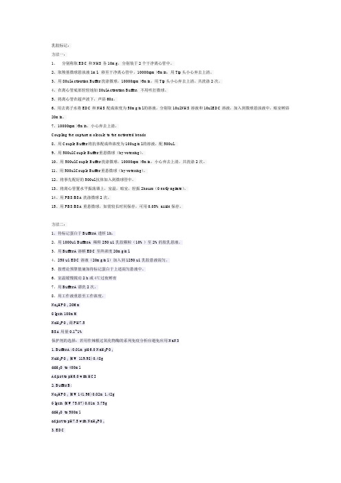
方法一:1、分别称取EDC和NHS各10mg,分别装于2个干净离心管中。
2、取羧基微球悬浊液1ml,移至干净离心管中,10000rpm×5min,用Tip头小心弃去上清。
3、用80ul Activation Buffer洗涤微球,10000rpm×5min,用Tip头小心弃去上清。
共洗涤2次。
4、在离心管底部轻轻地加80ul Activation Buffer,不用吹打微球。
5、将离心管在超声波下,声浴60s。
6、用去离子水将EDC和NHS配成浓度为50mg/ml的溶液。
分别取10ul NHS溶液和10ul EDC溶液,加入到微球悬浊液中,暗室孵浴20min。
7、10000rpm×5min,小心弃去上清。
Coupling the capture molecule to the activated beads8、用Couple Buffer将抗体配成终浓度为100ug/ml的溶液,配500ul。
9、用500ul Couple Buffer重悬微球(by votexing)。
10、用500ul Couple Buffer洗涤微球,10000rpm×5min,小心弃去上清。
共洗涤2次。
11、用500ul Couple Buffer重悬微球(by votexing)。
12、将事先配好的500ul抗体加入到微球管中。
13、将离心管置水平振荡器上,室温,暗室,轻振2hours(Gently agitate)。
14、用PBS/BSA洗涤微球2次。
15、用PBS/BSA重悬微球,如需较长时间保存,可用0.05% azide保存。
方法二:1、待标记蛋白于Buffer A透析1h。
2、用1000u1 Buffer A 稀释250 u1乳胶颗粒(10%)至2%的胶乳悬液。
3、用Buffer A溶解EDC至终浓度20mg/ml.4、250 u1 EDC溶液(20mg/ml.)加入到1250 u1乳胶悬液混匀。
EDC NHS 说明书
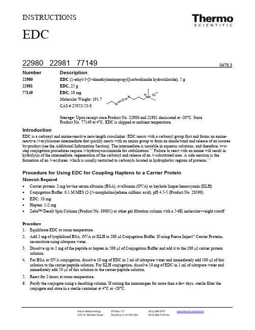
INSTRUCTIONSNumber Description22980 EDC (1-ethyl-3-[3-dimethylaminopropyl]carbodiimide hydrochloride), 5 g 22981 EDC, 25 g77149 EDC, 10 mgCAS # 25952-53-8Storage: Upon receipt store Product No. 22980 and 22981 desiccated at -20°C. Store Product No. 77149 at 4°C. EDC is shipped at ambient temperature. IntroductionEDC is a carboxyl and amine-reactive zero-length crosslinker. EDC reacts with a carboxyl group first and forms an amine-reactive O -acylisourea intermediate that quickly reacts with an amino group to form an amide bond and release of an isourea by-product (see the Additional Information Section). The intermediate is unstable in aqueous solutions, and therefore, two-step conjugation procedures require N -hydroxysuccinimide for stabilization.1,2 Failure to react with an amine will result in hydrolysis of the intermediate, regeneration of the carboxyl and release of an N -substituted urea. A side reaction is the formation of an N -acylurea, which is usually restricted to carboxyls located in hydrophobic regions of proteins.1,3Procedure for Using EDC for Coupling Haptens to a Carrier ProteinMaterials Required• Carrier protein: 2 mg bovine serum albumin (BSA), ovalbumin (OVA) or keyhole limpet hemocyanin (KLH) • Conjugation Buffer: 0.1 M MES (2-[N -morpholino]ethane sulfonic acid), pH 4.5-5 (Product No. 28390) • EDC: 10 mg • Hapten: 1-2 mg•Zeba™ Desalt Spin Column (Product No. 89891) or other gel filtration column with a 5-6K molecular-weight cutoffProcedure1. Equilibrate EDC to room temperature.2. Add 2 mg of lyophilized BSA, OVA or KLH to 200 µl Conjugation Buffer. If using Pierce Imject ® Carrier Proteins,reconstitute using ultrapure water. 3. Dissolve up to 2 mg of the peptide or hapten in 500 µl of Conjugation Buffer and add it to the 200 µl carrier proteinsolution. 4. For BSA or OVA conjugation, dissolve 10 mg of EDC in 1 ml of ultrapure water and immediately add 100 µl of thissolution to the carrier-peptide solution. For KLH conjugation, dissolve 10 mg of EDC in 1 ml of ultrapure water and immediately add 50 µl of this solution to the carrier-peptide solution. 5. React for 2 hours at room temperature.6. Purify the conjugate using a desalting column. If storing the immunogen for more than a few days, sterile filter theconjugate and store in a sterile container at 4°C or -20°C.EDCProcedure for Two-step Coupling of Proteins Using EDC and NHS or Sulfo-NHSThe following protocol, adapted from a procedure described by Grabarek and Gergely, allows sequential coupling of two proteins without affecting the second protein’s carboxyls by exposing them to EDC. This procedure requires quenching the first reaction with a thiol-containing compound.The activation reaction with EDC and Sulfo-NHS is most efficient at pH 4.5-7.2; however, the reaction of Sulfo-NHS-activated molecules with primary amines is most efficient at pH 7-8. For best results, perform the first reaction in MES buffer (or other non-amine, non-carboxylate buffer) at pH 5-6, then raise the pH to 7.2-7.5 with phosphate buffer (or other non-amine buffer) immediately before reaction to the amine-containing molecule. For quenching the first reaction, use2-mercaptoethanol, or the excess reagent can be simply removed (as well as the reaction pH adjusted) by buffer-exchange column.desaltingawithMaterials Required•Activation Buffer: 0.1 M MES, 0.5 M NaCl, pH 6.0•Coupling Buffer: Phosphate-buffered saline (PBS), 100 mM sodium phosphate, 150 mM NaCl; pH 7.2 (Product No.28372)•Protein # 1: Prepared in Activation Buffer at 1 mg/ml•Protein # 2•NHS or Sulfo-NHS (Product No. 24500 and 24510, respectively)•2-Mercaptoethanol (Product No. 35600)•(Optional) Zeba™ Desalt Spin Column (Product No. 89891) or other gel filtration column •Hydroxylamine•HCl (Product No. 26103)Procedure1.Equilibrate EDC and NHS to room temperature before opening bottles.2.Add 0.4 mg EDC (~2 mM) and 0.6 mg of NHS or 1.1 mg of sulfo-NHS (~5 mM) to 1 ml of protein #1 solution and reactfor 15 minutes at room temperature.3.Add 1.4 µl of 2-mercaptoethanol (final concentration of 20 mM) to quench the EDC.4.Optional: Separate the protein from excess reducing agent and inactivated crosslinker using a desalting column.Equilibrate the column with Coupling Buffer (PBS).5.Add protein #2 to the activated protein prepared in Coupling Buffer at an equal molar ratio with protein #1. Allow theproteins to react for 2 hours at room temperature.6.To quench the reaction, add hydroxylamine to a final concentration of 10 mM. This method hydrolyzes nonreacted NHSpresent on protein #1 and results in hydroxamate. Other quenching methods involve adding 20-50 mM Tris, lysine, glycine or ethanolamine; however, these primary amine-containing compounds modify carboxyls on protein #1.7.Remove excess quenching reagent using a desalting column.Additional InformationEDC reacts with a carboxyl group first and forms an amine-reactive O-acylisourea intermediate that quickly reacts with anamino group to form an amide bond and release of an isourea by-product (Figure 1).Information from Our Website•Tech Tip #15: Biotinylate carboxyl groups with EDC and Biotin Hydrazide•Tech Tip #5: Attach an antibody onto glass, silica or quartz surface•Tech Tip #18: Block amino groups to prevent polymer formation in peptide-carrier protein conjugations•Tech Tip #30: Modify and label oligonucleotide 5´ phosphate groups•Tech Tip #3: Determine reactivity of NHS-ester biotinylation and crosslinking reagents•Tech Tip #46: Preferentially biotinylate N-terminal alpha-amino groups in peptidesRelated Products20002 Bioconjugate Techniques, 785 pages, softcover89889 Zeba Desalt Spin Columns, 2 ml, 5 each, for 200-700 µl samples89890 Zeba Desalt Spin Columns, 2 ml, 25 each, for 200-700 µl samples89891 Zeba Desalt Spin Columns, 5 ml, 5 each, for 500-2,000 µl samples89892 Zeba Desalt Spin Columns, 5 ml, 25 each, for 500-2,000 µl samples89893 Zeba Desalt Spin Columns, 10 ml, 5 each, for 700-4,000 µl samples89894 Zeba Desalt Spin Columns, 10 ml, 25 each, for 700-4,000 µl samples(succinimidyl 4-[N-maleimidomethyl]cyclohexane-1-carboxylate), 50 mg22360 SMCC22322 Sulfo-SMCC (sulfosuccinimidyl 4-[N-maleimidomethyl]cyclohexane-1-carboxylate), 50 mg22622 Sulfo-SMCC, No-Weigh™ Format, 8 × 2 mg microtubes21555 DSS (disuccinimidyl suberate), 1 g21655 DSS, 50 mg21658 DSS in No-Weigh™ Format, 8 × 2 mg microtubes21580 BS3 (bis[sulfosuccinimidyl] suberate), 50 mg21585 BS3 in No-Weigh™ Format, 8 × 2 mg microtubesReferences1.Grabarek, Z. and Gergely, J. (1990). Zero-length crosslinking procedure with the use of active esters. Anal Biochem185:131-5.2.Staros, J.V., et al. (1986). Enhancement by N-hydroxysulfosuccinimide of water-soluble carbodiimide-mediated coupling reactions. Anal Biochem156:220-2.3.Timkovich, R. (1977). Detection of the stable addition of carbodiimide to proteins. Anal Biochem79:135-43.This product (“Product”) is warranted to operate or perform substantially in conformance with published Product specifications in effect at the time of sale, as set forth in the Product documentation, specifications and/or accompanying package inserts (“Documentation”) and to be free from defects in material and workmanship. Unless otherwise expressly authorized in writing, Products are supplied for research use only. No claim of suitability for use in applications regulated by FDA is made. The warranty provided herein is valid only when used by properly trained individuals. Unless otherwise stated in the Documentation, this warranty is limited to one year from date of shipment when the Product is subjected to normal, proper and intended usage. This warranty does not extend to anyone other than the original purchaser of the Product (“Buyer”).No other warranties, express or implied, are granted, including without limitation, implied warranties of merchantability, fitness for any particular purpose, or non infringement. Buyer’s exclusive remedy for non-conforming Products during the warranty period is limited to replacement of or refund for the non-conforming Product(s).There is no obligation to replace Products as the result of (i) accident, disaster or event of force majeure, (ii) misuse, fault or negligence of or by Buyer, (iii) use of the Products in a manner for which they were not designed, or (iv) improper storage and handling of the Products.Current versions of product instructions are available at /pierce. For a faxed copy, call 800-874-3723 or contact your local distributor.© 2007 Thermo Fisher Scientific Inc. All rights reserved. Unless otherwise indicated, all trademarks are property of Thermo Fisher Scientific Inc. and its subsidiaries. Printed in the USA.。
1、胶乳偶联方案的输出
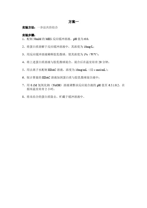
实验方法:一步法共价结合实验步骤:1、配制50mM的MES反应缓冲溶液,pH值为6.0;2、将蛋白质溶解于反应缓冲溶液中,其浓度为10mg/L;3、用反应缓冲溶液稀释胶乳微球,使其浓度为1%(W/V);4、将上述蛋白质溶液与胶乳微球混合,混合后在温室培育20分钟;5、用去离子水配制EDAC溶液,浓度为10mg/mL(52μmol/mL);6、取计算量的EDAC溶液加到蛋白质与胶乳微球混合液中;7、用0.1M氢氧化钠(NaOH)溶液调整该反应混合液的pH值至6.5±0.2,在摇床温室培育2小时;8、将未结合的蛋白质除去,贮藏于缓冲溶液中。
实验方法:简单二步法共价结合实验步骤:1、配制50mM的MES反应缓冲溶液,pH值为6.0;2、将蛋白质溶解于反应缓冲溶液中,其浓度为10mg/L;3、用反应缓冲溶液稀释胶乳微球,使其浓度为1%(W/V);4、1mL胶乳微球悬浮液,加入20mgEDAC,在室温培育40分钟,在20分钟后第二次加入20mg/mL;5、用相等体积的该缓冲溶液来洗涤胶乳微球,离心,用1倍体积的缓冲溶液重复处理;6、将蛋白质溶解于50-100mM的MES缓冲溶液中,蛋白质浓度为1mg/mL;7、再将胶乳微球悬浮于去离子水或结合的缓冲溶液中,迅速加入溶解的蛋白质,37℃搅拌反应3小时;8、按1mL反应混合液加入2.5μL乙醇胺混合,搅拌反应10分钟;9、除去未结合的蛋白质,贮藏与贮存的缓冲溶液中实验方法:生成NHS-酯的中间体二步法共价结合实验步骤:1、每ml反应混合物加入下列各物:a.去离子水是最后容积为1.0mL;b.0.1mL的10x的贮备缓冲溶液,其pH值为6.0-6.5(一般可用0.5M MES缓冲溶液);胶乳微球最终浓度为1%(W/V);c.0.23mL的50mg/ml的NHS在去离子水中的溶液;d.19.2mg/mL(100mM)的EDAC在去离子水中的溶液2、在室温将此混合物反应15-30分钟,不断搅拌;3、用MES缓冲液或纯化水洗涤胶乳微球悬浮物,除去未反应的NHS和EDAC;4、重新将胶乳微球在去离子水中悬浮,使其浓度为1%(w/v);5、将结合的蛋白质溶于50-100mM的MES缓冲溶液中,浓度为1mg/mL;6、立即加入蛋白质溶液(胶乳微球、蛋白质和缓冲溶液的浓度分别为0.5%(w/v)、0.5mg/mL、25-50mM);7、将混合物反应至少2小时,轻轻搅拌;8、按1mL反应混合液加入2.5μL乙醇胺混合,搅拌反应10分钟;9、除去未结合的蛋白质和乙醇胺,贮于适宜的贮存缓冲溶液中。
胶乳微球偶联蛋白的使用中常见问题及解答
胶乳微球偶联蛋白的使用中常见问题及解答以下问题是我们用户在使用过程中遇到的问题,我们咨询了国外的技术人员,这是他们的答复)问:产品说明书中给出的胶乳粒径是平均粒径吗?答:产品说明书中给出的胶乳粒径(Diameter)是平均粒径。
问:如何选择胶乳微球的粒径?答:一般选择粒径小的胶乳微球,则需要的抗体量就多,精密度和线性相对较好,而选择粒径大的胶乳微球性,则所需抗体少,精密度和线性相对较差,相对于小球,大球的灵敏度较好。
问:一般情况下我们所使用的微球的浓度大概是多少?答:整个测定体系中,微球的浓度大约在0.01%左右,当然这与试剂本身规定的线性有关系。
问:在使用离心方法偶联乳胶微球,一般需要多大的(相对)离心力?答:需要多大的离心力和所使用的乳胶微球有关,一般70nm左右的微球大概13000g/min30min以上,粒径越小所需时间越长,离心力越大。
问:蛋白(抗体)与微球偶联前,是不是微球的活化时间越长,效率越好?答:羧基微球的活化所需时间很短,一般以10-20分钟为宜,长时间的活化反而会降低偶联效率。
问:由于抗体不只是FC的氨基酸上有NH2,是不是表示它可以在任何方向与EDCA活化微球偶联呢?如果是这样,是不是会影响抗体与抗原的特异性反应?答:事实的确如此,当然由于抗体的空间折叠方式和FC端疏水性强,因此偶联反应的绝大多数发生在Fc端,对抗体与抗原的结合的影响不大。
问:胶乳微球与抗体偶联后,当时没发现有凝集,但隔夜后发现有凝集,这是什么原因?如何控制和避免?答:这种情况一般是偶联效率低的原因。
当体系中蛋白不足或是其它原因,使微球表面在交联后还空出许多反应基团时,这些基团又可以与相连微球上的蛋白反应,结果是把球又拉在一起了,所以有聚集。
可以加一些阻断剂解决,常见的是BSA,另外,也可提高微球的交联率。
但为什么是过一段时间后才出现凝集呢,这是因为微球偶联上蛋白后,相互之间由于携带同种电荷的关系,比较稳定(所以能以胶体样存在),只有当偶尔相互碰撞,遇上彼此的反应基团时才能结合。
羧基乳胶微球偶联方法
羧基乳胶微球偶联方法
羧基乳胶微球的偶联方法一般包括以下步骤:
1. 将羧基乳胶微球与含化学修饰剂的活化缓冲液混合,以对羧基乳胶微球进行活化。
2. 经重悬、分散后,将待偶联的蛋白与活化后的羧基乳胶微球混合,进行偶联反应。
3. 偶联反应后,进行封闭、保存。
在偶联过程中,常用的化学修饰剂为edc 和 sulfo-nhs 或 nhs 的混合物,edc 与sulfo-nhs 或 nhs 的混合摩尔比为1∶2-4,edc 与羧基乳胶微球的羧基量的摩尔比为5-30∶1。
实际操作中,偶联条件可能需要根据具体情况进行调整。
如果你想了解更多关于羧基乳胶微球偶联方法的信息,请补充相关细节继续向我提问。
EDC NHS 说明书
INSTRUCTIONSNumber Description22980 EDC (1-ethyl-3-[3-dimethylaminopropyl]carbodiimide hydrochloride), 5 g 22981 EDC, 25 g77149 EDC, 10 mgCAS # 25952-53-8Storage: Upon receipt store Product No. 22980 and 22981 desiccated at -20°C. Store Product No. 77149 at 4°C. EDC is shipped at ambient temperature. IntroductionEDC is a carboxyl and amine-reactive zero-length crosslinker. EDC reacts with a carboxyl group first and forms an amine-reactive O -acylisourea intermediate that quickly reacts with an amino group to form an amide bond and release of an isourea by-product (see the Additional Information Section). The intermediate is unstable in aqueous solutions, and therefore, two-step conjugation procedures require N -hydroxysuccinimide for stabilization.1,2 Failure to react with an amine will result in hydrolysis of the intermediate, regeneration of the carboxyl and release of an N -substituted urea. A side reaction is the formation of an N -acylurea, which is usually restricted to carboxyls located in hydrophobic regions of proteins.1,3Procedure for Using EDC for Coupling Haptens to a Carrier ProteinMaterials Required• Carrier protein: 2 mg bovine serum albumin (BSA), ovalbumin (OVA) or keyhole limpet hemocyanin (KLH) • Conjugation Buffer: 0.1 M MES (2-[N -morpholino]ethane sulfonic acid), pH 4.5-5 (Product No. 28390) • EDC: 10 mg • Hapten: 1-2 mg•Zeba™ Desalt Spin Column (Product No. 89891) or other gel filtration column with a 5-6K molecular-weight cutoffProcedure1. Equilibrate EDC to room temperature.2. Add 2 mg of lyophilized BSA, OVA or KLH to 200 µl Conjugation Buffer. If using Pierce Imject ® Carrier Proteins,reconstitute using ultrapure water. 3. Dissolve up to 2 mg of the peptide or hapten in 500 µl of Conjugation Buffer and add it to the 200 µl carrier proteinsolution. 4. For BSA or OVA conjugation, dissolve 10 mg of EDC in 1 ml of ultrapure water and immediately add 100 µl of thissolution to the carrier-peptide solution. For KLH conjugation, dissolve 10 mg of EDC in 1 ml of ultrapure water and immediately add 50 µl of this solution to the carrier-peptide solution. 5. React for 2 hours at room temperature.6. Purify the conjugate using a desalting column. If storing the immunogen for more than a few days, sterile filter theconjugate and store in a sterile container at 4°C or -20°C.EDCProcedure for Two-step Coupling of Proteins Using EDC and NHS or Sulfo-NHSThe following protocol, adapted from a procedure described by Grabarek and Gergely, allows sequential coupling of two proteins without affecting the second protein’s carboxyls by exposing them to EDC. This procedure requires quenching the first reaction with a thiol-containing compound.The activation reaction with EDC and Sulfo-NHS is most efficient at pH 4.5-7.2; however, the reaction of Sulfo-NHS-activated molecules with primary amines is most efficient at pH 7-8. For best results, perform the first reaction in MES buffer (or other non-amine, non-carboxylate buffer) at pH 5-6, then raise the pH to 7.2-7.5 with phosphate buffer (or other non-amine buffer) immediately before reaction to the amine-containing molecule. For quenching the first reaction, use2-mercaptoethanol, or the excess reagent can be simply removed (as well as the reaction pH adjusted) by buffer-exchange column.desaltingawithMaterials Required•Activation Buffer: 0.1 M MES, 0.5 M NaCl, pH 6.0•Coupling Buffer: Phosphate-buffered saline (PBS), 100 mM sodium phosphate, 150 mM NaCl; pH 7.2 (Product No.28372)•Protein # 1: Prepared in Activation Buffer at 1 mg/ml•Protein # 2•NHS or Sulfo-NHS (Product No. 24500 and 24510, respectively)•2-Mercaptoethanol (Product No. 35600)•(Optional) Zeba™ Desalt Spin Column (Product No. 89891) or other gel filtration column •Hydroxylamine•HCl (Product No. 26103)Procedure1.Equilibrate EDC and NHS to room temperature before opening bottles.2.Add 0.4 mg EDC (~2 mM) and 0.6 mg of NHS or 1.1 mg of sulfo-NHS (~5 mM) to 1 ml of protein #1 solution and reactfor 15 minutes at room temperature.3.Add 1.4 µl of 2-mercaptoethanol (final concentration of 20 mM) to quench the EDC.4.Optional: Separate the protein from excess reducing agent and inactivated crosslinker using a desalting column.Equilibrate the column with Coupling Buffer (PBS).5.Add protein #2 to the activated protein prepared in Coupling Buffer at an equal molar ratio with protein #1. Allow theproteins to react for 2 hours at room temperature.6.To quench the reaction, add hydroxylamine to a final concentration of 10 mM. This method hydrolyzes nonreacted NHSpresent on protein #1 and results in hydroxamate. Other quenching methods involve adding 20-50 mM Tris, lysine, glycine or ethanolamine; however, these primary amine-containing compounds modify carboxyls on protein #1.7.Remove excess quenching reagent using a desalting column.Additional InformationEDC reacts with a carboxyl group first and forms an amine-reactive O-acylisourea intermediate that quickly reacts with anamino group to form an amide bond and release of an isourea by-product (Figure 1).Information from Our Website•Tech Tip #15: Biotinylate carboxyl groups with EDC and Biotin Hydrazide•Tech Tip #5: Attach an antibody onto glass, silica or quartz surface•Tech Tip #18: Block amino groups to prevent polymer formation in peptide-carrier protein conjugations•Tech Tip #30: Modify and label oligonucleotide 5´ phosphate groups•Tech Tip #3: Determine reactivity of NHS-ester biotinylation and crosslinking reagents•Tech Tip #46: Preferentially biotinylate N-terminal alpha-amino groups in peptidesRelated Products20002 Bioconjugate Techniques, 785 pages, softcover89889 Zeba Desalt Spin Columns, 2 ml, 5 each, for 200-700 µl samples89890 Zeba Desalt Spin Columns, 2 ml, 25 each, for 200-700 µl samples89891 Zeba Desalt Spin Columns, 5 ml, 5 each, for 500-2,000 µl samples89892 Zeba Desalt Spin Columns, 5 ml, 25 each, for 500-2,000 µl samples89893 Zeba Desalt Spin Columns, 10 ml, 5 each, for 700-4,000 µl samples89894 Zeba Desalt Spin Columns, 10 ml, 25 each, for 700-4,000 µl samples(succinimidyl 4-[N-maleimidomethyl]cyclohexane-1-carboxylate), 50 mg22360 SMCC22322 Sulfo-SMCC (sulfosuccinimidyl 4-[N-maleimidomethyl]cyclohexane-1-carboxylate), 50 mg22622 Sulfo-SMCC, No-Weigh™ Format, 8 × 2 mg microtubes21555 DSS (disuccinimidyl suberate), 1 g21655 DSS, 50 mg21658 DSS in No-Weigh™ Format, 8 × 2 mg microtubes21580 BS3 (bis[sulfosuccinimidyl] suberate), 50 mg21585 BS3 in No-Weigh™ Format, 8 × 2 mg microtubesReferences1.Grabarek, Z. and Gergely, J. (1990). Zero-length crosslinking procedure with the use of active esters. Anal Biochem185:131-5.2.Staros, J.V., et al. (1986). Enhancement by N-hydroxysulfosuccinimide of water-soluble carbodiimide-mediated coupling reactions. Anal Biochem156:220-2.3.Timkovich, R. (1977). Detection of the stable addition of carbodiimide to proteins. Anal Biochem79:135-43.This product (“Product”) is warranted to operate or perform substantially in conformance with published Product specifications in effect at the time of sale, as set forth in the Product documentation, specifications and/or accompanying package inserts (“Documentation”) and to be free from defects in material and workmanship. Unless otherwise expressly authorized in writing, Products are supplied for research use only. No claim of suitability for use in applications regulated by FDA is made. The warranty provided herein is valid only when used by properly trained individuals. Unless otherwise stated in the Documentation, this warranty is limited to one year from date of shipment when the Product is subjected to normal, proper and intended usage. This warranty does not extend to anyone other than the original purchaser of the Product (“Buyer”).No other warranties, express or implied, are granted, including without limitation, implied warranties of merchantability, fitness for any particular purpose, or non infringement. Buyer’s exclusive remedy for non-conforming Products during the warranty period is limited to replacement of or refund for the non-conforming Product(s).There is no obligation to replace Products as the result of (i) accident, disaster or event of force majeure, (ii) misuse, fault or negligence of or by Buyer, (iii) use of the Products in a manner for which they were not designed, or (iv) improper storage and handling of the Products.Current versions of product instructions are available at /pierce. For a faxed copy, call 800-874-3723 or contact your local distributor.© 2007 Thermo Fisher Scientific Inc. All rights reserved. Unless otherwise indicated, all trademarks are property of Thermo Fisher Scientific Inc. and its subsidiaries. Printed in the USA.。
一种羧基乳胶微球与抗体的偶联方法
一种羧基乳胶微球与抗体的偶联方法引言:羧基乳胶微球是一种常用的载体材料,在生物技术、生物医学研究等领域有着广泛的应用。
将羧基乳胶微球与抗体偶联可以实现抗体的定向、高效地与靶分子结合,从而用于生物分析、诊断和治疗等应用。
本文将介绍一种羧基乳胶微球与抗体的偶联方法,以及该方法的优势和应用前景。
方法:1.材料准备:- 羧基乳胶微球:可通过合适的乳化聚合方法制备,其粒径一般控制在100-1000 nm之间。
-交联剂:羧基乳胶微球一般表面具有丰富的羧基官能团,可通过与交联剂反应实现微球的交联,增强抗体的稳定性。
-免疫活性分子:选择与目标抗体匹配的免疫活性分子,如胺基酸、蛋白质等。
2.羧基乳胶微球表面修饰:使用化学方法将胺基酸、蛋白质等与羧基乳胶微球反应,实现羧基乳胶微球表面的修饰和激活,增加偶联反应的效率和稳定性。
3.抗体偶联:将修饰后的羧基乳胶微球与目标抗体进行偶联,可采用传统的化学偶联方法,如活化的EDC/NHS法、硫酸亚铁/亚砜法等。
将抗体与羧基乳胶微球充分混合,在适当的反应条件下(pH、温度等),进行偶联反应。
4.反应后的处理:偶联反应后,通过洗涤等方法去除未偶联的抗体和副产物,得到羧基乳胶微球与抗体稳定结合的产物。
优势和应用前景:-选择合适的乳化聚合方法和交联剂,可控制羧基乳胶微球的粒径和形貌,使其具有较大比表面积和较好的稳定性。
-通过表面修饰,增加羧基乳胶微球与抗体之间的亲和力和稳定性,提高偶联效率。
-该方法操作简单、成本较低,适用于大规模生产。
-偶联的羧基乳胶微球具有良好的生物相容性和稳定性,可用于体内外生物学研究、诊断和治疗等领域。
-偶联的羧基乳胶微球可用于靶向传递药物、检测靶分子、研究生物分子交互作用等应用。
总结:本文介绍了一种羧基乳胶微球与抗体的偶联方法,该方法可以实现抗体的定向、高效地与靶分子结合。
该方法具有操作简单、成本低、适用性广等优点,并在生物学研究、诊断和治疗等领域具有广阔的应用前景。
抗体,胶乳基本偶联步骤
抗体,胶乳基本偶联步骤免疫胶乳偶联试验方案一试剂准备:MES缓冲液500mM,pH5-6,4℃储存;EDAC 配置浓度为52μmol/mL(称取10mg EDAC,加入1mL去离子水);NHS 配制浓度为50mg/mL水溶液;蛋白质溶液缓冲液稀释至浓度为1-10mg/mL。
二步偶联法:1. 将配好的溶液按以下顺序滴加入离心管①100μL 500mM MES 缓冲液;②加水稀释至1mL;③200μL 5%微球;④230uL NHS 溶液⑤EDAC 水溶液(计算量)2. 放置在恒温摇床上,37℃,120rpm,30min;3. 13000rpm离心30min,沉降后,弃上清,收集离心沉淀;用1mL 50mM MES 缓冲液洗涤2次;离心沉降后,弃上清。
4. 将配好的溶液按以下顺序加入离心管中,混匀①100ul 500mM MES 缓冲液;②加水稀释至1mL;③蛋白质溶液。
5. 放置在恒温摇床上,37℃,120rpm,2h;6. 13000rpm离心20min沉降后,弃上清,收集离心沉淀;用1mL 50mM MES 缓冲液(或者调节pH至8.0左右),同时加入BSA 溶液至其终浓度为1%,洗涤3 次;离心沉降后,弃上清。
7. 加入0.97mL MES 缓冲液(或者调节pH至8.0左右),将微球浓度调为1%,并重新悬浮,然后搅拌,接着温和的超声分散;8. 重新悬浮后加入一定量的BSA溶液(比如终浓度为1%),保存。
9.福林酚法测定偶联蛋白质含量(可选)。
(如要测蛋白质含量,则前面的方法中不可加入BSA。
)1. 每ml反应混合物加入以下成分:加入DIW,定容最终体积为1ml;0.1ml 10X的MES buffer,ph6.0~6.5(10X的buffer通常用0.5M);0.1ml 10%的微球(终浓度为1%);0.23ml NHS水溶液(50mg/mL); 11.5mg一定体积的19.2mg/ml(100mM)的EDAC水溶液;2. 室温下搅拌反应15~30min;(Note7)3. 清洗:MES buffer或DIW清洗两次;(Note3);4. 用MES buffe或者DIW悬浮微球到浓度为1%;5. 同时,用MES buffe r溶解稀释抗体。
EDC/NHS法制备免疫磁珠及其效果验证
作者: 张尔力
作者机构: 湖南警察学院,湖南长沙410138
出版物刊名: 化工管理
页码: 104-105页
年卷期: 2016年 第30期
主题词: 新型材料;免疫磁珠;细胞分离与提纯;PCR;STR
摘要:免疫磁珠又叫免疫磁性微球,是免疫学和磁性微球结合而发展的一类新型材料,一般用于物质的提取和分离,由于其高效、低毒等特性,目前在生物化学领域、医药领域、食品检测领域应用广泛。
本文主要应用EDC/NHS法活化羧基磁珠,再将人类精子特异性抗体与之偶联,最后制备成免疫磁珠,进行细胞捕获实验,分别捕获人类的上皮细胞和精子,提取DNA,进行PCR(聚合酶链式反应),进行电泳,根据STR(短串联重复)图谱的对比验证了这种方法制备的免疫磁珠的偶联效果。
实验证实,用EDC/NHS法活化制备的免疫磁珠能选择性地捕获到精子而不能捕获到上皮细胞,该方法制备磁珠效果良好。
- 1、下载文档前请自行甄别文档内容的完整性,平台不提供额外的编辑、内容补充、找答案等附加服务。
- 2、"仅部分预览"的文档,不可在线预览部分如存在完整性等问题,可反馈申请退款(可完整预览的文档不适用该条件!)。
- 3、如文档侵犯您的权益,请联系客服反馈,我们会尽快为您处理(人工客服工作时间:9:00-18:30)。
Activation
1. Wash particles (e.g., 100 mg of 1 μm carboxylated latex beads) into coupling buffer (50 mM MES, pH 6.0). Suspend in 5 ml coupling buffer. The addition of a dilute detergent solution may be done to increase bead stability and prevent clumping (e.g., 0.01 percent SDS). Avoid the addition of any components containing carboxylates or amines (such as acetate, glycine, Tris, imidazole, etc.). Also, avoid the presence of thiols (e.g., DTT, 2-mercaptoethanol, etc.), as these will react with EDC and effectively inactivate it.
2. Add 100 mg of EDC and 100 mg of sulfo-NHS. Mix to dissolve. To facilitate faster dissolution, EDC and sulfo-NHS may be dissolved immediately before use as a concentrated stock solution in reaction buffer and then an aliquot of this solution added to the particle suspension to obtain the correct final concentration.
3. React for 15 minutes at room temperature.
4. Quickly wash beads 2 times with coupling buffer using centrifugation and resuspend using a sonic probe in 5 ml of the same buffer.
Coupling
5. Dissolve protein to be coupled in 5 ml coupling buffer at a concen tration suffi cient to provide 1- to 10-fold molar excess of ligand over the maximal calculated monolayer concentration for the amount and type of beads used. For particle manufacturers reporting a carboxylate concentration in meq/g, this is equivalent to μmol/mg. The optimal protein concentration should be optimized. Note: Too low a protein concentration may result in particle crosslinking. For coupling of expensive antibodies that may not be available in enough quantity to reach the optimal molar ratio on the particles, the addition of another protein (i.e., bovine gamma globulin or BSA) may be done to take up remaining reactive sites.
6. Combine the protein solution with particles and mix thoroughly.
7. React at room temperature 2–4 hours with mixing.
8. Wash beads with coupling buffer and resuspend in the same buffer containing 100 mM of an amine-containing hydrophilic quenching molecule to block excess reactive sites (i.e., ethanolamine or Tris).
9. Wash beads and resuspend in an appropriate buffer for storage.。