ESC2015 非ST段抬高型ACS指南
ESC非ST段抬高型急性冠脉综合征管理指南

感谢观看
冠状动脉血运重建建议
冠状动脉血运重建建议
MINOCA
MINOCA 存在许多异质性的潜在病因,涉及冠状动脉和非冠状动脉 相关的病理机制,MINOCA 的诊断不包括心肌炎和 Takotsubo综合征。 心脏磁共振检查是诊断 MINOCA 的关键手段,可确定 85%以上该类患 者的潜在病因并有助于指导治疗。
NSTE-ACS的定义及流行病学
(4)快速的“纳入”和“排除”方法:建议使用0 h / 1 h方法(最佳选择)或0 h / 2 h方法(次佳选择)。选择排除和纳入的最佳阈值,将0 h / 1 h和0 h / 2 h 方法与临床和ECG结果结合使用,可识别出适合早期出院和门诊管理的患者。 (5)hs-cTn的混杂因素: 年龄(个体浓度差异,最高可达300%),肾功能不 全(健康患者的eGFR很高或极低水平之间的hs-cTn浓度差异,最高可达300%) 和胸痛发作(>300%),性别差异是适度的(约等于40%)。 (6)缺血风险评估:hs-cTn水平越高,死亡风险越大。所有NSTE-ACS患者应 检测血清肌酐和eGFR水平,利钠肽可能会提供更多的预后信息,并可能有助于 风险分层。
无房颤者PCI术后抗栓策略
双联抗血小板/抗栓药物延长策略的评估
降低PCI术相关出血因素的推荐策略
口服联用抗凝和抗血小板药物的推荐
口服联用抗凝和抗血小板药物的推荐
合并房颤患者PCI术后抗栓治疗流程
接受口服抗凝治疗患者出血和输血相关的推荐
NSTE-ACS的其他药物推荐
NSTE-ACS的侵入性治疗
ESC 非ST 段抬高型急性冠脉综合征管理指 南—解读
目录
➢ NSTE-ACS的定义及流行病学 ➢ 2020年相对于2015年指南的更新 ➢ NSTE-ACS的诊断及风险预测 ➢ NSTE-ACS的药物治疗 ➢ NSTE-ACS➢ NSTE-ACS的特殊人群 ➢ NSTE-ACS的长期管理
2015 ESC NSTE-ACS指南解读-林先和
413.341,022 有效期至2016/09/06, 仅供医疗专业人士参考
使用hs-cTn T 再评估, 可增加NSTEMI诊断率,降低UA诊断率
前瞻性国际多中心研究,连续入选1124例疑似AMI患者。由2位心脏病专家使用不同的诊断指标分别对 患者先后进行2次诊断。第一次使用普通cTn T指标,第二次使用高敏cTnT
413.341,022 有效期至2016/09/06, 仅供医疗专业人士参考
hs-cTn有助于区分AMI与其他急性胸痛性疾病1
前瞻性国际多中心研究,连续入选887例急性胸痛患者,使用盲法三种方法(hs-cTnT,罗氏诊断; hs-cTnI,贝克曼库尔特仪器; hscTnI,西门子仪器)检测基线hs-cTn及0-1小时的hs-cTn改变。最终127例(15%)确诊为AMI,124例(14%)确诊为非冠脉心脏病。
• 抗栓治疗:优选新型ADP受体抑制剂,疗程突破一年限制
• 其他更新:房颤相关抗血小板治疗、 CABG术后抗血小板 治疗及二级预防管理
413.341,022 有效期至2016/09/06, 仅供医疗专业人士参考
新指南对口服抗血小板药物的推荐
口服抗血小板治疗推荐
阿司匹林推荐用于所有无禁忌症的NSTE-ACS患者,负荷剂量150-300mg(之前未使用阿 司匹林者),维持剂量75-100mg/日,无论何种治疗策略长期使用。
指南推荐使用 0h/3h hs-cTn算法进行早期诊断
急性胸痛 hs-cTn < ULN
胸痛>6h 胸痛<6h
hs-cTn > ULN
欧洲心脏病学会(ESC)关于非ST段抬高急性冠脉综合征管理指南
欧洲心脏病学会(ESC)关于非ST段抬高急性冠脉综合征管理指南伦敦会展中心主会场,欧洲心脏病学会(ESC)正式发布了在非ST段抬高急性冠脉综合征(NSTE-ACS)管理指南。
新指南是指南发布4年后的首次更新,新指南推荐内容更为简洁和明确,多采用流程图,实用性更强,对指导NSTE-ACS患者的规范化诊治具有重要意义。
新指南在以下六个方面做了更新或者新增,1)冠脉造影和PCI经桡动脉入路获得最高级别推荐;2)高敏肌钙蛋白(hs-cTnT)1小时检测流程(algorithm)获得推荐;3)根据患者出血或缺血风险,给予更个体化的DAPT时间;4)不推荐P2Y12抑制剂预处理:反对NSTEMI患者使用普拉格雷进行预处理,支持或反对氯吡格雷或替格瑞洛进行预处理的证据均不充分;5)首次对NSTE-ACS患者根据临床表现进行心律监测时程(0、<24h、>24h)作出了推荐。
对监护病房患者管理进行了简化,有助于缩短住院时间、降低费用;6)新增对长期口服抗凝药患者使用抗血小板治疗的新章节。
根据MATRIX及相关荟萃分析结果,新指南首次对血管入路做出推荐。
对于有经验的中心,建议在冠脉造影和PCI时选择经桡动脉入路(I/A)。
同时,指南强调,血管入路的选择仍应考虑术者经验和中心习惯。
对于多支血管病变, 强调要根据具体病情及当地血管团队的流程来制定具体的血远重建策略。
这对于我国90%以上的医院外科搭桥水平较国外差距巨大的现状尤为有指导意义。
对于使用Hs-cTnT 1小时检测流程,我国存在的问题是检测方法众多,医院之间、城市之间标准值不同,无法直接对比,迫切需要标准化,更要注重结合临床情况判读。
基于大量新型支架证据,新指南建议应用新一代DES(I/A)。
此外,对于高出血风险计划接受短时程双联抗血小板治疗(30天)的患者,新一代DES可能优于BMS(IIb/B)。
2015年新指南的危险分层更为细化,分为极高危、高危、中危、低危4个级别,与2014年美国心脏协会(AHA)/美国心脏病学会(ACC)NSTE-ACS管理指南趋于一致。
非ST段抬高急性ACS的诊断治疗NSTEACS临床指南解读
药物治疗 早期介入治疗
Multimarker Strategy in ACS
Myocyte Necrosis Troponin
Inflammation hs-CRP, CD40L
Hemodynamic Stress BNP, NT-proBNP
北京朝阳医院
Beijing Chaoyang Hospital
STEMI & NSTEMI冠状动脉病变支数的比较
ST
ST
No. diseased vessels
(n=1864)
(n=2170)
0
10%
11%
1
45%
26%
2
27%
28%
3
18%
36%
Savonitto S, et al. J Am Med Asoc. 1999; 281:707-713.
病因及病理
HISTORICAL Age 65
POINTS 1
3 CAD risk factors
(FHx, HTN, chol,
1
DM, active smoker)
Known CAD (stenosis 50%) 1
ASA use in past 7 days
1
PRESENTATION Recent (24H) severe angina 1
15% 10% 5%
0
0
5
10
15
20
WBC Count (x103)
Cannon CP, et al. Am J Cardiol. 2001;87:636-639. (with permission)
ESC2015 NSTEACS指南
ESC2015 指南: 非 ST 段抬高型急性冠脉综合征(中文版)2015-09-01 09:09来源:丁香园作者:Tylen Chen字体大小-|+关于可疑非 ST 段抬高型急性冠脉综合征(ACS)患者的诊断、风险分层、影像学检查和心律监测的若干建议1. 诊断和风险分层(1)建议结合患者的病史、症状、重要体征、其他体格检查发现、 ECG 和实验室检查结果等,对患者进行基本诊断以及行短期的缺血和出血风险分层。
(I,A)(2)建议患者就诊后 10 min 内迅速行 12 导联 ECG 检查,并立即让有经验的医生查看结果。
为了防止症状复发或者诊断不明确,有必要再次行 12 导联 ECG 检查。
(I,B)(3)如果标准导联 ECG 结果阴性,但仍然高度怀疑缺血性病灶的存在,建议增加 ECG 导联(V3R、V4R、V7-V9)。
(I,C)(4)建议检测心肌钙蛋白(敏感或者高敏法),且在 60 min 内获取结果。
(I,A)(5)如果有高敏肌钙蛋白的结果,建议行 0 h 和 3 h 的快速排查方案。
(I,B)(6)如果有高敏肌钙蛋白的结果以及确认可用 0 h/1 h 算法,建议行 0 h 和 1 h 的快速排查和确诊方案。
如果前两次肌钙蛋白检测结果阴性但临床表现仍然提示 ACS,建议在 3-6 h 之后再做一次检查。
(I,B)(7)建议使用现有的风险分数来诊断评估患者病情。
(I,B)(8)如果患者预行冠脉造影,可考虑使用 CRUSADE 分数量化出血风险。
(IIb,B)2. 影像学检查(1)如果患者无复发胸痛、ECG 结果正常、心肌钙蛋白检查结果正常(最好是高敏),但仍然怀疑存在 ACS,建议行无创性的负荷试验诱发缺血,结果不理想再进一步考虑有创性的检查。
(I,A)(2)建议行超声心动图以评估局部和全左心室功能,以及确诊和排查鉴别诊断。
(I,C)(3)如果心肌钙蛋白和 / 或 ECG 结果阴性,但仍怀疑低中度 CAD,可考虑行 MDCT 冠脉造影检查。
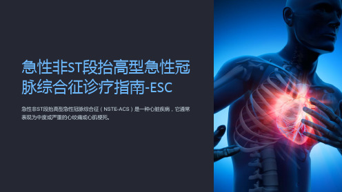
急性非ST段抬高型急性冠脉综合征诊疗指南-ESC
1 诊断标准
NSTE-ACS的诊断标准 包括临床表现、心电图 和血液生化指标。
2 诊断流程
通过患者症状、体征、 心电图和生化标记进行 综合评估,以确定诊断 结果。
ห้องสมุดไป่ตู้
3 诊断工具
常用诊断工具包括心电 图、血液生化标记、超 声心动图等。
急性非ST段抬高型急性冠脉综合征的治 疗
初步治疗
初步治疗措施包括静脉血栓 溶解治疗、抗血小板治疗、 镇痛等。
介入治疗
介入治疗主要指冠状动脉血 管成形术或支架植入,以恢 复冠状动脉通畅。
药物治疗
药物治疗常用药物包括抗血 小板药、抗凝药和抗心绞痛 药。
后续管理及预后评估
监护
患者在急性期之后需要定期监测心电图、心功能等指标,以评估病情变化和效果。
急性非ST段抬高型急性冠 脉综合征诊疗指南-ESC
急性非ST段抬高型急性冠脉综合征(NSTE-ACS)是一种心脏疾病,它通常 表现为中度或严重的心绞痛或心肌梗死。
急性非ST段抬高型急性冠脉综合征简介
定义
NSTE-ACS 是一组心脏疾 病,包括不稳定型心绞痛 和非ST段抬高型心肌梗死。
分类
根据心电图和心肌酶的变 化,NSTE-ACS被分为高 危型和非高危型。
流行病学特征
NSTE-ACS发病率在全球 范围内很高,尤其是对于 中老年人和存在危险因素 的人。
临床表现
1
症状
常见症状包括胸痛或胸闷、呼吸困难、恶心或呕吐。
2
体征
体检时可能出现心率不规则、心音减弱、杂音等异常体征。
3
实验室检查
血液检查可以检测心肌酶和肌钙蛋白等指标,以评估心肌损伤程度。
急性非ST段抬高型急性冠脉综合征的诊 断
欧洲心脏病学会(ESC)关于非ST段抬高急性冠脉综合征管理指南

欧洲心脏病学会(ESC)关于非ST段抬高急性冠脉综合征管理指南伦敦会展中心主会场,欧洲心脏病学会(ESC)正式发布了在非ST段抬高急性冠脉综合征(NSTE-ACS)管理指南。
新指南是指南发布4年后的首次更新,新指南推荐内容更为简洁和明确,多采用流程图,实用性更强,对指导NSTE-ACS患者的规范化诊治具有重要意义。
新指南在以下六个方面做了更新或者新增,1)冠脉造影和PCI经桡动脉入路获得最高级别推荐;2)高敏肌钙蛋白(hs-cTnT)1小时检测流程(algorithm)获得推荐;3)根据患者出血或缺血风险,给予更个体化的DAPT时间;4)不推荐P2Y12抑制剂预处理:反对NSTEMI患者使用普拉格雷进行预处理,支持或反对氯吡格雷或替格瑞洛进行预处理的证据均不充分;5)首次对NSTE-ACS患者根据临床表现进行心律监测时程(0、<24h、>24h)作出了推荐。
对监护病房患者管理进行了简化,有助于缩短住院时间、降低费用;6)新增对长期口服抗凝药患者使用抗血小板治疗的新章节。
根据MATRIX及相关荟萃分析结果,新指南首次对血管入路做出推荐。
对于有经验的中心,建议在冠脉造影和PCI时选择经桡动脉入路(I/A)。
同时,指南强调,血管入路的选择仍应考虑术者经验和中心习惯。
对于多支血管病变, 强调要根据具体病情及当地血管团队的流程来制定具体的血远重建策略。
这对于我国90%以上的医院外科搭桥水平较国外差距巨大的现状尤为有指导意义。
对于使用Hs-cTnT 1小时检测流程,我国存在的问题是检测方法众多,医院之间、城市之间标准值不同,无法直接对比,迫切需要标准化,更要注重结合临床情况判读。
基于大量新型支架证据,新指南建议应用新一代DES(I/A)。
此外,对于高出血风险计划接受短时程双联抗血小板治疗(30天)的患者,新一代DES可能优于BMS(IIb/B)。
2015年新指南的危险分层更为细化,分为极高危、高危、中危、低危4个级别,与2014年美国心脏协会(AHA)/美国心脏病学会(ACC)NSTE-ACS管理指南趋于一致。
2015+ESC指南:非ST段抬高型急性冠脉综合征的管理

ESC GUIDELINES2015ESC guidelines for the management of acute coronary syndromes in patients presenting without persistent ST-segment elevationTask Force for the Management of Acute Coronary Syndromes in Patients Presenting without Persistent ST-Segment Elevation of the European Society of Cardiology (ESC)Authors/Task Force Members:Marco Roffi*(Chairperson)(Switzerland),Carlo Patrono *(Co-Chairperson)(Italy),Jean-Philippe Collet †(France),Christian Mueller †(Switzerland),Marco Valgimigli †(The Netherlands),Felicita Andreotti (Italy),Jeroen J.Bax (The Netherlands),Michael A.Borger (Germany),Carlos Brotons (Spain),Derek P.Chew (Australia),Baris Gencer (Switzerland),Gerd Hasenfuss (Germany),Keld Kjeldsen (Denmark),Patrizio Lancellotti (Belgium),Ulf Landmesser (Germany),Julinda Mehilli (Germany),Debabrata Mukherjee (USA),Robert F.Storey (UK),and Stephan Windecker (Switzerland)Document Reviewers:Helmut Baumgartner (CPG Review Coordinator)(Germany),Oliver Gaemperli (CPG Review Coordinator)(Switzerland),Stephan Achenbach (Germany),Stefan Agewall (Norway),Lina Badimon (Spain),Colin Baigent (UK),He´ctor Bueno (Spain),Raffaele Bugiardini (Italy),Scipione Carerj (Italy),Filip Casselman (Belgium),Thomas Cuisset (France),Çetin Erol (Turkey),Donna Fitzsimons (UK),Martin Halle(Germany),*Corresponding authors:Marco Roffi,Division of Cardiology,University Hospital,Rue Gabrielle Perret-Gentil 4,1211Geneva 14,Switzerland,Tel:+41223723743,Fax:+41223727229,E-mail:Marco.Roffi@hcuge.chCarlo Patrono,Istituto di Farmacologia,Universita`Cattolica del Sacro Cuore,Largo F.Vito 1,IT-00168Rome,Italy,Tel:+390630154253,Fax:+39063050159,E-mail:carlo.patrono@rm.unicatt.it&The European Society of Cardiology 2015.All rights reserved.For permissions please email:journals.permissions@.†Section Coordinators affiliations listed in the Appendix.ESC Committee for Practice Guidelines (CPG)and National Cardiac Societies document reviewers listed in the Appendix.ESC entities having participated in the development of this document:Associations:Acute Cardiovascular Care Association (ACCA),European Association for Cardiovascular Prevention &Rehabilitation (EACPR),European Association of Cardiovas-cular Imaging (EACVI),European Association of Percutaneous Cardiovascular Interventions (EAPCI),Heart Failure Association (HFA).Councils:Council on Cardiovascular Nursing and Allied Professions (CCNAP),Council for Cardiology Practice (CCP),Council on Cardiovascular Primary Care (CCPC).Working Groups:Working Group on Cardiovascular Pharmacotherapy,Working Group on Cardiovascular Surgery,Working Group on Coronary Pathophysiology and Microcir-culation,Working Group on Thrombosis.The content of these European Society of Cardiology (ESC)Guidelines has been published for personal and educational use only.No commercial use is authorized.No part of the ESC Guidelines may be translated or reproduced in any form without written permission from the ESC.Permission can be obtained upon submission of a written request to Oxford University Press,the publisher of the European Heart Journal and the party authorized to handle such permissions on behalf of the ESC.Disclaimer:The ESC Guidelines represent the views of the ESC and were produced after careful consideration of the scientific and medical knowledge and the evidence available at the time of their publication.The ESC is not responsible in the event of any contradiction,discrepancy and/or ambiguity between the ESC Guidelines and any other official recom-mendations or guidelines issued by the relevant public health authorities,in particular in relation to good use of healthcare or therapeutic strategies.Health professionals are encour-aged to take the ESC Guidelines fully into account when exercising their clinical judgment,as well as in the determination and the implementation of preventive,diagnostic or therapeutic medical strategies;however,the ESC Guidelines do not override,in any way whatsoever,the individual responsibility of health professionals to make appropriate and accurate decisions in consideration of each patient’s health condition and in consultation with that patient and,where appropriate and/or necessary,the patient’s caregiver.Nor do the ESC Guidelines exempt health professionals from taking into full and careful consideration the relevant official updated recommendations or guidelines issued by the competent public health authorities,in order to manage each patient’s case in light of the scientifically accepted data pursuant to their respective ethical and professional obligations.It is also the health professional’s responsibility to verify the applicable rules and regulations relating to drugs and medical devices at the time of prescription.European Heart Journaldoi:10.1093/eurheartj/ehv320European Heart Journal Advance Access published August 29, 2015by guest on August 29, 2015Downloaded fromChristian Hamm(Germany),David Hildick-Smith(UK),Kurt Huber(Austria),EfstathiosIliodromitis(Greece),Stefan James(Sweden),Basil S.Lewis(Israel),Gregory Y.H.Lip(UK),Massimo F.Piepoli(Italy),Dimitrios Richter (Greece),Thomas Rosemann(Switzerland),Udo Sechtem(Germany),Ph.Gabriel Steg(France),Christian Vrints (Belgium),and Jose Luis Zamorano(Spain)The disclosure forms of all experts involved in the development of these guidelines are available on the ESC website /guidelines------------------------------------------------------------------------------------------------------------------------------------------------------Keywords Acute cardiac care†Acute coronary syndromes†Angioplasty†Anticoagulation†Apixaban†Aspirin†Atherothrombosis†Beta-blockers†Bivalirudin†Bypass surgery†Cangrelor†Chest pain unit†Clopidogrel†Dabigatran†Diabetes†Early invasive strategy†Enoxaparin†European Society ofCardiology†Fondaparinux†Glycoprotein IIb/IIIa inhibitors†Guidelines†Heparin†High-sensitivitytroponin†Myocardial ischaemia†Nitrates†Non-ST-elevation myocardial infarction†Platelet inhibition†Prasugrel†Recommendations†Revascularization†Rhythm monitoring†Rivaroxaban†Statin†Stent†Ticagrelor†Unstable angina†VorapaxarTable of ContentsAbbreviations and acronyms (4)1.Preamble (5)2.Introduction (7)2.1Definitions,pathophysiology and epidemiology (7)2.1.1Universal definition of myocardial infarction (7)2.1.1.1Type1MI (7)2.1.1.2Type2MI (7)2.1.2Unstable angina in the era of high-sensitivity cardiactroponin assays (7)2.1.3Pathophysiology and epidemiology(see Web addenda) (7)3.Diagnosis (7)3.1Clinical presentation (7)3.2Physical examination (8)3.3Diagnostic tools (8)3.3.1Electrocardiogram (8)3.3.2Biomarkers (9)3.3.3‘Rule-in’and‘rule-out’algorithms (10)3.3.4Non-invasive imaging (11)3.3.4.1Functional evaluation (11)3.3.4.2Anatomical evaluation (11)3.4Differential diagnosis (12)4.Risk assessment and outcomes (12)4.1Clinical presentation,electrocardiogram and biomarkers124.1.1Clinical presentation (12)4.1.2Electrocardiogram (12)4.1.3Biomarkers (13)4.2Ischaemic risk assessment (13)4.2.1Acute risk assessment (13)4.2.2Cardiac rhythm monitoring (13)4.2.3Long-term risk (14)4.3Bleeding risk assessment (14)4.4Recommendations for diagnosis,risk stratification,imagingand rhythm monitoring in patients with suspected non-ST-elevation acute coronary syndromes (14)5.Treatment (15)5.1Pharmacological treatment of ischaemia (15)5.1.1General supportive measures (15)5.1.2Nitrates (15)5.1.3Beta-blockers (15)5.1.4Other drug classes(see Web addenda) (16)5.1.5Recommendations for anti-ischaemic drugs inthe acute phase of non-ST-elevation acute coronary syndromes (16)5.2Platelet inhibition (16)5.2.1Aspirin (16)5.2.2P2Y12inhibitors (16)5.2.2.1Clopidogrel (16)5.2.2.2Prasugrel (16)5.2.2.3Ticagrelor (17)5.2.2.4Cangrelor (18)5.2.3Timing of P2Y12inhibitor administration (19)5.2.4Monitoring of P2Y12inhibitors(see Web addenda) (19)5.2.5Premature discontinuation of oral antiplatelet therapy (19)5.2.6Duration of dual antiplatelet therapy (19)5.2.7Glycoprotein IIb/IIIa inhibitors (20)5.2.7.1Upstream versus procedural initiation(see Web addenda) (20)5.2.7.2Combination with P2Y12inhibitors(see Web addenda) (20)5.2.7.3Adjunctive anticoagulant therapy(see Web addenda) (20)5.2.8Vorapaxar(see Web addenda) (20)5.2.9Recommendations for platelet inhibition innon-ST-elevation acute coronary syndromes (20)5.3Anticoagulation (21)5.3.1Anticoagulation during the acute phase (21)5.3.1.1Unfractionated heparin (21)5.3.1.2Low molecular weight heparin (22)5.3.1.3Fondaparinux (22)5.3.1.4Bivalirudin (22)5.3.2Anticoagulation following the acute phase (23)ESC GuidelinesPage2of59by guest on August 29, 2015Downloaded fromnon-ST-elevation acute coronary syndromes (23)5.4Managing oral antiplatelet agents in patients requiringlong-term oral anticoagulants (24)5.4.1Patients undergoing percutaneous coronary intervention (24)5.4.2Patients medically managed or requiring coronaryartery bypass surgery (26)5.4.3Recommendations for combining antiplatelet agentsand anticoagulants in non-ST-elevation acute coronary syndrome patients requiring chronic oral anticoagulation.26 5.5Management of acute bleeding events(see Web addenda) (27)5.5.1General supportive measures(see Web addenda)..27 5.5.2Bleeding events on antiplatelet agents(see Web addenda) (27)5.5.3Bleeding events on vitamin K antagonists(see Web addenda) (27)5.5.4Bleeding events on non-vitamin K antagonist oral anticoagulants(see Web addenda) (27)5.5.5Non-access-related bleeding events(see Web addenda) (27)5.5.6Bleeding events related to percutaneous coronary intervention(see Web addenda) (27)5.5.7Bleeding events related to coronary artery bypass surgery(see Web addenda) (27)5.5.8Transfusion therapy(see Web addenda) (27)5.5.9Recommendations for bleeding management andblood transfusion in non-ST-elevation acute coronary syndromes (27)5.6Invasive coronary angiography and revascularization (28)5.6.1Invasive coronary angiography (28)5.6.1.1Pattern of coronary artery disease (28)5.6.1.2Identification of the culprit lesion (28)5.6.1.3Fractionalflow reserve (29)5.6.2Routine invasive vs.selective invasive approach (29)5.6.3Timing of invasive strategy (29)5.6.3.1Immediate invasive strategy(,2h) (29)5.6.3.2Early invasive strategy(,24h) (29)5.6.3.3Invasive strategy(,72h) (30)5.6.3.4Selective invasive strategy (30)5.6.4Conservative treatment (30)5.6.4.1In patients with coronary artery disease (30)5.6.4.1.1Non-obstructive CAD (30)5.6.4.1.2CAD not amenable to revascularization (31)5.6.4.2In patients with normal coronary angiogram(see Web addenda) (31)5.6.5Percutaneous coronary intervention (31)5.6.5.1Technical aspects and challenges (31)5.6.5.2Vascular access (31)5.6.5.3Revascularization strategies and outcomes (32)5.6.6Coronary artery bypass surgery (32)5.6.6.1Timing of surgery and antithrombotic drugdiscontinuation(see Web addenda) (32)5.6.6.2Recommendations for perioperativemanagement of antiplatelet therapy in non-ST-elevationartery bypass surgery (32)5.6.6.3Technical aspects and outcomes(see Web addenda) (33)5.6.7Percutaneous coronary intervention vs.coronaryartery bypass surgery (33)5.6.8Management of patients with cardiogenic shock (33)5.6.9Recommendations for invasive coronary angiographyand revascularization in non-ST-elevation acute coronary syndromes (34)5.7Gender specificities(see Web addenda) (34)5.8Special populations and conditions(see Web addenda).345.8.1The elderly and frail patients(see Web addenda)..345.8.1.1Recommendations for the management ofelderly patients with non-ST-elevation acute coronarysyndromes (34)5.8.2Diabetes mellitus(see Web addenda) (35)5.8.2.1Recommendations for the management ofdiabetic patients with non-ST-elevation acute coronarysyndromes (35)5.8.3Chronic kidney disease(see Web addenda) (35)5.8.3.1Dose adjustment of antithrombotic agents(see Web addenda) (35)5.8.3.2Recommendations for the management ofpatients with chronic kidney disease and non-ST-elevation acute coronary systems (35)5.8.4Left ventricular dysfunction and heart failure(seeWeb addenda) (36)5.8.4.1Recommendations for the management ofpatients with acute heart failure in the setting of non-ST-elevation acute coronary syndromes (36)5.8.4.2Recommendations for the management ofpatients with heart failure following non-ST-elevationacute coronary syndromes (36)5.8.5Atrialfibrillation(see Web addenda) (37)5.8.5.1Recommendations for the management of atrialfibrillation in patients with non-ST-elevation acutecoronary syndromes (37)5.8.6Anaemia(see Web addenda) (37)5.8.7Thrombocytopenia(see Web addenda) (37)5.8.7.1Thrombocytopenia related to GPIIb/IIIainhibitors(Web addenda) (37)5.8.7.2Heparin-induced thrombocytopenia(Webaddenda) (37)5.8.7.3Recommendations for the management ofthrombocytopenia in non-ST-elevation acute coronarysyndromes (37)5.8.8Patients requiring chronic analgesic or anti-inflammatory treatment(see Web addenda) (37)5.8.9Non-cardiac surgery(see Web addenda) (37)5.9Long-term management (38)5.9.1Medical therapy for secondary prevention (38)5.9.1.1Lipid-lowering treatment (38)5.9.1.2Antithrombotic therapy (38)5.9.1.3ACE inhibition (38)5.9.1.4Beta-blockers (38)by guest on August 29, 2015Downloaded from5.9.1.7Glucose-lowering therapy in diabetic patients ..385.9.2Lifestyle changes and cardiac rehabilitation .......385.9.3Recommendations for long-term management after non-ST-elevation acute coronary syndromes .........386.Performance measures ..........................397.Summary of management strategy ...................398.Gaps in evidence ..............................419.To do and not do messages from the guidelines..........4110.Web addenda and companion documents.............4211.Acknowledgements.........................4212.Appendix ..................................4213.References ..............................43Abbreviations and acronymsACCAmerican College of CardiologyACCOASTComparison of Prasugrel at the Time of Percutaneous Coronary Intervention or as Pretreatment at the Time of Diagnosis in Patients with Non-ST Elevation Myocardial InfarctionACE angiotensin-converting enzyme ACS acute coronary syndromes ACT activated clotting timeACTION Acute Coronary Treatment and Intervention Outcomes NetworkACUITY Acute Catheterization and Urgent Interven-tion Triage strategYADAPT-DES Assessment of Dual AntiPlatelet Therapy with Drug-Eluting Stents ADP adenosine diphosphateAHAAmerican Heart AssociationAPPRAISE Apixaban for Prevention of Acute Ischaemic EventsaPTT activated partial thromboplastin time ARBangiotensin receptor blockerATLAS ACS 2-TIMI 51Anti-Xa Therapy to Lower Cardiovascular Events in Addition to Aspirin With or With-out Thienopyridine Therapy in Subjects with Acute Coronary Syndrome –Thrombolysis in Myocardial Infarction 51ATP adenosine triphosphateBARC Bleeding Academic Research Consortium BMS bare-metal stentCABG coronary artery bypass graft CADcoronary artery diseaseCHA 2DS 2-VAScCardiac failure,Hypertension,Age ≥75(2points),Diabetes,Stroke (2points)–Vascular disease,Age 65–74,Sex category CHAMPIONCangrelor versus Standard Therapy to Achieve Optimal Management of Platelet InhibitionCI confidence interval CKcreatine kinaseCOX cyclooxygenaseCMR cardiac magnetic resonanceCPG Committee for Practice GuidelinesCREDOClopidogrel for the Reduction of Events During ObservationCRUSADE Can Rapid risk stratification of Unstable an-gina patients Suppress ADverse outcomeswith Early implementation of the ACC/AHA guidelinesCT computed tomography CURE Clopidogrel in Unstable Angina to PreventRecurrent EventsCURRENT-OASIS 7Clopidogrel and Aspirin Optimal Dose Usage to Reduce Recurrent Events–Seventh Organ-ization to Assess Strategies in IschaemicSyndromesCV cardiovascular CYP cytochrome P450DAPT dual(oral)antiplatelet therapy DES drug-eluting stent EARLY-ACS Early Glycoprotein IIb/IIIa Inhibition inNon-ST-Segment Elevation Acute Coronary SyndromeECG electrocardiogram eGFR estimated glomerular filtration rate EMA European Medicines Agency ESC European Society of Cardiology FDA Food and Drug Administration FFR fractional flow reserve FREEDOM Future Revascularization Evaluation inPatients with Diabetes Mellitus:Optimal Management of Multivessel DiseaseGPIIb/IIIa glycoprotein IIb/IIIa GRACE 2.0Global Registry of Acute Coronary Events 2.0GUSTO Global Utilization of Streptokinase and TPAfor Occluded ArteriesGWTG Get With The Guidelines HAS-BLED hypertension,abnormal renal and liver func-tion (1point each),stroke,bleeding historyor predisposition,labile INR,elderly (.65years),drugs and alcohol (1point each)HIT heparin-induced thrombocytopenia HORIZONS Harmonizing Outcomes with Revasculariza-tiON and Stents in Acute Myocardial Infarction HR hazard ratio IABP-Shock II Intra-Aortic Balloon Pump in CardiogenicShock IIIMPROVE-IT IMProved Reduction of Outcomes:VytorinEfficacy International TrialINR international normalized ratio ISAR-CLOSURE Instrumental Sealing of ARterial puncturesite –CLOSURE device versus manual compressionISAR-REACT Intracoronary stenting and AntithromboticRegimen –Rapid Early Action for Coronary Treatmentby guest on August 29, 2015Downloaded from ISAR-TRIPLE Triple Therapy in Patients on Oral Anticoagula-tionAfter Drug Eluting Stent Implantationi.v.intravenousLDL low-density lipoproteinLMWH low molecular weight heparinLV left ventricularLVEF left ventricular ejection fractionMACE major adverse cardiovascular event MATRIX Minimizing Adverse Haemorrhagic Events byTRansradial Access Site and Systemic Imple-mentation of angioXMDCT multidetector computed tomography MERLIN Metabolic Efficiency With Ranolazine for LessIschaemia in Non-ST-Elevation Acute Coron-ary SyndromesMI myocardial infarctionMINAP Myocardial Infarction National Audit Project NOAC non-vitamin K antagonist oral anticoagulant NSAID non-steroidal anti-inflammatory drug NSTE-ACS non-ST-elevation acute coronary syndromes NSTEMI non-ST-elevation myocardial infarction NYHA New York Heart AssociationOAC oral anticoagulation/anticoagulantOASIS Organization to Assess Strategies for Ischae-mic SyndromesOR odds ratioPARADIGM-HF Prospective comparison of ARNI with ACEIto Determine Impact on Global Mortalityand morbidity in Heart FailurePCI percutaneous coronary intervention PEGASUS-TIMI54Prevention of Cardiovascular Events in Pa-tients with Prior Heart Attack Using Ticagre-lor Compared to Placebo on a Background ofAspirin-Thrombolysis in Myocardial Infarction54PLATO PLATelet inhibition and patient Outcomes POISE PeriOperative ISchemic EvaluationRCT randomized controlled trialRIVAL RadIal Vs femorAL access for coronaryinterventionRR relative riskRRR relative risk reductionSAFE-PCI Study of Access Site for Enhancement of PCIfor Womens.c.subcutaneousSTEMI ST-segment elevation myocardial infarction SWEDEHEART Swedish Web-system for Enhancement andDevelopment of Evidence-based care inHeart disease Evaluated According to Recom-mended TherapiesSYNERGY Superior Yield of the New Strategy of Enoxa-parin,Revascularization and Glycoprotein IIb/IIIa Inhibitors trialSYNTAX SYNergy between percutaneous coronaryintervention with TAXus and cardiac surgery TACTICS Treat angina with Aggrastat and determineCost of Therapy with an Invasive or Conser-vative Strategy TIA transient ischaemic attackTIMACS Timing of Intervention in Patients with AcuteCoronary SyndromesTIMI Thrombolysis In Myocardial InfarctionTRA2P-TIMI50Thrombin Receptor Antagonist in SecondaryPrevention of Atherothrombotic IschemicEvents–Thrombolysis in Myocardial Infarc-tion50TRACER Thrombin Receptor Antagonist for ClinicalEvent Reduction in Acute CoronarySyndromeTRILOGY ACS Targeted Platelet Inhibition to Clarify the Op-timal Strategy to Medically Manage AcuteCoronary SyndromesTRITON-TIMI38TRial to Assess Improvement in TherapeuticOutcomes by Optimizing Platelet InhibitioNwith Prasugrel–Thrombolysis In MyocardialInfarction38TVR target vessel revascularizationUFH unfractionated heparinVKA vitamin K antagonistWOEST What is the Optimal antiplatElet and anti-coagulant therapy in patients with OAC andcoronary StenTingZEUS Zotarolimus-eluting Endeavor Sprint Stent inUncertain DES Candidates1.PreambleGuidelines summarize and evaluate all available evidence on a par-ticular issue at the time of the writing process,with the aim of assist-ing health professionals in selecting the best management strategies for an individual patient with a given condition,taking into account the impact on outcome,as well as the risk–benefit ratio of particu-lar diagnostic or therapeutic means.Guidelines and recommenda-tions should help health professionals to make decisions in their daily practice.However,thefinal decisions concerning an individual patient must be made by the responsible health professional(s)in consultation with the patient and caregiver as appropriate.A great number of Guidelines have been issued in recent years by the European Society of Cardiology(ESC)as well as by other soci-eties and organisations.Because of the impact on clinical practice, quality criteria for the development of guidelines have been estab-lished in order to make all decisions transparent to the user.The re-commendations for formulating and issuing ESC Guidelines can be found on the ESC website(/Guidelines-&-Education/Clinical-Practice-Guidelines/Guidelines-development/ Writing-ESC-Guidelines).ESC Guidelines represent the official pos-ition of the ESC on a given topic and are regularly updated. Members of this Task Force were selected by the ESC to re-present professionals involved with the medical care of patients with this pathology.Selected experts in thefield undertook a com-prehensive review of the published evidence for management (including diagnosis,treatment,prevention and rehabilitation)of a given condition according to ESC Committee for Practice Guide-lines(CPG)policy.A critical evaluation of diagnostic and therapeutic procedures was performed,including assessment of theESC Guidelines Page5of59by guest on August 29, 2015Downloaded fromrisk–benefit ratio.Estimates of expected health outcomes for larger populations were included,where data exist.The level of evidence and the strength of the recommendation of particular management options were weighed and graded according to predefined scales,as outlined in Tables1and2.The experts of the writing and reviewing panels provided declar-ation of interest forms for all relationships that might be perceived as real or potential sources of conflicts of interest.These forms were compiled into onefile and can be found on the ESC website(http:// /guidelines).Any changes in declarations of interest that arise during the writing period must be notified to the ESC and updated.The Task Force received its entirefinancial support from the ESC without any involvement from the healthcare industry.The ESC CPG supervises and coordinates the preparation of new Guidelines produced by task forces,expert groups or consensus pa-nels.The Committee is also responsible for the endorsement pro-cess of these Guidelines.The ESC Guidelines undergo extensive review by the CPG and external experts.After appropriate revi-sions the Guidelines are approved by all the experts involved in the Task Force.Thefinalized document is approved by the CPG for publication in the European Heart Journal.The Guidelines were developed after careful consideration of the scientific and medical knowledge and the evidence available at the time of their dating.The task of developing ESC Guidelines covers not only integration of the most recent research,but also the creation of educational tools and implementation programmes for the recom-mendations.To implement the guidelines,condensed pocket guidelines versions,summary slides,booklets with essential mes-sages,summary cards for non-specialists and an electronic version for digital applications(smartphones,etc.)are produced.These versions are abridged and thus,if needed,one should always refer to the full text version which is freely available on the ESC website.The National Societies of the ESC are encouraged to endorse, translate and implement all ESC Guidelines.Implementation pro-grammes are needed because it has been shown that the outcome of disease may be favourably influenced by the thorough applica-tion of clinical recommendations.Surveys and registries are needed to verify that real-life daily prac-tice is in keeping with what is recommended in the guidelines,thus completing the loop between clinical research,writing of guidelines, disseminating them and implementing them into clinical practice. Health professionals are encouraged to take the ESC Guidelines fully into account when exercising their clinical judgment,as well as in the determination and the implementation of preventive,diagnos-tic or therapeutic medical strategies.However,the ESC Guidelines do not override in any way whatsoever the individual responsibility of health professionals to make appropriate and accurate decisions in consideration of each patient’s health condition and in consult-ation with that patient and the patient’s caregiver where appropriate and/or necessary.It is also the health professional’s responsibility to verify the rules and regulations applicable to drugs and devices at the time of prescription.by guest on August 29, 2015Downloaded from 2.Introduction2.1Definitions,pathophysiology and epidemiologyThe leadingsymptom that initiates the diagnostic and therapeutic cascade in patients with suspected acute coronary syndromes (ACS)is chest pain.Based on the electrocardiogram (ECG),two groups of patients should be differentiated:(1)Patients with acute chest pain and persistent (.20min)ST-segment elevation.This condition is termed ST-elevation ACS and generally re-flects an acute total coronary occlusion.Most patients will ultim-ately develop an ST-elevation myocardial infarction (STEMI).The mainstay of treatment in these patients is immediate reperfusion by primary angioplasty or fibrinolytic therapy.1(2)Patients with acute chest pain but no persistent ST-segmentelevation.ECG changes may include transient ST-segment elevation,persistent or transient ST-segment depression,T-wave inver-sion,flat T waves or pseudo-normalization of T waves or the ECG may be normal.The clinical spectrum of non-ST-elevation ACS (NSTE-ACS)may range from patients free of symptoms at presentation to individuals with ongoing ischaemia,electrical or haemodynamic instability or cardiac arrest.The pathological correlate at the myocardial level is cardiomyocyte necrosis [NSTE-myocardial infarction (NSTEMI)]or,less frequently,myocardial ischaemia without cell loss (unstable angina).A small proportion of patients may present with ongoing myocardial ischaemia,characterized by one or more of the follow-ing:recurrent or ongoing chest pain,marked ST depression on 12-lead ECG,heart failure and haemodynamic or electrical instabil-ity.Due to the amount of myocardium in jeopardy and the risk of malignant ventricular arrhythmias,immediate coronary angiography and,if appropriate,revascularization are indicated.2.1.1Universal definition of myocardial infarctionAcute myocardial infarction (MI)defines cardiomyocyte necrosis in a clinical setting consistent with acute myocardial ischaemia.2A combination of criteria is required to meet the diagnosis of acute MI,namely the detection of an increase and/or decrease of a cardiac biomarker,preferably high-sensitivity cardiac troponin,with at least one value above the 99th percentile of the upper reference limit and at least one of the following:(1)Symptoms of ischaemia.(2)New or presumed new significant ST-T wave changes or leftbundle branch block on 12-lead ECG.(3)Development of pathological Q waves on ECG.(4)Imaging evidence of new or presumed new loss of viable myo-cardium or regional wall motion abnormality.(5)Intracoronary thrombus detected on angiography or autopsy.2.1.1.1Type 1MIType 1MI is characterized by atherosclerotic plaque rupture,ulcer-ation,fissure,erosion or dissection with resulting intraluminal thrombus in one or more coronary arteries leading to decreasedmyocardial blood flow and/or distal embolization and subsequent myocardial necrosis.The patient may have underlying severe coron-ary artery disease (CAD)but,on occasion (i.e.5–20%of cases),there may be non-obstructive coronary atherosclerosis or no angio-graphic evidence of CAD,particularly in women.2–52.1.1.2Type 2MIType 2MI is myocardial necrosis in which a condition other than cor-onary plaque instability contributes to an imbalance between myo-cardial oxygen supply and demand.2Mechanisms include coronary artery spasm,coronary endothelial dysfunction,tachyarrhythmias,bradyarrhythmias,anaemia,respiratory failure,hypotension and se-vere hypertension.In addition,in critically ill patients and in patients undergoing major non-cardiac surgery,myocardial necrosis may be related to injurious effects of pharmacological agents and toxins.6The universal definition of MI also includes type 3MI (MI resulting in death when biomarkers are not available)and type 4and 5MI (related to percutaneous coronary intervention [PCI]and coronary artery bypass grafting [CABG],respectively).2.1.2Unstable angina in the era of high-sensitivity cardiac troponin assaysUnstable angina is defined as myocardial ischaemia at rest or minimal exertion in the absence of cardiomyocyte necrosis.Among unse-lected patients presenting with suspected NSTE-ACS to the emer-gency department,the introduction of high-sensitivity cardiac troponin measurements in place of standard troponin assays resulted in an increase in the detection of MI ( 4%absolute and 20%relative increase)and a reciprocal decrease in the diagnosis of unstable an-gina.7–10Compared with NSTEMI patients,individuals with unstable angina do not experience myocardial necrosis,have a substantially lower risk of death and appear to derive less benefit from intensified antiplatelet therapy as well as early invasive strategy.2–4,6–132.1.3Pathophysiology and epidemiology (see Web addenda)3.Diagnosis3.1Clinical presentationAnginal pain in NSTE-ACS patients may have the following presentations:†Prolonged (.20min)anginal pain at rest;†New onset (de novo)angina (class II or III of the Canadian Car-diovascular Society classification);21†Recent destabilization of previously stable angina with at least Canadian Cardiovascular Society Class III angina characteristics (crescendo angina);or †Post-MI angina.Prolonged and de novo/crescendo angina are observed in 80%and 20%of patients,respectively.Typical chest pain is character-ized by a retrosternal sensation of pressure or heaviness (‘angina’)radiating to the left arm (less frequently to both arms or to the right arm),neck or jaw,which may be intermittent (usually lasting several minutes)or persistent.Additional symptoms such as sweating,nau-sea,abdominal pain,dyspnoea and syncope may be present.AtypicalESC GuidelinesPage 7of 59by guest on August 29, 2015Downloaded from 。
《ESC2015:非 ST 段抬高型急性冠脉综合征指南》解读
《ESC2015:非 ST 段抬高型急性冠脉综合征指南》解读摘要介绍于下。
1疑似NSTE ACS患者的风险评估和预后1.1诊断和危险分层3①推荐结合病史,症状,生命体征,体检其他发现,ECG和实验室检查,作为诊断和初始短期缺血及出血的风险分层I A。
②推荐在首次医疗接触后10分钟内获得12导联ECG,并立即由有经验的内科医师解读。
并推荐在症状复发或诊断不确定时复查12导联ECG I B。
③当怀疑进行性缺血而心电图常规导联又不能确诊时,推荐附加导联(V3R,V4R,V7–V9)I C。
④推荐用敏感或高灵敏度方法测定肌钙蛋白,并在60分钟内获得结果I A。
⑤如果高灵敏度肌钙蛋白测定可用,推荐在0和3h快速复查排除I B。
⑥如果高灵敏度肌钙蛋白经过验证的0h/1h算法可用,推荐在0h和1h快速复查以排除及确定诊断。
如果最初2个肌钙蛋白测定不能下结论,同时临床表现仍提示ACS诊断,适应在3-6小时后复查I B。
⑦推荐使用已建立的风险评分估计预后I B。
⑧经受冠状动脉造影的,推荐可考虑用CRUSADE评分量化出血风险IIb。
1.2影像学4①对于无复发胸痛,心电图结果正常和心脏肌钙蛋白水平正常(优选高灵敏度的),但怀疑ACS的,决定侵入性策略之前,推荐非侵入性的激发试验(最好与影像结合)以诱导缺血IA。
②推荐用超声心动描记术评估区域和整体的左心室功能,并肯定或否定鉴别诊断IC。
③当有低至中度冠心病的可能性,而肌钙蛋白和(或)ECG又不能确定时,应考虑多层螺旋CT冠状动脉血管成像(MDCTA)作为替代有创血管造影排除ACS IIaA。
1.3监测①推荐持续进行心律监测,直到NSTEMI的诊断确定或排除I C。
②推荐接受NSTEMI患者进入监控病房I C。
③心律失常低风险的NSTEMI患者,应考虑心律监测至24小时或PCI(如首先进行)IIa C。
④心律失常中至高风险的NSTEMI患者,应考虑心律监测24小时IIa C。
非ST段抬高性急性冠脉综合症(NSTE-ACS )ESC指南(全文)
非ST段抬高性急性冠脉综合症(NSTE-ACS )ESC指南(全文)ESC组织在EuroPCR2007会上正式公布了对于非ST段抬高的ACS 的新治疗指南。
Jean-Pierre Bassand教授—ESC副主席说到:“这些指南内容体现了ESC主力近两年的辛勤劳作。
以前的指南从2002年起就存在大量的药理学方面的新资料,包括抗血小板药和抗凝药,及普通肝素和低分子肝素中新出现的fondaparinux(新型的选择性Ⅹa抑制剂)和比伐卢定。
在起草这些指南的过程中,介入方面的资料和治疗策略也得到了深入研究。
”指南中覆盖的临床事件不仅仅是以前所涉及的,还包括并发症的治疗。
在众多新出现的临床事件中,出血事件至关重要。
另外,糖尿病、高龄、贫血、慢性肾病、性别等问题都是新指南中的重要组成部分。
关于诊断和危险分层的推荐--对NSTE-ACS的诊断和短期危险分层应该综合考虑其病史、症状、ECG、生物标志物和危险积分(I-B)--对个人危险性的评估是一个动态过程,应随临床状况及时更新。
--患者就诊时,要在10分钟内为其做12导联心电图,并由有经验的内科医生查看心电图(I-C)。
对附加导联(V3R 及V4R, V7-V9)也应该有记录。
症状再发时及之后6小时、24小时和出院前都应有心电图记录(I-C)。
--应迅速检查肌钙蛋白(cTnT或cTnL),最好能在60分钟内得到检验结果(I-C)。
如果初次检验结果阴性,则应在6-12小时之后重复检验(I-A)。
--对初期和后期的危险评估应使用确立的危险积分评估法(如GRACE)(I-B)。
--推荐超声心动图常规用于鉴别诊断(I-C)。
--对于没有再发胸痛、心电图正常、肌钙蛋白阴性的患者,推荐应用非侵入性的负荷试验诱导心肌缺血的方法检查(I-A)。
--危险分层中用来评估远期死亡或心梗的预测因素应含有:临床指标(年龄、心率、血压、killip分级、糖尿病、心梗或冠心病史),心电图(ST 段压低),实验室检查(肌钙蛋白、GFR/CrCI、胱抑素C、脑钠肽\N末端、脑钠肽前体和C反应蛋白),影像学检查结果(射血分数低、左主干病变、3支病变),危险积分结果(I-B)。
非ST段抬高ACS指南未解决问题课件
加强医务人员的培训和教育,提高他们 对非ST段抬高ACS的诊疗和救治能力。
感谢观看
THANKS
加强患者教育与管理
提高患者认知度
向患者详细介绍非ST段抬高ACS 的疾病特点、治疗方法及注意事 项,以提高患者的认知度和自我
管理能力。
定期随访与监测
对患者进行定期随访与监测,了 解患者的病情变化和治疗效果,
以便及时调整治疗方案。
促进生活方式改变
鼓励患者积极改变不良的生活方 式,如戒烟、限酒、合理饮食等
非ST段抬高ACS指南 未解决问题课件
• 非ST段抬高ACS概述 • 指南中存在的问题 • 未解决问题及研究方向 • 临床实践建议 • 未来展望与总结
目录
01
非ST段抬高ACS概述
定义与分类
定义
非ST段抬高急性冠脉综合征(Non-ST-segment Elevation Acute Coronary Syndrome,NSTE-ACS)是一种急性心肌 缺血综合征,其特征为胸痛、心肌缺血但无ST段抬高。
,以降低心血管事件的风险。
05
未来展望与总结
继续深入研究病因与机制
深入研究非ST段抬高ACS的发病 机制,包括病理生理、分子机制 等方面,为预防和治疗提供科学
依据。
加强病因学研究,探索环境、遗 传等因素对非ST段抬高ACS的影
响,为个体化治疗提供依据。
深入研究非ST段抬高ACS的病程 发展,包括疾病进展、并发症等 方面的研究,为早期干预提供依
总结词
新型药物的研发是解决非ST段抬高ACS未解决问题的重要途径,但目前仍面临许多挑战。
详细描述
目前用于治疗非ST段抬高ACS的药物存在一定的局限性,因此,研发新型药物是必要的。然而,新药的研发过程 漫长且耗资巨大,同时还需要在临床试验中证明其有效性和安全性。因此,需要加大投入并加强合作,以加速这 一进程。
- 1、下载文档前请自行甄别文档内容的完整性,平台不提供额外的编辑、内容补充、找答案等附加服务。
- 2、"仅部分预览"的文档,不可在线预览部分如存在完整性等问题,可反馈申请退款(可完整预览的文档不适用该条件!)。
- 3、如文档侵犯您的权益,请联系客服反馈,我们会尽快为您处理(人工客服工作时间:9:00-18:30)。
ESC2015 指南: 非 ST 段抬高型急性冠脉综合征关于可疑非ST 段抬高型急性冠脉综合征(ACS)患者的诊断、风险分层、影像学检查和心律监测的若干建议1. 诊断和风险分层(1)建议结合患者的病史、症状、重要体征、其他体格检查发现、ECG 和实验室检查结果等,对患者进行基本诊断以及行短期的缺血和出血风险分层。
(I,A)(2)建议患者就诊后10 min 内迅速行12 导联ECG 检查,并立即让有经验的医生查看结果。
为了防止症状复发或者诊断不明确,有必要再次行12 导联ECG 检查。
(I,B)(3)如果标准导联ECG 结果阴性,但仍然高度怀疑缺血性病灶的存在,建议增加ECG 导联(V3R、V4R、V7-V9)。
(I,C)(4)建议检测心肌钙蛋白(敏感或者高敏法),且在60 min 内获取结果。
(I,A)(5)如果有高敏肌钙蛋白的结果,建议行0 h 和 3 h 的快速排查方案。
(I,B)(6)如果有高敏肌钙蛋白的结果以及确认可用0 h/1 h 算法,建议行0 h 和 1 h 的快速排查和确诊方案。
如果前两次肌钙蛋白检测结果阴性但临床表现仍然提示ACS,建议在3-6 h 之后再做一次检查。
(I,B)(7)建议使用现有的风险分数来诊断评估患者病情。
(I,B)(8)如果患者预行冠脉造影,可考虑使用CRUSADE 分数量化出血风险。
(Iib,B)2. 影像学检查(1)如果患者无复发胸痛、ECG 结果正常、心肌钙蛋白检查结果正常(最好是高敏),但仍然怀疑存在ACS,建议行无创性的负荷试验诱发缺血,结果不理想再进一步考虑有创性的检查。
(I,A)(2)建议行超声心动图以评估局部和全左心室功能,以及确诊和排查鉴别诊断。
(I,C)(3)如果心肌钙蛋白和/或ECG 结果阴性,但仍怀疑低中度CAD,可考虑行MDCT 冠脉造影检查。
(IIa,A)3. 监测方法(1)建议持续监测心律,直到排除或确诊NSTEMI。
(I,C)(2)建议将NSTEMI 患者收入监护病房。
(I,C)(3)对于临床表现为心脏性心律失常的低危NSTEMI 患者,建议行24 h 心律监测或者PCI。
(IIa,C)(4)对于临床表现为心脏性心律失常的中高危NSTEMI 患者,建议行至少24 h 的心律监测。
(IIa,C)(5)如果患者缺乏持续缺血的体征或症状,或许部分患者应考虑行不稳定型心绞痛的心律监测(例如疑似冠脉痉挛或提示相关心律失常的症状)。
(IIb,C)关于非ST 段抬高型ACS 的抗缺血药物的若干建议1. 如果患者持续表现缺血症状且无β受体阻滞剂的禁忌症,建议早期开始阻剂治疗。
(I,B)2. 除非患者的心功能进展为Kilip III 或者更高,建议持续使用β受体阻滞剂。
(I,B)3. 对于反复发作心绞痛的患者,建议舌下含服或者静脉给药,以快速缓解症状;对于反复发作的心绞痛、难控性高血压或者有心衰的体征的患者,建议静脉给药。
(I,C)4. 对于疑似或确诊冠脉痉挛性心绞痛的患者,建议选用钙通道阻滞剂和硝酸酯类药物,避免使用β受体阻滞剂。
(IIa,B)关于非ST 段抬高型ACS 患者应用抗血小板药物的若干建议1.口服抗血小板药物治疗2.3.(1)对于所有没有禁忌症的患者,建议使用口服阿司匹林,初始计量为150-300 mg 以及维持剂量为75-100 mg/天,长期给药,与治疗策略无关。
(I,A)(2)如果没有如重度的出血风险之类的禁忌症,建议在阿司匹林的基础上添加P2Y12 抑制剂,维持治疗12 个月。
(I,A)对于所有中高缺血风险(如心肌钙蛋白升高)的患者,无论初始治疗如何,即使前期已使用了氯匹格雷进行预治疗,若无禁忌症,建议停用氯匹格雷,换用替卡格雷(180 mg 符合剂量,90 mg,bid)。
(I,B)对于接下来准备做PCI 的患者,建议使用普拉格雷(60 mg 符合剂量,10 mg/天)。
(I,B)对于无法服用替卡格雷或普拉格雷或者同时需要口服抗凝药物的患者,建议使用氯匹格雷(300-600 mg 负荷剂量,75 mg,qd)。
(I,B)(3)对于疑似有高出血风险且行DES 植入的患者,建议在植入手术后行3-6 短期的P2Y12 抑制剂治疗方案。
(IIb,A)(4)对于冠脉解剖影像学资料尚未完善的患者,不建议使用普拉格雷。
(III,B)2. 静脉内抗血小板治疗(1)若在PCI 术间出现紧急情况或者血栓栓塞,建议使用GPIIb/IIIa 抑制剂。
(Iia,C)(2)对于预行PCI 治疗,且之前未使用P2Y12 抑制剂的患者,建议使用坎格瑞洛。
(Iib,A)(3)对于冠脉解剖影像学资料尚未完善的患者,不建议使用GPIIb/IIIa 抑制剂。
(III,A)3. 长期P2Y12 抑制剂治疗在仔细衡量患者的出血和缺血风险之后,可考虑在阿司匹林的基础上添加P2Y12 抑制剂,持续1 年。
(Iib,A)4. 一般治疗建议(1)对于有高胃肠出血风险的患者,建议在DAPT 方案的基础上添加质子泵抑制剂。
(I,B)(2)除非患者有缺血事件的高危因素且临床实施困难,若服用P2Y12 抑制剂的患者预行非紧急非心脏的大手术,建议延期手术,替卡格雷或氯匹格雷停药后至少5 天,普拉格雷至少7 天。
(Iia,C)(3)如果非心脏手术无法推迟或者合并出血,建议停用P2Y12 抑制剂,PCI 手术中植入裸金属支架和新一代的药物涂层支架分别停用药物至少 1 个月和 3个月。
(Iib,C)关于非ST 段抬高型ACS 患者抗凝药物的若干建议1. 诊断期间,考虑到缺血和出血风险,建议肠道外抗凝药物。
(I,B)2. 无论管理策略如何,建议使用璜达肝癸钠(2.5 mg,皮下注射,qd),可取得最理想的效果和安全性。
(I,B)3. PCI 手术期间,建议将普通肝素+ GPIIb/IIIa 抑制剂换成比伐卢定(0.75 mg/Kg,静脉注射;术后4 h 内注射剂量为 1.75 mg/Kg/h)。
(I,A)4. 若患者预行PCI 且未服用任何抗凝药物,建议使用普通肝素,70-100 IU/Kg,静脉注射(如果同时使用GPIIb/IIIa 抑制剂,则将剂量调整为50-70 IU/Kg)。
(I,B)5. 对于正在服用璜达肝癸钠且预行PCI 的患者,建议单独使用普通肝素,静脉注射(如果同时使用GPIIb/IIIa 抑制剂,则将剂量调整为50-60 IU/Kg 或者70-80 IU/Kg)。
(I,B)6. 如果璜达肝癸钠的效果不佳,建议换成低分子肝素(1 mg/Kg,bid)或者普通肝素。
(I,B)6. 对于预行PCI 手术且术前皮下注射过了低分子肝素的患者,可以考虑继续使用低分子肝素。
(Iia,B)7. 在普通肝素治疗后,且有活化凝血时间作为参考的情况下,可考虑PCI 术间大剂量给予普通肝素。
(IIb,B)8. 除非有其他用药指征,否则PCI 术后都应考虑停止抗凝药物。
(Iia,C)9. 不建议切换普通肝素和低分子肝素。
(III,B)10. 对于既往无卒中或TIA,但处于高缺血风险和低出血风险的NSTEMI 患者,在停止胃肠外抗凝药物时候可以考虑使用利伐沙班(2.5 mg,bid,持续用药 1 年)。
(Iib,B)关于非ST 段抬高型ACS 患者联合使用抗血小板药物和抗凝药物的若干建议1. 对于有确切口服抗凝药物(OAC)使用指征的患者,建议在抗血小板治疗的基础上添加OAC。
(I,C)2. 不管治疗方案中OAC 如何使用,建议对中高危患者早期行冠脉造影检查(24h 之内)。
(Iia,C)3. 不建议在冠脉造影前在OAC 的基础上添加使用「阿司匹林+ P2Y12 抑制剂」的双联抗血小板疗法(DAPT)。
(III,C)对于预行冠脉支架植入的患者,建议如下:1. 抗凝药物(1)不管上一次非口服抗凝药物(NOAC)的服用时间如何,或者使用维生素K 拮抗剂(VKA)治疗的患者的INR<2.5,建议PCI 术间添加胃肠外抗凝药物治疗。
(I,C)(2)围手术期间,应考虑连续使用VKA 或者NOAC 行抗凝治疗。
(I,C)2. 抗血小板治疗(1)对于NSTE-ACS和房颤患者,在冠脉支架植入术后,可以考虑将三联疗法更换为包括P2Y12 抑制剂的DAPT。
(Iia,C)(2)如果出血风险较低,可以考虑在维持「OAC+阿司匹林(75-100 mg/天)或氯匹格雷(75 mg/天)」双联疗法12 个月之后,行「OAC+阿司匹林(75-100 mg/天)+氯匹格雷(75 mg/天)」三联疗法,维持治疗 6 个月。
(Iia,C)(3)如果出血风险较高,不管植入支架的类型如何,可以考虑在维持「OAC+阿司匹林(75-100 mg/天)或氯匹格雷(75 mg/天)」双联疗法12 个月之后,行「OAC+阿司匹林(75-100 mg/天)+氯匹格雷(75 mg/天)」三联疗法,维持治疗1 个月。
(Iib,C)(4)对于部分特殊患者,可以考虑将三联疗法更换为「OAC+氯匹格雷(75 mg/天)」双联疗法。
(Iib,B)(5)不建议将替卡格雷或者普拉格雷列入三联疗法方案。
(III,C)3. 血管穿刺路径和支架类型(1)对于冠脉造影和PCI 手术,桡动脉路径优于股动脉。
(I,A)(2)对于需要服用OAC 的患者,新型药物洗脱支架(DES)优于裸金属支架(BMS)。
(Iia,B)4. 对于一般患者,可以考虑在OAC 的基础上添加一种抗血小板药物,维持1 年。
(Iia,C)关于非ST 段抬高型ACS 患者出血管理和输血的若干建议1. 对于因VKA 相关出血事件而面临生命危险的患者,建议使用IV 因子凝血酶原复合物快速逆转抗凝药物的作用,而不是选用新鲜冰冻血浆或者重组激活因子VII。
另外,若需要反复静脉注射维生素K(10 mg),建议缓慢注射给药。
(Iia,C)2. 对于因NOAC 相关持续出血事件而面临生命危险的患者,可以考虑使用凝血酶原复合物或者激活凝血酶原复合物。
(Iia,C)3. 对于贫血但无活动性出血证据的患者,如果出现血液动力学受损、血细胞比容<25% 或者血红蛋白水平低于7 g/dL,可以考虑输血。
(Iib,C)关于非ST 段抬高型ACS 患者预行冠脉搭桥手术(CABG)围手术期的抗血小板治疗的若干建议1. 无论血管再通的策略如何,如果没有过分的出血风险等禁忌症,建议使用「阿司匹林+ P2Y12 抑制剂」的双联抗血小板疗法,维持治疗12 个月。
(I,A)2. 建议组织一个心脏团队,权衡缺血和出血风险,指导CABG 手术时间和DAPT 管理。
(I,C)3. 如果患者的血流动力学不稳定、进行性心肌梗死或者极高危冠脉结构异常,无论抗血小板治疗如何,建议立即行CABG 治疗,不予延期。
