promega celltiter-fluor cell viability assay protocol
碧云天CellTiter-Lumi

CellTiter-Lumi™ Plus II 发光法细胞活力检测试剂盒产品编号 产品名称包装 C0057S CellTiter-Lumi™ Plus II 发光法细胞活力检测试剂盒 100次 C0057M CellTiter-Lumi™ Plus II 发光法细胞活力检测试剂盒 500次 C0057L CellTiter-Lumi™ Plus II 发光法细胞活力检测试剂盒 2500次 C0057XLCellTiter-Lumi™ Plus II 发光法细胞活力检测试剂盒10000次产品简介:碧云天生产的CellTiter-Lumi™ Plus II 发光法细胞活力检测试剂盒(CellTiter-Lumi™ Plus II Luminescent Cell Viability Assay Kit),简称CTL Plus II 发光法细胞活力检测试剂盒或CTL Plus II ,是一种通过化学发光法测定细胞内ATP 含量从而用于超高灵敏度、超宽线性范围定量检测活细胞数目的试剂盒。
本试剂盒的性能达到甚至在有些方面优于国外同类产品。
本产品是CellTiter-Lumi™ Plus 发光法细胞活力检测试剂盒(简称CTL Plus ,产品编号为C0068)的不同包装版本,两者的检测效果完全一致。
CTL Plus 为即用型液体,优点是无需配制即可以直接使用,缺点是长期保存需要置于-80ºC ,如果在-20ºC 保存时间较长后检测效果会逐渐下降;本产品,即CTL Plus II ,为CTL Plus 的冻干粉版本,使用前需要使用提供的缓冲液充分溶解底物冻干粉后才能使用,优点是在-20ºC 保存特别稳定。
本产品线性范围宽,96孔板中在12个至10万个细胞范围内有良好线性关系。
不同细胞的检测数量上限会有显著不同。
如果检测的细胞数量不超过3万,也可使用性价比更高但线性范围略窄的CellTiter-Lumi™发光法细胞活力检测试剂盒(C0065)。
3D Cell Analysis说明书

欢迎关注Promega官方微信目录1. 什么是3D细胞培养 (3)2. 3D细胞培养的应用 (4)3. 3D细胞培养的分类 (5)4. 3D培养细胞的检测 (6)1)细胞健康检测 (7)• 细胞活性检测 (8)• 细胞凋亡检测 (10)• 细胞毒性检测 (12)2)代谢检测 (14)• 二核苷酸检测系统 (15)• 能量代谢检测系统 (16)• 氧化应激检测系统 (17)5. 检测仪器 (19)33D 细胞培养是能在细胞培养过程中为细胞提供一个更加接近体内生存条件的微环境的细胞培养技术。
■ 什么是3D 细胞培养?很长一段时间以来,科学家们一直依靠平板培养的2D 细胞来研究细胞和疾病的机制。
2D 细胞模型对于细胞培养和处理当然简单且经济。
然而,我们可以看到在过去的十年里,3D 细胞培养越来越受欢迎,因为它们在生理上更为相关,更能代表体内组织。
仔细思考,我们体内没有一种细胞以独立于其他细胞或组织的形式进行单层生长。
相反,大多数细胞自然存在于复杂的三维结构中,包括细胞外基质中的不同细胞类型。
众多的细胞-细胞和细胞-基质相互作用都对它们的行为有着深刻的影响。
此外,2D 单分子膜可以均匀地获得营养和氧气,而肿瘤等细胞团则不是这样。
3D 肿瘤球体更能代表体内肿瘤,与外层相比,内部细胞获得营养和氧气的机会更少,形成自然梯度。
类器官、球状体和3D 细胞模型研究在包括疾病建模和再生医学在内的许多应用中表现出了巨大的潜力。
相对于2D 模型,类器官和球状体等3D 细胞模型使我们有机会在生理学相关背景下更好地理解生物学的复杂性。
经过验证的实验方案和教育资源增强了我们对于培养和分析类器官和球状体的信心,引领3D 模型取得成功。
Quiescence2, nutrientsand assay reagentsDifferences in Cellular Responses“compound is non -toxic”“compound is toxic”4■ 为何要使用3D 培养细胞模型?监测3D 培养物的生物学变化处理iPS 细胞肿瘤活检建好的普通细胞系分化的iPS 细胞CRISPR 转染支持培养敲除一个蛋白(siRNA )用蛋白处理表达一个蛋白用miRNA 处理小分子抑制剂物理学变化(如缺氧)可能在治疗前和/或后发生微球体培养3D Culture真皮成纤维细胞细胞工程细胞健康变化代谢变化表达变化基因组分析细胞模型越来越多地被用来了解疾病机制和药物研发治疗。
GloMaxMulti安装培训
• Kinase-Glo and Kinase-Glo Plus Assays
Cell Viability
• CellTiter-Glo® Assay • CellTiter
Cytotoxicity Assays
• CytoTox-Glo Assay
Apoptosis
• Caspase-Glo 3/7® & Caspase-Glo® 8 & 9 Caspase-Glo 2 & 6
Proteases Assays
• DPPIV Assay • Proteasomes Assays • Calpain Assays
ADME Assays
• P450-Glo Assays • PGP-Glo Assay • MAO-Glo Assay
荧光检测元件
• 两个元件
▪ 荧光模块 • 用户通过2个螺丝 即可安装
GloMax-Multi Detection System
Training
Promega Confidential! For Internal Use Only!
GloMax-Multi检测系统 构造及配件简介
GloMax-Multi Detection System
Color LCD Touch Screen 彩色LCD触摸屏
• 光源:发光二极管 (LED)
• 光谱范围: 400-800nm
• 滤光片:
-厂家预置滤光片 (450, 560, 600,
750 nm)
-2个位置留给客户添加其他的滤
光片
检测器
LED
吸光度检测应用
应用
ELISA QuantiCleave™ Protease Assay
Agilent Cell Viability Workstation 应用笔记
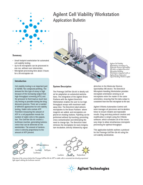
Agilent Cell Viability Workstation Application BulletinAgilent Cell ViabilityWorkstation consisting of aMolecular Devices SpectraMaxM5 (left), an Agilent BenchCelMicroplate HandlingWorkstation integrated with anAgilent Bravo AutomatedLiquid Handling Platform (right)=+LuciferinLuciferinCell lysisLuciferaseATPase inhibitorsOverview of the science behind the Promega CellTiter-Glo kit. ATP in viable cells is converted into directly proportionallight signal, utilizing the luciferase reaction.Summary•Small footprint workstation for automatedcell viability testing•Up to 65 microplates can be processed inone run, without user intervention•Microplate processing time about 3 hoursfor a 50-microplate runIntroductionCell viability testing is an important partof ADME/Tox compound profiling. Thedemand for this type of assay is highbecause of the increasing output fromhigh-throughput screening (HTS) andthe pressure to front load as much toxi-city testing as possible during the drugdiscovery process. There are a numberof different approaches for cell viabilitytesting. Viable cells contain ATP.Therefore measuring the amount ofATP in a cell population reveals thenumber of viable cells in this popula-tion. The CellTiter-Glo kit creates aluciferase reaction, generating lumines-cence that can be detected on theworkstation. The amount of lumines-cence is directly proportional to theamount of ATP present.System DescriptionThe Promega CellTiter-Glo kit is ideally suit-ed for adaptation on automated worksta-tions. The integration of the Agilent BravoPlatform with the Agilent BenchCelWorkstation enables the user to run high-throughput assays with maximum walk-away time. The BenchCel robot deliversmicroplates to the Bravo Platform wherereagents are added, and microplates areplaced on shaking stations. Pipetting can beperformed without tip touching, preventingcross-contamination and eliminating theneed to change tips. The BenchCel robotretrieves the microplates for room-tempera-ture incubation,directly followed by signaldetection in the Molecular DevicesSpectraMax M5 device. The BenchCelMicroplate Handling Workstation providesmicroplate-orientation sensing so allmicroplates enter the reader in the sameorientation, ensuring that the data output isconsistent from the first microplate to the last.Agilent VWorks Automation Control soft-ware manages all processes and incubationtimes to ensure reliable and repeatableresults. Drag-and-drop protocol creation andmodification is simple using the VWorkssoftware, which schedules all of the neces-sary steps to allow simultaneous microplateprocessing for optimal throughput.This application bulletin outlines a protocolfor the Promega CellTiter-Glo kit using thecell viability workstation.Instrument LayoutAgilent Bravo deck layout: locations 5 and 8 are configured with Orbital Shaking Stations (shaker) for enhanced throughput. A reservoir and a tipbox are placed manually at locations 2 and 6, respec-tively, before the protocol starts.Agilent BenchCel stacker layout: stacker 1 contains microplate A (can store up to 65 microplates), stackers 2 and 3 are used for incubation, and stacker 4 receives the processed microplates.MaterialsComponent List•Agilent BenchCel Workstation(R-series with 4 stackers)•Agilent Bravo Platform with gripper, 384ST disposable-tip pipette head, reservoir, 2 Orbital Shaking Stations•Molecular Devices SpectraMax M5•Agilent VWorks AutomationControl softwareLabware List•Microplate A: Greiner 96PS black,tissue-culture treated•Tipbox A: Agilent Tips 384 ST 70 µL Reagent List•Reservoir A: CellTiter-Glo reagent Protocol Workflow1.Move microplate A from BenchCelstacker 1 to Bravo deck location 7.2.Press on tips at Bravo decklocation 6.3.Aspirate 25 µL CellTiter-Glo fromreservoir A and dispense intomicroplate A.4.Move microplate A from decklocation 7 to 5 (shaker).5.Shake for 2 min.6.Move microplate A from decklocation 5 to 7.7.Move microplate A from Bravo decklocation 7 to BenchCel stacker 2.8.Incubate for 10 min.9.Move microplate A from BenchCelstacker 2 to the SpectraMax device.10.Read microplate A on theSpectraMax device.11.Move microplate A from theSpectraMax device to BenchCelstacker 4./lifesciences/automationThis item is intended for Research Use Only.Not for use in diagnostic procedures. Information,descriptions, and specifications in this publicationare subject to change without notice.Agilent Technologies shall not be liable for errorscontained herein or for incidental or consequentialdamages in connection with the furnishing,performance, or use of this material.Promega and CellTiter-Glo are registered trade-marks of Promega Corporation. Molecular Devicesand SpectraMax are registered trademarks of MDSAnalytical Technologies.© Agilent Technologies, Inc., 2009Published in the U.S.A., February 26, 2009Publication Number 5990-3555EN ConclusionsThe Agilent Cell Viability Workstation usingthe Promega CellTiter-Glo kit provides a reli-able, high-throughput solution for analyzingcell viability. The integration of microplatehandling, liquid handling, and microplatereading enables up to 65 microplates to beprocessed in one run without user interven-tion. Following the guidelines set by Promega,the typical throughput for this setup is about3 hours for 50 microplates, depending onexact protocol and liquid-handling steps.。
用于CRISPR文库筛选的N2a-Cas9细胞系的构建
中国预防兽医学报Chinese Journal of Preventive Veterinary Medicine第42卷第12期2020年12月V ol.42No.12Dec.2020doi :10.3969/j.issn.1008-0589.202002012用于CRISPR 文库筛选的N2a-Cas9细胞系的构建张纪文,王露露,李芳,刘杏,赵东明*,步志高*(中国农业科学院哈尔滨兽医研究所兽医生物技术国家重点实验室,黑龙江哈尔滨150069)摘要:为了进行CRISPR 文库筛选,本研究采用慢病毒转导的方法利用N2a 细胞构建表达Cas9蛋白的N2a-Cas9单克隆细胞系。
实验将LentiCas9-Blast 、pSPA X2和pMD 2.G 3种质粒共转染人胚胎肾细胞(HEK293T )获取高滴度的重组慢病毒,收集重组慢病毒并感染N2a 细胞,经杀稻瘟菌素(Blasticidin )筛选,通过有限稀释法获得含有Cas9基因的多个单克隆细胞株。
Western blot 结果显示候选细胞系高水平表达Cas9蛋白;利用慢病毒表达报告基因载体系统检测显示候选细胞系的Cas9蛋白具有很高的切割活力;利用CellTiter-Glo 试剂检测结果显示,Cas9基因在候选细胞系内高表达但不影响细胞增殖活性。
本研究利用慢病毒转导系统构建了稳定表达Cas9蛋白的N2a-Cas9单克隆细胞系,为基于CRISPR/Cas9全基因组高通量筛选技术探究神经细胞内关键宿主基因调控狂犬病病毒嗜神经性提供研究基础。
关键词:基因编辑;慢病毒;高通量筛选;鼠神经瘤母细胞中图分类号:S852.65文献标识码:A文章编号:1008-0589(2020)12-1292-04Construction of N2a-Cas9cell line for genome-wide CRISPR screeningZHANG Ji-wen,WANG Lu-lu,LI Fang,LIU Xing,ZHAO Dong-ming *,BU Zhi-gao *(State Key Laboratory of Veterinary Biotechnology,Harbin Veterinary Research Institute,Chinese Academy of Agricultural Sciences,Harbin 150069,China)Abstract :In this study,mouse neuroblastoma (N2a)cells were used to construct N2a-Cas9cell line which stably expresses Cas9protein by using lentivirus transfection technology.This constrcted cells line laid the foundation for CRISPR library screening.First,three plasmids including LentiCas9-Blast,pSPA X2,and pMD 2.G were co-transfected into human renal epithelial cells (HEK293T)to obtain recombinant lentivirus with high titers.N2a cells were infected with the recombinant lentivirus,and then screened by Blasticidin.Finally,several monoclonal cell lines expressing Cas9protein were isolated by limiting dilution.The expression of Cas9expression in N2a-Cas9cell line was determined by western blot,and the high cleavage activity of Cas9was also examined by using lentivirus-based reporter vector system.By using CellTiter-Glo ®reagent,the cell proliferation activity was well detectable and not decreased in N2a-Cas9cell lines,where Cas9gene was expressed in a high level.Therefore,N2a-Cas9cell line was successfully constructed using a lentiviral transfection system,which can provide a basis for genome-wide CRISPR screening to identify crucial host factors regulating Rabies virus neuro-tropism.Key words :gene editing;lentivirus;genome-wide CRISPR screening;murine neuroblastoma收稿日期:2020-02-11基金项目:兽医生物技术国家重点实验室自主课题(SKLVBP201801)作者简介:张纪文(1993-),男,河南驻马店人,硕士研究生,主要从事兽医微生物及其分子生物学研究.*通信作者:E-mail :****************;*********************Corresponding author张纪文,等.用于CRISPR文库筛选的N2a-Cas9细胞系的构建第12期CRISPR-Cas9(Clustered regularly interspaced short palindromic repeat sequences/CRISPR-associated protein 9)是存在于细菌或古生细菌中的一种抵御外源DNA 侵入的适应性免疫反应系统[1]。
promega 实时荧光 mt 细胞活力检测试剂盒说明书

G9711, G9712 and G97132021版 CTM645原英文技术手册TM645中文说明书适用产品目录号: W6010, W6011 和 W6012Lumit™ Human IL-1βImmunoassay普洛麦格(北京)生物技术有限公司Promega (Beijing) Biotech Co., Ltd 地址:北京市东城区北三环东路36号环球贸易中心B座907-909电话:************网址:技术支持电话:400 810 8133(手机拨打)技术支持邮箱:*************************CTM6452021制作1所有技术文献的英文原版均可在/ protocols 获得。
请访问该网址以确定您使用的说明书是否为最新版本。
如果您在使用该试剂盒时有任何问题,请与Promega 北京技术服务部联系。
电子邮箱:*************************1. 产品描述 (2)2. 产品组分和储存条件 (4)3. 开始实验前 (5)4. 培养细胞的直接(无转移)方案 (7)4. A. 细胞铺板和处理 (7)4. B. 制备人IL-1β标准品稀释液 (8)4. C. 将5X抗hIL-1β抗体混合物添加至检测孔 (9)4. D. 将 Lumit™检测试剂B添加至检测孔 (10)5. 可选样品转移方案 (11)5. A. 细胞铺板和处理 (11)5. B. 制备人IL-1β标准品稀释液 (12)5. C. 将2X抗hIL-1β抗体混合物添加至样品孔 (13)5. D. 将 Lumit™检测试剂B添加至样品孔 (14)6. 结果计算 (15)7. 代表性数据 (15)8. 疑难解答 (20)9. 附录 (21)9. A. 加工后的人IL-1β选择性的示例数据 (21)9. B. 炎症小体抑制多重检测的示例数据 (22)9. C. 参考文献 (23)9. D. 相关产品 (24)Lumit™ Human IL-1β Immunoassay普洛麦格(北京)生物技术有限公司Promega (Beijing) Biotech Co., Ltd 地址:北京市东城区北三环东路36号环球贸易中心B座907-909电话:************网址:技术支持电话:400 810 8133(手机拨打)技术支持邮箱:*************************CTM6452021制作21. 产品描述Lumit™ Human IL-1β Immunoassay(a,b)是可用于检测从细胞中释放的白介素1β(IL-1β)的均质生物发光检测试剂盒,操作过程中无需样品转移或洗涤。
Promega--双萤光素酶报告基因演示教学

1.98
处理A
1
5,1376 4,467
2
40,712 3,574
3
88,787 7,654
4
6,0292 5,232
5
处理B
1
587,635 5,144
2
988,347 8,832
3
3
409,881 3,564
661,954 5,847
113.21
第二天 对照
1 2 •
处理C
1
2 3
处理D
1
2 3 4
荧光 - Fluorescence
Fire
萤光 - Luminescence
Worm
萤光 (Bioluminescence)
荧光 (Fluorescence)
是化学发光(Chemilumi- nescence) 吸收来自光源的光,再发射
的一种,激发能量来自化学反应
另一光子
萤光发光计(Luminometer)
载体 - 萤火虫萤光素酶载体
•pGL3 家族
✓pGL3-Basic ✓pGL3-Control ✓pGL3-Enhancer ✓pGL3-Promoter
pGL3-Basic Map
pGL3-Control Map
pGL3-Enhancer Map
pGL3-Promoter Map
内对照载体可能存在的问题
GloMax ™ 96 luminometer
White microplates for luminescence
Promega公司出品的双报告基因实验产品
• 质粒
– 萤火虫萤光素酶质粒 – 海肾萤光素酶质粒 – 叩头虫萤光素酶质粒
MTS promega 说明书
MTS promega 说明书普洛麦格(北京)生物技术有限公司地址: 北京市东城区北三环东路36号环球贸易中心B座907-909CellTiter 96? AQ ueous One SolutionCell Proliferation AssayI. 描述 (1)II. 产品组分和保存条件 (4)III. 操作步骤 (5)A. 一般操作步骤 (5)B. 应用举例:用B9细胞检测IL-6生物活性的操作程序 (5)IV. 通常需考虑的因素 (6)A. 背景吸光度值 (6)B. 读取数据的可选波长 (6)C. 淋巴细胞检测 (7)D. 试剂优化 (7)E. 细胞数的优化 (7)V. 相关产品 (8)VI. 参考文献 (11)I. 描述CellTiter 96? AQ ueous One Solution Cell Proliferation Assay(a)是一种用比色法来检测细胞增殖和细胞毒实验中的活细胞数量的检测试剂。
此试剂含有一个新型的四唑化合物[3-(4,5-dimethylthiazol-2-yl)-5-(3-carboxymethoxyphenyl)-2-(4-sulfophenyl)-2H-tetrazolium, inner salt; MTS(a)]和一种电子偶联剂(phenazine ethosulfate; PES)。
PES具有增强的化学稳定性,这使它可与MTS混合形成稳定的溶液。
这种方便的“单溶液”模式,是在第一代CellTiter 96?AQ ueous Assay的基础上的改进,CellTiter96?AQueous Assay中使用的电子偶联剂PMS与MTS溶液是分开提供的。
MTS (Owen’s reagent) 被细胞生物还原成为一种有色的甲臜产物,可直接溶解于培养基中(图1, 1)。
这种转化很可能是在代谢活跃的细胞中的脱氢酶产生的NADPH或NADH的作用下完成的(2)。
细胞活力检测celltiter 96 aqueous one solution cell proliferation assay system protocol
Revised 12/12TB245T E C H N I C A L B U L L E T I NCellTiter 96® AQueousOne SolutionCell Proliferation AssayInstrucƟ ons for Use of ProductsG3580, G3581 and G3582Promega Corpora Ɵ on · 2800 Woods Hollow Road · Madison, WI 53711-5399 USA · Toll Free in USA 800-356-9526 · 608-274-4330 · Fax 608-277-2516 1 TB245 · Revised 12/12All technical literature is available at: /protocols/Visit the web site to verify that you are using the most current version of this Technical Bulletin. E-mail Promega Technical Services if you have questions on use of this system: techserv@CellTiter 96® AQ ueous One Solution Cell Proliferation Assay1. Description .........................................................................................................................................1 2. Product Components and Storage Conditions ........................................................................................4 3. Protocols .. (5)3.A. General Protocol ........................................................................................................................53.B. Example of a Protocol for Bioassay of IL-6 Using B9 Cells ..............................................................5 4. General Considerations .. (6)4.A. Background Absorbance ..............................................................................................................64.B. Optional Wavelengths to Record Data ..........................................................................................74.C. Lymphocyte Assays .....................................................................................................................74.D. Reagent Optimization .................................................................................................................84.E. Cell Number Optimization ...........................................................................................................8 5. References ..........................................................................................................................................9 6. Related Products . (10)1. DescriptionThe CellTiter 96® AQ ueous One Solution Cell Proliferation Assay (a) is a colorimetric method for determining the number of viable cells in proliferation or cytotoxicity assays. The CellTiter 96® AQ ueous One Solution Reagent contains a novel tetrazolium compound [3-(4,5-dimethylthiazol-2-yl)-5-(3-carboxymethoxyphenyl)-2-(4-sulfophenyl)-2H-tetrazolium, inner salt; MTS (a)] and an electron coupling reagent (phenazine ethosulfate; PES). PES has enhanced chemical stability, which allows it to be combined with MTS to form a stable solution. This convenient “One Solution” format is an improvement over the fi rst version of the CellTiter 96® AQ ueous Assay, where phenazine methosulfate (PMS) is used as the electron coupling reagent, and the PMS Solution and MTS Solution are supplied separately.The MTS tetrazolium compound (Owen’s reagent) is bioreduced by cells into a colored formazan product that is soluble in tissue culture medium (Figure 1, 1). This conversion is presumably accomplished by NADPH or NADH produced by dehydrogenase enzymes in metabolically active cells (2). Assays are performed by adding a small amount of theCellTiter 96® AQ ueous One Solution Reagent directly to culture wells, incubating for 1–4 hours and then recording the absorbance at 490nm with a 96-well plate reader (3,4).2Promega Corpora Ɵ on · 2800 Woods Hollow Road · Madison, WI 53711-5399 USA · Toll Free in USA 800-356-9526 · 608-274-4330 · Fax 608-277-2516TB245 · Revised 12/121. Description (continued)N N NNSO 3OCH 2COOHMTS+SN CH 3CH 3N N NNH SO 3OCH 2COOHFormazanSNCH 3CH 31605M B 09_6AFigure 1. Structures of MTS tetrazolium and its formazan product.The quantity of formazan product as measured by absorbance at 490nm is directly proportional to the number of living cells in culture (Figure 2). Because the MTS formazan product is soluble in tissue culture medium, the CellTiter 96® AQ ueous One Solution Assay requires fewer steps than procedures that use tetrazolium compounds such as MTT or INT (5,6). The formazan product of MTT reduction is a crystalline precipitate that requires an additional step in the procedure to dissolve the crystals before recording absorbance readings at 570nm (7).If you currently use a [3H]thymidine incorporation assay, addition of the CellTiter 96® AQ ueous One Solution Reagent can be substituted for the pulse of [3H]thymidine at the time point in the assay when the pulse of radioactive thymidine is usually added. Bioassay data comparing [3H]thymidine incorporation to the MTS-based CellTiter 96® AQ ueous Assay and the original MTT-based CellTiter 96® Assay demonstrate that tetrazolium reagents can be substituted for [3H]thymidine incorporation (4,7).Advantages of the CellTiter 96® AQ ueous One Solution Assay include:• Easy-to-Use: Add the CellTiter 96® AQ ueous One Solution Reagent to cells, incubate and read absorbance.• Convenient: Supplied as a single solution, fi lter-sterilized and ready for adding to assay plates.• Fast: Perform the assay in a 96-well plate with no washing or cell harvesting. Also eliminates solubilization steps normally required for MTT assays.• Non-Radioactive: Requires no scintillation cocktail or radioactive waste disposal.• Flexible: Plates can be read and returned to incubator for further color development.•Safe: Requires no volatile organic solvent to solubilize the formazan product (unlike MTT).Promega Corpora Ɵ on · 2800 Woods Hollow Road · Madison, WI 53711-5399 USA · Toll Free in USA 800-356-9526 · 608-274-4330 · Fax 608-277-2516 3 TB245 · Revised 12/122 × 1044 × 1046 × 1048 × 1041 × 1051.2 × 1051.4 × 1051.6 × 105A b s o r b a n c e (490n m )Cells/Well1606M A 10_9AFigure 2. Eff ect of cell number on absorbance at 490nm measured using the CellTiter 96® AQ ueous One Solution Assay. Various numbers of B9 hybridoma cells were added to the wells of a 96-well plate in RPMI containing 50µM 2-mercaptoethanol and supplemented with 5% FBS and 2ng/ml IL-6. The medium was allowed to equilibrate for 1 hour; then 20µl/well of CellTiter 96® AQ ueous One Solution Reagent was added. After 1 hour at 37°C in a humidifi ed, 5% CO 2 atmosphere, the absorbance at 490nm was recorded using an ELISA plate reader. Each point represents the mean ± SD of 4 replicates. The correlation coeffi cient of the line was 0.993, indicating a linearresponse between cell number and absorbance at 490nm. The background absorbance shown at zero cells/well was not subtracted from these data.4Promega Corpora Ɵ on · 2800 Woods Hollow Road · Madison, WI 53711-5399 USA · Toll Free in USA 800-356-9526 · 608-274-4330 · Fax 608-277-2516TB245 · Revised 12/12 2. Product Components and Storage ConditionsP R O D U C TS I Z E C AT.#CellTiter 96® AQ ueous One Solu Ɵ on Cell Prolifera Ɵ on Assay200 assays G3582Includes:• 4ml CellTiter 96® AQ ueous One Solution ReagentP R O D U C TS I Z E C AT.#CellTiter 96® AQ ueous One Solu Ɵ on Cell Prolifera Ɵ on Assay1,000 assays G3580Includes:• 20ml CellTiter 96® AQ ueous One Solution ReagentP R O D U C TS I Z E C AT.#CellTiter 96® AQ ueous One Solu Ɵ on Cell Prolifera Ɵ on Assay5,000 assays G3581Includes:• 100ml CellTiter 96® AQ ueous One Solution ReagentStorage Conditions: For long-term storage, store the CellTiter 96® AQ ueous One Solution Reagent at –20°C, protected from light. See the expiration date on the Product Information Label. For frequent use, solutions may be stored at 4°C, protected from light, for up to 6 weeks.Note: The performance of CellTiter 96® AQ ueous One Solution Reagent after 10 freeze-thaw cycles was demonstrated to be equal to that of freshly prepared solution.Safety: To the best of our knowledge, the chemical, physical and toxicological properties of this product have not been thoroughly investigated; therefore, we recommend the use of gloves, lab coats and eye protection when working with these or any chemicals.Light-Sensitivity: The CellTiter 96® AQ ueous One Solution Reagent is light-sensitive and is supplied in an amber container. Discoloration may occur if solutions are exposed to light outside of the container for several hours. This discoloration may cause slightly higher background 490nm absorbance readings, but it should not aff ect performanceof the CellTiter 96®AQ ueous One Solution Assay.Promega Corpora Ɵ on · 2800 Woods Hollow Road · Madison, WI 53711-5399 USA · Toll Free in USA 800-356-9526 · 608-274-4330 · Fax 608-277-25165 TB245 · Revised 12/123. ProtocolsMaterials to Be Supplied by the User • 96-well plates suitable for tissue culture • repeating pipettes, digital pipettes or multichannel pipettes • 96-well plate reader 3.A. General Protocol 1. Thaw the CellTiter 96® AQ ueous One Solution Reagent. It should take approximately 90 minutes at roomtemperature, or 10 minutes in a water bath at 37°C, to completely thaw the 20ml size.2. Pipet 20µl of CellTiter 96® AQ ueous One Solution Reagent into each well of the 96-well assay plate containing the samples in 100µl of culture medium.Note: We recommend repeating pipettes, digital pipettes or multichannel pipettes for convenient delivery of uniform volumes of CellTiter 96® AQ ueous One Solution Reagent to the 96-well plate.3. Incubate the plate at 37°C for 1–4 hours in a humidifi ed, 5% CO 2 atmosphere.Note: To measure the amount of soluble formazan produced by cellular reduction of MTS, proceed immediately to Step 4. Alternatively, to measure the absorbance later, add 25µl of 10% SDS to each well to stop the reaction. Store SDS-treated plates protected from light in a humidifi ed chamber at room temperature for up to 18 hours. Proceed to Step 4.4.Record the absorbance at 490nm using a 96-well plate reader.3.B. Example of a Protocol for Bioassay of IL-6 Using B9 Cells 1.Maintain stock cultures of B9 cells in RPMI 1640 medium containing 5% FBS, 50µM 2-mercaptoethanol (2-ME) supplemented with 5ng/ml human recombinant IL-6 . Subculture the stock cultures of cells to 2 × 10⁴ cells/ml, and refeed with human recombinant IL-6 every 3 days or when a density of 2 × 10⁵ cells/ml is reached. Note: B9 cells used for the bioassay should be from stock cultures 2 days after the last subculture (feeding with IL-6).2.Add 50µl/well of IL-6 samples or standards to be measured, diluted in RPMI 1640 medium containing 5% FBS and 50µM 2-ME. Start the titration of the IL-6 standard at 4ng/ml in column 12, and perform serial twofold dilutions across the plate to column 2 (to 4pg/ml). (After the cell suspension is added in Step 5 below, the fi nal concentration of the titrated standard will be 2ng/ml in column 12 to 2pg/ml in column 2.) Use column 1 for the negative control: RPMI 1640 medium (and supplements) without IL-6. Equilibrate the plate at 37°C in a humidifi ed, 5% CO 2 atmosphere while harvesting the cells for assay.6Promega Corpora Ɵ on · 2800 Woods Hollow Road · Madison, WI 53711-5399 USA · Toll Free in USA 800-356-9526 · 608-274-4330 · Fax 608-277-2516TB245 · Revised 12/12 3.B. Example of a Protocol for Bioassay of IL-6 Using B9 Cells (continued)3. Wash the B9 cells twice in RPMI 1640 containing 5% FBS and 50µM 2-ME by centrifugation at 300 × g for 5 minutes.4. Determine cell number and viability (by trypan blue exclusion), and resuspend the cells to a fi nal concentration of 1 × 10⁵ cells/ml in RPMI 1640 supplemented with 5% FBS and 50µM 2-ME.5. Dispense 50µl of the cell suspension (5,000 cells) into all wells of the plate prepared in Step 2. The total volume in each well should be 100µl.6. Incubate the plate at 37°C for 48–72 hours in a humidifi ed, 5% CO 2 atmosphere.7. Add 20µl per well of CellTiter 96® AQ ueous One Solution Reagent.8. Incubate the plate at 37°C for 1–4 hours in a humidifi ed, 5% CO 2 atmosphere.Note: To measure the amount of soluble formazan produced by cellular reduction of MTS, proceed immediately to Step 9. Alternatively, to measure the absorbance at a later time, add 25µl of 10% SDS to each well to stop the reaction. Store SDS-treated plates protected from light in a humidifi ed chamber at room temperature for up to 18 hours. Proceed to Step 9.9.Record the absorbance at 490nm using a 96-well plate reader.10. Plot the corrected absorbance at 490nm (Y axis) versus concentration of growth factor (X axis). Determine theX-axis value corresponding to one-half the diff erence between the maximum (plateau) and minimum (no growth factor control) absorbance values; this is the ED 50 value (ED 50 = the concentration of growth factor necessary to give one-half the maximum response).4. General Considerations 4.A. Background AbsorbanceA small amount of spontaneous 490nm absorbance occurs in culture medium incubated with CellTiter 96® AQ ueous One Solution Reagent. The type of culture medium used, type of serum, pH and length of exposure to light are variables that may contribute to the background 490nm absorbance. Background absorbance is typically0.2–0.3 absorbance units after 4 hours of culture. Background absorbance may result from chemical interference of certain compounds with tetrazolium reduction reactions. Strong reducing substances, including ascorbic acid, or sulfhydryl-containing compounds, such as glutathione, coenzyme A and dithiothreitol, can reduce tetrazolium salts nonenzymatically and lead to increased background absorbance values. Culture medium at elevated pH or extended exposure to direct light also may cause an accelerated spontaneous reduction of tetrazolium salts and result inincreased background absorbance values. If phenol red containing medium is used, an immediate change in color may indicate a shift in pH caused by the test compounds. Specifi c chemical interference of test compounds can be confi rmed by measuring absorbance values from control wells containing medium without cells at various concentrations of test compound.Promega Corpora Ɵ on · 2800 Woods Hollow Road · Madison, WI 53711-5399 USA · Toll Free in USA 800-356-9526 · 608-274-4330 · Fax 608-277-2516 7 TB245 · Revised 12/12Background 490nm absorbance may be corrected as follows: Prepare a triplicate set of control wells (without cells) containing the same volumes of culture medium and CellTiter 96® AQ ueous One Solution Reagent as in the experimental wells. Subtract the average 490nm absorbance from the “no cell” control wells from all other absorbance values to yield corrected absorbances.4.B. Optional Wavelengths to Record DataFigure 3 shows an absorbance spectrum of the formazan product resulting from reduction of MTS. We recommend recording data at the absorbance peak of 490nm; however, if your 96-well plate reader does not have a 490nm fi lter, data can be recorded at wavelengths of 450–540nm. Absorbance may be recorded at other wavelengths if necessary, but loss in sensitivity will result. A reference wavelength of 630–700nm may be used to subtract background contributed by excess cell debris, fi ngerprints and other nonspecifi c absorbance.A b s o r b a n c eWavelength (nm)3.002.502.001.501.000.500.00-0.503004005006007002284M A 07_8AFigure 3. Absorbance spectrum of MTS/formazan. The absorbance spectrum of the formazan product resulting from reduction of the MTS tetrazolium compound shows an absorbance maximum at 490nm. The negative absorbance values (382nm) correspond to the disappearance of MTS as it is converted into formazan.4.C. Lymphocyte AssaysLymphocytes may produce less formazan than other cell types (8). To achieve signifi cant absorbance changes withlymphocytes, increase the number of cells to approximately 2.5–10 × 104cells/well and incubate the plate with CellTiter 96® AQ ueous One Solution Reagent for the entire 4-hour period.8Promega Corpora Ɵ on · 2800 Woods Hollow Road · Madison, WI 53711-5399 USA · Toll Free in USA 800-356-9526 · 608-274-4330 · Fax 608-277-2516TB245 · Revised 12/12 4.D. Reagent OptimizationThe concentrations of tetrazolium and electron transfer reagents have been optimized for general use with a wide variety of cell lines cultured in 96-well plates containing 100µl of medium. If diff erent volumes of culture medium areused, adjust the volume to maintain a ratio of 20µl CellTiter 96®AQ ueous One Solution Reagent per 100µl culture medium. This reagent:medium ratio results in a fi nal concentration of 317µg/ml MTS in the assay wells. Minor variations in the optimum concentrations of tetrazolium and electron transfer reagents occur with diff erent cell lines; however, assay sensitivity is seldom compromised using the formulation in the CellTiter 96® AQ ueous One Solution Reagent. If reagent optimization is critical to your assay procedure, we recommend using the CellTiter 96® AQ ueous Non-Radioactive Cell Proliferation Assay (Cat.# G5421, G5430, G5440) or the CellTiter 96® AQ ueous MTS Reagent Powder products (Cat.# G1111, G1112) that supply the chemicals separately.4.E. Cell Number OptimizationCell proliferation assays require cells to grow over a period of time. Therefore, choose an initial number of cells per well that produces an assay signal near the low end of the linear range of the assay. This helps to ensure that the signal measured at the end of the assay will not exceed the linear range of the assay. This cell number can be determined by performing a cell titration as shown in Figure 2.Diff erent cell types have diff erent levels of metabolic activity. Factors that aff ect the metabolic activity of cells may aff ect the relationship between cell number and absorbance. Anchorage-dependent cells that undergo contact inhibition may show a change in metabolic activity per cell at high densities, resulting in a nonlinear relationship between cell number and absorbance. Factors that aff ect the cytoplasmic volume or physiology of the cells will aff ect metabolic activity.For most tumor cells, hybridomas and fi broblast cell lines, 5,000 cells per well is recommended to initiate proliferation studies, although fewer than 1,000 cells can usually be detected. The known exception to this is blood lymphocytes, which generally require 25,000–250,000 cells per well to obtain a suffi cient absorbance reading.Promega Corpora Ɵ on · 2800 Woods Hollow Road · Madison, WI 53711-5399 USA · Toll Free in USA 800-356-9526 · 608-274-4330 · Fax 608-277-2516 9 TB245 · Revised 12/125. References1. Barltrop, J.A. et al. (1991) 5-(3-carboxymethoxyphenyl)-2-(4,5-dimenthylthiazoly)-3-(4-sulfophenyl)tetrazo-lium, inner salt (MTS) and related analogs of 3-(4,5-dimethylthiazolyl)-2,5-diphenyltetrazolium bromide (MTT)reducing to purple water-soluble formazans as cell-viability indicators. Bioorg. Med. Chem. Lett. 1, 611–4.2.Berridge, M.V. and Tan, A.S. (1993) Characterization of the cellular reduction of 3-(4,5-dimethylthiazol-2-yl)-2,5-diphenyltetrazolium bromide (MTT): Subcellular localization, substrate dependence, and involvement of mitochondrial electron transport in MTT reduction. Arch. Biochem. Biophys. 303, 474–82.3. Cory, A.H. et al. (1991) Use of an aqueous soluble tetrazolium/formazan assay for cell growth assays in culture.Cancer Commun. 3, 207–12.4. Riss, T.L. and Moravec, R.A. (1992) Comparison of MTT, XTT, and a novel tetrazolium compound for MTS for in vitro proliferation and chemosensitivity assays. Mol. Biol. Cell (Suppl.) 3, 184a.5.Mosmann, T. (1983) Rapid colorimetric assay for cellular growth and survival: Application to proliferation and cytotoxicity assays. J. Immunol. Methods 65, 55–63.6. Bernabei, P.A. et al. (1989) In vitro chemosensitivity testing of leukemic cells: Development of a semiautomatedcolorimetric assay. Hematol. Oncol. 7, 243–53.7. CellTiter 96® Non-Radioactive Cell Proliferation Assay Technical Bulletin #TB112, Promega Corporation.8.Chen, C.-H., Campbell, P.A. and Newman, L.S. (1990) MTT colorimetric assay detects mitogen responses of spleen but not blood lymphocytes. Int. Arch. Allergy Appl. Immunol. 93, 249–55.10Promega Corpora Ɵ on · 2800 Woods Hollow Road · Madison, WI 53711-5399 USA · Toll Free in USA 800-356-9526 · 608-274-4330 · Fax 608-277-2516TB245 · Revised 12/12 6. Related ProductsMTS/MTT-Based Cell Viability Assay Systems ProductSize Cat.#CellTiter 96® AQ ueous Non-Radioactive Cell Proliferation Assay 1,000 assays G5421 5,000 assays G543050,000 assaysG5440CellTiter 96® AQ ueous MTS Reagent Powder* 250mg G11121g G1111CellTiter 96® Non-Radioactive Cell Proliferation Assay 1,000 assays G40005,000 assaysG4100*PMS is not supplied with MTS Reagent Powder and must be obtained separately.Luminescent-Based Cell Viability Assay System ProductSize Cat.#CellTiter-Glo ® Luminescent Cell Viability Assay 10ml G7570 10 × 10ml G7571 100ml G757210 × 100mlG7573Resazurin-Based Cell Viability Assay System ProductSize Cat.#CellTiter-Blue ® Cell Viability Assay 20ml G8080 100ml G808110 × 100mlG8082Fluorescent-Based Cell Viability Assay ProductSize Cat.#CellTiter-Fluor™ Cell Viability Assay 10ml G6080 5 × 10ml G60812 × 50mlG6082Promega Corpora Ɵ on · 2800 Woods Hollow Road · Madison, WI 53711-5399 USA · Toll Free in USA 800-356-9526 · 608-274-4330 · Fax 608-277-2516 11 TB245 · Revised 12/12Cytotoxicity Assay Systems (LDH)ProductSize Cat.#CytoTox-ONE™ Homogeneous Membrane Integrity Assay 200–800 assays G7890 1,000–4,000 assays G7891CytoTox 96® Non-Radioactive Cytotoxicity Assay 1,000 assaysG1780CytoTox-Glo™ Cytotoxicity Assay 10ml G9290 5 × 10ml G92912 × 50mlG9292Apoptosis Assay Systems ProductSize Cat.#Apo-ONE ® Homogeneous Caspase-3/7 Assay 1mlG779210ml G7790100ml G7791Caspase-Glo ® 2 Assay 10ml G094050ml G0941Caspase-Glo ® 6 Assay 10ml G097050ml G0971Caspase-Glo ® 3/7 Assay 2.5mlG809010ml G8091100ml G8092Caspase-Glo ® 8 Assay 2.5mlG820010ml G8201100ml G8202Caspase-Glo ® 9 Assay 2.5mlG821010ml G8211100ml G8212CaspACE™ Assay System, Colorimetric 50 assays G7351100 assays G7220DeadEnd™ Fluorometric TUNEL System 60 reactions G3250DeadEnd™ Colorimetric TUNEL System 40 reactions G713020 reactionsG73606. Related Products (continued)Apoptosis ReagentsProduct Size Cat.# CaspACE™ FITC-VAD-FMK In Situ Marker 50µl G7461125µl G7462 Anti-ACTIVE® Caspase-3 pAb 50µl G7481 Anti-Cytochrome c mAb 100µg G7421 Anti-pS473 Akt pAb 40µl G7441 Anti-PARP p85 Fragment pAb 50µl G7341 Caspase Inhibitor Z-VAD-FMK 125µl G723250µl G7231 Caspase Inhibitor, Ac-DEVD-CHO 100µl G5961Viability and Cytotoxicity AssayProduct Size Cat.# MultiTox-Fluor Multiplex Cytotoxicity Assay 10ml G9200 (live/dead cell protease activity determination) 5 × 10ml G92012 × 50ml G9202 CytoTox-Fluor™ Cytotoxicity Assay 10ml G9260 (dead cell protease activity determination) 5 × 10ml G92612 × 50ml G9262 MultiTox-Glo Multiplex Cytotoxicity Assay 10ml G9270 (live/dead cell protease activity determination) 5 × 10ml G92712 × 50ml G9272(a)The MTS tetrazolium compound is the subject of U.S. Pat. No. 5,185,450 assigned to the University of South Florida and is licensed exclusively to Promega CorporaƟ on.© 1996–2012 Promega CorporaƟ on. All Rights Reserved.AnƟ -ACTIVE, Apo-ONE, Caspase-Glo, CellTiter 96, CellTiter-Blue, CellTiter-Glo and CytoTox 96 are registered trademarks of Promega CorporaƟ on. CaspACE, CellTiter-Fluor, CytoTox-Fluor, CytoTox-Glo, CytoTox-ONE and DeadEnd are trademarks of Promega CorporaƟ on.NEN is a registered trademark of NEN Life Science Products, Inc.Products may be covered by pending or issued patents or may have certain limitaƟ ons. Please visit our Web site for more informaƟ on.All prices and specifi caƟ ons are subject to change without prior noƟ ce.Product claims are subject to change. Please contact Promega Technical Services or access the Promega online catalog for the most up-to-dateinformaƟ on on Promega products.12Promega CorporaƟ on · 2800 Woods Hollow Road · Madison, WI 53711-5399 USA · Toll Free in USA 800-356-9526 · 608-274-4330 · Fax 608-277-2516 TB245 · Revised 12/12 。
GFER抑制四氯化碳对HepG2细胞的损伤

GFER抑制四氯化碳对HepG2细胞的损伤高见;董凌月;安威【摘要】目的探讨HepG2细胞内生长因子ERV1样基因(growth factor Erv1-gene,GFER)表达降低后对四氯化碳(CCl4)诱导的细胞损伤的影响,以进一步明确GFER对于肝细胞的保护作用.方法首先将GFER siRNA转染入HepG2细胞,72 h 后收集细胞并通过Western Blot检测GFER的表达以明确沉默效率.再次将GFER siRNA转染入HepG2细胞72 h后,用CCl4处理细胞6h和24 h,检测细胞内ATP 的含量,caspase-3的活性,并应用MTS方法测定细胞的增殖能力以及TUNEL方法检测细胞凋亡.结果 Western blot结果显示转染GFER siRNA后细胞内GFER的表达降低.CCl4处理细胞6h后,GFER表达降低使细胞的增殖能力下降,细胞内ATP 含量增加,细胞凋亡更为明显.CCl4处理24 h后,GFER表达降低使细胞的增殖能力进一步下降,Caspase 3活性进一步升高,凋亡细胞数目显著增多,而ATP的含量明显下降.结论 GFER表达降低促进CCl4对HepG2细胞的损伤.【期刊名称】《中国组织化学与细胞化学杂志》【年(卷),期】2015(024)005【总页数】5页(P447-451)【关键词】生长因子样基因;四氯化碳;HepG2;caspase-3;凋亡【作者】高见;董凌月;安威【作者单位】首都医科大学细胞生物学系,肝脏保护与再生调节北京市重点实验室,北京 100069;首都医科大学细胞生物学系,肝脏保护与再生调节北京市重点实验室,北京 100069;首都医科大学细胞生物学系,肝脏保护与再生调节北京市重点实验室,北京 100069【正文语种】中文【中图分类】R735.7肝脏是人体重要的器官,具有复杂的生物学功能。
肝脏具有很强的再生功能,肝大部分切除或受到各种毒物、药物损伤后均可以启动肝再生过程[1]。
细胞活力和细胞毒性的评价(Molecular Devices)

优势:利用SpectraMax i3x多功能微孔板读板机所具有的超灵敏化学发光检测功能进行细胞活力和细胞毒性的评价简介方法 准备试剂• 仪器具有超高灵敏度化学发光检测功能,最低至10个细胞/每孔;• 微孔板读板高度自动优化设置,可提高检测信号强度• 软件预置模板可以更快分析出检测结果SpectraMax i3x是Molecular Devices公司最新推出的一款多功能微孔板读板机,可利用仪器所具有的化学发光检测功能,进行细胞活力和细胞毒性相应检测。
仪器可灵敏、快速检测出培养基中活细胞的数目和经相应处理后细胞毒性情况。
Promega公司推出的CellTiter-Glo试剂是利用了萤火虫荧光素酶反应体系中需要ATP参与才能使其发光的特点,化学发光信号强弱取决于培养基中ATP含量的高低,也就是依赖于其中活细胞数目的多少。
来自于BioVision公司基于生物化学发光原理的细胞毒性检测试剂盒,目的是检测腺苷酸激酶(AK)的含量,AK为一种存在于所有细胞中的常见蛋白,当破坏了细胞膜完整性后其会释放至培养基中,AK可转化ADP至ATP,所以可以利用类似方式进行化学发光检测。
材料• CellTiter-Glo Luminescent Cell ViabilityAssay (Promega P/N G7570)• Bioluminescence Cytotoxicity AssayKit (BioVision P/N K312-500)• HeLa 细胞(ATCC P/N CCL-2)• 黑色底透 96孔细胞培养板 (Corning P/N3904)• 白色96孔细胞培养板(Corning P/N 3917)• SpectraMax i3x多功能微孔板读板机使用前预先将CellTiter-Glo缓冲液和底物解冻并且平衡其至室温,将CellTiter-Glo 缓冲液加至含有CellTiter-Glo底物的棕色小瓶中,按照试剂盒说明书提示,将试剂轻轻反复颠倒进行混匀。
CellTiter-Glo实验注意事项

CellTiter-Glo实验注意事项细胞增殖检测技术已广泛应用于分子生物学、遗传学、肿瘤生物学、免疫学、药理和药代动力学等研究领域。
细胞增殖是指细胞在周期调控因子的作用下,通过DNA复制、RNA转录和蛋白质合成等复杂反应而进行的分裂系列过程。
细胞通过分裂的方式增殖,细胞增殖是生物体的重要生命特征。
单细胞生物以细胞分裂的方式产生新个体,多细胞生物以细胞分裂的方式产生新的细胞,从而补充体内衰老和死亡的细胞。
细胞增殖的同时,在细胞群体中总有一些因各种原因而死亡的细胞,活细胞在总细胞中所占的百分比叫做细胞活力。
检测细胞存活与增殖的方法主要包括观察DNA合成含量和检测细胞代谢活性两种,前者主要是DNA前体物质(胸腺嘧啶核苷类似物)掺入法,比如BRDU、EDU法;后者主要为MTT、XTT、MTS、CCK-8、WST-1及WST-8法等,在后者中现在最主流的就是CTG(CELLTITER-GLO)发光法,此法可快速灵敏的检测活细胞数量,市场上常用的是Promega公司CellTiter-Glo化学发光细胞活性检测试剂盒。
ATP腺嘌呤核苷三磷酸(简称三磷酸腺苷)参与生物体内多种酶促反应,是活细胞新陈代谢的一个指标,其含量直接反应了细胞的数量及细胞状态:实验过程中向细胞培养基加入等体积CellTiter-Glo™试剂,测量发光值,在光信号和体系中,发光值与ATP量成正比,而ATP又和活细胞数正相关,因此可通过检测ATP含量得细胞活力。
同普通的MTT、CCK8法相比,CellTiter-Glo™发光活细胞检测系统的检测试剂具有最高灵敏度和较长的信号持续时间,此系统已经广泛地应用在生命科学研究领域中,如一些生物活性因子的活性检测、大规模的抗肿瘤药物筛选、细胞毒性试验以及肿瘤放射敏感性测定等,各实验室已普遍使用此技术。
此法还有很多优点,如细胞活性分析是高效的“加样-混合-测量”系统,实验操作方便快捷,同时节约细胞用量。
CellTiter Glo Luminescent Cell Viability Assay Protocol

Promega Corporation ·2800 Woods Hollow Road ·Madison, WI 53711-5399 USA Toll F ree in USA 800-356-9526·Phone 608-274-4330 ·F ax 608-277-2516 ·1.Description (1)2.Product Components and Storage Conditions (4)3.Performing the CellTiter-Glo ®Assay (5)A.Reagent Preparation (5)B.Protocol for the Cell Viability Assay (6)C.Protocol for Generating an ATP Standard Curve (optional) (7)4.Appendix (7)A.Overview of the CellTiter-Glo ®Assay..............................................................7B.Additional Considerations..................................................................................8C.References............................................................................................................11D.Related Products. (12)1.DescriptionThe CellTiter-Glo ®Luminescent Cell Viability Assay (a–e)is a homogeneous method to determine the number of viable cells in culture based on quantitation of the ATP present, which signals the presence of metabolically active cells. The CellTiter-Glo ®Assay is designed for use with multiwell-plate formats, making it ideal for automated high-throughput screening (HTS) and cell proliferation and cytotoxicity assays. The homogeneous assay procedure (Figure 1) involves adding a single reagent (CellTiter-Glo ®Reagent) directly to cells cultured in serum-supplemented medium. Cell washing, removal of medium or multiple pipetting steps are not required.The homogeneous “add-mix-measure” format results in cell lysis and generation of a luminescent signal proportional to the amount of ATP present (Figure 2).The amount of ATP is directly proportional to the number of cells present in culture in agreement with previous reports (1). The CellTiter-Glo ®Assay relies on the properties of a proprietary thermostable luciferase (Ultra-Glo™ Recombinant Luciferase), which generates a stable “glow-type” luminescent signal and improves performance across a wide range of assay conditions. The luciferase reaction for this assay is shown in Figure 3. The half-life of the luminescent signal resulting from this reaction is greater than five hours (Figure 4). This extended half-life eliminates the need for reagent injectors and provides flexibility for continuous or batch-mode processing of multiple plates. The unique homogeneous format reduces pipetting errors that may be introduced during the multiple steps required by other ATP-measurement methods.CellTiter-Glo ®Luminescent Cell Viability AssayAll technical literature is available on the Internet at: /protocols/ Please visit the web site to verify that you are using the most current version of this Technical Bulletin. Please contact Promega Technical Services if you have questions on useofthissystem.E-mail:********************Figure 1. Flow diagram showing preparation and use of CellTiter-Glo ®Reagent.Promega Corporation ·2800 Woods Hollow Road ·Madison, WI 53711-5399 USA Toll F ree in USA 800-356-9526·Phone 608-274-4330 ·F ax 608-277-2516 ·3170M A 12_0ACellTiter-Glo CellTiter-Glo MixerLuminometer®System Advantages•Homogeneous:“Add-mix-measure” format reduces the number of plate-handling steps to fewer than that required for similar ATP assays.•Fast:Data can be recorded 10 minutes after adding reagent.•Sensitive:Measures cells at numbers below the detection limits of standard colorimetric and fluorometric assays.•Flexible:Can be used with various multiwell formats. Data can be recorded by luminometer or CCD camera or imaging device.•Robust:Luminescent signal is very stable, with a half-life >5 hours,depending on cell type and culture medium used.•Able to Multiplex:Can be used with reporter gene assays or other cell-based assays from Promega (2,3).Figure 3. The luciferase reaction.Mono-oxygenation of luciferin is catalyzed byluciferase in the presence of Mg 2+, ATP and molecular oxygen.Promega Corporation ·2800 Woods Hollow Road ·Madison, WI 53711-5399 USA Toll F ree in USA 800-356-9526·Phone 608-274-4330 ·F ax 608-277-2516 ·3171M A 12_0A L u m i n e s c e n c e (R L U )Cells per Well10,00060,00020,00030,00040,00050,0000R² = 0.9990.5 × 1061.0 × 1061.5 × 1062.0 × 1062.5 × 1063.0 × 1063.5 × 1064.0 × 106r² = 0.99020,00010,00030,00040,00050,000r² = 0.9900100200300400HO SN S N O S N S N OCOOH +ATP+O 2Ultra-Glo™ Recombinant Luciferase +AMP+PP i +CO 2+LightBeetle Luciferin OxyluciferinMg 2+0Figure 2. Cell number correlates with luminescent output.A direct relationship exists between luminescence measured with the CellTiter-Glo ®Assay and the number of cells in culture over three orders of magnitude. Serial twofold dilutions of HEK293cells were made in a 96-well plate in DMEM with 10% FBS, and assays wereperformed as described in Section 3.B. Luminescence was recorded 10minutes after reagent addition using a GloMax ®-Multi+ Detection System. Values represent the mean ± S.D. of four replicates for each cell number. The luminescent signal from 50HEK293 cells is greater than three times the background signal from serum-supplemented medium without cells. There is a linear relationship (r 2= 0.99)between the luminescent signal and the number of cells from 0to 50,000 cells per well.Figure 4. Extended luminescent half-life allows high-throughput batchprocessing.Signal stability is shown for three common cell lines. HepG2 and BHK-21cells were grown and assayed in MEM containing 10% FBS, while CHO-K1 cells were grown and assayed in DME/F-12 containing 10% FBS. CHO-K1, BHK-21 and HepG2 cells, at 25,000 cells per well, were added to a 96-well plate. After an equal volume of CellTiter-Glo ®Reagent was added, plates were shaken and luminescence monitored over time with the plates held at 22°C. The half-lives of the luminescent signals for the CHO-K1, BHK-21 and HepG2 cells were approximately 5.4, 5.2 and5.8hours, respectively.2.Product Components and Storage ConditionsProduct Size Cat.#CellTiter-Glo ®Luminescent Cell Viability Assay 10ml G7570Substrate is sufficient for 100 assays at 100µl/assay in 96-well plates or 400 assays at 25µl/assay in 384-well plates. Includes:• 1 × 10mlCellTiter-Glo ®Buffer • 1 vial CellTiter-Glo ®Substrate (lyophilized)Product Size Cat.#CellTiter-Glo ®Luminescent Cell Viability Assay 10 × 10ml G7571Each vial of substrate is sufficient for 100 assays at 100µl/assay in 96-well plates or 400 assays at 25µl/assay in 384-well plates (1,000 to 4,000 total assays). Includes:•10 × 10mlCellTiter-Glo ®Buffer •10 vials CellTiter-Glo ®Substrate (lyophilized)Promega Corporation ·2800 Woods Hollow Road ·Madison, WI 53711-5399 USA Toll F ree in USA 800-356-9526·Phone 608-274-4330 ·F ax 608-277-2516 ·R e l a t i v e L u m i n e s c e n c e (%)Time (minutes)CHO-K101020304050607080901003173M A 12_0AProduct Size Cat.# CellTiter-Glo®Luminescent Cell Viability Assay100ml G7572 Substrate is sufficient for 1,000 assays at 100µl/assay in 96-well plates or 4,000assays at 25µl/assay in 384-well plates. Includes:•1 × 100ml CellTiter-Glo®Buffer• 1 vial CellTiter-Glo®Substrate (lyophilized)Product Size Cat.# CellTiter-Glo®Luminescent Cell Viability Assay10 × 100ml G7573Each vial of substrate is sufficient for 1,000 assays at 100µl/assay in 96-well plates or4,000 assays at 25µl/assay in 384-well plates (10,000to 40,000 total assays). Includes:•10 × 100ml CellTiter-Glo®Buffer•10 vials CellTiter-Glo®Substrate (lyophilized)Storage Conditions:For long-term storage, store the lyophilized CellTiter-Glo®Substrate and CellTiter-Glo®Buffer at –20°C. For frequent use, the CellTiter-Glo®Buffer can be stored at 4°C or room temperature for 48hours without loss of activity. See product label for expiration date information. ReconstitutedCellTiter-Glo®Reagent (Buffer plus Substrate) can be stored at room temperaturefor up to 8hours with <10% loss of activity, at 4°C for 48hours with ~5% lossof activity, at 4°C for 4days with ~20% loss of activity or at –20°C for 21weekswith ~3% loss of activity. The reagent is stable for up to ten freeze-thaw cycles,with less than 10% loss of activity.3.Performing the CellTiter-Glo®AssayMaterials to Be Supplied by the User•opaque-walled multiwell plates adequate for cell culture•multichannel pipette or automated pipetting station for reagent delivery•device (plate shaker) for mixing multiwell plates•luminometer, CCD camera or imaging device capable of reading multiwell plates •optional:ATP for use in generating a standard curve (Section 3.C)3.A.Reagent Preparation1.Thaw the CellTiter-Glo®Buffer, and equilibrate to room temperature priorto use. For convenience the CellTiter-Glo®Buffer may be thawed andstored at room temperature for up to 48hours prior to use.2.Equilibrate the lyophilized CellTiter-Glo®Substrate to room temperatureprior to use.Promega Corporation·2800 Woods Hollow Road ·Madison, WI 53711-5399 USA Toll F ree in USA 800-356-9526·Phone 608-274-4330 ·F ax 608-277-2516 ·3.A.Reagent Preparation (continued)3.Transfer the appropriate volume (10ml for Cat.# G7570 and G7571, or 100mlfor Cat.# G7572 and G7573) of CellTiter-Glo ®Buffer into the amber bottlecontaining CellTiter-Glo ®Substrate to reconstitute the lyophilizedenzyme/substrate mixture. This forms the CellTiter-Glo ®Reagent.4.Mix by gently vortexing, swirling or inverting the contents to obtain ahomogeneous solution. The CellTiter-Glo ®Substrate should go intosolution easily in less than 1minute.3.B.Protocol for the Cell Viability AssayWe recommend that you perform a titration of your particular cells todetermine the optimal number and ensure that you are working within thelinear range of the CellTiter-Glo ®Assay. Figure 2 provides an example of sucha titration of HEK293 cells using 0 to 50,000 cells per well in a 96-well format.1.Prepare opaque-walled multiwell plates with mammalian cells in culturemedium, 100µl per well for 96-well plates or 25µl per well for 384-wellplates.Multiwell plates must be compatible with the luminometer used.2.Prepare control wells containing medium without cells to obtain a value forbackground luminescence.3.Add the test compound to experimental wells, and incubate according toculture protocol.4.Equilibrate the plate and its contents at room temperature forapproximately 30 minutes.5.Add a volume of CellTiter-Glo ®Reagent equal to the volume of cell culturemedium present in each well (e.g., add 100µl of reagent to 100µl of mediumcontaining cells for a 96-well plate, or add 25µl of reagent to 25µl ofmedium containing cells for a 384-well plate).6.Mix contents for 2 minutes on an orbital shaker to induce cell lysis.7.Allow the plate to incubate at room temperature for 10 minutes to stabilizeluminescent signal.Note:Uneven luminescent signal within standard plates can be caused bytemperature gradients, uneven seeding of cells or edge effects in multiwellplates.8.Record luminescence.Note:Instrument settings depend on the manufacturer. An integration timeof 0.25–1 second per well should serve as a guideline.Promega Corporation ·2800 Woods Hollow Road ·Madison, WI 53711-5399 USA Toll F ree in USA 800-356-9526·Phone 608-274-4330 ·F ax 608-277-2516 ·3.C.Protocol for Generating an ATP Standard Curve (optional)It is a good practice to generate a standard curve using the same plate onwhich samples are assayed. We recommend ATP disodium salt (Cat.# P1132,Sigma Cat.# A7699 or GE Healthcare Cat.# 27-1006). The ATP standard curveshould be generated immediately prior to adding the CellTiter-Glo®Reagentbecause endogenous ATPase enzymes found in sera may reduce ATP levels.1.Prepare 1µM ATP in culture medium (100µl of 1µM ATP solution contains10–10moles ATP).2.Prepare serial tenfold dilutions of ATP in culture medium (1µM to 10nM;100µl contains 10–10to 10–12moles of ATP).3.Prepare a multiwell plate with varying concentrations of ATP standard in100µl medium (25µl for a 384-well plate).4.Add a volume of CellTiter-Glo®Reagent equal to the volume of ATPstandard present in each well.5.Mix contents for 2 minutes on an orbital shaker.6.Allow the plate to incubate at room temperature for 10 minutes to stabilizethe luminescent signal.7.Record luminescence.4.Appendix4.A.Overview of the CellTiter-Glo®AssayThe assay system uses the properties of a proprietary thermostable luciferase toenable reaction conditions that generate a stable “glow-type” luminescentsignal while simultaneously inhibiting endogenous enzymes released duringcell lysis (e.g., ATPases). Release of ATPases will interfere with accurate ATPmeasurement. Historically, firefly luciferase purified from Photinus pyralis(LucPpy) has been used in reagents for ATP assays (1,4–7). However, it hasonly moderate stability in vitro and is sensitive to its chemical environment,including factors such as pH and detergents, limiting its usefulness fordeveloping a robust homogeneous ATP assay. Promega has successfullydeveloped a stable form of luciferase based on the gene from another firefly,Photuris pennsylvanica(LucPpe2), using an approach to select characteristics thatimprove performance in ATP assays. The unique characteristics of this mutant(LucPpe2m) enabled design of a homogeneous single-reagent-addition approachto perform ATP assays with cultured cells. Properties of the CellTiter-Glo®Reagent overcome the problems caused by factors, such as ATPases, thatinterfere with ATP measurement in cell extracts. The reagent is physicallyrobust and provides a sensitive and stable luminescent output.Promega Corporation·2800 Woods Hollow Road ·Madison, WI 53711-5399 USA Toll F ree in USA 800-356-9526·Phone 608-274-4330 ·F ax 608-277-2516 ·4.A.Overview of the CellTiter-Glo®Assay (continued)Sensitivity and Linearity:The ATP-based detection of cells is more sensitivethan other methods (8–10). In experiments performed by Promega scientists,the luminescent signal from 50HEK293 cells is greater than three standarddeviations above the background signal from serum-supplemented mediumwithout cells. There is a linear relationship (r2= 0.99) between the luminescentsignal and the number of cells from 0 to 50,000 cells per well in the 96-wellformat. The luminescence values in Figure 2 were recorded after 10minutes ofincubation at room temperature to stabilize the luminescent signal as describedin Section3.B. Incubation of the same 96-well plate used in the experimentshown in Figure 2 for 360minutes at room temperature had little effect on therelationship between luminescent signal and number of cells (r2= 0.99).Speed:The homogeneous procedure to measure ATP using the CellTiter-Glo®Assay is quicker than other ATP assay methods that require multiple steps toextract ATP and measure luminescence. The CellTiter-Glo®Assay also is fasterthan other commonly used methods to measure the number of viable cells(such as MTT, alamarBlue®or Calcein-AM) that require prolonged incubationsteps to enable the cells’ metabolic machinery to convert indicator moleculesinto a detectable signal.4.B.Additional ConsiderationsTemperature:The intensity and decay rate of the luminescent signal from theCellTiter-Glo®Assay depends on the luciferase reaction rate. Environmentalfactors that affect the luciferase reaction rate will change the intensity andstability of the luminescent signal. Temperature is one factor that affects therate of this enzymatic assay and thus the light output. For consistent results,equilibrate assay plates to a constant temperature before performing the assay.Transferring eukaryotic cells from 37°C to room temperature has little effect onATP content (5). We have demonstrated that removing cultured cells from a37°C incubator and allowing them to equilibrate to 22°C for 1–2 hours hadlittle effect on ATP content. For batch-mode processing of multiple assayplates, take precautions to ensure complete temperature equilibration. Platesremoved from a 37°C incubator and placed in tall stacks at room temperaturewill require longer equilibration than plates arranged in a single layer.Insufficient equilibration may result in a temperature gradient effect betweenwells in the center and at the edge of the plates. The temperature gradientpattern also may depend on the position of the plate in the stack.Promega Corporation·2800 Woods Hollow Road ·Madison, WI 53711-5399 USA Toll F ree in USA 800-356-9526·Phone 608-274-4330 ·F ax 608-277-2516 ·Chemicals:The chemical environment of the luciferase reaction affects theenzymatic rate and thus luminescence intensity. Differences in luminescenceintensity have been observed using different types of culture media and sera.The presence of phenol red in culture medium should have little impact onluminescence output. Assaying 0.1µM ATP in RPMI medium without phenolred resulted in ~5% increase in luminescence output (in relative light units[RLU]) compared to assays in RPMI containing the standard concentration ofphenol red, whereas assays in RPMI medium containing twice the normalconcentration of phenol red showed a ~2% decrease in luminescence.Solvents for the various test compounds may interfere with the luciferasereaction and thus the light output from the assay. Interference with theluciferase reaction can be detected by assaying a parallel set of control wellscontaining medium without cells. Dimethylsulfoxide (DMSO), commonly usedas a vehicle to solubilize organic chemicals, has been tested at finalconcentrations of up to 2% in the assay and only minimally affects light output.Plate Recommendations:We recommend using standard opaque-walledmultiwell plates suitable for luminescence measurements. Opaque-walledplates with clear bottoms to allow microscopic visualization of cells also maybe used; however, these plates will have diminished signal intensity andgreater cross talk between wells. Opaque white tape may be used to decreaseluminescence loss and cross talk.Cellular ATP Content:Different cell types have different amounts of ATP,and values reported for the ATP level in cells vary considerably (1,4,11–13).Factors that affect the ATP content of cells may affect the relationship betweencell number and luminescence. Anchorage-dependent cells that undergocontact inhibition at high densities may show a change in ATP content per cellat high densities, resulting in a nonlinear relationship between cell numberand luminescence. Factors that affect the cytoplasmic volume or physiology ofcells also will affect ATP content. For example, oxygen depletion is one factorknown to cause a rapid decrease in ATP (1).Promega Corporation·2800 Woods Hollow Road ·Madison, WI 53711-5399 USA Toll F ree in USA 800-356-9526·Phone 608-274-4330 ·F ax 608-277-2516 ·4.B.Additional Considerations (continued)Mixing:Optimal assay performance is achieved when the CellTiter-Glo®Reagent is mixed completely with the cultured cells. Suspension cell lines (e.g., Jurkat cells) generally require less mixing to achieve lysis and extract ATP than adherent cells (e.g., L929 cells). Tests were done to evaluate the effect ofshaking the plate after adding the CellTiter-Glo® Reagent. Suspension cellscultured in multiwell plates showed only minor differences in light outputwhether or not the plates were shaken after adding the CellTiter-Glo®Reagent.Adherent cells are more difficult to lyse and show a substantial differencebetween shaken and nonshaken plates.Several additional parameters related to reagent mixing include the force ofdelivery of CellTiter-Glo®Reagent, sample volume and dimensions of the well.All of these factors may affect assay performance. The degree of reagent mixing required may be affected by the method used to add the CellTiter-Glo®Reagent to the assay plates. Automated pipetting devices using a greater or lesser force of fluid delivery may affect the degree of subsequent mixing required.Complete reagent mixing in 96-well plates should be achieved using orbitalplate shaking devices built into many luminometers and the recommended2-minute shaking time. Special electromagnetic shaking devices that use aradius smaller than the well diameter may be required to efficiently mixcontents of 384-well plates. The depth of medium and geometry of themultiwell plates may have an effect on mixing efficiency. We recommend that you take these factors into consideration when performing the assay andempirically determine whether a mixing step is necessary for the individualapplication.LuminometersFor highly sensitive luminometric assays, the luminometer model and settings greatly affect the quality of data obtained. Luminometers from differentmanufacturers will vary in sensitivities and dynamic ranges. We recommend the GloMax®products because these instruments do not require gainadjustments to achieve optimal sensitivity and dynamic range. Additionally, GloMax®instruments are preloaded with Promega protocols for ease of use.If you are not using a GloMax®luminometer, consult the operating manual for your luminometer to determine the optimal settings. The limits should beverified on each instrument before analysis of experimental samples. The assay should be linear in some portion of the detection range of the instrument used.For an individual luminometer there may be different gain settings. Werecommend that you optimize the gain settings.4.C.References1.Crouch, S.P. et al.(1993) The use of ATP bioluminescence as a measure of cellproliferation and cytotoxicity. J. Immunol. Methods160, 81–8.2.Farfan, A.et al.(2004) Multiplexing homogeneous cell-based assays. Cell Notes10, 2–5.3.Riss, T., Moravec, R. and Niles, A. (2005) Selecting cell-based assays for drugdiscovery screening. Cell Notes13, 16–21.4.Kangas, L., Grönroos, M. and Nieminen, A.L. (1984) Bioluminescence of cellular ATP:A new method for evaluating cytotoxic agents in vitro. Med. Biol.62, 338–43.5.Lundin, A. et al.(1986) Estimation of biomass in growing cell lines by adenosinetriphosphate assay.Methods Enzymol. 133, 27–42.6.Sevin, B.U. et al.(1988) Application of an ATP-bioluminescence assay in human tumorchemosensitivity testing. Gynecol. Oncol.31, 191–204.7.Gerhardt, R.T.et al.(1991) Characterization of in vitro chemosensitivity ofperioperative human ovarian malignancies by adenosine triphosphatechemosensitivity assay. Am. J. Obstet. Gynecol. 165, 245–55.8.Petty, R.D. et al.(1995) Comparison of MTT and ATP-based assays for themeasurement of viable cell number. J. Biolumin. Chemilumin.10, 29–34.9.Cree, I.A. et al.(1995) Methotrexate chemosensitivity by ATP luminescence in humanleukemia cell lines and in breast cancer primary cultures: Comparison of the TCA-100assay with a clonogenic assay. AntiCancer Drugs6, 398–404.10.Maehara, Y. et al.(1987) The ATP assay is more sensitive than the succinatedehydrogenase inhibition test for predicting cell viability. Eur. J. Cancer Clin. Oncol.23, 273–6.11.Stanley, P.E. (1986) Extraction of adenosine triphosphate from microbial and somaticcells. Methods Enzymol.133, 14–22.12.Beckers, B. et al.(1986) Application of intracellular ATP determination in lymphocytesfor HLA-typing. J. Biolumin. Chemilumin.1, 47–51.13.Andreotti, P.E. et al.(1995) Chemosensitivity testing of human tumors using amicroplate adenosine triphosphate luminescence assay: Clinical correlation forcisplatin resistance of ovarian carcinoma. Cancer Res. 55, 5276–82.4.D.Related ProductsCell Proliferation ProductsProduct Size Cat.# ApoLive-Glo™ Multiplex Assay10ml G6410 ApoTox-Glo™ Triplex Assay10ml G6320 CellTiter-Fluor™ Cell Viability Assay (fluorescent)10ml G6080 CellTiter-Blue®Cell Viability Assay (resazurin)20ml G8080 CellTiter 96®AQ ueous One SolutionCell Proliferation Assay (MTS, colorimetric)200 assays G3582 CellTiter 96®AQ ueous Non-RadioactiveCell Proliferation Assay (MTS, colorimetric)1,000 assays G5421 CellTiter 96®AQ ueous MTS Reagent Powder1g G1111 CellTiter 96®Non-RadioactiveCell Proliferation Assay (MTT, colorimetric)1,000 assays G4000 Additional sizes available.Cytotoxicity AssaysProduct Size Cat.# CytoTox-Glo™ Cytotoxicity Assay (luminescent)*10ml G9290Mitochondrial ToxGlo™ Assay*10ml G8000 MultiTox-Glo Multiplex Cytotoxicity Assay(luminescent, fluorescent)*10ml G9270 MultiTox-Fluor Multiplex Cytotoxicity Assay(fluorescent)*10ml G9200 CytoTox-Fluor™ Cytotoxicity Assay (fluorescent)*10ml G9260 CytoTox-ONE™ Homogeneous MembraneIntegrity Assay (LDH, fluorometric)*200–800 assays G7890 CytoTox-ONE™ Homogeneous MembraneIntegrity Assay, HTP1,000–4,000 assays G7892 CytoTox 96® Non-Radioactive Cytotoxicity Assay1,000 assays G1780 (LDH, colorimetric)*GSH-Glo™ Glutathione Assay10ml V691150ml V6912 GSH/GSSG-Glo™ Assay10ml V661150ml V6612 *Additional sizes available.LuminometersProduct Size Cat.# GloMax®-Multi+ Detection System with Instinct™ Software:Base Instrument with Shaking 1 each E8032 GloMax®-Multi+ Detection System with Instinct™ Software:Base Instrument with Heating and Shaking 1 each E9032 GloMax®-Multi+ Luminescence Module 1 each E8041Apoptosis ProductsProduct Size Cat.# Caspase-Glo®2 Assay*10ml G0940 Caspase-Glo®6 Assay*10ml G0970 Caspase-Glo®3/7 Assay* 2.5ml G8090 Caspase-Glo®8 Assay* 2.5ml G8200 Caspase-Glo®9 Assay* 2.5ml G8210Apo-ONE®Homogeneous Caspase-3/7 Assay1ml G7792 DeadEnd™ Fluorometric TUNEL System60 reactions G3250 DeadEnd™ Colorimetric TUNEL System20 reactions G7360Anti-ACTIVE®Caspase-3 pAb50µl G7481Anti-PARP p85 Fragment pAb50µl G7341Anti-pS473Akt pAb40µl G7441 Caspase Inhibitor Z-VAD-FMK, 20mM50µl G7231125µl G7232*Additional sizes available.(a)U.S. Pat. Nos. 6,602,677 and 7,241,584, European Pat. No. 1131441, Japanese Pat. Nos. 4537573 and 4520084 and other patents pending(b)U.S. Pat. No. 7,741,067, Japanese Pat. No. 4485470 and other patents pending.(c)U.S. Pat. No. 7,700,310, European Pat. No. 1546374 and other patents pending.(d)U.S. Pat. Nos 7,083,911, 7,452,663 and 7,732,128, European Pat. No. 1383914 and Japanese Pat. Nos. 4125600 and 4275715.(e)The method of recombinant expression of Coleoptera luciferase is covered by U.S. Pat. Nos. 5,583,024, 5,674,713 and 5,700,673.© 2001–2012 Promega Corporation. All Rights Reserved.Anti-ACTIVE, Apo-ONE, Caspase-Glo, CellTiter 96, CellTiter-Blue, CellTiter-Glo, CytoTox 96 and GloMax are registered trademarks of Promega Corporation. ApoTox-Glo, ApoLive-Glo, CellTiter-Fluor, CytoTox-Fluor, CytoTox-Glo, CytoTox-ONE, DeadEnd, GSH-Glo, GSH/GSSG-Glo, Instinct, Mitochondrial ToxGlo and Ultra-Glo are trademarks of Promega Corporation. alamarBlue is a registered trademark of Trek Diagnostic Ssystems, Inc.Products may be covered by pending or issued patents or may have certain limitations. Please visit our Web site for more information.All prices and specifications are subject to change without prior notice.Product claims are subject to change. Please contact Promega Technical Services or access the Promega online catalog for the most up-to-date information on Promega products.。
Cell-BasedAssay(细胞活性、毒性和凋亡)

•定量计算某种药物或某 荧光法: 刃天青
种因素对细胞的毒性或 促增殖的作用
生物发光法:ATP法
•作为其他实验的对照
同位素标记:H3Thymidine标记
高灵敏度,高通量,操 作简单
高灵敏度,高通量,操 作时间短
灵敏度高,但是操作复 杂,放射性方法,成本 高
MTT操作步骤
前期培养
加加DDyyeeSSooluluttioionn
Vijaya Ramachandran, Thiruvengadam Arumugam, Huamin Wang, and Craig D. Logsdon Cancer Res., Oct 2008; 68: 7811 - 7818.
5.NADPH Oxidase-dependent Signaling Involved In Endothelial Cell Survival And Proliferation: Role Of Nox2 And Nox4
出? • 需要叠加其他实验?
如何选择适合的检测方法
• 现有的仪器 • 通量和灵敏度 • 操作简便度和操作时间的要求
对实验效率的要求-高通量药物筛选
实验目的与常用方法
实验目的
方法
•初步观察细胞的生存状 态,镜下计算活细胞和死 台盼蓝染料排斥法 细胞比例
特点
镜下观察计数细胞
比色法:MTT,MTS 中低通量,操作简单
Lan H. Ly, Xiao-Yan Zhao, Leah Holloway, and David Feldman.Endocrinology, May 1999; 140: 2071.
4.Anterior Gradient 2 Is Expressed and Secreted during the Development of Pancreatic Cancer and Promotes Cancer Cell Survival
鱼藤酮对PC12细胞及大鼠脑线粒体的损伤作用

•304•沈阳医学院学报Journal of Shenyang Medical College第23卷第3期2021年5月-大学生科技园地-鱼藤酮对PC12细胞及大鼠脑线粒体的损伤作用刘雨-周青J李晓秀3-4*,刘波3*(1.沈阳医学院药学院2017级药学专业,辽宁沈阳110034; 2.基础医学院2015级医学影像学专业;3.沈阳医学院药学院;4.辽宁省行为与认知神经科学重点实验室)[摘要]目的:在细胞水平、脑组织水平,探讨鱼藤酮对线粒体的影响及机制。
方法:以PC12细胞为研究对象,经鱼藤酮处理后,使用细胞计数试剂盒(CCK-8)检测细胞活力,JC-1探针检测线粒体膜电位,DCFH-DA荧光探针检测细胞活性氧(ROS)水平,荧光素酶发光法检测线粒体ATP的含量。
以大鼠脑线粒体为研究对象,通过Clark氧电极法检测鱼藤酮对线粒体呼吸功能的影响。
结果:鱼藤酮不同程度的降低细胞存活率(P<0.05),2^M的鱼藤酮与PC12细胞共处理48h,细胞活力下降为对照组的47.57%。
2^M鱼藤酮使细胞内ROS增加、ATP含量减少、膜电位降低(P<0.05)。
经鱼藤酮处理后,大鼠脑线粒体皿态和W态呼吸速率及呼吸控制指数显著降低(P<0.05)。
结论:鱼藤酮在细胞水平、脑组织水平,均诱导线粒体损伤。
[关键词]鱼藤酮;线粒体功能;活性氧;膜电位[中图分类号]R965.1[文献标识码]A[文章编号]1008-2344(2021)03-0304-04doi:10.16753/ki.1008-2344.2021.03.030Effects of rotenone on PC12cells and mitochondria in rat brainLIU Yu1,ZHOU Qing2,LI Xiaoxiu3,4*,LIU Bo3*(1.Undergraduate of Grade2017,Shenyang Medical College,Shenyang110034,China; 2.Undergraduate of Grade2015; 3.School of Pharmacy, Shenyang Medical College; 4.Liaoning Key Laboratory of Behavior and Cognitive Neuroscience)[Abstract]Objective:To investigate the effect and mechanism of rotenone on mitochondria of cells and brain tissue.Methods: PC12cells were treated with rotenone.The survival rate of PC12cells was measured by CCK8.The intracellular ROS and mitochondrial membrane potential were detected by fluorescence probe.The mitochondrial ATP was detected by luciferase luminescence.Furthermore,the respiratory function of rat brain mitochondria was measured by Clark oxygen electrode.Results:Rotenone decreased the cell viability(P<0.05).The viability of PC12cells treated with2r M rotenone for48h decreased to47.57%of that in the control group.Rotenone at2r M increased ROS,decreased ATP and membrane potential(P<0.05).After rotenone treatment,V3,V4 and respiration control rate of rat brain mitochondria significantly decreased(P<0.05).Conclusion:Rotenone can induce mitochondrial damage on cells and brain tissue.[Key words]rotenone;mitochondrial function;reactive oxygen species;mitochondrial membrane potential帕金森病(Parkinson's disease,PD)是一种神经退行性疾病,多发生于老年人,主要症状包括静止性震颤、肌僵直、运动功能和姿势平衡障碍。
SAHA和TRAIL联合使用对乳腺癌雌激素受体阳性细胞MCF-7生长的影响

SAHA和TRAIL联合使用对乳腺癌雌激素受体阳性细胞MCF-7生长的影响韩翰;王敏【摘要】目的:实时观测SAHA和TRAIL联合使用后对乳腺癌雌激素受体阳性细胞MCF-7生长的影响。
方法实时无标记动态细胞分析系统( real time cell analysis, RTCA)动态监测各种处理因素对乳腺癌MCF-7细胞生长状况的影响,并通过BiostationIM活细胞工作站实时收集各种处理因素对乳腺癌MCF-7细胞增殖干预的形态学证据。
结果 xCELLi-gence RTCA 实时无标记动态细胞分析系统显示,随着TRAIL的加入,SAHA对MCF-7细胞的抑制作用明显增强。
SAHA和TRAIL的联合用药可能提高了SAHA对MCF-7细胞的敏感性。
实时活细胞工作站成像实验进一步证明SA-HA和TRAIL联合用药对MCF-7乳腺癌细胞的抑制作用强于SAHA或TRAIL单独用药。
结论 SAHA和TRAIL的联合使用对MCF-7细胞的生长具有协同抑制作用。
%Aim To investigate the effects of com-bined treatment of SAHA and TRAIL on human breast cancer ER positive cell line MCF-7 . Methods MCF-7 cells were treated with SAHA and/or TRAIL. The inhibitory rates were detected by real-time cell prolifer-ation assays. Morphology changes of MCF-7 cells were observed through time-lapse live cell imaging acquisi-tion. Results Real-time cell proliferation assays showed that the anti-tumor efficacy of SAHA was sig-nificantly enhanced in combination with TRAIL. The results of time-lapse live cell imaging acquisition dem-onstrated that, with treatment of SAHA and TRAIL, the growth inhibition of MCF-7 cells was more obvious than that of in TRAIL or SAHA treatment alone. Con-clusion The combination treatment of SAHAand TRAIL has a synergistic effect of growth inhibition on breast cancer MCF-7 cells.【期刊名称】《中国药理学通报》【年(卷),期】2016(000)002【总页数】6页(P223-228)【关键词】SAHA;TRAIL;乳腺癌;雌激素受体阳性;MCF-7;协同作用【作者】韩翰;王敏【作者单位】沈阳医学院生物化学教研室,辽宁沈阳 110034;沈阳医学院病原生物学教研室,辽宁沈阳 110034【正文语种】中文【中图分类】R329.24;R392.11;R737.9;R916.4;R979.1中国图书分类号:R329.24;R392.11;R737.9;R916.4;R979.1乳腺癌是威胁女性健康的常见恶性肿瘤之一,近年来发病率已居全球女性恶性肿瘤首位[1-2]。
2021-Promega萤光素酶技术讲座-生物发光技术的前世今生

Promega萤光素酶30周年庆技术讲座季生物发光技术的前世今生目录1.自然界的生物发光与萤光素酶的发现2.萤光素酶从自然界走向实验台3.萤光素酶为何是最理想的报告基因4.生物发光的检测1.自然界的生物发光⚫自然界的发光生物⚫发光生物,为何要发光⚫生物发光机理的探索之路-萤光素酶的发现自然界中的生物发光世界上有很多意想不到的地方都存在会“发光”的生物,比如发光的蘑菇、发光的海洋生物,以及发光的昆虫等,如我们最为熟悉的萤火虫。
发光生物为何要发光人们认为,在生物中产生光的原因根据每个物种不同,其原因各不相同。
目前估计大约有1500种不同的物种可以利用生物化学反应产生光,并且每天还有更多的具有这种能力的物种被发现。
生物发光机理发现约17世纪,爱尔兰化学家、物理学家和自然哲学家Robert Boyle 发现了能够产生微光的腐烂木材,经过研究发现,发光需要氧气的参与。
这是关于生物发光本质的第一份有文献记载的报道。
到20世纪40年代,约翰霍普金斯大学年轻的生物化学教授William McElroy 开始质疑生物发光的分子基础,他知道产生光需要消耗能量,经研究发现ATP 可能是该能量的来源。
他的开创性研究,鼓舞了年轻一代科学家们。
Luciferase 萤光素酶Luciferin萤光素2.萤光素酶从自然界走向实验台⚫萤光素酶商品化产品的诞生⚫萤光素酶发光的原理⚫生物发光与荧光的区别2012新型专利萤光素酶NanoLuc ®诞生2015基于NanoLuc ®萤光素酶的NanoBRET ™技术诞生2016基于NanoLuc ®萤光素酶的NanoBiT ®蛋白互补技术诞生2020基于NanoBiT ®的Lumit ™技术诞生Promega 正式进入免疫检测领域2017基于NanoLuc ®萤光素酶的HiBiT 蛋白标签技术诞生··························································································································································1991第一代萤光素酶产品诞生Luciferase Assay System & pGL2 Vectors1995第一代双萤光素酶检测系统DLR TM 诞生DLR TM & pGL3 Vectors1999UltraGlo ™Recombinant Luciferase 诞生& CellTiter-Glo ®细胞活力检测系统诞生2003Caspase-Glo ®3/7细胞凋亡检测系统诞生2007One-Glo ™Luciferase Assay System 诞生Celebrating 30 Years of Innovation and Discovery Using Bioluminescent TechnologyPromega 萤光素酶技术30年发展里程碑30 Years Of Bioluminescent Technology Innovations ()不同萤光素酶工具与发光机理萤火虫海肾深海细脚刺虾萤光素酶底物萤光素酶萤光素酶底物萤光素酶萤光素酶生物发光与荧光的区别什么是发光?发光是光子(光)发射的过程。
2021-Promega萤光素酶技术讲座-萤光素酶报告基因检测实验操作篇

/.,mnbv\\][plkjhgfds=Promega萤光素酶30周年庆技术讲座季萤光素酶报告基因技术使用操作详解篇目录1.萤光素酶的由来及作用机制解读2.萤光素酶报告基因能用于哪些研究4.实验数据如何处理3. 如何选择合适的检测方案•选择报告基因类型•选择单报告基因或双报告基因•选择载体类型•选择内参•选择转染方式•选择检测试剂•选择检测板•选择检测仪萤光素酶的由来Luciferin 萤光素•通常来说,能够加速自然发生的化学反应的生物分子被称作“酶”,科学家使用后缀“-ase ”作为此类蛋白质的通用命名法。
•针对luciferase 这个分子来说,“lucere ”这个名字的拉丁词根意思为“发光”。
报告基因:基因表达变化或者与基因表达变化相关的细胞事件的一个指示剂。
通常是一种编码易被检测的蛋白质或酶的基因,其表达产物非常容易被鉴定。
萤光素酶:一类能够产生生物发光信号的酶的统称。
萤光素酶可以催化自身底物氧化,反应中一部分能量以光子形式被释放,即生物发光信号。
萤火虫海肾深海细脚刺虾萤光素酶促进生物发光的机制萤火虫萤光素酶促进生物发光产生的反应示意图萤光素酶是一种作为化学反应催化剂的蛋白质,能在萤光素、ATP 和其它细胞离子存在的条件下促进光的产生。
化学反应本身包括两个步骤:1.在萤光素酶存在的条件下,萤光素+ATP 生成萤光素化腺苷酸+ADP 。
2.将萤光素化腺苷酸+氧气转化为氧化萤光素+AMP 以及光。
以萤火虫萤光素酶的生物发光反应为例:萤火虫海肾深海细脚刺虾报告基因:基因表达变化或者与基因表达变化相关的细胞事件的一个指示剂。
通常是一种编码易被检测的蛋白质或酶的基因,其表达产物非常容易被鉴定。
萤光素酶报告基因技术通常检测基因表达是通过RT-qPCR (mRNA 水平)或Western Blot (蛋白水平)引入报告基因,通过很容易被检测的报告基因的表达来判断启动子区域功能萤光素酶作为报告基因的优点:✓没有内源性表达✓易于定量✓高灵敏度、线性范围宽✓反应迅速✓可实现双萤光素酶报告基因检测Promoter Exon 12345EnhancerTranscription start site (转录起始位点)Enhancer Reporter GenePromoter基因编码区启动子区域外显子Transcription start site (转录起始位点)报告基因编码区萤光素酶报告基因的应用•免疫学检测•细胞信号通路分析•转录因子分析•蛋白-蛋白相互作用•转录后修饰•调控元件功能研究(启动子,增强子等)•病毒研究•细胞代谢研究•干细胞分化•小动物活体成像•siRNA/miRNA 研究•配基-受体相互作用•细胞健康研究•激酶研究•膜受体和细胞内受体激活/结合研究查看更多应用请访问:https://wechat.promeg /luc30years/NanoLuc ®萤光素酶技术,应用更广泛!调控元件研究Luciferase Promoter 转录调控-如:检测启动子活性Promoter转录后调控-如:剪接(Splicing )LuciferasePromoter Luciferase PromoterLuciferase Test Sequence转录后调控-如:检测蛋白稳定性转录后/翻译调控-如:检测RNA 干扰(RNAi)Translational fusionIntronTarget SequenceProtein of Interest细胞学事件研究◼膜受体和细胞内受体激活/信号传导◼细胞信号通路分析•GPCR 配基,激活剂,拮抗剂•核受体研究◼免疫应答•定义信号通路•蛋白:蛋白相互作用Activator/Pathway信号通路Transcription Factor转录因子Binding Site 转录因子结合位点INF-αSTAT1:STAT2Interferon Stimulated Response Element(ISRE)TGF-βSMAD3:SMAD4SMAD Binding Element (SBE)IL6STAT3:STAT3sis-Inducible Element (SIE)IL3STAT5:STAT5STAT5 Response Element•TCR PD-1/PD-L1•抗体药物等细胞或动物成像表达蛋白激酶C (PKC)-NanoLuc ®萤光素酶融合蛋白的HEK293 细胞在PMA 处理后,使用furmazine 底物20 分钟后进行检测。
- 1、下载文档前请自行甄别文档内容的完整性,平台不提供额外的编辑、内容补充、找答案等附加服务。
- 2、"仅部分预览"的文档,不可在线预览部分如存在完整性等问题,可反馈申请退款(可完整预览的文档不适用该条件!)。
- 3、如文档侵犯您的权益,请联系客服反馈,我们会尽快为您处理(人工客服工作时间:9:00-18:30)。
Promega Corporation ·2800 Woods Hollow Road ·Madison, W I 53711-5399 USA Toll Free in USA 800-356-9526·Phone 608-274-4330 ·Fax 608-277-2516 ·1.Description ..........................................................................................................12.Product Components and Storage Conditions ............................................43.Reagent Preparation and Storage ...................................................................54.Protocols for the CellTiter-Fluor™ Cell Viability Assay ..........................5A.Determining Assay Sensitivity, Method 1........................................................6B.Determining Assay Sensitivity, Method 2........................................................7C.Example Viability Assay Protocol.....................................................................8D.Example Multiplex Assay Protocol (with luminescent caspase assay).......9E.Recommended Controls (10)5.General Considerations ..................................................................................106.References .........................................................................................................117.Related Products ..............................................................................................121.DescriptionThe CellTiter-Fluor™ Cell Viability Assay (a)is a nonlytic, single-reagent-addition fluorescence assay that measures the relative number of live cells in a culture population after experimental manipulation (Figures 1 and 2). The CellTiter-Fluor™ Cell Viability Assay measures a conserved and constitutive protease activity within live cells and therefore serves as a marker of cell viability (1). Results obtained using the CellTiter-Fluor™ Cell Viability Assay correlate well with other established methods of determining cell viability(Figure 3). The live-cell protease activity is restricted to intact viable cells and is measured using a fluorogenic, cell-permeant, peptide substrate (glycyl-phenylalanyl-aminofluorocoumarin; GF-AFC). The substrate enters intact cells where it is cleaved by the live-cell protease activity to generate a fluorescent signal proportional to the number of living cells (Figure 4). This live-cell protease becomes inactive upon loss of cell membrane integrity and leakage into the surrounding culture medium.The CellTiter-Fluor™ Cell Viability Assay also can be used in a single-well,sequential, multiplex format with other downstream chemistries to normalize data by cell number. Data from the assay can serve as an internal control andCellTiter-Fluor™ Cell Viability AssayAll technical literature is available on the Internet at: /protocols/ Please visit the web site to verify that you are using the most current version of this Technical Bulletin. Please contact Promega Technical Services if you have questions on useof this system. E-mail: techserv@allow identification of errors resulting from cell clumping or compoundcytotoxicity. The CellTiter-Fluor™ Cell Viability Assay is compatible with most Promega luminescence assays or spectrally distinct fluorescence assay methods,such as assays measuring caspase activation, reporter gene expression or orthogonal measures of viability. However, some P450-Glo™ multiplexprotocols may require removing culture supernatant to a separate assay well before performing the assay because of isoform-specific competitive inhibition of the cytotochrome P450 enzymes by the coumarin product of the CellTiter-Fluor™ Cell Viability Assay reaction.Figure 1. Schematic diagram of the CellTiter-Fluor™ Cell Viability Assay.Promega Corporation ·2800 Woods Hollow Road ·Madison, W I 53711-5399 USA Toll Free in USA 800-356-9526·Phone 608-274-4330 ·Fax 608-277-2516 ·6868M ACellTiter-Fluor™ ReagentAdd GF-AFC Substrate to Assay Buffer to create the CellTiter-Fluor™ Reagent.MeasureAssay BenefitsMeasure the Relative Number of Live Cells in Culture: Nonlytic, single-reagent-addition, homogeneous, “add-mix-measure” protocol.Get More Data from Every Well: The CellTiter-Fluor™ Cell Viability Assay can be performed in multiplex with most Promega luminescence assays.Normalize Data for Cell Number: Normalizing data for live-cell number makes results more comparable well-to-well, plate-to-plate, day-to-day.Figure 3. The CellTiter-Fluor™ Cell Viability Assay shows strong correlation with established methods for measuring viability. Panel A. The GF-AFC Substrate signal from serial dilutions of live cells plotted against results from the CellTiter-Glo ®Luminescent Cell Viability Assay (Cat.# G7570), which measures cellular ATP.Panel B.The GF-AFC Substrate signal from serial dilutions of live cells plottedagainst results achieved using the CellTiter-Blue ®Cell Viability Assay (Cat.# G8080).Promega Corporation ·2800 Woods Hollow Road ·Madison, W I 53711-5399 USA Toll Free in USA 800-356-9526·Phone 608-274-4330 ·Fax 608-277-2516 ·6867M AA.B.R e s o r u f i n F l u o r e s c e n c e (R F U )2,5002,6002,7002,8002,9003,0003,100GF-AFC Fluorescence (RFU)A T P -B a s e d A s s a y L u m i n e s c e n c e (R L U )0GF-AFC Fluorescence (RFU)LIVE-CELL SUBSTRATE:cell-permeant fluorogenic substrate for the live-cellprotease (Gly-Phe-AFCoumarin)NucleusLive CellLive-cell protease substrate can cross the cell membrane.Active Live-CellProteaseO CF 3GF–N HOGFOCF 3OH 2N6995M AFigure 2. CellTiter-Fluor™ Cell Viability Assay chemistry. The cell-permeant substrate enters the cell, where it is cleaved by the live-cell protease activity to produce the fluorescent AFC. The live-cell protease is labile in membrane-compromised cells and cannot cleave the substrate.Figure 4. The CellTiter-Fluor™ Cell Viability Assay signal derived from viable cells (untreated) is proportional to cell number. Dead cells (treated) do not contribute appreciable signal in the assay.2.Product Components and Storage ConditionsProductSize Cat.#CellTiter-Fluor™ Cell Viability Assay10mlG6080Cat.# G6080 contains sufficient reagents for 100 assays at 100µl/assay in a 96-well plate format or 400 assays at 25µl/assay in a 384-well plate format. Includes:• 1 × 10ml Assay Buffer• 1 × 10µl GF-AFC Substrate (100mM in DMSO)ProductSize Cat.#CellTiter-Fluor™ Cell Viability Assay5 × 10mlG6081Cat.# G6081 contains sufficient reagents for 500 assays at 100µl/assay in a 96-well plate format or 2,000 assays at 25µl/well in a 384-well format. Includes:• 5 × 10ml Assay Buffer• 5 × 10µl GF-AFC Substrate (100mM in DMSO)ProductSize Cat.#CellTiter-Fluor™ Viability Assay2 × 50mlG6082Cat.# G6082 contains sufficient reagents for 1,000 assays at 100µl/assay in a 96-well plate format or 4,000 assays at 25µl/well in a 384-well format. Includes:• 2 × 50ml Assay Buffer• 2 × 50µl GF-AFC Substrate (100mM in DMSO)Storage Conditions: Store the CellTiter-Fluor™ Cell Viability Assaycomponents at –20°C. See product label for expiration date.Promega Corporation ·2800 Woods Hollow Road ·Madison, W I 53711-5399 USA Toll Free in USA 800-356-9526·Phone 608-274-4330 ·Fax 608-277-2516 ·6866M ACells or Cell Equivalents/WellA F C F l u o r e s c e n c e (R F U )3.Reagent Preparation and Storagepletely thaw the CellTiter-Fluor™ Cell Viability Assay components in a37°C water bath. Vortex the GF-AFC substrate to ensure homogeneity, thenbriefly centrifuge for complete substrate volume recovery.2.Transfer the GF-AFC Substrate (10µl for Cat.# G6080 and G6081; 50µl for Cat.#G6082) into the Assay Buffer container (10ml for Cat.# G6080 and G6081; 50mlfor Cat.# G6082) to form a 2X Reagent. Mix by vortexing the contents until thesubstrate is thoroughly dissolved.Note: The solution may initially appear “milky” when the GF-AFC substrate isdelivered to the buffer. This is normal. The substrate will dissolve withvortexing. The CellTiter-Fluor™ Reagent may be scaled to accommodate thevolumes required for downstream multiplexes. To do this, use 1/5 the volumeof buffer when you prepare the reagent (i.e., 10µl of the GF-AFC Substrate in2ml of Assay Buffer). Be sure to label the bottle to indicate that this is a moreconcentrated reagent, suitable for multiplex assays. Add the reagent at 1/5 thevolume of the cell culture.Storage: The CellTiter-Fluor™ Viability Reagent should be used within 24hours if stored at room temperature. Unused GF-AFC Substrate and AssayBuffer can be stored at 4°C for up to 7 days with no appreciable loss ofactivity.4.Protocols for the CellTiter-Fluor™ Cell Viability AssayMaterials to Be Supplied by the User•96-, 384-, or 1536-well opaque-walled tissue culture plates compatible with fluorometer (clear or solid bottom)•multichannel pipettor or liquid-dispensing robot•reagent reservoirs•fluorescence plate reader with filter sets for AFC (380–400nm Ex/505Em)•orbital plate shaker•compound known to cause 100% cytotoxicity or lytic detergent (digitonin, Calbiochem Cat.# 300410 or Sigma-Aldrich Cat.# D141 at 20mg/ml in DMSO).If you have not performed this assay on your cell line previously, werecommend determining assay sensitivity using your cells and one of the two methods described below (Section 4.A or 4.B). If you do not need to determineassay sensitivity for your cells, proceed to Section 4.C.Promega Corporation·2800 Woods Hollow Road ·Madison, W I 53711-5399 USA Toll Free in USA 800-356-9526·Phone 608-274-4330 ·Fax 608-277-2516 ·6.Dilute digitonin to 300µg/ml in water. Using a multichannel pipet,carefully add 10µl of the diluted digitonin to all wells of columns 7–12 to lyse cells (treated samples). Add 10µl of water to all wells of columns 1–6to normalize the volume (untreated cells).7.Add 100µl of the CellTiter-Fluor™ Reagent to all wells, mix briefly by orbital shaking and incubate at 37°C for at least 30 minutes.Note:Longer incubations may improve assay sensitivity and dynamic range. However, do not incubate longer than 3 hours, and be sure to shieldplates from ambient light.Promega Corporation ·2800 Woods Hollow Road ·Madison, W I 53711-5399 USA Toll Free in USA 800-356-9526·Phone 608-274-4330 ·Fax 608-277-2516 ·8.Measure resulting fluorescence with a fluorometer (380–400nm Ex /505nm Em )Note: You may need to adjust instrument gains (applied photomultiplier tube energy).9.Calculate the practical sensitivity for your cell type by making a signal-to-noise calculation for each dilution of cells (10,000 cells/well; 5,000cells/well; 2,500 cells/well, etc.).Viability S:N =(Average Untreated – Average Treated)Note: The practical level of assay sensitivity for the assay is a signal-to-noise ratio of greater than 3 standard deviations (derived from reference 1).4.B.Determining Assay Sensitivity, Method 21.Harvest adherent cells (by trypsinization, etc.), wash with fresh medium (to remove residual trypsin) and resuspend in fresh medium.Note:For cells growing in suspension, proceed to Step2.2.Determine the number of viable cells by trypan blue exclusion using a hemacytometer, then adjust the cells by dilution to 100,000 viable cells/ml in at least 20ml of fresh medium.Note:Concentrate the cells by centrifuging and removing medium if the pool of cells is less than 100,000 cells/ml.3.Divide the volume of diluted cells into separate tubes. Subject one tube to "moderate" sonication (empirically determined by postsonicationmorphological examination) to rupture cell membrane integrity and to simulate a 100% dead population. The second tube of untreated cells will serve as the maximum viable population.4.Create a spectrum of viability by blending sonicated and untreatedpopulations in 1.5ml microcentrifuge tubes as described in Table 2.Promega Corporation ·2800 Woods Hollow Road ·Madison, W I 53711-5399 USA Toll Free in USA 800-356-9526·Phone 608-274-4330 ·Fax 608-277-2516 ·4.B.Determining Assay Sensitivity, Method 2 (continued)5.After mixing each blend by gently vortexing, pipet 100µl of each blend into8 replicate wells of a 96-well plate. Add the 100% viable cells to column 1,95% viable to column 2, etc. Add cell culture medium only to column 10 toserve as a no-cell control.6.Add CellTiter-Fluor™ Reagent in an equal volume (100µl per well) to allwells, mix briefly by orbital shaking, then incubate for at least 30 minutesat 37°C.Note:Longer incubations may improve assay sensitivity and dynamicrange. However, do not incubate longer than 3 hours, and be sure to shieldplates from ambient light.7.Measure resulting fluorescence with a fluorometer (380–400nm Ex/505nm Em).Note: You may need to adjust instrument gains (applied photomultipliertube energy).8.Calculate the practical sensitivity for your cell type by making a signal-to-noise calculation for each blend of cell viability (X = 95, 90%, etc.).Viability S:N = (Average 100% – Average X%)Standard Deviation of 0% (viable cells)Note: The practical level of assay sensitivity for the assay is a signal-to-noise ratio of greater than 3 standard deviations (derived from reference 1).4.C.Example Viability Assay Protocol1.Set up 96-well assay plates containing cells in culture medium at desireddensity.2.Add test compounds and vehicle controls to appropriate wells so that thefinal volume is 100µl in each well (25µl for a 384-well plate).3.Culture cells for the desired test exposure period.4.Add CellTiter-Fluor™ Reagent in an equal volume (100µl per well) to allwells, mix briefly by orbital shaking, then incubate for at least 30 minutesat 37°C.Note:Longer incubations may improve assay sensitivity and dynamicrange. However, do not incubate more than 3 hours, and be sure to shieldplates from ambient light.5.Measure resulting fluorescence using a fluorometer (380–400nm Ex/505nm Em).Note:You may need to adjust instrument gains (applied photomultipliertube energy).Promega Corporation·2800 Woods Hollow Road ·Madison, W I 53711-5399 USA Toll Free in USA 800-356-9526·Phone 608-274-4330 ·Fax 608-277-2516 ·4.D.Example Multiplex Assay Protocol (with luminescent caspase assay)1.Set up 96-well assay plates containing cells in culture medium at the desired density.2.Add test compounds and vehicle controls to appropriate wells so that the final volume is 100µl in each well (25µl for a 384-well plate).3.Culture cells for the desired test exposure period.Note:Caspase activation is a transient event dictated by compound potency and cell cycle susceptibility. Time course experiments are often useful for defining peak caspase activity and cytotoxicity.4.Add 20µl of CellTiter-Fluor™ Reagent (prepared as 10µl substrate in 2ml Assay Buffer) to all wells, and mix briefly by orbital shaking. Incubate for at least 30 minutes at 37°C.Note: Longer incubations may improve assay sensitivity and dynamic range. However, do not incubate longer than 3 hours, and be sure to shield plates from ambient light.5.Measure resulting fluorescence using a fluorometer (380–400nm Ex /505nm Em ).Note: You may need to adjust instrument gains (applied photomultiplier tube energy).6.Add an equal volume of Caspase-Glo ®3/7 Reagent prepared as described in Technical Bulletin #TB323 to wells (100–120µl per well), incubate for 30minutes, then measure luminescence using a luminometer.Figure 5. Multiplex of CellTiter-Fluor™ Assay and Caspase-Glo ®3/7 Assay.The CellTiter-Fluor™ Reagent was added to wells and viability measured after incubation for 30 minutes at 37°C. Caspase-Glo ®3/7 Reagent was added andluminescence measured after a 30-minute incubation (10,000 cells/well).Promega Corporation ·2800 Woods Hollow Road ·Madison, W I 53711-5399 USA Toll Free in USA 800-356-9526·Phone 608-274-4330 ·Fax 608-277-2516 ·6865M A® 3/7 Assaylog 10[paclitaxel] ML u m i n e s c e n c e (R L U )F l u o r e s c e n c e (R F U )4.E.Recommended ControlsNo-Cell Control: Set up triplicate wells without cells to serve as a control to determine background fluorescence.Untreated Cells Control:Set up triplicate wells with untreated cells to serve as a vehicle control. Add the same solvent used to deliver the test compounds to the vehicle control wells.Optional Test Compound Control: Set up triplicate wells without cells but containing the vehicle and test compound to test for possible interference with the assay chemistry.Positive Control for Viability:Set up triplicate wells containing cells treated with a compound known to be toxic to the cells used in your model system.5.General ConsiderationsOptical Filters and Instrumentation:Fluorogenic dyes exhibit distinct absorption (excitation) and emission profiles when a light energy source is applied. Most fluorometers or multimode instruments contain optical band-pass filters that restrict the wavelengths of light used to excite a fluorophore and the wavelengths passing through to the detector. Note that deviation from the optimal filter set recommendations (Figure 6) may adversely affect assay sensitivity and performance.Figure 6. Optimal excitation and emission spectra for AFC.Background Fluorescence and Inherent Serum Activity:Tissue culturemedium that is supplemented with animal serum may contain detectable levels of the protease marker used to measure live-cells. This protease activity may vary among different lots of serum. To correct for variability, determine background fluorescence using samples containing medium plus serum without cells.Temperature: The generation of fluorescent product is proportional to the live-cell protease activity. The activity of this protease is influenced by temperature.Promega Corporation ·2800 Woods Hollow Road ·Madison, W I 53711-5399 USA Toll Free in USA 800-356-9526·Phone 608-274-4330 ·Fax 608-277-2516 ·6864M AWavelength (nm)F l u o r e s c e n c e (R F U )For best results, we recommend incubating at a constant controlled temperatureto ensure uniformity across the plate. After adding reagent and briefly mixing,we suggest one of two options:1.At 37°C in a water-jacketed incubation module (Me’Cour, etc.).Note:Incubation at 37°C in a CO2culture cabinet may lead to edge-effectsresulting from thermal gradients.2.At room temperature with or without orbital shaking.Note: Assays performed at room temperature may require more than30minutes of incubation for optimal sensitivity. However, do not incubatelonger than 3 hours.Assay Controls: In addition to a no-cell control to establish background fluorescence, we recommend including a maximum viability (untreated cells)and maximum cytotoxicity control in the experimental design. The maximum viability control is established by adding vehicle only (used to deliver the test compound to test wells). In most cases, this consists of a buffer system ormedium and the equivalent amount of solvent added with the test compound.The maximum cytotoxicity control can be determined using a compound thatcauses 100% cytotoxicity or a lytic compound added to compromise viability (digitonin). See Section 4.A.Viability Marker Half-Life:The activity of the protease marker found has nohalf-life in viable cells. Viable cells will process the substrate to liberate the AFC fluorophore. However, when cells lose membrane integrity, the protease activity declines very quickly. Therefore enzymatic instability of the live-cell proteaseoutside of viable cells establishes GF-AFC as a good marker for cell viability.Light Sensitivity: Although the GF-AFC Substrate demonstrates good general photostability, the liberated AFC fluorophore (after contact with protease) can degrade with prolonged exposure to ambient light sources. We recommend shielding the plates from ambient light at all times.Cell Culture Medium:The GF-AFC Substrate is introduced into the test wellusing an optimized buffer system that mitigates differences in pH fromtreatment. In addition, the buffer system supports protease activity in a host of different culture media with varying osmolarity. With the exception of media formulations with either very high serum content or phenol red indicator, no substantial performance differences will be observed among media.6.References1.Niles, A.L. et al. (2007) A homogeneous assay to measure live and dead cells in thesame sample by detecting different protease markers. Anal. Biochem.366, 197–206.2.Zhang, J-H. et al.(1999) A simple statistical parameter for use in evaluation andvalidation of high-throughput screening assays. J. Biomol. Screen. 4, 67–73.Promega Corporation·2800 Woods Hollow Road ·Madison, W I 53711-5399 USA Toll Free in USA 800-356-9526·Phone 608-274-4330 ·Fax 608-277-2516 ·7.Related ProductsCell Viability and CytotoxicityAssays ProductSize Cat.#MultiTox-Fluor Multiplex Cytotoxicity Assay 10ml G9200MultiTox-Glo Multiplex Cytotoxicity Assay 10ml G9270CytoTox-Glo™ Cytotoxicity Assay 10ml G9290CytoTox-Fluor™ Cytotoxicity Assay10ml G9260CellTiter-Glo ®Luminescent Cell Viability Assay 10mlG7570CytoTox-ONE™ Homogeneous Membrane Integrity Assay1,000–4,000 assaysG7891CellTiter-Blue ®Cell Viability Assay20mlG8080Additional Sizes Available.Apoptosis AssaysProductSize Cat.#Caspase-Glo ®2 Assay 10ml G0940Caspase-Glo ®6 Assay 10ml G0970Caspase-Glo ®3/7 Assay 10ml G8091Caspase-Glo ®8 Assay 10ml G8201Caspase-Glo ®9 Assay10ml G8211Apo-ONE ®Homogeneous Caspase 3/7 Assay10ml G7790Additional Sizes Available.Reporter Gene AssaysProductSize Cat.#Bright-Glo™ Luciferase Assay System 10ml E2610Steady-Glo ®Luciferase Assay System10ml E2510Additional Sizes Available.Promega Corporation ·2800 Woods Hollow Road ·Madison, W I 53711-5399 USA Toll Free in USA 800-356-9526·Phone 608-274-4330 ·Fax 608-277-2516 ·(a)Patent Pending.© 2007–2012 Promega Corporation. All Rights Reserved.Apo-ONE, Caspase-Glo, CellTiter-Blue, CellTiter-Glo and Steady Glo are registered trademarks of Promega Corporation.BrightGlo, CellTiter-Fluor, CytoTox-Fluor, CytoTox-Glo, CytoTox-ONE and P450-Glo and are trademarks of Promega Corporation.Products may be covered by pending or issued patents or may have certain limitations. Please visit our Web site for more information.All prices and specifications are subject to change without prior notice.Product claims are subject to change. Please contact Promega Technical Services or access the Promega online catalog for the most up-to-date information on Promega products.。
