研FMIS-Ch1-AlgebraicPreliminaries [兼容模式]
美国 Gibco 最小必要基质 MEM 产品说明书
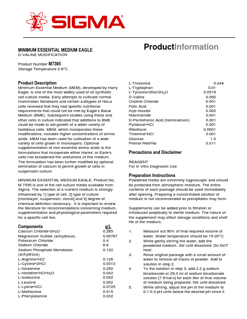
Product Information MINIMUM ESSENTIAL MEDIUM EAGLED-VALINE MODIFICATIONProduct Number M7395Storage Temperature 2-8°CProduct DescriptionMinimum Essential Medium (MEM), developed by Harry Eagle, is one of the most widely used of all synthetic cell culture media. Early attempts to cultivate normal mammalian fibroblasts and certain subtypes of HeLa cells revealed that they had specific nutritional requirements that could not be met by Eagle’s Basal Medium (BME). Subsequent studies using these and other cells in culture indicated that additions to BME could be made to aid growth of a wider variety of fastidious cells. MEM, which incorporates these modifications, includes higher concentrations of amino acids. MEM has been used for cultivation of a wide variety of cells grown in monolayers. Optional supplementation of non-essential amino acids to the formulations that incorporate either Hanks’ or Earle’s salts has broadened the usefulness of this medium. The formulation has been further modified by optional elimination of calcium to permit growth of cells in suspension culture.MINIMUM ESSENTIAL MEDIUM EAGLE, Product No. M 7395 is one of the cell culture media available from Sigma. The selection of a nutrient medium is strongly influenced by 1] type of cell, 2] type of culture [monolayer, suspension, clonal] and 3] degree of chemical definition necessary. It is important to review the literature for recommendations concerning medium, supplementation and physiological parameters required for a specific cell line.Components g/L Calcium Chloride•2H2O0.265 Magnesium Sulfate (anhydrous)0.09767 Potassium Chloride0.4 Sodium Chloride 6.8 Sodium Phosphate Monobasic0.122 (anhydrous)L-Arginine•HCl0.126L-Cystine•2HCl0.0313 L-Glutamine0.292L-Histidine•HCl•H2O0.042L-Isoleucine0.052L-Leucine0.052L-Lysine•HCl0.0725 L-Methionine0.015L-Phenylalanine0.032L-Threonine0.048L-Tryptophan0.01L-Tyrosine•2Na•2H2O0.0519D-Valine0.092 Choline Chloride0.001Folic Acid0.001myo-Inositol0.002 Niacinamide0.001D-Pantothenic Acid (hemicalcium)0.001 Pyridoxal•HCl0.001 Riboflavin0.0001 Thiamine•HCl0.001 Glucose 1.0 Phenol Red•Na0.011Precautions and DisclaimerREAGENTFor In Vitro Diagnostic UsePreparation InstructionsPowdered media are extremely hygroscopic and should be protected from atmospheric moisture. The entire contents of each package should be used immediately after opening. Preparing a concentrated solution of medium is not recommended as precipitates may form. Supplements can be added prior to filtration or introduced aseptically to sterile medium. The nature of the supplement may affect storage conditions and shelf life of the medium.1.Measure out 90% of final required volume ofwater. Water temperature should be 15-20°C.2.While gently stirring the water, add thepowdered medium. Stir until dissolved. Do NOTheat.3.Rinse original package with a small amount ofwater to remove all traces of powder. Add tosolution in step 2.4.To the solution in step 3, add 2.2 g sodiumbicarbonate or 29.3 ml of sodium bicarbonatesolution [7.5%w/v] for each liter of final volumeof medium being prepared. Stir until dissolved.5.While stirring, adjust the pH of the medium to0.1-0.3 pH units below the desired pH since itmay rise during filtration. The use of 1N HCl or1N NaOH is recommended.6.Add additional water to bring the solution tofinal volume.7.Sterilize immediately by filtration using amembrane with a porosity of 0.22 microns.8.Aseptically dispense medium into sterilecontainer.Storage/StabilityStore the dry powdered medium at 2-8°C under dry conditions and liquid medium at 2-8°C in the dark. Deterioration of the powdered medium may be recognized by any or all of the following: [1] color change, [2] granulation/clumping, [3] insolubility. Deterioration of the liquid medium may be recognized by any or all of the following: [1] pH change, [2] precipitate or particulate matter throughout the solution, [3] cloudy appearance [4] color change. The nature of supplements added may affect storage conditions and shelf life of the medium. Product label bears expiration date.ProcedureWater for tissue culture use [W-3500]Sodium Bicarbonate [S-5761] orSodium Bicarbonate Solution, 7.5% [S-8761]1N Hydrochloric Acid [H-9892]1N Sodium Hydroxide [S-2770]Medium additives as requiredProduct ProfileAppearance off-white powder Moisture content 2.0% Solubility clear solution at 1x concentrationpH at room temperature 5.8 ± 0.3 [without sodium bicarbonate]pH at room temperature 7.5 ± 0.3 [with sodium bicarbonate]Osmolality250 mOsm/kg H2O ± 5% [without sodium bicarbonate]Osmolality290 mOsm/kg H2O ± 5% [with sodium bicarbonate]Amino Acid Analysis Analysis has confirmedby HPLC that amino acids are present atconcentrations consistent withthe formula.Key Element Analysis Analysis has confirmed that by ICAP key elements are present atconcentrations consistent withthe formula.BIOLOGICAL PERFORMANCE CHARACTERISTICS Biological performance is assessed using an appropriate cell line(s). Growth studies are carried through 2 subculture generations. Cells are counted and growth is plotted as a logarithmic function of time in culture. Seeding efficiencies, doubling time, and final cell densities are determined. During the testing period cultures are examined microscopically for atypical morphology and evidence of cytotoxicity. Test results are available upon request.References1.Eagle, H. et al (1956) myo-Inositol as anEssential Growth Factor for Normal andMalignant Human Cells in Tissue Culture.J.Biol. Chem. 214, 845-847.2.Eagle, H.(1976) Media for Animal Cell Culture.Tissue Culture Association Manual. 3, 517-520.3.Eagle, H. (1959). Amino Acid Metabolism inMammalian Cell Cultures. Science. 130, 432-437.4.Eagle, H. (1955) Nutrition Needs of MammalianCells in Culture. Science. 122, 501.5.Gilbert, S.F. and Migeon, B.R. (1975) D-valineas a selective agent for normal human androdent epithelial cells in culture. Cell. 5, 11-17.7H027Sigma brand products are sold through Sigma-Aldrich, Inc.Sigma-Aldrich, Inc. warrants that its products conform to the information contained in this and other Sigma-Aldrich publications. Purchaser must determine the suitability of the product(s) for their particular use. Additional terms and conditions may apply. Please see reverse side ofthe invoice or packing slip.。
219525870_酥梨汁乳酸菌发酵工艺优化及挥发性成分分析

牛佳佳,赵彪,崔巍,等. 酥梨汁乳酸菌发酵工艺优化及挥发性成分分析[J]. 食品工业科技,2023,44(14):171−181. doi:10.13386/j.issn1002-0306.2022080321NIU Jiajia, ZHAO Biao, CUI Wei, et al. Optimization of Lactic Acid Bacteria Fermentation Process and Volatile Component Analysis of Suli Pear Juice[J]. Science and Technology of Food Industry, 2023, 44(14): 171−181. (in Chinese with English abstract). doi:10.13386/j.issn1002-0306.2022080321· 工艺技术 ·酥梨汁乳酸菌发酵工艺优化及挥发性成分分析牛佳佳1,赵 彪2,崔 巍1,郭超峰3,徐振玉3,鲁云风4,苗建银5,张四普1,*(1.河南省农业科学院园艺研究所,河南郑州 450002;2.河南农业大学园艺学院,河南郑州 450002;3.宁陵县农业农村局,河南商丘 476700;4.南阳师范学院生命科学与农业工程学院,河南南阳 473061;5.华南农业大学食品学院,广东省功能食品活性物重点实验室,广东广州 510642)摘 要:以宁陵县金顶谢花酥梨为主要原料,筛选适宜酥梨汁发酵的乳酸菌组合,在单因素实验的基础上,通过正交试验优化乳酸菌酥梨汁的发酵工艺,并对优化后的乳酸菌酥梨汁挥发性成分进行动态分析。
结果表明,最优发酵条件为:鼠李糖乳杆菌:嗜酸乳杆菌:乳双歧杆菌=1:1:1(V/V ),初始pH4.5,接种量4%,糖添加量10%,发酵时间50 h ,此条件下制得的乳酸菌酥梨汁V C 含量14.17 mg/100mL 、总酸含量7.66 g/L 、总酚含量3.68 mg/mL 、总黄酮含量6.25 mg/mL ,DPPH 自由基清除率90.05%,感官评分80.60。
Xofigo 放射性治疗药剂指南说明书
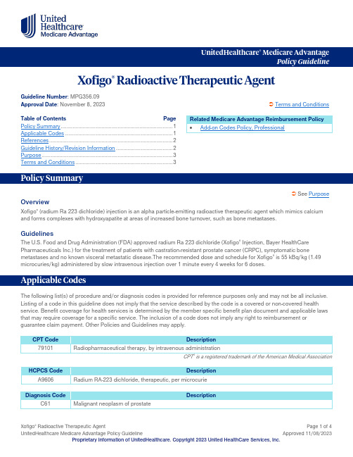
UnitedHealthcare ® Medicare AdvantagePolicy GuidelineXofigo ® Radioactive Therapeutic AgentGuideline Number : MPG356.09Approval Date : November 8, 2023 Terms and ConditionsTable of Contents Page Policy Summary ............................................................................. 1 Applicable Codes .......................................................................... 1 References ..................................................................................... 2 Guideline History/Revision Information ....................................... 2 Purpose .......................................................................................... 3 Terms and Conditions . (3)See PurposeOverviewXofigo ® (radium Ra 223 dichloride) injection is an alpha particle-emitting radioactive therapeutic agent which mimics calcium and forms complexes with hydroxyapatite at areas of increased bone turnover, such as bone metastases.GuidelinesThe U.S. Food and Drug Administration (FDA) approved radium Ra 223 dichloride (Xofigo ® Injection, Bayer HealthCare Pharmaceuticals Inc.) for the treatment of patients with castration-resistant prostate cancer (CRPC), symptomatic bonemetastases and no known visceral metastatic disease.The recommended dose and schedule for Xofigo ® is 55 kBq/kg (1.49 microcuries/kg) administered by slow intravenous injection over 1 minute every 4 weeks for 6 doses.The following list(s) of procedure and/or diagnosis codes is provided for reference purposes only and may not be all inclusive. Listing of a code in this guideline does not imply that the service described by the code is a covered or non-covered health service. Benefit coverage for health services is determined by the member specific benefit plan document and applicable laws that may require coverage for a specific service. The inclusion of a code does not imply any right to reimbursement or guarantee claim payment. Other Policies and Guidelines may apply.CPT Code Description 79101 Radiopharmaceutical therapy, by intravenous administrationCPT ® is a registered trademark of the American Medical AssociationHCPCS Code DescriptionA9606 Radium RA-223 dichloride, therapeutic, per microcurieDiagnosis Code DescriptionC61Malignant neoplasm of prostateRelated Medicare Advantage Reimbursement Policy • Add-on Codes Policy, ProfessionalDiagnosis CodeDescriptionAnd at least one of the following:C79.51 Secondary malignant neoplasm of boneC79.52 Secondary malignant neoplasm of bone marrowCMS Local Coverage Determinations (LCDs) and ArticlesLCDArticleContractor Medicare Part A Medicare Part BN/A A54559 Billing and Coding: Xofigo Billing Instructions PalmettoAL, GA, NC, SC,TN, VA, WV N/AA55052 Billing and Coding: Radiopharmaceutical Agents Retired 12/29/2022WPSAK, AL, AR, AZ, CA, CO, CT, DE, FL, GA, HI, IA, ID, IL, IN, KS, KY, LA, MA, MD, ME, MI, MO, MS, MT, NC, ND, NE, NH, NJ, NM, NV, OH, OK, OR, PA, RI, SC, SD, TN, TX, UT, VA, VT, WA, WI, WV, WYIA, IN, KS, MI, MO, NECMS Benefit Policy ManualChapter 15; § 50 Drugs and BiologicalsCMS Claims Processing ManualChapter 12; § 30.5 Payment for Codes for Chemotherapy Administration and Nonchemotherapy Injections and Infusions Chapter 14; § 10 General Ambulatory Surgical CenterChapter 17; § 90.2 Drugs, Biologicals, and RadiopharmaceuticalsOther(s)CGS Website (Submitting Claims for Xofigo/Radium 223)CMS HCPCS Codes for which ASP Reporting is in Units of Measure Other Than an NDC, Updated July 2023, CMS Website Xofigo Package Insert, Bayer Healthcare Pharmaceuticals WebsiteRevisions to this summary document do not in any way modify the requirement that services be provided and documented in accordance with the Medicare guidelines in effect on the date of service in question.Date Summary of Changes11/08/2023Policy Summary OverviewRemoved and relocated language pertaining to the U.S. Food and Drug Administration (FDA)approval of radium Ra 223 dichloride (Xofigo ® Injection, Bayer HealthCare Pharmaceuticals Inc.) usage (refer to the Guidelines section) GuidelinesRevised language to indicate:Date Summary of Changeso The U.S. Food and Drug Administration (FDA) approved radium Ra 223 dichloride (Xofigo®Injection, Bayer HealthCare Pharmaceuticals Inc.) for the treatment of patients with castration-resistant prostate cancer (CRPC), symptomatic bone metastases and no known visceralmetastatic diseaseo The recommended dose and schedule for Xofigo® is 55 kBq/kg (1.49 microcuries/kg)administered by slow intravenous injection over 1 minute every 4 weeks for 6 dosesSupporting InformationUpdated References section to reflect the most current informationArchived previous policy version MPG356.08The Medicare Advantage Policy Guideline documents are generally used to support UnitedHealthcare Medicare Advantage claims processing activities and facilitate providers’ submission of accurate claims for the specified services. The document can be used as a guide to help determine applicable:Medicare coding or billing requirements, and/orMedical necessity coverage guidelines; including documentation requirements.UnitedHealthcare follows Medicare guidelines such as NCDs, LCDs, LCAs, and other Medicare manuals for the purposes of determining coverage. It is expected providers retain or have access to appropriate documentation when requested to support coverage. Please utilize the links in the References section above to view the Medicare source materials used to develop this resource document. This document is not a replacement for the Medicare source materials that outline Medicare coverage requirements. Where there is a conflict between this document and Medicare source materials, the Medicare source materials will apply.The Medicare Advantage Policy Guidelines are applicable to UnitedHealthcare Medicare Advantage Plans offered by UnitedHealthcare and its affiliates.These Policy Guidelines are provided for informational purposes, and do not constitute medical advice. Treating physicians and healthcare providers are solely responsible for determining what care to provide to their patients. Members should always consult their physician before making any decisions about medical care.Benefit coverage for health services is determined by the member specific benefit plan document* and applicable laws that may require coverage for a specific service. The member specific benefit plan document identifies which services are covered, which are excluded, and which are subject to limitations. In the event of a conflict, the member specific benefit plan document supersedes the Medicare Advantage Policy Guidelines.Medicare Advantage Policy Guidelines are developed as needed, are regularly reviewed and updated, and are subject to change. They represent a portion of the resources used to support UnitedHealthcare coverage decision making. UnitedHealthcare may modify these Policy Guidelines at any time by publishing a new version of the policy on this website. Medicare source materials used to develop these guidelines include, but are not limited to, CMS National Coverage Determinations (NCDs), Local Coverage Determinations (LCDs), Medicare Benefit Policy Manual, Medicare Claims Processing Manual, Medicare Program Integrity Manual, Medicare Managed Care Manual, etc. The information presented in the Medicare Advantage Policy Guidelines is believed to be accurate and current as of the date of publication and is provided on an "AS IS" basis. Where there is a conflict between this document and Medicare source materials, the Medicare source materials will apply.You are responsible for submission of accurate claims. Medicare Advantage Policy Guidelines are intended to ensure that coverage decisions are made accurately based on the code or codes that correctly describe the health care services provided. UnitedHealthcare Medicare Advantage Policy Guidelines use Current Procedural Terminology (CPT®), Centers for Medicare andMedicaid Services (CMS), or other coding guidelines. References to CPT® or other sources are for definitional purposes only and do not imply any right to reimbursement or guarantee claims payment.Medicare Advantage Policy Guidelines are the property of UnitedHealthcare. Unauthorized copying, use, and distribution of this information are strictly prohibited.*For more information on a specific member's benefit coverage, please call the customer service number on the back of the member ID card or refer to the Administrative Guide.。
狼疮性肾炎患者肠道菌群变化及代谢物硫酸吲哚酚分析
Advances in Clinical Medicine 临床医学进展, 2021, 11(1), 235-242Published Online January 2021 in Hans. /journal/acmhttps:///10.12677/acm.2021.111034狼疮性肾炎患者肠道菌群变化及代谢物硫酸吲哚酚分析苏晗1,邢倩2*1青岛大学附属青岛市市立医院,山东青岛2青岛大学附属青岛市市立医院,风湿免疫科,山东青岛收稿日期:2020年12月25日;录用日期:2021年1月19日;发布日期:2021年1月26日摘要目的:分析狼疮性肾炎(Lupus nephritis, LN)患者与健康人群肠道菌群的结构及代谢产物差异,探讨肠道菌群在LN发病中的作用。
方法:选取系统性红斑狼疮患者51例,分为狼疮性肾炎组(LN组) 27例和狼疮非肾炎组(N-LN组) 24例,另选取53例健康志愿者作为健康对照组(H组)。
基于Illumina MiSeq平台分别对SLE患者和健康对照组的粪便样本进行Alpha多样性分析、Beta多样性分析以及属水平上的菌群差异性分析,采用ELISA双抗体夹心法测定SLE患者和健康对照组血清硫酸吲哚酚(IS)浓度,对LN患者的24h 尿蛋白定量与其血清IS浓度进行Pearson相关性分析,采用受试者工作曲线(ROC曲线),根据最佳敏感性和特异性确定IS诊断界点。
结果:LN组较健康对照组血IS浓度和肠杆菌含量均显著升高,差异有统计学意义(P < 0.05)。
在LN患者中,24 h尿蛋白定量与血清IS浓度呈正相关(r = 0.86, P < 0.01)。
结论:狼疮性肾炎患者存在肠道菌群失调,肠杆菌含量明显增加,相关代谢产物血清IS异常,可能参与LN发病。
关键词狼疮性肾炎,肠道菌群,24 h尿蛋白定量,硫酸吲哚酚Analysis of Intestinal Flora and IndoxylSulfate in the Patients with Lupus NephritisHan Su1, Qian Xing2*1Qingdao Municipal Hospital Affiliated to Qingdao University, Qingdao Shandong2Department of Rheumatic Immunity, Qingdao Municipal Hospital Affiliated to Qingdao University, Qingdao Shandong*通讯作者。
RNAscope_技术检测hTERC_RNA_组分在宫颈鳞状上皮内瘤变中的表达及其应用价值

ʌ文章编号ɔ1006-6233(2023)06-0928-05RNAscope技术检测hTERC RNA组分在宫颈鳞状上皮内瘤变中的表达及其应用价值胡倩岚,㊀张㊀爽,㊀李红娜,㊀毛㊀昕,㊀刘㊀芳,㊀吴晨阳,㊀宋旭东(华北理工大学附属医院病理科,㊀河北㊀唐山㊀063000)ʌ摘㊀要ɔ目的:探讨RNAscope技术检测hTERC RNA组分表达对宫颈鳞状上皮内瘤变分级诊断价值㊂方法:筛选2021年9月至2022年2月华北理工大学附属医院病理科已确诊为HR-HPV感染的宫颈活检标本80例,分3组:炎症组14例㊁低级别组34例㊁高级别组32例㊂分别采用荧光原位杂交技术及RNAscope技术检测石蜡包埋组织中hTERC基因的扩增状况及其RNA组分的表达㊂结果:FISH 检测hTERC基因扩增三组间具有差异性(P<0.05)㊂RNAscope检测三组间检出率具有统计学差异(P< 0.05)㊂组织学诊断分组中,炎症组14例中hTERC RNA组分阳性表达程度均为(-/++),低级别组中以hTERC RNA组分(++)为主(19/34),高级别组中以hTERC RNA组分(+++/++++)为主(27/32),上述染色模式表达情况在组织学分级诊断中具有统计学差异(P<0.05)㊂结论:RNAscope技术检测宫颈上皮内瘤变组织中hTERC RNA组分的表达有助于宫颈病变组织学诊断及进展预测㊂ʌ关键词ɔ㊀RNAscope技术;㊀荧光原位杂交;㊀宫颈上皮内瘤变;㊀人端粒酶RNAʌ文献标识码ɔ㊀A㊀㊀㊀㊀㊀ʌdoiɔ10.3969/j.issn.1006-6233.2023.06.09Expression of hTERC RNA Fraction in Cervical Squamous Intraepithelial Neoplasia by RNAscope Technique and Its Application ValueHU Qianlan,ZHANG Shuang,LI Hongna,et al(The Affiliated Hospital of North China University of Technology,Hebei Tangshan063000,China)ʌAbstractɔObjective:To investigate the diagnostic significance of hTERC RNA fractions expression de-tected by RNA scope technique in cervical squamous intraepithelial neoplasia.Methods:A total of80cervi-cal biopsy specimens diagnosed with HR-HPV infection in the Department of Pathology,Affiliated Hospital of North China University of Science and Technology from September2021to February2022were screened in3 groups:inflammation group(14cases),low-grade group(34cases),and high-grade group(32cases). The amplification status of hTERC gene and the expression of its RNA components in paraffin-embedded tis-sues were detected by fluorescence in situ hybridization and RNAscope techniques,respectively.Results: FISH detection of hTERC gene amplification was different among the three groups(P<0.05).Detection rate by RNAscope was statistically different among the three groups(P<0.05).In the histological diagnostic sub-groups,the degree of positive expression of hTERC RNA fraction was(-/++)in all14cases in the inflamma-tion group,the hTERC RNA fraction(++)was predominant in the low-grade group(19/34),and the hTERC RNA fraction(++++/++++)was predominant in the high-grade group(27/32),and the expression of the above staining patterns in the histological graded diagnosis had statistical differences(P<0.05).Con-clusion:The RNAscope technique to detect the expression of hTERC RNA fraction in cervical intraepithelial neoplasia tissue helps in the histological diagnosis and progression prediction of cervical lesions.ʌKey wordsɔ㊀RNAscope technique;㊀Fluorescence in situ hybridization;㊀Cervical squamous intraep-ithelial neoplasia;㊀Human telomerase RNA㊃829㊃ʌ基金项目ɔ2018年度河北省医学适用技术跟踪项目,(编号:G2018067)ʌ通讯作者ɔ宋旭东㊀㊀在全球女性恶性生殖性肿瘤中,宫颈癌位居第二,严重威胁女性健康[1]㊂据数据统计2020年全球新发病例超60万,死亡人数达34万[2]㊂宫颈癌是目前唯一明确病因的恶性肿瘤,高危型HPV(HR-HPV)感染是主要致癌因素,因此如何有效防止宫颈癌的发生发展,及早预防㊁精准诊治是目前研究热点㊂研究发现, HR-HPV可导致宫颈鳞状上皮细胞3号染色体长臂人端粒酶RNA组分(human telomericRNA component, hTERC)基因异常扩增,该基因异常表达是宫颈肿瘤发生的基础,也是宫颈癌形成的早期事件[3]㊂RNAscope 技术是一项新型RNA原位杂交检测方法,它利用 Z 型双探针引物与目标RNA序列互补结合,实现特异性信号放大,使其拥有高灵敏度及特异度,并能在显微镜下直接观察病变中RNA的表达情况[4,5]㊂本课题采用RNAscope技术检测宫颈上皮内瘤变患者hTERC RNA组分的表达,探讨该技术在宫颈鳞状上皮内瘤变病理诊断中的意义㊂1㊀资料与方法1.1㊀一般资料:筛选2021年9月至2022年2月华北理工大学附属医院病理科已确诊为HR-HPV感染宫颈活检标本80例,由两位高级职称病理医师按照2020年(第五版)WHO女性生殖器官肿瘤分类行组织病理诊断,其中炎性病例14例(简称炎症组)㊁低级别鳞状上皮内瘤变34例(简称低级别组)㊁高级别鳞状上皮内瘤变32例(简称高级别组)㊂所有病例均无相关治疗或其他恶性肿瘤放化疗史,且临床资料完备㊂1.2㊀荧光原位杂交技术(FISH):按照试剂盒操作说明对石蜡包埋宫颈组织学标本行FISH检测,橘红色荧光素标记的TERC探针检测TERC基因,绿色荧光素标记的CEP3探针检测3号染色体着丝粒序列,杂交信号结果记录为绿ʒ红,正常细胞为2ʒ2型,异常细胞包括2ʒ3㊁2ʒ4㊁3ʒ3㊁4ʒ4以及hTERC基因高拷贝型(N:5及以上)等㊂1.3㊀RNAscope技术:石蜡切片经二甲苯透明㊁梯度乙醇脱蜡至水,取出切片在室温下风干5min,滴加双氧水室温下10min后蒸馏水清洗,将切片浸入到接近沸腾的靶标修复试剂中,99ħ15min,然后用蒸馏水清洗切片,在无水乙醇中清洗3~5次,风干㊂疏水笔在每张切片样本周围画疏水圈,室温下放置至完全干燥㊂在上述每张切片上加约5滴蛋白酶放入预热至40ħ的HybEZ湿盒,加盖密封,放入杂交炉,40ħ孵育30min,蒸馏水清洗去除切片上的过量液体,在每张切片上加入4滴探针,置于杂交炉中,40ħ孵育2h㊂取出温度控制盘,用清洗缓冲液室温清洗切片2min㊂擦净阻水圈周围的液体,分别依次加入6次杂交反应液,反应温度40ħ,反应时间分别为30min㊁15min㊁15min㊁15min㊁30min㊁15min㊂每次反应结束均用缓冲液清洗玻片㊂加入配置好的Red工作液,室温显色20min,苏木素复染㊁去离子水冲洗㊂1.4㊀结果判读:FISH结果判读:在10ˑ物镜下查找FISH检测细胞区域,然后在40ˑ物镜下扫描整个杂交区域,观察标本的质量,随机计数100个细胞,分别记录正常信号及异常信号细胞的数目㊂正常细胞信号绿ʒ红为2ʒ2型,异常细胞包括2ʒ3㊁2ʒ4㊁2ʒ5㊁3ʒ3型等㊂以20例正常宫颈组织学标本中FISH检测结果建立阈值,阈值=均数+3ˑ标准差,计算结果阈值为6%,即异常细胞数/计数细胞总数ȡ6判断为hTERC 基因扩增,结果为阳性㊂RNAscope结果判读:hTERC RNA组分阳性反应为肿瘤细胞胞质及胞核中可见紫红色颗粒状染色,同时根据阳性细胞在宫颈鳞状上皮垂直切面的分布进行分级:(+)为基底和基底旁细胞染色;(++)为表皮下1/3层的细胞染色;(+++)为超过表皮下1/3层,但未达到2/3层的细胞染色;(+++ +)为超过表皮下2/3层和到全层的细胞染色㊂肿瘤细胞呈阴性反应或阳性反应仅局限于基底或基底旁者均视为hTERC RNA组分不表达,即(-)与(+)均判读为hTERC RNA组分阴性表达(Ⅰ级),(++)染色为Ⅱ级,(+++)和(++++)染色为Ⅲ级㊂1.5㊀统计学方法:采用SPSS26.0软件进行统计学分析,FISH和RNAscope检验结果采用配对χ2检验;一致性检验采用Kappa检验㊂检验水准α=0.05,P<0. 05为差异有统计学意义㊂2㊀结㊀果2.1㊀宫颈活检标本中hTERC基因扩增情况:80例标本中有24例呈hTERC基因扩增(见图1),总扩增率为30.0%(24/80),炎症组㊁低级别组及高级别组扩增阳性率分别为0%(0/14)㊁23.53%(8/34)及50%(16/ 32),统计分析三组阳性率比较具有统计学差异(χ2= 12.773,P=0.002),见表1㊂组间比较,仅高级别组与炎症组之间差异有显著统计学意义(P=0.01)㊂以上结果提示HR-HPV感染宫颈鳞状上皮可以引发hTERC基因扩增,并参与宫颈鳞状上皮瘤变,但hTERC基因扩增阳性结果对宫颈上皮内瘤变分级及预测病变进展的帮助有限㊂㊃929㊃图1㊀FISH检测hTERC基因扩增情况(ˑ100)A.正常宫颈组织无hTERC基因扩增(绿色信号ʒ红色信号=2ʒ2);B.宫颈高级别鳞状上皮内病变hTERC基因扩增阳性(绿色信号ʒ红色信号=8ʒ40)表1㊀FISH检测宫颈组织标本中hTERC基因扩增情况(n)分组例数扩增结果阳性㊀㊀㊀㊀阴性阳性率(%)炎症组140140低级别组3482625.53a高级别组32161650.00bc χ212.773P0.002㊀㊀注:三组之间两两进行比较,χ2分割(α=0.05/3=0. 0167);a示低级别组与炎症组比较,P=0.085;b示高级别组与炎症组比较,P=0.001;c示高级别组与低级别组比较,P=0. 0252.2㊀宫颈活检标本中hTERC RNA组分表达情况:80例HR-HPV感染的宫颈活检组织标本中有53例hTERC RNA组分呈阳性表达,总阳性率为66.25% (53/80),炎症组㊁低级别组及高级别组阳性率分别为0%(0/14)㊁67.65%(23/34)及93.75%(30/32)(见表2)㊂三组hTERC RNA组分阳性表达率的统计学差异显著(χ2=38.334,P=0.000),其中炎症组的阳性率显著低于宫颈鳞状上皮内瘤变组(低级别组及高级别组)(χ2分别为18.184㊁33.715,P值均为0.000),高级别组的阳性率显著高于低级别组(χ2=7.101,P=0. 008)㊂以上结果提示hTERC RNA组分表达增高参与宫颈癌的发生发展㊂参照组织学分级诊断标准,对hTERC RNA组分表达程度进行分级,其中炎症组hTERC RNA组分阳性表达程度均为Ⅰ级(-/+)(14/14),低级别组中以Ⅱ级(++)为主(19/34),高级别组中以Ⅲ级(+++/++++)为主(27/32)(见图2㊁表3)㊂统计学分析示RNAscope检测hTERC RNA组分分级染色模式在宫颈组织学分级中差异具有统计学意义(P<0.05),该结果提示hTERC RNA组分阳性表达的程度与宫颈病变级别呈正比,采用RNAscope检测宫颈组织中hTERC RNA组分的表达可辅助宫颈鳞状上皮内瘤变的组织学诊断㊂图2㊀RNAscope检测hTERC RNA组分表达情况(ˑ200) A.正常宫颈组织hTERC RNA组分(-)㊁B.慢性子宫颈炎hTERC RNA组分(+)㊁C.宫颈低级别鳞状上皮内瘤变hTERC RNA组分(++)㊁D.宫颈高级别鳞状上皮内瘤变hTERC RNA 组分(+++)㊁E.宫颈原位癌hTERC RNA组分(++++) 2.3㊀hTERC RNA组分在宫颈鳞状上皮内瘤变一致性分析:将宫颈鳞状上皮内瘤变中hTERC RNA组分阳性表达程度分Ⅰ级(-/+)㊁Ⅱ级(++)㊁Ⅲ级(+++/+++ +),分别对应组织学诊断中的慢性宫颈炎㊁低级别鳞状上皮内瘤变及高级别鳞状上皮内瘤变㊂根据宫颈鳞状上皮内瘤变临床分层管理共识,将宫颈鳞状上皮内瘤变及hTERC RNA组分阳性表达程度重新分为两级管理(见表4)㊂依据表4分析hTERC RNA组分表达㊃039㊃对宫颈高级别鳞状上皮内瘤变诊断的灵敏度为84. 37%(27/32)㊁特异度为100%(48/48)㊁阳性预测值为100%(27/27)㊁阴性预测值为90.57%(48/53)㊁准确率为93.75%(75/80),一致性检验Kappa值为0.866,一致性显著㊂表2㊀RNAscope检测宫颈组织标本中hTERCRNA组分表达情况组别例数阳性程度-㊀㊀㊀㊀㊀㊀+㊀㊀㊀㊀㊀㊀++㊀㊀㊀㊀㊀㊀+++㊀㊀㊀㊀㊀㊀++++阳性率(%)炎症组141130000低级别组3474194067.65a高级别组32203111693.75bc χ238.334P0.000㊀㊀注:三组之间两两进行比较,χ2分割(α=0.05/3=0.0167);a为低级别组与炎症组比较,P=0.000;b为高级别组与炎症组比较,P=0.000;c为高级别组与低级别组比较,P=0.008表3㊀RNAscope检测分级诊断和组织学分级诊断情况n(%)检测方法组别组织学诊断炎症组㊀㊀㊀㊀低级别组㊀㊀㊀㊀高级别组χ2PRNAscopeⅠ级14(100.0)11(29.7)2(6.3)38.3340.000Ⅱ级0(0.0)19(55.9)3(9.4)24.3200.000ⅢI级0(0.0)4(11.8)27(84.4)47.3550.000合计143432表4㊀RNAscope检测hTERC RNA组分一致性比较检测方法分级宫颈鳞状上皮内病变高级别病变㊀㊀㊀低级别及以下病变Kappa PⅡI级270RNAscopeⅠ/Ⅲ级5480.8660.000合计32483㊀讨㊀论端粒酶是一种维持端粒长度的核蛋白复合体㊂hTERC基因是端粒酶上具有11个碱基的模板序列,在端粒延伸过程中起模板作用,同时也是端粒延伸的重要组成部分㊂该基因异常扩增会导致端粒酶表达过度,致端粒酶延伸,最终造成细胞恶性循环增殖而产生肿瘤[3,6]㊂hTERC基因的表达与端粒酶活性一致,hTERC在许多正常组织和良性组织中均有表达,但其相对上调可反映恶性进展程度,在恶性肿瘤中高表达㊂在宫颈癌和癌前病变中,hTERC也有不同程度的扩增,被认为是宫颈癌的潜在肿瘤标志物[7]㊂HR-HPV 感染是导致hTERC重新激活的起始因子,两者的联合作用使宫颈细胞获得无限的增殖能力㊂目前国内外关于hTERC基因研究也证实了以上观点,Haizhen He[8]㊃139㊃及杨淑玲[9]等研究运用FISH技术检测hTERC基因扩增阳性率,结果显示在宫颈癌中最高,在CIN3中为中度,在CIN1/2类病变中最低,说明hTERC基因在不同宫颈病变的分级诊断中存在明显差异,即随着宫颈病变严重程度增加,hTERC基因表达也增加㊂本实验结果与上述研究一致㊂hTERC RNA组分是由DNA转录产生,在疾病的诊断与治疗中发挥重要作用,但极易降解,而RNA-scope技术所具备的高灵敏度及特异度优势可以显著提高RNA检出率并能提供有效的组织形态学特征㊂本研究中,hTERC RNA组分在各级宫颈病变中均有表达,但是在宫颈高级别鳞状上皮内瘤变中hTERC RNA 组分检出率达90%以上,且组间差异有显著统计学意义(P<0.05),显微镜原位观察hTERC RNA组分染色范围也随着宫颈鳞状上皮内瘤变程度的增加从基底层逐渐向表层覆盖,其染色模式在不同级别的宫颈鳞状上皮内瘤变中的表达具有统计学差异(P<0.05),且具有显著的诊断效能㊂以上统计分析示,RNAscope技术相比于目前主流的FISH技术,它具有更显而易见优势,如明了染色模式㊁判读方便㊁一致率高等,除此之外,在宫颈病变分级诊断中具有显著的灵敏度及特异度,并能直观反应病变进展情况㊂在本研究中,FISH 检测hTERC基因扩增阳性率无法象RNAscope一样进行分级比较,且组间阳性率比较无统计学意义,更加突出RNAscope技术优越性㊂hTERC RNA组分原位杂交染色最显著价值是对宫颈组织学标本进行染色定位㊂在本研究中,hTERC RNA组分在炎症组㊁低级别组及高级别组中显示了不同的染色模式,因此可利用hTERC RNA组分原位杂交为基础来对宫颈病变发展进行推测㊂众所周知, HPV感染从病毒损伤鳞状上皮黏膜基底细胞层开始,随后病毒将其基因整合入宿主基因组中并开始大量复制,最后扩散至整个上皮层,并表达大量E6㊁E7蛋白,破坏宿主DNA修复功能和细胞凋亡,同时上调端粒酶活性使hTERC基因扩增表达,导致上皮细胞无限增殖,最终导致癌的发生[10]㊂本研究中,低级别组大部分病例为表皮下1/3层表达模式,与复制期感染过程相符㊂在高级别病变中,大部分病例表现为表皮下2/ 3层至全层的染色模式,与转化期感染过程一致㊂因此可以通过hTERC RNA组分原位染色变化间接地反映了HPV感染过程㊂故在宫颈上皮内瘤变分流诊断中,对于病理诊断不明确或有分歧时,行hTERC RNA 组分检测可辅助组织学病理诊断以指导临床治疗,对hTERC RNA组分无阳性表达的CIN1及慢性宫颈炎病例,酌情考虑随诊,以避免过度治疗㊂当hTERC RNA 组分阳性表达级别增高时,极大可能为宫颈高级别病变或进展为高级别病变,而予以积极治疗防止漏诊㊂综上所述,采用RNA scope技术检测hTERC RNA 组分的表达,相对于FISH检测hTERC基因扩增状态,更有效地作为宫颈癌前病变的早期筛查辅助手段,并有助于宫颈病变组织学分级诊断㊂ʌ参考文献ɔ[1]㊀Bray F,Ferlay J,Soerjomataram I,et al.Global cancer statis-tics2018:GLOBOCAN estimates of incidence and mortalityworldwide for36cancers in185countries[J].CA:A CancerJournal for Clinicians,2020,70(4):133-133. [2]㊀Sung H,Ferlay J,Siegel RL,et al.Global cancer statistics2020:GLOBOCAN estimates of incidence and mortalityworldwide for36cancers in185countries[J].CA CancerClin,2021,71(3):209-249.[3]㊀Bruno Bernardes de Jesus,Maria A.Blasco.Telomerase atthe intersection of cancer and aging[J].Trends in Genetics,2013,29(9):513-520.[4]㊀Zavalov O,Irizarry R,Flamm M,et al.Mesoscale model ofthe assembly and cross-linking of HPV virus-like particles[J].Virology,2019,537(C):53-64.[5]㊀Deleage C,Chan CN,Busman-Sahay K,et al.Next-genera-tion in situ hybridization approaches to define and quantifyHIV and SIV reservoirs in tissue microenvironments[J].Retrovirology,2018,15(1):4.[6]㊀Armstrong C A,Tomita K.Fundamental mechanisms of te-lomerase action in yeasts and mammals:understanding telo-meres and telomerase in cancer cells[J].Open Biology,2017,7(3):160338-160338.[7]㊀Liu Y,Fan P,Yang Y,et al.Human papillomavirus and hu-man telomerase RNA component gene in cervical cancer pro-gression[J].Scientific Reports,2019,9(3):2675-2686.[8]㊀He H,Pan Q,Pan J,et al.Study on the correlation betweenhTREC and HPV load and cervical CINI/II/III lesions andcervical cancer[J].Journal of Clinical Laboratory Analysis, 2020,34(7):e23257.[9]㊀杨淑玲,张琳,张燕,等.荧光原位杂交法检测hTERC扩增在宫颈鳞状上皮内病变分级诊断中的价值[J].诊断病理学杂志,2020,27(8):533-538.[10]㊀Panczyszyn A,Boniewska-Bernacka E,Gtab G.Telomeresand telomerase during human papillomavirus-induced car-cinogenesis[J].Mol Diagn Ther.2018;22(4):421-430.㊃239㊃。
丹参多酚酸盐联合氯吡格雷对急性冠脉综合征病人心功能、内皮功能
细胞内钙离子水平,减少再灌注时心肌细胞损伤的程度㊂本研究对象为不伴有MD的AMI个体,研究思路及实验条件区别传统类似研究中MD的限制㊂本研究并非将氯硝西泮与舍曲林作为传统意义上的抗焦虑抗抑郁药物,将药物干预处理对心脏的积极作用作为附带效应,而是将此两种药物当作独立的具有心脏保护意义的药物研究,完全独立于焦虑障碍或抑郁障碍的背景㊂这一思想在同类研究中具有独创性与突破性,为减轻AMI病人PCI术后或溶栓后MIRI的程度提供新的治疗思路㊂参考文献:[1]Weir RA,McMurray JJ,Velazquez EJ.Epidemiology of heart fail-ure and left ventricular systolic dysfunction after acute myocardi-al infarction:prevalence,clinical characteristics,and prognosticimportance[J].The American Journal of Cardiology,2006,97(10):1325.[2]Gidron Y,Gilutz H,Berger R,et al.Molecular and cellular inter-face between behavior and acute coronary syndromes[J].Cardi-ovascular Research,2002,56(1):1521.[3]Oka J,Imamura M,Hatta E,et al.Carrier mediated norepi-nephrine release and reperfusion arrhythmias induced by pro-tracted ischemia in isolated perfused guinea pig hearts:effect ofpresynaptic modulation by2adrenoceptor in mild hypothermicischemia[J].Journal of Pharmacology and Experimental Thera-peutics,2002,303(2):681687.[4]Cǒt췍F,Fligny C,Fromes Y,et al.Recent advances in under-standing serotonin regulation of cardiovascular function[J].Trends in Molecular Medicine,2004,10(5):232238. [5]Kitayama S,Morita K,Dohi T,et al.Benzodiazepine inhibition ofthe stimulation evoked catecholamine release from bovine ad-renomedullary cells in culture[J].Neurochemistry International, 1988,13(2):265270.[6]Nijmeijer R,Krijnen PAJ,Assink J,et al.C reactive protein andcomplement depositions in human infarcted myocardium are more extensive in patients with reinfarction or upon treatment with reperfusion[J].European Journal of Clinical Investigation, 2004,34(12):803810.[7]Ganguly PK,Dhalla KS,Shao Q,et al.Differential changes insympathetic activity in left and right ventricles in congestive heart failure after myocardial infarction[J].American Heart Journal, 1997,133(3):340345.[8]Fordjour PA,Wang Y,Shi Y,et al.Possible mechanisms of Creactive protein mediated acute myocardial infarction[J].Euro-pean Journal of Pharmacology,2015,760:7280.[9]Frangogiannis NG,Smith CW,Entman ML.The inflammatory re-sponse in myocardial infarction[J].Cardiovascular Research, 2002,53(1):3147.[10]Kalawski R,Majewski M,Kaszkowiak E,et al.Transcardiac re-lease of soluble adhesion molecules during coronary artery by-pass grafting:effects of crystalloid and blood cardioplegia[J].CHEST Journal,2003,123(5):13551360.[11]Niccoli G,Burzotta F,Galiuto L,et al.Myocardial no reflow inhumans[J].Journal of the American College of Cardiology, 2009,54(4):281292.[12]Yellon DM,Hausenloy DJ.Myocardial reperfusion injury[J].NewEngland Journal of Medicine,2007,357(11):11211135. [13]Serebruany VL,Gurbel PA,O Connor CM.Platelet inhibition bysertraline and N desmethylsertraline:a possible missing linkbetween depression,coronary events,and mortality benefits ofselective serotonin reuptake inhibitors[J].Pharmacological Re-search,2001,43(5):453461.(收稿日期:20171024)(本文编辑薛妮)丹参多酚酸盐联合氯吡格雷对急性冠脉综合征病人心功能㊁内皮功能㊁炎症因子的影响石清1,程康2,田季雨1摘要:目的探讨丹参多酚酸盐联合氯吡格雷对急性冠脉综合征病人心功能㊁内皮功能㊁炎症因子的影响,并分析临床疗效与安全性㊂方法采用随机数字表法分为两组,对照组(n=93)和研究组(n=93),对照组给予氯吡格雷治疗,研究组在对照组治疗基础上给予丹参多酚酸盐治疗㊂观察并比较两组临床疗效,心功能,内皮功能,炎症因子及不良反应㊂结果治疗后,研究组总有效率高于对照组(P<0.05)㊂研究组左室收缩末期内径㊁左室舒张末期内径㊁左室射血分数㊁一氧化氮(NO)㊁依赖性舒张功能(FMD)高于对照组(P<0.05)㊂结论急性冠脉综合征病人采用丹参多酚酸盐联合氯吡格雷的临床效果优于单用氯吡格雷,可有效改善心功能㊁内皮功能及炎症因子㊂关键词:急性冠脉综合征;丹参多酚酸盐;氯吡格雷;炎症因子;心功能;内皮功能中图分类号:R541.4 R256.2文献标识码:B d o i:10.3969/j.i s s n.1672-1349.2017.23.025文章编号:1672-1349(2017)23-3026-04作者单位:1.中国人民解放军第五一八医院(西安710043),E mail:sunsumeme@;2.第四军医大学西京医院引用信息:石清,程康,田季雨.丹参多酚酸盐联合氯吡格雷对急性冠脉综合征病人心功能㊁内皮功能㊁炎症因子的影响[J].中西医结合心脑血管病杂志,2017,15(23):30263029.急性冠脉综合征为常见的临床综合征,主要由于冠状动脉粥样斑块破裂及松动,诱导血小板于受损表层发生聚集㊁黏附,导致血栓及病变血管出现不同程度闭塞所致,引起病人胸部不适及胸痛等表现,导致心功能下降,且造成心力衰竭及心律失常,严重者出现猝死[1]㊂国内外研究表示,急性冠脉综合征发病受到多种细胞因子影响,其中炎症因子㊁血管内皮受损等为主要诱因,通过测定其水平改变有利于病情进展及预后评估[23]㊂郭锦丽等[4]研究显示,早期保守治疗是其关键手段,临床多应用抗栓㊁缓解心肌氧供等方式治疗,其中抗血小板为抗栓的主要疗法,氯吡格雷为代表药物,能抑制血小板聚集㊂王静等[5]研究指出,中西医联合治疗能提高疗效,丹参多酚酸盐多用于心血管疾病治疗,具有扩张血管等作用㊂本研究旨在探讨丹参多酚酸盐联合氯吡格雷对急性冠脉综合征病人心功能㊁内皮功能㊁炎症因子的影响,并分析临床疗效与安全性㊂1 资料与方法1.1 一般资料 选择2015年1月 2016年1月于中国人民解放军第五一八医院接受治疗的186例急性冠脉综合征病人㊂入选标准:均符合急性冠脉综合征(不稳定型心绞痛)的相关诊断标准[6]:入院前48h 内心绞痛发作次数1次以上,同时发作时心电图显示2个相邻导联ST 段下移>0.1mV 2个以上,且T 波倒置并压低;属中医心血瘀阻证(胸闷㊁心悸㊁痛有定处㊁舌暗淡,脉结代);未接受冠状动脉造影介入治疗;近期未使用抗血小板药物;肝肾等主要器官未见明显病变;未见恶性肿瘤㊂排除标准:血液及免疫系统病变;近期伴感染或创伤;变异性心绞痛㊁恶性心律失常或先天性心脏病;药物或酒精依赖者㊂对照组男54例,女39例,年龄43岁~71岁(59.43岁ʃ3.65岁);研究组男50例,女43例,年龄42岁~70岁(60.21岁ʃ3.97岁)㊂两组性别等一般资料比较,差异无统计学意义(P >0.05)㊂1.2 方法 对照组给予氯吡格雷治疗,于每日睡前口服50mg 氯吡格雷(山东齐都药业有限公司,每片25mg ,产品批号:141021);研究组在对照组基础上加以丹参多酚酸盐治疗,将丹参多酚酸盐(涿州东乐制药有限公司,每瓶50mg ,产品批号:141209)200mg 加入5%葡萄糖注射液250mL 静脉输注,每日1次㊂两组均持续用药2个月,期间均接受降血脂㊁抗凝㊁溶栓㊁控制饮食等治疗㊂治疗结束后时评估疗效,并观察期间不良反应㊂1.3 观察指标1.3.1 临床疗效观察 显效:心前区不适㊁胸闷等心绞痛表现完全缓解或发作频率减少超过80%,心电图未见异常;好转:心绞痛发作频率减少超过50%,心电图可见明显改善;无效:心绞痛发作频率减少在50%以下,心电图未见缓解或加剧[7]㊂1.3.2 超声心动图观察 于治疗前和治疗2个月时采用超声心动图测定左室收缩末期内径㊁左室舒张末期内径㊁左室射血分数㊂1.3.3 指标观察 于治疗前后抽取晨起静脉血2mL ,将血清常规分离并于低温环境下保存待用㊂C 反应蛋白(CRP )㊁一氧化氮(NO )㊁白细胞介素6(IL 6)㊁内皮素1(ET1)及肿瘤坏死因子α(TNF α)均采用酶联免疫法测定,予以特定方法检测依赖性舒张功能(FMD ),试剂盒分别来自重庆明康科技有限公司㊁苏州新波生物技术有限公司㊁无锡市雷杜科技有限公司㊁郑州正泰生物技术有限公司㊁贵州汉方制药有限公司㊁长春市佳恒科技有限责任公司㊂1.4 统计学处理 采用SPPS18.0统计软件进行数据处理,计量资料以均数ʃ标准差(x ʃs )表示,采用t 检验;计数资料以率表示,采用χ2检验㊂以P <0.05为差异有统计学意义㊂2 结 果2.1 两组病人临床疗效比较 研究组治疗后总有效率高于对照组(P <0.05)㊂详见表1㊂表1 两组病人临床疗效比较例(%)组别n显效好转无效总有效对照组9339(41.94)40(43.01)14(15.05)79(84.94)研究组9358(62.37)30(32.25)5(5.38)88(94.62)注:两组总有效率比较,χ2=4.748,P =0.029㊂2.2 两组病人心功能比较 治疗前,两组心功能比较,差异无统计学意义(P >0.05);治疗后,两组超声心动图指标均有改善,研究组改善更明显(P <0.05)㊂详见表2㊂表2 两组病人心功能比较(x ʃs )组别n 左室收缩末期内径(mm )治疗前治疗后t 值P左室舒张末期内径(mm )治疗前治疗后t 值P 左室射血分数(%)治疗前治疗后t 值P对照组9340.32ʃ5.0434.51ʃ4.20 4.2000.00057.90ʃ7.1145.11ʃ5.6013.6280.00046.33ʃ5.7958.70ʃ7.20-12.9110.000研究组9340.83ʃ5.7330.29ʃ3.7914.7950.00058.32ʃ7.9040.29ʃ5.3118.2660.00045.80ʃ5.1261.20ʃ7.63-16.1620.000t 值-0.6447.193-0.381 6.0230.661-2.298P0.5200.0000.7030.0000.5090.0222.3 两组病人内皮功能比较 治疗前,两组内皮功能比较,差异无统计学意义(P >0.05)㊂治疗后,两组NO 及FMD 均有上升,研究组上升更明显(P <0.05);两组ET1均有降低,研究组下降更明显(P <0.05)㊂详见表3㊂表3 两组病人内皮功能比较(x ʃs )组别n NO (μg/L )治疗前治疗后t 值PET 1(ng/L )治疗前治疗后t 值P FMD (%)治疗前治疗后t 值P 对照组9335.08ʃ4.3743.29ʃ5.61-11.1330.00065.20ʃ8.1160.19ʃ7.50 4.3730.000 3.22ʃ0.42 4.85ʃ0.62-20.9900.000研究组9335.82ʃ4.1151.24ʃ6.39-19.5720.00064.78ʃ9.0555.21ʃ6.898.1130.0003.25ʃ0.43 6.50ʃ0.83-33.5280.000t 值-1.189-9.0160.333 4.7150.48115.359P0.2350.0000.7390.0000.6300.0002.4 两组病人炎症因子比较 治疗前,两组炎症因子比较,差异无统计学意义(P >0.05);两组炎症因子均较治疗前降低,研究组下降更明显(P <0.05)㊂详见表4㊂表4 两组病人炎症因子比较(x ʃs )组别n CRP (mg/L )治疗前治疗后t 值P IL6(ng/L )治疗前治疗后t 值P TNF α(ng/L )治疗前治疗后t 值P对照组9320.87ʃ2.5011.25ʃ1.4132.3220.00058.90ʃ7.2029.71ʃ3.6034.9690.000131.95ʃ16.3954.19ʃ6.7042.3510.000研究组9320.42ʃ2.977.51ʃ0.9340.0030.00058.35ʃ7.9516.25ʃ2.0449.4660.000132.04ʃ16.9126.51ʃ3.2359.1140.000t 值 1.11721.3530.49431.370-0.03635.888P0.2650.0000.6210.0000.9700.0002.5 安全性比较 对照组10例不良反应(4例皮下瘀斑,3例鼻出血,3例牙龈出血);研究组8例不良反应(3例头昏,2例头胀痛,3例腹胀)㊂两组不良反应比较,差异无统计学意义(P >0.05)㊂3 讨 论急性冠脉综合征是心血管疾病的主要类型,为临床死亡的主要诱因,有关研究表示,急性冠脉综合征病人及时㊁合理治疗后能明显降低病死率,缓解并发症,促进预后[89]㊂崔春便[10]研究指出,氯吡格雷为脑及心动脉循环异常的常用预防及治疗药物,抑制血小板受体与二磷酸腺苷结合,抑制相关糖蛋白复合物活化,影响血小板聚集,且可减小斑块面积,缓解内膜增生性改变,口服后能在短时间内吸收,且蛋白结合率较高㊂徐磊等[11]研究显示,部分急性冠脉综合征病人应用氯吡格雷疗效不理想,需辅以其他药物治疗㊂本研究显示单用氯吡格雷者总有效率相对较低㊂中医学认为急性冠脉综合征与中医 真心痛㊁胸痹 等病证相似,心血瘀阻是其主要证型,多由于血脉瘀滞所致脉络不畅,表现为胸闷㊁胸痛等症状,临床以活血祛瘀为主[12]㊂丹参多酚酸盐为丹参提取物,丹参性寒㊁味苦,入肝㊁心经,可痛经去痛㊁活血化瘀,药理研究显示其可抗心律失常,缓解微循环,避免血小板发生聚集,有利于冠脉血液灌注,促进冠脉扩张,增加心肌组织氧供[13]㊂张辉等[14]研究发现,丹参多酚酸盐能避免脂质发生过氧化,降低促炎症因子浓度,起到抗炎作用㊂本研究显示,联合丹参多酚酸盐组总有效率高于单用氯吡格雷组,提示二者联合治疗的应用效果较好,有利于提高疗效,但具体作用机制并不明确㊂急性冠脉综合征发病时产生过多氧自由基,引起钙超载,导致心肌细胞损伤,增加其通透性,诱导心肌代谢紊乱,引起心功能改变,超声心动图可客观评估病人心肌状态,且有利于发现并发症[15]㊂本研究显示,两组治疗后,超声心动图指标显著上升,且联合丹参多酚酸盐变化更明显,说明两者联合治疗有利于心功能改善,可能与丹参多酚酸盐能改善心肌氧供,有利于血液循环有关㊂张晨光等[16]研究表示,动脉粥样硬化是此类疾病的主要病变基础,血管内皮受损与动脉粥样硬化密切相关,NO 能诱导血管扩张,降低血压,导致血小板聚集㊁黏附㊁抑制,保证机体正常的血流灌注㊂ET1属活性生物多肽,多在肝㊁脑等器官组织的内皮细胞中分布,能诱导血管收缩㊂病理状态下,血管内皮功能紊乱,导致NO 降低,并促进ET1分泌,造成血管痉挛,诱导动脉粥样硬化形成,加剧心肌缺血[17]㊂股动脉FMD 存在内皮依赖性,能客观反映病人视网膜的一种血管内皮状态㊂国内外研究发现,炎症反应能参与急性冠脉综合征发病,炎症引起血管附近的单核细胞及中性粒细胞浸润,导致脂质出现沉积,引起血管斑块发生破裂[1819]㊂CRP 能参与机体的防御反应,可抑制激活补体后的炎症反应造成的破坏性,且造成内皮细胞的功能异常㊂IL6能参与机体的炎症反应与病理生理过程,加重机体的恶病质,加剧病情㊂有研究提示,适量浓度TNF α对机体有一定的保护作用,但其浓度过高时易造成机体受损,导致组织干酪样变化甚至坏死㊂本研究显示,两组治疗后NO㊁FMD明显上升, ET1㊁CRP㊁IL6㊁TNFα显著上升,但联合丹参多酚酸盐组改善程度均明显优于单用氯吡格雷组,可能与联合用药有利于内皮细胞的迁移,抑制炎症的表达,缓解内皮功能及炎症反应有关㊂进一步分析两组不良反应,差异无统计学意义(P>0.05),提示联合丹参多酚酸盐治疗未增加不良反应㊂综上所述,急性冠脉综合征病人应用丹参多酚酸盐联合氯吡格雷的临床效果优于单用氯吡格雷,能有效改善心功能㊁内皮功能及炎症因子㊂参考文献:[1]Ganyukov VI,Tarasov RS,Neverova YN,et al.Long term re-sults of different approaches to revascularization in non STsegment elevation acute coronary syndrome and multiple coro-nary atherosclerosis[J].Ter Arkh,2017,89(4):2934. [2]Moreira HG,Lage RL,Martinez DG,et al.Sympathetic nervousactivity in patients with acute coronary syndrome:a comparativestudy of inflammatory biomarkers[J].Clin Sci(Lond),2017,131(9):883895.[3]李艳,李小荣.急性冠脉综合征患者炎性因子水平变化及其与室性心律失常的关系研究[J].实用心脑肺血管病杂志,2016,24(12):171174.[4]郭锦丽,冯雅君.氯吡格雷对急性冠脉综合征患者炎性因子和血小板活化因子的影响[J].中国基层医药,2012,19(16):25142515.[5]王静,姚朱华,尹浩晔,等.中西医结合配合急诊介入治疗急性冠状动脉综合征临床分析[J].现代中西医结合杂志,2014,23(17):19011902.[6]中国医师协会急诊医师分会.急性冠脉综合征急诊快速诊疗指南[J].中华急诊医学杂志,2016,25(4):7380.[7]杨士伟,周玉杰.2011年美国不稳定型心绞痛和非ST段抬高心肌梗死治疗指南 解读与实践[J].中国医学前沿杂志电子版,2011,3(5):100107.[8]Soeiro AM,Scanavini Filho MA,Bossa AS,et plete treat-ment versus residual lesion long term evolution after acutecoronary syndrome[J].Arq Bras Cardiol,2016,107(6):550556.[9]Soeiro AM,Scanavini Filho MA,Bossa AS,et al.The use of textmessaging to improve the hospital to community transition inacute coronary syndrome patients(Txt2Prevent):interventiondevelopment and pilot randomized controlled trial protocol[J].JMIR Res Protoc,2017,6(5):e91.[10]崔春便.瑞舒伐他汀联合氯吡格雷对急性冠脉综合征患者血管内皮功能及TNFα㊁IL1的影响[J].中西医结合心脑血管病杂志,2015,13(3):344345.[11]徐磊,陈燕萍,裴东,等.氯吡格雷治疗与急性冠脉综合征患者预后相关性研究[J].中国生化药物杂志,2016,36(3):7072. [12]张洋.中西医结合治疗急性冠脉综合征(心血瘀阻证)临床研究[J].中西医结合心血管病电子杂志,2016,4(26):180. [13]唐群中,张学频,陈学智,等.丹参多酚酸盐联合阿托伐他汀对老年急性冠状动脉综合征患者经皮冠状动脉介入治疗术后血管内皮功能及炎性因子的影响[J].中国介入心脏病学杂志,2015,23(5):282285.[14]张辉,张杨,杨蓉,等.丹参多酚酸盐对急性冠脉综合征患者炎症因子影响的相关研究[J].中国中西医结合杂志,2013,33(5):598601.[15]胡菁,周全魁,谢艳丽.替罗非班联合氯吡格雷对老年急性冠脉综合征患者血小板功能㊁炎症因子及心功能的影响[J].中国老年学杂志,2017,37(5):11361138.[16]张晨光,张新岭,张英.丹红注射液联合硫酸氢氯吡格雷片治疗对急性冠脉综合征患者血清炎性因子㊁血小板活化及血管内皮功能的影响[J].海南医学院学报,2017,23(7):894897. [17]张天鹏,刘冰,王国兴,等.急性冠脉综合征患者胰岛素抗性与血管内皮功能的相关性研究[J].临床和实验医学杂志,2015,14(10):795799.[18]仇昌智,陆永光,黄军章,等.丹参多酚酸盐对ACS患者PCI术后内皮细胞微粒和C反应蛋白的影响[J].重庆医学,2016,45(19): 26682671.[19]Moreira HG,Lage RL,Martinez DG,et al.Inflammation and rup-ture of a congenital pericardial cyst manifesting itself as an acutechest pain syndrome[J].Tex Heart Inst J,2016,43(6):537540.(收稿日期:20171028)(本文编辑薛妮)。
益生菌对阿尔茨海默病作用的研究进展
益生菌对阿尔茨海默病作用的研究进展发布时间:2021-12-14T06:08:15.523Z 来源:《中国结合医学杂志》2021年12期作者:宋鑫萍1,2,李盛钰2,金清1[导读] 阿尔茨海默病已成为威胁全球老年人生命健康的主要疾病之一,患者数量逐年攀升,其护理的经济成本高,给全球经济造成重大挑战。
近年来研究显示,益生菌在适量使用时作为有益于宿主健康的微生物,在防治阿尔茨海默病方面具有积极影响,其作用机制可能通过调节肠道菌群,影响神经免疫系统,调控神经活性物质以及代谢产物,通过肠-脑轴影响该病发生和发展。
宋鑫萍1,2,李盛钰2,金清11.延边大学农学院,吉林延吉 1330022.吉林省农业科学院农产品加工研究所,吉林长春 130033摘要:阿尔茨海默病已成为威胁全球老年人生命健康的主要疾病之一,患者数量逐年攀升,其护理的经济成本高,给全球经济造成重大挑战。
近年来研究显示,益生菌在适量使用时作为有益于宿主健康的微生物,在防治阿尔茨海默病方面具有积极影响,其作用机制可能通过调节肠道菌群,影响神经免疫系统,调控神经活性物质以及代谢产物,通过肠-脑轴影响该病发生和发展。
本文综述了近几年来国内外益生菌对阿尔茨海默病的作用进展,以及其预防和治疗阿尔茨海默病的潜在作用机制。
关键词:益生菌;阿尔茨海默病;肠道菌群;机制Recent Progress in Research on Probiotics Effect on Alzheimer’s DiseaseSONG Xinping1,2,LI Shengyu2,JI Qing1*(1.College of Agricultural, Yanbian University, Yanji 133002,China)(2.Institute of Agro-food Technology, Jilin Academy of Agricultural Sciences, Chanchun 130033, China)Abstract:Alzheimer’s disease has become one of the major diseases threatening the life and health of the global elderly. The number of patients is increasing year by year, and the economic cost of nursing is high, which poses a major challenge to the global economy. In recent years, studies have shown that probiotics, as microorganisms beneficial to the health of the host, have a positive impact on the prevention and treatment of Alzheimer’s disease. Its mechanism may be through regulating intestinal flora, affecting the nervous immune system, regulating the neuroactive substances and metabolites, and affecting the occurrence and development of the disease through thegut- brain axis. This paper reviews the progress of probiotics on Alzheimer’s disease at home and abroad in recent years, as well as its potential mechanism of prevention and treatment.Key words:probiotics; Alzheimer’s disease; gut microbiota; mechanism阿尔茨海默病(Alzheimer’s disease, AD),系中枢神经系统退行性疾病,属于老年期痴呆常见类型,临床特征主要包括:记忆力减退、认知功能障碍、行为改变、焦虑和抑郁等。
全球材料类SCI收录期刊影响因子排名-投稿必备
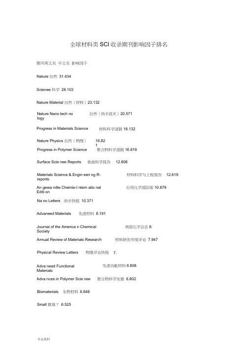
全球材料类SCI 收录期刊影响因子排名期刊英文名 中文名 影响因子 Nature 自然 31.434 Scienee 科学 28.103Nature Material 自然(材料)23.132Nature Nano tech no logy自然(纳米技术)20.571Progress in Materials ScienceProgress in Polymer Science聚合物科学进展16.819Nature Physics 自然(物理)16.821Surface Scie nee Reports 表面科学报告 12.808Materials Science & Engin eeri ng R-reports 材料科学与工程报告 12.619 An gewa ndte Chemie-I ntern atio nal Editi on 应用化学国际版10.879Na no Letters 纳米快报 10.371 Advaneed Materials 先进材料 8.191Journal of the America n Chemical Society 美国化学会志8. Annual Review of Materials Research 材料研究年度评论 7.947 Physical Review Letters 物理评论快报 7. Adva need FunctionalMaterials 先进功能材料6.808 Adva nces in Polymer Scie nee 聚合物科学发展 6.802Biomaterials 生物材料 6.646 Small 微观? 6.525材料科学进展18.132Progress in Surface Scie nee 表面科学进展5.429Chemical Communications 化学通信5.34MRS Bulletin 材料研究学会(美国)公告5.290Chemistry of Materials 材料化学5.Advances in Catalysis 先进催化4.812Journal of Materials Chemistry 材料化学杂志4.646Carbon 碳4.373Crystal Growth & Desig n 晶体生长与设计4.215Electrochemistry Communications 电化学通讯4.194The Journal of Physical Chemistry B 物理化学杂志,B辑:材料、表面、界面与生物物理4.Inorganic Chemistry 有机化学4.147Langmuir 朗缪尔4.Physical Chemistry Chemical Physics 物理化学4.064In ternatio nal Journal of Plasticity 塑性国际杂志3.875Acta Materialia 材料学报3.729Applied Physics Letters 应用物理快报3.726Journal of power sources 电源技术3.477Journal of the Mecha nics and Physics of Solids 固体力学与固体物理学杂志 3.467 International Materials Reviews 国际材料评论3.462Na notech nology 纳米技术3.446Journal of Applied Crystallography 应用结晶学3.212Journal of the American Ceramic Society 美国瓷学会杂志2.101Microscopy and Microa nalysis 2.992Current Opinion in Solid State & Materials Science Scripta Materialia材料快报 2.887The Jour nal of Physical Chemistry A Biometals 生物金属 2.801 Ultramicroscopy超显微术 2.629Microporous and Mesoporous Materials Composites Science and Tech no logy Curre nt Na noscie nee 当代纳米科学 Journal of the Electrochemical Society Solid State Ionics固体离子 2.425IEEE Journal of Qua ntum Electro nicsIEEE 量子电子学杂志 2.413Mechanics of Materials材料力学 2.374Journal of nan oparticle research 纳米颗粒研究 2.299CORROSION SCIENCE 腐蚀科学 2.293 Journal of Applied Physics应用物理杂志 2.201Journal of Biomaterials Scie nce-Polymer Editio n 生物材料科学一聚合物版 2.158IEEE Tran sacti ons on Na notech nologylEEE纳米学报 2.154Progress in Crystal Growth and Characterization of Materials 晶体生长和材料表征进展 2.129Journal of Physics D-Applied Physics物理杂志 D ------ 应用物理 2.104固态和材料科学的动态 2.976物理化学杂志,A 辑2.871多和类材料2.555 复合材料科学与技术 2.533 2.437电化学界2.437Diam ond and Related Materials金刚及相关材料2.092Journal of Chemical & En gi neeri ng Data 化学和工程资料杂志2.In termetallics 金属间化合物2.034Electrochemical and Solid State Letters 固体电化学快报2.001Sy nthetic Metals 合成金属1.962Composites Part A-Applied Scienee and Manufacturing 复合材料A 应用科学与制备1.951Journal of Nan oscie nee and Nano tech no logy 纳米科学和纳米技术 1.929Journal of Solid State Chemistry 固体化学1.91Journal of Physics: Co nde nsed Matter 物理学学报:凝聚态物质 1.9Urnal of Bioactive and Compatible Polymer 生物活性与兼容性聚合物杂志 1.896In ternatio nal Journal of Heat and Mass Tran sfer 传热与传质1.894Applied Physics A-Materials Scienee & Processing 应用物理A —材料科学和进展 1.884 Thin Solid Films 固体薄膜1.884Surface & Coat ings Techn ology 表面与涂层技术1.860Materials Science & Engin eeri ng C-Biomimetic and Supramolecular Systems 材料科学与工程C —仿生与超分子系统 1.812Materials Research Bullet in 材料研究公告1.812In ternatio nal Journal of Solids and Structures 固体与结构1.809Materials Science and Engin eeri ng A-Structural Materials Properties Microst 材料科学和工程A —结构材料的性能、组织与加工 1.806Journal of the American Ceramic Society 美国瓷学会杂志2.101Materials Chemistry and Physics 材料化学与物理1.799Powder Techn ology 粉末技术1.766Materials Letters 材料快报1.748Journal of Materials Research 材料研究杂志1.743Smart Materials & Structures 智能材料与结构 1.743Solid State Scie nces 固体科学1.742Polymer Testi ng 聚合物测试1.736Nan oscale Research Letters 纳米研究快报1.731Surface Scie nee 表面科学1.731Optical Materials 光学材料1.714In ternatio nal Journal of Thermal Scie nces 热科学1.683 Thermochimica Acta 热化学学报1.659Journal of Biomaterials Applicati ons 生物材料应用杂志 1.635 Jour nal of Thermal An alysis and Calorimetry1.63Journal of Solid State Electrochemistry 固体电化学杂志1.597 Journal of the European Ceramic Society 欧洲瓷学会杂志1.58Materials Science and Engin eeri ng B-Solid State Materials for Adva need Tech 科学与工程B —先进技术用固体材料 1.577Applied Surface Scie nee 应用表面科学1.576Europea n Physical Jour nal BSolid State Communi cati onsIntern ati onal Jour nal of Fatigue 材料欧洲物理杂志B1.568固体物理通信1.557疲劳国际杂志1.556Computational Materials Science 计算材料科学1.549Philosophical Magaz ine A-Physics of Conden sed Matter Structure Defects andCeme nt and Con crete Research Philosophical Magazi ne Letters哲学杂志(包括材料)1.548Curre nt Applied Physics 当代应用物理 1.526 Jour nal of Alloys and Compo unds Wear 磨损 1.509Journal of Materials Scie nce-Materials in Medici ne Adva need En gi neeri ng Materials 先进工程材料 1.506 Journal of Nuclear Materials核材料杂志 1.501Intern ati on al J ournal of Applied Ceramic Tech no logy 应用瓷技术 1.488 Chemical Vapor Depositi on化学气相沉积 1.483COMPOSITES PART B-ENGINEERING 复合材料 B 工程 1.481Composite Structures复合材料结构 1.454Journal of Non-crystalli ne Solids非晶固体杂志 1.449Journal of Vacuum Scie nee & Techn ology B 真空科学与技术杂志 B1.445 Semico nductor Scie nee and Tech no logy 半导体科学与技术1.434Journal of SOL-GEL Scie nee and TEch no logy 溶胶凝胶科学与技术杂志1.433Scie nee and Tech nology of Weldi ng and Joi ning焊接科学与技术 1.426Metallurgical and Materials Tran sact ions A-Physical Metallurgy and Material 冶金与材料会刊A ――物理冶金和材料1.389Modelli ng and Simulation in Materials Scie nee and Engin eeri ng 材料科学与工程中的建模与模拟1.388水泥与混凝土研究 1.549 合金和化合物杂志 1.51材料科学杂志一医用材料1.508Journal of Materials Scie nee材料科学杂志1.哲学杂志A 凝聚态物质结构缺陷和机械性能物理 1.384Philosophical Magaz ine 哲学杂志1.384 Ceamics Intern ati onal 国际瓷1.369 Oxidation of Metals材料氧化1.359Modern Physics Letters A现代物理快报 A1.334Ceme nt & Con crete Composites水泥与混凝土复合材料 1.312Mecha nical Properties Jour nal of In tellige nt Material Systems and Structures 智能材料系统与结构 1.293Jour nal of Magn etism and Magn etic Materials 磁学与磁性材料杂志 1.283Journal of Electr onic Materials 电子材料杂志 1.283 Surface and In terface An alysis表面与界面分析1.272Science and Tech no logy of Adva need Materials1.267 Jour nal of Computatio nal and Theoretical Nano scie nee 计算与理论纳米科学 1.256 IEEE TRANSACTIONS ON ADVANCED PACKAGINGIEEE 高级封装会刊1.253Materials Characterizatio n材料表征 1.225Intern ati onal Journal of Refractory Metals & Hard Materials 耐火金属和硬质材料国际 杂志1.221Physica Status solidi A-Applied Research 固态物理A ——应用研究 1.205PHASE TRANSITIONS 相变 1.201 Jour nal of Thermal Spray Tech no logy 热喷涂技术杂志1.2Intern ati onal Jour nal of Nano tech no logy 纳米工程1.184Journal of Vacuum Scie nee & Techno logy A-VACUUM Surfaces and Films真空科学与技术A真空表面和薄膜1.PHYSICA STATUS SOLIDI B-BASIC RESEARCH 固态物理B —基础研究1.166 MATERIALS SCIENCE IN SEMICONDUCTOR PROCESSING 半导体加工的材料科学1.158In ternatio nal Journal of Fracture 断裂学报1.147Journal of Materials Processi ng Techno logy 材料加工技术杂志1.143Metals and Materials In ternatio nal 国际金属及材料1.IEEE TRANSACTIONS ON MAGNETICSIEEE 磁学会刊1.129Vacuum 真空1.114Journal of Applied Electrochemistry 应用电化学1.111Materials & Desig n 材料与设计1.107JOURNAL OF PHYSICS AND CHEMISTRY OF SOLIDS 固体物理与化学杂志1.103Journal of Experime ntal Na noscie nee 实验纳米科学1.103POLYMER COMPOSITES 聚合物复合材料1.054Journal of Materials Scie nce-Materials in Electro nics 材料科学杂志一电子材料 1.054 Journal of Composite Materials 复合材料杂志1.034Journal of the Ceramic Society of Japa n 日本瓷学会杂志1.JOURNAL OF ELECTROCERAMICS 电子瓷杂志0.99ADVANCES IN POLYMER TECHNOLOGY 聚合物技术发展0.979IEEE TRANSACTIONS ON COMPONENTS AND PACKAGING TECHNOLOGIESIEEE 元件及封装技术会刊0.968Journal of Porous Materials 多材料0.959In ternatio nal Journal of Materials Research 材料研究杂志 0.819专业资料IEEE TRANSACTIONS ON SEMICONDUCTOR MANUFACTURINGIEEE 0.957CONSTRUCTION AND BUILDING MATERIALS结构与建筑材料 0.947Journal of Engin eeri ng Materials and Tech no logy-Tra nsact ions of The ASME 工程材料与技术杂志一美国机械工程师学会会刊0.938FATIGUE & FRACTURE OF ENGINEERING MATERIALS & STRUCTURES 工程材料与结构的疲劳与断裂0.934IEEE TRANSACTIONS ON APPLIED SUPERCONDUCTIVITYIEEE 应用超导性会刊 0.919ACI STRUCTURAL JOURNAL美国混凝土学会结构杂志0.895Materials Scie nee and Techn ology 材料科学与技术 0.894Materials and Structures 材料与结构 0.892 Reviews on Adva need Materials Scie nee 先进材料科学评论 0.891 In ternatio nal Journal of Thermophysics热物理学国际杂志 0.889JOURNAL OF ADHESION SCIENCE AND TECHNOLOGY 粘着科学与技术杂志 0.869Journal of Materials Scie nee & Tech no logy 材料科学与技术杂志 0.869High Performa nee Polymers高性能聚合物 0.86BULLETIN OF MATERIALS SCIENCE材料科学公告 0.858Mecha nics of Adva need Materials and Structures 先进材料结构和力学0.857PHYSICA B 物理 B0.822EUROPEAN PHYSICAL JOURNAL-APPLIED PHYSICS 欧洲物理杂志一应用物理 0.822CORROSION 腐蚀 0.821半导体制造会刊JOURNAL OF NONDESTRUCTIVE EVALUATIONMETALLURGICAL AND MATERIALS TRANSACTIONS B-PROCESS METALLURGY ANDMATERIALS 冶金和材料会刊 B —制备冶金和材料制备科学 0.798Materials Transactions材料会刊 0.753Aerospace Science and Tech no logy Journal of Energetic Materials Adva need Powder Tech no logy Applied Composite Materials Adva nces in Applied CeramicsMaterials and Manu facturi ng Processes Composite In terfaces 复合材料界面 0.69 JOURNAL OF ADHESION 粘着杂志 0.685INTERNATIONAL JOURNAL OF THEORETICAL PHYSICS 理论物理国际杂志 0.675 JOURNAL OF NEW MATERIALS FOR ELECTROCHEMICAL SYSTEMS 电化学系统新材料杂志0.67Journal of Thermophysics and Heat Tran sfer 热物理与热传递 0.647Materials and Corrosion-Werkstoffe Und Korrosion 材料与腐蚀 0.639 RESEARCH IN NONDESTRUCTIVE EVALUATION无损检测研究 0.630JOURNAL OF COMPUTER-AIDED MATERIALS DESIGN 计算机辅助材料设计杂志 0.605JOURNAL OF REINFORCED PLASTICS AND COMPOSITES 增强塑料和复合材料杂志0.573ACI MATERIALS JOURNAL美国混凝土学会材料杂志0.568无损检测杂志0.808航空科学技术0.74材料与制造工艺 0.706金属学杂志0.723 先进粉末技术0.716 应用复合材料0.712 先进应用瓷0.708SEMICONDUCTORS 半导体0.565FERROELECTRICS 铁电材料0.562INTERNATIONAL JOURNAL OF MODERN PHYSICS B 现代物理国际杂志B0.558 MATERIALS RESEARCH INNOVATIONS 材料研究创新0.54GLASS TECHNOLOGY -PART A 玻璃技术0.529JOURNAL OF MATERIALS IN CIVIL ENGINEERING 土木工程材料杂志0.526NEW DIAMOND AND FRONTIER CARBON TECHNOLOGY 新型金刚和前沿碳技术0.500SCIENCE IN CHINA SERIES E-TECHNOLOGICAL SCIENCES 中国科学E 技术科学0.495 ATOMIZATION AND SPRAYS 雾化和喷涂0.494SYNTHESE 合成0.477HIGH TEMPERATURE 高温0.469Journal of Phase Equilibria and Diffusion 相平衡与扩散0.457INORGANIC MATERIALS 无机材料0.455MECHANICS OF COMPOSITE MATERIALS 复合材料力学0.453BIO-MEDICAL MATERIALS AND ENGINEERING 生物医用材料与工程0.446PHYSICS AND CHEMISTRY OF GLASSES 玻璃物理与化学0.429JOURNAL OF WUHAN UNIVERSITY OF TECHNOLOGY-MATERIALS SCIENCE EDITION理工大学学报-材料科学版0.424ADVANCED COMPOSITE MATERIALS 先进复合材料0.404Journal of Materials En gi neering and Performa nee 材料工程与性能杂志0.403专业资料Solid State Techno logy 固体物理技术0.400FERROELECTRICS LETTERS SECTION 铁电材料快报0.375JOURNAL OF POLYMER MATERIALS 聚合物材料杂志0.373JOURNAL OF INORGANIC MATERIALS 无机材料杂志0.37GLASS SCIENCE AND TECHNOLOGY-GLASTECHNISCHE BERICHTE 玻璃科学与技术0.365POLYMERS & POLYMER COMPOSITES 聚合物与聚合物复合材料0.355Surface Engineering 表面工程0.354RARE METALS 稀有金属0.347HIGH TEMPERATURE MATERIAL PROCESSES 高温材料加工0.34JOURNAL OF TESTING AND EVALUATION 测试及评价杂志0.324AMERICAN CERAMIC SOCIETY BULLETIN 美国瓷学会公告0.324MATERIALS AT HIGH TEMPERATURES 高温材料0.323MAGAZINE OF CONCRETE RESEARCH 混凝土研究杂志0.315SURFACE REVIEW AND LETTERS 表面评论与快报0.309Journal of Ceramic Processing Research 瓷处理研究0.294JSME INTERNATIONAL JOURNAL SERIES A-SOLID MECHANICS AND MATERIAL ENGINEERIN日本机械工程学会国际杂志系列 A —固体力学与材料工程0.291 MATERIALS TECHNOLOGY 材料技术0.288ADVANCED COMPOSITES LETTERS 先进复合材料快报0.27HIGH TEMPERATURE MATERIALS AND PROCESSES 高温材料和加工0.268INTEGRATED FERROELECTRICS 集成铁电材料0.242MATERIALS SCIENCE 材料科学0.226MATERIALS EVALUATION 材料评价0.21POWDER METALLURGY AND METAL CERAMICS 粉末冶金及金属瓷0.201RARE METAL MATERIALS AND ENGINEERING 稀有金属材料与工程0. INTERNATIONAL JOURNAL OF MATERIALS & PRODUCT TECHNOLOGY 材料与生产技术国际杂志0.157METAL SCIENCE AND HEAT TREATMENT 金属科学及热处理0.157JOURNAL OF ADVANCED MATERIALS 先进材料杂志0.14ADVANCED MATERIALS & PROCESSES 先进材料及工艺0.129MATERIALS WORLD 材料世界0.122SCIENCE AND ENGINEERING OF COMPOSITE MATERIALS 复合材料科学与工程0.098 MATERIALS PERFORMANCE 材料性能0.Journal of Materials En gi neering and Performa nee 材料工程与性能杂志0.403专业资料。
Keap1

非小细胞肺癌(non-small cell lung cancer,NSCLC)发病率占据肺癌的75%~80%。
肿瘤细胞进展快且易扩散转移,临床常采用手术、放化疗等进行治疗,但5年生存率低于60%[1-2]。
氧化应激是由活性氧(ROS)生成量增加所致,ROS积累可诱导肺癌细胞凋亡,清除ROS 可阻止癌细胞凋亡,即肺癌细胞存活依赖于癌细胞自身抗氧化能力[3]。
Kelch样环氧氯丙烷相关蛋白-1 (kelch-like epichlorohydrin-associated protein-1,Keap1)/核因子E2相关因子2(nuclear factor E2related factor 2,Nrf2)信号通路在癌症中发挥重要调控作用,氧化应激可激活Keap1,促使Keap1-Nrf2复合物裂解,Nrf2转移至细胞核内,可激活下游靶基因表达,参与肺癌发生发展过程[4]。
Nrf2可维持氧化还原稳态,ROS侵袭细胞时,Nrf2可进入细胞核,结合抗氧化反应元件(ARE)转录编码各种抗氧化蛋白、代谢酶基因,抑制氧化应激反应[5-6]。
目前氧化应激、Keap1/Nrf2信号通路在NSCLC发生过程中的机制尚未明确。
基于此,本研究尝试分析Keap1/Nrf2信号通路与临床病理参数、氧化应激指标的相关性,探讨其在NSCLC氧化应激机制中的作用,为临床研制新药提供参考依据。
1资料与方法1.1一般资料选取2017年4月至2020年4月郑州市第三人民医院收治的100例NSCLC患者为研究对象。
纳入标准:符合NSCLC诊断标准[7];术前未接受放化疗、免疫治疗者;预计生存期≥6个月;符合手术适应证、禁忌证;Karnofsky功能状态评分≥70分;签署知情同意书。
排除标准:合并凝血功能障碍、肝肾功能障碍、其他恶性肿瘤者;伴有急/慢性感染者;伴有精神疾病者;既往腹部相关外科手术史者。
所有患者均行肺癌根治性切除术,术中收集癌组织、癌旁组织(距离癌组织5cm范围内正常组织),其中男性63例,女性37例;年龄46~67岁,平均(56.32±3.16)岁;体质量指数(BMI)17~30kg/m2,平均(23.16±2.03)kg/m2;病理类型:鳞癌58例、腺癌42例;病理分级[8]:Ⅰ~Ⅱ级51例、Ⅲ级49例;T分期[9]:T1~T253例、T3~T447例;N分期:N055例、N1~N245例。
研FMIS-Ch1-AlgebraicPreliminaries西交大第一章

西南交通大学2012级硕/博研究生课程有限域与计算数论Finite Field and ComputationalNumber Theory张文芳信息科学与技术学院wfzhang@1有限域与计算数论Finite Field and ComputationalNumber TheoryPart 1 Algebraic FoundationsZhang WenfangEmail: wfzhang@School of Information Science & TechnologySouthwest Jiaotong University2Subring and characteristicz Theorem 1.2.1 A ring R ≠{0} of positive characteristic having an identity and no zero divisors must have prime characteristic. Proof R ≠{0}, characteristic n ≥0 of R If n is not prime ⇒ n=km with k,m∈Z, 1<k, m<n Then 0=ne=(km)e =(ke)(me) ⇒ke=0 or me=0 (since R has no zero divisors ) ⇒kr=(ke)r=0 for all r∈R or mr=(me)r=0 for all r∈R ⇒ contradiction to the definition of the characteristic n!21Subring and characteristicz Theorem 1.2.2 Let R be a commutative ring of prime characteristic p. Then(a + b) = a + b for a,b∈R and n∈N.pn pn pnand ( a − b )pn=apn−bpn22Chapter 1 Algebraic Preliminaries1.1 Groups 1.2 Rings 1.3 Fields23Definition of Fieldz Definition 1.3.1 A field (域), denoted by (F, +, ∗), or simply F, is a set at least two elements with two binary operation + and ∗, which we call addition and multiplication, defined on F such that the following properties are satisfied: (1) The set is closed under the operation + : a+b∈R, ∀ a, b∈F. (2) Associative law holds for +: a+(b+c)=(a+b)+c, ∀ a, b, c∈F. (3) The commutative law holds for +: a+b=b+a, ∀ a, b∈F. (4) There is a special (zero) element 0∈F, called the additive identity of F, such that 0+a=a+0=a, ∀a∈F.24Definition of Field(5) For each a∈F, there is a corresponding element −a∈F, called the additive inverse of a, such that a+(−a)=0, ∀a∈F. (6) The set is closed under the operation ∗: a∗b∈R, ∀ a, b∈F. (7) Associative law holds for ∗: a∗(b∗c)= (a∗b)∗c, ∀ a, b, c∈F. (8) The commutative law holds for ∗: a∗b=b∗a, ∀ a, b∈F. (9) There is an element 1∈F, called the multiplicative identity of F, such that 1≠0 and a∗1=1*a=a, ∀a∈F.25Definition of Field(10) For each nonzero a∈F, there is a corresponding element a−1∈F, called the multiplicative inverse of a, such that a∗a−1=1. (11) The distributive law holds: a∗(b+c)= a∗b+a∗c, ∀ a, b, c∈F, (b+c)∗a = b∗a+c∗a, ∀ a, b, c∈F.26Definition of Fieldz Remark 1 A field (F, +, ∗) is a division ring with commutative multiplication.z Remark 2 A field (F, +, ∗) is an Abelian group with respect to addition and also the non-zero field elements form an Abelian group with respect to multiplication. (1) (F, +) is an Abelian group (2) (F∗, ∗) is an Abelian group, where F∗={a∈F: a ≠0}.27Definition of Fieldz Example 1.3.1 Q, R and C are all infinite fields Z is a ring but not a field, since 2, for example, has no multiplicative inverse28Definition of Fieldz Containment for algebraic structure having two binary operationsIntegral Domains Commutative Rings Rings with IdentityFields29A Finite Fieldz Definition 1.3.2 A finite field is a field that has a finite number of elements in it; we called the number the order of the fields. z Theorem 1.3.1 There exists a field of order q if and only if q is a prime power (i.e., q=pr) with p prime and r∈Z. Moreover, if q is a prime power, then there is, up to relabeling, only one field of that order. z Remark A field of order q with q a prime power is often called a Galois field, and is denoted by GF(q), or just Fq.30。
IEEE 519-1992 谐波分析指南说明书

Harmonic Analysis and IEEE 1992 GuidelinesIntroductionIEEE 519-1992 provides guidelines for applying limits to the level of harmonic distortion that a utilitycustomer may inject into the power system. This is a concern, since Adjustable Frequency Drives (AFDs) can contribute significant harmonic distortion to a power system. The guidelines pertain to percentharmonic current and voltage distortion at the point of common coupling (PCC), which is defined as the point where the utility connects to multiple customers.Although many customers and system designers interpret the PCC to be at the AFD input or various locations within the 480V distribution, this is not consistent with the intent of IEEE guidelines. There are no limits recommended for individual loads, only for the overall system. Customers and system designers can choose the point of analysis (POA) where they desire, but it may add substantial filtering costs if the POA is downstream of the PCC.Current distortion drawn through an impedance (transformer, cable resistance) causes voltagedistortion.The distorted current will also cause additional heating of the input cables and the transformer. Excessive voltage distortion is a concern, since it may cause interference with other electronic equipment and additional motor heating.IEEE 519-1992 recommends different limits on Individual Harmonics (I h ) and Total Demand Distortion (TDD), depending on the I SC /I L ratio. I SC is the short circuit current at the PCC, and I L is the maximum demand load current (fundamental) at the PCC. More current distortion is allowed at higher I SC /I L ratios, since voltage distortion decreases as the ratio increases.The voltage distortion guidelines for IEEE-1992 (at 480V) remain the same as IEEE 519-1981:∙ 3% — Special systems (i.e. hospitals or universities) ∙ 5% — General systems∙ 10% —Dedicated systems (AFDs only)Application Note AP04014002EHarmonic Analysis and IEEE 1992 GuidelinesEffective July 20142 EATON CORPORATION The best way to estimate AFD harmonic contribution to an electrical system is to perform a harmonicanalysis based on known system characteristics. An individual AFD may meet the IEEE guidelines in one system and not meet the guidelines in another system depending on the pre-existing characteristics of the specific system.Some AFD vendors, upon seeing a specification requirement for IEEE 519-1992, will simply add a line reactor. This is the wrong approach, since some systems will not require a line reactor and others will not benefit sufficiently to meet the guidelines (or the specification).For a free computerized harmonic analysis of AFD contribution to system harmonics or for additional information, contact your local Eaton sales office. A one-line drawing of the electrical distribution system and specification criteria will be required. A harmonic analysis worksheet for required data is attached.Any additional harmonic mitigation equipment requirements will be determined during the analysis. If there are harmonic constraints during AFD operation on a standby generator, a separate analysis will be required for the generator, and assumptions on load-shedding strategies during generator operationshould be provided. Several data runs may be required to evaluate various harmonic mitigation methods. Resultant recommendations may include 1%, 3%, or 5% line reactors, phase-shifting transformers, filters, or CPX9000 Clean Power.Helpful Facts∙ Harmonics are supply system dependent. As the short circuit amps available (SCA) increase, %voltage distortion decreases, and % current distortion increases.∙ For each PCC or POA analysis required, provide SCA and IL (load current) values for that point. SCAs used must be without motor contribution to SCA.∙Current distortion percentages are dependent on overall system loading. As linear loads (non-harmonic loads such as AC motors on line power) increase, the percent current distortion decreases through dilution.∙ % current distortion is the same across a transformer. Voltage distortion percentage is lower on the primary than on the secondary, assuming the harmonic loads are on the secondary. ∙A harmonic analysis is only as accurate as the assumptions made for the analysis!Harmonic Analysis Data Worksheet(Use separate sheets if necessary. Provide a 1-line drawing or sketch.)lated voltage distortion and higher calculated current distortion on the transformer secondary.Distribution Transformer(s) Data:Generator Data:Describe load-shedding scheme for generator operation in “AFD Data” below.AFD Data:kVAImpedanceX/R Ratio#1#2kW kVAVoltsX"dI L (amps)#1#2AFD hpType (SVX9000,Clean Power, etc.)Quantity Operated on Line / GeneratorDesired, existing or specified line reactor or isolation transformer (none, 1% / 3% / 5%)//////Application Note AP04014002E Effective July 2014Harmonic Analysis and IEEE 1992 Guidelines3 EATON CORPORATION Application Note AP04014002E Harmonic Analysis and IEEE 1992 Guidelines Effective July 2014Additional HelpIn the US or Canada: please contact the Technical Resource Center at 1-877-ETN-CAREor 1-877-326-2273 option 2, option 6.All other supporting documentation is located on the Eaton web site at /DrivesEaton1000 Eaton BoulevardCleveland, OH 44122 USA© 2014 EatonAll Rights ReservedPrinted in USAPublication No.AP04014002EJuly 2014Eaton is a registered trademarkof Eaton Corporation.All other trademarks are propertyof their respective owners。
生物学变异系数

3.7
9.1
载脂蛋白A1
49
血清
Apolipoprotein B
9
6.9
22.8
3.5
6
11.6
载脂蛋白B
50
血浆
Arginine
1
19.3
34.1
9.7
9.8
25.7
精氨酸
51
血清
Arilestearase activity, non inhibited
1
3.8
37.2
1.9
9.3
12.5
Arilestearase活动,非抑制
13
12.3
23.1
6.15
6.54
16.69
天冬氨酸转氨酶(AST)
56
血浆
Aspartic acid
1
31.2
55.1
15.6
15.8
41.6
天冬氨酸
57
血清
β-2-Microglobulin
1
5.9
15.5
3
4.1
9
β-2-Microglobulin
58
血浆
β-Carotene
1
18
48
9
12.8
5.1
16.5
α生育酚
24
血清
Acid phosphatase
2
8.9
8
4.5
3
10.3
酸性磷酸酶
25
血清
Acid phosphatase tartrate-resistant (TR-ACP)
2
8
13.3
4
3.9
Bia-core
Analytical Biochemistry 361 (2007) 1–6/locate/yabioANALYTICALBIOCHEMISTRYReviewHigher-throughput, label-free, real-time molecular interaction analysisRebecca L. Rich, David G. Myszka¤Center for Biomolecular Interaction Analysis, School of Medicine, University of Utah, Salt Lake City, UT 84132, USAMolecular interaction analysis involves characterizinghow molecular complexes form and break down over time.This dynamic information about biological processes at themolecular level has become an essential component of drugdiscovery and life science research. Interest in, and applica-tion of, label-free interaction technology has grown consid-erably over the past 10 years. Label-free assays can be fasterto set up than traditional methods such as X uorescent orradioligand assays, and they also minimize the worry thatthe label itself may alter the binding interaction. Althoughlabel-free biosensor technology is a well-established analyt-ical tool for kinetic, a Y nity, and concentration analyses, itsapplication in a high-throughput setting has been limited.This is about to change as a number of instrument manu-facturers have released or are developing next-generationbiosensors geared at increasing sample throughput.The basicsIn an optical biosensor experiment, a population of onebinding partner is tethered to a sensor surface and an ali-quot of the other partner is injected across this surface(Fig.1A). Fig.1B shows a schematic of the main features ofa biosensor binding pro W le, often referred to as a sensor-gram. During the association phase, analyte is exposed tothe sensor surface. The optical detection system measuresthe change in refractive index of the bu V er near the sensorsurface as analyte mass accumulates on the surface. If thereaction is allowed to proceed long enough, equilibriummay be achieved. A key advantage of biosensor technologyis that the amount of complex formed is measured in thepresence of free material, unlike W lter-binding or pull-downassays that require washing steps before quantitation. Thisavoids perturbing the reaction equilibrium and makes itpossible to characterize very weak or transient interactions.During the dissociation phase, the surface is washed withbu V er and the signal is monitored as the complexes breakdown over time. By tracking binding events in real time, it ispossible to derive kinetic information about the interactionmechanism. Also, by changing analysis parameters (e.g.,bu V er, temperature, pH, analyte concentration) and testinga family of analytes (e.g., related compounds or, proteinmutants), we can characterize an interaction in detail. Thesefeatures make optical biosensor technology a popular bio-physical tool.Although there has been an exponential increase in thenumber and range of available biosensors, we focus thistechnology update primarily on higher throughput sys-tems. Even though the instruments we describe mayemploy di V erent optical systems, we do not describe herethe details on how each detection system works. The rea-son for this is rather simple: one does not need to under-*Corresponding author. Fax: +1 801 585 3015.E-mail address:dmyszka@(D.G. Myszka).stand the principles behind any of the detectors to implement the technology correctly. For our purposes, it is su Y cient to know that all of these biosensors measure changes in refractive index of the solution very near the sensor surface. If one is interested in the underlying opti-cal phenomena of each instrument, we suggest visiting the manufacturers’ websites (listed in Table 1). I nstead,we focus our attention on the di V erences and similarities in how samples are delivered and processed by the vari-ous technologies since sample delivery correlates directly with throughput and throughput is the most critical unmet need of label-free interaction analysis.I nstrument manufacturers are employing a variety of formats to improve throughput—one analyte against many targets, many analytes against one target, and many ana-lytes against many targets. Here we subdivide the instru-ments we review by these di V erent analysis formats. To appreciate today’s biosensor technology, we brie X y review the evolution of the technology with an eye on sample delivery.One-on-one approachIn 1990, Pharmacia Biosensor released the W rst commer-cially viable optical biosensor platform called Biacore. This platform was truly revolutionary in its assemblage of micro X uidic, optic, chemical, and robotic technologies. ThisW rst-edition instrument had four X ow cells, but they could only be addressed separately (Fig.2A): one analyte could be studied over one target at a time. Typically, the same sample would be tested over the target surface and then over a reference surface in a separate injection. Although this was a bit tedious, the instrument was fully automated and capable of overnight unattended operation. This tech-nology may seem primitive compared with later-generation platforms, but it is important to recognize that this technol-ogy launched the W eld of label-free real-time molecular interaction analysis. Over the past 16 years, optical biosen-sors have evolved from a niche instrument directed toward examining a handful of straightforward macromolecular interactions to a widely accepted technology suitable for studying biological systems ranging from small molecules,carbohydrates, oligonucleotides, receptors, and antibodies to lipids, viruses, and whole cells. A good gauge of the suc-cess of biosensor technology is that more than 1000 publi-cations each year include data collected from commercial biosensors [1,2].One-on-several approachWhereas the W rst label-free real-time biosensor revolu-tionized molecular interaction analysis, the next break-through made it impossible to turn back. Around 1996,Pharmacia spun the biosensor division out as an indepen-Fig.1. Elements of an optical biosensor analysis. (A) Assay design. Analyte in solution binds to a target ligand immobilized on the sensor surface. (B) Bio-sensor output. The response increases during the association phase as analyte X ows across, and binds to, the immobilized ligand. A plateau in the response at the end of the association phase indicates that the system has reached equilibrium. The response decreases during the dissociation phase as the surface is washed with bu V er and the analyte dissociates from the surface.Table 1Array biosensor platforms aTechnology (or technologies) discussed in this review (not necessarily all platforms o V ered by a manufacturer).Manufacturer WebsiteTechnology aBiacoreBiacore, 2000, 3000, T100, Flexchip, S51, and A100Axela Biosensors dotLabToyobo www.toyobo.co.jp/bio MultiSPRinterLumera Proteomic Processor GenOptics SPRi-Plex SRU Biosystems BIND Corning Epic ForteBIO OctetBio-RadProteOn XPR36dent company called Biacore AB and released a new plat-form called Biacore 2000. Although it was similar to the original biosensor in its core features, the major advance in the technology was a micro X uidic design that allowed the user to run one analyte sample over four X ow cell surfaces independently or in series (Fig.2B). This made it possible to automatically immobilize separate target ligands and then collect data from a reference surface and three reaction sur-faces at the same time. The result was increased throughput and signi W cantly improved data quality, making it possible to detect low-molecular-weight analytes (»200Da). A few years later, an updated system called Biacore 3000 was released with a similar X ow cell design but having a detec-tor with an expanded dynamic range that made it possible to monitor interactions in the presence of highly refractive index bu V ers such as crude sera and dimethyl sulfoxide (DMSO)1. Both instruments are capable of processing sam-ples from two standard 96-well plates. Together, the Bia-core 2000 and 3000 series currently are the most used biosensors in the W eld.Biacore AB continues to make improvements to its four-X ow cell con W guration sensor technology. One of Biacore AB’s most recently released platforms is called the T100. In this instrument, two pairs of X ow cells are connected within the same plane and form “U-shaped” X ow paths (Fig.2C)[3]. By using a combination of three ports in this X ow cell, it is possible to immobilize a target on one arm of the “U”and use the unmodi W ed surface of the other arm as a refer-ence. This redesign shortens the distance between the target and reference surface, thereby improving referencing. I n contrast, the X ow cells in Biacore 2000 and 3000 instru-ments are joined through more complex X uidics between and under the X ow cells (Fig.2B). The use of the U X ow cell improves data quality while retaining many of the robust features of the previous four-channel X ow cell designs.Realizing that referencing was an important way of improving data quality, Biacore AB developed a novel approach to X ow cell design in its S51 systems, a platform that was engineered speci W cally to support small molecule drug discovery and development. The S51 uses “Y-shaped”X ow cells (Fig.2D) and the technique of hydrodynamic addressing to immobilize target molecules along the outer edges of the X ow cell while maintaining an unmodi W ed region in the center [4]. Small detector windows are posi-tioned in the middle of the X ow path within each region.The center region serves as an internal reference, making it possible to collect high-resolution data for very small ana-lytes. This increase in sensitivity also makes it possible to collect binding data on targets with low activity. The S51technology has been implemented by a number of pharma-ceutical companies as a secondary screening tool. The Bia-core T100 and S51 instruments both can accommodate samples from one 96- or 384-well plate while processing one sample at a time.One-on-many approachGiven the positive impact that Biacore’s technology has had on interaction analysis, it is not surprising to see other manufacturers developing systems for the label-free mar-ket. One approach to improving throughput is to increase the number of targets examined at a time—the one-on-many approach. For example, Axela Biosensors’ dotLab system involves producing a linear array of eight target spots within a X ow channel (Fig.2E). A single analyte is tested simultaneously for binding to all of the target spots.Axela Biosensors is working on producing addressable arrays by using a variety of capturing surfaces within the channel to immobilize a variety of tagged ligands at one time. These disposable cartridges could be particularly use-ful in diagnostic applications.Following the adage that more is more, several manufac-turers are signi W cantly expanding the number of targets into two-dimensional arrays. In this approach, one analyte is X owed across targets that have been spotted in a large-format X ow cell (Fig.2F). Using imaging technology, bind-ing data can be collected from both the target spots and unmodi W ed regions between the spots that are used for internal referencing. Typical applications for these instru-ments include monoclonal antibody screening and charac-terization of peptide and oligonucleotide arrays.Biacore’s Flexchip system was the W rst commercially available high-density array platform and is capable of ana-lyzing one analyte against 400 target spots at one time.1Abbreviations used: DMSO, dimethyl sulfoxide; cDNA, complementa-ry DNA; ELISA, enzyme-linked immunosorbent assay.Fig.2. Schematic of X ow cell con W gurations in the original Biacore instrument (A), the Biacore 2000 and 3000 (B), the Biacore T100 (C), the Biacore S51(D), the Axela Biosensors dotLab (E), and the Biacore Flexchip (F). In panels D–F, the reference surfaces are shown in yellow. (For interpretation of the reference to color in this W gure legend, the reader is referred to the Web version of this article.)Recently, researchers have begun to demonstrate Flexchip’sutility in higher-throughput biosensor-based screeningapplications, including epitope mapping [5], concentration analyses [6], and protein expression pro W ling [7]. As the W rststep toward fully recognizing this platform’s potential formonitoring interactions in real time, researchers havereported Flexchip-obtained kinetic and a Y nity parameters for various antibody systems [8,9].Toyobo’s MultiSPRinter is an array-based sensor plat-form distributed in Japan. This system is interestingbecause it provides the customer with a complete package, including a spotter and detection system. Exemplifying the X exibility of this biosensor, researchers have used this plat-form to monitor DNA hybridization [10], track proteinexpression [11], and obtain kinetics for protein binding to an array of related double-stranded DNAs [12].The pin-spotting methods commonly used to array tar-gets work well for immobilizing peptides, oligonucleotides,and stable proteins such as antibodies. However, because spotting uses small transfer volumes and requires drying the target on the chip, this method might not be universally applicable to preparing all protein arrays. Many proteins become denatured when dried onto surfaces, are not avail-able at high enough concentrations, and/or often are in crude preparations. To tackle this problem, researchers at GenOptics have developed novel in-solution target immo-bilization chemistries to complement their SPRi-Plex bio-sensor: with electrocopolymerization, pyrrole-conjugated targets are covalently coupled to pyrrole-modi W ed spots on the sensor surface. Using this system, which tracks binding at up to 300 target spots, investigators have characterized DNA, peptide, and antibody interactions [13–16].Lumera is taking the quest to produce protein arrays tothe next level. This optics company is in the W nal phases oftesting its array-based system, called the Proteomic Proces-sor, which is capable of examining more than 1000 target spots at one time [17]. Lumera’s future plans include launching an on-chip in vitro translation system in which complementary DNA (cDNA) will be spotted on the sensor surface to translate protein with a coil fusion tag that inter-acts with a coil peptide copartner prepatterned on the chip surface (NanoCapture microarrays). This translation sys-tem has the potential to eliminate laborious protein pro-duction and puri W cation steps. There are, of course, challenges in the routine application of in vitro translation, but this approach could provide a simple method of gener-ating content for protein arrays.Plate-based systemsTo move biosensor technology to the fast lane of drugdiscovery, it is apparent that the number of analytes thatcan be tested needs to increase signi W cantly. Two compa-nies, Corning and SRU Biosystems, have developed similar approaches to increasing sample throughput in their Epic [18–20] and BI ND [21,22] systems, respectively. I n these technologies, the sensor surface is incorporated directly into the bottom of specially modi W ed, but standard-sized, 96- or 384-well plates. Targets are loaded into these plates and can be tested simultaneously against many analytes. These plate systems meld well with existing plate-based high-throughput robotics that are common in pharmaceu-tical companies for e Y cient sample delivery and sample tracking. Although binding data can be collected in a real-time mode, this feature is used primarily for assay development. For high-throughput operation, data output is primarily end-point measurements, so it is better to think of these systems as label-free enzyme-linked immunosor-bent assays (ELI SAs). These types of systems could be good for handling assays for targets that are di Y cult to prepare in an EL SA format or for compounds that interfere with X uorescent-based systems.ForteBIO, with its Octet technology, has taken a di V er-ent approach to testing plates of samples. I n this system, sensors are incorporated into disposable detector tips, which are coated with target and dipped into wells contain-ing analyte solutions. In this con W guration, the binding of eight analyte–target pairs can be tracked at one time. After each set of analyses, the used tips are discarded and a fresh set of tips is used to examine the next eight analyte solu-tions. The system uses standard well plates and the analyte solutions can be fully recovered. Another advantage of each of these plate-based technologies is that they can be used to test binding in very crude material because clogging is not an issue, as it can be with micro X uidic X ow-through systems.High-resolution parallel processingThe W nal set of instruments we highlight involves novel developments of X ow-through systems. Biacore’s A100 and Bio-Rad’s ProteOn technologies can perform parallel sam-ple analyses while maintaining high data quality.Biacore’s A100 is a 4£4 parallel array processing instru-ment that represents an expansion of the S51 technology. The A100 has four independent X ow systems that transfer analytes from plates (96- or 384-well) to four independent hydrodynamic addressing X ow cells like those shown in Fig.2D. In each A100 X ow cell, however, four targets can be immobilized and an unmodi W ed spot in the center serves as the reference. The system can be used for applications ranging from antibody screening to small molecule analysis [23,24].Bio-Rad’s ProteOn XPR36 instrument incorporates a unique approach to generating a two-dimensional array. The system contains six parallel X ow channels that can be used to uniformly immobilize strips of six targets on the sensor surface (Fig.3A). The X uidic system can automati-cally rotate 90° to create a 36-target array (Fig.3B). Refer-ence spots are automatically formed before and after each reaction spot to permit internal referencing. The ProteOn system also introduces a novel concept in kinetic analysis referred to as “one-shot” kinetics. With this approach, one can simultaneously test six di V erent concentrations ofanalyte over the di V erent target surfaces, thereby generat-ing a full analyte concentration series with one injection [25,26]. This reduces analysis time signi W cantly and elimi-nates the need for surface regeneration. Alternatively, the six channels can be used to screen a single concentration of six compounds against six di V erent target proteins with each injection or any combination in between. The ProteOn XPR36 currently accommodates samples from two 96-well plates.SummaryThe burgeoning number of commercial biosensors geared toward higher throughput analysis clearly demon-strates that the market for this technology is expanding. Beyond throughput, the second most important issue is sensitivity. At this time, we have tried to steer clear of comparing systems based on sensitivity, principally because it is di Y cult to do so without a benchmark study using the same molecular system to evaluate every instru-ment. Su Y ce it to say that there is likely to be a trade-o V between resolution and throughput. For example, the sensitivity of the two-dimensional array-based systems that we discussed likely is not high enough at this stage to allow direct detection of the binding of small molecules to immobilized targets. So, in many ways, more could in fact mean less.It is, however, important to remember that the primary factor that determines the quality of any biosensor data set is the quality of the target itself and how it is immobilized. None of these sensors can yet take a denatured and precipi-tated protein and turn it into a fully active target on the sensor surface. (If there was such a sensor, we would have highlighted it.) In contrast, low-percentage activity often is not a hindrance for enzymatic-, radioligand-, and X uores-cent-based assays. Because these assays can be 1000 times more sensitive than optical biosensors, they often are used to analyze targets that are not especially pure or highly active (whether this is actually a good idea is debatable).It is also important to recognize that most optical bio-sensor assays are performed as direct binding assays. For small molecule work, for instance, we can use the sensor to determine whether a compound actually interacts with a particular target. These direct binding assays, on their own, do not tell us whether the compound actually inhib-its or activates a particular target. For this reason, most pharmaceutical companies will continue to use activity-based assays as a primary screening tool. I n this setting, biosensors are ideal tools to be used for secondary screen-ing to con W rm whether the hit is real and, if so, to provide dynamic information about its interaction with the target. Information on how fast something binds and the half-life of the complex is becoming an essential part of the opti-mization process for both small molecules and biophar-maceuticals.We applaud these biosensor manufacturers for develop-ing tools that will increase the speed of interaction research, ultimately leading to a better understanding of both basic biology and improved health care.References[1]R.L. Rich, D.G. Myszka, Survey of the year 2004 commercial opticalbiosensor literature, J. Mol. Recognit. 18 (2005) 431–478.[2]R.L. Rich, D.G. Myszka, Survey of the year 2005 commercial opticalbiosensor literature, J. Mol. Recognit. 19 (in press).[3]G.A. Papalia, M. Baer, K. Luehrsen, H. Nordin, P. Flynn, D.G.Myszka, High-resolution characterization of antibody fragment/ antigen interactions using Biacore T100, Anal. Biochem. 359 (2006) 112–119.[4]D.G. Myszka, Analysis of small molecule interactions using BiacoreS51 technology, Anal. Biochem. 329 (2004) 316–323.[5]G.J. Carven, S. Chitta, I. Hilgert, M.M. Rushe, R.F. Baggio, M.Palmer, J.E. Arenas, J.L. Strominger, V. Horejsi, L. Santambrogio, L.J. Stern, Monoclonal antibodies speci W c for the empty conforma-tion of HLA–DR1 reveal aspects of the conformational change asso-ciated with peptide binding, J. Biol. Chem. 279 (2004) 16561–16570.[6]D.W. Unfricht, S.L. Colpitts, S.M. Fernandez, M.A. Lynes, Grating-coupled surface plasmon resonance: a cell and protein microarray platform, Proteomics 5 (2005) 4432–4442.[7]K. Usui-Aoki, K. Shimada, M. Nagano, M. Kawai, H. Koga, A novelapproach to protein expression pro W ling using antibody microarrays combined with surface plasmon resonance technology, Proteomics 5 (2005) 2396–2401.[8]R. Baggio, G.J. Carven, A. Chiulli, M. Palmer, L.J. Stern, J.E. Arenas,Induced W t of an epitope peptide to a monoclonal antibody probed with a novel parallel surface plasmon resonance assay, J. Biol. Chem.280 (2005) 4188–4194.[9]D. Wassaf, G. Kuang, K. Kopacz, Q.L. Wu, Q. Nguyen, M. Toews, J.Cosic, J. Jacques, S. Wiltshire, J. Lambert, C.C. Pazmany, S. Hogan, R.C. Ladner, A.E. Nixon, D.J. Sexton, High-throughput a Y nity rank-ing of antibodies using surface plasmon resonance microarrays, Anal.Biochem. 351 (2006) 241–253.[10]K. Nakatani, S. Hagihara, Y. Goto, A. Kobori, M. Hagihara, G. Hay-ashi, M. Kyo, M. Nomura, M. Mishima, C. Kojima, Small-moleculeFig.3. Flow cell con W guration for Bio-Rad ProteOn XPR36. (A) Ligand targets are immobilized in six parallel X ow channels. (B) After the chip is rotated90°, six analytes are injected perpendicular to the channels of immobilized targets. Interstitial spaces in each channel are used for referencing.ligand induces nucleotide X ipping in (CAG)n trinucleotide repeats, Nat. Chem. Biol. 1 (2005) 39–43.[11]M. Kyo, K. Usui-Aoki, H. Koga, Label-free detection of proteins incrude cell lysate with antibody arrays by a surface plasmon resonance imaging technique, Anal. Chem. 77 (2005) 7115–7121.[12]M. Kyo, T. Yamamoto, H. Motohashi, T. Kamiya, T. Kuroita, T.Tanaka, J.D. Engel, B. Kawakami, M. Yamamoto, Evaluation of MafG interaction with Maf recognition element arrays by surface plasmon resonance imaging technique, Genes Cells 9 (2004) 153–164.[13]I. Mannelli, L. Lecerf, M. Guerrouache, M. Goossens, M-C. Millot,M. Canva, DNA immobilization procedures for surface plasmon res-onance imaging (SPRI) based microarray systems, Biosens. Bioelec-tron. 119 (2006) 583–591.[14]E. Maillart, K. Brengel-Pesce, D. Capela, A. Roget, T. Livache, M.Canva, Y. Levy, T. Soussi, Versatile analysis of multiple macromolec-ular interactions by SPR imaging: application to p53 and DNA inter-action, Oncogene 23 (2004) 5543–5550.[15]L. Grosjean, B. Cherif, E. Mercey, A. Roget, Y. Levy, P.N. Marche,M-B. Villiers, T. Livache, A polypyrrole protein microarray for anti-body–antigen interaction studies using a label-free detection process, Anal. Biochem. 347 (2005) 193–200.[16]B. Cherif, A. Roget, C.L. Villiers, R. Calemczuk, V. Leroy, P.N. Mar-che, T. Livache, M-B. Villiers, Clinically related protein–peptide inter-actions monitored in real time on novel peptide chips by surface plasmon resonance imaging, Clin. Chem. 52 (2006) 255–262.[17]C. Boozer, G. Kim, S. Cong, H. Guan, T. Londergan, Lookingtowards label-free biomolecular interaction analysis in a high-throughput format: a review of new surface plasmon resonance tech-nologies, Curr. Opin. Biotechnol. 17 (2006) 400–405.[18]Y. Fang, A.M. Ferrie, N.H. Fontaine, P.K. Yuen, Characteristics ofdynamic mass redistribution of epidermal growth factor receptor sig-naling in living cells measured with label-free optical biosensors, Anal.Chem. 77 (2005) 5720–5725.[19]Y. Fang, A.M. Ferrie, G. Li, Probing cytoskeleton modulation byoptical biosensors, FEBS Lett. 579 (2005) 4175–4180.[20]Y. Fang, G. Li, J. Peng, Optical biosensor provides insights for bra-dykinin B2 receptor signaling in A431 cells, FEBS Lett. 579 (2005) 6365–6374.[21]B.T. Cunningham, P. Li, S. Schulz, B. Lin, C. Baird, J. Gerstenmaier,C. Genick, R. Wang, E. Fine, L. Laing, Label-free assays on the BINDsystem, J. Biomol. Screen. 9 (2004) 481–490.[22]B.T. Cunningham, L. Laing, Microplate-based label-free detection ofbiomolecular interactions: applications in proteomics, Expert Rev.Proteomics 3 (2006) 271–281.[23]P. Säfsten, S.L. Klakamp, A.W. Drake, R. Karlsson, D.G. Myszka,Screening antibody–antigen interactions in parallel using Biacore A100, Anal. Biochem. 353 (2006) 181–190.[24]H. Takeda, A. Fukumoto, A. Miura, N. Goshima, N. Nomura,High-throughput kinase assay based on surface plasmon resonance suitable for native protein substrates, Anal. Biochem. 357 (2006) 262–271.[25]D. Reichmann, O. Rahat, S. Albeck, R. Meged, O. Dym, G. Schreiber,The modular architecture of protein–protein interfaces, Proc. Natl.Acad. Sci. USA 102 (2005) 57–62.[26]T. Bravman, V. Bronner, K. Lavie, A. Notcovich, G.A. Papalia, D.G.Myszka, Exploring “one-shot” kinetics and small molecule analysis using the ProteOn XPR36 array biosensor, Anal. Biochem. 358 (2006) 281–288.。
线粒体自噬

1
(2)IRE1-XBP1信号通路: IRE1a:一种跨膜胞命运的变应原的关键分子,可促进自噬小泡的形成。 IRE1可通过与下游的XBP1来激活自噬,还可以通过IRE1-TRAF2-JNK 来激活自噬
有研究证明,XBP1mRNA剪接可通过Beclin-1的转录激活参与巨噬细胞增殖、凋亡和自噬。
1
UPR信号通路
未折叠蛋白反应(unfolded protein response,UPR):主要作用是下调 未折叠蛋白和错误折叠蛋白,恢复细胞稳态功能以及维持内质网稳态。
主要的相关蛋白:蛋白激酶R样内质网激酶(PERK)、肌醇需求酶1 (IRE1) 、转录激活因子6 (ATF6) 蛋白、免疫球蛋白结合蛋白葡萄糖调节 蛋白78(GRP78/Bip)
3
抑制剂
4-苯基丁酸(4-phenylbutyric acid, 4-PBA)在临床上主要用于治 疗尿素循环障碍,在科学研究中普遍用作内质网应激抑制剂。
P53抑制剂pifithrin-α通过线粒体凋亡途径和线粒体自噬途径减弱百草枯诱导的人肺上皮样细胞A549 凋亡[D].中国医科大学,2018.
P53可以通过与胞质中Parkin的直接相互作用阻断其向线粒体的转 位进而抑制线粒体自噬过程,而P53小分子抑制剂PFT-α的干预可 以恢复正常自噬。
1
Ca2+依赖的自噬
内质网应激导致Ca2+泄露至胞浆中, (1)通过钙调蛋白性激酶β (CAMKβ) 激活AMPK依赖性途径, 抑制 mTOR活性, (2)激活蛋白激酶C (PKC) 通过不依赖mTOR的途径诱导自噬, (3)活化DAPK,参与Beclin-1磷酸化和破坏Bcl-2/Beclin-1复合物
细胞自噬相关蛋白:
(1)标记蛋白:微管相关蛋白轻链3(LC3),有LC3-I和LC3-II两种 (2)酵母Atg6同源蛋白B细胞淋巴瘤蛋白质-2-相互作用蛋白质-1(beclin1) (3)3-磷酸磷脂酰肌醇(PI3K),分为I型、II型、III型 (4)自噬相关蛋白6(Atg6)、Atg14、空泡蛋白质分选蛋白质(Vps15)等
- 1、下载文档前请自行甄别文档内容的完整性,平台不提供额外的编辑、内容补充、找答案等附加服务。
- 2、"仅部分预览"的文档,不可在线预览部分如存在完整性等问题,可反馈申请退款(可完整预览的文档不适用该条件!)。
- 3、如文档侵犯您的权益,请联系客服反馈,我们会尽快为您处理(人工客服工作时间:9:00-18:30)。
6
Definition of Group
Definition 1.1.2 A group (群), denoted by (G, ), or simply G, is a nonempty set G of elements together with a binary operation , such that the following three properties hold: (1) Associativity: For any a, b, cG, a(bc)= (ab)c. (2) Existence of identity (恒等元)(or unity: 单位元): There is a unique element eG, called the identity, such that ea= ae=a, aG. (3) Existence of inverse (逆元): For every aG there is a unique element bG such that ab= ba=e. This b is denoted by a1 and called the inverse of a.
10
Definition of Group
The operation rules For any n, mZ, aG,
Multiplicative Notation an=(a1)n anam =an+m (an)m =anm a0=1 Additive Notation (n)a=n(a) na+ma=(m+n)a m(na)=(mn)a 0a=0
西南交通大学 2013级硕/博研究生课程
有限域与计算数论
Fundamentals of Mathematics in Information Security
信息科学与技术学院
1
Part 1 Algebraic Foundations
2
Chapter 1 Algebraic Preliminaries 1.1 Groups 1.2 Rings 1.3 Fields
18
Definition of Ring
Definition 1.2.5 An division ring(除环) is a ring R with identity 1 0 that satisfies: for each a 0R, the equation ax=1 and xa=1 have solutions in R. Example 1.2.1 (Z,+,), (Q,+,), (R,+,) and (C,+,) are all rings The even integers (Ze,+,) form a commutative ring without identity. Let (R, +) be any Abelian group. Define ab =0 for all a, bR, then (R, +, ) is a ring.
7
Definition of Group
Definition 1.1.2 If the group (G, ) also satisfies (4) Commutativity: For any a, bG, ab=ba, then the group is called a commutative group (交换群) (or Abelian group).
5
Definition of Group
Definition 1.1.1 A binary operation (二元运算) on a set S is a rule that assigns to each ordered pair (a, b) of elements of S a unique element of S.
15
Chapter 1 Algebraic Preliminaries
1.1 Groups 1.2 Rings 1.3 Fields
16
Definition of Ring
Definition 1.2.1 A ring (环), denoted by (R, +, ), or simply R, is a set R, together with two binary operation + and , which we call addition and multiplication, such that the following properties hold: (1) R is a commutative group with respect to +; (2) Associative law holds for : a(bc)= (ab)c, a, b, cR. (3) The distributive law holds : a(b+c)= ab+ac, a, b, cR, (b+c)a = ba+ca, a, b, cR.
8
Definition of Group
Example 1.1.2 (Z,+), (Q,+), (R,+) and (C,+) are commutative groups (Z+, +) is not a group: since there is no identity element for + in Z+. (Z+, ) is not a group: there is an identity element 1, but no inverse of 3. (Q+, ) and (R+, ) are commutative groups (Q, ), (R, ) and (C, ) are not groups (Q*, ), (R*, ) and (C*, ) are commutative groups
11
Finite Group, Order, Subgroup
Definition 1.1.3 A group is called a finite group (有限 群) if it has a finite number of elements; otherwise it is called a infinite group (无限群). Definition 1.1.4 The order (阶) of a group G, denoted by | G | (or by #(G)), is the number of elements in G. Example The order of Z is |Z|=.
Example 1.1.1 Ordinary addition + is a binary operation on the set N, Z, R, C. Ordinary multiplication is another binary operation on the same sets.
12
Finite Group, Order, Subgroup
Definition 1.1.5 A nonempty set H of a group G which is itself a group, under the same operation, is called a subgroup (子群) of G. Subgroups of G other than trivial subgroup {e} and G itself are called nontrivial subgroup of G. Example Z is a subgroup of Q.
14
The order of the element
Definition 1.1.7 Let a be an element of a group G. If <a> is finite, then its order is called the order of the element a. Otherwise, a is called an element of infinite order.
17
Definition of Ring
Definition 1.2.2 A commutative ring(交换环) is a ring that further satisfies: ab = ba, a, bR. Definition 1.2.3 A ring with identity is a ring that contains an element 1 satisfying: a1 = 1a = a, a R. Definition 1.2.4 An integral ring (整环) is a commutative ring with identity10 that satisfies: a, bR ab=0 a =0 or b=0. Remark: There are no zero divisors.
3
Notations
Notations The set of natural numbers: N={1, 2, 3,…} The set of integers: Z={0, 1, 2, 3,…}; Z+= {1, 2, 3,…}= N; Z*={aZ: a 0}={1, 2, 3,…} ; The set of all residue classes modulo a positive integer n: Zn=Z/nZ ={0,1,2,…, n1}. The set of rational numbers: Q={a/b: a,bZ and b0}; Q+={aQ: a>0}; Q*={aQ: a 0}.