supersignal west pico chemiluminescent substrate 定影 显影 及注意事项
exosomes外泌体实验方案
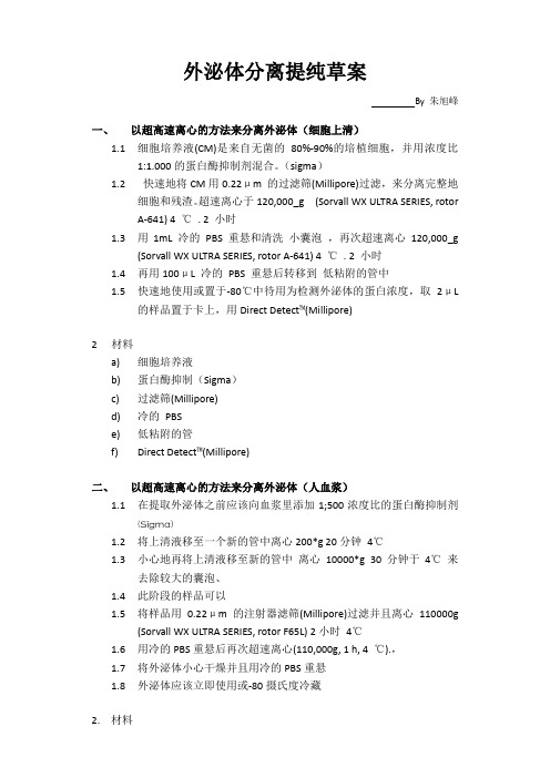
外泌体分离提纯草案By 朱旭峰一、以超高速离心的方法来分离外泌体(细胞上清)1.1细胞培养液(CM)是来自无菌的80%-90%的培植细胞,并用浓度比1:1.000的蛋白酶抑制剂混合。
(sigma)1.2快速地将CM用0.22μm 的过滤筛(Millipore)过滤,来分离完整地细胞和残渣。
超速离心于120,000_g (Sorvall WX ULTRA SERIES, rotorA-641) 4 ℃. 2 小时1.3用1mL 冷的PBS 重悬和清洗小囊泡,再次超速离心120,000_g(Sorvall WX ULTRA SERIES, rotor A-641) 4 ℃. 2 小时1.4再用100μL 冷的PBS 重悬后转移到低粘附的管中1.5快速地使用或置于-80℃中待用为检测外泌体的蛋白浓度,取2μL的样品置于卡上,用Direct Detect™(Millipore)2材料a)细胞培养液b)蛋白酶抑制(Sigma)c)过滤筛(Millipore)d)冷的PBSe)低粘附的管f)Direct Detect™(Millipore)二、以超高速离心的方法来分离外泌体(人血浆)1.1在提取外泌体之前应该向血浆里添加1;500浓度比的蛋白酶抑制剂(Sigma)1.2将上清液移至一个新的管中离心200*g 20分钟4℃1.3小心地再将上清液移至新的管中离心10000*g 30分钟于4℃来去除较大的囊泡、1.4此阶段的样品可以1.5将样品用0.22μm的注射器滤筛(Millipore)过滤并且离心110000g(Sorvall WX ULTRA SERIES, rotor F65L) 2小时4℃1.6用冷的PBS重悬后再次超速离心(110,000g, 1 h, 4 ℃).,1.7将外泌体小心干燥并且用冷的PBS重悬1.8外泌体应该立即使用或-80摄氏度冷藏2.材料a)蛋白酶抑制(Sigma)b)过滤筛(Millipore)c)冷的PBSd)低粘附的管e)Direct Detect™(Millipore)三、ExoQuick TM化学沉淀法1.1ExoQuick TM的使用要按照厂家提供的说明书来实行1.2该试剂盒可以简单地提取CM,人血浆,和血清里的外泌体,只需用倒相管按照所指示的量来加入ExoQuick TM试剂盒里的溶液1.3溶液孵化过夜,在4℃温和的摇晃1.4在流式细胞仪缓冲液中洗涤1.5试剂盒里的带荧光的免疫磁珠已经被绑定,用流式细胞仪鉴定即可(BD Biosciences) 配合使用CellQuest 的软件。
EZECLPico化学发光液(皮克级)使用说明书

EZ ECL Pico化学发光液(皮克级)使用说明书产品描述《EZ ECL Pico化学发光液》是一款增强型化学发光(ECL)HRP底物,可帮助用户在免疫印迹分析过程中实现低皮克级的蛋白检测(<10-12g),极其节省抗体。
本产品一抗浓度范围0.2-0.1μg/ml(以1µg/mL储存液稀释1:1,000至1:5,000倍);二抗浓度10-50ng/ml (以1mg/mL储存液稀释1:20,000至1:100,000倍)。
EZ Pico底物,为使用辣根过氧化物酶(HRP)偶联物的免疫印迹实验提供了明亮的信号、低至皮克级检测灵敏度。
该ECL底物能够兼容各种膜、封闭液和宽范围抗体稀释液,以出色性能、通用性和高性价比,满足用户的免疫印迹应用需求。
EZ Pico底物的特点:•ECL—用于辣根过氧化物酶(HRP)的增强型化学发光底物•低至皮克级灵敏度—检测硝化纤维素膜或PVDF膜上皮克级的蛋白条带•长信号持续时间—在条件优化情况下,经底物孵育的印迹条带能够持续输出6至8小时的可检测光信号•稳定试剂—工作液在24小时内保持稳定;试剂盒在室温下可稳定放置长达1年•价格经济—针对稀释的抗体浓度条件进行了优化:2)取A液和B液一定要用不同的枪头,工作液现配现用,室温放置数小时后仍可使用但灵敏度略有降低。
3.用平头镊取出膜,搭在滤纸上沥干洗液,勿使膜完全干燥。
用移液器将工作液加到膜上(推荐100μl发光液/cm2膜),使之充分接触。
室温孵育5分钟,准备立即压片曝光。
4.用平头镊子夹起膜,膜的下缘轻轻接触吸水纸,去除膜上多余的液体,留下少量工作液,不可让膜完全干燥。
5.在X光胶片暗盒内表面铺一张面积大于膜的保鲜膜。
将印迹膜贴在保鲜膜上,将保鲜膜折起来完全包裹印迹膜,去除气泡和皱褶,可剪去边缘部多余的保鲜膜。
用滤纸吸去多余的发光工作液。
用胶带将覆盖印迹膜的保鲜膜固定在暗盒内,蛋白条带面向上。
6.将X光胶片置于膜的上面。
显影剂说明——精选推荐

重要提醒:SuperSignal West Pico化学发光底物是一个高敏感性的物质,它比大多数化学发光产物(包括ECL,鲁米诺)都要敏感,为了超敏感底物的最佳性能,抗体必须比其他使用的溶液浓度要稀。
如果你一直用以上的底物或者另外初级的化学发光底物,那么,用此化学发光底物稀释一抗和二抗要5倍以上。
例如,如果你用ECL溶液的时候,以1:100稀释一抗。
那么,你用SuperSignalWest Pico溶液的时候,就要以1:500稀释。
建议使用Table1的稀释范围。
Table1. 用SuperSignalWest Pico化学发光底物的抗体稀释度。
一抗:1:1000-1:5000或者0.2-1.0ul/mL二抗:1:20000-1:100000或者10-50ng/Ml说明Thermo Scientific SuperSignal West Pico化学发光底物是检测辣根过氧化物酶标记的抗体的一种高度敏感性增强的溶液。
这种溶液能够强烈的检测到皮微克的抗原总量。
信号的灵敏性、强度、持续时间允许用摄影或其他的成像方法来检测辣根过氧化物酶。
免疫印记可以重复的曝光以获取最佳的结果,或者重新洗膜,再敷上另一种抗体,检测另一种蛋白。
重要产物的信息为了最好的结果,优化Western Blot中所有的组成部分是必不可少的,包括样品总量、一抗及二抗的浓度、膜的选择、封闭液。
因为溶液是极端敏感的,所以SuperSignalWest Pico溶液比其他市场上可以买得的液体来说,需要更少的样品总量、一抗及二抗,通常减少10-20倍。
同时应用Thermo Scientific SuperSignal Western Blot增强剂可以使该溶液敏感性增强,背景减少,抗体特异性提高。
抗体浓度要比用沉淀比色合系统的浓度更加稀释,为了选择最适当的浓度,可以应用斑点印迹分析法。
没有任何一种封闭液是所有系统的最佳选择,所以对于Western Blot的来说,实验检测是选择适合封闭液的必要步骤。
Acta Biochim Biophys Sin-2011-Wen-96-102
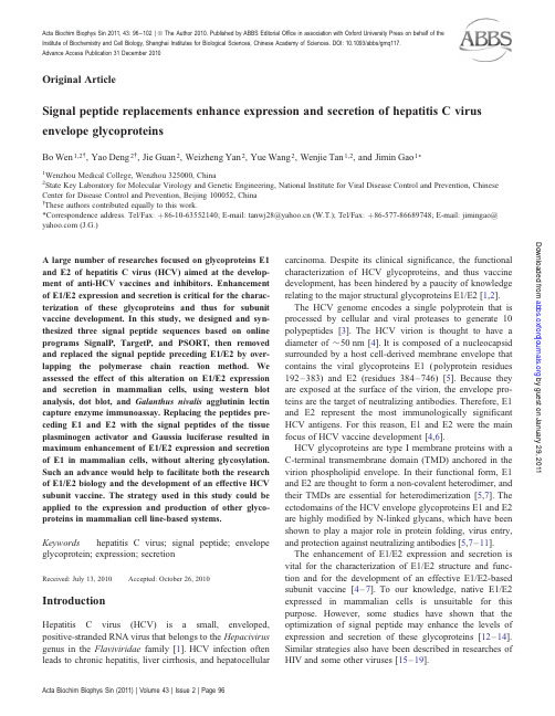
Original ArticleSignal peptide replacements enhance expression and secretion of hepatitis C virus envelope glycoproteinsBo Wen1,2†,Yao Deng2†,Jie Guan2,Weizheng Yan2,Yue Wang2,Wenjie Tan1,2,and Jimin Gao1*1Wenzhou Medical College,Wenzhou325000,China2State Key Laboratory for Molecular Virology and Genetic Engineering,National Institute for Viral Disease Control and Prevention,Chinese Center for Disease Control and Prevention,Beijing100052,China†These authors contributed equally to this work.*Correspondence address.Tel/Fax:þ86-10-63552140;E-mail:tanwj28@(W.T.);Tel/Fax:þ86-577-86689748;E-mail:jimingao@ (J.G.)A large number of researches focused on glycoproteins E1 and E2of hepatitis C virus(HCV)aimed at the develop-ment of anti-HCV vaccines and inhibitors.Enhancement of E1/E2expression and secretion is critical for the charac-terization of these glycoproteins and thus for subunit vaccine development.In this study,we designed and syn-thesized three signal peptide sequences based on online programs SignalP,TargetP,and PSORT,then removed and replaced the signal peptide preceding E1/E2by over-lapping the polymerase chain reaction method.We assessed the effect of this alteration on E1/E2expression and secretion in mammalian cells,using western blot analysis,dot blot,and Galanthus nivalis agglutinin lectin capture enzyme immunoassay.Replacing the peptides pre-ceding E1and E2with the signal peptides of the tissue plasminogen activator and Gaussia luciferase resulted in maximum enhancement of E1/E2expression and secretion of E1in mammalian cells,without altering glycosylation. Such an advance would help to facilitate both the research of E1/E2biology and the development of an effective HCV subunit vaccine.The strategy used in this study could be applied to the expression and production of other glyco-proteins in mammalian cell line-based systems. Keywords hepatitis C virus;signal peptide;envelope glycoprotein;expression;secretionReceived:July13,2010Accepted:October26,2010 IntroductionHepatitis C virus(HCV)is a small,enveloped, positive-stranded RNA virus that belongs to the Hepacivirus genus in the Flaviviridae family[1].HCV infection often leads to chronic hepatitis,liver cirrhosis,and hepatocellular carcinoma.Despite its clinical significance,the functional characterization of HCV glycoproteins,and thus vaccine development,has been hindered by a paucity of knowledge relating to the major structural glycoproteins E1/E2[1,2]. The HCV genome encodes a single polyprotein that is processed by cellular and viral proteases to generate10 polypeptides[3].The HCV virion is thought to have a diameter of 50nm[4].It is composed of a nucleocapsid surrounded by a host cell-derived membrane envelope that contains the viral glycoproteins E1(polyprotein residues 192–383)and E2(residues384–746)[5].Because they are exposed at the surface of the virion,the envelope pro-teins are the target of neutralizing antibodies.Therefore,E1 and E2represent the most immunologically significant HCV antigens.For this reason,E1and E2were the main focus of HCV vaccine development[4,6].HCV glycoproteins are type I membrane proteins with a C-terminal transmembrane domain(TMD)anchored in the virion phospholipid envelope.In their functional form,E1 and E2are thought to form a non-covalent heterodimer,and their TMDs are essential for heterodimerization[5,7].The ectodomains of the HCV envelope glycoproteins E1and E2 are highly modified by N-linked glycans,which have been shown to play a major role in protein folding,virus entry, and protection against neutralizing antibodies[5,7–11]. The enhancement of E1/E2expression and secretion is vital for the characterization of E1/E2structure and func-tion and for the development of an effective E1/E2-based subunit vaccine[4–7].To our knowledge,native E1/E2 expressed in mammalian cells is unsuitable for this purpose.However,some studies have shown that the optimization of signal peptide may enhance the levels of expression and secretion of these glycoproteins[12–14]. Similar strategies also have been described in researches of HIV and some other viruses[15–19].Acta Biochim Biophys Sin2011,43:96–102|ªThe Author2010.Published by ABBS Editorial Office in association with Oxford University Press on behalf of the Institute of Biochemistry and Cell Biology,Shanghai Institutes for Biological Sciences,Chinese Academy of Sciences.DOI:10.1093/abbs/gmq117.Advance Access Publication31December2010Acta Biochim Biophys Sin(2011)|Volume43|Issue2|Page96 by guest on January 29, 2011 Downloaded fromIn this study,we constructed several plasmids encoding E1and E2to assess the effect of the replacement of the signal peptide sequences located upstream of the E1and E2genes.Our results suggested that replacing the signal sequence preceding the E1and E2genes with three signal peptide sequences significantly enhanced expression and secretion of eE1(a secreted form of E1)and eE2(a secreted form of E2)in mammalian cells.Such modifi-cations represent a significant advance in the technique available to investigate the E1and E2structure and their function and will facilitate the development of an HCV entry inhibitor and/or effective vaccine.Materials and MethodsCell culture and plasmid constructionHuman embryo kidney293T cells were grown in Dulbecco’s modified essential medium(Invitrogen, Carlsbad,USA)supplemented with10%fetal bovine serum(FBS)and100U/ml penicillin/streptomycin.Cells were routinely maintained at378C with5%CO2.Wild-type HCV1b E1and E2genes were amplified from the plasmid as described previously[20].To replace their signal peptide regions,the E1and E2genes minus the signal peptide were amplified by polymerase chain reaction(PCR)using long upstream primers.These primers were used to add the signal peptide of self-designed [21,22],tissue plasminogen activator(tPA)[23,24]or Gaussia luciferase(Gluc)[25](Table1),to the E1and E2 genes.Each of these primers contained15nucleotides of the E1or E2sequence spanning the signal peptidase clea-vage site.An Eco RV site and a Bst EII site were added in those primes for clone construction.To facilitate down-stream purification,a Bam HI restriction site and a DNA sequence encoding a His6-tag peptide were synthesized and added to the30end of the E1and E2sequences by PCR (Fig.1).Standard recombinant DNA techniques were used to generate all constructs.After purification,fragments con-taining the desired signal sequence were digested with Eco RV/Bam HI and then cloned into the same sites in the pVRC8301vector.The general scheme of plasmid con-struction is shown in Fig.1and Table2.Cell transfectionTransient transfections were performed by plating2Â105cells/well in a12-well plate12h prior to initiation of the study.When cells were 80–90%confluent,the experiments were performed.On the day of transfection,the original growth medium was replaced with a serum-free medium and DNA transfection was performed using the FUGENE HD Transfection Reagent(Roche,Basel, Switzerland)according to the standard protocols with slight modifications.Briefly,DNA solution containing1m g of pVRC-E1/E2was diluted in50m l of Opti-MEM;and the ratio of transfection reagent:DNA was6:2.Mixtures were incubated at room temperature for15min.Culture medium in12-well plates was removed and thecells were washed with Opti-MEM.A total of900m l of Table1Prediction results for different signal peptidesSignalpeptide(Sp)Sequence and cleavesite prediction aExtracellular rate ink-NN predictionSpþE1(%)SpþE2(%)E1M G C S F S I F L L AL L S C L T T P A SA ...44.4E2M V G N W A K V L IV M L L F A G V D G...55.6Self-design(sd)MD A M K V L L L VF V S P S Q V TG ...66.777.8Gluc M G V K V L F A L IC I A V A E V T G...b66.766.7tPA M D A M K R G L CC V L L L C G A V FV D S V T G ...66.766.7a Protein cleavage sites are indicated by .b The SignalP-NN result is AVA EV,but the SignalP-HMM result isVTG ,and the SIG-Pred result conforms toHMM.Figure1Schematic representation of HCV E1(A)and E2(B)expression vectors Arrows indicate signal peptide cleavage sites.Signal peptide replacements enhance HCV envelope glycoprotein expression and secretionActa Biochim Biophys Sin(2011)|Volume43|Issue2|Page97by guest on January 29, 2011Downloaded fromfresh serum-free Opti-MEM and50m l of transfection mixture were mixed and added to each well.After48h of incubation,the culture medium was collected and cell pellets were separated by centrifugation at1200g for 5min.Pellets were washed with PBS and then lysed in 200m l of lysis buffer for30min on ice and centrifuged at 14,500g for5min;the clarified cell lysate was collected. Clarified lysate and culture medium were used for expression analysis[20].Identical cDNA amount of pVRC was transfected as a control.PNGase F digestion for analysis of E1and E2 glycosylationsCell lysates and culture medium were diluted in sodium dodecyl sulfate–polyacrylamide gel electrophoresis(SDS–PAGE)loading buffer and heated at1008C for10min. PNGase F(NEB)digestion was performed in1ÂG7 buffer,with1%NP-40at378C for1h.Enzyme-treated samples were analyzed by resolving on10–13%SDS–PAGE gels and western blot analysis.Western blot analysisAfter separation by SDS–PAGE(10–13%),proteins were electroblotted onto a nitrocellulose membrane(Whatman, Florham Park,USA).Monoclonal antibodies to E1(A4)or E2(AP33)(GeneTech,Redwood,USA)were used as the primary antibody with appropriate dilution(1:1500).The horseradish peroxidase(HRP)-conjugated AffiniPure goat anti-rabbit immunoglobulin G(IgG;1:5000dilution; Sigma,St Louis,USA)was used as the secondary anti-body.Proteins were revealed on Thermo film in a dark room using the SuperSignal West Pico Chemiluminescent Substrate(Pierce,Rockford,USA)as recommended by the manufacturer.Dot blot assayA nitrocellulose membrane was pre-wetted with PBS and the Dot blot apparatus(Bio-Rad,Hercules,USA)was assembled according to the manufacturer’s introductions. Culture media and diluted cell lysates were added to wells and vacuum was applied to load products onto the mem-brane.Wells were washed twice with200m l of PBS in a low vacuum.The apparatus was disassembled and the membranes were blocked with blocking buffer(PBSþ3% FBS).After extensive washing,the primary antibody, HCV-infected human serum diluted1:200in FBS buffer, was added.Then,the secondary antibody,HRP-conjugated goat anti-mouse IgG(Sigma)diluted1:5000,was added. Galanthus nivalis agglutinin lectin capture EIA Galanthus nivalis agglutinin(GNA)lectin at1m g/ml (Sigma)was used to coat enzyme immunoassay/radio-immunoassay(EIA/RIA)plates(Costar Group,Cambridge, USA)overnight at48C.After being washed with PBS, plates were blocked with5%milk powder,and then super-natants or cell lysates were added for binding at room temperature.Plates were washed with a washing buffer (PBSþ0.05%Tween-20)four times,then100m l of primary antibody monoclonal antibody(MAbs)A4or AP33,dilution at1:1500)was added,followed by incubat-ing for90min at378C.Unbound antibody was washed off with the washing buffer.HRP-conjugated goat anti-mouse IgG(secondary antibody,100m l)was added to each well at1:5000dilution in5%milk powder and incubated for 45min at378C.Finally developed with a tetramethylbenzi-dine substrate and absorbance values at450nm were determined.ResultsReplacement of the sequence encoding the signal peptide of E1and E2The online programs SignalP,TargetP,and PSORT were used to determine cleavage sites and the expression localiz-ation of various signal peptides[21,22].Our goal was to select the peptide that could facilitate higher extracellular expression of the E1and E2proteins compared with the native signal peptide.E1and E2genes were modified to enable a series of translational fusions with different signalTable2Plasmids used in this studyPlasmid Relevant characteristicpVRC8301Efficient expression vector in eukaryotic cellsE1pVRC-E101pVRC8301derivative containing the wild-typeHCV(1b)E1(amino acids170–311)pVRC-E102pVRC-E101derivative with the signal peptidesequence replaced with a self-designed signalsequencepVRC-E103pVRC-E101derivative with the signal peptidesequence replaced by that of tPApVRC-E103pVRC-E101derivative with the signal peptidesequence replaced by that of GlucE2pVRC-E201pVRC8301derivative containing the wild-typeHCV(1b)E2(amino acids364–661)pVRC-E202pVRC-E201derivative with the signal peptidesequence replaced with a self-designed signalsequencepVRC-E203pVRC-E201derivative with the signal peptidesequence replaced by that of tPApVRC-E203pVRC-E201derivative with the signal peptidesequence replaced by that of GlucSignal peptide replacements enhance HCV envelope glycoprotein expression and secretionActa Biochim Biophys Sin(2011)|Volume43|Issue2|Page98 by guest on January 29, 2011 Downloaded frompeptide-encoding regions.An Eco RV site and a Bst EII site were inserted at each end of the signal peptide-encoding region(Fig.1).The Bst EII insertion created a mutation in the C-terminal of each signal peptide,in which the terminal amino acid was replaced with V-T-G(Fig.1).Software analysis indicated that these replacements did not alter the cleavage site of E1/E2(Table1).For expression,the frag-ment obtained by digesting each PCR product with Eco RV/Bam HI was inserted at the same sites in the pVRC8301vector.Plasmids containing the wild-type signal peptide sequence were named pVRC-E101and pVRC-E201;plasmids containing the computer-designed signal peptide sequence were named pVRC-E102and pVRC-E202;plasmids containing the Gluc signal peptide sequence were named pVRC-E103and pVRC-E203;and plasmids containing the tPA signal peptide sequence were named pVRC-E104and pVRC-E204(Table2).In each case,successful insertion was confirmed by sequencing. Effects of signal peptide replacement on intracellular expression of E1and E2The signal peptide sequence of the HCV E1/E2glyco-proteins was substituted with three heterologous signal peptide sequences.Intracellular and extracellular expression levels of E1and E2proteins were assessed using a mono-clonal antibody specific for either E1or pared with the control(pVRC-E101/pVRC-E201),all of the clones (pVRC-E102/pVRC-E202,pVRC-E103/pVRC-E203,and pVRC-E104/pVRC-E204)successfully expressed the E1or E2glycoproteins.HCV E1expressed in untreated cells had an apparent molecular mass of33kDa and the molecular weight(MW)was reduced to18kDa after deglycosylation, and the MW of E2is60–70kDa in nature and31kDa after deglycosylation[3].Our results showed that the MW of each modified signal peptide attached to E1and E2did not differ significantly from the wild type(Fig.2).Furthermore, after digestion with N-glycosidase F,the MWs of E1and E2with the modified signal peptide were not significantly different from that of wild type,suggesting a pattern of identical glycosylation(Fig.3).Native E1glycoprotein was detected with the least expression,despite its expression conditions was identical with others.Only by increasing the loading quantity to 30m g(six times of the loading quantity than that of other samples),the band of native E1glycoprotein can be detected(Fig.2).However,band intensity remained notice-ably lower than any of the modified E1proteins.Thus,pro-duction of E1with a wild-type signal sequence was particularly low,at least less than one-sixth,that of E1 with either the tPA or Gluc signal peptide.Effect of signal peptide replacement on secretionof E1/E2After confirming the intracellular expression of E1/E2with the replaced signal peptide,we assessed the extracellular concentrations of the E1and E2proteins.As predicted by software,the ratios of secretion were different among the various signal peptides.To assess the true expression level of the modified E1and E2glycoproteins,we chose methods that could enrich the non-denature productions for more sen-sitive detection,such as dot blot and GNA capture EIA, both the semi-quantitative methods.They were used to detect glycoproteins E1and E2in all cell lysates and culture media in parallel.Results from these two methods showed no obvious disparities(Fig.4).The expression level of the E1native signal peptide was particularly low,and three of the modified signal peptides,particularly tPA and Gluc, showed significantly increased expression than thenative Figure2Western blot analysis of E1/E2produced by293T transfected with different plasmids Cell lysate samples(L,5m g/sample)and supernatants(S,20m g/sample)were electrophoresed separately.All samples were collected after48h cultivation with non-BSA opti-MEM.The blot was stripped and re-probed with b-actin.Asterisk denotes that the loading volume of this sample was six times greater(30m g)than that of other lysate samples.293T cells transfected without a plasmid was used as a control(mock).Signal peptide replacements enhance HCV envelope glycoprotein expression and secretionActa Biochim Biophys Sin(2011)|Volume43|Issue2|Page99 by guest on January 29, 2011 Downloaded fromsignal peptide.Such distinction was not so obvious in E2,but the expression level of HCV E2glycoprotein with a native signal peptide was still lower than that of E2modified with any of the exogenous signal peptides.Otherwise,the expression level of pVRC-E102/pVRC-E202,whose signal peptide was predicted to have the greatest secretion ability,was lower than the sample with tPA or Gluc signal peptides.Furthermore,modified with the tPA or Gluc signal peptides,neither the expression nor the secretion levels of glyco-proteins showed any marked differences (Fig.4).In our experiment,E2could be more easily detected than E1.This might be caused by different primary antibody,or because E1/E2were glycosylated at different levels leading to dis-tinct GNA-binding ability.DiscussionMore than 120million people worldwide are chronically infected with HCV,making HCV infection to be the leading cause of liver transplantation in developed countries.Treatment options are limited,and the efficacy depends on both the infecting strain and the initial viral load [1].During the translation of HCV,the nascent E1and E2polypeptides are targeted to the host endoplasmic reticulum membrane for modification by N-linked glycosylation [5].E1and E2are released from the polyprotein through cleavage by a host signal peptidase [3]and are anchored in the viral lipid envel-ope as a heterodimer,which plays a major role in the HCV entry [7,11].Deletion of the TMDs of E1and E2results in the secretion of these truncated forms of HCV glycoproteins into the extracellular medium.E2antigen in itssecretedFigure 4Analysis of the production levels of E1and E2glycoproteins containing altered signal peptides Human embryo kidney 293T cells contained the following plasmids were indicated,and 293T cells transfected without a plasmid was used as a control (mock).Cell lysates (L)and supernatants (S)were collected 48h after transfection.(A)and (B)represent the dot blot results of E1and E2glycoproteins detected using HCV-infected human serum as the primary antibody.GNA lectin capture EIA data are shown in (C)and (D).Data obtained from both the dot blot and EIA methods showed no markeddiscrepancies.Figure 3Different signal peptide have no apparently effect on the expression of E1and E2in 293T cells 293T cells contain pVRC-E101/104and pVRC-E201/204plasmids were collected 48h after transfection.Cell lysate (L)and supernatant (S)of each samples were treated with PNGase F (W)or left untreated (W/O).After being separated by SDS–PAGE (E1in 13%and E2in 10%acrylamide)under reducing conditions,E1and E2were immunoprecipitated with monoclonal antibodies A4and AP33.(A)In the same loading quantity (20m g),bands detected from E101(arrow indict)were much fainter than those of E104,but their distributions were the same under the similar treatment.(B)The distinction of band grayscale between E201and E204is not obvious,but the MW and distribution of their bands are identical under the same treatment.Signal peptide replacements enhance HCV envelope glycoprotein expression and secretionActa Biochim Biophys Sin (2011)|Volume 43|Issue 2|Page 100by guest on January 29, 2011 Downloaded fromform leads to an increased humoral response in mice[26]. HCV E1and E2are primary determinants of entry and pathogenicity.HCV E2glycoproteins are involved in recep-tor binding,virus–cell fusion,and entry into host cells[11]. However,its role in membrane fusion and immune evasion remains uncharacterized.The function of E1is unknown; however,it is a target for neutralizing antibodies and its association with E2is essential for viral entry[11]. Increasing knowledge of the nature and the function of HCV E1and E2glycoproteins help to develop antiviral drugs and vaccine candidates.The production of large quantities of functional and secreted E1and E2proteins will allow us to perform compre-hensive biochemical and biophysical analysis,which will lead to the development of a vaccine that is effective against HCV infection.In this study,we developed a novel expression system for producing the secreted form of E1(eE1)and E2 (eE2)ectodomains from mammalian cells via a signal peptide sequence replacement strategy and performed a comprehen-sive biochemical characterization.These data enhance our understanding of HCV envelope glycoproteins and may assist in the design of HCV vaccines and entry inhibitors. Our data showed that the replacement of the signal peptide sequences located at the upstream of E1and E2 genes altered their secretion and expression levels,and most importantly,the replacement did not affect glycosyla-tion.Furthermore,replacing them with tPA and Gluc resulted in the greatest increases at expression and secretion levels of eE1and eE2in mammalian cells.We believed that the strategy used in this study could be applied to the expression and production of other glycoproteins in mam-malian cell line-based systems.The next challenge is to use our novel strategy to gener-ate large quantities of HCV E1and E2envelope glyco-proteins.This study will allow us to elucidate their structural biology and the mechanism(s)involved in the interactions between these glycoproteins and other cellular factors,eventually to facilitate the development of an HCV vaccine and/or entry inhibitor. AcknowledgementsThe authors thank Dr Gary Nabel(Vaccine Research Center,National Institute of Allergy and Infectious Diseases,National Institutes of Health,Bethesda,USA)for the pVRC plasmid and Dr Jean Dubuisson(Universite´Lille Nord de France,CNRS-UMR8161,Institut Pasteur de Lille,Lille,France)for the A4antibody.FundingThis work was supported by the grants from the Hi-Tech Research and Development Program of China(2007AA02Z455,2007AA02Z157)and the State KeyLaboratory for Molecular Virology and Genetic Engineering. References1Lindenbach BD,Thiel HJ and Rice CM.Flaviviridae:the viruses and theirreplication.In:Knipe DM and Howley PM eds.Fields Virology.Philadelphia,PA:Lippincott Williams&Wilkins,2007,1101–1152.2Houghton M and Abrignani S.Prospects for a vaccine against the hepatitisC virus.Nature2005,436:961–966.3Dubuisson J.Hepatitis C virus proteins.World J Gastroenterol2007,13:2406–2415.4Wakita T,Pietschmann T,Kato T,Date T,Miyamoto M,Zhao Z andMurthy K,et al.Production of infectious hepatitis C virus in tissue culturefrom a cloned viral genome.Nat Med2005,11:791–796.5Lavie M,Goffard A and Dubuisson J.Assembly of a functional HCV gly-coprotein heterodimer.Curr Issues Mol Biol2007,9:71–86.6Stamataki Z,Coates S,Evans MJ,Wininger M,Crawford K,Dong C andFong YL,et al.Hepatitis C virus envelope glycoprotein immunization ofrodents elicits cross-reactive neutralizing antibodies.Vaccine2007,25:7773–7784.7Ciczora Y,Callens N,Penin F,Pecheur EI and Dubuisson J.Transmembrane domains of hepatitis C virus envelope glycoproteins:resi-dues involved in E1/E2heterodimerization and involvement of thesedomains in virus entry.J Virol2007,81:2372–2381.8Drummer HE,Maerz A and Poumbourios P.Cell surface expression offunctional hepatitis C virus E1and E2glycoproteins.FEBS Lett2003,546:385–390.9Falkowska E,Kajumo F,Garcia E,Reinus J and Dragic T.Hepatitis Cvirus envelope glycoprotein E2glycans modulate entry,CD81binding,and neutralization.J Virol2007,81:8072–8079.10Helle F,Goffard A,Morel V,Duverlie G,McKeating J,Keck ZY and Foung S,et al.The neutralizing activity of anti-hepatitis C virus antibodiesis modulated by specific glycans on the E2envelope protein.J Virol2007,81:8101–8111.11Cocquerel L,Voisset C and Dubuisson J.Hepatitis C virus entry:potential receptors and their biological functions.J Gen Virol2006,87:1075–1085.12Martoglio B and Dobberstein B.Signal sequences:more than just greasy peptides.Trends Cell Biol1998,8:410–415.13Futatsumori-Sugai M and Tsumoto K.Signal peptide design for improving recombinant protein secretion in the baculovirus expression vector system.Biochem Biophys Res Commun2010,391:931–935.14Zhang L,Leng Q and Mixson AJ.Alteration in the IL-2signal peptide affects secretion of proteins in vitro and in vivo.J Gene Med2005,7:354–365.15Li Y,Luo L,Thomas DY and Kang CY.The HIV-1Env protein signal sequence retards its cleavage and down-regulates the glycoprotein folding.Virology2000,272:417–428.16Bruno M,Roland G and Bernhard D.Signal peptide fragments of prepro-lactin and HIV-1p-gp160interact with calmodulin.EMBO J1997,16:6636–6645.17Robert E,Oliver L,Thomas S,Markus E,Hans-Dieter K and WolfgangG.Identification of Lassa virus glycoprotein signal peptide as a trans-acting maturation factor.EMBO Rep2003,4:1084–1088.18Lobigs M,Zhao HX and Garoff H.Function of Semliki forest virus E3 peptide in virus assembly:replacement of E3with an artificial signalpeptide abolishes spike heterodimerization and surface expression of E1.J Virol1990,64:4346–4355.19Paul AR,Mark LH,Toni W,Helen LR and Alan P.Antigenic and genetic characterization of the haemagglutinins of recent cocirculating strains ofinfluenza B virus K.J General Virol1992,73:2737–2742.Signal peptide replacements enhance HCV envelope glycoprotein expression and secretionActa Biochim Biophys Sin(2011)|Volume43|Issue2|Page101by guest on January 29, 2011Downloaded from20Bian T,Zhou Y,Bi S,Tan W and Wang Y.HCV envelope protein func-tion is dependent on the peptides preceding the glycoproteins.Biochem Biophys Res Commun2009,378:118–122.21Nakai K and Horton P.PSORT:a program for detecting sorting signals in proteins and predicting their subcellular localization.Trends Biochem Sci 1999,24:34–36.22Emanuelsson O,Brunak S,von Heijne G and Nielsen H.Locating proteins in the cell using TargetP,SignalP and related tools.Nat Protoc2007,2: 953–971.23Pennica D,Holmes WE,Kohr WJ,Harkins RN,Vehar GA,Ward CA and Bennett WF,et al.Cloning and expression of human tissue-type plasmino-gen activator cDNA in E.coli.Nature1983,301:214–221.24Qiu JT,Liu B,Tian C,Pavlakis GN and Yu XF.Enhancement of primary and secondary cellular immune responses against human immunodefi-ciency virus type1gag by using DNA expression vectors that target Gag antigen to the secretory pathway.J Virol2000,74:5997–6005.25Knappskog S,Ravneberg H,Gjerdrum C,Tro¨sse C,Stern B and Pryme IF.The level of synthesis and secretion of Gaussia princeps luciferase in transfected CHO cells is heavily dependent on the choice of signal peptide.J Biotechnol2007,128:705–715.26Abraham JD,Himoudi N,Kien F,Berland JL,Codran A,Bartosch B and Baumert T,et parative immunogenicity analysis of modified vacci-nia Ankara vectors expressing native or modified forms of hepatitis C virus E1and E2glycoproteins.Vaccine2004,28:3917–3928.Signal peptide replacements enhance HCV envelope glycoprotein expression and secretionActa Biochim Biophys Sin(2011)|Volume43|Issue2|Page102 by guest on January 29, 2011 Downloaded from。
哺乳动物细胞全蛋白提取试剂盒说明书
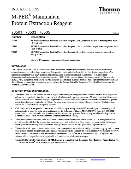
INSTRUCTIONSM-PER ®Mammalian Protein Extraction ReagentNumber Description78503 M-PER Mammalian Protein Extraction Reagent , 25mL, sufficient reagent to extract protein from ~2.5g of cells78501 M-PER Mammalian Protein Extraction Reagent , 250mL, sufficient reagent to extract protein from ~25g of cells78505M-PER Mammalian Protein Extraction Reagent , 1L, sufficient reagent to extract protein from ~100g of cellsStorage: Upon receipt store product at room temperature.IntroductionThe Thermo Scientific M-PER Mammalian Protein Extraction Reagent extracts cytoplasmic and nuclear protein from cultured mammalian cells using a proprietary detergent in 25mM bicine buffer (pH 7.6). The simple composition of this reagent is compatible with many different applications, such as reporter assays (e.g., luciferase, β-galactosidase,chloramphenicol acetyltransferase), protein assays (e.g., PKA, PKC, tyrosine kinase), immunoassays (e.g., Western blot, ELISA, RIA) and protein purification. M-PER Reagent enables rapid, mild and efficient lysis. The reagent is dialyzable and the cell lysate is compatible with protein assays such as the Thermo Scientific Coomassie Plus (Bradford) Assay and the Pierce BCA Protein Assay.Important Product Information•Adherent Cells vs. Cell Pellets: M-PER Reagent effectively lyses both plated cells and cells pelleted from suspension cultures or scraped cells. For direct, in-plate lysis of adherent cells, protein extraction efficiency using M-PER Reagent is similar to freeze/thaw methods. For lysis of pelleted cells, either from cell suspension or scraped adherent cells, protein extraction efficiency is typically 25% higher than that achieved with freeze-thaw (three cycles) and 20% higher than sonication (2 minutes with 50% pulse) methods.•Cell Lines: M-PER Reagent has been tested on cell lines representing several different cell types. Complete lysis of adherent cells is observed with, but is not limited to, the following cell lines: COS-7, NIH3T3, Hepa 1-6, 293, CHO, MDA, MB 231 and FM2 cells. For protein extraction from tissues, greater efficiency may be achieved using Thermo Scientific T-PER Tissue Protein Extraction Reagent (Product No. 78510).•Additives: Protease inhibitors, such as Thermo Scientific Halt Protease Inhibitor Cocktail, EDTA-Free (Product No. 87785) may be added to the reagent. For immunoassays, such as ELISA or RIA, extracts prepared in M-PER Reagent alone generate satisfactory results; however, adding 150mM NaCl to the cell lysate often improves results.•Volume for Cell Lysis: Volumes indicated in Table 1 are optimal for maximum cell lysis without scraping cells. If more concentrated extracts are preferred, use a smaller volume; however, scraping the cells is necessary for maximal recovery. If cell volume is unknown, it may be estimated. For example, 2 × 10⁶ of HeLa cells equals ~10μL of a packed cell volume, which is equivalent to 20 mg of cells and requires 200 μl of M-PER Reagent.• Compatibility with Protein Assays: M-PER Reagent is compatible with Coomassie Plus (Bradford) Assay (Protein No. 23236) and the Pierce ® BCA Protein Assay Kit (Product No 23225).Procedure for Lysis of Monolayer-cultured Mammalian CellsNote: M-PER Reagent does not contain protease inhibitors. If desired, add Halt™ Protease Inhibitor Cocktail, EDTA-Free (Product No. 87785) to the reagent.1.Carefully remove (decant) culture medium from adherent cells.Note: If the culture medium contained phenol red or other reagents that could interfere with subsequent protein analysis,wash cells once in wash buffer (e.g., PBS).2.Add the appropriate amount of M-PER Reagent to the plate or to each plate well (see Table 1). Shake gently for5 minutes.Table 1. Suggested volume of Thermo Scientific M-PER Reagent to use for different sizes ofstandard culture plates.Plate Size/Surface Area M-PER Reagent Volume100mm* 500-1,000μL60mm 250-500µL 6-well plate 200-400µL per well24-well plate 100-200µL per well96-well plate 50-100µL per well*Cells grown in 100mm plates typically contain 10 cells (50mg) and yield ~3mg total protein depending oncell type.3.Collect the lysate and transfer to a microcentrifuge tube. Centrifuge samples at ~14,000 ×g for 5-10 minutes to pellet thecell debris.4.Transfer the supernatant to a new tube for analysis.Procedure for Lysis of Suspension-cultured Mammalian Cells1.Pellet the suspension of cells by centrifugation at 2,500 ×g for 10 minutes. Discard the supernatant.2.Optional Wash: If the culture medium contained phenol red or other reagents that could interfere with subsequent proteinanalysis, wash the cells once by resuspending the cell pellet in wash buffer (e.g., PBS). Pellet cells by centrifugation at2,500 ×g for 10 minutes.3.Add M-PER Reagent to the cell pellet. Use at least 1mL of M-PER Reagent for each 100 mg (~100μL) of wet cell pellet.If a large amount of cells is used, first add 1/10 the final recommended volume of M-PER Reagent to the cell pellet.Pipette the mixture up and down to resuspend pellet. Add the rest of the M-PER Reagent to the cell suspension.Note: Total protein yield for 100mg of wet cell pellet is approximately 6 mg depending on cell type.4.Shake mixture gently for 10 minutes. Remove cell debris by centrifugation at ~14,000 ×g for 15 minutes.5.Transfer the supernatant to a new tube for analysis.TroubleshootingProblem PossibleCause SolutionProtein expression was low Optimize the transfection procedureInsufficient amount of M-PER Reagent was used Add more M-PER ReagentLow protein yieldM-PER Reagent was unable to penetrate the cell membrane Increase incubation time and shake more vigorously during incubationUnable to retrieve membrane protein M-PER Reagent extracts only nuclear andcytoplasmic proteinsUse Thermo Scientific Mem-PER MembraneProtein Extraction Reagent (Product No.89826)Related Thermo Scientific Products87785 Halt Protease Inhibitor Cocktail, EDTA-Free (100X), 1mL87786 Halt Protease Inhibitor Cocktail, contains sufficient reagents to treat 100mL of sample78248 B-PER® Bacterial Protein Extraction Reagent, 500mL78990 Y-PER® Yeast Protein Extraction Reagent, 500mLProtein Extraction Reagent Kit89826 Mem-PERMembrane23236 Coomassie Plus (Bradford) Assay23227 Pierce BCA Protein Assay Kit78833 NE-PER® Nuclear and Cytoplasmic Extraction Kit26148 Pierce Direct IP Kit34080 SuperSignal® West Pico Chemiluminescent Substrate, 500mL, Western blot substrate for HRP 34076 SuperSignal West Dura Extended Duration Substrate, 200mL, Western blot substrate for HRP Product ReferencesCampa, M.J., et al. (2003). Protein expression profiling identifies macrophage migration inhibitory factor and cyclophilin A as potential molecular targets in non-small cell lung cancer. Cancer Res63:1652-6.Deng, W., et al. (2003). LPA protects intestinal epithelial cells from apoptosis by inhibiting the mitochondrial pathway. Amer J Physiol-Gastrointest L 284:821-9.Phiel, C.J., et al. (2001). Differential binding of an SRF/NK-2/MEF2 transcription factor complex in normal versus neoplastic smooth muscle tissues. Biol Chem.276(37):34637-50.Waite, K.A. and Eng, C. (2003). BMP2 exposure results in decreased PTEN protein degradation and increased PTEN levels. Hum Mol Genet12(6):679-84. B-PER® Technology is protected by U.S. Patent # 6,174,704.SuperSignal® Technology is protected by U.S. Patent #6,432,662.This product (“Product”) is warranted to operate or perform substantially in conformance with published Product specifications in effect at the time of sale, as set forth in the Product documentation, specifications and/or accompanying package inserts (“Documentation”) and to be free from defects in material and workmanship. Unless otherwise expressly authorized in writing, Products are supplied for research use only. No claim of suitability for use in applications regulated by FDA is made. The warranty provided herein is valid only when used by properly trained individuals. Unless otherwise stated in the Documentation, this warranty is limited to one year from date of shipment when the Product is subjected to normal, proper and intended usage. This warranty does not extend to anyone other than the original purchaser of the Product (“Buyer”).No other warranties, express or implied, are granted, including without limitation, implied warranties of merchantability, fitness for any particular purpose, or non infringement. Buyer’s exclusive remedy for non-conforming Products during the warranty period is limited to replacement of or refund for the non-conforming Product(s).There is no obligation to replace Products as the result of (i) accident, disaster or event of force majeure, (ii) misuse, fault or negligence of or by Buyer, (iii) use of the Products in a manner for which they were not designed, or (iv) improper storage and handling of the Products.Current versions of product instructions are available at /pierce. For a faxed copy, call 800-874-3723 or contact your local distributor.© 2010 Thermo Fisher Scientific Inc. All rights reserved. Unless otherwise indicated, all trademarks are property of Thermo Fisher Scientific Inc. and its subsidiaries. Printed in the USA.。
【doc】Skp2参与HeLa细胞G2/M周期检查点的调控

Skp2参与HeLa细胞G2/M周期检查点的调控ISSNl007—7626CN11-3870/Q中国生物化学与分子生物ChineseJournalofBiochemistryandMolecularBio——l2005年l0月21(5):575~580研究快报?Skp2参与HeLa细胞G2/M周期检查点的调控钱俊杰,孙国敬,孟祥兵,宋宜,梅柱中,刘斌,董燕,孙志贤(军事医学科学院放射医学研究所,北京100850)摘要具癌基因特性的Skp2在大多数肿瘤组织和肿瘤细胞中异常高表达,它作为scF’s啦复合物的底物识别亚基调控p27蛋白的稳定性而促进细胞G./S期转换.为进一步明确Skp2与G:/M周期检查点的关系,在HeLa细胞中过表达Skp2以及通过反义寡核苷酸抑制Skp2表达.结果发现:Skp2能促进细胞周期运转,表现为s期细胞增多和G,/M期细胞减少,其中F—box结构域具有重要的功能意义;反义寡核苷酸抑制Skp2表达后,HeLa细胞发生显着的G:/M 期阻滞;MTr检测结果表明,400nmol/L的Skp2的反义寡核苷酸能明显抑制HeLa细胞的增殖活性;Western印迹结果表明,HeLa细胞中Skp2可能通过负调控p21的稳定性来参与G:/M检查点调控,这在用放线菌素D处理HeLa细胞的实验中得到验证.这些结果初步揭示了Skp2参与HeLa 细胞G:/M周期检查点调控的分子机制.关键词Skp2,G,/M周期检查点,p21WA中图分类号Q55EffectofSkp2onG2/MCheckpointofHeLaCellsQIANJun—Jie,SUNGuo—Jing,MENGXiang—Bing,SONGYi,MEIZhu —Zhong,LIUBin,DONGYan,SUNZhi-Xian(Beltingm啪ofRadiationMedcine,Beijing100850,China) AbstractOverexpressionofoncogenicSkp2hasbeenobservedinvaioustype sofhumantumortissuesandcelllines.Skp2positivelyregulatestheGl-StransitionasamemberoftheF—b oxfamilyofsubstrate-regulation subunitsofSCFubiquitin—proteinligasecomplexes.TodeterminethefuaherrelationshipbetweenSkp2andtheG2/Mcheckpoint,ectopicexpressionofSkp2orinhibitionofSkp2expressio nbyantisenseoligodeoxynucleotideswasperformed,Ourresultsindicatedthatoverexpres sionofSkp2maystimulate progressionofcellcycleofHeLacellsdependentofitsF—boxstructure,chara cterizedbyincreaseintheSpopulationanddecreaseinG2/Mpopulation.Skp2一antisensetreatmentnotonlyinhibitsproliferationofHeLa cellsasdeterminedbyMTr,butalSOinducesG,/MarrestdeterminedbyFCM. TheresultbyWesternblottingshowsp21,animportantG2/Mcheckpointregulator,invovledincellcyclepro gressionasaneffectorofSkp2.Furthermore,theinverserelationshipbetweenSkp2andp21Wasconsistantw iththeresultfromHeLacellstreatedwithactinomycinD.Thisstudywillbehelpfultorevealmolecularmec hanismsofSkp2regulationG2/McheckpointinHeLacells.KeywordsSkp2,G2/Mcheckpoint,p21Skp2(S-phasekinase—associatedprotein2)是1995年Zhang等最初发现的细胞周期中s期激酶相关蛋白,它在多种实体瘤和白血病细胞中异常高表达,表现为癌基因特性.后来研究表明,细胞中Skp2可作为泛素一蛋白酶体通路(ubiquitinproteasomepathway)中E3连接酶一SCF复合物(Skp—cullin.F—box proteincomplex)的底物识别亚基发挥其功能.作为与收稿13期:2005.04—04,接收13期:2005—05—27国家自然科学基金资助项目:(No.30100223;No.30170291)联系人Tel:010—66932212,E—mail:************* Received:April4,2005;Accepted:Mav27,2005 SupposedbyNationalNaturalScienceFoundationofChina(No.30100223 andNo.3O170291),CorrespondingauthorTel:Tel:010.66932212E—mail:Su调控,并与肿瘤的发生,发展关系密切.研究发现,细胞内Skp2的表达水平直接反映细胞的增殖状态,肺小细胞癌细胞中SKP2基因在染色体水平上表现为高度扩增,导致细胞中高水平的Skp2蛋白表达,而使细胞恶性增殖;相反,借助Skp2的反义寡核苷酸抑制Skp2的表达,则能诱导肺小细胞癌细胞的周期阻滞或凋亡’.以往研究表明,作为SCFs复合物中的底物识别亚基,Skp2识别磷酸化的p27,使其通过泛素.蛋白酶体通路降解,从而释放了周期素依赖激酶cyclin细胞/M检查点调控,从而导致细胞失去G2/M检查点的监控作用而使细胞恶性增殖.本文旨在通过HeLa细胞中过表达Skp2及其F.box缺失突变体或抑制Skp2的表达, 用流式细胞术检测Skp2的表达水平与细胞周期的关系,Western印迹分析相关周期检查点蛋白的表达水平.研究证明,HeLa细胞中高表达的Skp2可能通过调节p21的稳定性参与细胞中/M检查点调控,这在放线菌素D处理的HeLa细胞实验中得到验证.这些研究结果初步揭示了Skp2参与肿瘤细胞G2/M周期检查点调控的分子机制.1材料与方法1.1材料1.1.1细胞系和菌株HeLa细胞和E.coliDH5a菌株均由本实验室保存.1.1.2试剂RPMl1640,DMEM培养基购自Gibco公司;Skp2 (H.435),p21(C.19),p27(C.19)抗体购自Santa公司; Myc抗体购自Clontech公司;Lipofectin和Lipofectamine2000购自Invitrogen公司;BCAProtein AssayReagentKit和SupersignalWestPico ChemiluminescentSubstrate购自Pierce公司;proteaseinhibitorcocktailtablets购自Roche公司;其它试剂均为国产分析纯试剂.1.2方法1.2.1skp2F.box突变体及plRES.GFP-Skp2载体的构建设计PCR引物(skp2上游5’-CGGAA TI’CCTA TGCACAGGAAGCACCTC.3;skp2下游:GCTCGA GCTA TAGACAACTGGGCTIT一3),以pcDNA3.1-Skp2 和pcDNA3.1.Skp2AF(LifangLu博士惠赠)¨们为模板,扩增后酶切,构人pCMV.Myc和pIRES.hrG载体.Skp2AF为skp2的F.box缺失突变体,缺失的是skp2mRNA的289~480之间的132个碱基序列.质粒转染细胞所用转染试剂为Lipofectamine2000.具体方法见说明书.1.2.22反义寡核苷酸(AS)的设计及合成2反义寡核苷酸是针对人2mRNA起始密码子后180~196bp的碱基序列,设计与其互补的l7个碱基寡聚物,TGGGGGA TGTTCTCA.3 (forSkp2cDNA180~196);无义寡核苷酸:5.CGGGCA-3(controloligonucleotides),下划线为硫代修饰,由上海生工生物公司合成.反义寡核苷酸用Lipofectin转染试剂转染HeLa细胞,转染方法见说明书.1.2.3细胞增殖的检测MTT法检测细胞活性.计算细胞数,并调整细胞浓度为1×10个/ml,按5000个细胞/;E接种96孔板,培养24h.转染的反义寡核苷酸AS浓度分别为200,400,600,800nmol/L,在0.2#l/;fLLipofectin介导下转染后48h,加20lMTT,37.I=,5%CO培养箱中放置4h,每孔加入200l二甲基亚砜(DMSO),充分溶解.选择492nm波长,在酶联免疫检测仪上测各孔的吸光值,计算增殖抑制率.1.2.4细胞周期变化的检测流式细胞仪检测细胞周期的变化.取对数生长的HeLa细胞,转染有GFP,skp2.GFP和Skp2AF.GFP的HeLa细胞,37~C,5%Co2培养24h后,胰酶消化,先用1%甲醛/PBS溶液于4.I=固定10min,离心后加第5期钱俊杰等:Skp2参与HeLa细胞G2/M周期检查点的调控577 人300l5%小牛血清PBS后,加入700l无水乙醇,于一20~C固定过夜,加503l含500t~g/ml的RNaseA的磺化丙啶溶液,37~C温育30min后上机,检测时以GFP为分选标记,即分析GFP表达阳性细胞的周期变化.转染有200,400,6O0nmol/LAS的HeLa细胞周期测定方法基本同上,但省去1%甲醛固定步骤.1.2.5Western印迹收集转染有AS或.s2及.s2AF的HeLa细胞,裂解,蛋白定量后,SDS—PAGE,转移至硝酸纤维素膜,5%脱脂奶粉封闭过夜,一抗Skp2,Myc和肌动蛋白稀释倍数为1:1000,p21和p27为1:500,室温温育2h;二抗稀释倍数均为1:1000,室浊孵育1h,曝光显色.1.2.6Northern印迹分析用Trizo1分离经0,5,10,25ng/ml放线菌素D处理16h的HeLa细胞mRNA,定量后,用含甲醛的1% 琼脂糖凝胶电泳分离,转移至尼龙膜,与a一弛P标记的p21基因cDNA探针杂交.同一张膜再与I3肌动蛋白探针杂交,作mRNA上样量的对照,杂交后x光片放射自显影,分析结果.2结果2.1plRES—GFP-Skp2和pCMV-Myc-Skp2及其突变体的真核表达载体的构建.s切2AF是skp2缺失F—box结构的突变体,即缺失.s2的cDNA的289~420位碱基,编码44个氨基酸,失去了结合S切1的能力.以pcDNA3.1一Skp2 和pcDNA3.1一.s2AF为模板,高保真酶扩增.s2和.s2AF,引入EcoRI和XhoI酶切位点,构建到真核表达载体pIRES—hrG和pCMV.Myc中,酶切鉴定得到1.3kb.s2和1.2kbskp2AF片段,测序结果表明序列和读码框正确(Fig.1).2.2过表达的Skp2促进HeLa细胞周期转换将plRES—GFP,plRES.GFP-Skp2昶plRES—GFP—S如2AF用Lipofectamine2000转染HeLa细胞,24h后,收集细胞固定,PI染色,流式细胞仪检测结果表明:在所有的GFP阳性细胞中,表达Skp2AF的细胞和转染pIRES—GFP空载体以及GFP阴性细胞相比Fig.1IdentificationofplRES?GFP-Skp2(A)andpCMV?Myc-S~2(B) byrestrictiondigestion(A)1:pIRES?GFP-Skp2/EcoRI+XhoI;2:pIRES?GFP-Skp2AF/ EcoRI+XhoI:3:plRES.GFP/EcoRI+XhoI:4:DNAmarker;(B)1:pCMV?Myc-Skp2AFIEcoRI+XhoI;2:pCMV?Myc-Skp2/EcoR I+XhoI:3:pCMV.HA/EcoRI+XhoI:4:DNAmarker较,细胞周期没有变化;然而,过表达全长Skp2的HeLa细胞中,G2/M期细胞明显减少,s期细胞比例明显升高达20%,说明过表达的Skp2促进细胞通过G2/M检查点,促进细胞由G.期向s期的转变,同时,Skp2的F—box结构具有重要的功能意义(Table1).Table1AnalysisofcellcyclesofHeLacellstransfectedwithplRES-GFP.pIRES?GFP-Skp2andpIRES.GFP-S~2AFbyFCM(%,±s).P<0.01?comparedwithexpressionofGFPorGFPSkp2AF2.3Skp2的反义寡核苷酸诱导HeLa细胞G:/M期阻滞HeLa细胞转染.s2的反义寡核苷酸As(control,对照反义寡核苷酸),转染浓度分别为200,400,600nmol/L,转染48h后,流式细胞仪检测其周期变化,结果表明:As抑制Skp2表达后,细胞有明显的G:/M期阻滞,并表现为浓度依赖性,600nmol/L的As诱导的G:/M期阻滞最明显,能达到80%多,对照寡核苷酸在同样浓度梯度下对细胞周期影响不大.这一结果表明,Skp2的表达参与调控HeLa细胞的G2/M周期检查点(Table2).Table2AnalysisofcellcyclesofHeI.acellstransfeetedwithantisenseoligonu eleotidesagainstSkv2byFCM(%,;±s)P<O.01,comparedwithcontrololigonucleotides.‘Concentrationofantisens eoligonueleotide姗抛m∞∞∞21752578中国生物化学与分子生物2l卷2.4Skp2的反义寡核苷酸抑制HeLa细胞的增殖200,400,600,800nmol/LSkp2的反义寡核苷酸(AS)和对照寡核苷酸转染HeLa细胞,用MTT法检测HeLa细胞的增殖活性,结果表明:AS能显着抑制HeLa细胞的增殖,抑制率达到28%,并且有良好的浓度依赖性;同时,同样浓度的无义寡核苷酸,对HeLa细胞的增殖活性影响不大,这说明,AS抑制细胞增殖的活性是特异性的,跟反义寡核苷酸及转染试剂的毒性无关,这一结果同样证实了Skp2的表达能促进细胞周期的运转(Fig.2).-—一一▲/产/.:r/一————r200400600800c/nmolL-Fig.2Inhibitoryratesofantisense olignueleotidesagainsts2inHeLacellsMyc姒I——国一lMyc—Skp2AFl粕黼lp2lp27A~tin2.5HeLa细胞中Skp2主要调控p21的降解来参与G2/M期调控HeLa细胞转染全长的.s2和突变的SkD2AF, Western印迹结果表明:过表达全长skp2后细胞中p21蛋白水平下降,而p27表达水平变化不明显;转染空载体和缺失F—box的突变体后,细胞中p21水平没有变化(Fig.3A),所以说,Skp2负调控p21的稳定性同样需要Fbox结构域;反义寡核苷酸抑制Skp2的表达后,Western印迹结果表明:对应的p21蛋白水平升高,而p27表达水平没有变化(Fig.3B).Skp2过表达时,p21表达水平降低,细胞顺利通过G,/M检查点;抑制表达时p21表达水平升高,细胞有明显的G/M期阻滞,故Skp2可能通过调节周期素依赖激酶抑制因子一p21的稳定性变化,来调控HeLa细胞的G/M检查点.2.6放线菌素D抑制肿瘤细胞增殖过程中.Skp2负调控p21的稳定性放线菌素D是RNA合成抑制剂,通过抑制RNASkp2p21p27ActinASl23COiltrOl23Fig?3Theendogenousp2landp27proteinlevelinHeLacellstransfectedwith Skp2,Skp2AF(A)orantisenseoligonucleotidesagainstSkp2(B)(A)1:LysatesofHeLacells,2:LysatesHeLacellstransfectedwithemptyveet or;3:LysatesofHeLacelltransfectedwithpCMV—Myc-Skp2AF;4—6:LysatesofHeLacellstransfeetedwithpCMV—Myc-Skp2for24.48or 72hours.(B)LysatesofHeLacellstranfeetedwithantisenseoligonueleotidesagainstS kp2(AS)orControloligonucleotides(contro1)1:200nmol/L;2:4OOnmol/L;3:6OOnmol/L(A)AD(n~m1)051025E三二[二E三三AD(ng/m1)051025[]MmE二]Fig?4Negativeregulationofp2lbySkp2afteraetinomyeinD(AD)treatment (A)WesternblotanalysisoflevelsofSkp2andp21proteininHeLacellstreated withAD(B)Northemblotanalysisofp21mRNAlevelsinHeLacellstreatedwithAD第5期钱俊杰等:Skp2参与HeLa细胞G2/M周期检查点的调控579 聚合酶Ⅲ的活性影响蛋白合成,抑制细胞增殖和诱导细胞凋亡.不同浓度的放线菌素D处理HeLa细胞16h后,Western印迹检测结果表明:随着放线菌素D浓度的升高,Skp2表达水平逐渐降低,而对应的p21的表达水平逐渐升高(Fig.4A);Northem印迹检测结果表明,随着放线菌素D浓度的升高,p21的mRNA水平略有变化,但此时的p21蛋白表达水平显着升高(Fig.4B),这表明,放线菌素D抑制HeLa细胞增殖过程中,p21表达水平的变化体现在其稳定性的升高,有可能是Skp2负调控的结果.3讨论癌基因Skp2在肿瘤细胞中异常高的表达,它是作为泛素化蛋白酶降解途径的E3s酶一sc复合物的底物识别亚基来参与细胞周期调控的.大量肿瘤组织的免疫组化实验结果表明:Skp2负调控p27,下调的p27使细胞顺利通过R限制点,完成G.一s期的转变,同时,Nakayama等最新研究认为,Skp2调控p27的稳定性同样参与细胞G,一M期的调控.但细胞中p27并不是SCF复合物的唯一底物],周期检查点蛋白中的另外一个重要分子一p21同样是sC复合物的底物,它是细胞中G,/M期检查点关键的负调控因子,我们以前的研究结果也表明,Skp2 的表达能够调控p21的稳定性,所以我们认为:sC复合物是否能同样通过调节p21的稳定性参与细胞G:/M检查点的调控,而肿瘤组织中高表达的Skp2将导致p21的异常降解,从而使细胞丧失这一周期检查点,促进了细胞的恶性增殖.本实验结果证明了Skp2的表达与HeLa细胞G,/M检查点之间的相关性.Skp2AF是Skp2的F—box缺失突变体,丧失了结合SCF复合物中其它亚基的能力,所以没有生物学功能.全长的Skp2能使细胞顺利通过G:/M期,促进G.一s转变,向Skp2AF对细胞周期没有影响,这表明Skp2行使功能是通过SCF复合物来实现的,并且依赖于它的F—box结构.同时,我们选用的真核表达载体plRES—hrGFP能同时表达目的蛋白和GFP蛋白,而且转录出的mRNA中间含有一个核糖体进入位点,故翻译时两种蛋白以非融合的形式表达,这样,在表达GFP蛋白的同时目的蛋白的生物学活性不受影响,所以我们用GFP作为流式细胞仪细胞分选信号来分析GFP阳性细胞中过表达的Skp2对细胞周期的影响,这样能解决由于转染效率低而不能分析细胞周期的问题.同样的,Skp2的反义寡核苷酸抑制Skp2表达后,能诱导HeLa细胞显着的G,/M期阻滞.这些结果表明,Skp2参与HeLa细胞G,/M周期检查点调控.HeLa细胞中,过表达Skp2或抑制Skp2表达的WesteI13.印迹结果表明:Skp2负调控的是p21而非p27的稳定性!这提示Skp2是通过p21来完成G:/M 检查点调控的.放线菌素D为RNA聚合酶的抑制剂,是有效的肿瘤治疗药物,我们的研究结果表明,它可能是通过降低Skp2的表达来增强p21稳定性,从而抑制HeLa细胞的增殖.研究发现,小细胞肺癌细胞中由于Skp2基因高度扩增,Skp2异常表达,而使细胞恶性增殖;但通过Skp2的反义寡核苷酸抑制Skp2的表达后,小细胞性肺癌细胞有凋亡发生,所以Skp2不仅与细胞的增殖状态密切相关,而且在肿瘤的发生,发展与治疗中有重要的意义.目前,泛素蛋白酶体一通路在肿瘤预防与治疗过程中的作用日益受到人们的重视,其中针对E3连接酶的靶标药物具有较强的特异性¨,我们针对Skp2的反义寡核苷酸,或者通过RNAi技术来抑制Skp2表达,诱导肿瘤细胞周期阻滞和凋亡的研究,将可能为Skp2作为肿瘤治疗的分子靶标研究提供有益指导.参考文献(References)1ZhangH,KobayashiR,GalaktionovK,BeachD.pl9Skplandp45Skp2 areessentialelementsofthecyclinA-CDK2Sphasekinase.Cell, 1995,82(6):915~9252KornitzerD,CiechanoverA.Modesofregulationofubiquitin-mediated proteindegradation.JCellPhysiol,2000,182(1):1—113DeSalleLM,PagaI1oM.RegulationofG1toStransitionbythe ubiquitinpathway.FEBSLett,2001,490(3):179~1894Y okoiS,Y asuiK,Saito-OharaF,KoshikawaK,lizasaT,FujisawaT. TerasakiT,HoriiA,TakahashiT,HirohashiS,InazawaJ.Anoveltargetgene,Skp2,withinthe5p13ampliconthatisfrequentlydetectedin smallcelllungcance~.AmJPathol,2002,161(1):207~2165SanaY,KohichirohY,ToshihikoI,TakashiT,TakehikoF.JohjiI. Down-regulationofSkp2inducesapoptosisinlung-cancercells.Cancer Sci,2003,94(4):344~3496LimMS,AdamsonA,LjnZ,Perez-OredonezB,JordanRC.TrippS. PerkinsSL,Elenitoba-JohnsonKS.ExpressionofSkp2,ap27(Kip1) ubiquitinligase,inmalignantlymphoma:correlationwithp27(Kip1)and proliferationindex.B/ood,2002,100(8):2950~29567LatresE,ChiarleR,SehulmanBA,PavletichNP,PellicerA,Inghirami G,PaganoM.RoleoftheF-boxproteinSkp2inlymphomagenesis.Proc NatlAcadSciUSA,2001,98:2515~25208钱俊杰,孙国敬,孟祥兵,宋宜,梅柱中,刘斌,董燕,孙志贤.580中国生物化学与分子生物21卷9HeLa细胞中skD2的表达调控p21稳定性.中国生物化学与分子生物(QianJun—Jie,SunGuo—Jing,MengXiang-Bin,Song Yi,MeiZhu-Zhong,LiuBin,DangYan,SunZhi—xian.Stabilityofp21regnlatedbySkp2inHeLacellsChinJBiochemMolBid),2004,20(6):732—737宋宜,盂祥兵,梅柱中,刘斌,董燕,孙国敬,孙志贤.DNA损伤生物学反应中A TM对p21WAF/CIPI蛋白的直接磷酸化.中国生物化学与分子生物(SongYi,MengXiang—Bin,MeiZhu—Zhong,Liu Bin,DangYan,SunGuo—Jing,SunZhi—XianDirectphosphorylationof p21withA TMinDNAdamageresponseChinJBiochemMolBio1),2002,18(3):277—281LuL,SohulzH.WolfDA.TheF-boxproteinSkp2mediatesandrogen controlofp27stabilityinLNCaPhumanprostatecancercells.BMC CellBiol,2002,3(1):22—34SignorettiS,DiMarcotullioL,RichardsonA,RamaswamyS,IsaacB, RueM,MontiF,LodaM,PaganoM.Oncogenicroleoftheubiquitinl4ligasesubunitSkp2inhumanbreastcancer.JClinInvest,2002,I10 (5):633—641NakayamaK,NagahamaH,MinamishimaY A,MiyakeS,IshidaN, HatakeyamaS,KitagawaM,IemuraS,NatsumeT,NakayamaKI.Skp2一mediateddegradationofp27regulatesprogressionintomitosis.DevCell, 2004.6(5):661—672WidlundHR,HorstmannMA,PriceER,CuiJ,LessnickSL,WuM. HeX,FisherDE.Beta—catenin—inducedmelanomagrowthrequiresthe downstreamtargetMicrophthalmiaassociatedtranscriptionfactor.JCell Biof,2002,158(6):1079—1087RossigL,BadorffC,HolzmannY,ZeiherAM,DimmelerS.Glycogensynthasekinase?-3couplesAKT-?dependentsignalingtotheregulationof p21Cipldegradation.JBiolChem.2002,277(12):9684—9689 NalepaG,WadeHarperJ.Therapeuticanti—cancertargetsupstreamof theproteasome.CancerTreatRev,2003,29(1):49—57。
c-Myc标签Co-IP试剂盒中文说明
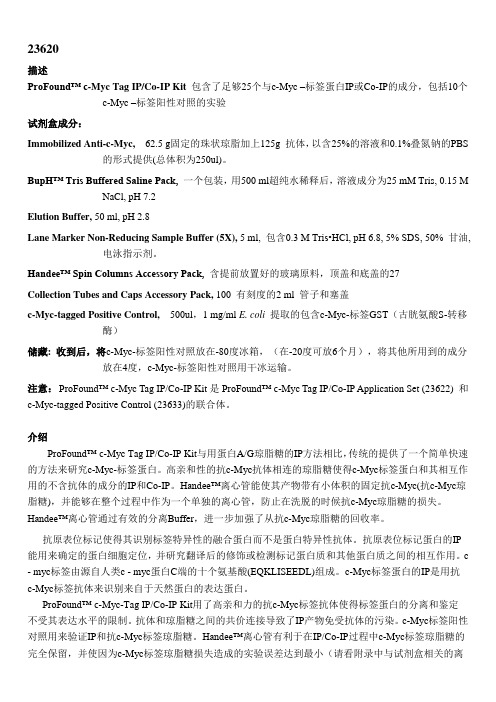
23620描述ProFound™ c-Myc Tag IP/Co-IP Kit 包含了足够25个与c-Myc –标签蛋白IP或Co-IP的成分,包括10个c-Myc –标签阳性对照的实验试剂盒成分:Immobilized Anti-c-Myc, 62.5 g固定的珠状琼脂加上125g 抗体,以含25%的溶液和0.1%叠氮钠的PBS 的形式提供(总体积为250ul)。
BupH™ Tris Buffered Saline Pack,一个包装,用500 ml超纯水稀释后,溶液成分为25 mM Tris, 0.15 M NaCl, pH 7.2Elution Buffer, 50 ml, pH 2.8Lane Marker Non-Reducing Sample Buffer (5X), 5 ml, 包含0.3 M Tris•HCl, pH 6.8, 5% SDS, 50% 甘油, 电泳指示剂。
Handee™ Spin Columns Accessory Pack, 含提前放置好的玻璃原料,顶盖和底盖的27Collection Tubes and Caps Accessory Pack, 100 有刻度的2 ml 管子和塞盖c-Myc-tagged Positive Control, 500ul,1 mg/ml E. coli 提取的包含c-Myc-标签GST(古胱氨酸S-转移酶)储藏: 收到后,将c-Myc-标签阳性对照放在-80度冰箱,(在-20度可放6个月),将其他所用到的成分放在4度,c-Myc-标签阳性对照用干冰运输。
注意:ProFound™ c-Myc Tag IP/Co-IP Kit是ProFound™ c-Myc Tag IP/Co-IP Application Set (23622) 和c-Myc-tagged Positive Control (23633)的联合体。
介绍ProFound™ c-Myc Tag IP/Co-IP Kit与用蛋白A/G琼脂糖的IP方法相比,传统的提供了一个简单快速的方法来研究c-Myc-标签蛋白。
Western Blot为什么必须要用内参
Western Blot为什么必须要用内参?一、背景:GAPDH(甘油醛-3-磷酸脱氢酶)是参与糖酵解的一种关键酶,由4个30-40kDa的亚基组成,分子量146kDa。
GAPDH基因几乎在所有组织中都高水平表达,广泛用作Western blot蛋白质标准化的内参。
GAPDH检测。
Western Blotting(检测条带大约在36kDa,稀释比例达10,000倍)、ELISA、亲和纯化、免疫荧光及免疫组化。
注:因为GAPDH 作为管家基因在同种细胞或者组织中的蛋白质表达量一般是恒定的。
在实验中,可能存在总蛋白浓度测定不准确;或者蛋白质样品在电泳前上样时产生的样品间的操作误差;这些误差需要通过测定每个样品中实际转到膜上的GAPDH的含量来进行校正,所以一般的western 实验都需要进行内参设置。
具体校正的方法就是将每个样品测得的目的蛋白含量与本样品的GAPDH含量相除,得到每个样品目的蛋白的相对含量。
然后才进行样品与样品之间的比较。
反应:抗GAPDH单抗(clone 6C5)能够与鱼、蛙、鸡、兔、小鼠、大鼠及人组织来源的GAPDH反应,但不能与酵母GAPDH反应。
应用:GAPDH检测。
Western Blotting(检测条带大约在36kDa,稀释比例达10,000倍)、ELISA、亲和纯化、免疫荧光及免疫组化。
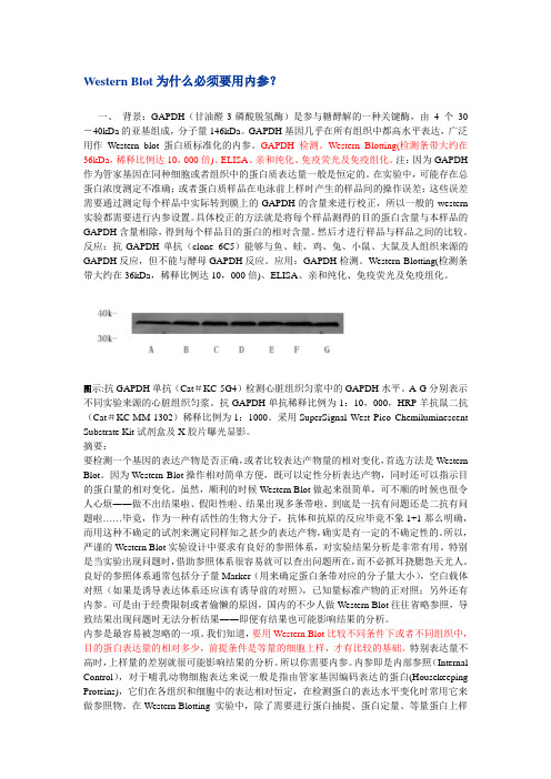
图示:抗GAPDH单抗(Cat#KC-5G4)检测心脏组织匀浆中的GAPDH水平。
A-G分别表示不同实验来源的心脏组织匀浆。
抗GAPDH单抗稀释比例为1:10,000,HRP羊抗鼠二抗(Cat#KC-MM-1302)稀释比例为1:1000。
采用SuperSignal West Pico Chemiluminescent Substrate Kit试剂盒及X胶片曝光显影。
摘要:要检测一个基因的表达产物是否正确,或者比较表达产物量的相对变化,首选方法是Western Blot。
因为Western Blot操作相对简单方便,既可以定性分析表达产物,同时还可以指示目的蛋白量的相对变化。
光动力疗法对两种黑素瘤细胞迁移与侵袭的影响及作用机制

光动力疗法对两种黑素瘤细胞迁移与侵袭的影响及作用机制作者:黎松张敏李蔼谕吴亚光来源:《中国美容医学》2024年第02期[摘要]目的:初步探究光动力疗法(Photodynamic therapy,PDT)对两种黑素瘤细胞迁移、侵袭的影响及作用机制。
方法:取生长至对数期黑素瘤细胞A375和B16-F10,胰酶消化离心后计数后接种至六孔板内,培养使细胞融合度达到80%~90%以上后孵育光敏剂1 mmol/L 5-氨基酮戊酸(5-ALA)2 h后使用635 nm红光照射治疗,根据照射能量的不同分为0 J(对照组)、2.4 J(PDT 2.4J组)、4.8 J(PDT 4.8 J组)。
采用划痕愈合实验和Transwell侵袭实验分别检测不同水平的PDT对B16-F10及A375系两种细胞迁移和侵袭能力的影响。
采用蛋白质印迹法检测迁移和侵袭相关蛋白[Ki-67、血管内皮生长因子(Vascular endothelial growth factor,VEGF)、基质金属蛋白酶9(Matrix metalloproteinase 9,MMP9)]的相对表达量。
结果:细胞划痕和Transwell实验的结果显示,PDT处理24 h后,相比对照组,PDT 2.4 J组及PDT 4.8 J组在B16-F10及A375两种细胞系中,划痕愈合率及侵袭细胞数目均明显减少(P<0.01);相比于PDT 2.4 J组,PDT 4.8 J组的划痕愈合率及侵袭细胞数目明显减少(P<0.01)。
两PDT组在Ki-67、VEGF及MMP9三种蛋白的表达水平上均明显低于对照组(P<0.01);相比于PDT 2.4 J组,PDT 4.8 J组在三种蛋白的表达水平上更低(P<0.01)。
结论:光动力治疗可显著抑制黑素瘤细胞系A375和B16-F10的侵袭与迁移能力,其机制可能是光动力治疗能够抑制促肿瘤生长相关因子的释放有关。
[关键词]光动力疗法;黑素瘤细胞;迁移;侵袭[中图分类号]R739.7 [文献标志码]A [文章编号]1008-6455(2024)02-0056-04Effect of Photodynamic Therapy on Migration and Invasion of Two Melanoma Cells and Mechanism of ActionLI Song1,ZHANG Min1,LI Aiyu2,WU Yaguang1[1.Department of Dermatology,the First Affiliated Hospital of Army Medical University (South West Hospital),Chongqing 400038,China; 2.Department of Dermatology,People's Hospital of Shapingba District,Chongqing 400030,China]Abstract: Objective A preliminary investigation of the effects and mechanisms of photodynamic therapy (PDT) on the migration and invasion of two types of melanoma cells. Methods Melanoma cells A375 and B16-F10, which grew to the log stage, were digested and centrifuged by pancreatic enzyme, counted, and inoculated into six-well plates. After culture,cell fusion reached more than 80% to 90%, the cells were incubated with photosensitizer 1 mmol/L 5-aminoketovalerate (5-ALA) for 2 h, and then treated with 635 nm red lightirradiation.According to the different irradiation energy, it was divided into 0 J (control group),2.4J (PDT 2.4J group), 4.8J (PDT 4.8J group). Scratch healing test and transwell invasion test were used to detect the effects of photodynamic therapy on the migration and invasion of melanoma cell lines A375 and B16-F10. Western blotting was used to detect the relative expression of migration and invasion-related proteins [Ki-67, vascular endothelial growth factor (VEGF), matrix metalloproteinase 9 (MMP9)]. Results The results of cell scratches and Transwell experiments showed that after 24 hours of PDT treatment, compared with the control group, the scratch healing rate and the invasion of cells in the PDT 2.4 J group and PDT 4.8 J group were in the B16-F10 andA375 cell lines. The number was significantly reduced (P<0.01), compared with the PDT 2.4 J group, the scratch healing rate and the number of invaded cells in the PDT 4.8 J group were significantly reduced (P<0.01). Compared with the PDT 2.4 J group, the PDT 4.8 J group had the expression levels of the three proteins is lower (P<0.01). Conclusion Photodynamic therapy can significantly inhibit the invasion and migration of melanoma cell lines A375 and B16-F10. The mechanism may be related to the inhibition of the expression of migration and invasion-related proteins.Key words: photodynamic therapy; melanoma cells; migration; invasion恶性黑素瘤约占皮肤癌的4%,发病率较低,但其具有高致死率[1]。
显影剂说明
重要提醒:SuperSignal West Pico化学发光底物是一个高敏感性的物质,它比大多数化学发光产物(包括ECL,鲁米诺)都要敏感,为了超敏感底物的最佳性能,抗体必须比其他使用的溶液浓度要稀。
如果你一直用以上的底物或者另外初级的化学发光底物,那么,用此化学发光底物稀释一抗和二抗要5倍以上。
例如,如果你用ECL溶液的时候,以1:100稀释一抗。
那么,你用SuperSignalWest Pico溶液的时候,就要以1:500稀释。
建议使用Table1的稀释范围。
Table1. 用SuperSignalWest Pico化学发光底物的抗体稀释度。
一抗:1:1000-1:5000或者0.2-1.0ul/mL二抗:1:20000-1:100000或者10-50ng/Ml说明Thermo Scientific SuperSignal West Pico化学发光底物是检测辣根过氧化物酶标记的抗体的一种高度敏感性增强的溶液。
这种溶液能够强烈的检测到皮微克的抗原总量。
信号的灵敏性、强度、持续时间允许用摄影或其他的成像方法来检测辣根过氧化物酶。
免疫印记可以重复的曝光以获取最佳的结果,或者重新洗膜,再敷上另一种抗体,检测另一种蛋白。
重要产物的信息为了最好的结果,优化Western Blot中所有的组成部分是必不可少的,包括样品总量、一抗及二抗的浓度、膜的选择、封闭液。
因为溶液是极端敏感的,所以SuperSignalWest Pico溶液比其他市场上可以买得的液体来说,需要更少的样品总量、一抗及二抗,通常减少10-20倍。
同时应用Thermo Scientific SuperSignal Western Blot增强剂可以使该溶液敏感性增强,背景减少,抗体特异性提高。
抗体浓度要比用沉淀比色合系统的浓度更加稀释,为了选择最适当的浓度,可以应用斑点印迹分析法。
没有任何一种封闭液是所有系统的最佳选择,所以对于Western Blot的来说,实验检测是选择适合封闭液的必要步骤。
常用的ecl发光液使用方法

常用的ecl发光液使用方法ECL(Electrochemiluminescence)发光液是一种常用于生物医学研究中的发光试剂。
它以电化学反应产生荧光信号,广泛应用于酶免疫检测、DNA检测、核酸杂交等实验中。
以下是使用ECL发光液的常用方法:1.材料准备:-ECL发光液:根据实验需要选择合适的ECL发光液,常用的有SuperSignal West Pico、SuperSignal West Dura等。
-靶分子:根据研究的目的,选择合适的抗体或探针。
-蛋白提取液:根据需要提取样品中的蛋白质。
2.准备蛋白质样品:-根据实验要求,准备合适的样品。
例如,细胞提取物、组织提取物或纯化的蛋白样品。
-将样品用适当的缓冲液稀释或洗涤以去除干扰物质。
3.确定最佳的抗体浓度:-根据之前的实验经验或者文献参考,初步决定使用的抗体浓度。
-进行一系列的试验,尝试不同的抗体浓度,确定最佳浓度。
4.准备蛋白质样品与抗体的混合物:-根据需要的实验体系,将蛋白质样品和抗体按照最佳浓度混合。
-加入适量的洗涤缓冲液,使得最终体积达到试管容积的一半。
5.孵育:-将混合物在适当的条件下进行温育,一般温度为4℃至室温,时间可按实验需要从数分钟到几小时不等。
6.增强剂的添加:-根据实验所使用的ECL发光液,加入适量的增强剂,如硝酸盐等。
增强剂的种类和使用浓度因ECL发光液的不同而不同,应根据产品说明进行操作。
7.发光显影:-将上述混合物与ECL发光液按照1:1的比例混合,混匀后立即添加在样品上。
-不同的ECL发光液有不同的发光时间窗口,可根据需要调整照相机或化学发光检测仪的参数。
8.显示和记录结果:-将发光结果通过照片或者相关设备记录下来。
-根据实验需要,可以对结果进行定量或半定量分析。
总结起来,使用ECL发光液的方法主要包括准备蛋白质样品、酶标抗体的配制、增强剂的加入、显影和结果分析等步骤。
在操作时应仔细阅读产品说明书,根据实验需要调整操作流程和使用浓度。
WB试验每步原理和技术及试剂的分析
试验每步原理和技术及试剂的分析WB试验每步原理和技术及试剂的分析WB蛋白印迹在生物化学这一块是常规实验,就像有人说炒菜中境界最高也最难的是蛋炒饭一样,实验中常规实验也是很考验技能的,除了潜心研究原理、认真揣摩技巧以外,对于新实验试剂的信息把握,勇于尝试新方法也是非常之重要的——各大厂家都在不断开发更方便更灵敏的新产品,“idea是生产力”嘛,因此生物通这里介绍一些能把我们从日常操作中解脱出来,获得“升级版”效果WB产品。
讲完了Western Blotting的电泳转移仪器,蛋白分子量标准和转移膜之后,我们最后来探讨一下WB的检测系统。
一般的WB检测过程中,都会有封闭、一抗、二抗和底物显色这四道工序要“加工”。
我个人觉得底物显色这最后一步是最关键的,也是最有文章可以作的一个部分。
你看,光标记方式就有生物素标记,地高辛标记,各种酶标记等等,酶标的底物又有各种生色底物、化学发光法底物和荧光底物可供选择;就连识别一抗的配体也不一定非要二抗不可,也可以是抗生物素蛋白,链亲和素或者Protein A或G等,更别说各种试剂盒琳琅满目,叫人好像无从下手!不过不急,生物通帮你作个参考,让你对各种产品的优点缺点一览无遗,真正成为一个WB高手。
另外如果实验室有已经建立的固定的显色方法,那也没关系,有时候不经意浏览到的方法可能就会对你的实验有莫大的帮助——这也就达到我们的目的了。
封闭和一抗的选择没有太多的选择余地——封闭前面介绍过了;一抗?现在的抗体产品说明书一般都有注明应用范围的,说明可以用于WB的就OK。
如果没有这些信息可以遵从以下原则:单抗专一性高,但是经过SDS-PAGE变性胶电泳的蛋白质可能由于原来的识别位点构象发生改变而不被识别,多抗不如单抗专一性高但更容易得到结果。
如果还有多种抗体选择,那当然是来源于兔或者小鼠抗体为好,因为后继的检测试剂盒一般都是针对兔鼠的居多,因而选择范围更大通用性也更强。
除了无标记的一抗,还有生物素等各种标记一抗。
Thermo-蛋白互作

6
Proprietary & Confidential
蛋白质相互作用技术
Genetic
Two Hybrid Phage Display
Chemical
Crosslinking Label-transfer
Mutational analysis
FeBABE mapping
Biochemical Immunoprecipitation (IP) Co-Immunoprecipitation (Co-IP)
Conclusion利用Protein A/G 可以满足绝大部分抗体结合的需要.
17
Proprietary & Confidential
Pain Points: 实验的时间比较长
• 传统Batch法在同一个Ep管中操作
• 细胞裂解液和珠子和抗体在一个管子
中进行孵育
• 通过离心沉淀珠子,分离固相和液相 • 取样不慎易导致样品损失或污染
Ag
Ab + Ag
抗体重链 抗原
抗体轻链
二抗检测
21
Proprietary & Confidential
Clean-Blot™ IP Detection Reagent
• Clean-Blot™ IP 检测试剂
• • • • 只结合未变性的一抗 替代二抗 用于IP结果检测或检测含内源IgG的组织细胞裂解液 去除变性的抗体对Western Blotting结果的干扰
1
Proprietary & Confidential
服务科学,世界领先
我们是提供分析仪器,设备,试剂和耗材,软件与服 务的全球领导者,为科学研究,分析测试,发明和诊 断等提供全套产品、服务和解决方案。
WB protocol

蛋白质技术培训一、组织总蛋白的提取材料:C57BL/6鼠(12w)肝脏或脾脏组织(-70℃保存);配制细胞裂解液所需各种溶液的试剂:Tirs(国药, 30188336), NaCl(国药, 10019318), Na3VO4(Sigma, S-6508), EDTA(国药, 10009719KP), EGTA(上海生工, E0732-5g), NP-40(上海生工, 9016-45-9), PMSF(Bio Basic Inc, 329-98-6), NaF(SCRC, 10019618)工具和仪器:研钵步骤:1.2.取-70℃保存的组织,根据EP管上的质量加入细胞裂解液,每100mg组织加入0.5ml裂解液,在研钵中磨碎。
(样品若为细胞,则按照每1×10⁶个细胞加入100-200μl裂解液,且不用研磨)3.将研磨的匀浆用加样枪吸入EP管中,冰上放置30min裂解细胞。
4.16000g,10min,4℃离心,取上清。
(上清中可能会有一些漂浮物,尽量吸取清澈的部分)二、蛋白定量材料:BSA(牛血清白蛋白)标准溶液(1mg/ml),考马斯亮蓝G-250染色液(Bio-Rad, 500-0006),组织细胞裂解上清。
工具和仪器:OneDrop OD-1000分光光度计步骤:1.取1ml考马斯亮蓝G-250染色液加入4ml三蒸水混匀,作为工作液。
2.取5个EP管将BSA用三蒸水稀释成浓度分别为0 mg /ml,0.1 mg /ml,0.2mg/ml,0.3mg/ml,0.4mg/ml,0.5mg/ml的标准品各10μl。
3.将蛋白样品用三蒸水稀释100倍后,取出10μl。
4.分别向6管标准品和10μl待测样品中加入200μl的工作液混匀,室温静置5min。
5.用OneDrop OD-1000分光光度计的Bradford法测量组织细胞裂解液的蛋白浓度。
三、SDS-聚丙烯酰胺凝胶电泳材料:2×SDS-PAGE loading buffer(配方见33页);电泳缓冲液(配方见34页);Prestained protein marker(Fermentas, SM1811);配制SDS-聚丙烯酰胺凝胶所需试剂:Acryl/Bis Solution (29:1) 40%(w/v)(上海生工, SD6013), APS(上海生工, 7727-54-0), 4×Tris-HCl(pH6.8)(上海生工, SD6022), 4×Tris-HCl(pH8.8)(上海生工, SD6021), TEMED(Bio Basic Inc, 110-18-9)工具和仪器:蛋白电泳槽(Mini-PROTEAN ⅡCell),DYY-6C电泳仪步骤:1.组装配胶的玻璃板,并验漏。
RIPA buffer pierce 细胞裂解缓冲液
INSTRUCTIONSRIPA Buffer89900 RIPA Buffer, 100mL89901 RIPA Buffer, 250mLContents: 25mM Tris•HCl pH 7.6, 150mM NaCl, 1% NP-40, 1% sodium deoxycholate, 0.1% SDSStorage: Upon receipt store at 4°C. Product shipped at ambient temperature.IntroductionThe Thermo Scientific RIPA buffer is one of the most reliable buffers used to lyse cultured mammalian cells from both plated cells and cells pelleted from suspension cultures. This buffer enables protein extraction from cytoplasmic, membrane and nuclear proteins and is compatible with many applications, including reporter assays, protein assays, immunoassays and protein purification.Important Product Information•RIPA Buffer does not contain protease or phosphatase inhibitors. If desired, add protease inhibitors, such as Thermo Scientific Halt Protease Inhibitor Cocktail (Product No. 78410) and Halt™ Phosphatase Inhibitor Cocktail (Product No.78420) to the reagent to prevent proteolysis and maintain phosphorylation status of proteins. Add protease andphosphatase inhibitors immediately before use.•Use 1mL of cold RIPA Buffer for every 5 × 10⁶ of HeLa or A431 cells (~20µL of packed cells, which is equivalent to ~40mg of cells). To obtain concentrated protein extracts, directly lyse cells on plate and use less buffer.•Some protein kinases and other enzymes may be sensitive to the components of the RIPA Buffer, resulting in their decreased activity. In such cases, prepare a RIPA buffer that does not contain sodium deoxycholate and SDS.•RIPA Buffer is compatible with the Thermo Scientific Pierce BCA Protein Assay Kit (Product No 23225).Procedure for Lysis of Monolayer-cultured Mammalian CellsNote: If desired, add protease and phosphatase inhibitors to the RIPA Buffer immediately before use.1.Carefully remove (decant) culture medium from adherent cells.2.Wash cells twice with cold PBS.3.Add cold RIPA Buffer to the cells. Use 1mL of buffer per 75cm2 flask containing 5 × 10⁶ HeLa or A431 cells. Keep onice for 5 minutes, swirling the plate occasionally for uniform spreading.4.Gather the lysate to one side using a cell scraper, collect the lysate and transfer to a microcentrifuge tube. Centrifugesamples at ~14,000 ×g for 15 minutes to pellet the cell debris.Note: To increase yields, sonicate the pellet for 30 seconds with 50% pulse.5.Transfer supernatant to a new tube for further analysis.Procedure for Lysis of Suspension-cultured Mammalian CellsNote: If desired, add protease and phosphatase inhibitors to the RIPA Buffer immediately before use.1.Pellet the cells by centrifugation at 2500 ×g for 5 minutes. Discard the supernatant.2.Wash cells twice in cold PBS. Pellet cells by centrifugation at 2500 ×g for 5 minutes.3.Add RIPA Buffer to the cell pellet. Use 1mL of RIPA buffer for 40mg (~5 × 10⁶ of HeLa cells) of wet cell pellet. Pipettethe mixture up and down to suspend the pellet.Note: To increase yields, sonicate the pellet for 30 seconds with 50% pulse.4.Shake mixture gently for 15 minutes on ice. Centrifuge mixture at ~14,000 ×g for 15 minutes to pellet the cell debris.5.Transfer supernatant to a new tube for further analysis.TroubleshootingProblem Possible Cause SolutionLow total protein yield Some cells are more resistantto lysis than others Make sure the cell pellet is thoroughly suspended in RIPA Buffer and incubate for longer with occasional swirling − sonicate the pellet to increase yieldLow concentration of proteins Excess buffer used Use less buffer (e.g., 0.25-0.5mL per 75cm2 flask containing5 × 106 cells) − use a sufficient amount to cover the entire plateProteolysis No protease inhibitors added Add Halt Protease Inhibitor Cocktail to the buffer before useLow phosphorylation of proteins Phosphatase activity Add Halt Phosphatase Inhibitor Cocktail to the buffer before use Protein is non-phosphorylatedor poorly phosphorylatedNoneRelated Thermo Scientific Products78410 Halt Protease Inhibitor Cocktail Kit78420 Halt Phosphatase Inhibitor Cocktail, 1mL78248 B-PER® Bacterial Protein Extraction Reagent, 500mL78990 Y-PER® Yeast Protein Extraction Reagent, 500mL89826 Mem-PER® Membrane Protein Extraction Reagent Kit78833 NE-PER® Nuclear and Cytoplasmic Extraction Kit23227 Pierce® BCA Protein Assay Kit26148 Pierce Direct IP Kit34080 SuperSignal® West Pico Chemiluminescent Substrate, 500mL34076 SuperSignal® West Dura Extended Duration Substrate, 200mLGeneral ReferencesCao, F., et al. (2005). Identification of an essential molecular contact point on the duck hepatitis B virus reverse transcriptase. J Virol79(16):10164-70. Pfrepper, K.I. and Flugel R.M. (2005). Molecular characterization of proteolytic processing of the gap proteins of human spumaretrovirus. Methods in Mol Biol304:435-44.Sefton, B.M. (2005). Labeling cultured cells with 32Pi and preparing cell lysates for immunoprecipitation. Unit 18.2. F. M. Ausubel, R. Brent, R.E.Kingston, D.D. Moore, J.G. Seidman, J.A. Smith, and K. Struhl (eds.) Current Protocols in Molecular Biology. John Wiley & Sons, Inc.This product (“Product”) is warranted to operate or perform substantially in conformance with published Product specifications in effect at the time of sale, as set forth in the Product documentation, specifications and/or accompanying package inserts (“Documentation”) and to be free from defects in material and workmanship. Unless otherwise expressly authorized in writing, Products are supplied for research use only. No claim of suitability for use in applications regulated by FDA is made. The warranty provided herein is valid only when used by properly trained individuals. Unless otherwise stated in the Documentation, this warranty is limited to one year from date of shipment when the Product is subjected to normal, proper and intended usage. This warranty does not extend to anyone other than the original purchaser of the Product (“Buyer”).No other warranties, express or implied, are granted, including without limitation, implied warranties of merchantability, fitness for any particular purpose, or non infringement. Buyer’s exclusive remedy for non-conforming Products during the warranty period is limited to replacement of or refund for the non-conforming Product(s).There is no obligation to replace Products as the result of (i) accident, disaster or event of force majeure, (ii) misuse, fault or negligence of or by Buyer, (iii) use of the Products in a manner for which they were not designed, or (iv) improper storage and handling of the Products.Current product instructions are available at /pierce. For a faxed copy, call 800-874-3723 or contact your local distributor.© 2011 Thermo Fisher Scientific Inc. All rights reserved. Unless otherwise indicated, all trademarks are property of Thermo Fisher Scientific Inc. and its subsidiaries. Printed in the USA.。
碧云天 特超敏ECL化学发光试剂盒说明书

碧云天生物技术/Beyotime Biotechnology订货热线:400-1683301或800-8283301订货e-mail:******************技术咨询:*****************碧云天网站微信公众号网址:BeyoECL Star (特超敏ECL化学发光试剂盒)产品编号产品名称包装P0018AS BeyoECL Star (特超敏ECL化学发光试剂盒) 100mlP0018AM BeyoECL Star (特超敏ECL化学发光试剂盒) 500ml产品简介:碧云天生产的Western萤光检测试剂BeyoECL Star是一种特超敏ECL化学发光试剂盒,发光效果显著优于BeyoECL Plus,可与二抗上偶联的辣根过氧化物酶(horseradish peroxidase, HRP)发生化学反应,发出萤光,从而可以通过用X光片压片或其它化学发光成像设备检测样品。
碧云天生产的Western萤光检测试剂目前共有三种,分别是P0018S/P0018M BeyoECL Plus、P0018AS/P0018AM BeyoECL Star 和P0018FS/P0018FM BeyoECL Moon。
常规的Western检测,优先推荐使用BeyoECL Star。
对于丰度比较高的目的蛋白的检测,例如内参蛋白等的检测,推荐使用性价比更高的BeyoECL Plus。
对于低丰度较难检测的目的蛋白,优先推荐使用检测灵敏度最高的BeyoECL Moon。
但对于丰度适中的目的蛋白的检测,不太推荐使用BeyoECL Moon,因为使用BeyoECL Moon时由于检测灵敏度特别高,容易产生过曝的现象。
BeyoECL Star灵敏度极高,比DAB显色的灵敏度至少高1000倍,比Amersham公司的ECL或碧云天以前生产的BeyoECL的灵敏度高100-500倍左右,比碧云天生产的BeyoECL Plus的灵敏度高约5-10倍(参考图1),比原Pierce公司(现Thermo公司)的SuperSignal West Pico Substrate的灵敏度高10倍以上,实际检测效果与原Pierce公司的SuperSignal West Dura和SuperSignal West Femto的检测灵敏度相近。
pull-down试剂盒说明书翻译
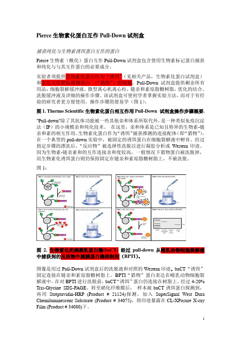
Pierce生物素化蛋白互作Pull-Down试剂盒捕获纯化与生物素诱饵蛋白互作的蛋白Pierce生物素(酰化)蛋白互作Pull-Down试剂盒包含使用生物素标记蛋白捕获和纯化与与其互作蛋白的必要成分。
实验者须提供生物素化蛋白作为“诱饵”(见相关产品,生物素化蛋白试剂盒)和表达互作靶标推测蛋白(“猎物”)的细胞。
Pull-Down试剂盒提供剩余所有用品:细胞裂解缓冲液,微型离心机离心柱,链亲和素琼脂糖树脂,优化的结合、洗脱缓冲液及详细的操作步骤。
该试剂盒可使初学者掌握实验方法,而对于有经验的研究者更方便使用。
操作步骤简便易学(图1)。
图1. Thermo Scientific生物素化蛋白相互作用Pull-Down 试剂盒操作步骤概要."Pull-down"除了其抗体功能被一些其他亲和体系所取代外,是一种类似免疫沉淀法(IP)的小规模亲和纯化技术。
在这里,亲和体系是已知且特异的生物素-链亲和素的相互作用。
生物素化蛋白作为“诱饵”捕获推测的连接配体(即“猎物”)。
在一个典型的pull-down实验中,被固定的诱饵蛋白在细胞裂解液中孵育。
经过指定步骤的漂洗后,“反应物”被选择性洗脱以进行凝胶分析或Western印迹。
因为生物素-链亲素和的互作连接亲和度较高,一般情况下猎物蛋白被洗脱掉,而生物素化诱饵蛋白则仍保持固定在链亲和素琼脂糖树脂上,不被洗脱。
图1:图2. 生物素化的胰凝乳蛋白酶(baCT) 经过pull-down从哺乳动物细胞裂解液中捕获到的反应物牛胰胰蛋白酶抑制剂(BPTI)。
图像是用过Pull-Down试剂盒后的洗脱液和对照的Western印迹。
baCT“诱饵”固定连接在链亲和素琼脂糖树脂上。
BPTI“猎物”蛋白表达在哺乳动物细胞裂解液中。
在对BPTI进行洗脱前,baCT“诱饵”蛋白仍连接在树脂上。
经过4-20% Tris-Glycine SDS-PAGE。
转至硝化纤维膜后,样本被baCT诱饵蛋白探测到,再用Streptavidin-HRP (Product # 21124)探测,加入SuperSignal West Dura Chemiluminescent Substrate (Product # 34075),将印迹暴露在CL-XPosure X-ray Film (Product # 34080)下。
蛋白质印迹技术

蛋白质印迹技术蛋白质印迹(Western blotting)是一种检测固定在固相基质上的蛋白质的免疫化学办法,又称免疫印迹(immunoblotting )。
1979年,Towbin等将分析DNA的Southern blotting技术扩展到蛋白质的讨论领域,并且和特异、敏捷的免疫分析技术相结合,称之为“Western blotting"(蛋白质印迹)。
免疫印迹可分为两个步骤:将蛋白质由凝胶转移至固相基质上;特异性抗体检测。
试验原理蛋白质印迹法是将蛋白质混合样品经SDS-PAGE后,分别为不同的条带,其中含有能与特异性抗体相结合的待检测的蛋白质(抗原蛋白),将凝胶上的蛋白质以电转印的方式,转印至固相支持物(如硝酸纤维素膜,NC膜)上,再用特异抗体作为探针对靶蛋白举行检测的办法。
因为该办法结合了PAGE的高辨别率和固相免疫反应的高特异性等多种优点,可检测到低至1-5 ng的中等分子量大小的靶蛋白。
仪器和材料电转移装置(电源);摇床;暗匣;转印夹;浅盘(30cm×15cm×3cm);玻璃棒;杂交袋或杂交盒;胶片;保鲜膜;滤纸;0. 22μm孔径的硝酸纤维素膜。
试剂 (1) Running Buffer (Tris-甘氨酸电泳缓冲液,1×2500ml) ; 25 mmol/L Tris 7. 55g 250mmol/L甘氨酸 47. 0g 10%SDS 25m1 (2) Transfer Buffer ( Tris-甘氨酸电转膜缓冲液,1× 2500m1,pH=8.3):48 mmol/L Tris 14. 50g 39mmol/L 甘氨酸 7. 25g 10% SDS 9. 25ml 20%甲醇 500m1 (3) TTBS缓冲液(pH 7.5,2 ×2500ml): 20 mmol/L Tris 12. l l g 0. 5 M NaCl 146. 1 g 0.05%Tween 20 2. 5 ml (4) Stripping Buffer(pH2. 0,1000m1):甘氨酸 1. 8768 SDS 10. 0g (5) 10%牛奶封闭液:称取10. 0g脱脂奶粉,溶于80m1 TTBS(pH7.5 )中,搅拌彻低溶解,后加入TTBS定容至100m1,再加入10μl Thimerosal混匀。
ECL免疫发光试剂使用常见问题及解决方案
ECL免疫发光试剂使用常见问题及解决方案Q: 你公司生产的ECL与市场上的其他ECL产品相比,灵敏度如何?A: 在最优条件下,我公司的ECL灵敏度比Pierce的SuperSignal West Pico Substrate高3-5倍,发光时间也更长可持续约8小时。
Q:你公司的ECL可检测到的最低抗原是多少?A: 本公司的ECL可检测到低达1pg的抗原。
Q: 为什么与我用过的其他品牌的ECL相比,你公司的ECL会出来更多的条带?A: 因为我公司的ECL比其他公司的化学发光试剂灵敏度更高,从而可以检测到低丰度的蛋白。
当遇到这种情况时,可优化封闭条件和抗体使用浓度。
但当目的蛋白表达丰度很低时,选择灵敏度合适的ECL就能得到理想的结果。
Q:如何判断ECL是否失效?A:准备ECL工作液1ml 到一个干净的Ep管中,加入1μl未稀释的HRP 酶标物(二抗)到Ep管中,混匀后即可看到管内发出蓝光,并在随后的几分钟内持续发光,表示底物活性正常。
若不发光则说明ECL失效了。
Q:为什么Western Blot化学发光(ECL)会出现高背景?A:Western Blot发光经常有“背景太高”问题,导致“背景高”的原因有很多,主要有以下几个方面:①. 抗体浓度过高,洗涤不充分,可以造成抗体在膜上的非特异结合,导致高背景。
可通过降低抗体稀释度,增加洗膜次数和Buffer用量,或在Wash Buffer中添加终浓度0.05%的Tween-20加以改善。
②. 封闭不充分,会造成抗体在膜上的非特异结合,导致高背景。
可通过4℃封闭过夜或增加封闭液中的蛋白浓度加以改善。
③. 使用了不恰当的封闭剂,可尝试不同的封闭剂。
④. 曝光过度,可减少底物显色时间,缩短曝光时间。
⑤. 操作过程中膜干了。
确保膜完全浸没于buffer中,始终杜绝膜干。
Q:Western Blot发光时为什么会发生荧光“淬灭”?A:荧光淬灭是Western Blot发光时常见的现象,即加ECL后立刻可以看到很明显的亮光,但亮光很快就消逝了,压完片后条带却很弱,甚至没有条带,发生这种情况的原因可能有以下几个方面:①. 抗原浓度太高,局部过多的HRP会快速消耗底物,导致荧光信号过强,条带呈灼烧样。
- 1、下载文档前请自行甄别文档内容的完整性,平台不提供额外的编辑、内容补充、找答案等附加服务。
- 2、"仅部分预览"的文档,不可在线预览部分如存在完整性等问题,可反馈申请退款(可完整预览的文档不适用该条件!)。
- 3、如文档侵犯您的权益,请联系客服反馈,我们会尽快为您处理(人工客服工作时间:9:00-18:30)。
INSTRUCTIONSNumberDescription34079 SuperSignal West Pico Chemiluminescent Substrate, sufficient for 500cm 2 of membrane Kit Contents:SuperSignal West Pico Luminol/Enhancer Solution, 25mLSuperSignal West Pico Stable Peroxide Solution, 25mL34077 SuperSignal West Pico Chemiluminescent Substrate, sufficient for 1000cm 2 of membrane Kit Contents:SuperSignal West Pico Luminol/Enhancer Solution, 50mL SuperSignal West Pico Stable Peroxide Solution, 50mL34087 SuperSignal West Pico Chemiluminescent Substrate, sufficient for 2000cm 2 of membrane Kit Contents:SuperSignal West Pico Luminol/Enhancer Solution, 100mLSuperSignal West Pico Stable Peroxide Solution, 100mL34080 SuperSignal West Pico Chemiluminescent Substrate, sufficient for 5000cm 2 of membrane Kit Contents:SuperSignal West Pico Luminol/Enhancer Solution, 250mL SuperSignal West Pico Stable Peroxide Solution, 250mL34078 SuperSignal West Pico Chemiluminescent Substrate, sufficient for 10,000cm 2 of membrane Kit Contents:SuperSignal West Pico Luminol/Enhancer Solution, 500mL SuperSignal West Pico Stable Peroxide Solution, 500mLStorage: Upon receipt store reagents at room temperature. Products shipped at ambient temperature.Table of ContentsIntroduction ................................................................................................................................................................................. 2 Important Product Information .................................................................................................................................................... 2 Procedure Summary ..................................................................................................................................................................... 3 Additional Materials Required ..................................................................................................................................................... 3 Detailed Western Blotting Procedure .......................................................................................................................................... 4 Troubleshooting ........................................................................................................................................................................... 5 Additional Information ................................................................................................................................................................ 5 General References . (6)IMPORTANT NOTE: Thermo Scientific™ SuperSignal™ West Pico Chemiluminescent Substrate is a high-sensitivity substrate that is more sensitive than most chemiluminescent products including ECL, LumiGLO™, Renaissance™ and Western Lightning™ Substrates. For optimal performance of SuperSignal Substrate, antibodies must be more dilute than those used with these other substrates. If you have been using one of the substrates listed above or another “entry-level” chemiluminescent substrate, dilute both primary and secondary antibodies at least 5-fold more. For example: If you have been using the primary antibody at 1:100 dilution with ECL Substrate, then use a ~1:500 dilution with SuperSignal West Pico Substrate. Recommended dilution ranges are listed in Table 1.SuperSignal™ West Pico Chemiluminescent SubstrateTable 1. Antibody dilution ranges to use with SuperSignal West Pico Chemiluminescent Substrate.Primary Antibody Dilution Range from a 1mg/mL stock Secondary Antibody Dilution Range from a 1mg/mL stock1:1000-1:5000 or 0.2-1.0µg/mL 1:20,000-1:100,000 or 10-50ng/mLIntroductionThe Thermo Scientific SuperSignal West Pico Chemiluminescent Substrate is a highly sensitive enhanced substrate for detecting horseradish peroxidase (HRP) on immunoblots. This substrate’s extremely intense signal output enables detection of picogram amounts of antigen. The sensitivity, intensity and duration of the signal allow for easy detection of HRP using photographic or other imaging methods. Blots can be repeatedly exposed to film to obtain optimal results or stripped of the immunodetection reagents and reprobed.Important Product Information•For best results, it is ESSENTIAL to optimize all components of the system including sample amount, primary and secondary antibody concentration, and the choice of membrane and blocking reagents. Because the substrate isextremely sensitive, SuperSignal West Pico Substrate requires the use of much less sample and primary and secondary antibodies than most commercially available substrates, usually by a factor of at least 10-20. Further enhancement of sensitivity, reduction of background, and improvement in antibody specificity can be achieved by incorporating the use of Thermo Scientific SuperSignal Western Blot Enhancer (Product No. 46640).•The antibody concentrations required are much more dilute than those used with precipitating colorimetric HRP systems.To optimize the appropriate concentrations, perform a systematic dot blot analysis.•Because no blocking reagent is optimal for all systems, empirical testing is essential to determine the appropriate blocking buffer for each Western blot system. Determining the proper blocking buffer can help increase sensitivity and prevent nonspecific signal caused by cross-reactivity between the antibody and the blocking reagent. Furthermore, when switching from one substrate to another, a diminished signal or increased background sometimes results because the blocking buffer was not optimal for the new system.•Avoid using milk as a blocking reagent when using avidin/biotin systems because milk contains variable amounts of endogenous biotin.•Use a sufficient volume of wash buffer, blocking buffer, antibody solution and Substrate Working Solution to cover blot and ensure that it never becomes dry. Large blocking and wash buffer volumes may result in reduced nonspecific signal. •For optimal results, use a shaking platform during incubation steps.•Add Tween™-20 Detergent (final concentration of 0.05%) to the blocking buffer and when preparing all antibody dilutions to reduce nonspecific signal. Use only high-quality products such as Thermo Scientific™ Surfact-Amps™ 20 (Product No. 28320), which is a purified detergent packaged in ampules and guaranteed to be low in peroxides and other contaminants.•Do not use sodium azide as a preservative for buffers. Sodium azide is an inhibitor of HRP and could interfere with this system.•Do not handle membrane with bare hands. Always wear gloves or use clean forceps.•All equipment must be clean and free of foreign material. Metallic devices (e.g., scissors) must have no visible signs of rust. Rust may cause speckling and/or high background.•The Substrate Working Solution is stable for 24 hours at room temperature. Exposure to the sun or any other intense light can harm the Working Solution. For best results keep the Working Solution in an amber bottle and avoid prolonged exposure to any intense light. Short-term exposure to typical laboratory lighting will not harm the Working Solution. •We offer a variety of protein transfer membranes, blocking buffers, primary antibodies, enzyme-labeled secondary antibodies, buffers and detergents. Please consult our website or catalog for product and ordering information.Procedure SummaryNote: Optimize the antigen and antibody concentrations. Recommended antibody dilutions must be used to consistently obtain positive results. For recommended dilution ranges please see the Additional Materials Required section.1.Dilute primary antibody to 0.2-1.0µg/mL or 1:1000-1:5000 dilution from a 1mg/mL stock.*2.Dilute secondary antibody to 10-50ng/mL or 1:20,000-1:100,000 dilution from a 1mg/mL stock.*3.Mix the two substrate components at a 1:1 ratio to prepare the substrate Working Solution.Note: Exposure to the sun or any other intense light can harm the Working Solution. For best results keep the Working Solution in an amber bottle and avoid prolonged exposure to any intense light. Short-term exposure to typical laboratory lighting will not harm the Working Solution.4.Incubate blot 5 minutes in SuperSignal West Substrate Working Solution.5.Drain excess reagent. Cover blot with clear plastic wrap.6.Expose blot to X-ray film.*See Important Note on page 1.Additional Materials Required•Completed Western blot membrane: Use any suitable protocol to separate proteins by electrophoresis and transfer them to a nitrocellulose membrane. Other membrane types can be used; however, optimization may be required.1•Dilution Buffer: Use either Tris-buffered saline (TBS, Product No. 28376) or phosphate-buffered saline (PBS, Product No. 28374). Alternatively, dilute primary antibody in SuperSignal Western Blot Enhancer (Product No. 46640) to further enhance sensitivity and specificity, while minimizing background.•Wash Buffer: Add 5mL of 10% Tween-20 Detergent (Surfact-Amps 20 Detergent Solution, Product No. 28320) to 1000mL of Dilution Buffer. (The final concentration of Tween-20 Detergent will be 0.05%.)•Blocking Reagent: Add 0.5mL of 10% Tween-20 to 100mL of a blocking buffer such as Thermo Scientific™ SuperBlock™ (PBS) Blocking Buffer (Product No. 37515) or SuperBlock (TBS) Blocking Buffer (Product No. 37535).Choose a blocking buffer with the same base component as the Dilution Buffer.•Primary Antibody:* Choose an antibody that is specific to the target protein(s). Prepare a 1mg/mL stock solution of this antibody in Dilution Buffer. Use the Blocking Reagent to make all working dilutions of this antibody stock. Prepare a working dilution between 1:1000 and 1:5000 or 0.2-1µg/mL. The optimal dilution to use depends on the specific primary antibody and the amount of antigen on the membrane.•HRP-conjugated Secondary Antibody:* Choose an HRP-conjugate that specifically binds to the primary antibody.Prepare a 1mg/mL stock solution in Dilution Buffer. Use the Blocking Reagent to make all working dilutions of this antibody stock. Prepare a working dilution between 1:20,000 and 1:100,000 or 10-50ng/mL. This range also applies when using either Thermo Scientific Streptavidin-HRP (Product No. 21124) or NeutrAvidin™-HRP (Product No.31001). The optimal dilution to use varies depending on the HRP conjugate and the amount of antigen on the membrane. •Film cassette, developing and fixing reagents: For processing autoradiographic film.•Rotary platform shaker: For agitation of membrane during incubations.*See Important Note on page 1.Detailed Western Blotting Procedure1.Remove blot from the transfer apparatus and block nonspecific sites with Blocking Reagent for 20-60 minutes at roomtemperature (RT) with shaking. For best results, block for 1 hour at RT.Please Note: It is critical to use the recommended antibody dilution indicated in theprevious Additional Materials Required Section2.Remove the Blocking Reagent and add the appropriate primary antibody dilution. Incubate blot for 1 hour with shaking.If desired, blots may be incubated with primary antibody overnight at 2-8°C.3.Wash membrane by suspending it in Wash Buffer and agitating for ≥ 5 minutes. Replace Wash Buffer at least 4-6 times.Increasing the wash buffer volume and/or the number of washes may help reduce background.Note: Briefly rinsing membrane in wash buffer before incubation will increase wash efficiency.Please Note: It is critical to use the recommended HRP-conjugate dilution indicated in theprevious Additional Materials Required Section4.Incubate blot with the appropriate HRP-conjugate dilution for 1 hour at RT with shaking.5.Repeat Step 3 to remove non-bound HRP-conjugate.Note: Membrane MUST be thoroughly washed after incubation with the HRP-conjugate.6.Prepare Working Solution by mixing equal parts of the Stable Peroxide Solution and the Luminol/Enhancer Solution.Use 0.1mL Working Solution per cm2 of membrane. The Working Solution is stable for 24 hours at room temperature.Note: Exposure to the sun or any other intense light can harm the Working Solution. For best results keep the Working Solution in an amber bottle and avoid prolonged exposure to any intense light. Typical laboratory lighting will not harm the Working Solution.7.Incubate blot with Working Solution for 5 minutes.8.Remove blot from Working Solution and place it in a plastic membrane protector; a plastic sheet protector or plasticwrap may be used. Use an absorbent tissue to remove excess liquid and to carefully press out any bubbles from between the blot and surface of the membrane protector.9.Place the protected membrane in a film cassette with the protein side facing up. Turn off all lights except thoseappropriate for film exposure (e.g., a red safelight).Note: Film must remain dry during exposure. For optimal results, perform the following precautions: •Make sure excess substrate is removed from the membrane and the membrane protector.•Use gloves during the entire film-handling process.•Never place a blot on developed film, as there may be chemicals on the film that will reduce signal.10.Carefully place a piece of film on top of the membrane. A recommended first exposure time is 60 seconds. Exposuretime may be varied to achieve optimal results. Enhanced or pre-flashed film is unnecessary.Caution: Light emission is intense and any movement between the film and membrane can cause artifacts on the film.Note: The exposure time may be varied to achieve optimal results. If the signal is too intense, reduce exposure time or optimize the system by decreasing the antigen and/or antibody concentrations. Alternatively, use Thermo Scientific Pierce Background Eliminator (Product No. 21065).Light emission is most intense during the first 5-30 minutes after substrate incubation. Light emission will continue for several hours, but will decrease with time. Longer exposure times may be necessary as the blot ages.If using a storage phosphor imaging device (e.g., Bio-Rad’s Molecular Imager System) or a CCD Camera (e.g., Cell Bioscience’s ChemiImager System), longer exposure times may be necessary.11.Develop film using appropriate developing solution and fixative. Blot may be stripped and reprobed if necessary. Forbest results, use Thermo Scientific™ Restore™ Western Blot Stripping Buffer (Product No. 21059).TroubleshootingProblem Possible Cause SolutionReverse image on film (i.e., whitebands with a black background)Too much HRP in the system Dilute HRP-conjugate at least 10-foldMembrane has brown or yellowbandsBlot glows in the darkroomSignal duration is less than 8 hoursWeak or no signal Too much HRP in the system depletedthe substrate and caused the signal tofade quicklyDilute HRP-conjugate at least 10-foldInsufficient quantities of antigen or antibody Increase amount of antibody or antigen Use SuperSignal Western Blot Enhancer(Product No. 46640)Inefficient protein transfer Optimize transferReduction of HRP or substrate activity **See note belowHigh background Too much HRP in the system Dilute HRP-conjugate at least 10-foldInadequate blocking Optimize blocking conditionsInappropriate blocking reagent Try a different blocking reagentInadequate washing Increase length, number or volume ofwashesFilm has been overexposed Decrease exposure time or use PierceBackground Eliminator (Product No.21065)Concentration of antigen or antibodyis too highDecrease amount of antigen or antibodyPoor antibody specificity Use SuperSignal Western Blot Enhancer(Product No. 46640)Spots within the protein bands Inefficient protein transfer Optimize transfer procedureUnevenly hydrated membrane Perform manufacturer’s recommendationsfor hydrating membrane properlyBubble between the film and the membrane Remove bubbles before exposing blot to filmSpeckled background on film Aggregate formation in the HRP-conjugateFilter conjugate through a 0.2µm filter Nonspecific bands Too much HRP in the system Dilute HRP-conjugate at least 10-foldSDS caused nonspecific binding to protein bands Do not use SDS during the Western blot procedurePoor antibody specificity Use SuperSignal Western Blot Enhancer(Product No. 46640)test tube. With the lights turned off, add 1µL undiluted HRP-conjugate to the Working Solution. The solution should immediately emit a blue light that will fade over the next several minutes.Additional InformationVisit our web site for additional information relating to this product including the following:•Tech Tip #67: Chemiluminescent Western blotting technical guide and protocols•Tech Tip #21: Convert to SuperSignal West Pico Substrate from ECL Substrate•Tech Tip #23: Strip and reprobe Western blots•Tech Tip #24: Optimize antigen and antibody concentrations for Western blots•Tech Tip #32: Guide to enzyme substrates for Western blotting•Tech Tip #43: Protein stability and storage•Western Blotting Handbook and Troubleshooting GuideRelated Thermo Scientific Products46640 SuperSignal Western Blot Enhancer, 500mL kit, sufficient for 25 mini blots or 2000cm246641 SuperSignal Western Blot Enhancer, 50mL kit, sufficient for 2 mini blots or 160cm234090 CL-XPosure™ Film, 5” × 7” sheets, 100 sheets/pkg34075 SuperSignal West Dura Extended Duration Substrate, 100mL34095 SuperSignal West Femto Maximum Sensitivity Substrate, 100mL21059 Restore Western Blot Stripping Buffer, 500mL21065 Pierce Background Eliminator Kit, for eliminating background from X-ray film37515 SuperBlock (PBS) Blocking Buffer, 1L37535 SuperBlock (TBS) Blocking Buffer, 1L88018 Nitrocellulose Membrane, 0.45µm, 33cm × 3m, 1 roll77010 Nitrocellulose Membrane, 0.45µm, 8cm × 12cm, 25/pkg.88025 Nitrocellulose Membrane, 0.45µm, 8cm × 8cm, 15/pkg.88600 Western Blotting Filter Paper, 8cm × 10.5cm, 100 sheets24580 Pierce Reversible Protein Stain Kit for Nitrocellulose Membranes24585 Pierce Reversible Protein Stain Kit for PVDF MembranesGeneral ReferencesCRC Handbook of Immunoblotting of Proteins: Volume 1 Technical Description. Eds Ole J. Bjerrum, Ph.D., M.D. and Niels H.H. Heegaard, M.D. CRC Press, Inc.: Boca Raton, FL, 1988.Kaufmann, S.H., et al. (1987). The erasable Western blot. Anal Biochem161:89-95.Mattson, D.L. and Bellehumeur, T.G. (1996). Comparison of three chemiluminescent horseradish peroxidase substrates for immunoblotting. Anal Biochem 240:306-8.Walker, G.R., et al. (1995). SuperSignal CL-HRP: A new enhanced chemiluminescent substrate for the development of the horseradish peroxide label in Western blotting applications. J of NIH Research7:76.Products are warranted to operate or perform substantially in conformance with published Product specifications in effect at the time of sale, as set forth in the Product documentation, specifications and/or accompanying package inserts (“Documentation”). No claim of suitability for use in applications regulated by FDA is made. The warranty provided herein is valid only when used by properly trained individuals. Unless otherwise stated in the Documentation, this warranty is limited to one year from date of shipment when the Product is subjected to normal, proper and intended usage. This warranty does not extend to anyone other than Buyer. Any model or sample furnished to Buyer is merely illustrative of the general type and quality of goods and does not represent that any Product will conform to such model or sample.NO OTHER WARRANTIES, EXPRESS OR IMPLIED, ARE GRANTED, INCLUDING WITHOUT LIMITATION, IMPLIED WARRANTIES OF MERCHANTABILITY, FITNESS FOR ANY PARTICULAR PURPOSE, OR NON INFRINGEMENT. BUYER’S EXCLUSIVE REMEDY FOR NON-CONFORMING PRODUCTS DURING THE WARRANTY PERIOD IS LIMITED TO REPAIR, REPLACEMENT OF OR REFUND FOR THE NON-CONFORMING PRODUCT(S) AT SELLER’S SOLE OPTION. THERE IS NO OBLIGATION TO REPAIR, REPLACE OR REFUND FOR PRODUCTS AS THE RESULT OF (I) ACCIDENT, DISASTER OR EVENT OF FORCE MAJEURE, (II) MISUSE, FAULT OR NEGLIGENCE OF OR BY BUYER, (III) USE OF THE PRODUCTS IN A MANNER FOR WHICH THEY WERE NOT DESIGNED, OR (IV) IMPROPER STORAGE AND HANDLING OF THE PRODUCTS.Unless otherwise expressly stated on the Product or in the documentation accompanying the Product, the Product is intended for research only and is not to be used for any other purpose, including without limitation, unauthorized commercial uses, in vitro diagnostic uses, ex vivo or in vivo therapeutic uses, or any type of consumption by or application to humans or animals.Current product instructions are available at /pierce. For a faxed copy, call 800-874-3723 or contact your local distributor.© 2013 Thermo Fisher Scientific Inc. All rights reserved. LumiGlo is a trademark of KPL, Inc. Western Lightning is a trademark of PerkinElmer, Inc. Renaissance is a trademark of NEN Life Science Products, Inc. Tween is a trademark of Croda International Plc. Molecular Imager is a trademark of Bio-Rad Laboratories, Inc. ChemiImager is a trademark of Cell Biosciences, Inc. All (other) trademarks are the property of Thermo Fisher Scientific Inc. and its subsidiaries. Printed in the USA.。
