Correlativity study on mammographic features and c-erbB-2 of breast cancer
氧化型胆固醇诱导兔血管平滑肌细胞凋亡

氧化型胆固醇诱导兔血管平滑肌细胞凋亡崔鸣;陈凤荣;朱应葆【期刊名称】《北京大学学报(医学版)》【年(卷),期】2001(033)002【摘要】Objective:To investigate the apoptosis of rabbit vascular smooth muscle cells (VSMC) induced by oxysterols and observe the time and dose effect. Methods: Light miroscope, electron microscope, DNA agarose gel electrophoresis and TUNEL were uesed to detect the apoptosis of rabbit aortic VSMC. Results:The characteristic morphological features of apoptosis were observed under light and electron microscope; DNA electrophoresis showed “DNA Ladder”; TUNEL showed the apoptoticrate of normal rabbit VSMC was 3.62%. While treated with either Triol or25-OH by different dose (5, 10, 15, 20 mg*L-1) and at different times (0,12,24,36 h), the apoptotic rate increased significantly. Conclusion: Both Triol and 25-OH can induce apoptosis of vascular smooth muscle cells in a dose and time dependent manner.%目的:研究氧化型胆固醇(Triol与25-OH)对兔主动脉血管平滑肌细胞(vascular smooth muscle cell,VSMC)凋亡的诱导,并观察其时效与量效关系。
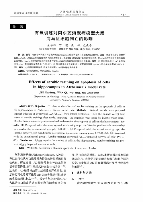
有氧训练对阿尔茨海默病模型大鼠海马区细胞凋亡的影响
21 0 0年第 1 4卷第 1 5期
实 用 临 床 医 药 杂 志
J u n l fCl c 1 e iiei atc o r a i a dcn nPrcie o ni M
有 氧 训 练 对 阿 尔 茨 海 默 病 模 型 大 鼠 海 马 区 细 胞 凋 亡 的 影 响
金 华锋 , 万
JN H afn , A i 、, ig HI h oc u I u — g W N Q , ) T n ,S a —h n e  ̄ U Z
Dea t e t fN uo g p rm n o e rl y,Fr 钰 it o isA{ l e l a dHo i l fNajn s t p ao nigMe cl di a
摘 要 :目的
琪, 吴
婷, 史兆春
侧脑室 注射 8 粉样 淀
( 南京医科大学第一 附属 医院 神经 内科 , 江苏 南京 , 1 0 9 202 )
观察有氧训练对阿尔茨海默病( l eme) Az i r模型大鼠海马区细胞凋 亡的影 响。方 法 h
蛋 白2 A3 ) 5 ( /5 制备认知功能障碍的 A 鼠动物模型 。模型制备成功后给予四周 的有 氧训练 ,Mor 水迷宫试验检测大 鼠的 2 D rs i
认 知 功 能 , eht 色 检 测 海 马 区 细 胞 凋 亡情 况 , Hocs 染 以确 定 有 氧 训练 对 细 胞 存 活 的影 响 。 结 果 ① 同对照组相 比 , AD 鼠海 马
23699772_那米鸡早期生长发育规律研究

DOI: 10.12101/j.issn.1004-390X(n).201907029那米鸡早期生长发育规律研究*胡 瑀1,2, 邓 俊3, 邱立华1, 范新阳1, 黄 静1, 王荣平2 **, 苗永旺1 **(1. 云南农业大学 动物科学技术学院,云南 昆明 650201;2. 云南农业职业技术学院 畜牧兽医学院,云南 昆明 650212;3. 云南省畜牧总站,云南 昆明 650224)摘要: 【目的】研究那米鸡的生长发育规律。
【方法】选用 Gompertz 、Logistic 和 Von Bertalanffy 3 种常用的非线性曲线模型对那米鸡0~210日龄体质量进行了生长曲线拟合与分析。
【结果】那米鸡体质量的累积生长曲线呈现“S”形,表明其生长发育正常;表现为60日龄之前生长速度较慢,60~120日龄阶段为该鸡的生长旺盛期,120日龄以后生长速度逐渐减缓。
60日龄以前,公鸡、母鸡的体质量累积生长曲线基本一致,60日龄以后,公鸡生长速度明显快于母鸡。
3种曲线模型均能较好的拟合公、母鸡的生长发育,拟合度均达0.99以上,其中Gompertz 模型对公鸡的拟合值与实际值最为接近,Von Bertalanffy 模型对母鸡的拟合值与实际值最接近,表明Gompertz 模型对那米鸡公鸡体质量的拟合度更高(R 2=0.998),Von Bertalanffy 模型对母鸡体质量的拟合度更高(R 2=1.000)。
【结论】本研究揭示了那米鸡的生长发育规律,表明运用Gompertz 和Von Bertalan-ffy 模型对那米鸡进行生长曲线的拟合与分析是可行的,可为那米鸡的饲养管理及选育利用提供依据。
关键词: 那米鸡;生长发育;生长曲线;拟合度中图分类号: S 831.4 文献标志码: A 文章编号: 1004–390X (2021) 02−0229−06Study on the Characteristics of the Early Growth andDevelopment of Nami ChickenHU Yu 1,2,DENG Jun 3,QIU Lihua 1,FAN Xinyang 1,HUANG Jing 1,WANG Rongping 2,MIAO Yongwang 1(1. Faculty of Animal Science and Technology, Yunnan Agricultural University, Kunming 650201, China;2. Department of Animal Husbandry & Veterinary medicine, Yunnan Agricultural College of Vocational Education, Kunming 650212, China; 3. Yunnan Animal Husbandry Station, Kunming 650224, China)Abstract: [Purposes ]In order to study the growth and development characteristics of Nami chick-en.[Methods ]Three nonlinear curve model commonly used Gompertz, Logistic and Von Bertalan-ffy, were used to fit and analyze the weight of Nami chicken at the age of 0-210 days.[Results ]The cumulative growth curve of Nami chicken showed “S” shape, indicating that its growth and develop-ment was normal. This curve showed that the growth rate was slow before 60 days of age, and fast at the age of 60 to 120 days, and slowed down gradually after 120 days of age. Before 60 days old, the accumulative growth weight of cocks and hens was basically the same, and after 60 days old, the云南农业大学学报(自然科学),2021,36(2):229−234Journal of Yunnan Agricultural University (Natural Science)E-mail:********************收稿日期:2019-07-13 修回日期:2020-11-13 网络首发时间:2021-03-10 17:10:04*基金项目:迪庆州科技局项目(KX141626);国家自然科学基金项目(31760659)。
遗传学英语文献
遗传学英语文献Genetics has been a field of study that has captivated the minds of scientists and laypeople alike for centuries. The intricacies of the genetic code and its influence on the development and behavior of living organisms have been the subject of extensive research and literature. In the realm of English literature, the topic of genetics has been explored in various forms, from scientific treatises to fictional narratives.One of the seminal works in the field of genetics is Charles Darwin's "On the Origin of Species," published in 1859. This groundbreaking publication laid the foundation for the theory of evolution through natural selection, which has had a profound impact on our understanding of genetics and the diversity of life on Earth. Darwin's work not only presented his scientific findings but also engaged in a broader philosophical discourse on the implications of his theory, sparking debates and conversations that continue to this day.Another notable contribution to the literature on genetics is the work of Gregor Mendel, an Augustinian friar whose experiments with peaplants in the mid-19th century laid the groundwork for our understanding of heredity. Mendel's laws of inheritance, which describe the patterns of genetic inheritance, have become a cornerstone of modern genetics. While Mendel's work was not widely recognized during his lifetime, it has since been celebrated as a pivotal moment in the history of science.In the realm of fiction, genetics has been a recurring theme, often used as a tool to explore the ethical and social implications of scientific advancements. One such example is Aldous Huxley's "Brave New World," published in 1932, which presents a dystopian future where human beings are genetically engineered and society is strictly controlled. Huxley's novel raises questions about the potential consequences of genetic manipulation and the impact it could have on individual autonomy and societal structures.Similarly, Mary Shelley's "Frankenstein," published in 1818, can be interpreted as an exploration of the ethical boundaries of scientific experimentation, particularly in the realm of creating life. The story of Victor Frankenstein's creation of a sentient being, and the subsequent consequences of his actions, has become a classic in the science fiction genre and continues to be analyzed and discussed in the context of genetics and the limits of scientific inquiry.In more recent years, the field of genetics has been further exploredin popular fiction, such as Michael Crichton's "Jurassic Park," which explores the potential of genetic engineering to resurrect extinct species. This novel, and the subsequent film adaptations, have captured the public's imagination and sparked discussions about the ethical and practical implications of such advancements.Beyond fiction, the field of genetics has also been the subject of various scientific texts and scholarly works, which have helped to advance our understanding of the genetic mechanisms that govern the development and function of living organisms. These works range from textbooks and research papers to more accessible popular science books, which aim to bridge the gap between the scientific community and the general public.One such example is James Watson and Francis Crick's "The Double Helix," a firsthand account of their groundbreaking discovery of the structure of DNA, which revolutionized our understanding of the genetic code. This book not only presents the scientific findings but also provides insights into the personalities and dynamics of the scientists involved in the research, offering a glimpse into the human side of scientific discovery.Another notable work in the field of genetics literature is "The Selfish Gene" by Richard Dawkins, published in 1976. This book presents a gene-centric view of evolution, which has had a significant impact onour understanding of the mechanisms of natural selection and the role of genetics in shaping the natural world. Dawkins' engaging writing style and thought-provoking ideas have made this book a classic in the field of evolutionary biology and genetics.In conclusion, the field of genetics has been the subject of a rich and diverse body of English literature, spanning from scientific treatisesto imaginative works of fiction. These literary contributions have not only advanced our understanding of the genetic mechanisms that govern living organisms but have also explored the ethical, social, and philosophical implications of our growing knowledge in this field. As the field of genetics continues to evolve, it is likely that we will see new and innovative perspectives emerge in the literature, further enriching our understanding of this captivating and ever-expanding area of study.。
发酵肉制品中的特征风味与微生物之间的关系研究进展
张鹏,赵金山,臧金红,等. 发酵肉制品中的特征风味与微生物之间的关系研究进展[J]. 食品工业科技,2024,45(2):380−391. doi:10.13386/j.issn1002-0306.2023030223ZHANG Peng, ZHAO Jinshan, ZANG Jinhong, et al. Progress of Research on the Relationship between Characteristic Flavor and Microorganisms in Fermented Meat Products[J]. Science and Technology of Food Industry, 2024, 45(2): 380−391. (in Chinese with English abstract). doi: 10.13386/j.issn1002-0306.2023030223· 专题综述 ·发酵肉制品中的特征风味与微生物之间的关系研究进展张 鹏1,2,赵金山2,3, *,臧金红1,2, *,彭传涛1,2(1.青岛农业大学食品科学与工程学院,山东青岛 266109;2.青岛特种食品研究院,山东青岛 266109;3.青岛农业大学动物科技学院,山东青岛 266109)摘 要:发酵肉制品是指在自然或者人工的控制条件下,以新鲜的肉类为原料,通过微生物或酶的发酵作用制成的一类产品。
发酵肉制品中特征风味物质种类繁多,其中主要有酯类、醛类、醇类、酸类以及游离氨基酸等。
由于消费者对发酵肉制品风味品质要求的提高,风味物质的控制已经成为提升发酵肉制品品质的关键指标之一。
发酵肉制品中的部分微生物特别是乳酸菌、酵母菌和葡萄球菌促进了肉制品中碳水化合物、蛋白质和脂肪的分解,从而促进发酵肉制品独特风味的形成。
本文详细介绍了国内外不同发酵肉制品中的主要风味物质、风味形成途径和检测方法,进一步归纳总结了能够产生这些风味物质的关键微生物以及产风味物质的微生物筛选方法,以期为后续发酵肉制品风味品质的提升提供参考。
Occurrence, Synthesis, and Mammalian
Occurrence,Synthesis,and Mammalian Cell Cytotoxicity and Genotoxicity of Haloacetamides:An Emerging Class of Nitrogenous Drinking Water Disinfection ByproductsM I C H A E L J.P L E W A,*,†M A R K G.M U E L L N E R,†S U S A N D.R I C H A R D S O N,‡F R A N C E S C A F A S A N O,‡,⊥K A T H E R I N E M.B U E T T N E R,‡Y I N-T A K W O O,§ A.B R U C E M C K A G U E,|A N D E L I Z A B E T H D.W A G N E R†College of Agricultural,Consumer and Environmental Sciences,Department of Crop Sciences,University of Illinois at Urbana–Champaign,1101West Peabody Drive,Urbana,Illinois61801,National Exposure Research Laboratory,U.S.Environmental Protection Agency,Athens, Georgia30605,Risk Assessment Division,Office of Pollution Prevention and Toxics,U.S.Environmental Protection Agency, Washington,DC20460,and CanSyn Chemical Corp.,200College Street,Toronto,Canada M5S3E5Received July16,2007.Revised manuscript received October 24,2007.Accepted October29,2007.The haloacetamides,a class of emerging nitrogenous drinking water disinfection byproduct(DBPs),were analyzed for their chronic cytotoxicity and for the induction of genomic DNA damage in Chinese hamster ovary cells.The rank order for cytotoxicity of13haloacetamides was DIAcAm>IAcAm> BAcAm>TBAcAm>BIAcAm>DBCAcAm>CIAcAm> BDCAcAm>DBAcAm>BCAcAm>CAcAm>DCAcAm> TCAcAm.The rank order of their genotoxicity was TBAcAm >DIAcAm≈IAcAm>BAcAm>DBCAcAm>BIAcAm> BDCAcAm>CIAcAm>BCAcAm>DBAcAm>CAcAm> TCAcAm.DCAcAm was not genotoxic.Cytotoxicity and genotoxicity were primarily determined by the leaving tendency of the halogens and followed the order I>Br>>Cl.With theexceptionofbrominatedtrihaloacetamides,mostofthetoxicity rank order was consistent with structure–activity relationship expectations.For di-and trihaloacetamides,the presence of at least one good leaving halogen group(I or Br but not Cl) appears to be critical for significant toxic activity.Log P was not a factor for monohaloacetamides but may play a role in the genotoxicity of trihaloacetamides and possible activation of dihaloacetamides by intracellular GSH and-SH compounds.With the advent of the U.S.EPA Stage2DBP regulations, water utilities are considering the use of disinfectants that are alternativestochlorine.Theuseofthesealternativedisinfectants willshiftthedistributionofDBPchemicalclasses.Theemergence of new,highly toxic iodinated,nitrogenous DBPs,as illustrated by the discovery of bromoiodoacetamide as a new DBP, underscores the importance of comparative toxicity studies to assist in the overall goal of safer drinking water practice. IntroductionWhen a disinfectant reacts with natural organic matter and/ or bromide/iodide in raw water,drinking water disinfection byproducts(DBPs)are unintentionally formed(1).Although the acute benefits of drinking water disinfection are uni-versally acknowledged,there is concern about adverse health risks due to long-term DBP exposure(2,3).Epidemiology studies provide moderate evidence for DBP associations with adverse pregnancy outcomes(4–8);specific DBPs are mu-tagens,carcinogens,teratogens,or developmental toxicants (9–18).In2006,the U.S.Environmental Protection Agency (EPA)promulgated the Stage2Disinfectants/Disinfection Byproducts Rule(19).In order to comply with this Rule,a number of utilities are switching from chlorine to chloramine disinfection;this may increase nitrogenous disinfection byproducts(N-DBPs),such as acetamides.N-DBPs were cited as research priorities by the U.S.EPA(20,21)and in a recent review on the occurrence,toxicity,and research priorities on emerging DBPs(22).Acetamides have been identified as DBPs from drinking water treatment plants and from laboratory studies(23–26). Haloamides were recently quantified in a Nationwide Oc-currence Study of priority,unregulated DBPs(21).Chloro-, bromo-,dichloro-,dibromo-,and trichloroacetamide were found infinished drinking water from several locations (maximum of14µg/L)from water treated with chlorine dioxide-chlorine-chloramines.As with the formation of haloacetonitriles(27),there is preliminary evidence that chloramination may increase their formation.Because nitriles can hydrolyze to form haloamides(28,29),it is possible that the haloamides are hydrolysis products of the corresponding haloacetonitriles,which are commonly found as DBPs.The analyzed haloacetamides and their abbreviations are listed in Table1.There is little information on the genotoxicity and carcinogenicity of haloamides.These agents react with cellular protein thiols and are prototypical alkylating agents inducing a multitude of biological responses,including apoptosis and necrosis(30).IAcAm was a cocarcinogen in a mouse skin assay(31)and enhanced nitrosamide-induced tumors in rats(32,33).The objective of our research was to quantitatively analyze and compare the chronic cytotoxicity and the acute genomic DNA damaging capacity of13haloacetamides using in vitro mammalian cell assays extensively used in DBP research (27,34–38).In addition,we identified the occurrence of a new iodinated DBP,bromoiodoacetamide,in drinking water. Materials And MethodsReagents.General reagents were purchased from Fisher Scientific Co.(Itasca,IL)and Sigma Chemical Co.(St.Louis, MO).Media and fetal bovine serum(FBS)were purchased from Hyclone Laboratories(Logan,UT).The haloacetamides were dissolved in dimethyl sulfoxide(DMSO)and stored at -22°C in sterile glass vials.Sources and purities of thehaloacetamides are presented in Table1.*Corresponding author phone:(217)333-3614;e-mail mplewa@.†University of Illinois at Urbana–Champaign.‡National Exposure Research Laboratory,U.S.EnvironmentalProtection Agency.§Office of Pollution Prevention and Toxics,U.S.EnvironmentalProtection Agency.|CanSyn Chemical Corp.⊥Currently at the University of Torino,Torino,Italy10125.Environ.Sci.Technol.2008,42,955–96110.1021/es071754h CCC:$40.75 2008American Chemical Society VOL.42,NO.3,2008/ENVIRONMENTAL SCIENCE&TECHNOLOGY9955 Published on Web12/12/2007Preparation of Haloacetamides.Eight haloacetamides were synthesized.DBAcAm was prepared from ethyl dibro-moacetate by reaction with ammonium hydroxide(39). BCAcAm was prepared from bromochloroacetic acid by conversion to the ethyl ester followed by reaction with ammonium hydroxide(39).BDCAcAm and DBCAcAm were prepared from the corresponding acids(40,41)by conversion to the methyl esters with BF3/MeOH followed by reaction with ammonium hydroxide(40).TBAcAm was prepared from the acid in a similar manner.BIAcAm was similarly prepared from bromoiodoacetic acid to give colorless material,mp 181–183°C.The purity of the product by gas chromatography (GC)usingflame ionization detection was85%and contained 7.5%each of DBAcAm and DIAcAm as impurities.CIAcAm was prepared from methyl chloroiodoacetate(42)and DIAcAm was prepared from diiodoacetic acid(43)via the methyl ester,by similar reaction with ammonium hydroxide.Chinese Hamster Ovary Cells.Chinese hamster ovary (CHO)cells,line AS52,clone11-4-8were used(44).We employed this cell line in previous DBP toxicity studies (27,34–38,45,46).The CHO cells were maintained in Ham’s F12medium containing5%FBS at37°C in a humidified atmosphere of5%CO2.CHO Cell Chronic Cytotoxicity Assay.This calibrated assay measures the reduction in cell density as a function of DBP concentration over a period of approximately3cell divisions(72h)(27,34–38,45,46).Detailed procedures with statistical analysis are presented in the Supporting Informa-tion.Within each experiment,10haloacetamide concentra-tions were analyzed with8replicate microplate wells for each concentration.Each experiment was repeated2–3×.Thus, the cytotoxicity of each concentration was evaluated with 16-24individual cell cultures.Single Cell Gel Electrophoresis Assay.Single cell gel electrophoresis(SCGE)quantitatively measures genomic DNA damage induced in individual nuclei of treated cells (47).We employed this calibrated assay to determine the genotoxicity of DBPs(27,34–38,45,46).Detailed procedures and statistical analysis are presented in the Supporting Information.CHO cells were exposed to a haloacetamide for 4h at37°C,5%CO2.Each experiment consisted of a negative control,a positive control(3.8mM ethylmethanesulfonate), and9haloacetamide concentrations.The concentration range was determined by the acute cytotoxicity of the haloacetamide.In general,each concentration was evaluated with2microgels with25randomly chosen nuclei per microgel.The experiments were repeated a minimum of3×with6microgels per concentration.Drinking Water Analysis.Drinking water samples were collected from full-scale drinking water treatment plants in the United States that use chloramines for disinfection.One plant used chlorine for disinfection.Details regarding the drinking water treatment,concentration,and analysis can be found in the Supporting Information.GC/mass spec-trometry(MS)analyses with electron ionization(EI)were carried out on an Agilent6890GC interfaced to a Waters-Micromass Autospec II high resolution,double focusing mass spectrometer at1000resolution.Results and DiscussionIdentification and Occurrence of a New Iodinated Aceta-mide.We identified BIAcAm as a DBP for thefirst time in drinking water from12treatment plants(of a total of23 analyzed)and located in6U.S.states.One plant used chlorine for disinfection;22plants used chloramination.These plants had source waters with relatively high natural bromide and iodide levels.Three of the plants had very small amounts of BIAcAm in their raw waters,at levels500×lower than the finished waters.The other9plants only had BIAcAm in their finished waters,and not in their corresponding raw waters. Using GC with selected ion monitoring-MS,we did not detect IAcAm,CIAcAm,or DIAcAm.The EI mass spectrum of BIAcAm is shown in Figure1. Selected ion monitoring of5key ions(m/z127,136,138,220, and263)was used to identify this compound in the drinking water extracts.A match of these ions with a match of the GC retention time was used to confirm its presence.All haloa-mides measured expressed distinctive GC/MS chromato-graphic peak shapes(Figure1,Supporting Information).This distinctive“tailing”peak shape appears to be due to surface reactions of the haloamides in the EI ion source and provided further confirmation of BIAcAm.All of the haloamides show a prominent peak at m/z44,which represents the amide group(Figure1).The presence of bromine and iodine is evident in the mass spectrum of BIAcAm,with1-bromine doublets present at m/z263/265,220/222,and136/138,and the presence of iodine at m/z127and loss of iodine at m/z 136/138.Chlorinated and brominated forms were measured in drinking water previously(21),but this is thefirst report of an iodinated amide.Naturally occurring bromide and iodide contribute to the formation of brominated and iodinated DBPs(25,37,38,48–51).There is evidence that chlorami-nation increases the formation of iodinated DBPs(37,52,53). Therefore,while this is thefirst report of an iodinated amide DBP,it is not surprising that iodo-amides would form in source waters with high bromide/iodide and chloramine disinfection.As mentioned earlier,it is possible that BIAcAm and other haloamides are hydrolysis products of the cor-responding haloacetonitriles,which are commonly found as DBPs.The amides can undergo further hydrolysis to form carboxylic acids,but this reaction requires longer reaction times and higher temperatures than the initial conversion to the amide(29).TABLE1.Characteristics of the Haloacetamides Analyzed in This Studyhaloacetamide abbreviation CAS no.chemical formula MW a source and purity iodoacetamide IAcAm144–48–9C2H4INO184.96Sigma-Aldrich,>97% diiodoacetamide DIAcAm5875–23–0C2H3I2NO310.85CanSyn Chem Corp.,99% bromoiodoacetamide BIAcAm62872–36–0C2H3BrINO263.856CanSyn Chem Corp.,85%b chloroiodoacetamide CIAcAm62872–35–9C2H3ClINO219.405CanSyn Chem Corp.,>95% bromoacetamide BAcAm683–57–8C2H4BrNO137.96Sigma-Aldrich,98% dibromoacetamide DBAcAm598–70–9C2H3Br2NO216.86CanSyn Chem Corp.,>95% tribromoacetamide TBAcAm594–47–8C2H2Br3NO295.75CanSyn Chem Corp.,>95% bromochloroacetamide BCAcAm62872–34–8C2H3BrClNO172.41CanSyn Chem Corp.,95% dibromochloroacetamide DBCAcAm855878–13–6C2H2Br2ClNO251.305CanSyn Chem Corp.,>95% bromodichloroacetamide BDCAcAm98137–00–9C2H2BrCl2NO206.85CanSyn Chem Corp.,>95% chloroacetamide CAcAm79–07–2C2H4ClNO93.51Sigma-Aldrich,>95% dichloroacetamide DCAcAm683–72–7C2H3Cl2NO127.96Sigma-Aldrich,98% trichloroacetamide TCAcAm594–65–0C2H2Cl3NO162.40Sigma-Aldrich,99%a MW)molecular weight.b7.5%each of dibromoacetamide and diiodoacetamide as impurities.9569ENVIRONMENTAL SCIENCE&TECHNOLOGY/VOL.42,NO.3,2008CHO Cell Cytotoxicity.Data from individual experiments were normalized to the averaged percent of the concurrent negative control;these data were plotted as a concentra-tion–response curve (Figure 2).An ANOVA test was conducted with normalized data representing each microplate well.If a significant F value of P e 0.05was obtained,a Holm -Sidak multiple comparison analysis was conducted.The power of the test statistic (1- )was maintained at g 0.8at R )0.05.The lowest concentration that induced a significant cytotoxic response ranged from 25nM (DIAcAm)to 800µM (DCAcAm)(Table 2).Regression analysis was conducted on each concentration–response curve;the coefficient of determi-nation (R 2)ranged from 0.95to 0.99.From these concen-tration–response curves,the %C½value was calculated (Table 2).The %C½value is the concentration that induced a cell density of 50%as compared to the concurrent negative control.The %C½values ranged from 678nM (DIAcAm)to 2.05mM (TCAcAm).The rank order for cytotoxicity of the 13haloacetamides based on their %C½values was DIAcAm>IAcAm >BAcAm >TBAcAm >BIAcAm >DBCAcAm >CIAcAm >BDCAcAm >DBAcAm >BCAcAm >CAcAm >DCAcAm >TCAcAm.CHO Cell Genotoxicity.Acute genomic DNA damage was measured as SCGE tail moment values.Acute cytotoxicity was determined with trypan blue vital dye;data were used within the range that contained g 70%viable cells (Table 2,Figure 3).The data were plotted,and regression analysis was used to fit the curve;the coefficient of determination (R 2)ranged from 0.97to 0.99.SCGE tail moment values are not normally distributed,thus the median tail moment value for each microgel was determined and averaged.Averaged median values express normal distributions according to the Central Limit theorem (54)and were used with an ANOVA test.If a significant F value of P e 0.05was obtained,a Holm -Sidak multiple comparison analysis was conducted (1- g 0.8at R )0.05,Table 2).The lowest concentration that induced a significant SCGE genotoxic response ranged from 25µM for DIAcAm,BIAcAm,BAcAm,andDBCAcAmFIGURE 1.EI mass spectrum ofbromoiodoacetamide.FIGURE parison of the CHO cell chronic cytotoxicity concentration–response curves of 13haloacetamides.VOL.42,NO.3,2008/ENVIRONMENTAL SCIENCE &TECHNOLOGY9957to 5mM for TCAcAm.The SCGE genotoxic potency was calculated at the midpoint of the concentration–response curve,and ranged from 32.5µM for TBAcAm to 6.5mM for TCAcAm (Table 2).The rank order of genotoxic potency from most potent to least was TBAcAm >DIAcAm ≈IAcAm >BAcAm >DBCAcAm >BIAcAm >BDCAcAm >CIAcAm >BCAcAm >DBAcAm >CAcAm >TCAcAm.DCAcAm was not genotoxic (Table 2).Structure–Activity Relationships (SARs)and Factors Affecting the Toxicity of Haloacetamides.The haloaceta-mides have or may generate a number of electrophilic reactivities:(i)for monohaloacetamides,alkylation by the S N 2reaction,inducing the displacement of a halogen atomat the R carbon,(ii)for dihaloacetamides,the potential generation of highly reactive R -halothioether electrophilic intermediates by cellular glutathione GSH or -SH compounds,(iii)for trihaloacetamides,nucleophilic attack at the elec-trophilic carbonyl carbon to yield trihalomethyl carbanions,which in turn,may lead to trihalomethanes as well as electrophilic dihalocarbene intermediates.In addition to chemical reactivity,the capacity to cross cell membranes is an important factor for toxicity.The logarithm of the octanol–water partition coefficient (log P)is a measure of lipophilicity which correlates with cell permeability.Log P increased with the degree of halogenation and with the sizeTABLE 2.Summary Comparison of the CHO Cell Chronic Cytotoxicity and Acute Genotoxicity of the Haloacetamideschemicallowest toxic conc.(M)a %C½Value (M)b R 2c lowest genotox.conc.(M)d genotox.potency (M)eR 2f iodoacetamide 5.00×10⁻⁷ 1.42×10-60.98 3.00×10⁻⁵ 3.41×10-50.99diiodoacetamide2.50×10-8 6.78×10-70.98 2.50×10-53.39×10-50.98bromoiodoacetamide 2.00×10⁻⁶ 3.81×10⁻⁶g 0.98 2.50×10-57.21×10-5h 0.99chloroiodoacetamide 2.00×10⁻⁶ 5.97×10-60.96 2.00×10⁻⁴ 3.02×10-40.99bromoacetamide 0.50×10⁻⁶ 1.89×10-60.99 2.50×10⁻⁵ 3.68×10-50.99dibromoacetamide 2.50×10⁻⁶ 1.22×10-50.99 5.00×10-47.44×10-40.99tribromoacetamide2.00×10-63.14×10-60.97 3.00×10⁻⁵ 3.25×10-50.97bromochloroacetamide 1.00×10⁻⁶ 1.71×10-50.984.00×10-45.83×10-40.99dibromochloroacetamide 1.00×10⁻⁶ 4.75×10-60.96 2.50×10-56.94×10-50.98bromodichloroacetamide 2.00×10-68.68×10-60.987.50×10-5 1.46×10-40.99chloroacetamide 7.50×10⁻⁵ 1.48×10-40.987.50×10-4 1.38×10-30.99dichloroacetamide8.00×10-4 1.92×10-30.95NANS >1×10⁻²NA trichloroacetamide5.00×10-42.05×10-30.965.00×10-36.54×10-30.98aLowest toxic concentration was the lowest concentration of the haloacetamide in the concentration–response curve that induced a significant reduction in cell density as compared to the negative control.b The %C 1/2value is the concentration of the haloacetamide,determined from a regression analysis of the data,that induced a cell density of 50%as compared to the concurrent negative control.c R 2is the coefficient of determination for the regression analysis upon which the %C 1/2value was calculated.d The lowest genotoxic concentration was the lowest concentration of the haloacetamide in the concentration–response curve that induced a significant amount of genomic DNA damage as compared to the negative control.e The SCGE genotoxic potency is the haloacetamide concentration that was calculated,using regression analysis,at the midpoint of the curve within the concentration range that expressed above 70%cell viability of the treated cells.f R 2is the coefficient of determination for the regression analysis upon which the genotoxic potency value was calculated.gThe calculated %C 1/2value for BIAcAm alone assuming an additive model for the DIAcAm and DBAcAm contaminants was 3.35×10⁻⁶M.h The calculated SCGE genotoxic potency value for BIAcAm alone assuming an additive model for the DIAcAm and DBAcAm contaminants was 1.62×10⁻⁵M.NA )not applicable;NS )not statistically significant from the negativecontrol.FIGURE parison of the CHO cell acute genotoxicity concentration–response curves of 13haloacetamides.9589ENVIRONMENTAL SCIENCE &TECHNOLOGY /VOL.42,NO.3,2008of the halogen.Only the estimated log P values were used in the present study (Table 3).For the 13haloacetamides,CHO cell chronic cytotoxicity and acute genotoxicity were highly and significantly cor-related (r )0.99;P <0.001).The rank order and relative activities follow:monohaloacetamides (cytotoxicity and genotoxicity),I >Br >>Cl;dihaloacetamides (cytotoxicity),I 2>IBr >ICl >Br 2>BrCl >>Cl 2,and (genotoxicity),I 2>IBr >ICl >BrCl >Br 2;Cl 2was inactive;trihaloacetamides (cytotoxicity and genotoxicity)Br 3>Br 2Cl >BrCl 2>>Cl 3.The rank order and relative activity of the monohaloac-etamides are related to their S N 2reactivity.Owing to increasing bond length and decreasing dissociation energy,the leaving tendency of the halogen in alkyl halides followed the order I >Br >>Cl.The S N 2reactivity of an alkyl iodide was 3–5×greater than alkyl bromide,which was 50×greater than alkyl chloride (37,55).The cytotoxicity of IAcAm was 1.3×greater than BAcAm,which was 78×greater than CAcAm.IAcAm was more genotoxic than BAcAm,which was 38×more potent than CAcAm.Log P does not play a major role;the small difference in the log P of BAcAm vs CAcAm cannot account for the large difference in relative activity.Consistent with the relative leaving tendencies of the halogen,dihaloacetamides containing the most iodo group(s)expressed the greatest combined cyto-and genotoxicity indices,followed by bromo group(s)and chloro group(s)(Table 3).DCAcAm was weakly cytotoxic and was not genotoxic.These results are difficult to explain by S N 2reactivity alone but may involve the activation of dihaloac-etamides by intracellular GSH or -SH compounds,which displace one halogen and form highly reactive R -halothio-ether electrophilic intermediates.The key element of this reaction is the presence of at least one halogen with good leaving tendency.With GSH-mediated activation,the weak activity of DCAcAm and similar combined toxicity indices between DBAcAm vs BCAcAm may be expected (Table 3).The estimated log P values followed the order I 2>IBr >ICl >Br 2>BrCl >Cl 2.This relative order is identical to their cytotoxicity and genotoxicity.Log P may play a more important role in the activity of dihaloacetamides by affecting cellular uptake.The cytotoxicity and genotoxicity of trihaloacetamides decreased with a decrease in the number of bromo groups.The cytotoxicity of TCAcAm was lower than TBAcAm by almost 3orders of magnitude;this confirmed results in human leukemia P388cells (56).Only one bromo group was required for potent cytotoxicity;the %C 1/2values of TBAcAm,DBCAcAm,and BDCAcAm were within the same order of magnitude.In contrast,the decrease in genotoxic potency with a decrease in the number of bromo groups was more gradual (Table 2).The cytotoxicity and genotoxicity of trihaloacetamides could be partially explained by electro-philic reactivity at the carbonyl carbon as well as the possible release of electrophilic dihalocarbene intermediates (see discussion above).Alternatively,it is possible that reductive dehalogenation may yield cytotoxic free radicals;this pathway and the metabolic competency of the CHO cells have only been partially defined (57).Glutathione S-transferase theta 1-1(GST T1-1)catalyzes preferential activation of brominated trihalomethanes to genotoxic intermediates (58,59);the possible role of GST T1-1in the activation of trihaloaceta-mides in CHO cells remains to be explored.Comparison of the Toxicity of Haloacetamides to Other DBP Classes.We compared the mammalian cell cytotoxic potencies,and genotoxic potencies between the haloaceta-mides and other DBP chemical classes,by calculating the CHO cell chronic cytotoxicity and acute genotoxicity indices (Figure 4).The cytotoxicity index was determined by calculating the median %C 1/2value of all of the individual members of a single class of DBPs.The reciprocal was taken of this number so that a larger value was equated with that of higher cytotoxic potency.The genotoxicity index was determined by calculating the median SCGE genotoxicity potency value from the individual members within a single class of DBPs.The reciprocal was taken of this number so that a larger value was equated with that of higher geno-toxicity.As a class,the haloacetamides were 99×more cytotoxic than 13haloacetic acids (60),142×more cytotoxic than the 5regulated haloacetic acids (35),2×more cytotoxic than the haloacetonitriles (27),and 1.4×more cytotoxic than the halonitromethanes (36).The haloacetamides were 19×more genotoxic than 13haloacetic acids (60),12×more genotoxic than the 5regulated haloacetic acids (35),and 2.2×more genotoxic than the halonitromethanes (36).The haloacetamides were slightly less genotoxic (0.9×)than the haloacetonitriles (27).With the enforcement of the U.S.EPA Stage 2DBP regulations,water utilities are considering the use of dis-infectants that are alternatives to chlorine.The use of these alternative disinfectants will shift the distribution of DBPTABLE 3.Estimated and Measured log P and Combined Toxicity Values of the Haloacetamides Studiedcompoundestimated log P a measured log P combined toxicity value bmonohaloacetamides iodoacetamide -0.08-0.19c ;-0.15d 5.63×10⁴bromoacetamide -0.49-0.52c ,d 5.17×10⁴chloroacetamide -0.58-0.53c ;-0.59d1.31×103dihaloacetamides diiodoacetamide 0.92 5.78×10⁴dibromoacetamide 0.090.18d2.64×103dichloroacetamide -0.090.19c ;-0.03d 1.68×10²bromoiodoacetamide 0.50 1.02×10⁵e chloroiodoacetamide 0.41 6.49×10³bromochloroacetamide 0.003.33×103trihaloacetamides tribromoacetamide1.10 5.61×10⁴dibromochloroacetamide 1.012.70×104bromodichloroacetamide 0.92 1.29×10⁴trichloroacetamide0.83 1.04c ;0.79d2.33×102aCalculated using the KOWWIN program (version 1.67)developed by U.S.EPA using the atom/fragment approach(program available at /oppt/newchems/pubs/sustainablefutures.htm).b The Combined Toxicity Index is the reciprocal of the averaged %C 1/2and the SCGE genotoxic potency values.A larger Toxicity Index value indicates greater overall toxicity.c Based on data compiled by (62).d Based on data compiled by (63).e Calculation based on the estimated BIAcAm values alone.VOL.42,NO.3,2008/ENVIRONMENTAL SCIENCE &TECHNOLOGY9959chemical classes (21,37,61).The emergence of new,highly toxic iodinated,nitrogenous DBPs,such as BIAcAm,under-scores the importance of comparative toxicity studies to assist in the overall goal of safer drinking water practice.AcknowledgmentsThis paper is dedicated to Professor Saburo Matsui in celebra-tion of his service to and retirement from Kyoto University and to recognize his outstanding contributions in environmental chemistry and engineering.We thank Gene Crumley for assistance with drinking water extractions and MS analyses,Ed Sverko of Environment Canada for providing an analysis using aninertsource,andFredMengerofEmoryUniversityforhelpful discussions.We are especially grateful to Prof.Marco Vincenti of the University of Torino (Italy)for his support of F.F.and promoting her collaboration with EPA-Athens on this study.This research was funded in part by AwwaRF Grant 3089,U.S.EPA Cooperative Agreement CR83069501,and Illinois-Indiana Sea Grant R/WF-09-06(M.J.P.and E.D.W.).M.M.was supported by T32ES07326(NIEHS).We appreciate the support by the Center of Advanced Materials for the Purification of Water with Systems,a National Science Foundation Science and Technol-ogy Center,under Award CTS-0120978.This paper has been reviewed in accordance with the U.S.EPA’s peer and admin-istrative review policies and approved for publication.Mention of trade names or commercial products does not constitute endorsement or recommendation for use by the U.S.EPA.Note Added after ASAP PublicationThis paper was published ASAP December 12,2007with an errorinthefirstparagraphoftheResultsandDiscussionsection;the corrected version published ASAP December 27,2007.Supporting Information AvailableDetailed information on the sampling,extraction and concentration of drinking water;the subsequent GC/MS analysis;and the biological assays.This information is available free of charge via the Internet at .Literature Cited(1)Richardson,S.D.Drinking water disinfection by-products.InThe Encyclopedia of Environmental Analysis and Remediation ;Wiley:New York,1998;Vol.3,pp 1398-1421.(2)Richardson,S.D.;Simmons,J.E.;Rice,G.Disinfection byprod-ucts;the nextgeneration.Environ.Sci.Technol.2002,36,198A–205A.(3)Betts,K.Growing concern about disinfection by-products.Environ.Sci.Technol.1998,32,546.(4)Waller,K.;Swan,S.H.;DeLorenze,G.;Hopkins,B.Trihalom-ethanes in drinking water and spontaneous abortion.Epide-miology 1998,9,134–140.(5)Swan,S.H.;Waller,K.;Hopkins,B.;Windham,G.;Fenster,L.;Schaefer,C.;Neutra,R.R.A prospective study of spontaneous abortion:relation to amount and source of drinking water consumed in early pregnancy.Epidemiology 1998,9,126–133.(6)Waller,K.;Swan,S.H.;Windham,G.C.;Fenster,L.Influenceof exposure assessment methods on risk estimates in an epidemiologic study of total trihalomethane exposure and spontaneous abortion.J.Expos.Anal.Environ.Epidemiol.2001,11,522–531.(7)Bove,F.;Shim,Y.;Zeitz,P.Drinking water contaminants andadverse pregnancy outcomes:a review.Environ.Health Perspect.2002,110Suppl 1,61–74.(8)Savitz,D.A.;Singer,P.C.;Hartmann,K.E.;Herring,A.H.;Weinberg,H.S.;Makarushka, C.;Hoffman, C.;Chan,R.;Maclehose,R.Drinking Water Disinfection By-products and Pregnancy Outcome ;AWWA Research Foundation:Denver,CO,2005.(9)Bull,R.J.;Meier,J.R.;Robinson,M.;Ringhand,H.P.;Laurie,R.D.;Stober,J.A.Evaluation of mutagenic and carcinogenic properties of brominated and chlorinated acetonitriles:by-products of chlorination.Fundam.Appl.Toxicol.1985,5,1065–1074.(10)Ahmed,A.E.;Campbell,G.A.;Jacob,S.Neurological impairmentin fetal mouse brain by drinking water disinfectant byproducts.Neurotoxicology 2005,26,633–640.(11)Daniel,F.B.;Schenck,K.M.;Mattox,J.K.;Lin,E.L.;Haas,D.L.;Pereira,M.A.Genotoxic properties of haloacetonitriles:drinking water by-products of chlorine disinfection.Fundam.Appl.Toxicol.1986,6,447–453.(12)Lin,E.L.;Daniel,F.B.;Herren-Freund,S.L.;Pereira,M.A.Haloacetonitriles:metabolism,genotoxicity,and tumor-initiat-ing activity.Environ.Health Perspect.1986,69,67–71.(13)Muller-Pillet,V.;Joyeux,M.;Ambroise, D.;Hartemann,P.Genotoxic activity of five haloacetonitriles:Comparative in-vestigations in the single cell gel electrophoresis (Comet)assay and the Ames-fluctuation test.Environ.Mol.Mutagen.2000,36,52–58.(14)Smith,M.K.;George,E.L.;Zenick,H.;Manson,J.M.;Stober,J. A.Developmental toxicity of halogenated acetonitriles:drinking water by-products of chlorine disinfection.Toxicology 1987,46,83–93.(15)Villanueva,C.M.;Cantor,K.P.;Cordier,S.;Jaakkola,J.J.;King,W.D.;Lynch,C.F.;Porru,S.;Kogevinas,M.Disinfection byproducts and bladder cancer:a pooled analysis.Epidemiology 2004,15,357–367.(16)Villanueva,C.M.;Cantor,K.P.;Grimalt,J.O.;Malats,N.;Silverman,D.;Tardon,A.;Garcia-Closas,R.;Serra,C.;Carrato,A.;Castano-Vinyals,G.;Marcos,R.;Rothman,N.;Real,F.X.;Dosemeci,M.;Kogevinas,M.Bladder cancer and exposure to water disinfection by-products through ingestion,bathing,showering and swimming in pools.Am.J.Epidemiol.2007,165,148–156.(17)Cantor,K.P.;Villanueva,C.;Garcia-Closas,M.;Silverman,D.;Real,F.X.;Dosemeci,M.;Malats,N.;Yeager,M.;Welch,R.;Chanock,S.Bladder cancer,disinfection byproducts,and markers of genetic susceptibility in a case-control study from Spain.Epidemiology 2006,17,S150.(18)Villanueva,C.M.;Cantor,K.P.;Grimalt,J.O.;Castaño-Vinyals,G.;Malats,N.;Silverman,D.;Tardon,A.;Garcia-Closas,R.;Serra,C.;Carrato,A.Assessment of lifetime exposure to trihalom-ethanes through different routes.Occup.Environ.Med.2006,63,273–277.(19)U.S.Environmental Protection Agency National Primary Drink-ing Water Regulations:Stage 2Disinfectants and Disinfection Byproducts Rule.Fed.Regist.2006,71,387–493.(20)Woo,Y.T.;Lai,D.;McLain,J.L.;Manibusan,M.K.;Dellarco,e of mechanism-based structure-activity relationships analysis in carcinogenic potential ranking for drinking water disinfection by-products.Environ.Health Perspect.2002,110Suppl 1,75–87.(21)Krasner,S.W.;Weinberg,H.S.;Richardson,S.D.;Pastor,S.J.;Chinn,R.;Sclimenti,M.J.;Onstad,G.D.;Thruston,A.D.,Jr.The occurrence of a new generation of disinfection by-products.Environ.Sci.Technol.2006,40,7175–7185.(22)Richardson,S.D.;Plewa,M.J.;Wagner,E.D.;Schoeny,R.;DeMarini,D.M.Occurrence,genotoxicity,and carcinogenicityFIGURE parison of the CHO cell chronic cytotoxicity index values and the CHO genotoxicity index values for a series of DBP chemical classes.9609ENVIRONMENTAL SCIENCE &TECHNOLOGY /VOL.42,NO.3,2008。
木犀草素预防高脂介导的小鼠胸主动脉血管内皮依赖性舒张功能损害
L t ol r v n s廿 I p i do MieAo t ue i nP e e t 埒 m ar e f c r aEn i t e l m De e d n V s e a a i nI d c d b i hF t e c hl o u p n e t a or lx t o n u e yH g a Dit
压 水 平显著高 于j 常饮食对 照组 及高脂 饮食+ 犀草 索组 ,P<O0 。② 长期商脂 膳食损 害小 鼠胸主 动脉 的血管 内皮依赖 性舒 张功 能,P . ;而 l 木 .5 <0 5 0 木犀 草素 预防性膳 食干 预可 防 I血管 内皮依赖 性舒 张功能 的损害 ③ 木犀 革素孵 茸可改 善高脂 小鼠胸主 动脉 的内皮 依赖 性舒 张功能 , 陔作用 可被 但 PARY的特 异性 阻断剂 GW96 P 62阻断 。结论 木犀 草索可预 防高 脂膳食介 导的 小 鼠胸 主动脉血 管 内皮 功能损 害,该作 用 可能 与激活 PARY 关 。 P 有
文章 编号 : 17・79 (0 1 2 ・160 622 7 2 1)-20 4-3 关键 词 :木犀 草素 ; 高脂膳 食 ;血 管 内皮 依 赖性 舒张 功 能;P A P RY d i 1. 6/i n17-7 9 0 1 213 o: 03 9 .s.622 7 . 1. . 9 js 2 2 0
Meh d 3 c eedvd d i o t e ru s ten r l h w g o p(omie,h i t i r u 】 c) tehg a de+ %L t l to s 0mi w r iie t reg o p ,h oma c o r u 1 c ) teh h f e go p(O mi ,h i ft it2 ue i e n h g ad t e h on
在颞叶癫痫大鼠模型中海马组织中即早基因c—fos的表达
在颞叶癫痫大鼠模型中海马组织中即早基因c—fos的表达该文主要综述现公认的颞叶癫痫模型(氯化锂-匹罗卡品大鼠模型),围绕选择该模型的依据进行分析,对该模型中大鼠的海马及脑脊液组织中即早基因的表达的研究分别分析,对颞叶癫痫的发病机制的探讨,以期指导癫痫的临床治疗。
标签:颞叶癫痫;海马;即早基因据国际抗癫痫联盟统计,癫痫的患病率高达23~190人/10 万,已成为最常见的神经科疾病之一[1]。
尽管随着抗癫痫药物不断更新,治疗方案不断规范,但仍有约30% 的患者接受抗癫痫治疗后痫性发作仍不能得到有效控制。
局灶性癫痫中颞叶癫痫最为常见,近五年研究显示,在新发的癫痫患者中,1/4~1/3左右为颞叶癫痫[2]。
研究氯化锂-匹罗卡品大鼠模型所致的癫痫持续状态所致的颞叶癫痫模型中即早基因的表达以期研究颞叶癫痫的发病机制,其研究现状与进展如下。
1 氯化锂-匹罗卡品致颞叶癫痫大鼠模型的建立早在1983 年Honchar等研究人员在Science 杂志上第一次发表了匹罗卡品大鼠模型,此模型可致痫鼠诱导自发性痫性发作,损伤海马结构中易损区,该鼠致痫模型被公认为是和人类的颞叶癫痫有着相近的特征。
该模型让匹罗卡品不良反应作用所导致的高死亡率得到下降,也提高了癫痫持续状态诱发率,是目前国外较普便使用的一种癫痫模型[3]。
苏曼[4]等实验验证锂-匹罗卡品颞叶癫痫模型建模成功后按照Racine分级和人类的颞叶癫痫具有相近行为学方面的特点,诱发快,发作时容易辨别,具有明确的分期,机体死亡率较低、致痫率较高、操作简单可控而且不会破坏正常的人脑组织结构,是一种较好研究人类癫痫病理学、神经学等多科学的方法。
2 致颞叶癫痫大鼠模型的脑电图在神经电生理领域,研究致颞叶癫痫大鼠模型急性期记录到致癫痫组SD大鼠在海马区爆发长程中出现的棘活动、尖活动及不规则的尖慢复合活动,较背景脑电活动明显突出,与人类癫痫全性发作时脑电图改变一致[4]。
3 致颞叶癫痫大鼠模型的病理学表现人类癫痫的脑部病理学改变表现为海马萎缩、硬化,包括胶质增生和神经元减少,以CA1区和齿状回区域最为明显[5]。
