{选篇}Quantitative ELISA Method Development for Detection of Insulin ln
总黄曲霉毒素ELISA定量检测方法的研制
82.
b 1J Pdemersma R A,Wood D A,Macintyre C C,et a1.Risk of
angina pectoris and plasma concentrations of vitamins A,C, E and carotene[J].Lancer,1991,(337):1-5.
№
'rsukida K, Saiki K,Takii, et a1.Separation and
determination of cis/trans-p-carotenes by hish-performance
liquid chromatography[J].J Chromatogr,1982,(245):359—
column in reversed—phase liquid chromatography[J].J Agric Food Chem,1996,(44):3887.3893.
眇 1J Emenhiser C,Sander L C,Schwartz S J.Capability of a polymeric C∞stationary phase tO resolve cis-trans carotenoid
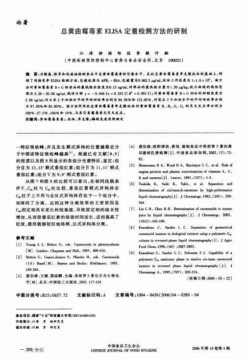
Aflatoxin(B+G)惴2.08 was Y=一O.446 3x+0.353 2(R2=0.991 5).The 50%inhibition concentration for
ng,m1.The
recovery rates of spiked maiT∞on the level of2.10腭,l‘g and 10.40/..tg/ks we托94.56%and 112.05%respectively.For peanut
elisa实验报告
竭诚为您提供优质文档/双击可除elisa实验报告篇一:eLIsA实验报告experiment:eLIsA(enzyme-linkedImmunosorbentAssay) Date:20XX.10.17objectives(1)LearneLIsAprinciple(2)mastereLIsAtechnology,quantitativelydetermineant ibodyandantigen.experimentalprincipleTheprincipleofeLIsAistomakeantigenorantibodycombine tothesurfaceofsomesolidphasecarrierandmaintainitsim munocompetence,andthenmaketheantibodyorantigentoconnectwithsomeenzymetobecomeenzymelabeledantigenorant ibodyinwhichtheimmunocompetenceofbothantigenorantibodyandenzymeisk ept.Inthedetermination,makethespecimentobetestedand enzymelinkedantigenreactwiththeantigenorantibodyont hesurfaceofsolidphasecarrierfollowingdifferenceproc edures.Throughwashing,antigen-antibodycomplexformed onsolidphasecarrierandothermaterialsareseparated.Fi nally,addingsubstrateintoenzymereaction,thesubstrat ewillbecatalyzedandbecomecoloredproduct.Theamountof producthasdirectrelationwiththeamountofenzymeonthes olidphasecarrier,inotherwords,theamountofthemateria ltobetestedinthespecimen.Quantitativeanalysiscanbec onductedbasedontheshadeofcolorreaction.Thisdetermin ationhasaveryhighsensitivity.eLIsAcanbedividedintomainlyfourtypes:thedirectmetho d,indirectmethod,doubleantibodysandwichmethodandcom petitiveinhibitionmethod.Also,differentkindsofenzym eandsubstratecanbeusedineLIsA.DoubleantibodymethodisakindofeLIsAtodetermineantige ninthespecimen.Inthismethod,antibodyiscoatedonthesu rfaceofsolidphasecarrier.Antigeninthesamplebindswit hantibodyonthesolidphase,whileothermaterialsarewash edaway.Thenaddenzymelinkedantibodytobindtheantigenw hichareboundonthesolidphase.Afterwashing,superfluou santibodyiswashedaway.Thesubstrateisthenaddedtoprod ucecolorandthequantitativetestsareconducted.Inourexperiment,weadopteddoubleantibodysandwichmeth odtodeterminemousetumornecrosisfactoralpha(TnFα)withanti-mouseTnFα.TheenzymeandsubstrateweusedareAvidin-hRpandTmbre spectively.Reagentsandequipment(1)equipments40-wellreactionplatecoatedwithanti-mouseTnF α;liquidtransfergun(1ml,200μl);thermotank(37℃).(2)Reagentswashingliquid(pbsT);sealingfluid;antigen(1000pg/ml);anti-mouseTnFα;Tmb;Avidin-hRp;1mh2so4.methodofoperation(1)emptyoutthesolutionintheplateandcleantheplatewit hwashingliquidforthreetimes.Topupeachholeandflapona bsorbentpapertocleanawaytheremainingliquidineachhol e.Add200μlsealingfluidineachholeandputtheplatein37℃for30mins.(2)washtheplatefor3times.Addreagentsin8holesaccordi ngtothetable:(numberrefertothe(3)washtheplatefor3times,add100μlantibodyineachhole,puttheplatein37℃for30mins.(4)washtheplatefor3times,add100μlenzymeineachhole,puttheplatein37℃for20mins.(5)washtheplatefor5times,add100μlsubstrateineachhole,puttheplatein37℃for5mins.observethecolorationandtakeapicture.(6)Add50μl1mh2so4ineachhole,observethecolorchangeandtakeapic ture.notes:(1)Allreagentsshallbekeptindarkplaceat2-8℃.(2)Liquidfeedingshallbelimitedatthebottomofholesins teadofwall,withoutspillage.(3)whencleaningtheplatemanually,donotspillouttheliq uidintheholesincasethattheadjacentholespolluteeachothertocausefalsepositive.(4)Theresultsareonlyvalidwithin30minsafterthetermin ationoftest.(5)experimentwastesshallbetreatedasinfectioussample s.Discussion(1)Fromtheresult,wecanseethegradientshadeofcolorati on,whichmeanstheshadeofcolorationisinproportiontoth econcentrateofantigen.Theexactshadeofcolorationcanb edetectedbyspectrophotometer,andbecomparedwiththest andardcurvetogettheconcentratevalue.(2)everytimewhencleaningtheplate,thewashingliquidmu stbecompletelyremoved.Questions(1)Analyzetheeffectofimpureenzymelabeledantibodytot hetestingresultofantigen.(DoubleAntibodysandwichmet hod)Iftheimpuritycannotbebondwithantigen,ithasnoaffectt otheresult;iftheimpuritycanbebondwithantigen,itwill causetheresultlowerthantherealconcentrate.(2)comparethemostsuitabletestingobjectsofIFAandeLIsA.IFAisusedinvivotolocateacertainantigen;whileeLIsAis usedinvitroconditiontodeterminethepresenceandconcen trateofacertainantigenorantibody.篇二:eLIsA英文版实验报告Test1.productionofantibody[principle]1.Inthespecialhumoralimmuneresponse,antigencanstimu lateimmunesystemtoproducespecificantibodies.eachant igenmoleculehasseveraldifferentantigenicdeterminant s,soitcanberecognizedbydifferentbcellsandthentheseb cellswillproducedifferentspecificantibodiesthatcanb indwithepitopesspecifically.Theimmunityserumproduce dbyusingantigentoimmuneanimalisamixtureofmanydiffer entantibodies,calledpolyclonalantibody.Themostcommo nmethodtoproducepolyclonalantibodyisusingpureantige ntoimmuneanimal.2.Thefactorsinfluencingproducingofantibodiesisrelat edtoantigenpurity,animal,amountofantigen,waysofinje ction,timesofimmunity,etc.primaryimmuneanimal,theantigenenteranimalforthefirs ttime,thebodywillproduceasmallamountantibodiesafter alatency,butwhensecondinjectionantigentotheanimal,t herewillbeafastresponsetotheantigenandalotantibodie swillbeproduced.sointhistest,useantigentoimmunemice 3times,thenwecandetectantibodiesintheserum.。
ELISA法定量检测各类商品中黄曲霉毒素B1时存在的基质效应

ELISA法定量检测各类商品中黄曲霉毒素B1时存在的基质效应Quantitation of Aflatoxin B1 by ELISA in Commodities that Pose a Matrix EffectThu Hyunh, Ph.D,, Chritina Ly, Peter Knight, Ph.D, and Wondu Wolde-Mariam (Presented at AOAC 126th Annual Meeting, Oct 1, 2012)摘要黄曲霉毒素B1是存在于各类食品和饮料中主要的真菌毒素残留和致癌物质。
酶联免疫吸附测定(ELISA)已长期用于定量检测真菌毒素的含量,因为它们操作简单、快速,可高通量检测。
另外,ELISA比较轻便,并可用于一些需要快速出具真菌毒残留检测结果的领域。
ELISA的一个缺点是基质效应,有些商品中的成分可能会干扰该测定,并导致比预期值高或低。
以前,我们开发了一种黄曲霉毒素B1低基质ELISA试剂盒用来定量检测小麦,snaplage,玉米,干草,辣椒,开心果,花生的黄曲霉毒素B1残留。
在此次研究中,我们扩展了可以用黄曲霉毒素B1低基质ELISA试剂盒测试的商品列表。
我们研究了使用这种试剂盒准确定量检测广泛的粮食商品包括常见的烹饪调料、油、烹饪酱油和动物饲料的黄曲霉毒素B1。
通过对各种商品的提取方法的稍作调整,这些商品的黄曲霉毒素B1 回收率可以达到80%或更高。
简言之,我们已经开发出最小基质效应的,可以检测多种商品的一种黄曲霉毒素B1试剂盒。
结论是,黄曲霉毒素B1低基质ELISA试剂盒是用来监测和确保世界范围内的食品安全的一种廉价的,并且非常有价值的工具。
材料和方法:材料:黄曲霉毒素B1低基质ELISA试剂盒是在实验室条件下准备的(HELICA生物系统公司,圣安娜,CA)。
乙腈和黄曲霉毒素B1(AFB 1)标准品采购自Sigma公司(圣路易斯,密苏里州)。
蛋白质组学定量研究常见方法
蛋白质组学定量研究常见方法蛋白质组学定量研究是通过测定蛋白质样本中蛋白质的相对或绝对含量来了解生物系统中蛋白质表达的变化。
在蛋白质组学定量研究中,有很多常见的方法,包括质谱法、免疫学法、色谱法和光谱法等。
以下将对其中几种常见方法进行介绍。
1.质谱法质谱法是蛋白质组学定量研究中应用最广泛的方法之一、质谱法可以利用质量比较准确测定蛋白质的绝对或相对含量。
常见的质谱方法包括二维凝胶电泳质谱法(2D-DIGE)、液相色谱-串联质谱法(LC-MS/MS)和同位素标记质谱法(SILAC),通过这些方法,可以高效准确地测定蛋白质的绝对或相对表达水平。
2.免疫学法免疫学法是一种广泛使用的定量蛋白质组学方法,其基本原理是利用特异性抗体与目标蛋白质结合,并通过与荧光或酶标记结合进行测定。
常见的免疫学方法包括Western blot、ELISA、流式细胞术和蛋白质芯片技术等。
这些方法具有高灵敏度和高特异性,可以快速准确地测定蛋白质的表达水平。
3.色谱法色谱法是一种常见的蛋白质组学定量方法,通过色谱柱的分离和去除杂质,从而获得纯净的蛋白质。
色谱法可以分为离子交换色谱、逆向相色谱、尺寸排除色谱和亲和层析等。
通过这些技术,可以高效准确地测定蛋白质的含量和纯度。
4.光谱法光谱法是一种快速准确测定蛋白质含量的方法。
在紫外-可见吸收光谱法中,通过测定蛋白质在特定波长下的吸光度,可以间接测定其含量。
此外,还有荧光光谱法和圆二色光谱法等。
这些光谱法可以快速定量蛋白质的含量,并了解蛋白质的构型和结构。
除了上述方法外,还有一些辅助分析方法,如蛋白质互作法(如蛋白质关联网分析)、功能学法(如蛋白质酶活测定)和结构分析法(如X射线晶体学)等,可以进一步了解蛋白质的功能和结构。
总结起来,蛋白质组学定量研究常见方法包括质谱法、免疫学法、色谱法和光谱法等。
这些方法在蛋白质组学研究中发挥重要作用,可以用于研究蛋白质的表达变化、功能与结构。
随着技术的不断发展,蛋白质组学定量研究方法也在不断更新和完善。
elisa方法学开发实践版-生物大分子检测的定量方法开发

elisa方法学开发实践版-生物大分子检测的定量方法开发Elisa is a widely used technique for the detection and quantification of biomolecules, such as proteins and peptides, in various biological samples. It stands for Enzyme-Linked Immunosorbent Assay and is based on the specific binding of an antigen and an antibody.Elisa方法是一种广泛应用的技术,用于检测和定量生物样本中的生物大分子,如蛋白质和肽。
它是酶联免疫吸附法的简称,基于抗原和抗体之间的特异性结合。
In order to develop a quantitative method using Elisa, several steps need to be followed. First, it is important to select the appropriate antibodies for the target biomolecule. These antibodies should specifically bind to the target molecule without cross-reactivity with other molecules present in the sample.为了开发使用Elisa的定量方法,需要遵循几个步骤。
重要的是选择适用于目标生物大分子的抗体。
这些抗体应当能够特异性地与目标分子结合,而不与样本中存在的其他分子发生交叉反应。
Next, a standard curve needs to be established. This can be done by preparing a series of known concentrations of the target biomolecule and measuring their signal intensity using Elisa. The signal intensity can be measured by using a colorimetric or fluorescent reporter system.接下来,需要建立一个标准曲线。
Methods of quantitatively measuring biomolecules
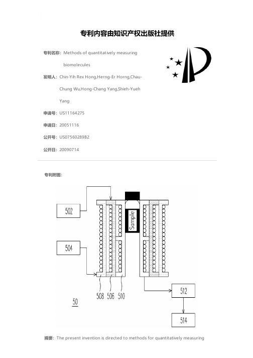
专利名称:Methods of quantitatively measuringbiomolecules发明人:Chin-Yih Rex Hong,Herng-Er Horng,Chau-Chung Wu,Hong-Chang Yang,Shieh-YuehYang申请号:US11164275申请日:20051116公开号:US07560289B2公开日:20090714专利内容由知识产权出版社提供专利附图:摘要:The present invention is directed to methods for quantitatively measuringbiomolecules by using magnetic nanoparticles. Through the use of the magnetic nanoparticles and the bioprobes coated to the magnetic nanoparticles, the biomolecules conjugated with the bioprobes result in the formation of particle clusters. By measuring the changes in magnetic properties resulting from the existence of the bio-targets, the amount of the bio-targets in a sample to be tested can be measured.申请人:Chin-Yih Rex Hong,Herng-Er Horng,Chau-Chung Wu,Hong-Chang Yang,Shieh-Yueh Yang地址:4F., No. 31, Lane 57, Ta Tze St., Ta Tze Taipei 104 TW,4F., No. 31, Lane 57, Ta Tze St., Ta Tze Taipei 104 TW,2F., No. 9, Lane 74, Aiguo E. Rd., Jhongjheng District Taipei 100 TW,4F., No. 31, Lane 57, Ta Tze St., Ta Tze Taipei 104 TW,12F., No. 427, Siyuan Rd. Sindian City, Taipei County 231 TW国籍:TW,TW,TW,TW,TW代理机构:Jianq Chyun IP Office更多信息请下载全文后查看。
Method for quantitative detection of biological to
专利名称:Method for quantitative detection ofbiological toxins发明人:Ekaterina Igorevna Dementieva,Veronika Igorevna Djukova,Alexander SergeevichZasedatelev,Alla Jurievna Rubina,AndreiAlexandrovich Stomakhin,VladimirAndreevich Nesmeyanov,Evgeny VasilievichGrishin申请号:US10589143申请日:20041124公开号:US20070172904A1公开日:20070726专利内容由知识产权出版社提供摘要:The invention relates to analytical chemistry and to quantitative immunochemical analysis, in particular, to a method for immunochemical quantitative detection of various biological toxins by using biological microchips. A biological microchip comprises an ordered array of three-dimensional hydrogel cells on a solid support, which are produced by a method of photo- or chemically induced polymerization and contain immobilized antibodies to various bacterial, plant and animal biotoxins, or biotoxins. The use of microchips makes it possible to analyze a sample simultaneously for the presence of several biotoxins. The proposed method for detecting biotoxins can be used in medicine, in food industry, and in environmental protection.申请人:Ekaterina Igorevna Dementieva,Veronika Igorevna Djukova,AlexanderSergeevich Zasedatelev,Alla Jurievna Rubina,Andrei Alexandrovich Stomakhin,Vladimir Andreevich Nesmeyanov,Evgeny Vasilievich Grishin地址:Moscow RU,Moscow RU,Moscow RU,Moscow RU,Moscow RU,Moscow RU,Moscow RU国籍:RU,RU,RU,RU,RU,RU,RU更多信息请下载全文后查看。
(完整版)乙肝表面抗原的ELISA法定量分析方法学验证毕业设计

毕业论文题目:乙肝表面抗原的ELISA 法定量分析方法学验证姓名:学号:专业:指导教师:教师职称:研究起止日期:研究提交日期:二○一三年十一月目录毕业论文乙肝表面抗原的ELISA 法定量分析方法学验证中文摘要 (1)英文摘要 (1)前言 (2)1. 材料与方法 (3)1.1 试剂盒 (3)1.2 仪器 (3)1.3 方法 (3)1.3.1 溶液配制 (4)铺板方4 案...........................................................................1.3.3 加样步5 骤 ...........................................................................2. 结6 果....................................................................................................2.1 标准曲线与线性6 范..................................................................2.2准确度(回收率)的验证 (6)2.3 精密度的验7 证..........................................................................2.3.1 板内精密度的验7 证.........................................................2.3.2 板间精密度的验8 证.........................................................2.4 检测限(LOD )计10 算.............................................................3. 讨10 论...................................................................................................3.1乙肝表面抗原定量分析的意义 (10)3.2常用定量分析方法 (11)3.3方法概述 (11)3.4方法学验证内容 (12)3.5结果分析及应用评价 (13)参考文献 (14)综述 (1)5 致谢 (1)8乙肝表面抗原的 ELISA 法定量分析方法学验证中文摘要目的:对福州蓝图生物工程有限公司生产的HBsAg 的 ELISA 法定量分析试剂盒进行方法学验证,以判断是否能用于临床样本分析。
工程蛋白分泌定量方法论
工程蛋白分泌定量方法论The quantification of secreted engineering proteins is a crucial step in biotechnology and pharmaceutical research. It is essential to accurately measure the amount of protein secreted into the culture medium to assess the efficiency of protein production and purification processes. However, there are various methods available for protein quantification, each with its own advantages and limitations. Therefore, it is important to carefully select the most appropriate method for a specific research project.工程蛋白的分泌定量是生物技术和制药研究中的关键步骤。
准确测量分泌到培养基中的蛋白质量是评估蛋白质生产和纯化过程效率的必要步骤。
然而,有许多可用于蛋白质定量的方法,每种方法都有其优点和局限性。
因此,在选择适用于特定研究项目的最合适方法时,需要认真考虑。
One commonly used method for protein quantification is the Bradford assay, which relies on the binding of Coomassie Brilliant Blue dye to proteins. The intensity of the dye-protein complex is proportional to the protein concentration, allowing for quantitative measurement. While the Bradford assay is relatively simple and quick,it is known to have limitations in terms of sensitivity and accuracy, particularly when used with certain types of proteins or in complex sample matrices.一种常用的蛋白质定量方法是布拉德福法,该方法依赖于考马斯亮蓝染料与蛋白质的结合。
定量体外诊断试剂临床试验统计方法探讨
定量体外诊断试剂临床试验统计方法探讨定量体外诊断试剂(quantitative in vitro diagnostic reagents)是指可以精确测量患者体内特定分子或物质浓度的试剂,因此在诊断疾病、监测疾病进展和评估治疗效果方面起到了重要作用。
而临床试验(clinical trials)则是评价这些试剂是否具有良好的性能和准确性的重要手段。
本文将探讨定量体外诊断试剂临床试验中的统计方法。
定量体外诊断试剂的临床试验需要明确的研究目的和研究设计,以确保结果的可靠性和有效性。
统计方法则是对试验数据进行分析和解释的重要工具。
一般来说,定量体外诊断试剂的临床试验中涉及到的统计方法主要包括以下几个方面。
首先是样本量的确定。
样本量的大小直接影响试验结果的可靠性和推广性。
在确定样本量时需要考虑试剂的准确性、效用和不良反应,以及研究结果的置信区间和统计功效等因素。
通常,样本量的确定会通过进行实际统计计算来得出。
其次是数据分析方法的选择。
定量体外诊断试剂的临床试验数据分析常用的方法包括描述性统计、方差分析、线性回归、相关分析等。
这些方法可以帮助研究人员对试验数据进行整体的统计描述、比较和解释。
还可以根据具体的研究问题选择适当的假设检验方法,以评估试剂的性能和准确性是否达到预期。
要注意的是,定量体外诊断试剂的临床试验中还需要进行统计显著性检验和置信区间估计。
统计显著性检验可以帮助判断试剂的效果和差异是否真实存在,而置信区间估计则可以对试验结果提供更为准确的界定。
还需要关注试验结果的统计解释和临床意义。
临床试验的统计分析结果需要与实际疾病诊断和治疗相关的临床指标进行比较和解释,以评估试剂的应用价值和临床实际意义。
还需要注意统计结果的稳定性和可靠性。
定量体外诊断试剂的临床试验中的统计方法是非常重要的,可以帮助评估试剂的性能和准确性,进而指导其在临床实践中的应用。
定量研究方法(Quantitative Research Method)

什么是定量研究?定量研究一般是为了对特定研究对象的总体得出统计结果而进行的。
定性研究具有探索性、诊断性和预测性等特点,它并不追求精确的结论,而只是了解问题之所在,摸清情况,得出感性认识。
定性研究的主要方法包括:与几个人面谈的小组访问,要求详细回答的深度访问,以及各种投影技术等。
在定量研究中,信息都是用某种数字来表示的。
在对这些数字进行处理、分析时,首先要明确这些信息资料是依据何种尺度进行测定、加工的,史蒂文斯(S. S. Stevens)将尺度分为四种类型,即名义尺度、顺序尺度、间距尺度和比例尺度。
[编辑]定量研究的四种测定尺度及特征名义尺度所使用的数值,用于表现它是否属于同一个人或物。
顺序尺度所使用的数值的大小,是与研究对象的特定顺序相对应的。
例如,给社会阶层中的上上层、中上层、中层、中下层、下下层等分别标为“5、4、3、2、1”或者“3、2.5、2、1.5、1”就属于这一类。
只是其中表示上上层的5与表示中上层的4的差距,和表示中上层的4与表示中层的3的差距,并不一定是相等的。
5、4、3 等是任意加上去的符号,如果记为 100、50、10 也无妨。
间距尺度所使用的数值,不仅表示测定对象所具有的量的多少,还表示它们大小的程度即间隔的大小。
不过,这种尺度中的原点可以是任意设定的,但并不意味着该事物的量为“无”。
例如,O°C 为绝对温度273°K,华氏32°F。
名义尺度和顺序尺度的数值不能进行加减乘除,但间距尺度的数值是可以进行加减运算的。
然而,由于原点是任意设定的,所以不能进行乘除运算。
例如,5℃和 10℃之间的差,可以说与15℃和20℃之间的差是相同的,都是5°C。
但不能说 20℃就是比5℃高4倍的温度。
比例尺度的意义是绝对的,即它有着含义为“无”量的原点0。
长度、重量、时间等都是比例尺度测定的范围。
比例尺度测定值的差和比都是可以比较的。
例如:5分钟与10 分钟之间的差和10分钟与15分钟之间的差都是5 分钟,10 分钟是2分钟的5倍。
资料 二级易考点 数量 定量分析 quantitative methods

定量分析:Quantitative Methods (1Case )题目内容形式:题目信息:Case 一般会先以一段文字介绍什么为dependent factor ,什么为independent factor ; 然后会给一些表格,可能包括回归表,ANOVA table (可能不是表格,直接给数值),Durbin-Waston /Dicky-Fuller testing table ; 紧随表格后可能会有一些关于表格数据得出的结论。
考察形式:题目主要是根据表格信息得出一些结论或者判断case 中的结论是否正确。
通常考察一个Case ,考题最多集中在Correlation Analysis 和Regression Analysis ;time-series 可能会涉及到1-2题。
易考点一:Correlation Analysis1.Sample Correlation Coefficient 的公式:考法:知道r,Sx 和Sy,求Cov(X ,Y)或者知道Sx,Sy 和Cov(X ,Y),求r (☆☆)r XY =0,no linear correlationr XY =1,完全正相关r XY =-1,完全负相关-1<r XY <0;0<r XY <1,Hypothesis testing of correlation2.Hypothesis testing of correlation:考法:知道n 和r ,求相关系数ρ的t 统计量,已经问是否线性相关(☆☆☆)H 0:ρ=0(两个变量相关系数为0,不相关)H 1:ρ≠0t-test :212r n r t --=df =n -2(two-taild )Decision rule :If t >|t critical |,reject H 0YX XY S S Y X Cov r ),(=易考点二:Linear regression model(以multiple 为例,simple 的区别就是少了X 2这一项,☆☆☆)CoefficientStandard Errort-statisticP-value Intercept b 00b S t 0p 0X 1b 11b S t 1p 1X 2b 22b S t 2p 21.Hypothesis testing of regression (当检测出某项系数为0,就说明对应的自变量对于表示Y is not statistically significant ,以b 1举例):H 0:Coefficient =0(b 1=0)H 1:Coefficient≠0Decision rule :If p <significance level (α),reject H 0(如果5%的significance ,p 1小于0.05,则reject H 0;如果不给significance level ,p-value 很小也reject )If t statistic >|t critical |,reject H 02.Predict Y :22110X b X b b Y++=3.Confidence interval for a coefficient (for b 1))(11b critical s t b⨯±易考点三:ANOVA analysis(simple regression 的情况就是k=1的情况,k 为自变量的个数,☆☆☆)ANOVA Degree of FreedomSum of SquareMean Square Regression k RSS MSR=RSS/k Residual n-k-1SSE MSE=SSE/(n-k-1)Totaln-1SST1.Coefficient of determination (R 2):R 2=RSS/SSTR 2判断的是有多少variation 被自变量所解释,if R 2=0.7735,意思为通过回归方程得出的dependent variable 有77.35%能被independent variable 所解释Adjusted R 2=)]1([(-1211R k n n -⨯---,Adjusted R 2≤R 2加入新的变量,R 2会增加;但如果增加的变量没有统计学意义的时候,Adjusted R 2不会增加,反而会减小。
酸水解同位素稀释质谱法测量基因组DNA含量_张玲

化学分析计量CHEMICAL ANALYSIS AND METERAGE第22卷,第5期2013年9月V ol. 22,No. 5Sept. 20139doi :10.3969/j.issn.1008–6145.2013.05.002酸水解同位素稀释质谱法测量基因组DNA 含量*张玲,陈大舟,武利庆,高运华,汤桦,王晶 刘新海 (中国计量科学研究院,北京 100013) (中国医学科学院整形外科医院,北京 100144)摘要 建立了测量噬菌体λ基因组DNA 含量的方法。
样品添加同位素标记碱基内标之后,用体积分数为88%的甲酸溶液在170℃水解30 min ,解离出的核酸碱基通过反向柱分离,电喷雾四级杆质谱法测定,用多反应监测模式分别检测碱基及其同位素标记物的母离子和碎片子离子,从而建立了基因组DNA 水解–同位素稀释质谱法测量长片段核酸含量的方法,并将DNA 浓度溯源至碱基浓度。
方法的线性范围为1~1 000 μg /g ,检出限可低至100 ng /g 。
测定的λDNA 含量标准物质为(2.51±0.06)μg /支 (k =2),该方法可用于长片段核酸含量标准物质定值。
关键词 噬菌体λDNA ;核酸碱基;核苷酸;高效液相色谱法;同位素稀释质谱法中图分类号:O657.6 文献标识码:A 文章编号:1008–6145(2013)05–0009–05Accurate Quantitation of Genome DNA Mass Concentration by Acid Hydrolysis–Isotope Dilution Mass SpectrometryZhang Ling ,Chen Dazhou, Wu Liqing, Gao Yunhua, Tang Hua, Wang Jing(National Institute of Metrology, Beijing 100013, China )Liu Xinhai(Plastic Surgical Hospital, Chinese Academy of Medical Science, Beijing 100144,China )Abstract The method for determining genomic DNA mass content in phage was set up. Phage λ genome DNA samples were added by isotope labeled nucleic bases internal standard , t hen hydrolyzed in 88% formic acid solution for 30 min under 170℃. The DNA hydrolysis products nucleic bases were separated in C 18 reverse phase column and analyzed by electrospray quadrupore isotope dilution mass spectrometry (HPLC–IDMS) by multiple reaction monitoring mode to scan parent ion and product ion of nucleic bases and the isotope labeled nucleic bases. The method was established for traceability of the genomic DNA to 4 nucleic bases mass content. The method linear range was 1–1 000 μg /g, and the detection limit could be as low as 100 ng /g. The DNA mass content in each vial was measure d (2.51±0.06) μg (k =2). This method could be used as a primary method for determining DNA mass as a reference material.Keywords phage λDNA; nucleic acid base; nucleotide; high performance liquid chromatography; isotope dilution mass spectrometry含量测量是核酸测量的首要问题,世界工业先进国家计量机构均已针对核酸含量测量开展了基础研究和应用研究。
酶识别支持向量机特征提取自检法留一法硕士论文

基于特征提取的酶识别问题研究【关键词】酶识别; 支持向量机; 特征提取; 自检法; 留一法;【英文关键词】enzyme identification; support vector machine; feature selection; self-consistency test; leave-one-out test;【中文摘要】在生物信息学中,将酶从蛋白质识别出来一直是对酶进行进一步研究的一个前提。
其研究方法都是将已知的酶作为研究对象,找出一种对已知酶进行准确识别的方法,然后推广到对未知酶识别的应用中。
传统的酶识别方法多是采用序列比对的方法,虽然后人对这种方法有不断地改进,但是仍需要较大的存储空间与比对时间。
近些年,机器学习的方法也开始的应用到这个领域中。
支持向量机(Support V ector Machine, SVM)——一种基于统计学理论的机器学习方法,借助自己的无局部最小点和防止过适应等优点,迅速成为研究的热点并且在酶识别领域表现出不错的效果。
为了得到好的机器学习效果,机器学习需要研究者根据实际问题的不同提出一套完整的机器学习方案。
本文以支持向量机为基础,采用了一种基于特征提取的机器学习方案,通过选取合适数量的特征作为训练数据形成分类精度最高的酶识别器。
之所以选用特征提取的方法主要是因为:在实验中,蛋白质的功能域被看做它的特征,并不是所有的功能域都对形成准确的分类器起到好的作用,并且我们推测这些功能域特征中存在噪声,因此应该剔除其中一些起到反作用的特征。
基于以上的原因,文中选用了1-rule法和信息增益法两种...【英文摘要】In bioinformatics, identifying enzymes from proteins is a prerequisite for further research in enzymes. Its method of research is thattaking known enzymes as research object and finding a method could identify enzymes with high accuracy, then applying in identifying unknown enzymes. The traditional method used in enzymes identification is alignment. Although many scientists do lots of work to improve alignment, the method still needs big storage space and computing time. In recent years, machine learning h...摘要5-6Abstract 6第1章绪论9-141.1 研究的背景、目的及意义9-101.2 国内外研究现状及评价10-121.3 本文的内容和章节安排12-131.4 本文的创新点13-14第2章基础理论14-242.1 支持向量机的理论知识14-172.1.1 线性可分14-162.1.2 线性不可分16-172.2 特征提取的原因17-182.2.1 什么是特征172.2.2 原因17-182.3 几种特征提取方法18-242.3.1 1-rule 18-202.3.2 信息增益法20-24第3章实验步骤24-323.1 实验数据24-253.1.1 蛋白质酶的获取243.1.2 非酶蛋白质的获取24-253.2 实验数据的筛选253.3 基于功能结构域组成的蛋白质数字化表示25-273.3.1 Pfam 数据库25-263.3.2 数字化表示26-273.4 特征信息计算27-283.4.1 1-rule 法特征信息计算27-283.4.2 信息增益法特征信息计算283.5 学习机的选择28-293.6 训练数据的选择与测试29-313.7 实验过程流程图31-32第4章实验结果分析32-394.1 误差率324.2 自检法32-334.3 留一法334.4 实验结果33-364.4.1 1-rule 法实验结果33-354.4.2 信息增益法实验结果35-364.5 实验结果分析36-394.5.1 对比对象36-374.5.2 分析37-39第5章总结与研究展望39-415.1 总结395.2 存在的问题395.3 展望39-41参考文献。
不同方法的蛋白质鉴定量英语

不同方法的蛋白质鉴定量英语Title: Comparative Analysis of Protein Identification Quantification MethodsIntroduction:Protein identification and quantification are crucial steps in biological research, particularly in the fields of molecular biology, biochemistry, and proteomics.various methods have been developed to identify and quantify proteins, each with its unique principles and applications.This document aims to provide an in-depth analysis of different methods used for protein identification and quantification, highlighting their advantages and limitations.1.Mass Spectrometry-Based Protein Identification:Mass spectrometry (MS) has become the gold standard for protein identification.It involves the ionization and separation of peptide fragments based on their mass-to-charge ratio, followed by the identification of proteins through database searching.Advantages:- High sensitivity and accuracy- Wide dynamic range- Suitable for complex sample analysisLimitations:- Sample preparation can be time-consuming- Requires expensive equipment and expertise- Limited by the availability of protein databasesQuantification Methods:- Label-free quantification: Measures the abundance of proteins based on the intensity of MS signals.- Stable isotope labeling: Uses heavy/light isotopes to compare protein abundance between samples.2.Gel Electrophoresis-Based Protein Identification:Gel electrophoresis is a widely used method for protein separation based on size and charge.After separation, proteins can be stained, and the bands can be excised for further identification using MS or other techniques.Advantages:- Cost-effective- Suitable for small-scale experiments- Visual representation of protein separationLimitations:- Low resolution and sensitivity compared to MS- Time-consuming- Limited to the analysis of a small number of proteinsQuantification Methods:- Densitometry: Measures the intensity of stained protein bands to estimate abundance.- Fluorescence-based quantification: Uses fluorescent dyes to label proteins and measure their abundance.3.Immunoassays for Protein Quantification:Immunoassays rely on the specific binding of antibodies to target proteins.They are widely used for protein quantification due to their simplicity and sensitivity.Advantages:- High specificity and sensitivity- Wide range of applications- Suitable for high-throughput analysisLimitations:- Cross-reactivity and antibody specificity issues- Time-consuming optimization of assay conditions- Limited dynamic rangeQuantification Methods:- ELISA (Enzyme-Linked Immunosorbent Assay): Uses enzymatic reactions to measure protein abundance.- Western blotting: Separates proteins by gel electrophoresis and detects them using specific antibodies.bel-Free Imaging Techniques:Label-free imaging techniques provide a non-invasive alternative for protein quantification, often used in live-cell imaging and tissue analysis.Advantages:- No need for sample labeling- Real-time monitoring of protein dynamics- High spatial resolutionLimitations:- Lower sensitivity compared to labeled methods- Limited to surface or thin-section analysis- Require specialized equipmentQuantification Methods:- Fluorescence lifetime imaging microscopy (FLIM)- Second-harmonic generation (SHG) microscopyConclusion:Each method for protein identification and quantification has its unique advantages and limitations.Researchers should choose the most appropriate method based on their specific experimental requirements, including sensitivity, sample complexity, and availablebining different techniques can often provide a more comprehensive understanding of protein abundance and function in biological systems.。
Quantitative Methods for Interlaboratory Testing –
7/30-8/10/2001
Uncertainty Workshop by Carl Lee
8
Module Ten: Planning Experiments – the general consideration and Comparative Study for more than
Quantitative Methods for Measuring and Assessing Uncertainties in Testing Process
and Outcomes
(Workshop from July 30 to August 10, 2001 at National Bureau of Standards & Metrology and
• The concept and Procedure for performing the one sample t-test
•Comparative Study for Inter-laboratory Testing : two-group cases
•Designing experiments for two-sample comparative study
•Data transformation techniques
•Post-Hoc Analysis and Sum of square decomposition
•Contrasts, simultaneous comparison, pair-wise comparison, comparison with control
体外抗体形成细胞的检测—定量溶血分光光度计法(Quantitative

②裂解红细胞:5ml Tris· NH4Cl重悬细胞,37℃孵育5 分钟。
③用含有5%小牛血清的Hanks液洗涤一次,用1.2ml Hank’s液重悬细胞。
5
正式实验
1. 取两支试管,标记 2. 按下表加试剂
脾细胞悬液 SRBC悬液
补体 1ml 1ml
Hank’s液
总体积 3ml 3ml
对照管 实验管
— 1ml
1ml 1ml
1ml —
3. 混匀,37℃水浴1小时。 4. 3000rpm 离心 5 min,取上清 5.721分光光度计测413nm处OD值(Hb量)
6
注意事项:
1.SRBC要新鲜,无溶血。
2.离体脾细胞的制备过程要迅速,以防细胞
死亡,细胞计数应准确。 3.红细胞裂解是本实验的关键,应用力吹打, 时间也要严格控制。
9
抗体制备
原理:
抗原免疫小鼠,激活免疫系统,B 细胞活化并分化为浆细胞,产生针对特 定抗原的特异性抗体。
10
方法
1.用移液管吸取脱纤维防凝处理的绵羊血适量
2.加2-3倍体积生理盐水,混匀,2000rpm离心
5分钟,小心吸尽上清。
3.重复第二步操作。
4.用生理盐水配20%、25%、30%、40%和50%
绵
羊红细胞(SRBC)
5.腹股沟皮下免疫小鼠,0.1ml/只,免疫三次。 6.取脾脏检测B细胞
11
1)免疫鼠制备:取上述稀释的SRBC,经尾静脉或腹腔 内注射免疫小鼠 (鼠龄8~10周),每只0.2ml。 2)取脾脏:72小时拉颈处死,取出脾脏用含5%小牛血 清的Hanks液清洗,去除脂肪和结缔组织。 3)收取脾细胞: ①在200目铜网上研磨,并用含5%小牛血清的冷Hanks液 冲洗,制备单个脾细胞悬液,转移滤液至试管内。 1500rpm离心5min。
利用胶体金免疫层析技术快速定量检测单增李斯特菌方法的建立

1:3.5 × 10⁸ cfu/ml;2:3.5 × 107 cfu/ml;3:3.5 × 106 cfu/ml;4:3.5 × 105 cfu/ml;5:3.5 × 104 cfu/ml;6:3.5 × 103 cfu/ml;7:3.5 × 102 cfu/ml; 8:3.5 × 101 cfu/ml;9:空白对照
1.7.1 定 性 检 测 将 处 理 后 的 样 品 和 样 品 处 理 液 (作为阴性样品)100 μl 加到制备好的胶体金免疫层 析试纸条样品垫端,静置 10 min。 检测带(T)和质控 带 (C)均 出 现 红 色 判 为 阳 性 , 仅 质 控 带 出 现 红 色 为 阴 性,检测带和质控带均不显色,则为试纸条失效。 1.7.2 定量检测
中国国境卫生检疫杂志 2010 年 4 月第 33 卷第 2 期 Chinese Frontier Health Quarantine Apr.2010,Vol 33,No.2
· 127 ·
(2) 单 增 李 斯 特 菌 P60 多 克 隆 抗 体 (Listeria monocytogenes, multiple cloning antibody of P60 由 吉 林出入境检验检疫局提供)、羊抗兔 IgG(志 2010 年 4 月第 33 卷第 2 期 Chinese Frontier Health Quarantine Apr.2010,Vol 33,No.2
〔检测技术〕
利用胶体金免疫层析技术快速定量检测 单增李斯特菌方法的建立
谢士嘉 1 王 静 1 王振国 2 (1.中国检验检疫科学研究院,北京 100123;2.吉林出入境检验检疫局,长春 130062)
quantitativedetection单核细胞增生李斯特菌isteriamonocytogenes单增李斯特菌是李斯特菌属isteria中最重要的人类食源性病原菌也是一种人畜共患的致病菌它可引起人和动物患脑膜炎脑炎败血症心内膜炎流产死胎及脓肿等死亡率可达34年来已有不少国家报道了由于污染单增李斯特菌发生的食物中毒事件因此世界各国政府部门对单增李斯特菌引起的食物中毒事件越来越重视目前单增李斯特菌的检测主要依靠传统的分离培养和生化鉴定费时费力45年代以来发展起来的一项新型体外诊断技术近年来该方法发展迅速在生物医学领域特别是医学检验中得到了广泛应用但较少用于食品卫生领域
- 1、下载文档前请自行甄别文档内容的完整性,平台不提供额外的编辑、内容补充、找答案等附加服务。
- 2、"仅部分预览"的文档,不可在线预览部分如存在完整性等问题,可反馈申请退款(可完整预览的文档不适用该条件!)。
- 3、如文档侵犯您的权益,请联系客服反馈,我们会尽快为您处理(人工客服工作时间:9:00-18:30)。
Antigenic Specificity on B cells. B cell receptor (BCR)
Secreted-bound Antibody
Humoral Immunity
BCR facilitates the activation of these cells and their subsequent differentiation into either antibody factories called plasma cell, or memory B cell that will survive in the body and remember that same antigen so the B cells can respond faster upon future exposure.
To determine the coating concentration generating the best signal-to-noise ratio while maintaining low background in the blank rows.
Formal experiment
Bacteria and virus
ห้องสมุดไป่ตู้
• Coating antibody: Insulin monoclonal antibody (Reactivity with rat)
Clone number, specificity (insulin/proinsulin/C-peptide), reactivity (rat), application (ELISA), price (200 – 300 $/100 µg ).
Fab Fc
Principle of ELISA
• ELISA: Enzyme-linked Immunosorbent Assay
Specificity of antibodies or antigens Sensitivity of simple enzyme assays
A high turnover number Qualitative analysis Quantitive analysis
• Coating concentration: depending on the optimized concentration. (100 L per well)
• Time and temperature: 4C , 16 -18 h
Blocking
• Blocking: BSA 1% - 5% in PBS, 4C , 16 h or RT, 2 – 3 h, kept before used
• Structure and Function of Antibody (Ig G)
V region: Highly variable sequence
C region: Constant sequence
Fab: Antigen-binding fragment
Fc: Crystallizable fragment
Negative control wells receive negative sample, serum without insulin;
to determine the matrix effect;
serum pretreatment
Conjugate control wells receive coating buffer solution without antibody; to ensure that non specific binding of the detection antibody to well surfaces;
Coating
• Coating buffer: 50mM Carbonate pH 9.6; 10mM Tris pH 8.5; 10mM PBS pH 7.2, depending on the storage condition of antibody. (no detergents and extraneous proteins, pH≥ pI)
Quantitative ELISA Method Development for Detection of Insulin
Purpose
ELISA method
• Large molecule drug: analysis method • Commercial market: method development
better washing
hydrophobic interactions van der Waals forces hydrogen bonding ionic interactions
Very hydrophobic Low binding (100-200 ng of IgG /cm2)
Modified to produce more COOH or OH
Antibody
• Antibodies are the antigen-binding and Y-shaped proteins present on the B-cell membrane and secreted by plasma cells.
Membrane-bound Antibody
The antibody recognizes antigen. Each tip of the "Y" of an antibody contains a paratope (a structure analogous to a lock) that is specific for one particular epitope (that is equivalent to a key) on an antigen, allowing these two structures to bind together with precision.
• Detection antibody: anti-rat insulin monoclonal antibody labeled with enzyme
Horseradish peroxidase enzyme is widely used. TMB: tetramethyl benzidine OPD: Ortho-phenylenediamine (OPD) AP: Alkaline phosphatase p-Nitrophenylphosphate (pnpp)
Formal experiment
Method validation
(Accuracy, Specificity, Repeatability)
Materials and reagents
• Solid phase: polystyrene surface, 96 well plate. • (Nunc, MaxiSorp)
(Storage: fill the well with sucrose 2% in PBS and incubate 2 – 3 min; remove all liquid and dry 1 – 2 h in RT)
Wash • Wash buffer: Tween-20 or Triton X-100 0.01% to 0.05% in
Insulin as the reference drug
• Mature skills on insulin ELISA kit • PK-PD model
Content
• Structure and Function of Antibody • Principle of ELISA • Structure and Property of Insulin • ELISA Method Development for Insulin • Method Validation
Materials and reagents
(solid phase, coating antibody and detection antibody)
Preliminary test on materials and reagents
(Several controls)
Optimization of coating antibody and detection antibody concentrations
Step 3 Add the detection antibody (100 L, 25C , 1 h) Wash (300 L)
Step 4 Add substrate (100 L, 37C, 20 min)
Step 5 Terminate the reaction and read the absorbance
All other conditions are referred to the common procedures.
Optimization of Antibody concentrations
• Coating concentration: 0.1-15 µg/ml (3 levels) • Detection concentration: 500 – 10 ng/ml for monoclonal, (3 levels)
High binding (400 – 500 ng of IgG /cm2)
Direct absorption to polystyrene
Proteins Peptides longer than 15-20 amino acids Small molecule epitopes attached to a protein
Substrate control wells receive BSA solution without detecting antibody; to ensure no other oxidases in reaction system
