Anti-DYKDDDDK Magarose beads
洋甘菊提取液MSDS英文版

1. IDENTIFICATION OF THE SUBSTANCE/TREPARATION AND THE COMPANY/UNDERTAKING3.HAZARDS IDENTIFICATION4. FIRST AID MEASURESMATERIAL SAFETY DATA SHEETProduct name:Supplier:Tel:EMERGENCY OVERVIEW: May cause skin irritation and/or dermatitisPrinciple routes of exposure: Inhalation: Ingestion: Skin contact: Eye contact:SkinMay cause irritation of respiratory tract May be harmful if swallowed May cause allergic skin reaction Avoid contact with eyesStatements of hazard MAY CAUSE ALLERGIC SKIN REACTION.Statements of Spill of Leak Label Eliminate all ignition sources. Absorb and/or contain spill with inert materials (e.g., sand, vermiculite). Then place in appropriate container. For large spills, use water spray to disperse vapors, flush spill area. Prevent runoff from entering waterways or sewers.General advice:POSITION/INFORMATION ON INGREDIENTSInhalation:Skin contact:Ingestion:Eye contact:Protection of first – aiders:Medical conditions aggravated by exposure: In the case of accident or if you fell unwell, seek medical advice immediately (show the label where possible).Move to fresh air, call a physician immediately.Rinse immediately with plenty of water and seek medical adviceDo not induce vomiting without medical advice.In the case of contact with eyes, rinse immediately with plenty of water and seek medical advice.No information availableNone knownSuitable extinguishing media:Specific hazards:Special protective equipment for firefighters:Flash point:Autoignition temperature:NFPA rating Use dry chemical, CO2, water spray or “alcohol” foam Burning produces irritant fumes.As in any fire, wear self-contained breathing apparatus pressure-demand, MSHA/NIOSH (approved or equivalent) and full protective gearNot determinedNot determinedNFPA Health: 1 NFPA Flammability: 1 NFPA Reactivity: 0Personal precautions: Environmental precautions: Methods for cleaning up: Use personal protective equipment.Prevent product from entering drains.Sweep up and shovel into suitable containers for disposalStorage:7. HANDLING AND STORAGE5.FIRE-FIGHTING MEASURES6. ACCIDENTAL RELEASE MEASURESRoom temperature Handling:Safe handling advice: Incompatible products:Use only in area provided with appropriate exhaust ventilation.Wear personal protective equipment.Oxidising and spontaneously flammable productsEngineering measures: Respiratory protection: Skin and body protection:Eye protection: Hand protection: Hygiene measures:Ensure adequate ventilation.Breathing apparatus only if aerosol or dust is formed. Usual safety precautions while handling the product will provide adequate protection against this potential effect. Safety glasses with side-shieldsPVC or other plastic material glovesHandle in accordance with good industrial hygiene and safety practice.Melting point/range: Boiling point/range: Density: Vapor pressure: Evaporation rate: Vapor density: Solubility (in water): Flash point:Autoignition temperature:No Data available at this time. No Data available at this time. No data available No data available No data available No data available No data available Not determined Not determinedStability: Stable under recommended storage conditions. Polymerization: None under normal processing.Hazardous decomposition products: Thermal decomposition can lead to release of irritating gases and vapours such as carbon oxides.Materials to avoid: Strong oxidising agents.10. STABILITY AND REACTIVITY9. PHYSICAL AND CHEMICAL PROPERTIES8. EXPOSURE CONTROLS/PERSONAL PROTECTION11. TOXICOLOGICAL INFORMATIONConditions to avoid: Exposure to air or moisture over prolonged periods.Product information Acute toxicityChronic toxicity:Local effects: Chronic exposure may cause nausea and vomiting, higher exposure causes unconsciousness.Symptoms of overexposure may be headache, dizziness, tiredness, nausea and vomiting.Specific effects:May include moderate to severe erythema (redness) and moderate edema (raised skin), nausea, vomiting,headache.Primary irritation: Carcingenic effects: Mutagenic effects: Reproductive toxicity:No data is available on the product itself. No data is available on the product itself. No data is available on the product itself. No data is available on the product itself.Mobility:Bioaccumulation: Ecotoxicity effects: Aquatic toxicity:No data available No data available No data availableMay cause long-term adverse effects in the aquatic environment.12. ECOLOGICAL INFORMATION13. DISPOSAL CONSIDERATIONSWaste from residues/unused products:Contaminated packaging:Waste disposal must be in accordance with appropriate Federal, State and local regulations. This product, if unaltered by use, may be disposed of treatment at a permitted facility or as advised by your local hazardous waste regulatory authority. Residue from fires extinguished with this material may be hazardous.Do not re-use empty containers.UN/Id No:Not regulated14. TRANSPORT INFFORMATIONDOTProper shipping name: Not regulatedTGD(Canada)WHMIS hazard class: Non - controlledIMDG/IMOIMDG – Hazard Classifications Not ApplicableIMO – labels:15. REGULATORY INFOTMATION International Inventories16. OTHER INFORMATIONPrepared by: Health & SafetyDisclaimer: The information and recommendations contained herein are based upon tests believed to be reliable.However, XABC does not guarantee the accuracy or completeness NOR SHALL ANY OF THIS INFORMATION CONSTITUTE A WARRANTY, WHETHER EXPRESSED OR IMPLIED, AS TO THE SAFETY OF THE GOOD, THE MERCHANTABILITY OF THE GOODS, OR THE FITNESS OF THE FITNESS OF THE GOODS FOR A PARTICULAR PURPOSE. Adjustment to conform to actual conditions of usage maybe required. XABC assumes no responsibility for results obtained or for incidental or consequential damages, including lost profits arising from the use of these data. No warranty against infringement of any patent, copyright or trademark is made or implied.End of safety data sheet。
常见蛋白质标签总结
常见蛋白质标签总结(Flag、HA、cMyc、CBP等)Protein tags are peptide sequences genetically grafted onto a recombinant protein. Often these tags are removable by chemical agents or by enzymatic means, such as proteolysis or intein splicing. Tags are attached to proteins for various purposes.一、氨基酸标签(含小肽标签)A stretch of amino acids is added to the protein and enables the recovery of the labelled protein by its unique affinity. Usually its easiest to add the tag to either end of the protein to ensure its accessibility and not to disturb the protein folding.1.组氨酸标签(His tag)一般为6个组氨酸,用Ni2+(Cu2+)亲和层析纯化2.FLAG tag :N-DYKDDDDK-C ,recovered with specific antibody3.HA tag: an epitope derived from the Influenza protein haemagglutinin(HA,禽流感病毒血凝素),e.g. N-YPYDVPDYA-C,recovery with anHA antibody4.MYC tag: an epitope derived from the human proto-oncoprotein MYC,e.g.N-ILKKATAYIL-C, N-EQKLISEEDL-C,recovery with an MYCantibody5.SBP tag:Streptavidin Binding Peptide,链霉亲合素结合肽,38 amino acidtag (MDEKTTGWRGGHVVEGLAGELEQLRARLEHHPQGQREP),更多参考在Sigma6.CBP tag:钙调蛋白结合肽(CBP; 26aa)钙调蛋白结合肽与钙调素结合是Ca2+依赖的,这种结合不受标签所处的位置影响(N端和C端均可),在中性pH条件下使用2mM EGTA可以很方便的将目标蛋白洗脱下来。
常见蛋白质标签总结

/bbs/home.php?mod=space&uid =34800&do=blog&id=38530常见蛋白质标签总结(Flag、HA、cMyc、CBP等)Protein tags are peptide sequences genetically grafted onto a recombinant protein. Often these tags are removable by chemical agents or by enzymatic means, such as proteolysis or intein splicing. Tags are attached to proteins for various purposes.一、氨基酸标签(含小肽标签)A stretch of amino acids is added to the protein and enables the recovery of the labelled protein by its unique affinity. Usually its easiest to add the tag to either end of the protein to ensure its accessibility and not to disturb the protein folding.1.组氨酸标签(His tag)一般为6个组氨酸,用Ni2+(Cu2+)亲和层析纯化2.FLAG tag :N-DYKDDDDK-C ,recovered with specific antibody3.HA tag: an epitope derived from the Influenza protein haemagglutinin (HA,禽流感病毒血凝素),e.g. N-YPYDVPDYA-C,recovery with an HAantibody4.MYC tag: an epitope derived from the human proto-oncoprotein MYC,e.g.N-ILKKATAYIL-C, N-EQKLISEEDL-C,recovery with an MYCantibody5.SBP tag:Streptavidin Binding Peptide,链霉亲合素结合肽,38 amino acidtag (MDEKTTGWRGGHVVEGLAGELEQLRARLEHHPQGQREP),更多参考在Sigma6.CBP tag:钙调蛋白结合肽(CBP; 26aa)钙调蛋白结合肽与钙调素结合是Ca2+依赖的,这种结合不受标签所处的位置影响(N端和C端均可),在中性pH条件下使用2mM EGTA可以很方便的将目标蛋白洗脱下来。
兰嘉丝汀
【兰嘉丝汀幻彩亮彩去角质面膜(橘色胶囊)】特别添加柳橙、柠檬、葡萄柚及杏桃核颗粒。
能有效使暗沉粗糙的肌肤,立即恢复柔嫩透亮,更好吸收保养品。
所有肤质适用。
使用方法:将脸打湿后,避开眼周,涂上薄薄一层,用指腹轻轻画圈按摩。
最后以温水冲洗干净。
一个胶囊的使用量为一次。
【兰嘉丝汀幻彩珍珠活力面膜(黄色胶囊)】(右一)特含有莲花及维他命A,可立即补充新鲜美丽能量。
针对疲累、压力型肌肤,可立即醒肤,恢复珍珠般明亮光采,适用于所有肤质。
使用方法:厚敷于全脸及颈部,停留5分钟,用面纸将多余的面膜轻轻擦拭。
一个胶囊的使用量为一次。
【兰嘉丝汀理肤银杏隔离霜SPF50/PA+++ 3ML】(左二)运用独家防晒技术,完美延展科技,全面覆盖肌肤,不堵塞毛孔,不生成粉刺,在肌肤形成透明保护膜,高效抵御各种肌肤伤害源。
使用方法:取适量用于面霜之后,均匀涂抹与面部,避开眼部。
出门前20分钟使用。
【兰嘉丝汀水润身体防晒液SPF30 30ml 】(左一)双重防护,高效预防UV A/UVB,专利天然RPF复合因子,可同时中和95%因外在环境所产生的自由基。
专为亚洲肌肤需求设计,防止肌肤晒黑,运用特殊水润配方。
使用方法:于外出前30分钟涂于身体暴露部位,每过一段时间加涂一次新七白美白嫩肤面膜洁肤后,取适量(两粒葡萄大小,约6g),轻柔均匀地涂抹于面部,停留10-15分钟,再以温水洗净,每周使用3-4次。
搭配佰草集新七白美白系列产品共同使用,效果尤佳。
清肌养颜太极泥先清:首先使用黑泥,清洁双手并擦干,用面膜调棒取适量黑泥,涂抹薄薄一层于面部,至掩盖肤色铺展均匀即可,注意避开眼部周围。
加轻轻按摩以,使黑泥中的本草成分渗透并作用于肌肤。
约5分钟后,用洁面棉擦去黑泥,再用温水冲洗干净;后补:接着使用白泥,黑泥洗去后,在面部半湿状态下,用面膜调棒取适量白泥,均匀涂抹于面部;用食指和中指以划圈的方式轻柔按摩面部2-3分钟,按摩后,再使白泥在脸部停留8分钟左右,使肌肤充分吸收营养成分。
FlowComp
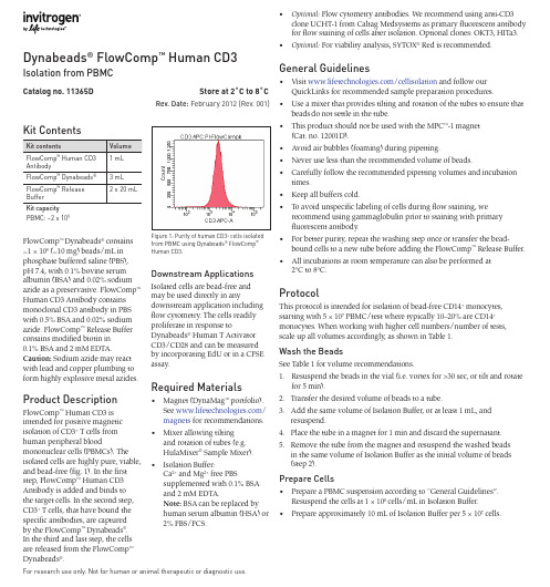
Kit ContentsFlowComp ™ Dynabeads ® contains ~1 × 10⁹ (~10 mg) beads/mL in phosphate buffered saline (PBS), pH 7.4, with 0.1% bovine serum albumin (BSA) and 0.02% sodium azide as a preservative. FlowComp ™ Human CD3 Antibody contains monoclonal CD3 antibody in PBS with 0.5% BSA and 0.02% sodium azide. FlowComp ™ Release Buffer contains modified biotin in 0.1% BSA and 2 mM EDTA.Caution: Sodium azide may react with lead and copper plumbing to form highly explosive metal azides.Product DescriptionFlowComp ™ Human CD3 is intended for positive magnetic isolation of CD3+ T cells from human peripheral bloodmononuclear cells (PBMCs). The isolated cells are highly pure, viable, and bead-free (fig. 1). In the first step, FlowComp ™ Human CD3 Antibody is added and binds to the target cells. In the second step, CD3+ T cells, that have bound the specific antibodies, are captured by the FlowComp ™ Dynabeads ®. In the third and last step, the cells are released from the FlowComp ™ Dynabeads ®.Dynabeads ® FlowComp ™ Human CD3Isolation from PBMCFor research use only. Not for human or animal therapeutic or diagnostic use.Catalog no. 11365D Store at 2˚C to 8˚CRev. Date: February 2012 (Rev. 001)Downstream ApplicationsIsolated cells are bead-free and may be used directly in anydownstream application including flow cytometry. The cells readily proliferate in response toDynabeads ® Human T Activator CD3/CD28 and can be measured by incorporating EdU or in a CFSE assay.Required Materials• Magnet (DynaMag ™ portfolio).See /magnets for recommendations.• Mixer allowing tiltingand rotation of tubes (e.g. HulaMixer ® Sample Mixer).• Isolation Buffer:Ca 2+ and Mg 2+ free PBSsupplemented with 0.1% BSA and 2 mM EDTA.Note: BSA can be replaced by human serum albumin (HSA) or 2% FBS/FCS.• Optional: Flow cytometry antibodies. We recommend using anti-CD3clone UCHT-1 from Caltag Medsystems as primary fluorescent antibody for flow staining of cells after isolation. Optional clones: OKT3, HITa3.• Optional: For viability analysis, SYTOX ® Red is recommended.General Guidelines• Visit /cellisolation and follow ourQuickLinks for recommended sample preparation procedures.• Use a mixer that provides tilting and rotation of the tubes to ensure thatbeads do not settle in the tube.• This product should not be used with the MPC ™-1 magnet(Cat. no. 12001D).• Avoid air bubbles (foaming) during pipetting.• Never use less than the recommended volume of beads.• Carefully follow the recommended pipetting volumes and incubationtimes.• Keep all buffers cold.• To avoid unspecific labeling of cells during flow staining, werecommend using gammaglobulin prior to staining with primary fluorescent antibody.• For better purity, repeat the washing step once or transfer the bead-bound cells to a new tube before adding the FlowComp ™ Release Buffer.• All incubations at room temperature can also be performed at2°C to 8°C.ProtocolThis protocol is intended for isolation of bead-free CD14+ monocytes, starting with 5 × 10⁷ PBMC/test where typically 10–20% are CD14+ monocytes. When working with higher cell numbers/number of tests, scale up all volumes accordingly, as shown in Table 1.Wash the BeadsSee Table 1 for volume recommendations.1. Resuspend the beads in the vial (i.e. vortex for >30 sec, or tilt and rotatefor 5 min).2. Transfer the desired volume of beads to a tube.3. Add the same volume of Isolation Buffer, or at least 1 mL, andresuspend.4. Place the tube in a magnet for 1 min and discard the supernatant.5. Remove the tube from the magnet and resuspend the washed beadsin the same volume of Isolation Buffer as the initial volume of beads (step 2).Prepare Cells• Prepare a PBMC suspension according to “General Guidelines”.Resuspend the cells at 1 × 10⁸ cells/mL in Isolation Buffer. • Prepare approximately 10 mL of Isolation Buffer per 5 × 10⁷cells.PBMC: ~2 × 10⁹Figure 1: Purity of human CD3+ cells isolated from PBMC using Dynabeads ® FlowComp ™Human CD3.For support visit /support or *****************************©2012 Life Technologies Corporation. All rights reserved. The trademarks mentioned herein are the property of Life Technologies Corporation or their respective owners, except where otherwise stated. LIFE TECHNOLOGIES AND/OR ITS AFFILIATE(S) DISCLAIM ALL WARRANTIES WITH RESPECT TO THIS DOCUMENT, EXPRESSED OR IMPLIED, INCLUDING BUT NOT LIMITED TO THOSE OF MERCHANTABILITY OR FITNESS FOR A PARTICULAR PURPOSE. IN NO EVENT SHALL LIFE TECHNOLOGIES AND/OR ITS AFFILIATE(S) BE LIABLE, WHETHER IN CONTRACT, TORT, WARRANTY, OR UNDER ANY STATUTE OR ON ANY OTHER BASIS FOR SPECIAL, INCIDENTAL, INDIRECT, PUNITIVE, MULTIPLE OR CONSEQUENTIAL DAMAGES IN CONNECTION WITH OR ARISING FROM THIS DOCUMENT, INCLUDING BUT NOT LIMITED TO THE USE THEREOF .SPEC-06682on labels is the symbol for catalog number.REF Table 1: Volumes for human CD3+ T cells. This protocol is scalable from 1 × 10⁷ to 5 × 10⁸ PBMC. larger tube than recommended in step 14 to successfully remove the biotin in the sample.** When incubating, tilt and rotate the vial so the cells and beads are kept in the bottom of the tube. Do not perform end-over-end mixing if the volume is small relative to the tube size.Description of MaterialsFlowComp ™ Dynabeads ® are uniform, superparamagnetic polystyrene beads (2.8 μm in diameter) coated with modified streptavidin. FlowComp ™ Human CD3 Antibody contains a DSB-X conjugated monoclonal mouse anti-human CD3. FlowComp ™ Release Buffer contains a modified biotin that displaces the modified biotin on the antibody to release cells from the beads.Limited Use Label LicenseThe purchase of this product conveys to the purchaser the limited, nontransferable right to use the purchased amount of the product only to perform internal research for the sole benefit of the purchaser. No right to resell this product or any of itscomponents is conveyed expressly, by implication, or by estoppel. This product is for internal research purposes only and is not for use in commercial applications of any kind, including, without limitation, quality control and commercial services such as reporting the results of purchaser’s activities for a fee or other form of consideration. For information on obtaining additional rights, please contact outlicensing@ or Out Licensing, Life Technologies, 5791 Van Allen Way, Carlsbad, California 92008.Manufactured by Life Technologies AS, Norway. Life Technologies AS complies with the Quality System Standards ISO 9001:2008 and ISO 13485:2003.Limited Product WarrantyLife Technologies Corporation and/or its affiliate(s) warrant their products as set forth in the Life Technologies' General Terms and Conditions of Sale found on Life Technologies' website at /termsandconditions . If you have any questions, please contact Life Technologies at /support .Isolate CellsThis protocol is based on 5 × 10⁷PBMC, but is directly scalable from 1 × 10⁷to 5 × 10⁸ cells, according to Table 1.1. Transfer 500 μL (5 × 10⁷ cells) prepared cells to a tube and add 25 μLFlowComp ™ Human CD3 antibody. 2. Mix well and incubate for 10 min at 2°C to 8°C.3. Wash by adding 2 mL Isolation Buffer and centrifuge for 8 min at350 × g.4. Remove the supernatant and resuspend in 1 mL Isolation Buffer.5. Add 75 μL washed FlowComp ™ Dynabeads ® and mix well(e.g. vortex 2–3 seconds).6. Incubate for 15 min at room temperature under rolling and tilting.7. Add 1 mL isolation buffer, pipet 2–3 times (or vortex 2–3 seconds) andplace the tube in a magnet for 2 min.8. While the tube is still in the magnet, carefully remove and discard thesupernatant containing the CD3 negative cells.9. Repeat steps 7–8 to wash the bead-bound CD3+ cells. These steps arecritical to obtain a high purity of isolated cells.Release Cells10. Resuspend the bead-bound cells in 1 mL Release Buffer.11. Incubate for 10 min with rolling and tilting at room temperature.12. Pipet 10 times to efficiently release the cells and place in a magnet for2 min. Avoid foaming.13. Transfer the supernatant containing the bead-free CD3+ cells to a newtube, and again place on the magnet for 1 min to remove any residual beads. Transfer again the supernatant containing the bead-free cells to a new tube.14. Add 2 mL Isolation Buffer followed by centrifugation for 8 min at350 × g. Discard the supernatant and resuspend the cell pellet in preferred medium.Keep the cells on 2°C to 8°C until further use in downstream applications.Related Products。
Halo-Trap Agarose 产品说明书

The ChromoTek Halo-Trap Agarose consists of an anti-Halo-tag Nanobody (VHH), which is covalently bound to agarose beads. Halo-Trap Agarose is used to immunoprecipitate Halo-tagged fusion proteins from cell extracts of various organisms like mammals, plants, bacteria, yeast, insects etc. in the presence or absence of a covalently bound ligand. The interaction between Halo-Trap and the Halo-tagged fusion protein is reversible.Ligand: Anti-Halo-tag single domain antibody fragment (VHH, Nanobody)Reactivity: Specifically binds to Halo-tag (modified variant of the bacterial haloalkane dehalogenase enzyme from Rhodococcus rhodochrous) in the absence or presence of covalently bound chloralkane-based ligands. Binding capacity: 7.5-10 µg of recombinant Halo-tag per 25 µL bead slurryBead size: 90 µm (cross-linked 4 % agarose beads)Buffer compatibility: See Wash buffer compatibility table.Storage buffer: 20 % ethanolStorage conditions: Upon receipt store at +4°C. Do not freeze!Stability: Stable for 1 year upon receipt.Shipment: Shipped at ambient temperature.RRID: AB_2827595Required buffer solutionsNEW: Update of Wash buffer components.Buffer CompositionLysis buffer10 mM Tris/Cl pH 7.5, 150 mM NaCl, 0.5 mM EDTA, 0.5 % Nonidet™ P40 Substitute (adjust the pH at +4°C)RIPA buffer10 mM Tris/Cl pH 7.5, 150 mM NaCl, 0.5 mM EDTA, 0.1 % SDS, 1 % Triton™ X-100, 1 %deoxycholate (adjust the pH at +4°C)Dilution buffer10 mM Tris/Cl pH 7.5, 150 mM NaCl, 0.5 mM EDTA (adjust the pH at +4°C)Wash buffer10 mM Tris/Cl pH 7.5, 150 mM NaCl, 0.05 % Nonidet™ P40 Substitute, 0.5 mM EDTA (adjust the pH at +4°C)2x SDS-sample buffer120 mM Tris/Cl pH 6.8, 20 % glycerol, 4 % SDS, 0.04 % bromophenol blue, 10 % β-mercaptoethanolAcidic elution buffer 200 mM glycine pH 2.5 or 100 mM citric acid pH 3.0 (adjust the pH at +4°C)Neutralization buffer 1 M Tris pH 10.4 (adjust the pH at +4°C)Note: Use your equivalent cell lysis buffer for other cell types like yeast, plants, insects, bacteria.Note: Consider using a Wash buffer without detergent for co-immunoprecipitation.Buffer ingredients Max. concentrationDTT10 mMNaCl 1 MNonidet™ P40 Substitute tested up to 2 %SDS0 %Triton™ X-100tested up to 1 %Urea 4 MProduct Product code SizeHalo-Trap Agarose ota-1010 reactions (250 µL slurry)ota-2020 reactions (500 µL slurry)ota-100100 reactions (2.5 mL slurry)ota-200200 reactions (5 mL slurry)ota-400400 reactions (10 mL slurry)Halo-Trap Agarose Kit otak-2020 reactions (500 µL slurry) including buffersCell materialThe following protocol describes the preparation of mammalian cell lysate!For other type of cells, we recommend using 500 µg of cell extract and start the protocol with step Bead equilibration.Mammalian cell lysisNote: Harvesting of cells and cell lysis should be performed with ice-cold buffers. We strongly recommend to add protease inhibitors to the Lysis buffer to prevent degradation of your target protein and its binding partners.For one immunoprecipitation reaction, we recommend using ~106-107 cells.1.Choice of lysis buffer:·For cytoplasmic proteins, resuspend the cell pellet in 200 µL ice-cold Lysis buffer by pipetting up and down. Supplement Lysis buffer with protease inhibitor cocktail and 1 mM PMSF (not included).·For nuclear/chromatin proteins, resuspend cell pellet in 200 µL ice-cold RIPA buffer supplemented(f.c. 2.5 mM), protease inhibitor cocktail and PMSF (f.c. 1with DNaseI (f.c. 75-150 Kunitz U/mL), MgCl2mM) (not included).2.Place the tube on ice for 30 min and extensively pipette the suspension every 10 min.3.Centrifuge cell lysate at 17,000x g for 10 min at +4°C. Transfer cleared lysate (supernatant) to a pre-cooled tube and add 300 µL Dilution buffer supplemented with 1 mM PMSF and protease inhibitor cocktail (not included). If required, save 50 µL of diluted lysate for further analysis (input fraction).Bead equilibration1.Resuspend the beads by gently pipetting up and down or by inverting the tube. Do not vortex the beads!2.Transfer 25 µL of bead slurry into a 1.5 mL reaction tube.3.Add 500 µL ice-cold Dilution buffer.4.Sediment the beads by centrifugation at 2,500x g for 5 min at +4°C. Discard the supernatant.Note: Alternatively, Spin columns (sct-10; -20; -50) can be used to equilibrate the beads.Protein binding1.Add diluted lysate to the equilibrated beads.2.Rotate end-over-end for 1 hour at +4°C.Washing1.Sediment the beads by centrifugation at 2,500x g for 5 min at +4°C.2.If required, save 50 µL of supernatant for further analysis (flow-through/non-bound fraction).3.Discard remaining supernatant.4.Resuspend beads in 500 µL Wash buffer.5.Sediment the beads by centrifugation at 2,500x g for 5 min at +4°C. Discard remaining supernatant.6.Repeat this step at least twice.7.During the last washing step, transfer the beads to a new tube.Optional: To increase stringency of the Wash buffer, test various salt concentrations e.g. 150-500 mM, and/or add a non-ionic detergent e.g. Triton™ X-100 (see Wash buffer compatibility table for maximal concentrations). Note: Alternatively, Spin columns (sct-10; -20; -50) can be used to wash the beads.Elution with 2x SDS-sample buffer (Laemmli)1.Remove the remaining supernatant.2.Resuspend beads in 80 µL 2x SDS-sample buffer.3.Boil beads for 5 min at +95°C to dissociate immunocomplexes from beads.4.Sediment the beads by centrifugation at 2,500x g for 2 min at +4°C.5.Analyze the supernatant in SDS-PAGE / Western Blot.Note: For Western blot detection we recommend Halo antibody [28A8] (28a8-20; -100).Elution with Acidic elution buffer1.Remove the remaining supernatant.2.Add 50-100 µL Acidic elution buffer and constantly pipette up and down for 30-60 sec at +4°C or roomtemperature.3.Sediment the beads by centrifugation at 2,500x g for 2 min at +4°C.4.Transfer the supernatant to a new tube.5.Immediately neutralize the eluate fraction with 5-10 µL Neutralization buffer.6.Repeat this step at least once to increase elution efficiency.Note: Elution at room temperature is more efficient than elution at +4°C. Prewarm buffers for elution at room temperature.Note: Alternatively, Spin columns (sct-10; -20; -50) can be used to separate the beads.Halo-tag toolbox Product code Halo-Trap Agarose ota-10; -20; -100 Halo-Trap Agarose Kit otak-20Binding Control Agarose bab-20Spin columns sct-10; sct-20; sct-50 Halo VHH, recombinant binding protein ot-250Halo antibody [28A8]28a8-20; -100For product details, information, and ordering visit .*********************ChromoTek GmbHAm Klopferspitz 1982152 Planegg-Martinsried Germanyphone: +49 89 124 148 80 fax: +49 89 124 148 811ChromoTek Inc.62-64 Enter Lane Islandia, NY 11749 USAphone: 631 501 1058 fax: 631 501 1060Only for research applications, not for diagnostic or therapeutic use!ChromoTek and GFP-Trap, RFP-Trap, Myc-Trap, Spot-Trap, Spot-Tag, Spot-Label, Spot-Cap, Nano-Secondary, F2H Kit, and Chromobody are registered trademarks of ChromoTek GmbH, part of Proteintech group. Nano-CaptureLigand and V5-Trap are trademarks of ChromoTek GmbH, part of Proteintech group. Nanobody is a registered trademark of Ablynx, a Sanofi company. Alexa Fluor is a registered trademark of Life Technologies Corporation, a part of Thermo Fisher Scientific Inc. Dynabeads is a trademark of Life Technologies AS, a part of Thermo Fisher Scientific Inc. SNAP-tag is a registered trademark and CLIP-tag is a trademark of New England Biolabs, Inc. Octet is a registered trademark of FortéBio, a Sartorius brand. Other suppliers’ products may be trademarks or registered trademarks of the corresponding supplier each. Statements on other suppliers’ products are given according to our best knowledge.。
Protein A G MagBeads 技术手册说明书

Protein A/G MagBeads Cat. No. L00277Technical Manual No. TM0249 Version 08212013 Index1.Product Description2. Instruction For Use3.Troubleshooting4. General Information1.Product Description1.1Intended UseGenScript Protein A/G MagBeads are ideal for small‐scale antibody purification and immunoprecipitation (IP) of proteins, protein complexes or other antigens.1.2PrincipleThe sample containing antibody is added to the Protein A/G MagBeads. The antibody will bind to beads during a short incubation. Then the beads‐bound antibody may be eluted from the beads by using a magnetic separation rack, or used for immunoprecipitation (IP). A cross‐linking procedure may be needed before IP to prevent co‐elution of the primary antibody. Magnetic separation eliminates the changes of micro tubes, minimizes the loss of sample and removes excessive steps of traditional centrifugation method.1.3Description of MaterialMaterial SuppliedGenScript Protein A/G MagBeads are super paramagnetic beads of average 40 μm in diameter, covalently coated with recombinant Protein A/G. The beads are supplied as 25% slurry in phosphate buffered saline (PBS), pH 7.4, containing 20% ethanol. The Protein A/G MagBeads have a binding capacity of more than 10 mg Goat IgG per 1 ml settled beads (e.g. 4 ml 25% slurry).Protein A/G is a genetically engineered protein (MW≈43 kDa) that combines the IgG binding sites of both Protein A and Protein G. 6×His‐tag was attached to its N‐terminal to facilitate the purification. The secreted Protein A/G contains four Fc‐binding domains from Protein A and two from Protein G, making it a more universal tool to bind and purify immunoglobulins.Cat. No. L00277 Size: 2 ml.Additional Material RequiredMixing/Rotation DeviceMagnetic Separation RackTest tubes and pipettesBuffers and solutions (see below)Additional Buffers NeededBinding/Wash Buffer: 20 mM Na2HPO4, 0.15 M NaCl, pH 7.0Elution Buffer: 0.1 M glycine, pH 2‐3Neutralization Buffer: 1 M Tris, pH 8.51×SDS Sample Buffer: 62.5 mM Tris‐HCl (pH 6.8 at 25°C), 2% w/v SDS, 10% glycerol, 50 mM DTT,0.01% w/v bromophenol blue2.Instruction For UseThe protocol uses 100 μl Protein A/G MagBeads, this may be scaled up or down accordingly.2.1Preparation of the MagBeadspletely resuspend the beads by shaking or vortexing the vial.2.Transfer 100 μl beads into a clean tube.3.Place the tube on a magnetic separation rack to collect the beads. Remove and discard the supernatant.4.Add 1 ml Binding/Wash Buffer to the tube and invert the tube several times to mix. Use the magnetic separationrack to collect the beads and discard the supernatant. Repeat this step twice.2.2Separation of Target IgG1.Resuspend the beads in 100 μl Binding/Wash Buffer.2.Add the sample containing target IgG to the tube and gently invert the tube to mix.3.Incubate the tube at room temperature with mixing (on a shaker or rotator) for 30 – 60 minutes.e the magnetic separation rack to collect the beads and discard the supernatant. If necessary, keep thesupernatant for analysis.5.Add 1 ml Binding/Wash Buffer to the tube and mix well, use the magnetic separation rack to collect the beads anddiscard the supernatant. Repeat the wash step three more times.6.Proceed to elution of isolated IgG (Section 2.3).2.3Elution of Isolated IgG1.Add 100 μl Elution Buffer to the tube and mix well. Incubate for five minutes at room temperature with occasionalmixing.e the magnetic separation rack to collect the beads and transfer the supernatant that contains the eluted IgG intoa clean tube.3.Repeat Step 1 and 2 twice.4.Add 10 μl of Neutralization Buffer to each 100 μl eluate to neutralize the pH. If needed, perform a buffer exchangeby dialysis or desalting.2.4ImmunoprecipitationBound IgG will be co‐eluted along with the target when using elution methods. If the presence of IgG does not disturb desired detection system, go directly to section 2.4.2 below. For applications where co‐elution of the IgG is not desired, the primary IgG can be cross‐linked to the Protein A/G MagBeads as described in section 2.4.1 below.2.4.1Cross‐linking IgG to the Beads1.Add 1 ml 0.2 M triethanolamine, pH 8.2 to the Protein A/G MagBeads with immobilised IgG. Wash twice using themagnetic separation rack with 0.2 M triethanolamine, pH 8.2 as the washing buffer.2.Resuspend the beads in 1 ml of 20 mM dimetyl pimelimidate dihydrochloride (DMP) in 0.2 M triethanolamine, pH 8.2(5.4 mg DMP/ml buffer). This cross‐linking solution must be prepared freshly.3.Incubate the beads with rotational mixing for 30 minutes at room temperature. Use the magnetic separation rack tocollect the beads and discard the supernatant.4.Resuspend the beads in 1 ml of 50 mM Tris, pH 7.5 to stop the reaction and incubate for 15 minutes at roomtemperature with rotational mixing.e the magnetic separation rack to collect the beads and discard the supernatant.6.Wash the cross‐linked beads three times with 1 ml PBS, pH7.4.2.4.2Binding Antigen to the IgG Cross‐linked Beads1.Add sample containing target antigen to the beads. For a 100 kD protein, use a volume containing approximate 25 µgtarget antigen/ml beads to assure an excess of antigen. If dilution of antigen is necessary, PBS or 0.1 M phosphate buffer (pH 7‐8) can be used as dilution buffer.2.Incubate with tilting and rotation for one hour at room temperature.3.Place the tube on the magnetic separation rack for 2 minutes to collect the IgG‐coated Beads‐target complex. Forviscous samples, double the time on the rack. Discard the supernatant.4.Wash the beads 3 times using 1 ml PBS.2.4.3Elution of Target ProteinA.Denaturing elution1.Place the tube from section2.4.2 on the magnetic separation rack to collect the beads and discard the supernatant.2.Add 100 µl 1XSDS Sample Buffer to the tube and mix well.3.Heat the tube at 100°C for five minutes.e the magnetic separation rack to collect the beads and transfer the supernatant containing desired sample into anew tube.5.Analyze the sample by SDS‐PAGE followed by Western blot analysis.B.Non‐denaturing elution1.Place the tube from section2.4.2 on the magnetic separation rack to collect the beads and discard the supernatant.2.Add 100 µl Elution Buffer to the tube and mix well. Incubate for five minutes at room temperature with occasionalmixing.e the magnetic separation rack to collect the beads and transfer the supernatant into a new tube.4.Repeat Step 2 and 3 twice.5.Add 10 μl Neutralization Buffer to each 100 μl of eluate to neutralize the pH.3.TroubleshootingReview the information below to troubleshoot your experiments using the GenScript Protein A/G MagBeads. Problem Possible Cause SolutionThe beads are difficult toimmobilize using the magneticseparation rack.Too many beads are used.Decrease the volume of MagBeadssuspension.A considerable amount of samplehas been added, but very fewspecific antibody of interest isdetected.The antibody of interest is at verylow concentration.Use a serum‐free medium for cellsupernatant samples.Affinity‐purify the antibody using itsspecific antigen coupled to anaffinity supporting material.The antibody of interest is purified,but it is degraded (as determined byloss of function in downstreamassay).The antibody is sensitive to low‐pHelution buffer.The downstream application issensitive to the neutralized elutionbuffer.Try another elution reagent, such as3.5 M MgCl2, 10 mM phosphate, pH7.2.Desalt or dialyze the eluted sampleinto a suitable buffer.No antibody is detected in anyeluate.The antibody in the sample cannotbind to Protein A/G.Try GenScript Protein A MagBeadsor Protein G MagBeads.4.General Information4.1Storage and StabilityThis product is stable until the expiration date stated on the COA, when stored unopened at 2–8°C. Do not freeze the product. Keep the MagBeads in liquid suspension during storage and all handling steps. Drying will cause loss of binding capacity and result in reduced performance. Resuspend the beads well before use. Be careful to avoid bacterial/fungal contamination.4.2Technical SupportPlease contact GenScript for further technical information (see contact details). Certificate of Analysis/Compliance is available upon request. The latest revision of the package insert/instructions for use is available on .4.3Warning and LimitationsThis product is for research use only. Not intended for any animal or human therapeutic or diagnostic use unless otherwise stated. This product contains 20 % EtOH as a preservative. Flammable liquid and vapor. Flash point 38°C. R‐10 flammable. Material Safety Data Sheet (MSDS) is available at .4.4Related MagBeads ProductsCat. No. Product NameL00273 Protein A MagBeadsL00274 Protein G MagBeadsL00295 Ni‐Charged MagBeadsL00327 Glutathione MagBeadsL00275 Mouse Anti‐His mAb MagBeadsL00336 Mouse Anti‐GST mAb MagBeadsGenScript USA Inc.860 Centennial Ave.,Piscataway, NJ 08854Toll‐Free: 1‐877‐436‐7274Tel: 1‐732‐885‐9188, Fax: 1‐732‐210‐0262Email: *********************Web: 。
巴斯夫露保康

安全技术说明书页: 1/14 巴斯夫安全技术说明书按照GB/T 16483编制日期 / 本次修订: 07.11.2022版本: 7.0日期/上次修订: 18.12.2021上次版本: 6.1日期 / 首次编制: 29.12.2005产品: 露保康®Product: Lupro-Grain®(30062123/SDS_GEN_CN/ZH)印刷日期 10.09.20231. 化学品及企业标识露保康®Lupro-Grain®推荐用途和限制用途: 饲料添加剂公司:巴斯夫(中国)有限公司中国上海浦东江心沙路300号邮政编码 200137电话: +86 21 20391000传真号: +86 21 20394800E-mail地址: **********************紧急联络信息:巴斯夫紧急热线中心(中国)+86 21 5861-1199巴斯夫紧急热线中心(国际):电话: +49 180 2273-112Company:BASF (China) Co., Ltd.300 Jiang Xin Sha RoadPu Dong Shanghai 200137, CHINA Telephone: +86 21 20391000Telefax number: +86 21 20394800E-mail address: ********************** Emergency information:Emergency Call Center (China):+86 21 5861-1199International emergency number: Telephone: +49 180 2273-1122. 危险性概述纯物质和混合物的分类:皮肤腐蚀/刺激: 分类2巴斯夫安全技术说明书日期 / 本次修订: 07.11.2022版本: 7.0产品: 露保康®Product: Lupro-Grain®(30062123/SDS_GEN_CN/ZH)印刷日期 10.09.2023易燃液体: 分类3急性毒性: 分类5 (口服)急性毒性: 分类5 (皮肤接触)严重损伤/刺激眼睛: 分类1特异性靶器官毒性-一次接触: 分类3 (对呼吸道系统有刺激性)标签要素和警示性说明:图形符号警示词:危险危险性说明:H226易燃液体和蒸气。
蔻天使
蔻天使导语:爱美之心人皆有之,如何绽放自己更美的青春呢?这就要说到皮肤的保养了,皮肤保养总得来说就是给肌肤充足的补水保湿,蔻天使面膜是补水保湿的不二选择。
一、蔻天使面膜( KOPFANGEL)的有效成分蔓越莓精华:此亚属的物种均为常绿灌木,主要生长在北半球的凉爽地带酸性泥炭土壤中。
蔓越莓具有一种非常强力的抵抗自由基物质生物黄酮,而且它的含量高居于一般常见的20种蔬果之冠,可帮助抵抗自由基,预防衰老,激活细胞活性,减少色素沉着的功效,同时具一定的抗菌功能。
葡萄酒提取物:选自法国卢瓦尔河谷产地红酒,含有原花青素、白藜芦醇、多酚、鞣花酸等抗氧化元素,可帮助抵抗自由基的损害,同时红酒中的原花青素、多酚、鞣花酸等也有助于减轻色斑、维护皮肤的弹性和白嫩的作用,加速血液循环,激活细胞活力,增强新陈代谢等功能。
二、蔻天使面膜的功效功效:特有的红酒多酚原液及蔓越莓精华,为细胞提供丰富养分,提高皮肤细胞的抗氧化能力,激活细胞年轻态,配合max-silky专有技术,可快速注入皮肤水分,加强细胞含氧量,净化肌肤吸收通道,帮助营养吸收,提亮肤色,改善暗沉,黄气,早衰,松弛等现象。
三、蔻天使“美丽方程式”第一步:疗效+保养;控制病变皮肤,针对治疗为先,加入保养元素,让皮肤在安全的状态下解除隐患。
第二步:保养+修复;保养肌肤,强健肌肤防御系统之后逐步靶向修复受损部位,提升皮肤抵抗力。
第三步:修复+活化。
国内首推阶梯式皮肤护理新概念,让皮肤短时间内迅速解决浅表问题,一段时期后彻底摆脱问题困扰。
第四步:精准定制。
让不同年龄,不同肤质,不同表象针对各个部位定制护理步骤,让皮肤无论从整理感观及内部环境同步得到相应的改善及修复。
四、蔻天使美白新概念:1、强效渗透力,全面调理:蔓越莓精华的独特小分子结构,,其植物特性,强大抗氧化能力,有效抵御紫外线侵害。
2、通透净化毛孔:调理皮层,通透毛孔污垢,通畅毛孔吸收,另毛孔变小而细洁,从内而外白皙透明3、美白不忘紧致毛孔:红酒多酚提取物,有效填充细胞间隙,促进蛋白合成,另肌肤弹性再现,细胞充满活性。
常见蛋白质标签

常见蛋白质标签总结2008-12-08 22:06Protein tags are peptide sequences genetically grafted onto a recombinant protein. Often these tags are removable by chemical agents or by enzymatic means, such as proteolysis or intein splicing. Tags are attached to proteins for various purposes.一、氨基酸标签(含小肽标签)A stretch of amino acids is added to the protein and enables the recovery of the labelled protein by its unique affinity. Usually its easiest to add the tag to either end of the protein to ensure its accessibility and not to disturb the protein folding.组氨酸标签(His tag)一般为6个组氨酸,用Ni2+ (Cu2+)亲和层析纯化FLAG tag :N-DYKDDDDK-C ,recovered with specific antibodyHA tag:an epitope derived from the Influenza protein haemagglutinin (HA,禽流感病毒血凝素),e.g. N-YPYDVP-C,recovery with an HA antibodyMYC tag:an epitope derived from the human proto-oncoprotein MYC,e.g.N-ILKKATAYIL-C, N-EQKLISEEDL-C,recovery with an MYC antibodySBP tag:Streptavidin Binding Peptide,链霉亲合素结合肽,38 amino acid tag (MDEKTTGWRGGHVVEGLAGELEQLRARLEHHPQGQREP),更多参考在SigmaCBP tag:钙调蛋白结合肽(CBP; 26aa)钙调蛋白结合肽与钙调素结合是Ca2+依赖的,这种结合不受标签所处的位置影响(N端和C端均可),在中性pH条件下使用2mM EGTA可以很方便的将目标蛋白洗脱下来。
牙齿抗敏所需的钾离子浓度
牙齿抗敏所需的钾离子浓度引言牙齿敏感是一种常见的口腔问题,引起牙齿敏感的原因有很多,其中之一就是牙齿表面的牙釉质受损。
钾离子在维持牙齿健康方面起着重要作用,它能够加强牙釉质的保护层,减少牙齿敏感问题。
本文将探讨牙齿抗敏所需的钾离子浓度以及如何通过合理的饮食和口腔护理来维持适当的钾离子水平。
钾离子在牙齿健康中的作用钾离子是一种重要的矿物质,在人体内有着多种功能。
对于牙齿来说,钾离子主要通过以下方式发挥作用:1.加强保护层:钾离子能够加强牙釉质表面的保护层,减少外界刺激对牙齿的影响。
2.控制神经传导:钾离子参与控制神经传导过程,有助于减轻牙齿敏感引起的不适感。
3.维持酸碱平衡:钾离子能够帮助维持牙齿表面的酸碱平衡,减少牙釉质的脱矿作用。
牙齿抗敏所需的钾离子浓度根据研究,牙齿抗敏所需的钾离子浓度应该在一定范围内。
过低或过高的钾离子浓度都可能对牙齿健康产生不利影响。
具体来说,合适的钾离子浓度应在0.5-2.5 mmol/L之间。
维持适当的钾离子水平有助于保护牙釉质,减少牙齿敏感问题。
但是,并非只有增加钾离子摄入量才能达到这个目标,还需要注意其他因素,如口腔卫生和饮食结构。
如何通过合理的饮食来维持适当的钾离子水平合理的饮食结构对于维持适当的钾离子水平至关重要。
以下是一些建议:1.多摄入富含钾离子的食物:富含钾离子的食物包括香蕉、土豆、菠菜、豆类等。
合理地增加这些食物的摄入量有助于提高钾离子水平。
2.控制盐的摄入量:高盐饮食会增加尿液中钠离子的排泄,导致钾离子流失。
因此,适度控制盐的摄入量对于维持适当的钾离子水平非常重要。
3.饮食均衡:保持饮食均衡是维持适当钾离子水平的关键。
多摄入各种蔬菜、水果和谷物,避免单一食物过度摄入。
如何通过口腔护理来维持适当的钾离子水平除了饮食结构外,口腔护理也对维持适当的钾离子水平起着重要作用。
以下是一些建议:1.定期刷牙:每天定期刷牙是保持口腔健康的基本要求。
正确刷牙可以去除口腔中残留的食物碎屑和细菌,减少牙齿表面受损的可能性。
贝伐珠单抗致罕见严重不良反应糖尿病酮症酸中毒的个案分析
贝伐珠单抗致罕见严重不良反应糖尿病酮症酸中毒的个案分析Δ徐保平1*,彭怀文1,喻怀斌1,沈男逊2,王晓涛3 #[1.安徽中医药大学第四临床医学院(六安市中医院)重症医学科,安徽六安 237000;2.安徽中医药大学第四临床医学院(六安市中医院)药剂科,安徽六安 237000;3.无锡市锡山人民医院鹅湖分院中医科,江苏无锡 214116]中图分类号 R969.3文献标志码 A 文章编号 1001-0408(2024)07-0877-04DOI 10.6039/j.issn.1001-0408.2024.07.19摘要目的为肿瘤患者安全使用贝伐珠单抗提供参考。
方法回顾分析1例65岁肺腺癌女性患者使用贝伐珠单抗治疗致糖尿病酮症酸中毒(DKA)的诊疗经过,结合文献复习分析其可能的发生机制和原因。
结果与结论分析患者诊疗经过,排除其他药物和疾病因素导致的DKA;依据诺氏药物不良反应评估量表,考虑DKA为使用贝伐珠单抗所致,予血液透析治疗1次后患者酸中毒迅速改善。
贝伐珠单抗致 DKA属临床罕见不良反应,临床医师需意识到贝伐珠单抗可能影响胰腺功能,诱发DKA,应做到早期发现和处理,从而改善患者预后。
关键词贝伐珠单抗;糖尿病酮症酸中毒;罕见不良反应;严重不良反应;胰腺功能Rare and severe adverse reaction diabetic ketoacidosis induced by bevacizumab: one case reportXU Baoping1,PENG Huaiwen1,YU Huaibin1,SHEN Nanxun2,WANG Xiaotao3[1. Dept. of ICU,the Fourth Clinical Medical College of Anhui University of Chinese Medicine (Lu’an Hospital of Traditional Chinese Medicine), Anhui Lu’an 237000,China;2. Dept. of Pharmacy,the Fourth Clinical Medical College of Anhui University of Chinese Medicine (Lu’an Hospital of Traditional Chinese Medicine),Anhui Lu’an 237000,China;3. Dept. of Traditional Chinese Medicine, Ehu Branch of Xishan People’s Hospital of Wuxi City, Jiangsu Wuxi 214116, China]ABSTRACT OBJECTIVE To provide reference for the safe use of bevacizumab in cancer patients.METHODS The diagnosis and treatment of a 65-year-old female lung adenocarcinoma patient with diabetic ketoacidosis (DKA)induced by bevacizumab was retrospectively analyzed,and the possible mechanisms and causes were analyzed based on literature review. RESULTS & CONCLUSIONS The diagnosis and treatment process of patients were analyzed,and DKA caused by other drugs and disease factors were excluded. DKA was considered to be caused by the use of bevacizumab according to Naranjo’s ADR evaluation scale;the acidosis of the patient improved rapidly after one hemodialysis treatment. DKA caused by bevacizumab is rare in clinic,clinicians should be aware that bevacizumab may affect pancreatic function and induce DKA,and early detection and treatment should be achieved to improve the prognosis.KEYWORDS bevacizumab; diabetic ketoacidosis; rare adverse reaction; severe adverse reaction; pancreatic function贝伐珠单抗是一种重组人源化免疫球蛋白G1(IgG1)单克隆抗体,于2004年获得美国FDA批准上市,是目前被研究得最多的抗血管生成药物,其可以靶向与肿瘤血管内皮生长因子(vascular endothelial growth fac‐tor,VEGF)结合,阻断VEGF与VEGF受体2发生作用,抑制肿瘤血管生成、生长及转移,从而发挥抗肿瘤作用[1]。
FLAG HA双重亲和性诱导净化套件说明书
FLAG®HA Tandem Affinity Purification KitCatalog Number TP0010Storage Temperature 2-8 °CTECHNICAL BULLETINProduct DescriptionThe FLAG HA Tandem Affinity Purification Kit is designed for the isolation of high purity FLAG-HA dual-tagged fusion proteins from complex matrices, such as cell lysates and tissue homogenates. The kit is particularly suitable for isolation of protein complexes using TAP (Tandem Affinity Purification) technology. TAP technology is a recent development in protein purification. This technology incorporates tandem-linked affinity tags into genes of interest so that fusion proteins can be isolated through two consecutive affinity purification steps. The technology is particularly useful for the isolation and identification of protein complexes by adding a tandem affinity tag to a known “bait” protein to pull down endogenous proteins that interact with the targeted protein of interest. Protein complexes isolated using TAP technology have a much higher purity compared to single purification and can be used for mass spectrometry (MS) analysis .1,2Although existing TAP systems using combinations of GST, calmodulin, nickel binding peptides, streptavidin, and Protein A have been used in various experimental systems, they suffer from several limitations: interference with complex assembly or protein function due to relatively large TAP tags (20kDa and larger), high rate of cross reactivities with non-targeted endogenous proteins, and/or elution requirement of TEV protease.The FLAG HA tandem epitope tagging system eliminates or minimizes these concerns. Both FLAG (DYKDDDDK)and HA (YPYDVPDYA)are small, non-eukaryotic derived tags. These features minimize interference with protein functions and provide superior specificity. The hydrophilic character of the FLAG HA peptides increases the likelihood that the dual epitope will be located on the surface of the fusion protein where it is accessible for antibody-antigen interaction. Antibodies against FLAG and HA tags and affinity resins derived from them have demonstrated a high specificity and affinity in pull-down assays compared to other existing epitope-tagging systems.As shown below, the protein purification procedure using the FLAG HA Tandem Affinity Purification Kit is simple and fast. The kit can be used for isolation of single tandem-tagged fusion proteins or for isolation of protein complexes by co-immunoprecipitation using tandem-tagged “bait” proteins.Outline of FLAG HA Fusion Protein Purification Precautions and DisclaimerFLAG HA Tandem Affinity Purification kit is for R&D use only, not for drug, household or other uses. Consult the MSDS for information regarding hazards and safe handling practices.Storage/StabilityThe kit is shipped on wet ice. Upon receipt, store the spin columns and collection tubes at room temperature. Store the remaining reagents at 2-8 °C.12(Different elution strategies depending onPrepare Cell Lysate(Presence of a FLAG-HA fusion protein required)Bind to ANTI-FLAG resin(Sample volume can be scaled up to 10 ml or larger)Transfer and Wash Resin(Transfer resin to a Spin column and wash)st Elution of FLAG-HA-tagged Protein(Competitive elution with 3x FLAG peptide)Bind to Anti-HA resin(Incubate in a Spin column)Wash Resinnd Elution of Protein/protein Complexdownstream applications)2Table 1Additional Reagents and Equipment Not Supplied • FLAG HA TAP Tag Generation Kit, Product CodeTP0020• Immunoprecipitation compatible extraction buffer • Protease inhibitor cocktail (see Table 2)• Conical vials and microcentrifuge tubes • Pipettes and tips• Refrigerated microcentrifuge • Shaker/agitator• Tris Buffered Saline TBS (0.138 M NaCl, 0.003 MKCl, 0.05 M Tris; pH 8.0)Product Code T6664• Water, Molecular Biology Reagent, Product CodeW4502o HA peptide, Product Code I2149o 2X Laemmli Sample Buffer (LSB), Product CodeS3401o Ammonium bicarbonate, Product Code A6141o C18 micro spin column (Vivapure ®fromVivascience)o Chemiluminescent Peroxidase Substrate-1,Product Code CPS1120o Monoclonal ANTI-FLAG ®M2-Peroxidase (HRP),Product Code A8592o Monoclonal Anti-HA-Peroxidase (HRP), ProductCode H6533o ProteoSilver ™Plus Silver Stain Kit, Product CodePROTSIL2˜required šoptionalGeneral NotesPlease read the entire protocol before proceeding with the procedure.Extract PreparationThe appropriate amount of starting material depends on the downstream protein analysis technique intended. The abundance of the tagged protein and theinteracting endogenous complexes will also determine the necessary amount of starting material. As a frame of reference, 15-50 g of transgenic Arabidopsisseedlings (fresh weight) and 1 X10⁷transfected COS-7 cells have been used successfully to isolate tandemly tagged protein for visualization and identification. The concentration of protein in the extracts should be 1-100mg/ml. For protein identification by massspectrometry (MS) and Edman sequencing, the protein concentration is measured in picomoles (pmols) and not micrograms (µg). The following formula can be used for picomole/microgram conversion:1000/MW of the protein in kDa = pmol/µgExtracts should be prepared in an immunoprecipitation-compatible buffer with added protease inhibitors (see table below). Removal of cellular debris by multiple filtration steps or by centrifugation is necessary. If protein-protein interactions mediated byphosphorylation are being investigated, suitablephosphatase inhibitors (such as P5726 and/or P2850) should be included. When performing experiments, samples should remain at 4 °C when possible.Table 23EZview ANTI-FLAG M2 and anti-HA affinity resinsThe ANTI-FLAG and anti-HA affinity resins have been created by covalently linking monoclonal antibodies to resin and are supplied as a 50% slurry suspension in phosphate buffered saline with an anti-microbial agent. The initial immunoprecipitation volume is relatively large and dependent on the experimental design. EZview ANTI-FLAG resin is provided to enhance visibility during incubation and transfer manipulations. The red color of EZview resin does not affect binding capacity. Using epitope tagged proteins for quantification, ANTI-FLAG and anti-HA resins were found to have binding capacities greater than or equal to 0.6mg/ml and 0.4mg/ml, respectively.There are many variations in immunoprecipitation procedures. The initial incubation volume affects the amount of ANTI-FLAG resin used. The user may need to adjust the amount of resin used to satisfy the specific experimental requirements. In general, 100µl of ANTI-FLAG resin slurry is appropriate for 10ml of extract. Approximately 300 to 500 µl of ANTI-FLAG resin is recommended for 30 ml of extract. Larger extract volumes (>30 ml) should be divided for processing. Carefully mix resins for homogeneous suspension before aliquots are taken. Resins should be washed 3X with the supplied RIPA buffer. The volume of eachwash should be at least 4X the packed-resin volume. In case of numerous immunoprecipitation samples, wash the resin needed for all the samples together. After washing, divide the resin according to the number of samples. Use a pipette tip with a wide bore to dispense slurry. Centrifugation should be done at 3,000 X ing SigmaPrep spin columns with breakable tips The use of spin columns ensures minimal loss of the affinity resins during washing. Break off the end tip of the spin column and plug column with tip. See Figure 1. The column has a capacity of 750 µl. Keep the tipinserted in the bottom of the column and the red screw cap secure on the column during the incubation steps. Remove the tip from the column during centrifugation and save for later steps. The column should be placed in a 2 ml collection tube for the washing step, or a microcentrifuge tube for the elution step. The columnand red screw cap should be labeled.ProcedureStep 1. Binding to ANTI-FLAG M2 resinA.Prepare RIPA + protease inhibitor (PI) cocktail (see Table 2 for suitable inhibitors). Each sample needsapproximately 4 ml of RIPA + PI (for Step 2.A, 2.B, 4.B, and 5.B).B.Aliquot an appropriate amount of ANTI-FLAG M2 resin into a clean tube or vial. Wash three times with RIPA buffer. For each of the washes, gently agitate the resin in the buffer then centrifuge at 3,000 X g for 30 seconds. Decant as much of the remaining final wash volume as possible without losing any resin. (See General notes on affinity resins for amount of ANTI-FLAG resin/sample and resin-washing directions.)C.At this point, a FLAG HA fusion protein must be present in the extract. It is worthwhile to confirm the presence of a FLAG HA protein with an appropriate immunodetection technique. If the FLAG HA protein is not endogenously expressed in the tissue or species being investigated, it is necessary to add purified FLAG HA protein to experimental samples to serve as a “bait” protein.Add washed ANTI-FLAG ®M2 resin (from Step B) to the sample extract (See Extract Preparation under General Notes). Conical vials may be used as incubation containers.D.Incubate two hours to overnight at 4 °C with gentle rocking or agitation. The resin must remain suspended during the incubation.4Step 2. Removing unbound protein.A.Spin the conical vial (from Step 1.D.) at 3,000 X g at4 °C for two minutes. The supernatant containsunbound protein. Remove as much of the supernatantas possible with a pipette without disturbing the resin.Discard the supernatant. Again, using a pipette, carefully transfer the remaining supernatant and resininto a spin column that has been placed into acollection tube. For adequate washing, the spincolumn’s packed-resin volume should not exceed 150 µl. Use multiple spin columns if needed. Add 500µl RIPA + PI cocktail to each spin column being used. Spin sample in microcentrifuge at 3,000 X g at 4 °C for30 seconds. (See page 3 for notes on using SigmaPrepspin columns).B.Reinsert tip into column(s) (See Fig. 1). Add 500 µlof RIPA + PI cocktail. Agitate samples for several minutes at 4 °C.C.Remove tip from column(s). Spin column(s) in the same 2 ml collection tube at 3,000 X g at 4 °C for30seconds. Discard the column washings whichcontains unbound proteinD.Repeat wash steps (B-C) two more times.Step 3. First elution of the protein complex (using3XFLAG peptide)A.Reinsert tip into column(s). Place column(s) intoclean microcentrifuge tube(s).B.Preparing the 3XFLAG peptide:Prepare a 5mg/ml 3XFLAG peptide stock solution by adding TBS, Product Code T6664, to the product vial and dissolve powder by vortexing. Aliquot and store at −20 °C. Repeated freezing and thawing is not recommended.For elution, add 3 µl of 5µg/µl 3XFLAG peptide stock solution to 100 µl of TBS (150 ng/µl final concentration).C.Elute the tandem tagged FLAG HA protein with2.5times the packed-resin volume of 150 ng/µl3XFLAG peptide (see Table 3). Incubate resin/elutionvolume for at least 10 minutes. The resin must remainsuspended during the incubation.D.Remove the tip and spin the column in a clean microcentrifuge tube at 3,000 X g for 1minute. Keep the eluate, which contains the eluted protein.E.Perform a second elution (Steps C-D) and pool the eluates.Table 3Step 4. Binding to anti-HA resinA.Determine the number of new spin columns needed from Table 3. Use 40 µl of washed anti-HA resin slurry for each column. See General notes on affinity resins for resin-washing directions.Final elutions can be concentrated by not splitting larger volumes (≥750 µl) into multiple spin columns and not increasing resin amount. During the anti-HA resin incubation (Step 4.B.), use a microcentrifuge tube instead of a spin column. After incubation with anti-HA resin, the sample needs to be sequentially added to the spin column (Step 5.A.).B.Break the tip(s) off new column(s), invert, and plug column(s) (See Fig. 1). Add xµl RIPA +PI, 20 µl packed-resin volume of washed anti-HA resin, and the 3XFLAG peptide elution (from Step 3). The maximum volume of the column is 700 µl. Incubate the column from 30minutes to 2 hours at 4°C with gentle rocking or agitation. The resin must remain suspended during the incubation.Step 5. Removing unbound proteinA.Remove tip and place the spin column(s) into a new 2 ml collection tube. Spin at 3,000 X g for 30 seconds without tip. Discard column washings..B.Reinsert column tip. Add 500µl of RIPA + PI cocktail. Agitate samples for several minutes. If the eluted sample will be assayed directly by mass spectrometry without running PAGE (see information under Step 6.A.3.) wash in TBS instead of RIPA. RIPA contains detergents that interfere with MS results; however, residual amounts of RIPA enhance urea elution.C.Remove the tip and spin column(s) in the same 2 ml collection tube at 3,000 X g for 30 seconds. Discard column washings, which contains unbound protein.D.Repeat wash steps (B-C) two more times.5Step 6. Final elution of the protein complexThree elution methods are recommended according to downstream applications and preference.A.Elution with ureaFor MS analysis of the protein, elute the sample with8M urea. Urea will preferentially elute the prey proteins that interact with the “bait” (dual tagged) protein. See Figure 2. This maximizes the detection sensitivity of unknown proteins by reducing contamination by the “bait” protein. The protein is denatured. Urea elution is applicable to PAGE gel and direct MS submission1.Add 16 ml of water (W 4502) to the bottlecontaining urea and vortex. The resulting solutionwill be 25 ml of 8 M urea.2.Insert tip into column outlet. Put the column withresin sample into a new microcentrifuge tube.Elute 20 µl of packed-resin from Step 5.D. with 50 µl of 8 M urea at room temperature. Incubateresin/elution volume for a minimum of 10 minutes.The resin must remain suspended during theincubation. Remove tip from column. Spin at 3,000 X g for 1minute. Sample can now be prepared to load on a PAGE gel.3.For direct MS submission, samples needed to bewashed with TBS in Step 5.B. Protein needs to be at a picomole level (See General Notes on Extract Preparation on page 2). Dilute the eluate (as inStep 6.A.2.) with 100mM ammonium bicarbonateuntil the final urea concentration is ≤1M. Sample is then ready to be digested with trypsin. For MSanalysis, the sample can be concentrated using aC18 micro spin column (such as Vivapure fromVivascience). Figure2: Urea preferentially elutes the prey complex from the bait protein attached to the resin. p53 and large T-antigen interact in COS-7 cells (a mammalian cell line). FLAG HA was incorporated into p53 using overlapping PCR (see FLAG HA Tandem TAP Tag Generation Kit TP0020), placed into an expression vector driven by the CMV2 promoter, and transiently expressed. Transfected and non-transfected samples were processed using ANTI-FLAG resin followed by anti-HA resin. Protein was eluted with 3XFLAG peptide (A-once, B-twice) from the ANTI-FLAG resin then urea (C) followed by LSB (D) from the anti-HA resin. Resins were washed with RIPA through out the procedure. The prey protein is predominantly eluted in urea, while the bait protein is not.B.Elution with HA peptide (I 2149).This elution strategy is the mildest elution, suitable for recovery of single dual-tagged proteins or protein complexes. HA peptide competitively elutes the tandem tagged protein from the HA resin with minimum interference to protein function.1.Prepare the HA peptide (I2149, not supplied)according to the product instructions. The finalworking dilution should be 1 µg/µl.2.Insert tip into column outlet. Put the column withresin sample into a new microcentrifuge tube.Elute 20 µl of packed-resin from Step 5.D. with 50µl of 1 µg/µl HA peptide. Incubate for at least 10minutes. The resin must remain suspended during the incubation. Remove tip from column. Spin at3,000 X g for 1 minute. Save eluate in amicrocentrifuge tube. Repeat elution. Combine theeluates.PreyDetecting12BaitFLAG HA p53T-antigenBait presence---++++st IP (FLAG)nd IP (HA)A B C DElutionsFigure 26C.Elution with 2X Laemmli Sample Buffer (LSB) Product Code S3401This is a harsh elution condition that elutes everything, including the antibody from the resin. It is useful for analysis of tagged proteins and protein complexes by PAGE gel and Western blotting.1.Insert tip into column outlet. Put the column withresin sample into a new microcentrifuge tube.Elute 20 µl of packed resin (from Step 5.D) with50µl 2X LSB. Incubate and mix for 5 to 10 minutes.The resin must remain suspended during theincubation. Remove plug from column. Spin at3,000 X g for 1minute. The eluate contains thesample. Heat sample at 95 °C for 5 minutes.2.Run on a PAGE gel. Heavy and light chains of theantibody (from the HA resin) are present and mayinterfere with visualizing the proteins of interest. References1.Honey, S., et al., A novel affinity purification tag andits use in identification of proteins associated with a cyclin-CDK complex. Nucleic Acids Res., 29, e24(2001).2.Rigaut, G., et al., A generic protein purificationmethod for protein complex characterization andproteome exploration. Nat. Biotechnol. 17, 1030 -1032 (1999).3.Rose, J. K. C., et al., Tackling the plant proteome:practical approaches, hurdles and experimentaltools. Plant J.,39, 715-733 (2004).4.Shevchenko, A., et al. Deciphering proteincomplexes and protein interaction networks bytandem affinity purification and Mass Spectrometry.Mol. Cell. Proteomics, 1, 204-212 (2002).5.Hall, D. B., and Struhl, K., The VP16 ActivationDomain Interacts with Multiple TranscriptionalComponents as Determined by Protein-ProteinCross-linking in vivo. J. Biol. Chem., 277, 46043-46050 (2002).7 Troubleshooting GuideIGEPAL is a registered trademark of Rhodia OperationsVivapure is a Registered Trademark of Vivascience AGFLAG, ANTI-FLAG, and 3XFLAG are Registered Trademarks of Sigma-Aldrich Biotechnology LLCEZview, ProteoSilver, FLAG-BAP, and SigmaPrep are trademarks of Sigma-Aldrich Biotechnology LLCAH,RS,PHC 08/10-1Sigma brand products are sold through Sigma-Aldrich, Inc.Sigma-Aldrich, Inc. warrants that its products conform to the information contained in this and other Sigma-Aldrich publications. Purchaser must determine the suitability of the product(s) for their particular use. Additional terms and conditions may apply. Please see reverse side ofthe invoice or packing slip.。
细胞角蛋白(广谱)抗体试剂(免疫组织化学)说明书
1. 供专业人员使用。 2. 该产品中含有叠氮钠 (NaN3),纯品具有高度的化学毒性。产品中叠氮钠的浓度虽然不能被认定为危险
性浓度,但是叠氮钠可以与铅、铜发生化学反应,形成具有爆炸性危险的叠氮化金属物质。处理时需 要用大量清水冲洗,防止管道中形成金属叠氮化物质。 3. 与所有生物来源的产品相同,必须遵循相关操作步骤。 4. 穿戴合适的个人防护装置,避免皮肤和眼睛接触。 5. 请按照当地、地区以及国家的相关法规,处理未使用的溶液。
【主要组成成份】
1. 提供的试剂 即用型单克隆小鼠抗体以液体形式提供,缓冲液中含有稳定蛋白和0.015 mol/L NaN3。 克隆:AE1/AE3 同型:IgG1,kappa 2.免疫原 人体表皮细胞愈伤组织 (1)。 3.特异性 AE1/AE3 包括两种单克隆抗体,采用人愈伤组织细胞角蛋白免疫小鼠的方法得到 (2)。研究显示, AE1/AE3 能够识别大部分人细胞角蛋白,因此可以作为 IHC 中单层和复层上皮来源的判定工具 (1,2,4)。 抗体 AE1 能够与大部分亚组 A 细胞角蛋白的抗原决定簇发生反应,包括 Moll 分型(4) 中的 10、13、14、 15、16 和 19(分子量分别为 56.5、54'、50、50'、48 和 40 kDa),但是不包括编号为 12、17 和 18(分 子量分别为 55、47 和 45 kDa)的细胞角蛋白(4)。抗体 AE3 能够与亚组 B 细胞角蛋白的抗原决定簇发生 反应,包括编号为 1 和 2、3、4、5、6、7 和 8(分子量分别为 65、67、64、59、58、56、54 和 52 kDa) 的细胞角蛋白(5)。
5. Eichner R, Bonitz P, Sun T-T. Classification of epidermal keratins according to their immunoreactivity, isoelectric point and mode of expression. J Cell Biol 1984; 98:1388
FLAG M1 Agarose Affinity Gel 产品说明书
Data SheetANTI-FLAG® M1 Agarose Affinity Gel A4596Product DescriptionThe FLAG® peptide sequence, Asp-Tyr-Lys-Asp-Asp-Asp-Asp-Lys (DYKDDDDK), is one of the most widely used protein tags in recombinant protein expression and purification.1-3 The ANTI-FLAG® M1 Agarose Affinity Gel is a covalent conjugate of a purified mouse IgG2B monoclonal antibody to agarose by a hydrazide linkage. The ANTI-FLAG® M1 antibody has a binding specificity for FLAG®-tagged proteins with the FLAG®-tag at their N-terminus. This product is useful for calcium-mediated purification of FLAG®fusion proteins.The binding specificity is at the free N-terminus of the FLAG® sequence (N-Asp-Tyr-Lys-Asp-Asp-Asp-Asp-Lys-C). Several theses4-5 and dissertations7-26 cite use of this product in their research protocols. ReagentThe ANTI-FLAG® M1 Affinity Gel is supplied as a suspension in 50% glycerol containing 10 mM sodium phosphate, 150 mM sodium chloride, pH 7.4, and0.02% (w/v) sodium azide.Storage/StabilityStore the resin as supplied at –20 °C. Store columns of ANTI-FLAG® Affinity Gel as indicated in the following procedure. Do not store the gel at freezing temperatures in the absence of glycerol.Precautions and DisclaimerFor R&D use only. Not for drug, household, or other uses. Please consult the Safety Data Sheet for information regarding hazards and safe handling practices.ProcedurePurification of FLAG® Fusion Proteins withANTI-FLAG® M1 Affinity Gel•Pre-equilibrate the column and all buffers.Perform all steps at room temperature.•If there is a problem with proteases, perform column chromatography at 2-8 °C. •Cellular debris and particulate matter can clog the column and must be removed prior to purification. •Highly viscous samples containing chromosomalDNA or RNA can also clog the column. Thesesamples should be sonicated or treated withnuclease to reduce the viscosity.•Amino-terminal-FLAG-BAP™ positive control proteins can be used to verify the functionality of the gel.•The ANTI-FLAG® M1 Affinity Gel is resistant to the following detergents: 5.0% TWEEN® 20, 5.0%TRITON® X-100, 0.1% NP-40®, 0.1% CHAPS, and0.2% digitonin. This gel can also be used with1.0 M NaCl or 1.0 M urea.•Do not use the gel in the presence of SDS, deoxycholate, or guanidine HCl. This is not acomprehensive list of interfering substances. Forgeneral guidance, a Reagent Compatibility Tableis provided on page 5.A.Isolation of FLAG® Fusion Proteins fromYeast BJ3505 Broth(Proceed to Section B, Column Set-Up, if you are not working in yeast.)1.Grow 50 mL of culture in YP expression medium(1% dextrose, 3% glycerol, 1% yeast extract, and 2% peptone) using optimized conditions forsecreted expression of FLAG® fusion protein.2.Divide the culture into two sterile plasticcentrifuge tubes and centrifuge at 10,000 × g for5 minutes.3.Pipette 20 mL of supernatant from eachcentrifuged tube into fresh sterile plasticcentrifuge tubes. Carefully pipette thesupernatant without transferring the cell pellet. 4.Centrifuge the supernatant at 15,000 × g for15 minutes.5.Pipette 15 mL from each centrifuged tube into asterile plastic 50 mL storage tube. Keep on ice.Note: If you do not wish to store your protein,you may proceed directly to Step 7.6. Storage of the FLAG ® fusion protein (Optional): • Sterile-filter the centrifuged supernatant by passing it through a 0.45 µm filter. •15 mL per filter may be processed before back pressure is too high from particulates clogging the filter.•Centrifuged culture broth from YP4 media cannot be sterilized using 0.45 µm filters, since they will become clogged.•The filtered supernatant may be stored on ice for up to a week before degradation of the FLAG ® fusion protein begins to occur.7. Buffer exchange into TBS/Ca buffer (50 mM Tris,pH 7.4, with 0.15 M NaCl and 10 mM CaCl 2) to insure high reproducible binding of the FLAG ® fusion protein. Two methods are available:•Add 9 mL of centrifuged culture broth to 1 mL of 10× TBS/Ca (0.5 M Tris, pH 7.4, with 1.5 M NaCl and 100 mM CaCl 2).•Take 10 mL of centrifuged culture broth and buffer exchange into TBS/Ca on a Sephadex ® G-25 desalting column.B. Column Set-Up1. Place the empty chromatography column on afirm support. 2. Attach a drainage tube to the column to controlthe flow rate. Limit the length of tubing to 25 cm. 3. Remove the top and bottom tabs and rinse thecolumn twice with TBS (50 mM Tris with 150 mM NaCl, pH 7.4). Allow the buffer to drain from the column and leave residual TBS in the column to aid in packing the ANTI-FLAG ® M1 Affinity Gel. C. Packing the Column1. Thoroughly suspend the vial of ANTI-FLAG ® M1Affinity Gel to make a uniform suspension of the gel beads. 2. Immediately transfer the suspension to thecolumn. 3. Allow the gel bed to drain and rinse the vial withTBS. 4. Add the rinse to the column and allow the columnto drain again. The gel bed will not crack when excess solution is drained under normalcircumstances, but do not let the gel bed dry. D. Washing the ColumnWash the gel by loading three sequential 5 mL aliquots of 0.1 M glycine HCl, pH 3.5, followed by three sequential 5 mL aliquots of TBS. Avoiddisturbing the gel bed while loading. Let each aliquot drain completely before adding the next. Do not leave the column in glycine HCl buffer for longer than 20 minutes.E. Binding FLAG ® Fusion Proteins to the Column1. Proper binding of FLAG ® fusion proteins to theANTI-FLAG ® M1 affinity column requires 0.15 M sodium chloride at pH 7.0 as well as the presence of calcium. Before loading the lysate or culture supernatant onto the ANTI-FLAG ® M1 affinity column, be sure that it contains at least 1 mM CaCl 2.Note : If the sample contains particulate material, centrifuge or filter prior to applying to the column. Viscous samples should be treated with DNase or sonicated prior to loading on the column.2. Load the supernatant onto the column undergravity flow. Fill the column completely several times or attach a 12 mL column reservoir prior to loading for larger volumes. Depending upon the protein and flow rate, all of the antigen may not bind. Multiple passes over the column will improve the binding efficiency. 3. Wash the column three times with 12 mL aliquotsof TBS/Ca (TBS containing 1 mM CaCl 2). F. Elution of FLAG ® Fusion ProteinsThree protocols are provided here as suggested protocols for elution of FLAG ®-tagged proteins from this ANTI-FLAG ® M1 Affinity Gel.1. Elution of FLAG ® Fusion Proteins by Acid Elutionwith Glycine:•Elute the bound FLAG ® fusion protein from the column with six 1 mL aliquots of 0.1 M glycine HCl, at pH 3.5, into vials containing 15-25 µL of 1 M Tris, pH 8.0.•Do not leave the column in glycine-HCl buffer for longer than 20 minutes.2. Elution of FLAG ® Fusion Proteins by EDTAChelating Agent:•Incubate the column with 1 mL of TBS/EDTA (TBS containing 2 mM EDTA) for 30 minutes to chelate the calcium ions.•Follow with 1 mL aliquots of TBS/EDTA at 10-minute intervals. Six elution aliquots are usually sufficient to elute the FLAG ® fusion protein.3. Elution of FLAG ® Fusion Proteins by Competitionwith FLAG ® Peptide:• Allow the column to drain completely. •Elute the bound FLAG ®-tagged protein by competitive elution with five one-columnvolume aliquots of a solution with 100 µg/mL FLAG ® peptide (Cat. No. F3290) in TBS.G. Storing the Column1. Wash the column three times with 5 mL of TBS/A(TBS containing 0.02 % sodium azide). 2. Then add another 5 mL of TBS/A. 3. Store at 2-8 °C without draining. H. Recycling the Column1. It is recommended the column be regeneratedimmediately after use by washing with three 5 mL aliquots of glycine HCl, pH 3.5. 2. The column should be immediatelyre-equilibrated in TBS until the effluent is at neutral pH. General Notes1. When E. coli periplasmic extracts are applied tothe column, it may be possible to reuse the column as many as 20 times. 2. When E. coli crude cell extracts are applied to thecolumn, the column may be reused 3 times before loss of binding capacity is observed. 3. The number of cycles observed will be dependenton variables such as sample condition. 4. Do not leave the column in glycine-HClbuffer for longer than 20 minutes .References1. Terpe, K., Appl. Microbiol. Biotechnol., 60(5),523-533 (2003). 2. Einhauer, A., and Jungbauer, A., J. Biochem.Biophys. Methods , 49(1-3), 455-465 (2001). 3. Chubet, R.G., and Brizzard, B.L., BioTechniques ,20(1), 136-141 (1996). 4. Munjal, Neera, “Bioprocessing of microalgae C.reinhardtii for production and purification of single chain antibody fragment”. Texas A&M University, M.S. thesis, p. 24 (December 2014). 5. Quinones, Kathryn Marie, “Bioprocessing ofrecombinant proteins produced in the chloroplast of Chlamydomonas reinhardtii ”. Texas A&M University, M.S. thesis, p. 32 (August 2015). 6. DeRango-Adem, Eva Francesca, “MacromolecularInteractome of Tetrahymena CHD FamilyChromatin Remodelers ”. University of Toronto, M.Sc. thesis, p. 50 (October 2017).7. Heymann, Gregory Seth, “C-Terminus of HSP70Interacting Protein as a Regulator of Parkin Translocation”. University of Toronto, M.S c. thesis, p. 22 (2019). 8. Jabbar, Javard, “POLR2A/RPB1 subunit of RNApolymerase II interacts with NTD-MED14containing core mediator complex to facilitate basal and activator driven transcription”. Bilke nt University, M.Sc. thesis, p. 21 (June 2020). 9. Direkze, Shamindra Gerald, “Characterisation ofTranscriptional Mediator Subunit, MED17 and its Regulation by Cyclins”. University of London, Ph.D. dissertation, p. 120 (2006). 10. Cabrera, Ilva Esther, “The Role of G ProteinSignaling Components in Growth and Development of the Filamentous Fungus, Neurospora crassa ”. University of CaliforniaRiverside, Ph.D. dissertation, p. 149 (December 2015).11. Ning, Wenjing, “Functionalized membranes forprotein purification and proteolysis prior to mass spectrometry analysis”. Michigan State University, Ph.D. dissertation, p. 28 (2016). 12. Ragazzini, Roberta, “Identification of a tissue-specific cofactor of polycomb repressive complex 2”. Université Pierre et Marie Curie, Ph.D. dissertation, p. 106 (2017). 13. DiChiara, Andrew Stephen, “Type I CollagenProteostasis”. Massachusetts Institute ofTechnology, Ph.D. dissertation, p. 149 (February 2018). 14. Kulkarni, Sayali Vishwas, “Bioprocessing ofmicroalgae for extraction of high-value pro ducts”. Texas A&M University, Ph.D. dissertation, p. 101 (August 2018). 15. Ravi, Ayswarya, “Evaluation of mixed -modechromatography resins for isolation ofrecombinant therapeutic proteins”. Texas A&M University, Ph.D. dissertation, p. 43 (August 2019). 16. Laurien, Lucie Henriette, “The role of RIPK1auto-phosphorylation at S166 in cell death and inflammatory signaling ”. Universität zu Köln, Ph.D. dissertation, p. 29 (2021).Troubleshooting GuideProblem Possible Cause SolutionNo signal is observed. FLAG® fusion proteinis not present inthe sample.•Make sure the protein of interest contains the FLAG®-tag by immunoblotor dot blot analyses.•Prepare fresh lysates. Avoid using frozen lysates.•Use appropriate protease inhibitors in the lysate or increase theirconcentrations to prevent degradation of the FLAG® fusion protein. Washes are too stringent.•Reduce the number of washes.•Avoid adding high concentrations of NaCl to the mixture.•Use solutions that contain less or no detergent.Incubation times areinadequate.Increase the incubation times with the affinity resin (from several hours toovernight).Interfering substance ispresent in sample.•Lysates with high concentrations of dithiothreitol (DTT),2-mercaptoethanol, or other reducing agents may destroy antibodyfunction, and must be avoided.•Excessive detergent concentrations may interfere with the antibody-antigen interaction. Detergent levels in buffers may be reduced bydilution.Detection system isinadequate.If Western blotting detection is used:•Check primary and secondary antibodies using proper controls to confirmbinding and reactivity.•Verify that the transfer was adequate by staining the membrane withPonceau S.•Use fresh detection substrate or try a different detection system.Background is too high. Proteins bind nonspecificallyto the ANTI-FLAG®monoclonal antibody, theresin beads, or themicrocentrifuge tubes.•Pre-clear lysate with Mouse IgG-Agarose (Cat. No. A0919) to removenonspecific binding proteins.•After suspending beads for the final wash, transfer entire sample to aclean microcentrifuge tube before centrifugation.Reagent Compatibility TableReagent Effect CommentsChaotropic agents (for example, urea, guanidine HCl) Denatures the immobilizedM1 antibody•Do not use any reagent that contains chaotropic agents, since chaotropicagents will denature the M1 antibody on the resin and destroy its abilityto bind the FLAG® fusion proteins.•If necessary, low concentrations of urea (1 M or less) can be used.Reducing agents (such as DTT, DTE, 2-mercapto-ethanol) Reduces the disulfide bridgesholding the M1 antibodychains togetherDo not use any reagent that contains reducing agents, since reducing agentswill reduce the disulfide linkages in the M1 antibody on the resin and destroyits ability to bind FLAG® fusion proteins.TWEEN® 20, 5% or less Reduces nonspecific proteinbinding to the resin May be used up to recommended concentration of 5%, but do not exceed.TRITON™ X-100, 5% or less Reduces nonspecific proteinbinding to the resin May be used up to recommended concentration of 5%, but do not exceed.IGEPAL® CA-630, 0.1% or less Reduces nonspecific proteinbinding to the resin May be used up to recommended concentration of 0.1%, but do not exceed.CHAPS, 0.1% or less Reduces nonspecific proteinbinding to the resinMay be used up to recommended concentration of 0.1%, but do not exceed.Digitonin, 0.2% or less Reduces nonspecific proteinbinding to the resin May be used up to recommended concentration of 0.2%, but do not exceed.Sodium chloride, 1.0 M or less Reduces nonspecific proteinbinding to the resin byreducing ionic interactionsMay be used up to recommended concentration of 1.0 M, but do not exceed.Sodium dodecyl sulfate Denatures the immobilizedM1 antibody•Do not use any reagent that contains sodium dodecyl sulfate in theloading and washing buffers, since sodium dodecyl sulfate will denaturethe M1 antibody on the resin and destroy its ability to bind FLAG® fusionproteins.•Sodium dodecyl sulfate is included in the sample buffer for removal ofprotein for immunoprecipitation. However, after contact with sodiumdodecyl sulfate, the resin cannot be reused.0.1 M glycine HCl, pH 3.5Elutes FLAG® protein fromthe resinDo not leave the column in glycine HCl for longer than 20 minutes. Longerincubation times will begin to denature the M1 antibody.Deoxycholate Interferes with M1 binding toFLAG® proteins Do not use any reagent that contains deoxycholate, since deoxycholate will inhibit the M1 antibody from binding to FLAG® fusion proteins.The life science business of Merck KGaA, Darmstadt, Germany operates as MilliporeSigma in the U.S. and Canada.NoticeWe provide information and advice to our customers on application technologies and regulatory matters to the best of our knowledge and ability, but without obligation or liability. Existing laws and regulations are to be observed in all cases by our customers. This also applies in respect to any rights of third parties. Our information and advice do not relieve ourcustomers of their own responsibility for checking the suitability of our products for the envisaged purpose. The information in this document is subject to change without notice and should not be construed as acommitment by the manufacturing or selling entity, or an affiliate. We assume no responsibility for any errors that may appear in this document.Technical AssistanceVisit the tech service page at /techservice .Standard WarrantyThe applicable warranty for the products listed in this publication may be found at /terms .Contact InformationFor the location of the office nearest you, go to /offices .。
搏般美肌隔离防护喷雾使用说明

搏般美肌隔离防护喷雾
使用说明
-CAL-FENGHAI.-(YICAI)-Company One1
搏般BOBAN美肌隔离防护喷雾
*清新水感质地不油腻,触肤温和。
*雾化技术护肤保湿双重功效。
*根据瓶盖变色深浅提升防护肌肤。
成份:水、库拉索芦荟提取物、环聚二甲基硅氧烷、甘油、红花籽油、万寿菊花提取物、生物糖胶-1、二氧化钛、鲸蜡硬脂醇、二鲸蜡磷酸酯、鲸蜡醇聚醚-10磷酸酯、抗坏血酸四异棕榈酸脂、苯氧乙醇、羟苯甲酯、六钛-9、香精、黄原胶、透明质酸钠、甘草酸二钾。
功效:
甄选万寿菊花提取物精华,为肌肤提供多重防护,帮助肌肤隔离外界有害物质侵入。
糅合天然植物滋养复合物和营养成分,配以感光变色瓶盖,冰霜清凉肤感,保护肌肤,舒缓因缺水引起的不适现象,同时具有提亮肤色,细致毛孔、防护隔离等多种护肤效果,令肌肤长久保持清爽、水润、细腻、光滑的状态。
使用方法:
出门随身携带,当瓶盖变色,则需要使用防护喷雾;在使用本产品前,请将产品摇晃均匀;距皮肤10-15cm处喷洒涂抹于全身裸露肌肤,涂抹均匀即可;用于面部时,请先喷于手心,然后再拍到脸上即可。
- 1、下载文档前请自行甄别文档内容的完整性,平台不提供额外的编辑、内容补充、找答案等附加服务。
- 2、"仅部分预览"的文档,不可在线预览部分如存在完整性等问题,可反馈申请退款(可完整预览的文档不适用该条件!)。
- 3、如文档侵犯您的权益,请联系客服反馈,我们会尽快为您处理(人工客服工作时间:9:00-18:30)。
Anti-DYKDDDDK Magarose Beads
目录
1.产品介绍 (1)
2.试剂准备 (1)
3.样品纯化 (2)
4.试剂兼容性 (3)
5.问题及解决方案 (3)
6.订购信息及相关产品 (4)
1.产品介绍
Flag标签是一个由八个亲水氨基酸组成的多肽片段,定位在融合蛋白表面,因此更易与抗体结合以及被肠激酶分解。
Magarose Beads系列产品具有超顺磁性、快速磁响应性、丰富羟基官能团和相对集中的粒径等特点,是医学与分子生物学研究中重要的载体工具。
Anti-DYKDDDDK Magarose Beads是以抗flag(DYKDDDDK)抗体为亲和配体,一步纯化原核、酵母或哺乳动物细胞表达的flag标签融合蛋白。
表1.Anti-DYKDDDDK Magarose Beads产品性能
性能指标
基质磁性琼脂糖微球
配体Anti-DYKDDDDK Antibody
结合能力(/ml磁珠)>1mg DYKDDDDK标签蛋白
粒径(μm)30-100
储存缓冲液PBS,0.01%Tween-20,0.02%NaN3
磁珠体积磁珠体积占悬浮液体积的20%
储存温度2°C-8°C
2.试剂准备
2.1样品准备
上柱之前要确保样品溶液有合适的离子强度和pH值,可以用平衡液对样品或细胞培养液稀释,或者用平衡液透析。
样品在上样前建议离心或用0.22μm或0.45μm滤膜过滤,减少杂质,提高蛋白纯化效率和防止堵塞柱子。
2.2缓冲液的准备
所用水和缓冲液在使用之前建议用0.22μm或0.45μm滤膜过滤。
平衡/洗杂液:50mM Tris,0.15M NaCl,pH7.4
洗脱液:0.1M glycine HCl,pH3.0
竞争性洗脱液:50mM Tris,0.15M NaCl,100-500ug flag多肽/ml,pH7.4
中和液:1M Tris-HCl,pH8.0
3.样品纯化
3.1磁珠预处理
将Anti-DYKDDDDK Magarose Beads颠倒数次,保证磁珠完全混匀,取计算量的磁珠悬浮液,转移至离心管中,放置在磁分离器上,静置大约1min,待溶液变澄清后,用移液器吸弃清液。
将离心管磁分离器上取下来,加入与悬浮液等体积的平衡液,使用枪头反复吹打5次,将离心管置于磁分离器上,大约1min,待溶液变澄清后,用移液器吸弃清液,重复洗涤2次。
3.2样品吸附
在步骤3.1预处理的磁珠管中加入样品溶液,漩涡振荡均匀,在室温下置于翻转混合仪或者手工轻轻翻转离心管,促使样品和磁珠充分接触并吸附,约30min后,置于磁分离器上,大约1min,待溶液变澄清后,吸弃上清液。
3.3洗杂
向离心管中加入5倍磁珠体积的洗杂液,振荡悬浮,置于磁分离器上,大约1min,待溶液变澄清后,吸弃上清液。
该操作重复两次以上。
3.4样品洗脱
A酸性洗脱:
在上述离心管中加入3-5倍磁珠体积的酸性洗脱液(0.1M glycine HCl,pH3.0),用移液器吹打5次,然后在室温下置于翻转混合仪或者手工轻轻翻转离心管,5-10min后,置于磁分离器上,大约1min,待溶液变澄清后,吸取上清液,收集洗脱组分,即为目标抗体。
该操作可重复一次。
向洗脱组分中加入洗脱体积十分之一的中和液,调节pH值至7.0-8.0。
注:酸性洗脱后磁珠要立即用平衡液平衡,Anti-DYKDDDDK Magarose Beads在洗脱液中不要超过20min。
B竞争性洗脱:
使用3-5倍磁珠体积的竞争性洗脱液flag多肽100µg/ml洗脱,2-8度孵育30min,将离心管置于磁分离器上,大约1min,待溶液变澄清后,小心取出上清,不要吸到填料。
洗脱样品放置4度,长时间放置-20度保存。
C:变性洗脱
样品缓冲液中含有β-巯基乙醇和DTT,可以使填料中抗体重链和轻链断开(50和25KD)。
含有SDS的样品缓冲液可以使Anti-Flag抗体变性,洗脱后的Anti-DYKDDDDK Magarose Beads没办法重复使用。
每管中加入磁珠体积等量的2X样品缓冲液,煮3min。
将离心管置于磁分离器上,大约1min,
待溶液变澄清后,吸取上清SDS-PAGE电泳检测。
注:Anti-DYKDDDDK Magarose Beads变性洗脱后不能重复使用。
4.试剂兼容性
表2、Anti-DYKDDDDK Magarose Beads试剂兼容性
试剂名称最大耐受浓度备注
β-巯基乙醇10mM纯化过程中应避免使用,如果在IP中
使用,填料不能回收重复利用。
DTT80mM
SDS--
EDTA5mM过高的EDTA会降低蛋白回收率。
Tween-205%过高浓度会影响标签蛋白结合效率。
Triton X-1005%
NP-404%
盐酸胍0.3M过高浓度会使抗体变性。
尿素 1.5M
甘油20%过高浓度会影响标签蛋白结合。
氯化钠1M减少非特异性吸附
5.问题及解决方案
问题原因分析推荐解决方案
流穿中有目的蛋白过载减少上样体积或增加磁珠体积。
结合时间太短延长样品和磁珠的结合时间。
标签未暴露可以加入低浓度的变性剂,上样前透析。
溶液中试剂不兼容样品上样前进行透析。
洗脱组分中没有目的蛋白目的蛋白不稳定使用新配制的样品。
低温操作。
细胞裂解液中加入蛋白酶抑制剂。
样品中无标签融合蛋白纯化前Western检测是否有flag标签融合蛋白目的蛋白表达量太低优化蛋白表达量。
增加上样量。
减少NaCl浓度。
背景太杂非特异性吸附减少上样量
洗杂不充分增加洗杂次数,每次清洗孵育5-10min。
增加洗杂液中盐离子浓度。
6.订购信息及相关产品
名称货号规格
Anti-DYKDDDDK Magarose Beads SM009011ml SM009055ml
Anti-DYKDDDDK Affinity Beads SA0420011ml
SA0420025ml。
