系统解剖 英文版 神经系统 脑干Brain stem
神经系统(英文版)课件
CENTRAL NERVOUS SYSTEM 中枢神经系统 脑 brain 脊髓 spinal cord
PERIPHERAL NERVOUS SYSTEM 周围神经系统 脑神经 cranial nerves 脊神经 spinal nerves 内脏神经 autonomic nervous system
内脏神经 autonomic nervous system 分布于心肌、平滑肌和 腺体,不受主观意识控 制,又称自主神经或植 物神经。又分为交感神 经sympathetic nerve 和付交感神经
parasympathetic
nerve
fundamental tissue of the nervous system:
Nissl bodies尼氏体
神经元纤 维 neurofibril
Nissl body Neurofibrils
树突棘 dendrite spine contacted by different types of synaptic terminals
types of neurons(1): morphologic types of neurons: classed by the configuration of their processes as unipolar, bipolar,or multipolar
the nervous tissue is made up of two classes of cells, the neurons and neurologia.
神经元
1.构造structure of neuቤተ መጻሕፍቲ ባይዱons:
胞体cell body
轴突axon 树突dendrites (Fig1:
系统解剖 英文版 神经系统 脊髓.Spinal cord
★ Basic Terminology in Nervous System
In the CNS White matter 白质 collection of 白质: nerve fibers, white color during fresh condition Medulla 髓质 a central core 髓质: of white matter beneath the cortex of cerebrum and cerebellum Fasciculus ( tract ) 纤维束: 纤维束 a bundle of nerve fibers which have the same origin, termination, pathway and function
Spinal Cord 脊髓
Zhaoxi Ding Department of Human Anatomy Medical School, Shandong University dingzhaoxi@
Position of Spinal Cord
★Position: :
Lies in the upper 2/3 of the vertebral canal Continuous above with medulla oblongata at level of foramen magnum Ends below at the lower border of L1 in adult or at level of L3 at birth About 40~45cm in length Weights about 30 g.
Vertebral levels (spines) = C1 ~ C4 -1 = C4 ~ T3 -2 = T3 ~ T6 -3 = T6 ~ T9 = T10 ~ T12 = L1
系统解剖学完整版

英译汉:Skeleton 骨骼Vertebra 椎骨Joint 关节Knee joint 膝关节Esophagus 食管Stomach 胃Pancreas 胰Rectum 直肠Colon 结肠Pharynx 咽Larynx 喉Kidney 肾Ureter 输尿管urethra尿道Testis 睾丸Ovary 卵巢Uterus 子宫Heart 心Artery 动脉Atrium 心房Ventricle 心室Thoracic trunk 胸导管Cornea 角膜Conjunctiva 晶状体Retina 视网膜Cortex 皮质Nucleus 神经核Neuron 神经元Ganglion 神经节Sciatic nerve 坐骨神经Cerebellum 小脑Hypothalamus 下丘脑Brain stem 脑干名词解释:胸骨角:胸骨柄与胸骨体得连接处,形成微向前凸得横嵴,可在体表摸到,第2肋恰与胸骨角侧方相连接,因此胸骨角可作为计数肋得标志。
翼点:颞窝底得前下部骨质较薄,最薄弱处在额、顶、颞、蝶四骨得汇合处,常构成“H”形得缝,称为翼点,其内面紧邻脑膜中动脉前支。
椎间盘:就是连接相邻两个椎体得纤维软骨盘(第1与第2颈椎之间除外),由两部分构成,中央部为髓核,由柔软并富有弹性得胶状物质构成。
周围部为纤维环,由多层纤维软骨按同心圆排列而成,富于坚韧性。
椎间盘不仅将相邻椎体紧密连接在一起,而且可承受压力,缓冲震荡,并允许脊柱作各个方向得运动。
咽峡:腭垂、腭帆游离缘、两侧愕舌弓及舌根共同围成咽峡,为口腔与咽得分界。
肝门:肝得脏面中部有呈似“H”形得沟,其中位于中间得横沟称肝门,就是肝左、右管,肝固有动脉左、右支,肝门静脉左、右支与肝得神经、淋巴管等出入肝得部位。
肺门:肺得内侧面中部偏后有一长椭圆形凹陷,称肺门,就是支气管、肺动脉、肺静脉、支气管动脉、支气管静脉、淋巴管与神经等进出肺之处。
肋隔隐窝:为肋胸膜与隔胸膜转折处,呈半环形,就是胸膜腔得最低点。
神经系统词汇中英对照

神经系统词汇中英对照神经系统是由神经元这种特化细胞的网络所构成的器官系统,调节动物的动作与在其身体的不同部位间传递讯号。
店铺为大家整理了神经系统词汇中英对照,希望对你有帮助哦!神经系统词汇中英对照一:dura mater 硬脑膜arachnoid 蛛网膜pia mater 软脑膜basilar artery 基底动脉cerebral arterial circle 大脑动脉环cerebrospinal fluid 脑脊液Nervous System 神经系统peripheral nervous system 周围神经系统neuron 神经元neuroglia 神经胶质nucleus 神经核tract 神经束ganglion 神经节nerve 神经spinal nerves 脊神经cervical plexus 颈丛phrenic nerve 膈神经brachial plexus 臂丛longthoracic nerve 胸长神经thoracodorsal nerve 胸背神经axillary nerve 腋神经musculocutaneous nerve 肌皮神经median nerve 正中神经ulnar nerve 尺神经radial nerve 桡神经thoracic nerves 胸神经intercostal nerve 肋间神经lumbar plexus 腰丛lumbosacral trunk 腰骶干femoral nerve 股神经obturator nerve 闭孔神经sacral plexus 骶丛sciatic nerve 坐骨神经tibial nerve 胫神经common peroneal nerve 腓总神经superficial peroneal nerve 腓浅神经deepp eroneal nerve 腓深神经cranial nerves 脑神经olfactory nerve 神经optic nerve 视神经oculomotor nerve 动眼神经trochlear nerve 滑车神经trigeminal nerve 三叉神经abducent nerve 展神经facial nerve 面神经vestibulocochlear nerve 前庭蜗神经glossopharyngeal nerve 舌咽神经vagus nerve 迷走神经accessory nerve 副神经hypoglossal nerve 舌下神经visceral nervous system 内脏神经系autonomic nervous system 自主神经系vegetative nervous system 植物性神经系sympathetic nerve 交感神经parasympathetic nerve 副交感神经central nervous system 中枢神经系统spinal cord 脊髓gray matter 灰质white matter 白质substantia gelatinosa 胶状质nucleus thoracicus 胸核fasciculus gracilis 薄束fasciculus cuneatus 楔束spinothalamic tract 脊髓丘脑束corticospinal tract 皮质脊髓束brain (encephalon) 脑brain stem 脑干medulla oblongata 延髓pons 脑桥midbrain (mesencephalon) 中脑pyramid 锥体神经系统词汇中英对照二:inferior cerebellar peduncle 小脑下脚fourth ventricle 第四脑室rhomboid fossa 菱形窝striae medullaris 髓纹hypoglossal trigone 舌下神经三角vagal trigone 迷走神经三角area postrema 最后区obex 闩medial eminence 内侧隆起facial colliculus 面丘vestibular area前庭区acoustic tubercle 听结节middle cerebellar peduncle 小脑中脚superior cerebellar peduncle 小脑上脚inferior colliculus 下丘superior colliculus 上丘crus cerebri 大脑脚cerebral aqueduct 中脑导水管medial lemniscus 内侧丘系spinal trigeminal tract 三叉神经脊髓束spinal trigeminal nucleus 三叉神经脊束核solitary nucleus 孤束核nucleus ambiguus 疑核pontine nuclei 脑桥核trapezoid body 斜方体lateral lemniscus 外侧丘系locus ceruleus 蓝斑substantia nigra 黑质red nucleus 红核reticular formation 网状结构raphe nuclei 中缝核cerebellum 小脑diencephalon 间脑thalamus 丘脑metathalamus 后丘脑epithalamus 上丘脑subthalamus 底丘脑hypothalamus 下丘脑opticchiasma 视交叉telencephalon (cerebrum) 端脑(大脑) temporal lobe 颞叶frontal lobe 额叶parietal lobe 顶叶occipital lobe 枕叶insula 岛叶limbic lobe 边缘叶corpus callosum 胼胝体central sulcus 中央沟lateral sulcus 外侧沟parieo-occipital sulcus 顶枕沟calcarine sulcus 距状沟precentral gyrus 中央前回postcentral gyrus 中央后回angular gyrus 角回supramarginal gyrus 缘上回lingual gyrus 舌回cuneus gyrus 楔回dentate gyrus 齿状回parahippocampal gyrus 海马旁回uncus (海马旁回)钩paracentral lobule 旁中央小叶cingulate gyrus 扣带回lateral ventricle 侧脑室basal nuclei 基底核caudate nucleus 尾状核lentiform nucleus 豆状核putamen 壳globus palidus 苍白球corpus striatum 纹状体fornix 穹窿internal capsule 内囊cerebral cortex 大脑皮质。
人体解剖-脑干brain stem

㈠、脑干灰质Grey matter
⒈ 脑神经核:
⑴. 横向分布于中脑、脑桥和 延髓三个水平; ⑵. 纵向排列成6个功能柱; ⑶. 6个柱,输入和传出7种纤 维成分; ⑷. 有后10对脑神经与之联系。
脑神经核(7种纤维成分)
1. 躯体运动核(柱) 支配肌节演化的骨骼肌 – 眼肌和舌肌(动 眼神经核、滑车神经核、 展神经核、舌下 神经核) 2. 一般内脏运动核(柱) 支配头颈胸腹平滑肌、心肌和腺体的副交感 核(动眼神经副核、上涎核、下涎核、迷走 神经背核) 3. 特殊内脏运动核(柱) 支配腮弓演化的骨骼肌 – 咀嚼肌、表情肌 和腭咽喉肌(三叉神经运动核、面神经核、 疑核、副神经核) 4. 一般内脏感觉核(柱) 接受内脏和心血管的感觉传入(孤束核) 5. 特殊内脏感觉核(柱) 接受舌等部位的味觉传入(孤束核)
General somatic afferent
⑦. 特殊躯体感觉
Special somatic afferent
Ⅴ 三叉神经
中脑核 脑桥核 脊束核
Ⅴ,VII, IX, X 脊束核
Nucleus of spinal trigeminal tract
Ⅷ
前庭神经核 蜗神经核
脊髓的发生
生发层 套层 缘层
复习题
• 脑可分哪几部分? • 脑干可分哪几部分? • 脊髓丘系、内侧丘系、三叉丘系和外侧丘系
的功能是什么? • 皮质脊髓束和皮质核束起止和功能是什么? • 与脑干相连的脑神经是哪几对?
中枢神经的种系发生
中枢神经来源于神经管。神经管的头端演变为脑,尾端演变为脊髓。 脑的发生经历体积由小变大,结构由简单到复杂,功能由低级到高级的演变过程。 动物等级越高,脑越发达,脊髓相对越短。
III 动眼神经核
中枢神经系统解剖(英文版)
Nervous system
Introduction
The elements of the nervous system
The nervous system composed of nervous tissue that consists of billions of nerve cells (neurons) and supported by a special variety of connective tissue known as neuroglia.(神经胶质)
Nervous system
Introduction
The structures of the neuron
The axon is a slender process. It may arise from the conical region of the cell body called axon hillock, or from the base of one of the main dendrites. The axon gives rise to several side branches or collaterals, usually oriented perpendicular(垂直的) to the main axon process. Distally, the axon breaks up into fine branches that end in swollen button called button terminal or axon terminal. The latter comes into contact with other neurons to form synapse, or with muscle to cause muscle contraction, or with the gland to cause secretion. The plasmic membrane of the axon is known as axolemma(轴膜) and the interior of axon called axoplasm(轴索原浆). The axoplasm differs from the cytoplasm of the dendrites by complete absence of the Nissl body. Components of the axoplasm consist of agranular endoplasmic reticulum, mitochondria, microtubules and neurofilments. The terminal segments of axon comprises numerous synaptic vesicles that contain neurotransmitter substances.
系统解剖英文名词(100)
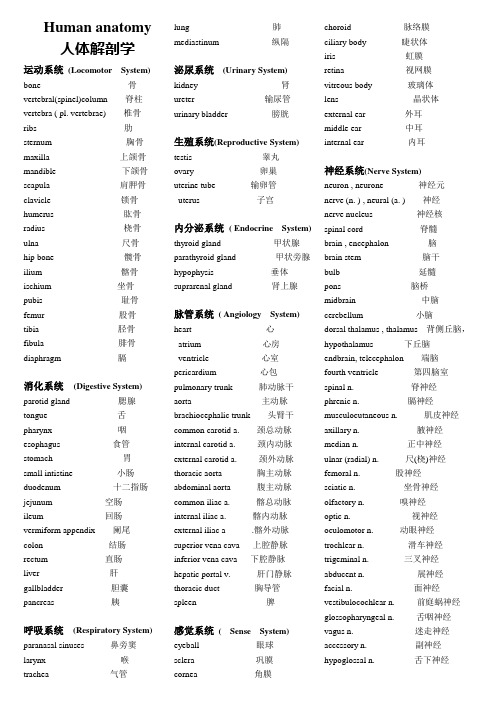
Human anatomy人体解剖学运动系统(Locomotor System) bone 骨vertebral(spinel)column 脊柱vertebra ( pl. vertebrae) 椎骨ribs 肋sternum 胸骨maxilla上颌骨mandible 下颌骨scapula肩胛骨clavicle锁骨humerus 肱骨radius 桡骨ulna尺骨hip bone 髋骨ilium 髂骨ischium 坐骨pubis 耻骨femur 股骨tibia 胫骨fibula 腓骨diaphragm 膈消化系统(Digestive System) parotid gland 腮腺tongue 舌pharynx 咽esophagus 食管stomach 胃small intistine 小肠duodenum 十二指肠jejunum 空肠ileum 回肠vermiform appendix 阑尾colon 结肠rectum 直肠liver 肝gallbladder 胆囊pancreas 胰呼吸系统(Respiratory System) paranasal sinuses鼻旁窦larynx喉trachea气管lung 肺mediastinum 纵隔泌尿系统(Urinary System)kidney 肾ureter 输尿管urinary bladder 膀胱生殖系统(Reproductive System)testis 睾丸ovary 卵巢uterine tube 输卵管uterus 子宫内分泌系统( Endocrine System)thyroid gland 甲状腺parathyroid gland 甲状旁腺hypophysis 垂体suprarenal gland 肾上腺脉管系统( Angiology System)heart 心atrium 心房ventricle 心室pericardium 心包pulmonary trunk 肺动脉干aorta 主动脉brachiocephalic trunk 头臂干common carotid a. 颈总动脉internal carotid a. 颈内动脉external carotid a. 颈外动脉thoracic aorta 胸主动脉abdominal aorta 腹主动脉common iliac a. 髂总动脉internal iliac a. 髂内动脉external iliac a .髂外动脉superior vena cava 上腔静脉inferior vena cava 下腔静脉hepatic portal v. 肝门静脉thoracic duct 胸导管spleen 脾感觉系统( Sense System)eyeball 眼球sclera 巩膜cornea 角膜choroid 脉络膜ciliary body 睫状体iris 虹膜retina 视网膜vitreous body 玻璃体lens 晶状体external ear 外耳middle ear 中耳internal ear 内耳神经系统(Nerve System)neuron , neurone 神经元nerve (n. ) , neural (a. ) 神经nerve nucleus 神经核spinal cord 脊髓brain , encephalon 脑brain stem 脑干bulb 延髓pons 脑桥midbrain 中脑cerebellum 小脑dorsal thalamus , thalamus 背侧丘脑,hypothalamus 下丘脑endbrain, telecephalon 端脑fourth ventricle 第四脑室spinal n. 脊神经phrenic n. 膈神经musculocutaneous n. 肌皮神经axillary n. 腋神经median n. 正中神经ulnar (radial) n. 尺(桡)神经femoral n. 股神经sciatic n. 坐骨神经olfactory n. 嗅神经optic n. 视神经oculomotor n. 动眼神经trochlear n. 滑车神经trigeminal n. 三叉神经abducent n. 展神经facial n. 面神经vestibulocochlear n. 前庭蜗神经glossopharyngeal n. 舌咽神经vagus n. 迷走神经accessory n. 副神经hypoglossal n. 舌下神经。
系统解剖学:12脑干
中枢
神经节
可进一步区分为六个核功能柱、七种核团
①躯体运动柱:动眼神经核、滑车神经核、展神经核、舌下 神经核
②特殊内脏运动柱:三叉神经运动核、面神经核、疑核、副 神经核
③一般内脏运动柱:动眼神经副核、上泌涎核、下泌涎核、 迷走神经背核
④内脏感觉柱:孤束核(头段为特殊感觉,尾段为一般感觉)
⑤一般躯体感觉柱:三叉神经中脑核、三叉神经脑桥核、三 叉神经脊束核
下可接脊髓中央管 并借正中孔和外侧孔通蛛网膜下隙
第四脑室
1)脉络丛 2)三个孔
小脑 上脚
中脑水管
上髓帆
第四脑室 脉络丛
第四脑室 外侧孔
第四脑室脉络组织 第四脑室正中孔
下髓帆
(二)脑干的内部结构
与脊髓相比,脑干内部结构特征: • 灰质呈核团状 • 白质交叉多 • 灰质核团由前、后排列关系转化为内、外关系 • 核团有脑神经核、非脑神经核两种 • 脑神经中共有七种纤维成分,故脑神经核团也分
面为前庭神经核) Acoustic tubercle 听结节 (深面为蜗神经背核) Medial eminence 内侧隆起
• Facial colliculus 面神经丘(深面为面神经膝和展神 经核)
• Hypoglossal triangle 舌下神经三角(深面为舌下 神经核)
• Vagal triangle 迷走神经三角(深面为迷走 神经背核)
三、 白 质
1. 下行(运动)传导束 (1)锥体束 (在脑干内)走行:中脑大脑脚底中间3/5→ 脑桥基底部→延髓锥体
皮质核束
皮质脊髓束
皮质脊 髓侧束
皮质脊 髓前束
(2)其他下行传导束
对侧中脑
红核 上丘
系统解剖学课件:新脑干

近似四棱锥形
Similar to diamond shape
底为菱形窝 尖向后上,朝向小脑
The bottom is rhomboid fossa The point exposition to vermis
顶盖
上髓帆 下髓帆
中脑水管
上髓帆
第四脑室脉络组织中孔 第四脑室外侧孔
(含蜗神经核)
中脑mid brain :
四叠体 quadrigeminal bodies 上丘superior colliculus :
下丘inferior colliculus :
附着有Ⅳ根。
第四脑室
fourth ventricle of cerebrum
延髓、脑桥和小脑之间
medulla oblongata、pons and cerebellum
(4)在灰质与白质之间的区域出现的网状结构面积 急剧扩大,结构更加复杂,其中包含了生命中枢中许 多重要的神经核团(网状核),如心跳、血压和呼吸 中枢等。
1、脑干的灰质 1) 脑神经核Nuclei of cranial nerves
根据其性质和功能,在脑干内按 照以下规律纵行排列成6个功能柱: 在第四脑室室底灰质中,运动 性神经核柱位于界沟内侧,感觉 性神经核柱位于界沟外侧; 由中线向两侧依次为一般躯体 运动核柱、一般内脏运动核柱、 一般和特殊内脏感觉核柱和特殊 躯体感觉核柱。 特殊内脏运动核柱和一般躯体 感觉核柱则位于室底灰质(或中 央灰质)的腹外侧,网状结构内。
Brain stem
脑的发育
前脑forebrum :端脑和间脑
脑
Brain
中脑midbrain :无明显的变化
后脑afterbrain :脑桥和小脑 菱脑hindbrain
人体解剖学:Brain stem脑干

Cerebral peduncle
Optic n. Oculomotor n. Trochlear n. Pons Trigeminal n. Vestibulocochear n. Abducent n. Olive Pyramid Spinal root of accessory n. Medulla oblongata
Temporal lobe颞叶 Optic n.视神经
Oculomotor n.动眼神经 Trochlear n.滑车神经 Motor & sensory roots of trigeminal n. 三叉神经运动和感觉根
Abducent n.展神经
Hypoglossal n.舌下神经
Cerebellum小脑
Inferior colliculus
Midbrain Trochlear nerve
Middle cerebellar peduncle
Pons
Medullary stria Hypoglossal trigone
Vagal trigone Obex
Inferior cerebellar peduncle Cuneate tubercle Medulla Gracile tubercle oblongata
Medullary stria髓纹 Vestibular area前庭区 Vagal trigone迷走三角
Gracile tubercle薄束结节
Pineal gland松果体 Trochlear n.滑车神经 Locus ceruleus蓝斑
4th ventricle第四脑室
Facial colliculus面丘 Hypoglossal trigone舌下三角
脑干(解剖)
薄束核和楔束核
gracile nucleus and cuneate nucleus 位于延髓背侧,是颈以下深感觉和精细触觉神经 通路的中继性核团。
脑干背侧面观
延髓水平切面(平内侧丘系交叉)
红核 Red nucleus
呈圆柱状,平上丘平面,富于血管,对 肢体远端的精细活动有重要调控作用。
脑干背侧面观
脑神经连脑的部位
Ⅰ Ⅱ 嗅三角 视交叉
Ⅲ
Ⅳ Ⅴ
脚间窝
下丘下方 脑桥基底 延髓脑桥沟
Ⅵ
Ⅶ Ⅷ Ⅸ Ⅹ Ⅺ Ⅻ 橄榄前方 橄榄后沟
第四脑室
底:菱形窝 顶:朝向小脑
前髓帆
后髓帆
脑干内部结构
概述:与脊髓比较,脑干有下列特点
1.经脑干的上下行纤维有不少在脑干内交叉 到对侧。 2.灰质被交叉的纤维分割成不连续的细胞团 块(神经核)。 3.由于中央管在延髓上部和脑桥背侧敞开成 为第四脑室底,运动核与感觉核从在脊髓的背、腹 关系变成为内、外关系。 4.脑干网状结构复杂。
内侧丘系
脊髓丘系
2、长下行纤维束
锥体束
红核脊髓束
顶盖脊髓束
前庭脊髓束
网状脊髓束
皮质脊髓束
皮质核束
脑干网状结构
reticular formation of brain stem
位置 脑N核、境界明确的非脑N核 和上、下行纤维束以外的区域。 结构 神经纤维交织成网,胞体散 落其中,有多群核团。 主要传出纤维和功能 上行网状激动系统,调节 觉醒睡眠周期 固有束和网状脊髓束,调 节躯体和内脏运动
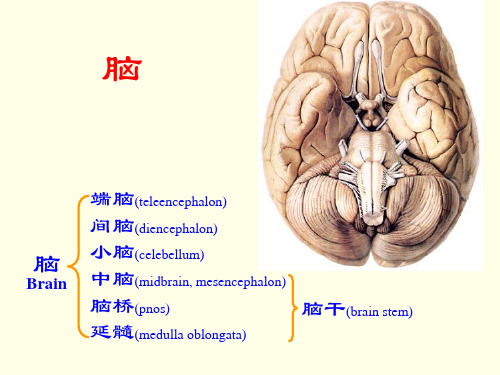
脑
端脑(teleencephalon) 间脑(diencephalon) 小脑(celebellum) 中脑(midbrain, mesencephalon) 脑桥(pnos) 脑干(brain stem) 延髓(medulla oblongata)
脑干的系统解剖

前庭神经核
• 在正中沟的外侧,各有一大致与之平行的纵行界沟sulcus limitans, 将每一半的菱形窝分为内、外侧区。外侧区呈三角区,称前庭区 vestibular area,深方有前庭神经核。
• 前庭区的外侧角有一小隆起称听结节acoustic tubercle,内藏蜗背侧 核。
面神经丘
第四脑室脉络组织
• 此丛呈U形分布,下部沿正中线两侧平行排列,上升至下髓帆附近时, 分别向两侧横行,最终向外延伸至第四脑室外侧隐窝,并经第四脑 室外侧孔突入蛛网膜下腔。
• 第四脑室向上经中脑导水管通第三脑室,向下续为延髓下部和脊髓 的中央管,并借脉络组织上的3个孔与蛛网膜界之间的圆弧形移行部称闩obex,与第 四脑室脉络组织相连。
3.第四脑室
• 第四脑室fourth ventricle位于延髓、脑桥和小脑之间,呈四棱锥形, 内容脑脊液。其底为菱形窝,两侧角为外侧隐窝,顶向后上朝向小 脑蚓。
• 第四脑室顶的前上部由两侧小脑上脚及上髓帆构成,后下部由下髓 帆及第四脑室脉络组织形成。
2.脑干的背侧面
• (1)延髓:延髓背侧面可分为上、下两部,上部形成菱形窝的下半 部;下部形似脊髓,在后正中沟的两侧各有两个膨大,内侧者为薄束 结节gracile tubercle,外上者为楔束结节cuneate tubercle;
• 二者与脊髓的薄束、楔束相延续,其深面分别含有薄束核和楔束核, 它们是薄束、楔束的终止核。
• 在下丘的下方与上髓帆之间有滑车神经根出脑,它是唯一自脑干背 侧面出脑的脑神经。
(4)菱形窝
• 菱形窝rhomboid fossa:是延髓上部和脑桥的背侧面,呈菱形,由延髓 上部和脑桥内的中央管于后壁中线处向后敞开而形成。因构成第 四脑室的底部,又称第四脑室底floorof fourth ventricle。此窝的外 上界为小脑上脚,外下界自内下向外上依次为薄束结节、楔束结节 和小脑下脚。
神经系统

脑干内部结构
2.反射的低级中枢:
3.网状结构:
脑干功能
(1)调节肌张力; (2)调节内脏活动; (3)影响大脑皮质的活动;
小脑位置
小脑外形
(4)对感觉传入影响:网状结构可修改各种传入信息。
总目录
末
页
系统解剖:小脑(cerebellum)
脑干外形
位置
脑干内部结构
脑干功能
小脑位置
小脑外形
约占全脑的 1/10,居颅后 窝、延髓、脑 桥的后方,借 小脑下脚与脑 干相连。
小脑位置
小脑外形
总目录
内脏感觉柱:1对
躯体感觉柱:5对
下页
末
页
系统解剖:脑干(brain stem)
脑干外形
内部结构
躯体运动柱
(1)动眼神经核:图片
脑干内部结构
居上丘平面,组成动眼神经,支配5块眼外肌。
(2)滑车神经核:图片 居下丘平面,组成滑车神经,支配上斜肌。
脑干功能
(3)展神经核:
小脑位置
图片
脑干外形
内部结构
脑干内部结构
脑干功能
小脑位置
小脑外形
上、下泌涎核
总目录
末
页
返回
系统解剖:脑干(brain stem)
脑干外形
内部结构
脑干内部结构
脑干功能
小脑位置
小脑外形
总目录
末
页
返回
系统解剖:脑干(brain stem)
脑干外形
内部结构
脑干内部结构
脑干功能 蜗神经核
小脑位置
小脑外形
总目录
末
页
返回
脑干内部结构
(7)疑核:
奈特神经解剖图谱 .奈特.英文版
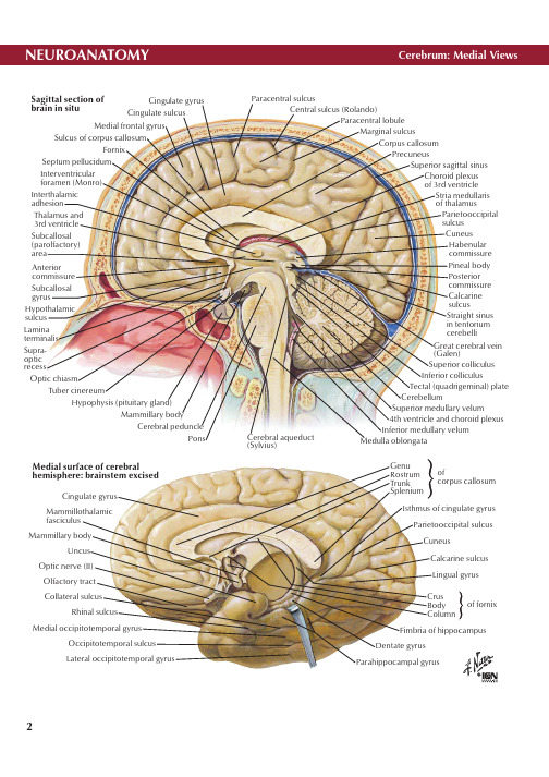
Cingulate sulcusMedial frontal gyrusSulcus of corpus callosumFornixSeptum pellucidum Central sulcus (Rolando)Interventricular foramen (Monro)Interthalamic adhesionThalamus and 3rd ventricle Subcallosal (parolfactory)areaAnterior commissure Subcallosal gyrusHypothalamic sulcus Lamina terminalis Supra- optic recessParacentral lobuleCorpus callosumPrecuneusSuperior sagittal sinusChoroid plexus of 3rd ventricle Stria medullaris of thalamusGreat cerebral vein (Galen)Superior colliculus Inferior colliculusTectal (quadrigeminal) plate CerebellumSuperior medullary velum4th ventricle and choroid plexus Inferior medullary velum Medulla oblongataHypophysis (pituitary gland)Mammillary bodyCerebral pedunclePonsCerebral aqueduct (Sylvius)Genu Rostrum Trunk Spleniumofcorpus callosumIsthmus of cingulate gyrus Parietooccipital sulcus CuneusCalcarine sulcus Fimbria of hippocampus Dentate gyrus Parahippocampal gyrusLateral occipitotemporal gyrusOccipitotemporal sulcus Medial occipitotemporal gyrusRhinal sulcusUncusMammillary bodyMammillothalamic fasciculus Cingulate gyrus Medial surface of cerebralhemisphere: brainstem excisedTuber cinereumOptic chiasmMarginal sulcusOrbital sulciOrbital gyriTemporal poleLateral sulcus (Sylvius)Inferior temporal sulcusInferior temporal gyrusCollateral sulcusParahippocampal gyrusMedial occipitotemporal gyrusCalcarine sulcusIsthmus of cingulate gyrus Longitudinal cerebral fissureOccipital pole of cerebrumApex of cuneusSplenium of corpus callosumCerebral aqueductPulvinar of thalamusRed nucleusOptic tractOptic nerve (II) (Optic chiasmOlfactory tractHypophysis(pituitary gland)Anteriorperforated substanceSuperior colliculus (ofcorpora quadrigemina)Horizontal sections through cerebrumGenu of corpus callosum Lateral ventricle Septum pellucidum Column of fornixof lateral ventriclecorpus callosumHead of caudate nucleusAnterior limbGenuPosterior limbPutamenGlobus pallidus3rd ventricleTail of caudate nucleusHippocampus and fimbriaOccipital (posterior) hornof lateral ventricleHabenulaPineal bodyCleft for internal capsuleOrganization of basal nuclei (ganglia)Putamen GlobuspallidusLentiformnucleusBasal nuclei (ganglia)CaudatenucleusLevels ofsectionsaboveBodyHeadLentiform nucleus(globus pallidus medialLateral geniculate bodyTail of caudate nucleusAmygdaloid bodyAInterrelationship of thalamus, lentiform nucleus, caudatenucleus and amygdaloid body (schema): left lateral viewBA BA BInterventricular foramen (Monro)3rd ventricle Corpus callosum (Head of caudate nucleus Interthalamic adhesion Lamina affixa Stria medullaris Habenular trigone Pulvinar (Lateral geniculate body Medial geniculate body Brachium of superior colliculus cut edge )Calcarine sulcus CerebellumPulvinarReticular nucleusMedian nuclei3rd ventriclePulvinarLateral geniculate body Medial geniculate body3rd ventricle Interthalamic adhesionIntralaminar nucleiExternal medullary laminaCM LD LP Schematic representation of thalamus(external medullary lamina and MDM CM VPL V PMLP L P L D V P V P L V P MV IV LV AM e di a n M e d i a ll a m in a m e dul l a r y I n t er na lA n t e r i o rL ami naAnterior cerebellar notchCentral lobule (II & III)Posterior cerebellar notchInferior surfaceCentral lobuleLingula (I)Superior medullary velumFlocculus (H X)Posterior cerebellar notch4th ventriclesuperior cerebellar pedunclesCerebral crusMedial longitudinal fasciculusHorizontal fissure Inferior semilunar(caudal) lobule (H VII B)Wing of central lobule Superior Middle InferiorAnterior lobeQuadrangular lobule (H IV-V)Primary fissure Inferior semilunar(caudal) lobule (H VII B)Posterolateral viewAnterior viewOptic chiasmOptic tractOlivePyramid Ventral roots of 1st spinal nerve (C1)Decussation of pyramidsAccessory nerve (XI)Trigeminal nerve (V)Middle cerebellar peduncleVestibulocochlear nerve (VIII)Facial nerve (VII)Inferior cerebellar peduncle Hypoglossal nerve (XII)Accessory nerve (XI)Superior medullary velum Lateral recess Superior fovea Sulcus limitans Inferior fovea Trigeminal tubercle Hypoglossal trigoneVagal trigoneObexGracile fasciculusInterthalamic adhesion Posterior commissureHabenular commissurePineal bodySplenium of corpus callosum Great cerebral vein (Galen)Cuneate fasciculusLateral funiculus Habenular trigone Geniculate bodiesDorsal median sulcus Superior cerebellar peduncle Locus ceruleus Medial eminence Facial colliculusVestibular areaSuperior Middle InferiorMedial LateralLingula (I)Central lobule (II-III)Culmen (IV-V)Declive (VI)Folium (VII A)Choroid plexus of 4th ventricle Tonsil of cerebellumCentral canal of spinal cordMedulla oblongataChoroid plexus of 4th ventricle4th ventricleMedial longitudinal fasciculusPonsInferior colliculusTectal (quadrigeminal) plateSuperior colliculus Cerebral aqueduct (Sylvius)Decussation of pyramidsMedian aperture (foramen of Magendie)Superior medullary velum Median sagittal section。
脑解剖专有名词
脑解剖专有名词(总2页)--本页仅作为文档封面,使用时请直接删除即可----内页可以根据需求调整合适字体及大小--Central nervous system (CNS) 中枢神经系统Peripheral nervous system (PNS) 周围神经系统Brain 脑Telencephalon 端脑Diencephalon 间脑Cerebellum 小脑Brain stem 脑干Midbrain 中脑Pons 脑桥Medulla oblongata 延髓Spinal cord 脊髓Cranial (12 pairs) 脑神经Spinal (31 pairs) 脊神经Visceral 内脏神经Sympathetic part 交感神经Parasympathetic part 副交感神经Neurons 神经元Cell body 胞体Axon 轴突Dendrites 树突Neuroglia -glial cell胶质细胞Bipolar neuron 双极神经元Pseudounipolar neuron 假单极神经元Multipolar neuron多极神经元Sensory (efferent) neuron 感觉神经元Motor (afferent) neuron 运动神经元Association neuron 联络神经元Gray matter 灰质Cortex皮质White matter白质Medulla髓质Nucleus神经核Fasciculus 纤维束Reticular formation 网状结构Ganglion 神经节Nerve 神经Central sulcus 中央沟Lateral sulcus 外侧沟Parietooccipital sulcus 顶枕沟Frontal lobe 额叶Parietal lobe 顶叶Temporal lobe 颞叶Occipital lobe 枕叶Insular lobe 岛叶Precentral sulcus 中央前沟Superior frontal sulcus 额上沟Inferior frontal sulcus 额下沟Precentral gyrus 中央前回Superior frontal gyrus 额上回Middle frontal gyrus 额中回Inferior frontal gyrus 额下回Postcentral sulcus 中央后沟Postcentral gyrus中央后回Intraparietal sulcus 顶内沟Superior parietal lobule 顶上小叶Inferior parietal lobule 顶下小叶Supramarginal gyrus 缘上回Angular gyrus 角回Superior temporal sulcus 颞上沟Inferior temporal sulcus 颞下沟Superior temporal gyrus 颞上回Middle temporal gyrus 颞中回Inferior temporal gyrus 颞下回Transverse temporal gyri 颞横回Corpus callosum 胼胝体Callosal sulcus 胼胝体沟Cingulate gyrus 扣带回Cingulate sulcus 扣带沟Marginal ramus 缘支Paracentral lobule 中央旁小叶Calcarine sulcus 距状沟Cuneus 楔叶Lingual gyrus 舌回Olfactory bulb 嗅球Olfactory tract 嗅束Olfactory trigone 嗅三角Anterior perforated substance 前穿质Collateral sulcus 侧副沟Occipitotemporal sulcus 枕颞沟Medial occipitotemporal gyrus 枕颞内侧回Lateral occipitotemporal gyrus 枕颞外侧回Hippocampal sulcus 海马沟Parahippocampal gyrus 海马旁回Uncus 钩Hippocampus 海马Dentate gyrus 齿状回Hippocampal formation 海马结构Archicortex 原皮质 (hippocampal formation 海马结构)Paleocortex 古皮质 (rhinencephalon 嗅脑) Neocortex 新皮质 (most of cerebral cortex) Vestibular area平衡觉区Olfactory area 嗅觉区Taste area 味觉区Motor speech area 运动性语言中枢Writing area 书写中枢Auditory speech area 听觉性语言中枢Visual speech area 视觉性语言中枢Lateral ventricle 侧脑室Corpus striatum 纹状体Lentiform nucleus 豆状核(Globus pallidus 苍白球——paleostriatum 旧纹状体;putamen 壳)Caudate nucleus 尾状核——Neostriatum 新纹状体Claustrum 屏状核Amygdaloid body 杏仁体Association fibers 联络纤维Cerebral arcuate fibers 大脑弓状纤维Superior longitudinal fasciculi 上纵束Inferior longitudinal fasciculi 下纵束Uncinate fasciculus 钩束Cingulum 扣带Commissural fibers 连合纤维Corpus callosum胼胝体Anterior commissure 前连合Fornix and commissure of fornix 穹隆和穹隆连合Projection fibers投射纤维Anterior limb of internal capsule 内囊前肢Genu of internal capsule 内囊膝Posterior limb of internal capsule 内囊后肢Limbic system边缘系统。
- 1、下载文档前请自行甄别文档内容的完整性,平台不提供额外的编辑、内容补充、找答案等附加服务。
- 2、"仅部分预览"的文档,不可在线预览部分如存在完整性等问题,可反馈申请退款(可完整预览的文档不适用该条件!)。
- 3、如文档侵犯您的权益,请联系客服反馈,我们会尽快为您处理(人工客服工作时间:9:00-18:30)。
Midbrain ➢ Attached to the 3rd and 4th cranial n. ➢ Divided into the cerebral peduncle and tectum
Brachium of inferior colliculus 下丘臂
Fourth ventricle 第四脑室
Central canal →fourth ventricle →mesencephalic aqueduct→third ventricle
Crus cerebri 大脑脚底 Interpeduncular fossa 脚间窝
Oculomotor nerve emerges on the medial side of crus cerebri Posterior perforated substance 后穿质
★ Dorsal Surface of Midbrain
Superior colliculus 上丘 Constitute the centers for visual reflexes
Inferior colliculus 下丘 Associated with auditory pathway
Brachium of superior colliculus 上丘臂
➢ Inferior cerebellar peduncle 小脑下脚
➢ Obex 闩
Upper portion: forms the lower half of rhomboid fossa 菱形窝
★ Ventral Surface of Pons
Bulbopontine sulcus 延髓脑桥沟
Gross Appearance of Brain Stem
★ Gross Appearance of Brain Stem
Medulla oblongata ➢ Attached to the 9th to 12th cranial n. ➢ Divided into an open(superior) and a closed (inferior) part
Forms the superior half of rhomboid fossa菱形窝
Superior cerebellar peduncle 小脑上脚
Superior medullary velum 上髓帆
Trochlear nerve 滑车神经
★ Ventral Surface of Midbrain
★ Dorsal Surface of Medulla oblongata
Lower portion ➢ Gracile tubercle 薄束结节 produced by underlying gracile nucleus 薄束核 ➢ Cuneate tubercle 楔束结节 produced by underlying cuneate nucleus 楔束核
★ Ventral Surface of Medulla oblongata
Pyramid 锥体 Composed of bundles of nerve fiber, called pyramidal tract 锥体束
Decussation of pyramid 锥体交叉 Formed by crossing fibers of pyramidal tract
➢ It connects the diencephalon anteriorlly, spinal cord inferiorlly and cerebellum posteriorlly.
➢ The space between the pons, upper half of medulla oblongata and cerebellum is the fourth ventricle.
Brain stem 脑干
学习方法
看外形,想内部,内外结合。 看断面,想立体,上下串联。 记重点,莫贪多,整理表格。
Brain stem 脑干
★ Component ➢ Midbrain 中脑 ➢ Pons 脑桥 ➢ Medulla oblongata 延髓
Position
➢ The brain stem occupies the posterior cranial fossa of the skull and lies on the clivus of the occipital bone
There emerge, from medial to lateral, the abducent, facial and vestibulocochlear nerves Basilar part 基底部 ➢ Basilar sulcus 基底沟 Middle cerebellar peduncle 小脑中脚 Trigeminal nerve 三叉神经
Emerges from the junction of basilar part and middle cerebellar peduncle Pontocerebellar trigone 脑桥小脑三角
The junction of medulla, pons and cerebellum
★ Dorsal Surface of Pons
Olive 橄榄 Produced by underlying inferior olivary nucleus 下橄榄核
Anterolateral sulcus 前外侧沟 Emerge the roots of hypoglossal nerve
Retroolivary sulcus 橄榄后沟 Emerge the roots of glossopharyngeal, vagus and accessory nerves