磁共振水成像技术对内耳的成像研究和应用
MRl内耳水成像技术的应用
贵州医药,2012,36(1):51-53. (收稿日期:2012-12Copyright©博看网 . All Rights Reserved.
内耳是 人体 听 觉 器官的 重 要 组 成 部 分,因其 结 构复 杂,也被 称 之为迷 路。内耳 在 人体的听 觉当中起 着重 要 作用,它维 持 着机体 的平衡,对人体 接受的外在声音 进行加工 后转换为神经冲动,并 将 其 从 蜗 后传 达 到大 脑 皮 层的听 觉中 枢,是 人体 能 够 对外在 声音 信息做出反 应的关 键 器官。一旦内耳发 生 病 变,出现 结 构 或 者功 能 上的异常,极易影响人的听力,造成各种不便。临床上,突破传统 X 射线及CT检查局限的M R I水成像(核磁共振成像术,简称为磁共 振成像 M R I)技术,在内耳水成像中具 有重要作用,对于内耳异常 的 检 查提 供了更 好的临床实践。本 次 选 取该院 2 011年3月间 — 2 012 年6月间进行检 查的病患83例,探 讨M R I 水成像技 术的临床应 用 价 值,并形成 如下报 道。 1 资料与方法 1.1 一般资料
从 此次临床 研 究的结果 来 看,M R I 水成像 技 术 在 检 查内耳 时,成像效 果较 好,检查较为全面,在临床应用上具 有广泛前景。 内耳是人体听力器官的重要组成部分,全 部埋藏于颞骨岩 部骨质 内,介于鼓室与内耳道底之间,由骨迷 路和膜 迷 路构成,分为两个 部分,一个叫做耳蜗是听觉器,另一个叫做前庭是 平衡器,因此内 耳又叫做平衡听 觉器。内耳因其组 成 结 构复杂,因此也有迷 路之 称。磁 共 振 成像是 断层成像中的一种发 射断层成像,它利用磁 共 振 现 象 从 人体中获得电磁信号, 并重 建出人体信息,但与 P E T 和 S P E C T不同的是 磁 共 振 成像 不用注 射 放 射 性同 位 素就可成像,这 一点也使 磁 共 振 成像 技 术更加安 全。由于内耳结 构细小复 杂,位 置特 殊,所以实体解剖标 本的获取比较困难,传 统的X 线及C T 在 成像上也存在很大 局限性,因此给内耳疾病的诊断带来了困难。 相较之下,M R I成像技术下,我们可以得到更多的关于内耳结构物 理特性的相关参数及图像,M R I成像技术相较于其他成像技术能 够 提 供 更多的信息量,在诊断患者病 情上具 有更 强的优势,比如 横 断 面、矢状 面、冠 状面 和 各 种 斜 面的 体 层图 像,但同时又 不 会 产 生伪 影,辐 射少,对 机体影响 小,应 用 优 势 非常明显。
正常内耳内听道磁共振水成像观测
关键 词 : 内耳 ; 内听道 ;3 F E;磁 共 振 成像 D— S
中 图分 类 号 :R7 4 6
文献标 识 码 :A
岩 骨 深部 , 构细 微复 杂 , 度 曾是 影像 学检 结 一
1 材 料 与 方 法
11 临床 资料 .
查的盲区. 近年来 , 随着高分辨率磁共振的出现和 相应软件平 台的研发利用 , 特别是利用静态液成像
在 后 台处理 工 作站 A ., 3 F E采 集 的 W42 对 D—S
逐层 勾勒 出感 兴趣 区轮 廓. 耳蜗的图层勾勒方法
如图 1 图 2 示 , 和 所 阴影 部分 即 为被 勾 勒后 的感 兴 趣 区 . 积重 建 : 容 在后 处理 工作 站 A v nae r. d at k g Wo s t n42 t i .,对每 一 图层感 兴 趣 区叠加 计算 . ao
共振 内耳 内 听道影 像解 剖 测量 数据 报道 仍然 较 少 , 选 择 磁 共 振 三 维 快 速 自旋 回 波 ( redmes nl t e—i ni a h o fs si co D—S ) 描 序列 ,三 维 容积 再现 at pn eh ,3 F E扫 ( red ninl ou n eig 3 R 和 多平 t e—i s a vlmer dr , DV ) h me o e n 面 重建 ( ut lnrrcnt ci ,MP ) 处理 技 m lpaa eo s t n i u r o R后
第 2 卷 第 3期 1 20 0 8年 9月
宁 波 大 学 学 报 (理 工 版 )
J UR O NALO NG O U V R I Y( E FNI B NI E S T NS E)
V l l NO 3 o 2 .
磁共振成像对耳聋病因的诊断与判断

病例描述
诊断与鉴别 诊断
突发性聋的发病原因尚不完全清楚,但磁共振成像检 查可排除内耳结构异常,为临床治疗提供重要依据。
临床意义
结合患者病史和磁共振成像表现,可诊断为突发性聋 。需与内耳结构异常导致的耳聋进行鉴别诊断,如先 天性内耳发育异常、中耳炎等。
老年性聋病例
病例描述
老年患者逐渐出现听力下降,经磁 共振成像检查发现内耳结构退行性 改变,如耳蜗萎缩、毛细胞减少等 。
聋。
两种病因同时存在
03
如外耳道闭锁合并内耳发育畸形等。混合性耳聋的听力损失程
度通常较重,治疗也较为困难。
03
磁共振成像在耳聋诊断 中应用
内耳结构显示技术
高分辨率磁共振成像
能够清晰显示内耳细微结构,如耳蜗、前庭、半规管等。
三维重建技术
通过对内耳结构的三维重建,提供更直观、立体的内耳形态信息 。
水成像技术
扫描参数
包括磁场强度、射频脉冲频率、扫描 层厚、扫描时间等,影响图像质量和 扫描时间。
临床应用范围与优势
01
临床应用范围
适用于头部、颈部、胸部、腹部、盆腔、脊柱、四肢等部位的检查,特
别对软组织层次显示效果好。
02
耳聋病因诊断
可观察内耳结构,如耳蜗、前庭等,判断是否存在发育异常、肿瘤、炎
症等病变,为耳聋病因提供重要诊断依据。
扫描参数优化策略
序列选择
根据内耳不同组织结构和病变特点,选择最合适的扫描序 列。
分辨率与扫描时间平衡
在保证图像分辨率的同时,尽量缩短扫描时间,减少患者 不适和运动伪影。
噪声控制
降低扫描过程中的噪声,提高患者舒适度和图像质量。
人工智能在磁共振成像中应用
探讨MR内耳水成像技术的应用
探讨 MR 内耳 水 成像 技 术 的应用
刘 良卿 ,郑建 刚 ,梅 莉 ,许 建兴 ,周 鸿雁 ,顾 勇 坚,吴刘 萍
(江 苏 大 学 附 属 武 进 人 民 医 院 放 射 科 江 苏 常 州 2I3002)
hydrography features,to discuss the technology of magnetic resonance hydrography in clinical applications. Methods U— sing siemens 1.5 T supercondu ting scanners,we adopted three dimensional fast sequence(3D-TSE). In 35 cases suspec— ted internal ear disease by hydrography,all patients were antecedent general M R scanning.The reconstruction included maximum intensity projection(MIP)and multi—planar reconstruction (MPR).Results Both sides image can be satisfied w ith the display of the inner ear anatom ical structures:vestibular,cochlear im plants,sem icircular canals,and internal au— ditory cana1. Conclusion H ydrography is an effective and non-invasive display bilateral labyrinth and the internal auditory canal fine structure. It is not only able display 3-dimensional anatomical structures of the labyrinth and internal auditory canal, but aeso able to clearly dem onstrate its interna1. It can help clinical analysis of illness, and treat by surgery, espe— cially provide im portant inform ation to electronics cochlear im plant,w hich could be excluded som e absolute or relative con— traindications,greatly increases accuracy. [Key words] Magnetic resonance imaging;Inner ear;Membranous labyrinth;Internal auditory canal
MR水成像技术及其临床应用

进一步拓展MR水成像在临床领域的应用范围,如神经、腹部、盆 腔等部位的病变诊断和评估。
未来发展趋势展望
01
人工智能辅助诊断
结合人工智能技术,实现自动化、智能化的图像分析和诊断辅助,提高
诊断准确性和效率。
02
定量评估与功能成像
பைடு நூலகம்
发展定量评估和功能成像技术,提供更准确的病变信息和生理功能评估。
02
MR水成像技术种类
静态水成像
01
02
03
原理
利用长TR和长TE技术,使 体内静态水分呈现高信号, 而周围组织呈现低信号, 从而形成对比。
应用
主要用于胆道系统、泌尿 系统、内耳迷路等含液器 官的检查。
优点
无需注射造影剂,无辐射 损伤,可多角度成像。
动态水成像
01
原理
采用快速扫描序列和动态增强技 术,实时观察体内水分子的动态 变化。
无创检查
MR水成像技术是一种非侵入性的检 查方法,不需要穿刺或注射造影剂, 避免了传统检查方法可能带来的并发 症和风险。
无辐射
该技术不使用放射性物质,因此不会 对患者和医护人员产生辐射危害,安 全性高。
多方位、多角度观察病变情况
多方位成像
MR水成像技术能够在任意方向上进行成像,提供多方位的观察视角,有助于 全面评估病变的范围和程度。
现状
目前,MR水成像技术已经成为临床影像学检查的重要手段之一,广泛应用于颅脑、脊柱、腹部、盆腔等多个部 位的检查。同时,随着人工智能、大数据等技术的不断发展,MR水成像技术的图像处理和诊断水平也得到了进 一步提升。
技术特点与优势
高分辨率
无创性
MR水成像技术能够提供高分辨率的图像, 清晰显示含水器官和组织的细微结构和病 变。
MRH在内耳成像及病变诊断中的应用研究

李 英 , 敬 霞 , 剑 羽 . D. I S 列 在 内 耳 及 内 听 道 谢 刘 3 CS序 磁共振成像中的应用. 中国医学影像技 术, 0 3 1() 2 0 , 94 :
41 . 7. 5 41
构 ,对于 内淋 巴管和位 于颞骨 内外 的 内淋 巴囊显示 M i tr e ia r s , 0 7 2 3 l a y M dc l e s 2 0 : 5 . i P 不 佳 ,并且大 多数前庭 导水 管扩大 综合征 的患者均 杨 正汉 , 逢, 霄 英 . 共 振 成 像 技术 指 南 . 京 : 民 冯 王 磁 北 人 军 医 出版 社 ,0 7 2 3 2 0 :5 . 为婴幼儿 ,矢状面C 扫 描很难配合 ,但在横 断面 上 T 9 B u , i JYm ,t1 J . mai fh i r l n tn 测 量又会造成假 阴性 ,3 S 2 能准确的发现扩 [] y n S Km H , i YJe a MRi g go tene a DT ET WI a d t r a a n n e a t3 c mp rs n b t e u i y c n l d i n r e r a T: o a i o e we n o a 大 的内淋 巴囊 ,其优 越性显 而易见 ,本组 患者利 用 3 d i e q i b i m n D a a c d f s e d e h D rv n e u l ru a d 3 b l n e a tf l c o i i MR H不但 可观察到异 常扩大 的导水管 ,还 能观 察到 sq ecsKoenJ a il 0 893: 1—1 . eun e. ra R d , 0 ,()2 22 8 o2 扩大 的 内淋 巴囊及 内淋 巴管 。对 三种后 处理方 法 图 [0 e X. w t h ooiseerhadc ncl rci f 1】XiJ Ne c lge sa n ii at e en r c l ap c o 像进 行综合 分析 ,将 内耳的细微 结构进 行显 示 ,使 te ul r g ec eo ac. e i : u lhn ue h c a nt sn eB in P bi i Ho s n e ma ir n jg s g 引起S L NH 的病因得到显示 ,提高 了内耳病变诊 断的 o B in dcl nvr t 2 0 : 3 —4 . f e i Mei i sy 0 14 8 4 jg aU e i, 4
【doc】3D—重T2W水成像技术显示面听神经及内耳结构的研究

3D—重T2W水成像技术显示面听神经及内耳结构的研究放射学实践3D一重曾南林【择4O层厚3D-重TwIFSE序列与常规TwIFSE序列对两侧耳部同时进行扫描,比较两者对内听道面听神经分支和迷路解剖结构的显示率,并作统计分析.结果:3D-重TwIFSE序列与常规TwIFSE序列对其细微结构的显示率分别为螺旋板(98:0,P<0.01),前庭阶(98:0,P<0.01),鼓阶(98:0,P<O.01),椭圆囊(95:0,P<0.01),球囊(95:0,P<0.01),前庭嵴(95:0,P<0.01),前庭上神经(100:40,P<0.01),前庭下神经(98:20,P<0.01),其差异均有显着性意义,而在3个半规管,前庭,耳蜗,内听道面神经,蜗神经等结构的显示率方面差异无显着性意义(P>0.05).结论:薄层3D-重T2wFSE水威像技术在显示内听道前庭上下神经分支和迷路的螺旋板,前庭阶,鼓阶,椭圆囊,球囊等细微结构方面明显优于常规TwI.【关键词】内耳;内耳道;磁共振成像【中图分类号】R445.2;R745.12;R764【文献标识码】A【文章编号】1000—0313(2003)04-0293—04NormalanatomyofinnerearandIAConFSE3DheavyT2weightedMRimagingZENGNanli n,ZHAIZhaohua,ZHANGXiaoming.DepartmentofRadiology,AffiliatedHospitalofNorthSichuanMedicalCollege, Sichuan637000[Abstract]Objective:Tostudythedetectabilityforanatomystructuresofinternalauditoryca nal(IAC)andlabyrinthinnormalsubjectsonFSE3DHeavyT2WIMRimaging.Methods:MRimagesof80innerears in40normalsubjectswere studiedwithoutrelatedabnormalitiesorsymptoms.AllstudieswereperformedonGE1.5TM Rscannerwithacircularheadcoilor3inchcircularcoils,using0.5mmthicknessonbothFSE3DHeavyT,WIandFSET ,WIsequences.3Drecon—structedMRimagesoftheinnerearweredirectlysynthesizedwithmaximumintensityproject ion(MIP)technique.Thede—tectionratesoftheminuteanatomystructuresofIACandlabyrinthonFSE3DHeavyT2W1w erecomparedwiththatonFSET2WIMRimaging.Results:ThedetectionratesfortheanatomyonFSE3DHeavyT2WI andFSET2W1were95vs0(P<0.01)forsaccule,95vs0(P<0.01)forvestibularcrest,95vs0(P<0.01)forutricle, 98vs0(P<0.01)forscalavestibuli,98vs0(P<0.01)forscalatympani,98vs0(P<0.01)forspi rallamina,100%vs4O(P<0.01)fornervivestibularissuperior,and98vs20(P<0.01)fornervivestibularisi nferior,respectively. Thedifferenceofdetectionrateswasstatisticallysignificantfortheseminuteanatomicalstru ctures,butwasnotstatisticallysignificantforthreesemicircularducts.vestibule,cochieae,nervinochieaeandnervifacialis (P>0.05)betweenFSE3DHeavyT2WandFSET2WMRimages.Conclusion:FSE3DHeavyT2WIMRimagingisbett erthanFSET2WIMRimaging fordetectiontheanatomicalstructuresofinternalauditorycanalandlabyrinth. [Keywords]Innerear;Internalauditorycanal;MRI内耳位置深,且结构细小复杂,周围有信号强度差异较大的颞骨,空气和脑脊液.这些较大的信号差异可导致MRI上磁化敏感伪影而使其某些结构的信号丢失.近年来,随着多种MRI技术的开发成功和应用,活体显示内耳迷路的精细结构已成为现实_】.].国内有学者曾用0.6mm层厚3D~CISS序列对耳部正常解剖进行研究,能显示其3个半规管,耳蜗,前庭以及内听道等结构,但未能显示前庭阶,鼓阶和内听道四神经分支的精细结构,未能将椭圆囊与球囊分开.我们采用0.5mm层厚31%重T2WFSE序列(TR4000ms,TE900ms),用最大强度投影(maximumintensityprojection,MIP)对兴趣区进行多平面容积重建(multiplanarvolumerecostruction,MPVR),显示了内耳中作者单位:637000四J1j,川北医学院附属医院放射科作者简介:曾南林(1958~),男,四JII南充人,主管技师,主要从事医学影像技术及计算机应用研究.的骨螺旋板,前庭阶,鼓阶,椭圆囊,球囊,前庭嵴和内听道四神经分支的精细结构.为该部位病变尤其是微小病变的定位诊断以及内耳显微外科手术提供了有价值的影像解剖学依据.材料与方法本文搜集我院2000年~2002年共4O例(80侧)患者的颞部MR检查,其中健康志愿者3O例(60侧),其余1O例(20侧) 为临床怀疑脑内病变而行此检查者.4O例(80侧)中男25例(50侧),女15例(30侧),年龄15~55岁,平均32岁.所有患者均经临床问诊及听力测验等检查排除耳部病变.检查方法:均采用GE公司1.5T超导MR系统SignaHo—rizonIX1I,最大梯度磁场为22mT/M,最大切换速度为20T/M/s,环形头部线圈或3in环形线圈(3-inchcircularcoils),使用3D-重T2WFSE序列,TR4000ms,TE900ms,翻转角294放射学实践2003年4月第18卷第4期Radi—ol—Practic!!垒pr:30.,FOV18cm,矩阵256×16O,层厚0.5mm.无间隔,冠状位或轴位采集范围包括两侧岩骨区.待3D原始数据采集完毕并重建后,再用MIP技术对整个内听道及迷路兴趣区进行MPVR,重点展示各部位的细微结构,并与常规T2(TR4000ms,TElOOms,FOV18cm)进行比较,其扫描参数见表1.表1内耳扫描参数表重建方式:采用MIP的多轴位多视角多平面容积重建(MPVR).分别作绕左右轴,上下轴,前后轴旋转的MPVR图.在1象限轴位图上定上下轴,2象限冠状图上定前后轴,3象限矢状图上定左右轴,4象限为多平面容积重建MPVR图,轴芯在各象限位于前庭处使MPVR图像居中,从0.开始顺时针每15.旋转1次观察至一165.,重点展示各部位的细微结构.重建厚度视重建对象而定,在显示内听道四神经分支时选用薄平面(O.7mm)MPVR,在显示内耳迷路时选用厚平面(16~22mm) MPVR.DFOV选用3.2mm.显示情况比较:对细微结构显示情况分为两个等级:1无法显示,2可显示.对于无论在冠状位,轴位,矢状位,斜位扫描图像以及重建图像的任一层面均无法显示的定为无法显示,反之只要有一个层面能显示则定为可显示.4O例(80侧)的全部图像资料经3位有经验的影像专家双盲法评估.分别计算3D-重TzWFSE序列和常规T.FSE序列对其细微结构的显示率,列表比较,同时进行Y.检验.结果4O例(80侧)中3D-重TWFSE序列均能满意显示两侧内听道及内耳迷路的精细解剖结构,而常规TWFSE序列不能区分椭圆囊与球囊,也未能显示螺旋板和前庭神经分支.在31b-重T.WFSE序列中3个半规管,椭圆囊,球囊,耳蜗管及内听道均呈明显高信号,耳蜗螺旋板,前庭嵴及4条面听神经分支均为低信号.面听神经主要分支及内耳各结构的显示情况比较以及Y.检验结果见表2.4O例(80侧)正常内听道和迷路中,单幅常规TWFSE图像上均不能显示内耳的全貌(图1).3D-重T.WFSE水成像左右轴一165.MPVR图(TR4000ms,TE900ms),显示内听道及桥前池为明显高信号,迷路呈不规则形高信号,3个半规管同时显示呈"蝴蝶状",耳蜗呈"草帽状",三维立体感强,上为水平半规管,下为后半规管;蜗底直径为8mm,蜗顶直径为2mm,蜗轴底直径为3mm,蜗高为3mm,蜗管中间见横形条状低信号,为耳蜗骨螺旋板,螺旋板上方为前庭阶,下方为鼓阶;内听道内有2条并行排列的线条状低信号,分别为面神经和听神经(图2). 在上下轴_45.MPVR图上(图3),3个半规管呈半环状,表240例(80侧)内听道及内耳各结构显示情况比较上为前半规管,下为后半规管(全展开),中为外(水平)半规管, 蜗管呈盘旋的"蚊香状"清晰可见,前后半规管汇于总骨脚,球囊(saccule,S)呈高信号近似为圆球形,长短径约为1.2mm×0.9mm.椭圆囊(utricle,U)信号稍高,呈圆弧管状形,长短径约为4.5mm×1.1mm;椭圆囊与球囊之间有明显的较低信号分界,此低信号为骨性结构的前庭嵴(vestibularcrest,VC).前后轴一135.MPVR图上,前后半规管,总骨脚显示最佳,外(水平)半规管与椭圆囊重叠,耳蜗管显示清楚(图4).在上下轴一45.左耳内听道横截面(0.7mm)MPVR图上,可以同时清晰地显示4根神经分支(图5):1前庭上神经(nervi vestibularissuperior,NVS);2前庭下神经(nervivestibularisin- ferior,NVI);3蜗神经(nervicochlearis,NC);4面神经(nervifa—cialis,NF).在左右轴0.面神经薄平面(O.7mm)MPVR图上,左右内耳均可见两条平行的低信号神经,上为面神经,下为前庭上神经(图5a).在左右轴O.蜗神经薄平面(0.7mm)MPVR图上,可见蜗神经与前庭下神经呈"Y"型排列,上为蜗神经,下为前庭下神经(图5b).讨论内耳迷路包括骨迷路和膜迷路.骨迷路主要由耳蜗,前庭以及前,后,外3个骨半规管构成;膜迷路主要由耳蜗管,椭圆囊,球囊及与之相连的前,后,外3个膜半规管构成,其内充满内淋巴液;骨迷路的骨性结构与膜迷路间的腔内充满外淋巴液,膜迷路悬浮在外淋巴液中;内听道充满脑脊液,其内有面神经和听神经走行.上述结构细小复杂,位置深,且外围均有骨性结构封闭,以往的影像检查很难将这些结构显示出来.国内有学者_1_曾用0.6mm层厚31>CISS序列对耳部正常解剖进行研究,能显示3个半规管及内听道结构.我们采用0.5mm层厚3D-重T2FSE水成像技术(TR4000ms,TE900ms),用MIP对兴趣区进行MPVR,能较好显示内耳中的骨螺旋板,前庭阶,鼓阶,椭圆囊,球囊,前庭嵴和内听道四神经分支的更精细结构.迷路及内听道成像的基础是利用水成像原理,即使用长TR和特长TE的重T.W作用,以提高内听道内脑脊液和迷路的内,外淋巴液信号强度,使之成为极高信号,同时抑制周围放射学实践2003年4月箜鲞笙塑翌垒:295图1常规rr2一FSE图像(TR4000ms,TElOOms).水平半规管全部显示,前半规管部分显示,后半规管几乎不显示.图23D-重rr2FSE左右轴一165.MPVR图.3个半规管均较好显示,箭处为螺旋板.图3上下轴一45.MPVR图.u椭圆囊,s球囊,VC前庭嵴.图4前后轴一135.MPVR图.箭处为总骨脚,ASC前半规管,PSC后半规管.图5正常内听道4根神经分支的显示.左上插图示上下轴一45.左耳内听道横截面MPVR图,1~4分别为四神经分支横截面,白横线为薄平面重建位置示意.1前庭上神经,2前庭下神经,3蜗神经,4面神经.a)左右轴O.面神经薄平面MPVR 图;b)左右轴O.蜗神经薄平面MPVR图.的骨结构,从而达到内听道及迷路"造影"的效果.本组资料显示3个半规管,椭圆囊,球囊,蜗管以及内听道均呈明显高信号,耳蜗螺旋板,前庭嵴,位听神经和面神经均为低信号,图像清晰,效果满意,其形态与大体解剖基本一致.为获得清晰的迷路及内听道的精细解剖图像,在MR水成像时应特别注意以下几点.①患者头位必须十分准确,左右对称,扫描范围应包括整个岩骨,使两侧迷路及内听道同时一次性成像,便于两侧对比观察,更易发现病变.②MIP技术是保证图像质量的关键,重建时兴趣区不宜过大或过小,过大将有其他高信号结构与迷路重叠,影响观察;过小则会使部分正常迷路结构被删除.我们观察发现,在显示内听道四神经时宜选用薄平面(0.7mm)MPVR,在显示内耳迷路时宜选用厚平面(16~22ram)MPVR,耳蜗与3个半规管可同时清晰显示,如果要显示内淋巴囊宜选用更厚平面(32mm)MPvR.③多轴位多视角观察:为充分展示整个迷路及内听道的立体结构,待3D原始图像重建并MIP后,以电影形式将其沿一定方向旋转不同角度观察,可沿左右轴,上下轴,前后轴从O.开始顺时针每15.旋转1次观察至一165.(O.图与180.图相同).④线圈选择:Held等的研究表明,使用特殊表面线圈以替代环形头线圈可提高几何分辨率,非常小的充盈缺损也可观察到.但使用头线圈是重要的,因为脑干和桥小脑角的病变在单纯使用表面线圈时通常会漏掉.本组资料表明,用3in环形线圈扫描可获得满意效果,但换线圈费时且脑干部分的信号差,故主张先使用头部环形线圈,必要时用3in环形线圈扫描.⑤扫描序列:James等采用T!FSE序列(TR4000ms, TElOOms)对内耳进行高分辨成像,结果表明该序列明显较少受到磁化敏感伪影的影响.Christian等报道在内耳和桥小脑角的MR成像中,采用3D序列比2D序列能提供更多的细节,以便更准确地观察精细解剖结构和更多地发现病变.本组资料把3D序列的TE提高到800ms以上,与常规T.比较更能提高液体与骨的信号强度对比,在显示内耳的骨螺旋板,前庭阶,鼓阶,椭圆囊,球囊,前庭嵴和内听道四神经分支的精细结构方面明显优于常规T(P<0.01).本组资料表明,应用MR水成像技术能清晰显示内耳迷路及内听道四神经分支的精细结构和解剖关系,其图像立体直观,具有其他影像学方法不可替代的作用.笔者对迷路及内听道主要结构进行了观察与测量,其中3个半规管形态近似,呈296放射学实践2003年4月第18鲞堑塑!!竺!:垒1:!!: "C"或"U"字形,半规管边缘光滑,直径为1~2mm,半规管的脚稍膨隆,3个半规管共有5个脚与椭圆囊相连.椭圆囊和球囊显示为高信号囊,其长短径分别为4.5turn)<1.1mrn和l_2ram)<0.9mm. 耳蜗在左右轴--30.~--75.MIP图上呈盘旋的"蚊香状",在左右轴0.MIP图上呈"草帽状",蜗底较大,直径8mm,蜗顶较小2mrn,蜗高为3mrn,蜗轴底直径3mm,蜗管直径1.5~2.5mm,耳蜗螺旋板表现为条状低信号,螺旋板上方为前庭阶,下方为鼓阶.两侧内听道呈管状或锥形高信号,直径5--6ram,边缘光滑.其内可见4条低信号结构,为前庭上神经,前庭下神经,蜗神经和面神经.前庭下神经最细,直径约为0.9mrn,其余3条神经直径约为lmrn.结论本组资料表明薄层319-重TWFSE水成像技术在显示内听道面听神经分支和迷路的螺旋板,前庭阶,鼓阶,椭圆囊,球囊,前庭嵴等细微结构方面明显优于常规Tz.313-重TzWFSE序列的MPVR图像在显示内耳的全貌方面明显优于单幅常规,r2WFSE图像.(注:1in一2.54cm)参考文献1孔祥泉,刘定西,徐海波,等.国人内听道及内耳膜迷路的正常MR解剖研究[J].临床放射学杂志,1999,18(1):1卜13.2ShinjiN.TokikoI,ErikoI,eta1.MRimagingofthecochlearmodio—Ius:areameasurementinhealthysubjectsandinpatientswithalarge endolymphaticductandsac[J].Radiology,1999,213(3):819—823.3HeldP,FellnerC,FellnerF,eta1.MRIofinnerandfacialnervepa—thologyusing3D-MRGEand3DCISSsequences[J].BrJRadiol,l9977O(834):558—566.4HarnsbergerHR,RichardTD,CloughS,eta1.Advancedtechniques inmagneticresonanceimagingintheevaluationofthelargeen—dolymphaticductandsacsyndrome[J].Laryngoscope,1995,105(10):l037—1042.5ChristianC,ThomasR,WolfgangG,eta1.MRimagingoftheinnerandcerebeIl0p0ntineangle:comparisonofthree-dimensionalandtwo—dimensionalsequences[J].AJR,1998,170(3):791-796.6JamesNL,BrianDK,DennisLP,eta1.Highresolution3Dimagingof theinnerearwithamodifiedfastspin—echopulsesequence[J].JMRI,1996,6(1):223—225.'7HirschBE,WissmannJL,CurtinHD,eta1.Advancedmagneticreso—nanceimagingofthelargevestibularaqueduct[J].ArchOtolaryngol HeadNeckSurg,1992,118(10):1124—1127.(2002—08—26收稿2002—10—21修回)乳腺淋巴管瘤一例张笑魁牛淑祥【中图分类号】R445;R655.7【文献标识码】D【文章编号】1000—0313(2003)04—0296—01病例资料患者,女,43岁,20d前发现右乳下肿物,局部疼痛伴发热,体检发现右乳内下象限可触及约5.0cm×4.0cm包块,质硬,表面无扩张静脉,无波动感.右乳内上及外下象限分别触及约2.0cm×1.5cm和4.0cm×4.0cm大小的肿块,质硬有波动感,腋下未扪及肿大淋巴结.B超提示:右乳腺实性包块.CT平扫:右胸壁软组织隆起高于对侧,皮下及乳腺间脂肪间隙消失,被大量结节样层状分布的软组织影充填,其边界清楚,右侧乳腺不能识别,CT值为2~1OHU,软组织影沿肌肉间隙生长,肌肉,病例报道?图1乳腺淋巴管瘤.CT示右侧胸壁内大量软组织影,其内有线条状低密度影,大部呈节条状,层样分布,沿肌间隙蔓延(箭).a)右乳下部;b)右乳上部.骨胳未见浸润征象(图1).CT考虑为胸壁淋巴管瘤.手术及病理:术中见右乳内下象限包块与周围组织分界清,并有囊性分隔,内伴清亮液体.另于右乳内上及外下方乳腺腺体上各剥离1.5cm×1.5cm×1.0cm和3.5cm×3.0cm×1.5cm大小的肿块各一,其质韧,无分隔.病理诊断:右乳腺淋巴管瘤.作者单位:043014山西,侯马解放军第289医院放射科作者简介:张笑魁(1974~),男,内蒙古宁城人,技师,大专,主要从事普通放射技术工作.,讨论乳腺淋巴管网丰富,但发生淋巴管瘤者少见.淋巴管瘤系先天性病变,由增生的淋巴管构成,为淋巴管发育畸形所致,而非真正的肿瘤.多发于儿童,偶见成人,以颈部及腋部常见,发生于乳腺者较少见.组织学上分为毛细管型淋巴管瘤,海绵状淋巴管瘤及囊性淋巴管瘤3种,亦可为混合型.卢光明等认为淋巴管瘤壁薄CT多不能显示其分隔,且不强化.本例因患者对对比剂过敏未能做增强检查,但就其低密度肿块(密度近似于水),沿肌肉间隙蔓延生长及分布广泛的特点,在排除乳腺自身病变的基础上还应考虑为淋巴管瘤. (2002—10—04收稿2002—12—06修回)。
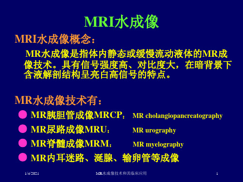
MR水成像技术和其临床应用
1/4/2021
MR对静止的液体在重T2加权成像时表现为明显 高信号强度,通过各种后处理技术以获得类似于各种X 线造影效果的液体MR影像。
实质器官——低信号 脂肪组织——利用技术被抑制 静止或相对静止液体——高信号
人体内液体的特性:具有长T2(300~500ms)
----观察脊髓解剖和病理形态的好方法
椎管内肿瘤、椎间盘突出、神经根周围囊肿蛛网膜粘连等
1/4/2021
MR水成像技术和其临床应用
11
神经鞘瘤
1/4/2021
MR水成像技术和其临床应用
12
四、MR内耳迷路成像
(1)检查技术:用3D傅立叶转换,重T2加权FSEMR成像 技术可增强有液体充盈的内耳迷路与周 围骨的对比。
1/4/2021
MR水成像技术和其临床应用
3
2、半傅立叶采集单次激发快速SE
序列基础:硬件设备:高梯度场、高切换率、 相共振线圈
软件功能:
成像特点: 屏气扫描速度快 可进行薄层(3mm) 厚层(40~70mm)采集 多角度成像 高分辨率
1/4/2021
MR水成像技术和其临床应用
4
MRI水成像优点
(2)临床应用:能够测量正常内耳及显示解剖变异,用 以诊断各种内耳疾病。
1/4/2021
MR水成像技术和其临床应用
13
五、MR涎腺成像
(1)检查技术:用3D傅立叶转换,重T2加权FSEMR成像 技术显示腺体内外大部分含唾液的管道。
(2)临床应用:评价涎管扩张、狭窄、脓腔、创伤性涎 管损伤,评估普通X线涎管造影不能评 价的受感染腺体及涎管闭塞平面以上部 分和涎管开口。
MR水成像技术和其临床应用
9
二、MRU检查技术及临床应用
水成像的原理及临床应用

水成像的原理及临床应用概述水成像技术是一种非侵入性的医学成像技术,通过对水分子的磁共振信号进行采集和处理,可以获取人体组织的结构和功能信息。
本文将从水成像的原理入手,探讨其在临床应用中的优势和前景。
水成像的原理水成像的原理基于核磁共振成像(MRI)技术,通过对水分子中的氢原子进行成像和分析。
具体原理如下:1.磁共振现象:水分子中的氢原子具有自旋,当处于外加磁场中时,原子的自旋会发生共振,产生特定的信号。
2.磁共振信号的采集:通过放射频(RF)线圈产生的磁场,激发水分子中氢原子的自旋共振,同时使用梯度磁场对不同空间位置的信号进行编码。
3.信号处理和图像重建:将采集到的磁共振信号进行数字化处理和图像重建,最终生成具有解剖结构和功能信息的图像。
临床应用1. 肿瘤诊断和定位•水成像技术可以辅助医生判断肿瘤的位置、大小和形态。
•通过分析水分子的弛豫时间等参数,可以提供肿瘤组织的代谢活性信息,从而帮助选择最佳治疗方案。
2. 心脏病诊断•水成像技术可以实时观察心脏功能,包括心脏的收缩和舒张过程。
•可以定量评估心脏功能参数,如心脏体积、射血分数等,为心脏病的诊断和治疗提供依据。
3. 神经科学研究•水成像技术可以观察大脑的结构和功能,帮助研究者理解神经系统的工作机制。
•可以观察神经元的连接方式和神经活动的脑区分布,研究脑功能障碍和神经退行性疾病等。
4. 运动损伤和康复评估•水成像技术可以通过观察骨骼肌水分分布和代谢活性,评估运动损伤的程度和影响。
•可以帮助康复医生设计个性化的康复方案,监测康复过程中肌肉的水分恢复情况。
5. 胎儿成像•水成像技术可以观察胎儿的器官发育和功能情况,评估胎儿的健康状况。
•可以检测胎儿早期发育异常,及时采取措施保障胎儿的健康发展。
未来发展趋势水成像技术在医学领域的应用前景广阔,未来的发展趋势有以下几个方面:1.高分辨率成像:随着技术的不断进步,水成像技术将能够实现更高的分辨率,更精细的解剖结构和功能信息。
MRI内耳水成像技术的应用

超导 型核 磁 共 振 ,按 照 常 规 序 列进 行检 查 ,标 准头 部 相 控 阵 线 圈 。 相 较 之下,MRI成 像 技 术 下,我 们 可 以得 到更 多 的关 于 内耳结 构 物
的 平 衡 ,对 人 体 接 受 的 外 在 声 音 进 行 加 一 后 转 换 为 神 经 冲 动 ,并 2结 果
将其从蜗后传达 到大 脑皮层的听觉中枢 ,是人体能够 对外在声音
83例 患 者 的 全 部 检 查 均 顺 利 完 成 ,从 检 查 图像 来 看 ,MRI水
信息 做 m 反 应 的关 键 器 官 。一旦 内耳发 生 病 变 , 现 结 构或 者功 能 成 像 技 术 能 够 很 好 的 显 示 双 侧 内耳 结 构形 态 ,经 过 图像 后处 理 ,
振 成 像 M RI)技 术 ,在 内耳 水 成 像中 具 有 重 要 作 用 ,对 于 内耳 异 常 3讨 论
的 检 查 提 供 了更 好 的 临床 实 践 。本 次选 取 该 院2011年3月问一 2Ol2
从 此 次 临 床 研 究 的 结 果 来 看 ,M RI水 成 像 技 术 在 检 查 内 耳
的 异常 ,极 易影 响 人 的 听 力,造 成 各 种不 便 。临 床 上 ,突 破 传 统 x 均 能 达 至 诊 断 病 情 的 目的 。83例 中66例 图像 检 查 结 果 均 正常 ,l7例
射 线 及 CT检 查 局 限 的M RI水 成 像 (核磁 共 振 成 像 术 ,简 称 为 磁 共 受 检 者 内耳 畸 形 。
1.2 方 法
置 特 殊 ,所 以实 体 解 剖 标 本 的 获 取 比较 困 难 ,传 统 的x线 及 CT在
MRI仪器 选 用 Texas Instruments公 司出产 的 SPRC137型 号 成 像 上 也 存 在 很 大 局 限 性 ,因 此 给 内耳 疾 病 的诊 断 带 来 了困 难 。
内耳膜迷路mr成像技术研究
内耳膜迷路MR成像技术研究华东医院叶春涛内耳作为位听器官的重要组成部分,并且位于多对颅神经的重要通路上,其功能正常与否,对于人体能否维持正常的听觉和位置觉,有着重要的意义。
然而,内耳系统因其结构精细、复杂而体积微小,且埋藏于颞骨岩部深处,一直是影像学检查的难点。
近年来,随着MR软、硬件技术的迅速发展,在活体上高分辨率三维显示内耳膜迷路的精细结构已成为现实。
国外对此已有文献报道,国内这方面研究亦有初步开展。
我们在此与各位同道探讨正常中国人内耳膜迷路的MR表现及成像技术。
内耳解剖内耳又称迷路,分为骨迷路和膜迷路,骨迷路是颞骨岩部内的骨性隧道,分为耳蜗、前庭、半规管三部;膜迷路为套在骨迷路内的膜性小管和小囊,对应骨迷路的三个组成部分分别为膜半规管、椭圆囊和球囊、蜗管。
骨迷路和膜迷路两者间充以外淋巴液,膜迷路内则充满内淋巴液;内听道亦充满液体,其内有面神经和听神经走行。
原始图像:显示脑脊液呈高亮信号,于桥小脑角区相当于内听道位置呈管状高信号,在其衬托下可见两条细线状低信号影,自桥脑延髓沟外侧发出,双侧对称,分别为面神经及听神经,在冠状位扫描的原始图像上面、听神经表现为在数个连续层面上断续显示。
MPR重建图像:MPR重建后的图像与原始层面相似,亦为断层显示,通过调整重建方向,可见双侧面、听神经全程在同一层面上得以最佳展示。
MIP重建图像:全部原始资料经过MIP重建后均能清晰、完整显示双侧内耳膜迷路的精细结构。
半规管:共三个,均呈“C”形,互相垂直,凸向上方者为前半规管,垂直位凸向后外方者为后半规管,水平位凸向外侧者为外半规管。
三个半规管近端两脚膨大,连于椭圆囊。
蜗管:呈螺旋状旋转约两圈半,状似蜗牛壳。
半规管与蜗管之间的膨大区域为椭圆囊及球囊。
因MR水成像无法区分内、外淋巴液,椭圆囊及球囊显示为一个高信号囊,外形上与骨迷路中的前庭吻合。
蜗管内侧为高信号管为内听道,其内见线状低信号影,此即为面、听神经,但较难象CISS-3D原始图像那样清晰的区分两者。
mr水成像的原理和应用

Mr水成像的原理和应用1. 概述Mr水成像是一种通过磁共振成像技术来观察和分析水分子在生物组织中的分布和运动方式的方法。
本文将介绍Mr水成像的原理和其在医学、生物学等领域的应用。
2. 原理Mr水成像是基于磁共振成像技术,其原理与普通的磁共振成像有所不同。
普通的磁共振成像是通过对组织中的氢原子核进行成像,而Mr水成像则是通过对组织中的水分子进行成像。
水分子由一个氧原子和两个氢原子组成。
在磁场中,氢原子核会产生自旋,这种自旋可以产生旋磁共振。
Mr水成像利用水分子中的氢原子核进行成像,在磁场中通过高频激励使氢原子核进入共振状态,然后通过检测氢原子核的共振信号来重建水分子的分布图像。
3. 应用Mr水成像在医学、生物学等领域有着广泛的应用。
以下是一些常见的应用场景:3.1 诊断和研究疾病Mr水成像可以用于诊断和研究各种疾病。
通过对水分子的成像,可以观察到生物组织中的水分子的分布和运动情况,从而了解疾病的发生和发展过程。
例如,在心脏病研究中,Mr水成像可以用来观察心脏组织中水分子的运动方式,进而评估心脏功能。
3.2 脑科学研究Mr水成像在脑科学研究中起着重要的作用。
通过对大脑中水分子的成像,可以观察到不同区域的水分子的分布和运动方式,从而了解大脑的结构和功能。
例如,通过Mr水成像可以研究大脑中的神经元连接情况,揭示大脑功能的机制。
3.3 纤维束追踪Mr水成像可以用于纤维束追踪,即追踪人体中的神经纤维束。
通过对水分子的成像,可以观察到神经纤维束的分布和连接情况,从而推断不同脑区之间的连接和通信方式。
纤维束追踪在神经外科手术中有重要意义,可以帮助医生规划手术路径,保护重要神经束的完整性。
3.4 药物研发Mr水成像还可以用于药物研发领域。
通过对水分子的成像,可以观察到药物在组织中的分布情况,从而评估药物在体内的吸收和分布特性。
这对于药物的研发和优化有着重要的指导意义。
此外,Mr水成像还可以用于评估药物的代谢和清除,以及对药物在体内的副作用进行评估。
磁共振水成像在人工耳蜗植入术中的应用

( D e p a r t m e n t o f O t o r h i n o l a r y n g o l o g y — H e a d a n d N e c k S u r g e r y , T h e F i m t H o s p i t a l A f il f i a t e d t o A n h u i Me d i c a l U n i v e r s i t y , H e f e i , A n h u i 2 3 0 0 2 2 , C h i n a )
A b s t r a c t : 0b j e c t i v e T o i n v e s t i g a t e t h e d i a g n o s t i c v a l u e o f t h e i n n e r m a g n e t i c r e s o n a n c e h y d r o g r a p h y f o r c o c h l e a r i m p l a n t a t i o n s u r g e r -
例, 漏诊 3例 ; 4例 ( 4耳) 内听道狭窄 M R I 可以诊 断出 , 而C T仅 发现 2例 ; 3例 ( 3耳 ) MR I 检 查发现耳 蜗纤维 化 的患者 C T扫 描 未发现异常 ; 在诊 断 内耳畸形 中, MR I 与C T相 比具 有显著 差异 ( P< 0 . 0 5 ) 。结 论
t i o n s c o n i f r m e d b y M R I h a d a C T o u t c o me o f 1 0 c a s e s , 3 c a s e s m i s s e d ; MR I s h o w e d a u d i t o y r c a n a l s t e n o s i s i n 4 c a s e s( 4 e a r s ) , w h i l e
内耳mri造影的成像原理
内耳MRI造影的成像原理是基于核磁共振(Nuclear Magnetic Resonance,NMR)技术。
核磁共振是一种利用原子核的自旋磁矩与外加磁场相互作用的物理现象。
在内耳MRI造影中,首先将患者放置在强磁场中,使得患者的原子核自旋与外加磁场方向相一致。
然后,通过向患者体内施加一系列的无线电频率脉冲,使得原子核自旋发生共振。
这些脉冲会导致原子核自旋从低能级跃迁到高能级,然后再返回低能级。
当原子核自旋从高能级返回低能级时,会释放出能量。
这些能量以无线电波的形式被接收器探测到,并转化为图像。
通过测量原子核自旋的共振频率和强度,可以得到内耳的详细结构信息。
内耳MRI造影的成像原理基于不同组织的核磁共振特性不同,可以区分不同组织的信号。
通过对这些信号进行处理和重建,可以生成内耳的高分辨率图像,用于诊断和治疗。
磁共振水成像技术对内耳的成像研究和应用
磁共振⽔成像技术对内⽿的成像研究和应⽤磁共振⽔成像技术对内⽿的成像研究及应⽤研究⽣:明洁导师:贾⽂霄教授摘要⽬的:探讨磁共振⽔成像技术(Magnetic resonance hydrography MRH)的三种后处理⽅法,即多平⾯重建(MPR)、最⼤密度投影(MIP)、容积再现(VR)对内⽿显⽰分别有各⾃优势,为不同内⽿原因导致的感⾳神经性⽿聋(SNHL)选择简单、合理、有效的扫描序列和最佳后处理⽅法,为临床⽆创评估内⽿病变及诊断治疗提供客观有效的影像学依据。
⽅法:对80例⽆听⼒障碍的正常⼈及45例临床确诊为SNHL的患者⾏⾼分辨率三维快速⾃旋回波T2加权序列(3D TSE T2WI)⾏横断⾯扫描,然后⾏3D TSE T2WI双侧斜⽮状⾯扫描。
对45例SNHL患者加扫T1WI,若该序列显⽰有异常信号则⾏T1WI扫描。
数据采集后传⾄⼯作站利⽤ViewForum后处理软件⾏MPR、MIP、VR 重建。
观察正常内⽿及内听道解剖结构的显⽰情况,并进⾏测量。
应⽤统计学⽅法对测量值及不同后处理技术对内⽿各解剖结构评分,分别⾏组间及不同后处理⽅法间的差异性检验。
结果:(1)正常志愿者组80例160⽿均能清晰地显⽰⽿蜗前庭神经、⾯神经、蜗神经及内⽿膜迷路的细微解剖结构。
(2)VR、MIP测量前、⽔平、后半规管最⼤径及管径,蜗管管径及最⼤径差异有统计学意义;VR、MIP图像质量的差异有统计学意义;VR、MIP、MPR显⽰膜迷路诸结构及神经的能⼒的差异具有统计学意义;(3)45例病⼈中MRI内⽿发现有9例异常(阳性率20%),共7种病变,其中⼉童(⼩于14岁)以先天性异常-前庭导⽔管扩⼤(3例)最常见。
结论:(1)内听道及膜迷路的细微结构可利⽤磁共振内⽿⽔成像技术得以⽴体⽽直观的显⽰,具有其他⽅法不可替代的作⽤;(2)MRH对SNHL病因的诊断有很好的指导作⽤,是有效的影像学检查⽅法;(3)在多种内⽿⽔成像技术的后处理⽅法中,MPR在显⽰神经⽅⾯具有优势,VR在观察膜迷路形态⽅⾯更具优势,其图像更清晰,与周围结构的毗邻关系显⽰的更加确切,能对内⽿形态是否具有改变进⾏更有效地评估。
MRI水成像检查对内耳眩晕的诊断价值

MRI水成像检查对内耳眩晕的诊断价值魏晓辉;刘冰芳;吕晓娟;黄煜;杨小飞;闫瑜池【期刊名称】《西部中医药》【年(卷),期】2017(030)006【摘要】目的:探讨MRI水成像检查对内耳眩晕的临床诊断价值.方法:将80例患者随机分为2组,每组各40例,观察组使用磁共振水成像检查,对照组使用64排螺旋CT检查,比较2组双侧内耳影像结果,了解半规管、耳蜗、前庭、内耳淋巴液等情况.结果:耳内结构如半规管、耳蜗、前庭及内耳淋巴液清晰显示率观察组均高于对照组,差异有统计学意义(P<0.05).结论:MRI水成像检查对内耳眩晕具有较高的诊断价值.【总页数】2页(P139-140)【作者】魏晓辉;刘冰芳;吕晓娟;黄煜;杨小飞;闫瑜池【作者单位】兰州市第一人民医院磁共振室,甘肃兰州 730050;兰州市第一人民医院磁共振室,甘肃兰州 730050;兰州市第一人民医院磁共振室,甘肃兰州 730050;兰州市第一人民医院磁共振室,甘肃兰州 730050;兰州市第一人民医院磁共振室,甘肃兰州 730050;兰州市第一人民医院磁共振室,甘肃兰州 730050【正文语种】中文【中图分类】R445.2【相关文献】1.MRI水成像检查对内耳眩晕的诊断价值 [J], 魏晓辉;刘冰芳;吕晓娟;黄煜;杨小飞;闫瑜池;;;;;;2.膀胱癌患者接受磁共振(MRI)常规检查以及磁共振泌尿系水成像(MRU)检查的临床效果分析 [J], 周连军3.MRI内耳水成像检查对位置性眩晕的诊断价值 [J], 陈豫; 龚左; 王泽尉; 李键4.低张水成像联合多模态MRI对十二指肠原发性肿瘤及肿瘤样病变的诊断价值 [J], 谢丽卿;邓燕芳;林蓉;许东波;林祺5.人工耳蜗植入术前MRI内耳水成像的诊断价值 [J], 赵荟;赵桂娇;周丽;张晶;孔德华;付旷因版权原因,仅展示原文概要,查看原文内容请购买。
- 1、下载文档前请自行甄别文档内容的完整性,平台不提供额外的编辑、内容补充、找答案等附加服务。
- 2、"仅部分预览"的文档,不可在线预览部分如存在完整性等问题,可反馈申请退款(可完整预览的文档不适用该条件!)。
- 3、如文档侵犯您的权益,请联系客服反馈,我们会尽快为您处理(人工客服工作时间:9:00-18:30)。
磁共振水成像技术对内耳的成像研究及应用研究生:明洁导师:贾文霄教授摘要目的:探讨磁共振水成像技术(Magnetic resonance hydrography MRH)的三种后处理方法,即多平面重建(MPR)、最大密度投影(MIP)、容积再现(VR)对内耳显示分别有各自优势,为不同内耳原因导致的感音神经性耳聋(SNHL)选择简单、合理、有效的扫描序列和最佳后处理方法,为临床无创评估内耳病变及诊断治疗提供客观有效的影像学依据。
方法:对80例无听力障碍的正常人及45例临床确诊为SNHL的患者行高分辨率三维快速自旋回波T2加权序列(3D TSE T2WI)行横断面扫描,然后行3D TSE T2WI双侧斜矢状面扫描。
对45例SNHL患者加扫T1WI,若该序列显示有异常信号则行T1WI扫描。
数据采集后传至工作站利用ViewForum后处理软件行MPR、MIP、VR 重建。
观察正常内耳及内听道解剖结构的显示情况,并进行测量。
应用统计学方法对测量值及不同后处理技术对内耳各解剖结构评分,分别行组间及不同后处理方法间的差异性检验。
结果:(1)正常志愿者组80例160耳均能清晰地显示耳蜗前庭神经、面神经、蜗神经及内耳膜迷路的细微解剖结构。
(2)VR、MIP测量前、水平、后半规管最大径及管径,蜗管管径及最大径差异有统计学意义;VR、MIP图像质量的差异有统计学意义;VR、MIP、MPR显示膜迷路诸结构及神经的能力的差异具有统计学意义;(3)45例病人中MRI内耳发现有9例异常(阳性率20%),共7种病变,其中儿童(小于14岁)以先天性异常-前庭导水管扩大(3例)最常见。
结论:(1)内听道及膜迷路的细微结构可利用磁共振内耳水成像技术得以立体而直观的显示,具有其他方法不可替代的作用;(2)MRH对SNHL病因的诊断有很好的指导作用,是有效的影像学检查方法;(3)在多种内耳水成像技术的后处理方法中,MPR在显示神经方面具有优势,VR在观察膜迷路形态方面更具优势,其图像更清晰,与周围结构的毗邻关系显示的更加确切,能对内耳形态是否具有改变进行更有效地评估。
关键词:内耳;磁共振水成像;3D TSE T2WI序列Imaging Study and Application of inner ear with Magnetic Resonance Hydrography techniquePostgraduate:Ming Jie Supervisor: Prof. Jia WenxiaoAbstractObjective: To probe into the advantage and disadvantage of the three kinds of post-processing method of Magnetic resonance hydrography(MRH)technology, i.e. Mutiplanar reformation(MPR), Maximum intensity projection(MIP) and V olume rendering(VR). Thus choose simple reasonable and effective scanning sequence and best post-processing method for different reasons SNHL, to offer the objective imaging basis for clinic non-invasive evaluation of inner ear. Methods: we performed high-resolution three-dimensional turbo spin echo T2 weighted image (3D TSE T2WI) axial scanning with 3D TSE T2WI and then performed bilateral oblique plan sagittal scanning with 3D TSE T2WI to 80 normal adults and 45 clinically suspected SNHL patients of inner ear. Then performed T1WI axial scanning to 45 patients, if we found abnormal in T1WI, we must performed enhanced T1WI scanning to them.After the data acquisition we transmitted them to the workstation for 2 or 3-dimensional reconstruction in MPR, MIP and VR by ViewForum. The ability of MPR, MIP and VR to showing and measuring anatomic structures in inner ear was studied. Results: (1) Subtle anatomical structure of cochleovestibular nerve, facial nerve and inner ear membrane labyrinth could be clearly shown in 160 ears of 80 eases. (2) There were post-procession-related differences in three semicircular canal height and diameter of upper、lateral semicircular canal, VR, MIP image quality difference was statistically significant, the ability of VR、MIP and MPR display the structure of inner and neural is differences and statistically significant; (3) In 45 cases, 9 cases were found to be abnormal by using MRI (positive rate abnalout 20%) . There were 7 kinds of diseases in these cases. Amongthese abnormal cases, a congenital abnormality-large vestibular aqueduct (3 cases) was the most common disease happened in children. Conclusion: (1) MRI can intuitively and three-dimensionally display therefined structures of the membranous labyrinth. It can not be replaced by other methods.(2) MRH is a reliable imaging method for diagnosing sensorineural hearing loss as it is helpful in finding its causes. (3) Compared with the several methods of post-processing, MPR is good to observe the endolymphatic labyrinth and nerves of the labyrinth, VR technology displayed image quality is higher and it is the best way to generally evaluate the inner ear.Key words: inner ear; Magnetic resonance hydrography; 3D TSE T2WI中英文缩略词对照表英文缩写 英文全名 中文译名MRI Magnetic resonanceimaging磁共振成像MRH Magnetic resonancehydrography磁共振水成像CTComputedtomography计算机体层摄影VR Volume rendering 容积再现MIP Maximum intensityProjection最大信号强度投影MPR Mutiplanarreformation多平面重建CPA Cerebellopontineangle桥小脑角IAC Internal auditorycanal内听道SNHL Sensorineural hearingloss感音神经性耳聋SNR Signal noise ratio 信噪比TR Time of repetition 重复时间TE Time of echo 回波时间TSE Turbo spin echo 快速自旋回波T1WI T1 weighted image T1加权相T2WI T2 weighted image T2加权相人工耳蜗植入implantCI cochlearFOV Field of view 视野论文独创性说明本人申明所呈交的学位论文是在我个人在导师的指导下进行的研究工作及取得的研究成果。
尽我所知,除了文中特别加以标注和致谢的地方外,论文中不包含其他人已经发表或撰写过的研究成果。
与我一同工作的同志对本研究所做的任何贡献均已在论文中作了明确的说明并表示了谢意。
学位论文作者签名: 签字日期: 导师签名: 签字日期:关于论文使用授权的说明本人完全了解学校关于保留、使用学位论文的各项规定, (选择“同意/不同意”)以下事项:1.学校有权保留本论文的复印件和磁盘,允许论文被查阅和借阅,可以采用影印、缩印或扫描等复制手段保存、汇编学位论文;2.学校有权将本人的学位论文提交至清华大学“中国学术期刊(光盘版)电子杂志社”用于出版和编入CNKI《中国知识资源总库》或其他同类数据库,传播本学位论文的全部或部分内容。
学位论文作者签名: 签字日期: 导师签名: 签字日期:前言耳由外耳、中耳及内耳构成(附图1),外耳和中耳起传导声音的作用,内耳有听、位觉感受装置(附图2)。