PCNA核内参抗体说明书
核蛋白内参

核蛋白内参我们知道,内参蛋白是指由管家基因(house keeping gene)编码表达的一类蛋白,它们在各组织和细胞中的表达相对恒定,在实验中检测蛋白的表达水平变化时常用它们来做参照物。
这些内参蛋白常应用于 Western Blot 中,Western Blot 不仅能证明样品中是否含有某种蛋白,还能比较不同条件、不同组织和细胞中目的蛋白的相对含量,从而衡量蛋白的表达水平。
内参蛋白作为一种参照物,在其中的作用为确定样本使用量的一致性,以及校正实验过程中可能出现的操作误差,从而进一步保证实验结果的准确性。
那么对于不同的蛋白,内参蛋白该如何选择呢?前两节介绍了几种常见内参蛋白的选择和应用。
本节将系统介绍不同内参蛋白的选择以及云克隆公司现有的内参蛋白抗体。
对于内参蛋白的选择首先是考虑分子量。
一般情况下,内参蛋白和检测的目的蛋白最好相差在 5kDa 以上。
如分子量为45kDa 的检测蛋白,我们可以选择 Tubulin Beta(54kDa),Lamin B1(66kDa)等,而不选择 ACTa2(42kDa)或者 beta-actin(45kDa)。
其次对于不同的组织,应选择不同的内参。
如肌动蛋白是一种细胞骨架蛋白,有六种不同的亚型,包括: alpha-skeletal muscle actin、alpha-cardiac muscle actin、alpha-smooth muscle actin、gamma-smooth muscle actin、beta-actin (β-non-muscle) 和 gamma-non-muscle actin。
其中常用作Western Blot 内参的 beta-actin 在肌肉和脂肪组织中分布较少,那么若样本是心肌或脂肪组织来源的,就可以考虑分别用 alpha-cardiac muscle actin (ACTC1) 和 GAPDH 作为内参蛋白。
最后,提取蛋白的部位不同,选择的内参蛋白也不同。
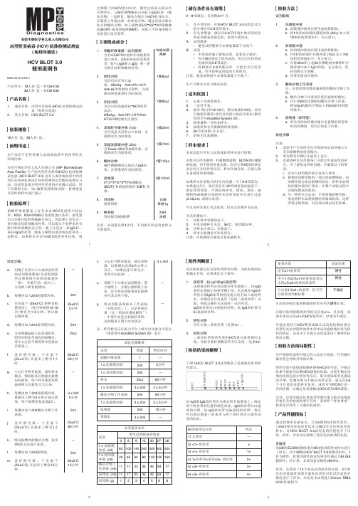
Roche MagNA Lyser 产品说明书

Ordering Information Cat. No. Product ***********MagNA Lyser Instrument (230 Volt)***********MagNA Lyser Instrument (110 Volt)(Instruments supplied with rotor and rotor cooling block)***********MagNA Lyser Green Beads (100 tubes)Related Products Cat. No. Product***********MagNA Pure LC DNA Isolation Kit II (Tissue)***********MagNA Pure LC mRNA Isolation Kit II (Tissue)03 330 591 001MagNA Pure LC RNA Isolation Kit III (Tissue)***********MagNA Pure LC DNA Isolation Kit III (Bacteria, Fungi)***********MagNA Pure LC RNA Isolation Tissue Lysis Buffer – Refill (70 ml)System DescriptionHomogenize up to 16 samples in just a few seconds.Save valuable lab space with a small benchtop instrument.Reduce hands-on time by replacing the mortar and pestle and other manual methods.Integrate your workflow with the automated nucleic acid isolation of the MagNA Pure LC Instrument.Perform consistent and reproducible sample disruption.Process many different sample types.Prevent nucleic acid degradation with the benchtop cooling unit.Ease your setup with a removable rotor and prefilled disposable vials.Automate with an easy-to-use instrumentVersatile, efficient, and rapid pre-preparationFigure 71. Add your sample and lysis buffer to the MagNA Lyser Green Beads.2. Homogenize with the MagNA Lyser Instrument.3. Centrifuge to pellet the debris.4. Proceeed with the supernatant to prepare nucleic acids or proteins.For detailed information,visit or contact your local representative.Trademarks:MagNA Pure, MagNA Lyser, LightCycler, and the MagNA Pure Logo are trademarks of a member of the Roche Group.The technology used for the LightCycler System is licensed from Idaho Technology Inc., Salt Lake City, UT, USA.Fully automated sample preparationon the PCR Workflow SystemRoche Diagnostics GmbH Roche Applied Science Nonnenwald 282372 Penzberg Germany0000Roche Applied Science Part of Roche DiagnosticsMagNA Lyser InstrumentStart the Ball Rollingwith Automated Tissue HomogenizationᕤᕣᕢᕡFigure 6Components of the system.The MagNA Lyser InstrumentAutomated tissue homogenizationProcessing conditionsRefer to the following tables for guidelines on setting up your homogenizationSample material(10 mg)*Time settings(seconds)Cooling(between the runs)Speed Average yield(µg)***Average purity(OD 280/260 nm)***Spleen 2 x 25 906,00030–40 1.9Liver 25-6,00016–18 1.8Lung 2 x 25906,00025 1.8Kidney25-6,000201.8Maize leaves **20-5,00010n.d.Maize polenta **20-5,0008n.d.Tortilla chips **20-5,0001n.d.*Aliqout containing 10 mg sample material (here mouse and food samples) was taken for the DNA purificationusing the MagNA Pure LC DNA Isolation Kit II (Tissue), (see pack insert)**Centrifugation after the homogenization for 5 minutes at 2,200 x g*** Yield and purity strongly depend on the condition of the sample material n.d.not determinedData kindly provided by Dr. Peterhänsel, RWTH Aachen, GermanyFigure 1Gel electrophoresis from genomic DNA isolated from tissue homogenized with the MagNA LyserInstrument, using the MagNA Pure LC DNA Kit II (Tissue).Marker: DNA Marker III*Aliquot containing 10 mg sample material (here mouse and human research samples) was taken to purify RNAeither with the MagNA Pure LC RNA Isolation Kit III (Tissue) or the MagNA Pure LC mRNA Isolation Kit II (Tissue) homogenized with the MagNA Lyser Instrument.** Yield and purity strongly depend on the condition of the sample material. The yield for mRNA was not determined.Sample material(10 mg)*Time settings(cycles/seconds)Cooling(between/afterthe runs in seconds)SpeedAverage yield (mg)(total RNA)**Average purity(OD 280/260 nm)**RNA/mRNARarely expressed targets in small numbers of target cells,as seen in experiments about minimalresidual diseases,are difficult to detect.Increasing the cell number can improve sensitivity and lead to accurate results.Without the MagNA Lyser pre-processing,the MagNA Pure mRNA HS Kit can efficiently obtain mRNA from a maximum of 1 x 10⁷white blood cells (WBCs),as shown in research studies with human samples.However,using greater cell numbers results in a saturation effect with quantitative assays (Figure 3).Homogenization of the lysate with the MagNA Lyser Instrument prior to the purification eliminatesthe amplification saturation at 1 x 10⁷cells and allows the use of up to 2.5 x 10⁷WBCs (Figure 4 and 5),enhancing the analytical sensitivity of the assay.Eliminate sensitivity barriers with increased sample inputFigure 3mRNA was purified from different amounts of human white blood cells with the MagNA Pure mRNA HS Kit. G6PDH was amplified using the LightCycler t(9;22) Quantification Kit (see text beside).Figure 4mRNA was purified from different amounts of human white blood cells with the MagNA Pure mRNA HS Kit. The lysates from 2.5 x 10⁷cells and 5 x 10⁷cells were homogenized with the MagNA Lyser Instrument (2x50 seconds with 90 seconds cooling in between) prior to the mRNA purification. G6PDH was amplified using the LightCycler t(9;22) Quantification Kit (see text beside).Figure 5Scalability from 1 x 10⁶cells to 2.5 x 10⁷cells is represented in the graph and the table of the relationship between crossing points and cell numbers. The limitation of cell input is indicated by no change in crossing point with increased cell number (see text beside).Cell number 5 x 1072.5 x 1071 x 1075 x 1061 x 106Log (cell number)7.77.47.06.76.0Crossing point 20.320.321.822.424.4crossingpointLog(cell number)252423222120195.86.36.87.37.8Figure 2Gel electrophoresis from total RNA isolated from tissue homogenized with the MagNA Lyser Instrument, using the MagNA Pure LC RNA Kit III (Tissue).Ma r k e rS p l e e nL i v e rL u n gK i d n e yM a r k e rMa i z e l e a v e sMa i z e l e a v e sS p l e e nL i v e r11 kb5 kb5 kb28 S rRNA 18 S rRNASpleen 2 x 50 90 6,500–7,000 30–40 1.9Liver 50 - 6,500–7,000 13–17 2.0Thymoid tissue60906,500n.d.n.d.Heart 60 90 6,500 n.d. n.d.Abdominal fat 60 90 6,500 n.d. n.d.Aorta 60 90 6,500 n.d. n.d. Other samples1+n x 50 90 6,500–7,000- -1 x 105 x 101 x 10- 5 x 101 x 105 x 105 x 10- 5 x 102.5 x 10 5 x 10。
本试剂仅供研究使用

本试剂仅供研究使用人抗增殖细胞核抗原抗体(PCNA)elisa试剂盒使用说明书试剂盒组成:48孔配置/96孔配置说明书:1份封板膜:2片(48)/2片(96)密封袋:1个目的:【人抗增殖细胞核抗原抗体(PCNA)elisa试剂盒说明书】本试剂盒用于测定人血清,血浆及相关液体样本中介素2(IL-2)的含量。
服务承诺:供货期:款到发货。
工作时间内免费的技术咨询和指导。
请来电咨询为客户提供来样检测服务,最大限度实验结果的有效性(免费代测)。
试剂盒性能:1.样品线性回归与预期浓度相关系数R值为0.95以上。
2.批内与批见应分别小于9%和11%保存条件及有效期:1.试剂盒保存:2-8℃。
2.有效期:6个月检测范围:0.2IU/L - 6IU/L实验原理:【人抗增殖细胞核抗原抗体(PCNA)elisa试剂盒说明书】本试剂盒应用双抗体夹心法测定标本中人抗增殖细胞核抗原抗体(PCNA)水平。
用纯化的人抗增殖细胞核抗原抗体(PCNA)抗体包被微孔板,制成固相抗体,往包被单抗的微孔中依次加入介素2(IL-2),再与HRP标记的介素2(IL-2)抗体结合,形成抗体-抗原-酶标抗体复合物,经过彻底洗涤后加底物TMB显色。
TMB在HRP酶的催化下转化成蓝色,并在酸的作用下转化成最终的黄色。
颜色的深浅和样品中的介素2(IL-2)呈正相关。
用酶标仪在450nm波长下测定吸光度(OD 值),通过标准曲线计算样品中人抗增殖细胞核抗原抗体(PCNA)浓度。
样本处理及要求:1. 血清:室温血液自然凝固10-20分钟,离心20分钟左右(2000-3000转/分)。
仔细收集上清,保存过程中如出现沉淀,应再次离心。
2. 血浆:应根据标本的要求选择EDTA或柠檬酸钠作为抗凝剂,混合10-20分钟后,离心20分钟左右(2000-3000转/分)。
仔细收集上清,保存过程中如有沉淀形成,应该再次离心。
3. 尿液:用无菌管收集,离心20分钟左右(2000-3000转/分)。
壮骨健膝方对脂多糖诱导兔滑膜成纤维细胞炎症模型的影响

·实验研究 ·福建中医药 2023 年 6 月 第 54 卷 第 6 期Fujian Journal of TCM June 2023,54(6)壮骨健膝方对脂多糖诱导兔滑膜成纤维细胞炎症模型的影响张英杰1,张鹏1,肖艳2,刘俊1,陈雨1,邱梦婷1,苏友新1*(1.福建中医药大学中医学院,福建 福州 350122;2.福建中医药大学康复医学院,福建 福州 350122)摘要: 目的 探讨壮骨健膝方对兔滑膜成纤维细胞炎症模型的影响及作用机制。
方法 采用脂多糖(LPS )刺激兔滑膜成纤维细胞(FLS )建立炎症模型,筛选壮骨健膝方干预炎症模型的最佳条件(浓度10%,时间48h )后,采用随机数字表法随机分为空白组、模型组和壮骨健膝方组,分别给予相应条件干预后采用ELISA 法检测各组细胞上清液中白细胞介素-1β(IL-1β)、肿瘤坏死因子-α(TNF-α)、白细胞介素-6(IL-6)含量,qPCR 法检测各组细胞中IL-1β、TNF-α、IL-6、增殖细胞核抗原(PCNA )、I κB α、NF-κB p65 mRNA 表达,Western blot 法检测各组细胞中PCNA 、核因子κB 抑制因子α(I κB α)及核内核因子κB (NF-κB )p65蛋白表达。
结果 与空白组比较,模型组IL-1β、TNF-α、IL-6含量,IL-1β、TNF-α、IL-6、PCNA 、NF-κB p65 mRNA 及核内NF-κB p65蛋白表达均显著升高(P 均<0.05),I κB α mRNA 及蛋白表达显著降低(P 均<0.05);与模型组比较,壮骨健膝方组IL-1β、TNF-α、IL-6含量,IL-1β、TNF-α、IL-6、PCNA 、NF-κB p65 mRNA 及核内NF-κB p65蛋白表达均显著降低(P 均<0.05),I κB α mRNA 及蛋白表达显著升高(P 均<0.05)。
免疫试验必备 内参抗体

品名 货号 价格 宿主
应用
His, HRP Labeled
CB101001 ¥3000/100ug
Rabbit
WB 1:1000 IHC 1:200 IP 1:200
北京西美杰科技有限公司 上海 电话:021-63599871 传真:021-63599873 Email:shanghai@
货号 25-01-01 24-01-01 25-00-01 24-00-02 24-00-01
品名 HisDetector Nickel-AP HisDetector Nickel-HRP HisDetector Western Blot Kit, AP Colorimetric HisDetector Western Blot Kit, HRP Chemiluminescent HisDetector Western Blot Kit, HRP Colorimetric
检测物种 Human, Mouse, Rat
品名 货号 价格 宿主
Rubisco Large Subunit AS03 037 ¥4770/100ug Rabbit
应用
WB 1:5000-1:10000 IF 1: 250
检测物种 plant and algal
北京西美杰科技有限公司 上海 电话:021-63599871 传真:021-63599873 Email:shanghai@
品名 货号 价格 宿主
β-actin CB100996 ¥1500/100ug Rabbit
应用
WB 1:1000 IP 5ug/mg lysate
检测物种 Human, Mouse
品名 货号 价格
β-actin 60008-1-Ig ¥1548/150ul
All-in-One qPCR Mix 产品说明书

All-in-One™qPCR MixFor universal quantitative real-time PCRCat.No.QP001(Old Cat.No.AOPR-0200,20μl×200reactions)Cat.No.QP002(Old Cat.No.AOPR-0600,20μl×600reactions)Cat.No.QP004(Old Cat.No.AOPR-1000,20μl×1000reactions)Cat.No.QP005(Old Cat.No.AOPR-4000,20μl×4000reactions)Performance optimized for All-In-One™qPCR Primers,All-In-One™miRNA qPCR Primers, miProfile™miRNA qPCR Arrays,ExProfile™Gene qPCR Arrays,All-In-One™First-Strand cDNA Synthesis Kit and All-In-One™miRNA First-Strand cDNA Synthesis KitUser ManualGeneCopoeia,Inc.9620Medical Center Drive,#101Rockville,MD20850USA301-762-0888866-360-9531***********************©2016GeneCopoeia,Inc.USER MANUALAll-in-One TM qPCR MixI.DescriptionII.Related ProductsIII.Contents and StorageIV.PreparationV.ProcedureVI.ExampleVII.Trouble Shooting GuideVIII.Limited Use License and WarrantyI.DescriptionThe All-in-One™qPCR Mix provides fast and efficient SYBR®Green-based real-time quantitative PCR.The qPCR Mix uses a high-fidelity hot-start DNA polymerase,optimized reaction buffer and high-quality dNTPs to enable specific and sensitive amplification of even low-copy genes or miRNAs.The All-in-One TM qPCR Mix reduces experimental design time by providing a universal reaction condition that can be used with almost all primers and most real-time PCR instruments.II.Related ProductsGeneCopoeia offers comprehensive solutions for studying gene expression.A careful process of co-development ensures that they work well together and provide robust and reproducible results.Product DescriptionAll-in-One™First-Strand cDNASynthesis KitReverse transcribe mRNA into first–stand cDNAAll-in-One™qPCR PrimersValidated,gene-specific primers ensure specificity and sensitivity (human,mouse and rat)ExProfile™Gene qPCR Arrays High-throughput or focused group profiling of gene expression All-in-One™miRNA First-StrandcDNA Synthesis KitReverse transcribe miRNA into first–stand cDNAAll-in-One™miRNA qRT-PCR Detection Kits SYBR®Green-based detection kit accurately quantifies miRNA expressionAll-in-One™miRNA qPCR Primers Validated human,mouse,rat miRNA primers for robust,reproducible and reliable quantitation of miRNA activitymiProfile™miRNA qPCR Arrays High-throughput or focused group profiling of miRNA expression RNAzol®RT RNA Isolation Reagent Easy isolation of mRNA,microRNA or total RNAIII.Contents and StorageContents and storage recommendations for the All-in-One TM qPCR Mix are provided in the following table. Cat.Nos.QP001,QP002,QP004,and QP005Contents Quantity Storage temperature/conditions2×All-in-One TM qPCR Mix 2×1ml3×(2×1ml)5×(2×1ml)20×(2×1ml)–20°C(Stable for at least12months)Alternatively,the solution can also bestored at–80°C in aliquots.Avoidrepeated freezing/thawing.ROX Reference Dye (30μΜ)1×80µl3×80µl5×80µl20×80µl–20°C(Stable for at least12months)Alternatively,the solution can also bestored at–80°C in aliquots.Avoidrepeated freezing/thawing.IV.PreparationWearing a lab coat,disposable gloves and protective goggles are recommended when handling chemicals.IMPORTANT NOTES:1.When using the All-in-One qPCR Mix with miProfile miRNA qPCR Arrays and All-in-One miRNAFirst-Strand cDNA Synthesis Kit for miRNA expression profiling,please follow the miProfile miRNA qPCR array user manual for the complete instruction.2.Store the kit at–20°C.Avoid storage or leaving reagents at4°C or room temperature.Avoid lightexposure at all times.3.Mix reagents thoroughly by gently inverting tubes several times avoiding bubbles and then brieflycentrifuge before use.4.Prepare the reaction mix with PCR grade water.5.Strictly follow standard procedures for PCR to avoid nucleic acid contamination and non-specificamplification.6.Read all procedures before setting up the PCR reactionV.Procedure1.Thaw the2×All-in-One TM qPCR Mix and ROX Reference Dye as needed.2.Prepare the PCR reaction mix on ice.See the example below.Reagent Volume Final concentration2×All-in-One TM qPCR Mix a10μl1×PCR forward primer(2µM)b2µl0.2µM cPCR reverse primer(2µM)2µl0.2µMTemplate d2μlROX Reference Dye e(30μΜ)ifneeded0.4-0.1μl600nM-150nMWater(double distilled)■Not using ROX Reference Dye4μl■Using ROX Reference Dye3.6-3.9μlTotal20μle the2×All-in-One TM qPCR Mix as half of the total reaction volume and adjust other reagentsaccordingly.If the total reaction volume is changed,maintain each component in the proper proportion. b.Primers are important considerations to ensure success with real-time PCR.All-in-One TM human,mouseand rat primer sets from GeneCopoeia have been validated to provide specific and sensitive amplification even with low copy number genes.For designing your own primers,you may wish to use Oligo primer analysis software(Molecular Biology Insights)or Primer Premier software(Premier Biosoft International).c.Primer concentration should be in the range of0.2to0.6µM.In general,a PCR reaction using0.2µMprimers produces good results.If the PCR efficiency is low,consider increasing primer concentration.However,keep in mind that non-specific PCR products may also increase with increased primer concentration.d.Generally,the amount of DNA template should be less than100ng.Because different templates containvarying copies of a target gene,it may be necessary to perform a gradient dilution to determine the optimal amount of DNA template to use.If reverse transcript cDNA is used as template,dilute before use.Do not add more than5%of the original cDNA solution volume to the total qPCR reaction solution.e.ROX Reference Dye is added only for qPCR instruments that require ROX for calibration.ROXReference Dye provides an internal reference to which the reporter-dye signal can be normalized during data analysis.Normalization is necessary to correct for fluorescence fluctuations due to changes in concentration or volume.Adjust the ROX Reference Dye to optimal concentration according to different qPCR instruments.Instrument ROX per20µl PCR Reaction Final Concentration BioRad iCycler,MyiQ,iQ5,CFX-96,CFX-384,Eppendorf Mastercyclerrealplex,Roche LightCycler480,LightCycler2.0None No ROXABI PRISM7000/7300/7700/7900HTand7900HTFast,ABI Step One,ABI Step One Plus0.4µl(0.2-0.4µl)600nM(300-600nM)ABI7500,7500Fast,ABI Viia7,Stratagene Mx3000P,Mx3005P,Mx4000,0.1µl(0.02-0.1µl)150nM(30-150nM)For other instruments which need calibration of ROX but have not been listed out in the table,please optimize the concentration of ROX according to the guide line of specific instrument.3.Mix the PCR reaction mix sufficiently and add to the PCR reaction tubes.4.Briefly centrifuge to make sure all the reagents are at the bottom of the reaction tubes.5.The following three-step method for programming the PCR reaction is recommended:Cycles Steps Temperature Time Detection 1Initial denaturation95°C10min No40Denaturation95°C10sec No Annealing55°C~60°C20sec No Extension72°C15sec YesNotesi.When using SYBR Green dye to monitor the qPCR reaction,a melting curve analysis should beperformed immediately at the end of cycling.(example adapted from the iQ5real-time PCRdetection system from Bio-Rad):Temperature range Heating rate Constant temperature Detection 72–95°C0.5°C/unit time6sec/unit time Yes25°C30sec NoThe conditions for your instrument may differ,consult the documentation of your qPCR instrument for instructions.ii.The DNA polymerase used in the2×All-in-One TM qPCR Mix is a special chemically modified hot-start enzyme.Incubation for10minutes at95°C will sufficiently activate the enzyme.iii.The actual annealing temperature should be adjusted around the primer melting temperature ranging from55°C~60°C.However,the optimal annealing temperature may be outside of thisrange.Adjust the temperature according to actual reaction conditionsiv.The optimal fragment length to use for amplification during real-time PCR is in the range of80-150bp.However,fragment lengths up to300bp are possible.v.The main condition for the above reaction are referred to in the iQ5qPCR instrument manual from Bio-Rad.If a qPCR instrument from another commercial source is used,please reference theinstrument manual and adjust the extention time and melting curve conditions accordingly.VI.ExampleObjective:The amplification efficiency and detection sensitivity of the2×All-in-One TM qPCR Mix are assessed by standard curves made by gradient dilution of plasmid DNA.The target fragment is102bp.Equipment:iQ5instrument(Bio-Rad Laboratories)Procedure:1.The plasmid is serially diluted to6concentrations ranging from105to1molecule/μl.2.PCR reaction mix preparation(on ice)Reagent components Volume2×All-in-One qPCR Mix10µlPCR forward primer(2µM)2µlPCR reverse primer(2µM)2µlddH201µlTotal15µl3.Mix the above reagents sufficiently.Aliquot to PCR tubes after a brief centrifugation.4.Add5μl of the diluted plasmid template to each PCR e5μl ddH2O as a negative control.5.Program the PCR reaction and corresponding reading conditions of the melting curve:Cycles Steps Temperature Time Detection1Initial denaturation95°C10min No45Denaturation95°C10sec No Annealing60°C20sec No Extension72°C15sec Yes Melting curve reading72°C~95°CHeating Rate0.5°C/6secYes Cooling25°C30sec No6.Analyze the amplification and corresponding melting curves after the qPCR experiment:Amplification curves of serially diluted plasmid DNA Peak values of amplified products in melting curves.7.Construct a standard curve using the Ct values from each amplification curve:Picture of a standard curve8.Conclusion:The peak values from the amplification and melting curves show that as low as5molecules can be detected when using plasmid DNA as a template and that there is only a single amplified product,showing that very high sensitivity can be attained using the All-in-One TM qPCR Mix.At the same time,high amplification efficiency is also shown by the good linear relationship among each concentration of serially diluted plasmid.VII.Trouble Shooting GuidePoor precision or failed qPCR reactions ∙Make sure the initial denature time was set as10min,sufficiently activating of the hot-start polymerase could avoid non-specific amplification and production of primer-dimers.∙The fluorescence detection temperature may not be appropriate.Adjust accordingly.∙The set up position for reaction samples in the real-time PCR instrument may not be right.Adjust accordingly.∙PCR cycle conditions,primer concentration and primer sequences may not be appropriate.Adjust the primer concentration and annealing temperature.If this does not work,redesign the primers.∙The template sample purity may not be adequate.Purify the template sample by phenol/chloroform extraction and ethanol precipitation.If the samples are reverse transcribed cDNA,set up the qPCR reaction with a diluted sample as other concentrated reagents in the RT reaction mixture may be interfering with the qPCR.∙Try to use 3.0%agarose gel electrophoresis to check the qPCR products.Check the purity of the primers by electrophoresis or use PAGE-purified primers if the bands are diffused.One may also use phenol/chloroform extraction and ethanol precipitation methods to treat the primers before the experiment.Abnormal meltingcurvesSignal in the blank(No Template Control)sample∙There may be contamination of the positive samples in the qPCR reaction system if the T m of the melting curve of the blank control is the same as the positive control.Eliminate sample application error first.If the situation still persists,replace the PCR grade water and/or primers and/or use a new2×All-in-One TM qPCR Mix.∙If the T m of the melting curve of the blank control is lower than the positive control,the qPCR reaction may have produced nonspecific amplification such as primer-dimers.Prepare the qPCR reaction mix on ice and increase the temperature of fluorescence detection.If this does not work,redesign the primers.Double peaks and multiple peaks in the melting curve of the positive control∙In the absence of other primers present in the reaction,double or multiple peaks in the melting curve of the positive control indicate that the qPCR reaction produced nonspecific amplification fragments.Prepare the qPCR reaction mix on ice;optimize the qPCR reaction conditions,for example,by increasing the annealing temperature, decreasing the primer concentration or increasing the fluorescence detection temperature(not more than the T m value of the expected product).If this does not work,redesign the forward primer.No peaks or abnormal peaks in the melting curve(or the amplification curves)of the positive control∙Adjust the ROX Dye to optimized concentration according to instrument.No signal(Ct)or late appearing signal ∙Not enough PCR cycles.For good sensitivity,one should generally set up more than35PCR cycles,but more than45cycles may result in too much background signal.∙The amount of template used may not be enough or the template may be e the highest concentration possible of diluted template samples to set up the qPCR.At the same time,avoid freezing and thawing the samples repeatedly.∙The amplification efficiency is low and the qPCR reaction conditions are not optimal.Redesign the primers and optimize the reaction conditions.VIII.Limited Use License and WarrantyLimited Use LicenseFollowing terms and conditions apply to use of all OmicsLink™ORF Expression Clones in all lentiviral vectors and Packaging Kit(theProduct).If the terms and conditions are not acceptable,the Product in its entirety must be returned to GeneCopoeia within5calendar days.A limited End-User license is granted to the purchaser of the Product.The Product shall be used by the purchaser for internal researchpurposes only.The Product is expressly not designed,intended,or warranted for use in humans or for therapeutic or diagnostic use.TheProduct must not be resold,repackaged or modified for resale,or used to manufacture commercial products without prior written consentfrom GeneCopoeia.This Product should be used in accordance with the NIH guidelines developed for recombinant DNA and genetice of any part of the Product constitutes acceptance of the above terms.Limited WarrantyGeneCopoeia warrants that the Product meets the specifications described in the accompanying Product Datasheet.If it is proven to the satisfaction of GeneCopoeia that the Product fails to meet these specifications,GeneCopoeia will replace the Product.In the event a replacement cannot be provided,GeneCopoeia will provide the purchaser with a refund.This limited warranty shall not extend to anyone other than the original purchaser of the Product.Notice of nonconforming products must be made to GeneCopoeia within30days of receipt of the Product.GeneCopoeia’s liability is expressly limited to replacement of Product or a refund limited to the actual purchase price.GeneCopoeia’s liability does not extend to any damages arising from use or improper use of the Product,or losses associated with the use of additional materials or reagents.This limited warranty is the sole and exclusive warranty.GeneCopoeia does not provide any other warranties of any kind,expressed or implied,including the merchantability or fitness of the Product for a particular purpose.GeneCopoeia is committed to providing our customers with high-quality products.If you should have any questions or concerns about anyGeneCopoeia products,please contact us at301-762-0888.©2016,GeneCopoeia,Inc.GeneCopoeia,Inc.9620Medical Center Drive,#101Rockville,MD20850Tel:301-762-0888Fax:301-762-3888Email:***********************Web:GeneCopoeia Products are for Research Use Only Copyright©2016GeneCopoeia,Inc. Trademarks:GeneCopoeia™,All-in-One™,ExProfile™,miProfile™(GeneCopoeia Inc.);RNAzol®(Molecular Research Center,Inc.);SYBR®(Molecular Probes);iQ™5(Bio-Rad);ROX®(Invitrogen).QP001020216。
Extract-N-Amp Tissue PCR Kit 产品说明书

Product InformationExtract-N-Amp™ Tissue PCR KitXNAT2, XNAT2RProduct DescriptionThe Extract-N-Amp™ Tissue PCR Kit for direct PCR contains the reagents needed to rapidly extract and amplify genomic DNA from mouse tails and other animal tissues, buccal swabs, hair shafts, and saliva. Briefly, the DNA is released from the starting material by incubating the sample with a mixture of the Extraction Solution and the Tissue Preparation Solution at room temperature for 10 minutes. There is no need for mechanical disruption, organic extraction, column purification, or precipitation of the DNA.After adding Neutralization Solution B, the extract is ready for PCR. An aliquot of the neutralized extract is then combined with the Extract-N-Amp™ PCR Reaction Mix and user-provided PCR primers to amplify target DNA. The Extract-N-Amp™ PCR Reaction Mix is a 2X ready mix containing buffer, salts, dNTPs, and Taq polymerase. It is optimized specifically for use with the extraction reagents. It also contains the JumpStart Taq antibody for hot start PCR to enhance specificity but does not contain the inert red dye found in the REDExtract-N-Amp™ PCR Reaction Mix.Reagents Provided Cat. No. XNAT2 100 Preps,100 PCRsXNAT2R 1000 Preps, 1000 PCRsExtraction SolutionE7526 24 mL 240 mL Tissue Preparation Solution T3073 3 mL 30 mL Neutralization Solution BN391024 mL240 mLExtract-N-Amp™ PCR Reaction Mix This is a 2X PCR reaction mix containing buffer, salts, dNTPs, Taq polymerase, and JumpStart™ Taq antibody.E30041.2 mL12 mLReagents and Equipment Required(Not Provided)•Microcentrifuge tubes (1.5 or 2 mL) or multi-well plate for extractions (200 μL minimal well volume) • Small dissecting scissors• Forceps (small to medium in size)• Buccal swab - Sterile foam tipped applicator (Cat. No. WHAWB100032)•Sample collection card - Bloodstain card (Cat. No. WHAWB100014)• Tubes or plate for PCR• Heat block or thermal cycler at 95 °C • PCR Primers (Cat. No. OLIGO) • Thermal cycler•Water, PCR Reagent (Cat. No. W1754)Precautions and DisclaimerThis product is for R&D use only. Not for drug, household, or other uses. Please consult the Safety Data Sheet for information regarding hazards and safe handling practices.StorageThe Extract-N-Amp™ Tissue PCR Kit can be stored at 2 to 8 °C for up to 3 weeks. For long-term storage, greater than 3 weeks, -20 °C is recommended. Do not store in a "frost-free" freezer.ProcedureAll steps are carried out at room temperature unless otherwise noted.DNA Extraction from Mouse Tails, Animal Tissues, Hair, or Saliva1.Pipette 100 μL of Extraction Solution into amicrocentrifuge tube or well of a multi-well plate.Add 25 μL of Tissue Preparation Solution to thetube or well and pipette up and down to mix.Note: If several extractions will be performed,sufficient volumes of Extraction and TissuePreparation Solutions may be pre-mixed in a ratio of 4:1 up to 2 hours before use.2.For fresh or frozen mouse tails: Rinse thescissors and forceps in 70% ethanol prior to useand between different samples. Place a 0.5–1 cm piece of mouse tail tip (cut end down) into thesolution. Mix thoroughly by vortexing or pipetting.Ensure the mouse tail is in solution.Note: For fresh mouse tails, perform extractions within 30 minutes of snipping the tail.For animal tissues: Rinse the scissors or scalpel and forceps in 70% ethanol prior to use andbetween different samples. Place a 2–10 mgpiece of tissue into the solution. Mix thoroughlyby vortexing or pipetting. Ensure the tissue is inthe solution.For hair shafts: Rinse the scissors and forceps in 70% ethanol prior to use and between differentsamples. Trim excess off of the hair shaft leaving the root and place sample (root end down) intosolution. Only one hair shaft, with root, isrequired per extraction.For Saliva: Pipette 10 μL of saliva into thesolution. Mix thoroughly by vortexing or pipetting.For saliva dried on card: Pipette 50 μL of saliva onto collection card and allow the card to dry.Rinse the punch in 70% ethanol prior to use andbetween different samples. Punch a disk(preferably 1/8 inch or 3 mm) out of the cardfrom the area with the dried saliva sample. Place disk into the solution. Tap tube or plate on hardsurface to ensure disk is in solution forincubation period.3.Incubate sample at room temperature for10 minutes.4.Incubate sample at 95 °C for 3 minutes.Note: Tissues will not be completely digested atthe end of the incubations. This is normal and will not affect performance.5.Add 100 μL of Neutralization Solution B to sampleand mix by vortexing.6.Store the neutralized tissue extract at 4 °C oruse immediately in PCR amplification.Note: For long term storage, remove theundigested tissue or transfer the extracts tonew tubes or wells. Extracts may now be storedat 4 °C for at least 6 months without notable loss in most cases.DNA Extraction for Buccal Swabs1.Collect buccal cells on swab and allow theswab to dry. Drying time is approximately10 to 15 minutes.Note: Due to the low volume of solution used for DNA extraction, a foam tipped swab should beused. Swabs with fibrous tips, such as cotton orDacron®, should be avoided because the solution cannot be recovered efficiently.2.Pipette 200 μL of Extraction Solution into amicrocentrifuge tube. Add 25 μL of TissuePreparation Solution to the tube and pipette upand down to mix.Note: If several extractions will be performed,sufficient volumes of Extraction and TissuePreparation Solutions may be pre-mixed ina ratio of 8:1 up to 2 hours before use.3.Place dried buccal swab into solution and incubateat room temperature for 1 minute.4.Twirl swab in solution 10 times and then removeexcess solution from the swab into the tube bytwirling swab firmly against the side of the tube.Discard the swab. Close the tube andvortex briefly.5.Incubate sample at room temperature for10 minutes.6.Incubate sample at 95 °C for 3 minutes.7.Add 200 μL of Neutralization Solution B to sampleand mix by vortexing.8.Store the neutralized extract at 4 °C or useimmediately in PCR. Continue to PCRamplification.Note: Extracts may be stored at 4 °C for at least6 months without notable loss in most cases. PCR AmplificationThe Extract-N-Amp™ PCR Reaction Mix contains JumpStart™ Taq antibody for specific hot start amplification. Therefore, PCR mixtures can be assembled at room temperature without premature Taq DNA polymerase activity.Typical final primer concentrations are approximately 0.4 μM each. The optimal primer concentration and cycling parameters will depend on the system being used.1.Add the following reagents to a thin-walled PCRmicrocentrifuge tube or plate:Reagent VolumeWater, PCR grade VariableExtract-N-Amp™ PCRreaction mix 10 μLForward primer VariableReverse primer VariableTissue extract 4 μL*Total volume 20 μL*The Extract-N-Amp™ PCR Reaction Mix isformulated to compensate for components in the Extraction, Tissue Preparation, and Neutralization Solutions. If less than 4 µL of tissue extract isadded to the PCR reaction volume, use a 50:50mixture of Extraction and Neutralization BSolutions to bring the volume of tissue extract upto 4 μL.2.Mix gently.3.For thermal cyclers without a heated lid, add20 μL of mineral oil on top of the mixture in eachtube to prevent evaporation.4.Perform thermal cycling. The amplificationparameters should be optimized for individualprimers, template, and thermal cycler.Common cycling parameters:Step Temperature Time Cycles InitialDenaturation 94 °C 3 minutes 1 Denaturation 94 °C 30 seconds Annealing 45 to 68 °C 30 seconds 30-35 Extension 72 °C 1-2 minutes(1 min/kb)FinalExtension 72 °C 10 minutes 1 Hold 4 °C Indefinitely5.The amplified DNA can be loaded onto an agarosegel after the PCR is completed with the addition ofa separate loading buffer/tracking dye such as GelLoading Solution, Cat. No. G2526.Note: PCR products can be purified, if desired, fordownstream applications such as sequencing withthe GenElute PCR Clean-Up Kit, Cat. No.NA1020.Troubleshooting GuideProblem Cause SolutionLittle or no PCR product is detected. PCR reaction may beinhibited due tocontaminants in thetissue extract.Dilute the tissue extract with a 50:50 mix of Extractionand Neutralization Solutions. To test for inhibition, includea DNA control and/or spike a known amount of template(100-500 copies) into the PCR along with the tissue extract. Extraction isinsufficient.Incubate samples at 55 °C for 10 minutes instead ofroom temperature.A PCR component maybe missing or degraded.Run a positive control to ensure that componentsare functioning. A checklist is also recommendedwhen assembling reactions.There may be too fewcycles performed. Increase the number of cycles (5-10 additional cycles at a time). The annealingtemperature maybe too high.Decrease the annealing temperature in 2-4 °C increments.The primers may notbe designed optimally.Confirm the accuracy of the sequence information. If theprimers are less than 22 nucleotides long, try to lengthen theprimer to 25-30 nucleotides. If the primer has a GC contentof less than 45%, try to redesign the primer with a GCcontent of 45-60%.The extension timemay be too short.Increase the extension time in 1-minute increments, especiallyfor long templates.Target templateis difficult.In most cases, inherently difficult targets are due to unusuallyhigh GC content and/or secondary structure. Betaine, Cat. No.B0300, has been reported to help amplification of high GCcontent templates at a concentration of 1.0-1.7 M.Multiple products JumpStart™ Taqantibody is notworking correctly.Do not use DMSO or formamide with Extract-N-Amp™ PCRReaction Mix. It can interfere with the enzyme-antibodycomplex. Other cosolvents, solutes (e.g., salts), and extremesin pH or other reaction conditions may reduce the affinity ofthe JumpStart™ Taq antibody for Taq polymerase and therebycompromise its effectiveness.TouchdownPCR maybe needed.“Touchdown” PCR significantly improves the specificity of manyPCR reactions in various applications. Touchdown PCR involvesusing an annealing/extension temperature that is higher thanthe TM of the primers during the initial PCR cycles. Theannealing/extension temperature is then reduced to the primerTM for the remaining PCR cycles. The change can be performedin a single step or in increments over several cycles.Negative control shows a PCR product or “false positive” result. Reagents arecontaminated.Include a reagent blank without DNA template be included asa control in every PCR run to determine if the reagents used inextraction or PCR are contaminated with a template froma previous reaction.Tissue is not digested after incubations. Tissue is not expectedto be completelydigested.The REDExtract-N-Amp™ Tissue PCR Kit does not require thetissue to be completely digested. Sufficient DNA is released forPCR without completely digesting the tissue.Buccal swab absorbed all the solution. The recommended typeof swab was not used.Due to the low volume of solution used for DNA extraction, afoam tipped swab should be used. Swabs with fibrous tips, suchas cotton or Dacron®, should be avoided because the solutioncannot be recovered efficiently.References1.Dieffenbach, C.W., and Dveksler, G.S. (Eds.), PCRPrimer: A Laboratory Manual, 2nd ed., Cold Spring Harbor Laboratory Press, New York (1995).2.Don, R.H. et al., ‘Touchdown' PCR to circumventspurious priming during gene amplification.Nucleic Acids Res., 19, 4008 (1991).3.Erlich, H.A. (Ed.), PCR Technology: Principles andApplications for DNA Amplification, StocktonPress, New York (1989).4.Griffin, H.G., and Griffin, A.M. (Eds.), PCRTechnology: Current Innovations, CRC Press,Boca Raton, FL (1994).5.Innis, M.A., et al., (Eds.), PCR Strategies,Academic Press, New York (1995).6.Innis, M., et al., (Eds.), PCR Protocols: A Guide toMethods and Applications, Academic Press, SanDiego, California (1990).7.McPherson, M.J. et al., (Eds.), PCR 2: A PracticalApproach, IRL Press, New York (1995).8.Newton, C.R. (Ed.), PCR: Essential Data, JohnWiley & Sons, New York (1995).9.Roux, K.H. Optimization and troubleshooting inPCR. PCR Methods Appl., 4, 5185-5194 (1995).10.Saiki, R., PCR Technology: Principles andApplications for DNA Amplification, Stockton, New York (1989). Product OrderingOrder products online at Related Products Cat. No.Ethanol E7148; E7023; 459836 Forceps,micro-dissecting F4267PCR Marker P9577PCR microtubes Z374873; Z374962;Z374881PCR multi-well plates Z374903Precast Agarose Gels P6097Sealing mats & tapes Z374938; A2350TBE Buffer T4415, T6400, T9525The life science business of Merck operatesas MilliporeSigma in the U.S. and Canada.Merck, Extract-N-Amp, REDExtract-N-Amp, JumpStart, GenElute and Sigma-Aldrich are trademarks of Merck KGaA, Darmstadt, Germany or its affiliates. All other trademarks are theproperty of their respective owners. Detailed information on trademarks is available via publicly accessible resources.NoticeWe provide information and advice to our customers on application technologies and regulatory matters to the best of our knowledge and ability, but without obligation or liability. Existing laws and regulations are to be observed in all cases by our customers. This also applies in respect to any rights of third parties. Our information and advice do not relieve our customers of their own responsibility for checking the suitability of our products for the envisaged purpose. The information in this document is subject to change without notice and should not be construed as a commitment by the manufacturing or selling entity, or an affiliate. We assume no responsibility for any errors that may appear in this document. Technical AssistanceVisit the tech service page at/techservice.Terms and Conditions of SaleWarranty, use restrictions, and other conditions of sale may be found at /terms. Contact InformationFor the location of the office nearest you, go to /offices.。
抗核抗体检测试剂盒(磁条码免疫荧光法)产品技术要求丽珠
1性能指标
1.1外观
试剂盒各组份应齐全、完整,标签清晰,易识别;液体无渗漏。
1.2准确度
对准确度参考品进行检测,双链DNA(dsDNA)IgG抗体的相对偏差应在±20%范围内。
1.3最低检测限
对检测限参考品进行检测,dsDNA IgG抗体的检测限应不高于10 IU/mL;其它定性指标对应的各指标的L1应为阳性、L2应为阳性或阴性、L3应为阴性。
1.4线性
dsDNA IgG抗体在10~ 600 IU/mL范围内,其线性相关系数(r)r2应不小于0.950。
1.5阴性参考品符合率
对10份阴性参考品进行检测,对应指标的阴性参考品符合率应为100%。
1.6阳性参考品符合率
对12份阳性参考品进行检测,对应指标的阳性参考品符合率应为100%。
1.7重复性
对重复性参考品检测10次,同一指标的变异系数(CV)应不大于15%。
1.8批间差
用三个批号试剂盒检测同一份重复性参考品,批间差应不大于20%。
1.9dsDNA 校准品均匀性
1.9.1瓶内均匀性
校准品的瓶内均匀性(变异系数,CV)应不大于15%。
1.9.2瓶间均匀性
校准品的瓶间均匀性(变异系数,CV)应不大于15%。
Abbkinecrownantibody

"皇冠"抗体因为验证,所以承诺结合完整的标签与内参抗体产品线,我们针对热门核心研究领域,如信号通路、神经科学、细胞骨架、癌症、表观遗传等,精心挑选具有文章热度的关键靶标,推出经过层层严苛质控和品质承诺的“皇冠”抗体。
简介Abbkine 技术专家多年来一直潜心研究、开发并生产高品质的免疫检测试剂。
我们为全世界范围内的生命科学领域科学家提供卓越的产品,成为了许多国际领先的研究机构和临床诊断公司值得信赖的可靠供应商。
90%以上的产品均属于Abbkine 持续创新抗体的生产工艺,以经济实惠的价格提供高品质的产品,以满足您的多样化的研究需求。
如有大订单,欢迎联系我们,您将获取更多的支持!产品目录其它抗体内参抗体内参简介. . . . . . . . . . . . . . . . 1内参抗体. . . . . . . . . . . . . . . . 2标签抗体WB:使用GAPDH单克隆抗体(组织,Hela细胞等样品。
样品类型内参抗体理论分子量全细胞/胞浆蛋白β-Actin43 kDaGAPDH37 kDaβ-Tubulin55 kDaα-Tubulin55 kDaCyclophilin B24 kDaCofilin19 kDaVinculin116 kDa线粒体COX IV16 kDaHSP6060 kDaVDAC1/Porin31 kDa核蛋白PCNA29 kDaHistone H315 kDaHDAC156 kDaLamin B166 kDaTBP/TATA38 kDa膜蛋白NaK ATPase113 kDa血清Transferrin77 kDa1产品名称产品货号应用类型Anti-β-Actin Mouse Monoclonal Antibody (1C7) A01010WB, IHC HRP Conjugated Anti-beta Actin Mouse Mab (1C7) A01015WBAnti-GAPDH Mouse Monoclonal Antibody (2B5) A01020WB, IHC HRP Conjugated Anti-GAPDH Mouse Mab (2B5) A01025WBAnti-β-Tubulin Mouse Monoclonal Antibody (3G6) A01030WB, IHC Anti-PCNA Mouse Monoclonal Antibody (1D7)A01040WB, IHC Anti-Plant Actin Mouse Monoclonal Antibody (3T3)A01050WBAnti-COX IV Mouse Monoclonal Antibody (14Y2)A01060WBAnti-Histone H3 Mouse Monoclonal Antibody (2D10)A01070WB, IF, IP Anti-α-Tubulin Monoclonal Antibody (3G5) A01080WB, IHC, IF, IP Anti-Lamin B1 Monoclonal Antibody (15T1) A01090WB, IPAnti-Rubisco (Large Chain) Monoclonal Antibody (9Y6) A01110WBAnti-TBP/TATA Binding Protein Mab (2C6) A01120WBAnti-Cyclophilin B Monoclonal Antibody (7B2) A01130WB Histone H3 Monoclonal Antibody (1G1)ABM40038WB, IHC, IF, IP Cofilin Polyclonal Antibody ABP51018WB, IHC, IF Histone deacetylase 1 Polyclonal Antibody ABP51519WB, IHC, IF HSP60 Polyclonal Antibody ABP51585WB, IHC, IF Na+/K+-ATPase α1 Polyclonal Antibody ABP51894WB, IHC, IF Vinculin Polyclonal Antibody ABP52701WB, IHC, IF Transferrin Polyclonal Antibody ABP52968WB, IHC VDAC1 Polyclonal Antibody ABP53121WB Na+/K+-ATPase α1 Polyclonal AntibodyWB:使用Na+/K+-ATPase α1检测293细胞裂解液。
western-blot-内参

Western Blot内参要检测一个基因的表达产物是否正确,或者比较表达产物量的相对变化,首选方法是Western Blot。
因为Western Blot操作相对简单方便,既可以定性分析表达产物,同时还可以指示目的蛋白量的相对变化。
虽然,顺利的时候Western Blot做起来很简单,可不顺的时候也很令人心烦――做不出结果啦、假阳性啦、结果出现多条带啦、到底是一抗有问题还是二抗有问题啦……毕竟,作为一种有活性的生物大分子,抗体和抗原的反应毕竟不象1+1那么明确,而用这种不确定的试剂来测定同样知之甚少的表达产物,确实是有一定的不确定性的。
所以,严谨的Western Blot实验设计中要求有良好的参照体系,对实验结果分析是非常有用。
特别是当实验出现问题时,借助参照体系很容易就可以查出问题所在,而不必抓耳挠腮怨天尤人。
良好的参照体系通常包括分子量Marker(用来确定蛋白条带对应的分子量大小),空白载体对照(如果是诱导表达体系还应该有诱导前的对照),已知量标准产物的正对照;另外还有内参。
内参是最容易被忽略的一项。
我们知道,要用Western Blot比较不同条件下或者不同组织中,目的蛋白表达量的相对多少,前提条件是等量的细胞上样,才有比较的基础。
特别表达量不高时,上样量的差别就很可能影响结果的分析。
所以你需要内参。
内参即是内部参照(Internal Control),对于哺乳动物细胞表达来说一般是指由管家基因编码表达的蛋白(Housekeeping Proteins),它们在各组织和细胞中的表达相对恒定,在检测蛋白的表达水平变化时常用它来做参照物。
在Western Blotting 实验中,除了需要进行蛋白抽提、蛋白定量、等量蛋白上样电泳、转膜、靶蛋白抗体孵育、显色等步骤以外,还需要进行内参的检测,以校正蛋白质定量、上样过程中存在的实验误差,保证实验结果的准确性。
在国外发表的文章中,Western Blotting 实验结果须进行内参校正已成为一种惯例。
Bioss讲堂|Westernblot内参抗体选择攻略大全

Bioss讲堂|Westernblot内参抗体选择攻略大全Western Blot除了能证明某样品中含有某种蛋白之外,其最为重要的作用是比较不同条件下或者不同组织中,目的蛋白表达量的相对多少——即为蛋白表达水平最直接的证据。
要衡量蛋白的表达水平,前提条件就是等量的上样量。
内参的意义就是保证上样量的一致。
内参即是内部参照(Internal Control),对于哺乳动物细胞来说一般是指由管家基因编码表达的蛋白(Housekeeping Proteins),它们在各组织和细胞中的表达相对恒定,在检测蛋白的表达水平变化时常用它来做参照物。
选择内参抗体应遵循的原则1、样本来源♠哺乳动物的组织或细胞:β-Actin、β-Tubulin、GAPDH、Lamin B1、PCNA 等;♠植物样本:plant actin、Rubisco 等;♠其他来源样本,应参照已发表文献,选择合适蛋白作为内参。
2、目的蛋白定位▶全细胞蛋白和细胞质:β-Actin、beta Tubulin、GAPDH 等;▶细胞核蛋白:PCNA、LaminA、LaminB、HistoneH3,K70、K80、Erk2、TATAbindingprotein(TBP)以及c-Jun、c-Fos等;▶胞膜蛋白:Na( )/K( ) ATPase 等;▶线粒体蛋白:VDAC1、COXIV等;▶血清:Transferrin等。
3、目的蛋白分子量目的蛋白分子量最好能与内参蛋白分子量相差5kd 以上,各内参蛋白分子量详见下方汇总表。
Tips: 内参的选择还需要考虑实际的试验环境:★在做多组织多细胞样本对比表达量时,最好选用 GAPDH作为内参,因为GAPDH是代谢类蛋白,在活组织中表达比较恒定。
而β-Actin和β-Tubulin是结构蛋白,不同组织的细胞结构会有差异性;★而在某些细胞中,由于组织缺氧、糖尿病等因素会导致GAPDH 的表达增高,不适合做内参;★涉及细胞增殖的相关试验中,c-Jun由于自身表达变化不适合做内参;★凋亡实验时,TBP、Lamin等也不适合作为内参;★做诱导后样本或检测磷酸化等修饰性抗体时,应选择结构蛋白作为内参,如β-Actin 和β-Tubulin。
核蛋白内参抗体的选择原则
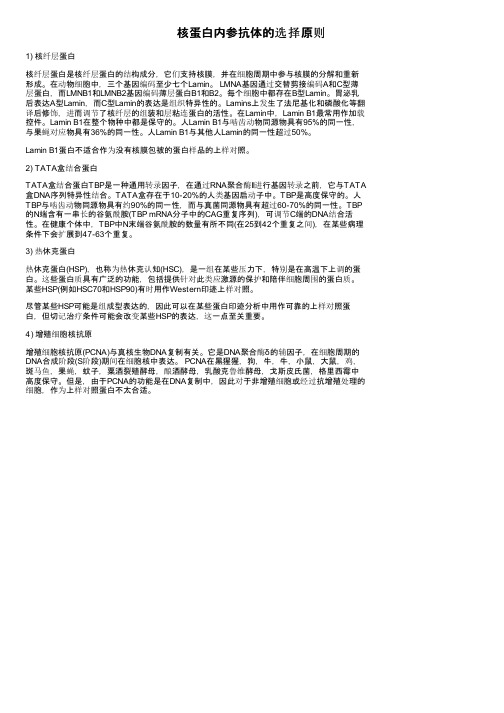
核蛋白内参抗体的选择原则1) 核纤层蛋白核纤层蛋白是核纤层蛋白的结构成分,它们支持核膜,并在细胞周期中参与核膜的分解和重新形成。
在动物细胞中,三个基因编码至少七个Lamin。
LMNA基因通过交替剪接编码A和C型薄层蛋白,而LMNB1和LMNB2基因编码薄层蛋白B1和B2。
每个细胞中都存在B型Lamin。
胃泌乳后表达A型Lamin,而C型Lamin的表达是组织特异性的。
Lamins上发生了法尼基化和磷酸化等翻译后修饰,进而调节了核纤层的组装和层粘连蛋白的活性。
在Lamin中,Lamin B1最常用作加载控件。
Lamin B1在整个物种中都是保守的。
人Lamin B1与啮齿动物同源物具有95%的同一性,与果蝇对应物具有36%的同一性。
人Lamin B1与其他人Lamin的同一性超过50%。
Lamin B1蛋白不适合作为没有核膜包被的蛋白样品的上样对照。
2) TATA盒结合蛋白TATA盒结合蛋白TBP是一种通用转录因子,在通过RNA聚合酶II进行基因转录之前,它与TATA 盒DNA序列特异性结合。
TATA盒存在于10-20%的人类基因启动子中。
TBP是高度保守的。
人TBP与啮齿动物同源物具有约90%的同一性,而与真菌同源物具有超过60-70%的同一性。
TBP 的N端含有一串长的谷氨酰胺(TBP mRNA分子中的CAG重复序列),可调节C端的DNA结合活性。
在健康个体中,TBP中N末端谷氨酰胺的数量有所不同(在25到42个重复之间),在某些病理条件下会扩展到47-63个重复。
3) 热休克蛋白热休克蛋白(HSP),也称为热休克认知(HSC),是一组在某些压力下,特别是在高温下上调的蛋白。
这些蛋白质具有广泛的功能,包括提供针对此类应激源的保护和陪伴细胞周围的蛋白质。
某些HSP(例如HSC70和HSP90)有时用作Western印迹上样对照。
尽管某些HSP可能是组成型表达的,因此可以在某些蛋白印迹分析中用作可靠的上样对照蛋白,但切记治疗条件可能会改变某些HSP的表达,这一点至关重要。
pBR322 产品说明书

HonorGene---专业的基因研究资源提供商(基因/载体/细胞/启动子/腺病毒/慢病毒/AA V/siRNA /miRNA)
pBR322产品说明书
产品信息
产品货号载体名称出品公司质粒用途原核抗性HG-VKY0280 pBR322 HonorGene 克隆载体Amp+、Tet+
质粒图谱
质粒简介
(1)pBR322是研究得最多,使用最早且应用最广泛的大肠杆菌质粒载体之一。
质粒名称pBR322中的“p”代表质粒,“BR”代表两位两位研究者Bolivar和Rogigerus姓氏的字首,“322”是实验编号。
(2)pBR322是一个人工构建的重要质粒,有万能质粒之称。
它是由pSF2124、pMB1及pSC101三个亲本质粒经复杂的重组过程构建而成的。
(3)pBR322具有很多优点,如相对分子质量较小、拷贝数较高等,是最常用的克隆载体之一。
Propagation in E.coli
(1)克隆菌株:DH5α、TOP10、XL1-Blue等均可。
(2)原核抗性:Amp+(工作浓度建议为100ug/ml)、Tet+(工作浓度建议为10ug/ml)。
(3)培养温度:37℃。
HonorGene(奥诺基因)---长沙艾碧维生物科技有限公司
地址:湖南省长沙市岳麓区桐梓坡西路229号麓谷国际工业园C栋12楼电话:155****6881
网址: E-mail:***************。
蛋白内参

GAPDH(glyceraldehyde-3-phosphate dehydrogenase)和细胞骨架蛋白beta-actin或beta-tubulin。
GAPDH分子量为146KD,beta-actin分子量为42KD,beta-tubulin分子量可能为100KDactin即肌动蛋白,是细胞的一种重要骨架蛋白。
actin大致可分为六种,其中四种是不同肌肉组织特异性的,包括alpha-skeletal muscle actin、alpha-cardiac muscle actin、alpha-smooth muscle actin和gamma-smooth muscle actin,其余两种广泛分布于各种组织中,包括beta-actin(β-non-muscle)和gamma-non-muscle actin。
这些不同的亚型组织分布是不一样的,在肌肉组织中的beta-actin分布就很少,心肌主要是alpha-cardiac muscle actin。
因此不同的组织本来就应该选择不同的内参,不能一概而论的。
beta-actin作为内参是得到了公认的,这是针对大多数组织和细胞来说的,它广泛分布于细胞浆内,表达量非常丰富。
尽管最近有一些文章已经开始质疑beta-actin作为内参的有效性(好像是对于上样量>20ug的蛋白区分能力下降,记不清楚了),但是发文章应该还是没有问题的。
至于其他的内参也是可以考虑用的,GAPDH(甘油醛-3-磷酸脱氢酶)是参与糖酵解的一种关键酶,而tubulin和actin类似,是细胞骨架的组成部分,但是不是肌肉的主要成分,应该是一个代替品。
细胞总蛋白的Western Blot 的内参蛋白一般用ACTIN, TUBLIN等细胞内较稳定表达的蛋白。
但是核蛋白的Western Blot 内参有LAMIN A, LAMIN B等;膜蛋白的Western Blot一般用α-tubulin或GAPDH为内参。
HCV BLOT 3.0 使用说明书
12HCV BLOT 3.0使用说明书MAD 0013-CHN-4 产品型号:18人份/盒―11139-018 36人份/盒―11139-0361. 通用名称:丙型肝炎病毒 (HCV) 抗体检测试剂 盒 (免疫印迹法)2. 英文名称:HCV BLOT3.018人份/盒,36人份/盒。
本产品用于定性检测人血清或血浆中的丙型肝炎病毒抗体。
安倍生物医学亚太私人有限公司 (MP Biomedicals Asia Pacific) 生产的丙型肝炎病毒(HCV) 抗体检测试剂盒 (HCV BLOT 3.0) 是对人血清或血浆中的丙型肝炎病毒抗体进行体外检测的定性酶免疫测定方法。
该试剂盒做为特异性更高的补充确认试验,用于对筛查方法 (如:酶联免疫吸附试验) 检测重复呈阳性的样本进行确认。
硝酸纤维素膜条上含有来自HCV 基因组中核抗原、NS3、NS4和NS5区的重组蛋白条带。
重组蛋白以谷胱甘肽转移酶融合表达,因此膜上也包含一条谷胱甘肽转移酶质控带,用以提示个别样本对谷胱甘肽转移酶的反应性。
膜上还包含一条IgG 和一条抗-IgG 质控带。
膜条与稀释的血清或血浆样本一起孵育。
如果样本中存在HCV 的特异性抗体,则2~8°C 储存,有效期24个月。
1.在不使用时,应将HCV BLOT 3.0试剂盒及其组分储存在2-8˚C 环境中。
2. 在失效期前,储存在2-8˚C 环境中的试剂组份和试剂膜条是稳定的,试剂不能冰冻。
A. 试剂膜条• 避免试剂膜条不必要地暴露于光线下。
B. 试剂• 用原装的瓶子盛装试剂,盖紧盖子储存。
• 在冷藏的情况下取用试剂,用完后尽快将试剂放回到2-8˚C 储存。
• 底物液在2-8˚C 储存时,可能会有沉淀形成,但不影响试剂盒的正常使用。
注意:避免底物液不必要地暴露于光线下。
生产日期及失效日期见标签。
1. 去离子水或蒸馏水。
2. 一次性手套。
3. 摇床(每分钟12-16次,摆动角度5-10度,以均匀地清洗膜条)或全自动蛋白免疫印迹仪(推荐使用型号AutoBlot System 20)。
Magna(SIGMA) RIP RNA IP 说明书 中文

RIP流程规划RIP实验的裂解物要求●计算所需免疫沉淀的数量。
样本包括目的抗体(用户提供)和与目的抗体相同种属来源的阴性对照IgG。
抗SNRNP70(Cat.CS203216)和阴性对照正常兔IgG(Cat#PP64B)可以用作RIP程序的控件,这两个组件都包含在EZ Magna RIP套件(目录#17-701)。
●获得浓缩的细胞裂解液对于免疫沉淀的成功至关重要。
通常,一次RIP反应(使用一种抗体的一次免疫沉淀)需要100μL来自约2.0 x 10⁷个细胞或一个15 cm平板的细胞裂解物。
RIP实验所需的裂解缓冲液体积的计算基于收获细胞的量。
该体积可能因使用的细胞类型而异。
请记住一旦你在某个细胞中用候选抗体证明RIP成功背景下,可以根据需要减少或进一步优化每个RIP的裂解物数量。
●必须优化每个RIP使用的细胞总数或蛋白质总量基于所研究的RNA结合蛋白的丰度以及RNA检测的计划方法。
一、裂解液制备完整RIP裂解缓冲液制备根据收获的细胞数量准备适量的完整的RIP裂解缓冲液。
(冰上操作)100μL RIP裂解缓冲液+ 0.5μL蛋白酶抑制剂鸡尾酒+ 0.25μL RNA酶抑制剂对于悬浮细胞:1.将细胞收获到15 mL锥形管中。
使用血细胞仪计数细胞。
2.在4°C下以1500 rpm的转速离心5分钟,收集细胞,并丢弃细胞上清液。
3、将细胞重新悬浮在10毫升冰冷的PBS中进行清洗。
通过离心收集细胞1500 rpm,在4°C下持续5分钟,并丢弃上清液。
4、重复步骤3进行一次额外清洗。
5、用等量的完整RIP裂解缓冲液重新悬浮细胞。
通过移液管混合直到混合物看起来均匀为止。
在冰上培养裂解物5分钟。
分配约200μL每种裂解液放入无核酸酶微量离心管中,并储存在-80°C下。
虽然每个抗体对应的裂解物分配量并不关键,但是每个RIP反应通常为100μL,每个RIP中通常使用阳性和阴性抗体。
实验表明,200μL细胞裂解液与单次使用的小份相关。
博雅生物 人凝血酶原复合物 说明书

博雅生物人凝血酶原复合物说明书
人凝血酶原复合物(PCC)是血液制品的一种,主要用于治疗凝血因子缺乏引起的出血症状。
博雅生物作为国内知名的血液制品生产企业,其人凝血酶原复合物在市场上具有一定的影响力。
以下是博雅生物人凝血酶原复合物的简要说明书:
成分与性状:
本品主要成分为人凝血酶原复合物,含有凝血因子Ⅱ、Ⅶ、Ⅸ、Ⅹ。
外观为黄色或淡黄色澄明液体,无异物、无沉淀。
适应症:
主要用于治疗凝血因子缺乏引起的出血症状,如血友病、肝脏疾病等。
用法用量:
根据患者病情和医生指导,确定使用剂量和次数。
通常采用静脉滴注给药,滴注速度不宜过快,以免引起过敏反应。
不良反应:
少数患者可能出现过敏反应、发热、寒战、头痛等不适症状,严重者应立即停止使用并就医。
注意事项:
1. 本品为血液制品,应严格遵守规定储存和运输。
2. 对本品过敏者禁用,过敏体质者慎用。
3. 孕妇及哺乳期妇女慎用。
4. 使用前应仔细检查药品外观,如有异常应立即停止使用。
5. 如有不适,请及时咨询医生或专业人士的建议。
请注意,以上信息仅供参考,具体使用方法和剂量等详细信息应遵医嘱。
如果您在使用过程中遇到任何问题,建议立即停止使用并与医生联系。
Western Blot实验常见问题的解决以及内参的选择

W estern Blot实验中为什么检测无信号?(白板)大概总结为一下几类原因:(1)样品中目的蛋白丰度很低,低于实验的检测下限(2)一抗不识别检测物种的蛋白,同时若有阳性参照的话提供阳性对照蛋白,若阳性对照正常则说明抗体及实验参照没有问题,可能是样本中目的蛋白含量比较低。
(3)蛋白降解(4)待测样品的确为阴性;(5)一抗失效;(6)抗原量不足,每泳道蛋白上样量不低于0.1ugWestern Blot实验中为什么检测的结果分子量大小与实际不符?(1)蛋白存在翻译后修饰—比如蛋白的磷酸化,糖基化等,这些都会增加蛋白的分子量大小。
(2)蛋白存在翻译后剪切—比如很多蛋白首先被合成为前体形式,然后通过剪切获得生物学活性(3)剪接变异体—相同的基因,经过选择性剪接会产生不同分子量大小的蛋白。
(4)相对电荷—氨基酸的组成(带电或不带电)(5)蛋白形成多聚体—比如蛋白二聚体。
为什么wb的结果与QPCR结果趋势不一样?(1)WB反映的是蛋白水平的结果,qpcr反映的是基因水平的结果。
理论上蛋白水平与基因水平是一致的,但是mRNA的高低不代表其表达蛋白的高低。
也有一些基因可能在组织样品中没有正常翻译(或发生泛素化修饰,修饰水平不一致),这样即使qpcr检测结果有表达,但是若蛋白没有正常翻译的话,WB则检测不到目的条带。
为什么检测结果有很多杂带?(1)可能是样品本身体内表达的蛋白具有多种修饰形式如糖基化、磷酸化、乙酰化等;(2)蛋白降解,导致蛋白分子量降低;(3)蛋白形成多聚体形式;为什么WB检测的背景比较高?(1)可能抗体浓度浓度较高,(2)抗原量较大。
(3)一张膜中有的目的条带较弱,为了能让较弱条带显示出来,曝光时间较长转膜后蛋白少或者没有可能原因(1)转膜板放置出问题(2)凝胶与膜接触不好(3)目的蛋白被其他高丰度蛋白掩盖,如白蛋白, IgG等Western Blot实验中为什么选择内参?内参即是内部参照(Internal Control),对于哺乳动物细胞表达来说一般是指由管家基因编码表达的蛋白(Housekeeping Proteins),它们在各组织和细胞中的表达相对恒定,在检测蛋白的表达水平变化时常用它来做参照物。
(整理)HCVRIBA中文说明书

编码抗原(重组蛋白C33C,NS5;合成多肽5-1-1,C100,C22)的丙肝病毒CHIRON®RIBA®HCV3.0SIA针对人血清或血浆中丙肝病毒抗体(anti-HCV)的条带免疫印迹试验(SIA)适用于体外诊断编码抗原(重组蛋白C33C,NS5;合成多肽5-1-1,C100,C22)的丙肝病毒CHIRON®RIBA®HCV3.0 SIA针对人血清或血浆中丙肝病毒抗体(anti-HCV)的条带免疫印迹试验(SIA)适用于体外诊断⋅产品编号930600命名和使用目的 (2)试验概述和说明 (2)试验的生化原理 (3)试剂 (4)警告 (4)试剂制备 (6)储存说明 (7)试剂变质指示 (8)样本收集和准备 (8)程序 (9)提供材料(30人份试剂盒;产品编号930600) (9)需要但不供给的材料 (9)主要陈述 (9)试验程序 (9)质量控制程序 (11)结果解释 (11)程序的局限性 (12)预期结果 (13)具体的性能特征 (17)关键符号 (19)参考文献 (20)发行日期:2007-12©诺华疫苗和诊断试剂公司10012512CHIRON®RIBA®HCV3.0 SIA是一种针对人血清或血浆(来自为输血或血液再造的捐赠者)中丙肝病毒编码的单个蛋白所对应的抗体的体外定性酶免印迹测定,它主要是一种对经类似ELISA等许可的抗HCV筛选步骤重复筛查的活性人血清或血浆样本进行辅助的,更具体的测试手段。
正如Ebeling等人所描述的,条带免疫印迹测试(SIA)已经在阐明丙肝对应的单个抗体方面展示出用性,通过SIA法对抗HCV的检测是基于传统的Western-blotting技术,在这项技术中,HCV基因组编码的特异性抗原被固定在一个膜支持物上。
因HCV编码的单个蛋白而产生的抗HCV活性的可视化是通过使用酶标记的抗人IgG对酶底物的比色实现的。
- 1、下载文档前请自行甄别文档内容的完整性,平台不提供额外的编辑、内容补充、找答案等附加服务。
- 2、"仅部分预览"的文档,不可在线预览部分如存在完整性等问题,可反馈申请退款(可完整预览的文档不适用该条件!)。
- 3、如文档侵犯您的权益,请联系客服反馈,我们会尽快为您处理(人工客服工作时间:9:00-18:30)。
PCNA核内参抗体,抗PCNA单抗,PCNA小鼠源单克隆抗体
Anti-PCNA Mouse Monoclonal Antibody ( 1D7)
增殖细胞核抗原(Proliferating Cell Nuclear Antigen简称PCNA)由Miyachi等于1978年在 SLE(系统性红斑狼疮)患者的血清中首次发现并命名,因其只存在于正常增殖细胞及肿瘤细胞内而得名。
PCNA是一种分子量为36KD的蛋白质,在细胞核内合成,并存在于细胞核内,为DNA聚合酶的辅助蛋白。
由于其在细胞核内稳定表达,因此其抗体被广泛应用于细胞核蛋白的内参抗体。
如何选择合适的细胞核内参抗体?
常用的细胞总蛋白质内参有GAPDH和细胞骨架蛋白β-Actin或β-Tubulin等。
对于一些植物的样本,则需要特别的植物Actin蛋白作为内参。
当实验样品中只是核蛋白,而不是细胞总蛋白提取液时,可以用组蛋白H(Histone H),或者增殖细胞核抗原(PCNA)等为核内参抗体。
除了这些,其它常见的核蛋白内参还有K70, K80, Lamin A和B,在一些文献报道中,Erk2、TATA binding protein(TBP)以及c-Jun、c-Fos等都有使用。
但是需要注意的问题是核蛋白内参的选择需要考虑实际的试验环境,比如在涉及细胞增殖相关试验中,c-Jun由于自身表达变化就不适合做内参;而在凋亡实验时,TBP、Lamin等也不适合作为内参。
应用类型:WB(1:1000-1:5,000)、IHC(1:100-1:500)
Fig.1.Immunohistochemistry staining (1:100) of Human breast cancer tissue paraffin-embedded with Anti-PCNA mouse monoclonal antibody (1D7).
Fig.2.Western blot analysis (1:2,000) of PCNA expression in Rat brain (lane A), A549 cell (lane B), NIH 3T3 cell (lane C) and 293T cell (lane D) with Anti-PCNA mouse monoclonal antibody (1D7).
免疫原:KLH偶联的PCNA部分多肽序列
来源宿主:小鼠
反应性:该PCNA核内参抗体可识别人、小鼠、大鼠等种属的PCNA蛋白。
保存建议:在发货后能在-20℃保存1年。
为最大限度的避免损失,请在打开管盖之前融化抗体并离心。
我们建议使用前分装以避免反复冻融,分装后可在4℃保存1个月。
背景资料:增殖细胞核抗原( Proliferating Cell Nuclear Antigen简称PCNA)由Miyachi等于1978年在 SLE(系统性红斑狼疮)患者的血清中首次发现并命名,因其只存在于正常增殖细胞及肿瘤细胞内而得名。
PCNA是一种分子量为36KD的蛋白质,在细胞核内合成,并存在于细胞核内,为DNA聚合酶的辅助蛋白。
由于其在细胞核内稳定表达,因此其抗体被广泛应用于细胞核蛋白的内参抗体。