Muse智能触控细胞分析仪-折页
Cellometer细胞计数分析仪
Insert chamber in Cellometer Output data generated instantly
One click to generate more data
18
Vision 结果分析:
Automatically save all results in a data table Fluorescence intensity distribution
PBMC
platelets
PBMC
应用:酵母计数
应用背景: 酵母广泛应用于酿酒、生物燃 料的领域,由于其具有直径 小、出芽(budding)等特性, 常用方法计数比较困难。
常用方法: 手动计数
荧光染料: PI Oxonol
部分客户:
市场:
• Auto T4 • Auto X4(Auto 2000) • Vision
Cell size histograms
Automatically save cell images
Size vs. intensity scatter plot Export all data to Excel
Concentration calculator
可更换滤光片组件:
Cellometer Vision 应用:
Vision提供更加简便,快速的定量
检测细胞表面标志物的方法.
应用:原代肝细胞计数
首次实现原代肝细胞存活率的自动检测
AO & PI Stain
Live
Live
Dead
Live AO
Dead PI
应用:PBMC计数
Using fluorescent stains for accurate concentration and viability: •Fresh PBMCs •Splenocytes •WBCs in whole blood •BAL •Primary hepatocytes
Millipore手持式自动细胞计数器
ScepterHandheld Automated Cell Counter 手持式自动细胞计数器手持式自动细胞计数器Handheld Pipette Form•像移液器一样方便使用•屏幕式使用指导•USB数据下载•72组图形数据的内存传感器前后•2 种类型的传感器•灵敏的电极•精确的进样室•高精度的电子感应检测孔•精确到亚微米的细胞直径•精确到来皮升的细胞体积传感器特点40 µm Sensor60 µm Sensor检测样品浓度范围50,000-1,500,000particles/mL10,000-500,000particles/mL检验颗粒直径范围3-17 um6-36 um建议的检测细胞平均直径4-13 microns8-25 microns目录号50-pack: PHCC40050250-pack: PHCC40250500-pack: PHCC4050050-pack: PHCC60050250-pack: PHCC60250500-pack: PHCC60500Sensor Selection GuideMeasured size (µm)40 µm60 µm NIH 3T315454 beadsAlgae (various)7-9 CHO14-17 Cos-715 Epithelia 14-15 HEK29311-15 HeLa12-14 HepG212 HT-2911 HUH7-Hepatoma lineB Cells6-11 Human ES Cells9-12 HUVEC14-15 Jurkat13K56222 Luminex beads5-6 MCF715-17 MDCK13-15Measuredsize (µm)40 µm60 µm Mouse Embryomic Stem Cell5-13Mesenchymal Stem Cell15-16PBMCs7-12PC129-13Primary Astrocytes7Primary Neuronal CellRat Whole Blood 4.6Rat Dorsal Root Ganglion Cells7Red Blood Cells5-7Rat Neural Stem Cell11-13SF913SH-SY5Y12Splenocytes7-9U26612U87-Human Glioblastoma cell line12-14Yeast-Pichia Pastoris5Yeast-S.cerevisiae6Merck Millipore ValidatedCustomer ValidatedRecommended based on size仪器软件Software Pro•强大的细胞分析平台•直方图叠加比较多组数据和多参数设置表格•构建和保存门的设置Software Pro Analysis PackageAn intuitive, intelligent platform to perform high level cell analysisData:data files from your Scepter device Current Plot:working plot and data file Multifunctional Plot: multiple data sets/ histogram overlaysAnalysis Templates: saved gating parameters Group Stats:customizable stats from your data files selected Export, print selectedgraphs/files, cut and paste订购信息PHCC00000Scepter Handheld Cell Counter-42525 PHCC20040Scepter 2.0 Handheld Cell Counter, with 50pk of 40uM Sensors42525PHCC20060Scepter 2.0 Handheld Cell Counter, with 50pk of 60uM Sensors42525 PHCC60050Scepter Sensors, 60uM, 50PK1186 PHCC60250Scepter Sensors, 60uM, 250 PK (5-50PKs)5747 PHCC60500Scepter Sensors, 60uM, 500PK (10-50PKS)10919 PHCC40050Scepter Sensors, 40um, 50PK2120 PHCC40250Scepter Sensors, 40uM, 250 PK (5-50PKs)10275 PHCC40500Scepter Sensors, 40uM, 500 PK (10-50PKs)19522 PHCC00KIT Scepter 2.0 Startup Kit, 1 Scepter 2.0 pls 12 PKs of Sensors, 60uM51576 PHCC01KIT 2 Scepter 2.0 Handheld Automated Cell Counters80798 PHCC02KIT Scepter 2.0 Start Up Kit, 1 Scepter 2.0 pls 5 packs of 60uM Sensors47307PHCC03KIT Scepter 2.0 Startup Kit, 1 Scepter 2.0 pls 12 PKs of Sensors, 40uM58708PHCC04KIT Scepter 2.0 Start up kit, 1 Scepter 2.0 pls 5 pks of 40uM Sensors51744PHCC05KIT Scepter 2.0 Startup Kit, 1 Scepter 2.0 pls 17 PKs of Sensors, 60uM54737计数器原理-精确记数的保证VoltageV=I RCurrent Resistance•Scepter感应器吸入精确的样本体积•细胞穿过感应器小孔时,电阻值增加。
Elo Touch Solutions ESY10i1 10.1” i 系列互动式标牌 ESY15i

Elo Touch Solutions ESY10i1 10.1” i系列互动式标牌ESY15i1 15.6” i系列互动式标牌ESY22i1 21.5” i系列互动式标牌SW602242 修订版ACopyright © 2015 Elo Touch Solutions, Inc. 版权所有。
未经Elo Touch Solutions, Inc.事先书面同意,不得以任何形式或通过任何方式,包括但不限于电子、磁性、光学、化学、手工或其他方式对本出版物的任何部分进行复制、传播、转录、存储于检索系统或翻译成任何语言或计算机语言。
免责声明本文档中的信息如有变更,恕不另行通知。
Elo Touch Solutions, Inc.及其附属公司(统称“Elo”)对本文中的内容不做声明或担保,特别是对特殊用途的适销性或适用性不做任何暗含的担保。
Elo保留修订本出版物以及随时更改其中内容的权利,如有变更,恕不另行通知。
商标确认AccuTouch、CarrollTouch、Elo、Elo(徽标)、Elo Touch、Elo Touch Solutions、Elo TouchSystems、IntelliTouch、iTouch 、SecureTouch、TouchTools和VuPoint均为Elo及其附属公司的商标。
Windows是Microsoft Corporation的商标。
Android是Google Corporation的商标。
目录第1章 – 简介 (4)第2章 – 开箱 (5)第3章 – i系列系统安装 (6)第4章 – 安装 (7)第5章 – 操作 (10)第6章 – 技术支持 (23)第7章 – 安全与维护 (24)第8章 – 法规信息 (27)第9章 – 保修信息 (30)第1章:简介产品描述新型i 系列互动式标牌系统将Elo Touch Solutions 的可靠性能与触摸屏技术和显示器设计的最新研发成果结合在一起。
全自动细胞计数和活力分析系统
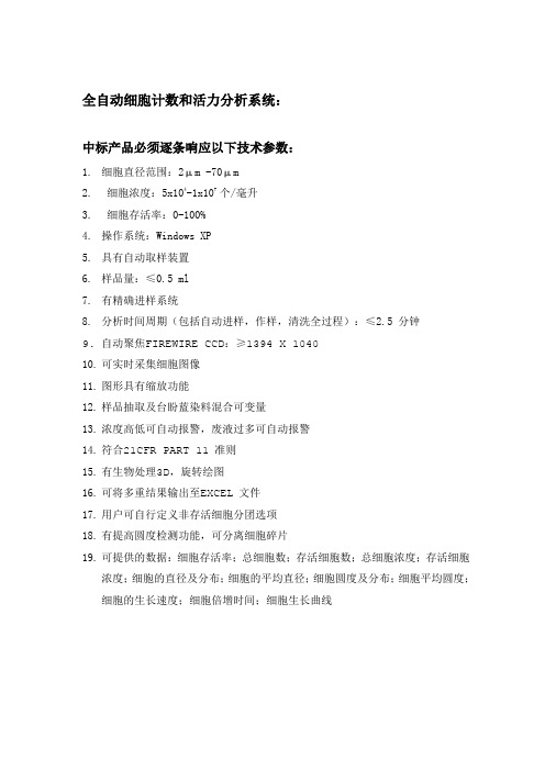
全自动细胞计数和活力分析系统:中标产品必须逐条响应以下技术参数:1.细胞直径范围:2μm -70μm2.细胞浓度:5x10⁴-1x10⁷个/毫升3.细胞存活率:0-100%4.操作系统:Windows XP5.具有自动取样装置6.样品量:≤0.5 ml7.有精确进样系统8.分析时间周期(包括自动进样,作样,清洗全过程):≤2.5 分钟9.自动聚焦FIREWIRE CCD:≥1394 X 104010.可实时采集细胞图像11.图形具有缩放功能12.样品抽取及台盼蓝染料混合可变量13.浓度高低可自动报警,废液过多可自动报警14.符合21CFR PART 11 准则15.有生物处理3D,旋转绘图16.可将多重结果输出至EXCEL 文件17.用户可自行定义非存活细胞分团选项18.有提高圆度检测功能,可分离细胞碎片19.可提供的数据:细胞存活率;总细胞数;存活细胞数;总细胞浓度;存活细胞浓度;细胞的直径及分布;细胞的平均直径;细胞圆度及分布;细胞平均圆度;细胞的生长速度;细胞倍增时间;细胞生长曲线连续波长微孔板分光光度计中标产品必须逐条响应以下参数:常规参数孔板类型:6-,12-,24,48,96和384-孔标准微孔板,大小128mm×86mm,最高0.8英寸。
可选配件:Take3TM微孔板适配器,可进行微量检测、BioCell TM、标准比色杯检测软件:Gen5软件控制,兼容各种版本,包括中文软件检测模式:终点法,动力学法,波长扫描发和孔域扫描法检测性能波长范围:200-999nm,可选1nm步进波长准确性:+/-2nm波长重复性:+/-0.2nm吸收光检测范围:0-4 OD吸收光分辨率:0.0001带宽:5nmOD准确性:0-2 OD:+/-1% +/-0.010 OD2-2.5OD:+/-3% +/-0.010 OD重复性:0-2 OD:+/-1% +/-0.005 OD0-2 OD:+/-3% +/-0.005 OD线性:0-2 OD:+/-1% +/-0.010 OD2-2.5 OD: +/-3% +/-0.010 OD读板速度:96孔≤50秒(标准);≤40秒(快速);≤15秒(扫描)384孔≤170秒(标准);≤135秒(快速);≤35秒(扫描)物理参数连接:USB串口连接电脑控制化学兼容性:所有暴露表面均耐受0.5%次氯酸钠、70%乙醇或异丙醇进行消毒符合条款:EN 61326-1:2006;EN61326-2-6:2006;EN61000-4-2; EN61000-4-3; EN61000-4-4; EN61000-4-5; EN61000-4-6; EN61000-4-11梯度PCR仪中标产品必须逐条响应以下技术参数:1主要技术指标1.1拥有应用生物系统公司颁发的PCR许可证。
Scepter 2.0 单细胞计数仪说明书

Scepter™ 2.0 Cell Counter Precise, handheld cell countingThe life science business of Merck KGaA,Darmstadt, Germany operates as/scepterupgrade Precise, handheld cell countingScepter™ 2.0 cell counters mark the next generation in Scepter™ technology, highlighted by:Compatibility with More Cell TypesIncreased Cell Concentration RangePowerful Software for Complex, Effortless Cell AnalysisScepter™ 2.0 Cell Counter• Recommended based on size • Customer Validated• MilliporeSigma Validated“At last, an alternative to lining up for the Coulter Counter ®, and far easier than sweating over fragile hemocytometers.”Amy A. Caudy is a Lewis-Sigler Fellow at Princeton University’s Lewis-Sigler Institute for Integrative GenomicsThe Scientist , Dec. 2010. Top Ten Innovations of 2010.The power of precisionTrust Scepter™ devices with your most valuable samples to get reproducible and reliable counts. The reliability of Scepter™ cell counters is particularly apparent with smallercell types. Because the Scepter™ cell counter measures volume using the Coulter Principle, it can quantify cells based on size and will discriminate larger cells from smaller debris, unlike vision-based techniques, which rely on object recognition software and cannot reliably detect small cells.Scepter™ sensor technologyCompatible with 60 μm and 40 μm sensors, theScepter™ 2.0 cell counter can meet even more of your cell- and particle-counting needs. Use the 60 μm sensor for particles between 6 and 36 μm. Use the 40 μm sensor for particles between 3 and 17 μm.• Precise volumes are drawn into the Scepter™ sensor.• As cells flow through the aperture in the sensor, resistance increases. This increase in resistance causes a subsequent increase in voltage.• Voltage changes are recorded as spikes with each passing cell.• Spikes of the same size are bucketed into a histogram and counted. This histogram gives you quantitative data on cell morphology that can be used to examine the quality and health of your cell culture.Particles are detected by Ohm’s Law V=IR (V=voltage, I=current, and R=resistance).Table 1.Cell types validated with the Scepter™ cell counter and the recommended Scepter™ sensor.Cell Type Measuredsize (μm)40 μm sensor 60 μm sensorMeg-0116-17•MG-6315-17•Mouse ES Cell5-13••Mesenchymal Stem Cell 15-16•MRC-5•NCI-H14610-13•NIH 3T315•NTERA2, clone D113••OK 17-18•PBMCs 7-12•PC129-13•Primary Astrocytes 7•Primary Neuronal Cell •Raji 12-15••Ramos11-12••Rat Dorsal Root Ganglion Cells 7•Rat Whole Blood 4.6•Red Blood Cells 5-7•Rat Neural Stem Cell 11-13•RAW 264.712-15•RBL 11-13••RIN-mF513-14•SF913•SH-SY5Y 12•Sk-Br-315-20•SK-MEL-2817-19•SK-N-MC 14-15•SK-N-SH 14-15•Splenocytes 7-9•SW-48015•SW-62013-14••T8414-18•T98G 17•TF-113-14••U25116-20•U2OS 16-19•U26612•U87-HumanGlioblastoma cell line 12-14•U93711-13••WI-3812-15•Y7913-14•Yeast- Pichia Pastoris 5•Yeast- S.cerevisiae 6•Cell Type Measuredsize (μm)40 μm sensor 60 μm sensor2102 Ep 15-19•454 beads •A17215•A25314-18•A37516•A43115-17•A549•Algae (various)7-9•B3513-16••B Cells6-11•C2C1212••C30512-14••C612-13•CA4610-12••Caco-217•CHO 14-17•COS-112••Cos-715•D28312•Daudi 10-12••DU-14515-17•Epithelia 14-15•HCT-11610••HEK29311-15•HeLa 12-14•HepG212•HFF 18-20•Hs2714••HT-108014-16•HT-2911•HUH7- Hepatoma line •Human ES Cells 9-12•HUVEC 14-15•IMR-3212-14••IMR-9015••Jurkat 13•K56222•KB 14•KG-110-13••L614-16•LNCaP15-16•Luminex ® beads 5-6•MCF715-17•MDCK13-15•Scepter™ cell counterHemocytometer= Average % CV= Coulter counter % CVVision-based counter1234567Figure 2.The average percent coefficient of variation (CV) for each counting method shown was calculated using cell concentration measurementsat 50,000 cells/mL samples of 19 different cell lines. The Scepter™ cell counter is more precise than vision-based counting and hemocytometry, and approaches the precision of the Coulter Counter ® standard (yellow bars). Error bars represent standard deviation.of cell diameter or cell volume FormatCounting methods Samplevolume needed Samplevolume counted Cells counted in a 100,000 cell/mL sample Average % CV Hemocytometer Slide and microscope Manual, vision-based 10 μL 0.1 μL /square 10/square 41.8Brand L Benchtop Automated vision-based system10 μL 0.4 μL 4032.1Scepter™ Cell Counter HandheldImpedance-based cell detection100 μL50 μL50009.1Intuitive analysis softwareFrom simple counts to complex volume measurements used to assess cell health parameters, Scepter™ Software Pro provides an intuitive, intelligent platform to perform high-level cell analysis based on the size measurements captured with the Scepter™ cell counter.Using the Scepter™ Software Pro on your computer, you can:• Compare several samples and data sets side by side using histogram overlay and multiparametric tables • Create and save gates to be used from one experiment to the next • Create attractive graphical presentations and reports with your dataA View of Scepter™ Software ProData:Data files from your Scepter™ cell counter Current Plot:Working plot and data file Group Stats:Customizable statistics from your selected data filesMultifunctional Plot:Multiple data sets/histogram overlays Reports:Export, print selected graphs/files, cut and pasteAnalysis Templates:Saved gating parametersPrepare the sample:Start with a single-cell suspension, diluted to a total volume of 100 μL (recommended) in phosphate buffered saline (such as EmbryoMax ® 1x DPBS) to 10,000-500,000 cells/mL (operating range for 60 μm sensor) in a 1.5 mL microcentrifuge tube.Perform cell count:• Turn on the Scepter™ cytometer by pressing the toggle on the back of the instrument and wait for on-screen instructions to appear.• When prompted, attach a sensor to the end of the Scepter™ unit with the electrode sensing panel facing toward the front of the instrument, and you’ll see detailed instructions for each step of the counting process.• Pipette once to draw sample into the sensor. 50 μL of your cell suspension is drawn into the microfabricated, precision-engineered channel embedded in the sensor. The cell sensing zone detects each cell drawn into the sensor and thus cell concentration is calculated.• The sensing zone also measures cell sizes and cell volumes with sub-micron and sub-picoliter resolution, enabling the Scepter™ cytometer to display a As easy as pipetting100MDMA231 cellsFlow cytometryScepter™Trypan blue + hemocytometerNIH 3T3 cells% V i a b i l i t y1009080706050403020100Flow cytometryScepter™Trypan blue + hemocytometer% V i a b i l i t yA.B.ApplicationsMilliporeSigma continues to expand the capabilities of Scepter™ technology, and the latest generation Scepter™ 2.0 device features enhanced analytical powers, enabling you to count even more cell types and sizes.Figure 3.Rapid cell analysis using the Scepter™ 2.0 device provides reliable assessments of cell viability compared to flow cytometry (ViaCount ® assay) and hemocytometry (using Trypan blue staining). MDMA231 cells (A) and NIH 3T3 cells (B) were treated with camptothecin 24 hours prior to analysis.“Cell counting is normally a very tedious process and usually only provides minimal information on the cell population. This instrument, which is only slightly larger than an automatic pipette, allows you to count cells in your tissue-culture hood, simplifies the procedure, and provides much useful data, such as the fraction of intact cells.”H. Steven Wiley is a lead biologist at the Environmental Molecular Sciences Laboratory at the Pacific Northwest National LaboratoryThe Scientist , Dec. 2010. Top Ten Innovations of 2010.Scepter™ 2.0 cell counter for counting heterogeneous cell populationsCount blood cells and other cells with small diameters with the highest precision. Biological samples such as primary isolates or cultured cells are often heterogeneous mixtures of cells that differ by type and/or function. Such differences in cellularattributes are most commonly determined by multicolor fluorescent antibody detection of cell type specific surface marker(s) using flow cytometry. Notably,in addition to variations in protein expression, many cell types and physiological states are also uniquely distinguishable on the basis of size alone. The ability to identify population subsets on the basis ofphenotypic differences and further determine their relative frequencies (and concentrations) is critical to many aspects of research.Scepter™ 2.0 cell counter for cell healthInstantly gauge the health of your cell cultures without even leaving the culturehood. Because the Scepter™ cell counter displays high-resolution histograms of entire cell populations, you can differentiate live cells from dead cells and debris by simply gating on the histogram peak corresponding to larger-diameter cells. No staining is required! The resulting calculation for % viable cells agrees with viability calculations obtained using flow cytometry (ViaCount ® reagent) and Trypan blue staining/hemocytometry (shown here with MDMA231 and NIH 3T3 cells).% C o e ffic i e n t o f V a r i a t i o n (%C V )(40 µm sensor)Vision-based Counter Counting MethodPBMCsAverage % CV Z2 Coulter Counter C o u n tC o u n tForward ScatterFlow CytometryPlot PO3, ungatedScepter™ (40 µm)Current AcquisitionDiameter (µm)100200300400500600700800900100001000200030004000500060005.6 µm Polystyrene MILLIPLEX ® Antibody-Conjugated MicrospheresC o u n tDiameter (µm)1,500,000500,000250,000125,00050,000750,0000.0E+00M e a s u r e d C e l l C o n c e n t r a t i o n (c e l l s /m L )Theoretical Cell Concentration (cells/mL)4.0E+058.0E+051.2E+061.6E+06Theoretical Cell ConcentrationZ2 Coulter Counter Scepter™ - 40 µm SensorsScepter™ 2.0 │ Precise, handheld cell countingScepter™ 2.0 cell counter for countingheterogeneous cell populations (continued)Distinguishing lymphocytes from monocytes in freshly isolated PBMCs . The assessment of immune profiles of the various immune cell subsets can help identify molecular signatures that may facilitate research. The Scepter™ cell counter, when used in combination withScepter™ Software Pro, provides a tool for rapid determination of lymphocyte and monocyte concentrations as well as the relative frequency of these cell types in PBMC isolates.Figure 4.The Scepter™ 2.0 cell counter counts PBMCs with greater precision than other counting methods, as reflected by low coefficients of variation. % CVs were calculated using average cell counts of four replicate samples.Figure 6.Scepter™ Software Pro displays imported size distribution histograms as either a single sample histogram or as overlaid histograms for multiple samples. Shown is an overlaid histogram for serially diluted 5.6 μm MILLIPLEX ® map microspheres.Figure 7.The Scepter™ cell counter counts yeast cells with good accuracy and linearity. Measured yeast cell concentrations were compared totheoretical concentrations. The solid blue line represents the theoretical values. Dotted lines represent best linear fit to data. Both the Scepter™ and Coulter Counter ® platforms show a loss of linearity and accuracy upon an increase in cell concentration.Figure 5.Representative comparison of histogram plots for human PBMCsamples acquired on the Scepter™ cell counter (diameter histogram on right) and guava ® easyCyte™ flow cytometry (forward scatterhistogram on left) platforms. Analysis plots derived from both platforms demonstrate three distinct peaks corresponding to 1) dead cell/debris, 2) lymphocyte and 3) monocyte fractions. The difference in counts displayed (Y-axis) is due to differences in sample dilution between the guava ® easyCyte™ flow cytometer and the Scepter™ cell counter.Cell Fraction Scepter™ 1Forward Scatter 2Staining 31Lymphocyte 586563Monocyte 4235372Lymphocyte 687271Monocyte 3228293Lymphocyte 666971Monocyte343129Precise and accurate bead counting with the Scepter™ 2.0 cell counterMicron-sized beads are used in a variety of biological applications, ranging from daily validation of flowcytometer performance to purification of fusion protein constructs from cell lysates. Accurate determination of bead counts at the onset of each assay allows for standardization of bead concentrations acrossmultiple samples and minimizes errors and variation in downstream results. The Scepter™ cell counter is well suited for precise counting for beads of numerous types and can improve reproducibility of bead-based assays, such as immunoprecipitation and multiplexed detection.Scepter™ devices can facilitate yeastcell counting for brewing and wine industriesIntroduction of a consistent yeast cell concentration is required for successful beer and wine fermentation as well as to maintain batch-to-batch reproducibility and ensures consistent fermentation over many cycles. Scepter™ cell counter can be used to monitor yeast size and concentration by yielding interpretable histograms that could be gated to provide this depth of information.Ordering InformationDownloadable Scepter™ Software 1O-Rings2Scepter™ Test Beads 1PHCCBEADS Scepter™ USB Cable 1PHCCCABLE Scepter™ Sensors, 60 μm50PHCC60050500PHCC60500Scepter™ Sensors, 40 μm 50PHCC40050500PHCC40500Universal Power Adapter1PHCCP0WER Scepter™ O-Ring Kit (includes 2 O-rings and 1 filter cover)1PHCC0CLIP Table 2.Lymphocyte and monocyte subset frequencies from three individual PBMC samples. Aliquots from each sample were analyzed using the guava ® easyCyte™ flow cytometry and Scepter™ platforms. 1 Values were derived from the diameter histogram plot. 2 Values were derived from the forward scatter histogram plot based on total events measured on guava ® easyCyte™ flow cytometry platform. 3 Staining frequencies derived as follows: % Lymphocytes = (% CD3+ T cells) + (%CD16/56+ NK cells) + (%CD19+ B cells); % Monocytes = % CD14+ cells.vMilliporeSigma400 Summit Drive Burlington, MA 01803。
VITEK MS仪器介绍

Desorption / Ionisation - Time of Flight) 最柔软的电离方式保证完整分子量信息,例如蛋白质 / 多肽; 直接从细菌核糖体中筛选蛋白质; 一般都使用飞行时间检测器(Time of Flight)来检测,故名。
29
细菌质量指纹图谱
扩展到新的菌种和应用(用户不断增加细菌数据库)
4000
m/z
8000
4000
m/z
➢检测细菌的核糖体蛋白
➢不同波形代表不同菌种
Pantoea agglomerans Acinetobacter lwoffi Burkholderia cepacia Raoultella ornithinolytica Staphylococcus aureus Escherichia coli 8000
#
1
=
>
A
dvຫໍສະໝຸດ BC(3
2
,
0
.
5
,
1
.
0
)
=
>
N
F
0
.
7
[
B
P
=
4
4
2
3
.
9
,
2
1
1
7
]
100
4424.2
9476.2
mass
2117. 3
90
4861.0
80
Countstar 细胞计数仪介绍

Countstar细胞计数仪 与人工计数对比
Countstar细胞计数仪与血球计数板计数细胞活率结果一致,两种方法 计数结果差异无统计学意义(Y=1.0072x;R2=0.9937)
生化进口品牌介绍
旋混合器的厂家,从设计到生产形成一套完整的流水线,其生产的Vortex-Genie 2 是所有涡旋混合器中应用最广范,用途最广,最耐用的品牌,已形成一种国际标准,成为该领域的国际知名品牌。
44 、SciGene 美国SciGene 公司主要致力于为基因芯片杂交实验提供自动化的样品前制备(标记,纯化),上样杂交和洗涤干燥仪器。
用自动化的仪器代替人工操作,减小了实验的批间批内差,极大的改善了实验的均一性。
并能减少背景干扰,为后期的芯片扫描提供良好的数据。
45 、SY-LAB 奥地利SY-LAB 公司是一家专业开发生产生物培养、冻存等产品的公司。
其旗下的ICECUBE 程式冷冻降温仪专为细胞冷冻操作设计,是一款细胞、组织、血液、骨髓、器官、胚胎等珍贵的生物样品超低温冷冻前处理的必备仪器。
46 、SYNGENE SYNGENE 是英国著名公司SYNOPTICS 公司的子公司,主要产品集中在凝胶图像的扫描成像和分析,包括凝胶成像、化学发光成像、化学发光与荧光成像以及2D 胶成像多种,设计精巧、操作简单、灵敏度高,并具备强大的功能分析软件,使其成为当今世界上最完善的成像分析系统之一47、SORENSON SORENSON 是生命科学领域塑料耗材革新的引领者,专业提供移液器和吸头等耗材。
其产品确保无RNA 酶、DNA 酶、无热源,广泛应用于分子生物学、基因测序,生塑料工艺的完美体现。
48 、SYNBIOSIS SYNBIOSIS 是SYNOPTICS 旗下的公司之一,是世界上知名的自动菌落计数仪生产商。
其全自动微生物分析仪具有自动的菌落计数和抑菌区测量功能,适合研究单位、医院、检验检疫机构和工业领域进行微生物菌落计数和含量分析、抗生素的抗菌性测试以及细菌的抗生素敏感性分析等工作。
Luminex Muse Ki67 Proliferation Kit 快速参考卡说明书
Expected ResultsThe figures below show examples of results obtained with the Muse® Ki67 Cell Proliferation Kit.Events in the histogram are as follows:•Proliferating cells (Ki67+)•Negative cells (Ki67–)Figures A and B. Example Data: Results obtained with the Muse® Ki67 Cell Proliferation Kit. Figure A shows results for healthy proliferating Jurkat cells (red) with an optional histogram overlay of the isotype control (grey). Figure B shows human PBMC cells stimulated with PHA (red and blue) with an optional histogram overlay of the isotype control (grey). The statistics show population percentages in the stained sample. The dot plot shows Ki67 staining vs Cell Size Index and the histogram shows Ki67 staining.For more information, refer to the comprehensive user’s guide, which can be found at /muse (search for Catalog Code, MCH100114).Related ProductsFor Research Use Only; not for use in diagnostic procedures. The latest version of Muse® software, which includes all assay modules, as well as the kit user’s guide, can be found at /muse.DescriptionCatalog No.Muse® System Check KitMCH100101Muse® Count & Viability Kit (100T)MCH100102Muse® Annexin V & Dead Cell Kit MCH100105Muse® Human Lymphocyte CD69 Kit MIM100104Muse® Human Lymphocyte CD25 KitMIM100105Muse and the M logo are trademarks of Merck KGaA, Darmstadt, Germany.Part No. 4600-3461 Rev A, 9/2013. Printed in the USA.© 2013 EMD Millipore Corporation. All rights reserved.。
ELISPOT入门知识
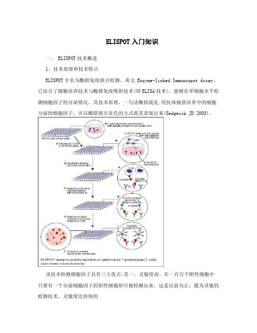
ELISPOT入门知识一、 ELISPOT技术概述1、技术原理和技术特点ELISPOT全名为酶联免疫斑点检测,英文:Enzyme-linked Immunospot Assay。
它结合了细胞培养技术与酶联免疫吸附技术(即ELISA技术),能够在单细胞水平检测细胞因子的分泌情况。
其技术原理,一句话概括就是:用抗体捕获培养中的细胞分泌的细胞因子,并以酶联斑点显色的方式将其表现出来(Sedgwick JD 2005)。
该技术检测细胞因子具有三大优点:其一,灵敏度高。
在一百万个阴性细胞中只要有一个分泌细胞因子的阳性细胞即可被检测出来。
这是目前为止,最为灵敏的检测技术,灵敏度比传统的第 1 页共 8 页的ELISA方法高2-3个数量级。
其二,单细胞水平,活细胞功能检测。
ELISPOT检测的是单个细胞分泌,而非细胞群体的平均分泌。
在检测的过程中,有活细胞培养与抗原刺激阶段,检测的是活细胞的功能,而非死细胞的遗留物。
其三,操作简便经济,可以进行高筒量筛选。
ELISPOT没有复杂的细胞体外扩增过程,不使用同位素,不需要大型的、专门的实验仪器设备。
按照标准化的实验操作,一个实验者可以同时处理数百个样品,效率远远高于其它检测方法(Kalyuzhny AE 2005)。
实验设计在96孔培养板上进行,直接以培养板的塑料板底或者PVDF膜以及硝酸纤维素膜为基质,包被上特异性的单克隆抗体,用以捕获细胞分泌的细胞因子。
(由于涉及到细胞培养的过程,对单克隆抗体的要求要远高于ELISA中的捕获抗体,该抗体需要无毒,不含内毒素,亲和力高等特点。
)之后,在培养板的孔内加入细胞培养基(现在无血清ELISPOT技术已经成熟,培养基中可以不再含有血清)、待检测的细胞以及抗原刺激物进行培养。
在特异性的抗原或者非特异性的有丝分裂原的刺激下,数小时之内,T细胞就会开始分泌各种细胞因子。
细胞因子当即就被位于细胞下方的膜上的单克隆抗体所捕获。
在洗去细胞之后,被捕获的细胞因子可以与生物素标记的第二抗体结合,然后用酶标亲和素再与生物素结合,进行化学酶联显色,就可以在膜的局部形成一个个圆形的斑点。
ASTREE味觉指纹分析仪操作简明步骤系统启动前请确认s
ASTREE味觉指纹分析仪操作简明步骤系统启动前请确认windows系统是否设置正确:确认windows控制面板里面的区域/语言选项是否正确:双击进入区域语言选项,点击自定义进入参数设置界面,按图片中格式设置。
A. 根据应用,选择所需的传感器种类(食品专用传感器,药品专用传感器),在软件的〞configure〞中定义传感器〔输入传感器型号和序列号,仅第一次安装传感器或更换新传感器时〕B. 定义自动进样器的样品盘的烧杯位数:一、开机Astree开机前请先将氯化银电极的保护套去掉,整个过程中注意带上防静电保护套,防止传感器被静电损害。
取下电极保护套后,分别开启自动进样器电源,电子舌主机电源,以及电子舌操作软件Alphasoft。
系统连接测试:翻开Alphasoft 软件,进入Configuration(设置)菜单,点击按钮,依图中所示顺序测试系统连接是否顺畅,完成连接检测。
假设连接失败,点击〞Verify验证〞旁边的〞Autodetect自动检测〞按钮自动检测通讯端口,待发现通讯端口后点击工具栏上面的〞Apply(应用)〞按钮保存设置。
同样完成自动进样器的通讯设置。
二、电子舌传感器活化1.被动活化电子舌系统开机、连接正常后,在样品盘1号位置放入盛有蒸馏水的烧杯。
使用自动进样器手动控制器,摁面板上面的〞→〞符号,将1号烧杯位置旋转至传感器下方,然后一直摁住〞↓〞符号,直到传感器泡在烧杯里面,被动活化时间为0.5-1h。
根据烧杯体积的不同倒入适宜体积的蒸馏水:100ml的烧杯倒入80ml,20ml烧杯倒入刻度线的位置。
2.主动活化(conditioning)按照如下列图中所述,主动活化中所用的HCl溶液为alphamos提供,用蒸馏水配置成浓度为0.01mol/L的HCL溶液,倒入三个烧杯,每个80ml,假设为20ml烧杯至刻度线;清洗溶液为蒸馏水(注意更换新的蒸馏水)。
3.传感器校准(calibration)校准溶液为alphamos提供的HCl校准溶液,使用时用蒸馏水配置成浓度为0.01mol/L的HCL溶液,与传感器主动活化中使用的溶液一致。
VITEK MS PRIME 产品说明书
©2022 bioMérieux, Inc.• BIOMÉRIEUX, the BIOMÉRIEUX logo, CLARION, FLEXPREP, MYLA, PICKME, AND VITEK are used pending and/or registered trademarks belonging to bioMérieux, or one of its subsidiaries, or one of its companies • Patents: ///patents • PRN 059452 Rev01.A
4
URGENT SLIDE PRIORITIZATION
• Prioritize critical samples in one click • Analysis automatically resumes after critical slide completion
5
FLEXIBLE RESULTS REVIEW
AT MULTIPLE BENCHES
• Automatically release high confidence results
• Easily view spot images for quick troubleshooting
6
AUTOMATED FINE-TUNING
• Increase uptime by initiating automated fine-tuning when instrument
is not in use
• Hands-free maintenance to free up technician time
1. Perrot N, Dauwalder O, Paris C, et al. New innovative tool for easy colony picking and sample preparation for MALDI-TOF. Poster presented at: ASM Microbe 2019; June 20-24, 2019; San Francisco, CA.
MasterScreen中文手册

德国耶格公司高级组合式肺功能仪MasterScreen系列中文操作手册系统简介耶格的MasterScreen肺功能仪是一个组合式、模块化的结构,其特点有灵活的扩展性、良好的经济性和长久性。
您可以在主机基础上根据自己的需要选择不同的部件构成通气、弥散、残气、气道阻力、顺应性、药物激发实验、舒张实验等等各项肺功能功能项目的检查。
MasterScreen的主机框架如下:硬件主要由移动式仪器车、隔离电源、计算机系统(包括计算机主机、显示器、打印机、键盘、鼠标)、肺功能专用接口板(EASI-CARD)、扩展电路模块、流速传感器模块、气体分析器模块(一氧化碳、氦气、氧气)、气路管道和各种阀门(气动阀门和电动阀门)等组成。
软件由运行在Windows操作系统下的肺功能测试软件和病人资料管理以及专用的维修测试软件等组成。
肺功能的核心部件为流速传感器和气体分析器,所以在日常维护中一定要保护好这些敏感部件。
气路管道设计的合理性会给临床使用带来很大的方便,MasterScreen在这方面作得不错。
所以整套系统非常稳定、可靠、而且故障率低。
仪器基本原理:流速传感器为压差筛网式结构,用铂金丝编制,双层90度交叉,其原理为气流经过压差筛网,由于筛网有阻力(阻力为0.36Kpa/L/S),两端会产生压差,其压差的大小与流速成正比。
这样就可以得到流速,通过对流速的积分就获得容量。
有了流速和容量者两个基本指标,我们就可以得到呼吸频率BF、潮气量VT、肺活量VC、补呼气量ERV、一秒量FEV1、峰流速PEF、每分最大通气量MVV和流速容量环等静态和动态肺参数。
一氧化碳分析器用于弥散检查,其原理为电化学分子膜渗透原理,理论上是长寿命的。
如果测试气体比较脏,那分子渗透膜可能会失效,这一点一定要注意。
氦分析器用于残气和肺容量的检查,其原理为热敏式惠斯通电桥,利用氦为热的良导体,氦浓度越高,热量被带走速度也越快。
理论上也是长寿命的。
所有这些长寿命的部件大大降低了使用成本。
Amicus血细胞分离机的使用效果评价

Amicus血细胞分离机的使用效果评价【摘要】Amicus血细胞分离机是一种用于分离血液中不同细胞类型的先进设备。
本文从设计和原理、使用效果评价方法、分离效率、细胞完整性和应用范围等方面对该设备进行评价。
通过对分离效率和细胞完整性的评价,我们可以得出Amicus血细胞分离机在分离血细胞方面具有较高的效率和保持细胞完整性的能力。
该设备在不同应用范围下也表现出色。
综合评价该设备的使用效果,可以看到其在血细胞分离领域具有较好的应用前景。
随着技术的不断发展,我们也期待Amicus血细胞分离机能够在未来实现更多的创新,使其在血细胞分离领域持续发挥重要作用。
【关键词】Amicus血细胞分离机、使用效果评价、细胞完整性、分离效率、应用范围、设计和原理、未来发展、研究背景1. 引言1.1 研究背景血细胞分离机在临床上起着非常重要的作用,可以帮助医生分离出不同类型的血细胞,用于疾病诊断和治疗。
Amicus血细胞分离机是一种先进的设备,能够高效地将血液中的各种血细胞进行分离,提高实验结果的准确性和可靠性。
对于这种设备的使用效果需要进行全面的评价,以确保其在临床实践中的有效性和可靠性。
本文将对Amicus 血细胞分离机的使用效果进行评价和分析,为临床医生提供参考和指导。
通过评价这种设备的设计和原理、分离效率、细胞完整性以及应用范围等方面的指标,可以全面了解Amicus血细胞分离机在实际应用中的表现,为其未来的发展提供有益的建议和展望。
2. 正文2.1 Amicus血细胞分离机的设计和原理Amicus血细胞分离机是一种高效、精准的细胞分离设备,其设计和原理决定了其在细胞分离领域的重要性和有效性。
该机器主要由抽血系统、分离系统和控制系统组成。
在使用过程中,先通过抽血系统获取样本血液,然后将血液通过分离系统进行离心分离,进而得到血浆、白细胞和红细胞等组分。
控制系统则负责监控整个分离过程,确保分离的准确性和稳定性。
Amicus血细胞分离机的设计理念是借助离心力将不同密度的细胞分离开来,从而实现快速、高效的血细胞分离。
MUSE产品介绍及成功案例列举(院内)

MUSE介绍
自动测量参数矩阵与手动测量工具
基于原始采样点采集技术,MUSE系统能够对心电图进行自动测量从而获得每导联 53个参数的参数矩阵。
同时,只需双击波形,便可任意对波形进行手动测量,精度达到毫秒、微伏级。
MUSE介绍
自动诊断功能
12SL自动分析诊断算法是Marquette公司历经数十年的探索和数十万份心电图报告 的积累所获得的专利算法,具备FDA认证(国内CFDA尚未对此类单一程序或算法开 放认证)。该算法是唯一具有第三方准确性论证报告的心电诊断算法,且在业界赢 得了“金标准”的称号。 自动诊断分析程序能够为读图医生节省大量时间,且同时提高诊断准确性等。具体 方式如下: 1、初筛病例,分级诊断:
Mac5500HD 高端心电图机(用于门诊、心内科)
▪ “中国人心电数据库”项目指定用机 ▪ 支持15导联同步采集 ▪ 支持心向量、晚电位、儿童专用模式采集 ▪ 业界最高端“真金白银”采集盒 ▪ 配备大屏幕、专用台车、条码扫描枪
Mac800 网络专用心电图机(用于普通科室病房)
▪ 12导联同步采集 ▪ 操作简易,一键采集、保存、发送 ▪ 配备轻装便携手柄,适于病房床旁检测 ▪ 无缝对接心电网络,支持无线WIFI连接,“秒传”心电图 ▪ 以红黄绿三色灯实时监测信号质量,辅助非专业人员保证采集质量
“现在,在MUSE无线环境中,当技师还在楼梯
Kim Scheumann,
上的时候,我已经阅完图了。这太美妙了!”
Cardiac Group Leade Indiana
Pocono医疗中心
➢ 232张床位 ➢ 提供急救及心血管救治服务 ➢ 2007年采集了27,093份心电图
• 绍兴市人民医院院内及绍兴市区域属两个相互独立的局域网,由两套MUSE 服务器分别管理;
Endress+Hauser Memosens Wave CAS80E 全光谱传感器操作手册说明书

Products Solutions Services操作手册Memosens Wave CAS80E 全光谱传感器数字式全光谱水质分析传感器BA02005C/28/ZH/05.22-00715972932022-04-25Memosens Wave CAS80E 全光谱传感器目录Endress+Hauser 3目录1文档信息 (4)1.1安全图标 (4)1.2信息图标 (4)2基本安全指南 (5)2.1人员要求 (5)2.2指定用途 (5)2.3工作场所安全 (5)2.4操作安全 (5)2.5产品安全 (6)3产品描述 (7)3.1产品设计 (7)4到货验收和产品标识 (9)4.1到货验收 (9)4.2产品标识 (9)4.3供货清单 (10)5安装 (11)5.1安装要求 (11)5.2安装设备 (13)5.3安装后检查 (19)6电气连接 (20)6.1连接设备 (20)6.2确保防护等级 (21)6.3连接后检查 (21)7调试 (23)7.1功能检查 (23)8操作 (24)8.1基于过程条件调节测量设备 (24)8.2循环清洗 (27)9诊断和故障排除 (28)9.1常见故障排除 (28)10维护 (29)10.1维护计划 (29)10.2维护任务 (29)11维修 (31)11.1概述 (31)11.2备件 (31)11.3返厂 (31)11.4处置 (31)12附件..............................3212.1设备专用附件........................3213技术参数..........................3413.1输入...............................3413.2电源...............................3513.3性能参数............................3513.4环境条件............................3913.5过程条件............................3913.6机械结构............................39索引.. (41)文档信息Memosens Wave CAS80E 全光谱传感器4Endress+Hauser1 文档信息1.1 安全图标1.2 信息图标附加信息,提示允许或推荐的操作禁止或不推荐的操作参见设备文档参考页面参考图 操作结果1.2.1 设备上的图标参见设备文档资料此类产品不可作为未分类城市垃圾废弃处置。
- 1、下载文档前请自行甄别文档内容的完整性,平台不提供额外的编辑、内容补充、找答案等附加服务。
- 2、"仅部分预览"的文档,不可在线预览部分如存在完整性等问题,可反馈申请退款(可完整预览的文档不适用该条件!)。
- 3、如文档侵犯您的权益,请联系客服反馈,我们会尽快为您处理(人工客服工作时间:9:00-18:30)。
I
lliJOlllPl4JlIIlIJ.ltA»oy• Jll J'" ..,,1Il1l-1it. - JIIIt: • • · !lIi\f*IIlf!;
=
~
c
y_0.973x+1813B71l'_0.9I28 1.2llE+07 -----'-----------.~~~ .:>--l.ooE+07
Mu,,-l!IftI!Mll!!IIIJ(;:Sl'~f)'(j§*l!i~m!ll~2-60um~
jj£!IIIJ(;~*:Sl'~=J!.
1.6OE+07 l.4OE+07 -
t
. Imag ...bllsed .utom,ttd d~ • M,nu,1
Hl'm-~
M~~n*. ~~*~~m*~~~~~
II
,
~~@m.~~-~~~~m*. ~~~ ~~~~m.~~. **7¥.tt~~
Muse-. ftl!Ml!i!!IIIJ(;:Sl'~f)'( J;.l_iIl~ lit*!l!!J'tl&ilut J;.l&
~~~~~~~!IIIW~~~~. **£~~.~~.~
&i'lIl"'I!""'lt,rorUlIJoit
~''''
<--+~.~Wicllil
lIlJ9litli'J1l:
Mu,,"!IIIlJ!iltllll..lli n
lIlJllitli'J 1l:1!III1J!i1/i t:)
liUI"J*,
lIlJ9litli'J1l:
tlfJPI~'@j;z
am.~J.*.-M,
IllfilJ!!II"lIl19"'IIi"
lIlttlll!ltlllI!IIIllIIlU!l!. fifltt'IUJl"ll•.
°0 Z
.0
"""'I!l
ICI
,~
III
'DO
lZ
140
~
· !IIIJ(;:Sl'~~JI< m. ~ ft . ll~ £ ~£m. ~~ ~~~ff~.
AnII,ipIllOO
5tlln~g
1'I1l1:'" ,nd Cdl511< I"de<
&i'lIl"'&~II9"~n~.l'_"'l!lmff.~J.lIll!
~~1Il.
,,"' _
Musc'". flI!tiINlftllMl1t*i'f.t 'iiIiI l>llllll!fill IIilI"l!llNlf",.n lIl. !Ii!~",,,,.;oJ.lItl!"JlIl
•.
","
",,@ ,i;j,
Dob..
•
ll!~~,*m~1"~~,t*ll:z-, 2012''FIi:ll!ll!Jn~~,t*ll:
t:P Doad-';'II.
• ;1<31'-'<'1'1 1!. ..Hlftili.
ftMuse-V AliMl!i!!IIIJ(;:Sl'~HII.. I"J iiP,dlli
MM~~&.~~~oo~~.!IIIJ(;:Sl'~~
JI<.
.....-I'-JOllllt:
(Annexin v)2iiJEtIII.~.fi
...tif~Hpilli**li12Bim~~ ./i1fij;Jl§88,,% "%~2~ ~i!: (021)38529000 ~A: (021)50803042 lII~tIli: 201203
=
~t*
r-m
r- #IiPitli~£U&33"%
E:$:r-~802~ ~i!: (020)83634531 ~A: (020)83634347 lII~tIli: 510060
lII~tIli: 100035
lII~tIli: 610061
,
iIl!.;;t: 1& tt~~Ii~ Ij:IIL': asiatechserv@
MUSE CN201205 V1
IILeabharlann Merck Millipore is a division of ' ,M R K EC
MBl'lli.~~nll<~:t'ltill,
ll&&Casp,se
~~~~.~~~~~.~~:Sl'm.*E
!IIIJ(;ffl:IJ:Sl'~. !IIIJ(;llI-5~l&. DNA~I9i~ ~l'~~em~ !li.~~nlli!~~!'HI-!Il .
• . mp~~$~.~~
·II"..... • "l!lill'OU
Ml
...t1fi
ff~Ii~i!:4008891988
red dat design award
:It*iPWJ~1l ~1Ii]'t~~1fI5"% JjlJll.I±t!!r-~A~~*~18~
~i!: (010)59898600 ~A: (010)57623560
. ." i'lC,
tJt(;:\5;~;jff
--
. = "J.... . _.- 1..... -
-_._ ..,.'--"_,..,
, . ,. -_.....-.. - .........
.....
"'
,,.._ Ie>"
-
--.
1 o ...' n
.. H
KS62, Hb, CHo-Kli1UurkarilllMl~l.mji}fjj.:=~ll$i\=.fflIIl MItt., • • s.jj.:, Muse.....MHRI!I1t.fiil.f11llHJlJtIlJlIt .:HJJafttl1!.l§IlEltllJdbtl:Kl,*,. MUsc'"I'U:J!!:!flIIMl1tm.JIJtIfJ ~*lIllll""!Il"'.lU;o;;:t!t.lf. ltlll:IU~lillillil. Mililt liE? M"~·~fII!;o;..alll1ilUJil~lII. It!ltlll~~IIIllI.
MI
Muse'" Cell Analyzer 0500-3115
Kits and Reagents
Muse'" Count Et Viability Kit (100 tests) Muse'" Count Et Viability Kit (600 tests) Muse'" Annexin V Et Dead Cell Kit Muse'" Cell Cycle Kit Muse'" Performance Check Kit Muse'" Count and Viability Kit (200X) (For difficult samples) Muse'" Cell Dispersal Reagent Accessories Muse'" Instrument Cleaning Fluid (ICF) Muse'" Flow Cell 4200-0140 0500-3120 MCH100102 MCH600103 MCH100105 MCH100106 MCH100101 MCH100104 MCH100107
----------+~~----
. ~ ~ ~-
-,-
Itdtz*.
Itl~@mllJfE~~Muse-'I't!'" ~*~£~!IIIJ(;:Sl'~~~.~~n*.~
~ lI.OOE+06 -----------; ~~.__'_-----
~
...~ ---~~:;;L--------
4.00E+06
JiUB
Jllt4lBiP~Ill)I~*j;:m1f~uq *1Jr-~C~11~7"%
~i!: (028)85288550 ~A: (028)85288553
'i]fJi
§~~ff!l&~J!3"%29~
~i!: (852)23766339
product design
~1t: (852)25761912
(MCF-7,
~*"'
•.
"'.'fi..
s..tIfJ~.*.
*~i' ..I!l>I!.~!lft.flt~~*.II9~.
it.ol. _*ilI~ lilJ~llIl
!IIIJ(;~*,~iIlJ'tIO-5lJl:iCli!~ ~IIiJllcl~. ~1II1¥i