分子生物学(英文版)
分子生物学---蛋白组学整理英文
proteomicsProteome: 细胞或组织或机体在特定时间和空间上表达的所有蛋白质。
Proteomics: 分析细胞内动态变化的蛋白质组成成分,表达水平于修饰状态,了解蛋白质之间的相互作用于联系,在整体水平上研究蛋白的组成与调控的活动规律。
研究蛋白组学希望达到的目标:By studying global patterns of protein content and activity and how these change during development or in response to disease, proteomics research is poised to boost our understanding of systems-level cellular behaviors. Clinical research also hopes to benefit from proteomics by both the identification of new drug targets and the development of new diagnostic markers.蛋白质组学研究内容:蛋白鉴定,蛋白定量,蛋白相互作用,蛋白修饰。
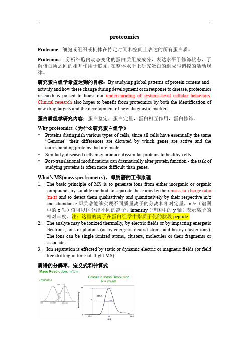
Why proteomics(为什么研究蛋白组学)•Proteins distinguish various types of cells, since all cells have essentially the same “Genome” their differences are dictated by which genes are active and the corresponding proteins that are made.•Similarly, diseased cells may produce dissimilar proteins to healthy cells.•Post-translational modifications can dramatically alter protein function - the task of studying proteins is often more difficult than genes.What’s MS(mass spectrometry),即质谱的工作原理1.The basic principle of MS is to generate ions from either inorganic or organiccompounds by suitable method, to separate these ions by their mass-to-charge ratio (m/z) and to detect them qualitatively and quantitatively by their respective m/z and abundance.即质谱能够实现不同质量离子的分离和相对定量,m/z(谱图中的x轴)值可以区分出不同的离子,intensity(谱图中的y轴)表示离子的相对丰度。
分子生物学词汇(中英文对照表 )
第一页A band|A带A chromosome|A染色体[二倍体染色体组中的正常染色体(不同于B染色体)] A site|[核糖体]A部位ABA|脱落酸abasic site|脱碱基位点,无碱基位点abaxial|远轴的abequose|阿比可糖,beta脱氧岩藻糖aberrant splicing|异常剪接aberration|象差;畸变;失常abiogenesis|自然发生论,无生源论ablastin|抑殖素(抑制微生物细胞分裂或生殖的一种抗体)abnormal distrbution|非正态分布abnormality|异常,失常;畸形,畸变ABO blood group system|ABO血型系统aboriginal mouse|原生鼠abortin|流产素abortion|流产,败育abortive egg|败育卵abortive infection|流产(性)感染abortive transduction|流产(性)转导ABP|肌动蛋白结合蛋白abrin|相思豆毒蛋白abscisic acid|脱落酸abscission|脱落absolute|绝对的absolute configuration|绝对构型absolute counting|绝对测量absolute deviation|绝对偏差absolute error|绝对误差absorbance|吸收,吸光度absorbed dose|吸收剂量absorbent|吸收剂absorptiometer|吸光计absorptiometry|吸光测定法absorption|吸收absorption band|吸收谱带absorption cell|吸收池absorption coefficient|吸收系数absorption spectroscopy|吸收光谱法absorption spectrum|吸收光谱;吸收谱absorptive endocytosis|吸收(型)胞吞(作用) absorptive pinocytosis|吸收(型)胞饮(作用) absorptivity|吸光系数;吸收性abundance|丰度abundant|丰富的,高丰度的abundant mRNAs|高丰度mRNAabzyme|抗体酶acaricidin|杀螨剂accedent variation|偶然变异accelerated flow method|加速流动法accepting arm|[tRNA的]接纳臂acceptor|接纳体,(接)受体acceptor site|接纳位点,接受位点acceptor splicing site|剪接受体acceptor stem|[tRNA的]接纳茎accessible|可及的accessible promoter|可及启动子accessible surface|可及表面accessory|零件,附件;辅助的accessory cell|佐细胞accessory chromosome|副染色体accessory factor|辅助因子accessory nucleus|副核accessory pigment|辅助色素accessory protein|辅助蛋白(质)accommodation|顺应accumulation|积累,累积accuracy|准确度acenaphthene|二氢苊acene|并苯acentric|无着丝粒的acentric fragment|无着丝粒断片acentric ring|无着丝粒环acetal|缩醛acetaldehyde|乙醛acetalresin|缩醛树脂acetamidase|乙酰胺酶acetamide|乙酰胺acetate|乙酸盐acetic acid|乙酸,醋酸acetic acid bacteria|乙酸菌,醋酸菌acetic anhydride|乙酸酐acetification|乙酸化作用,醋化作用acetin|乙酸甘油酯,三乙酰甘油酯acetoacetic acid|乙酰乙酸Acetobacter|醋杆菌属acetogen|产乙酸菌acetogenic bacteria|产乙酸菌acetome body|酮体acetome powder|丙酮制粉[在-30度以下加丙酮制成的蛋白质匀浆物] acetomitrile|乙腈acetone|丙酮acetyl|乙酰基acetyl coenzyme A|乙酰辅酶Aacetylcholine|乙酰胆碱acetylcholine agonist|乙酰胆碱拮抗剂acetylcholine receptor|乙酰胆碱受体acetylcholinesterase|乙酰胆碱酯酶acetylene|乙炔acetylene reduction test|乙炔还原试验[检查生物体的固氮能力] acetylglucosaminidase|乙酰葡糖胺糖苷酶acetylglutamate synthetase|乙酰谷氨酸合成酶acetylsalicylate|乙酰水杨酸;乙酰水杨酸盐、酯、根acetylsalicylic acid|乙酰水杨酸acetylspiramycin|乙酰螺旋霉素AchE|乙酰胆碱酯酶achiral|非手性的acholeplasma|无胆甾原体AchR|乙酰胆碱受体achromatic|消色的;消色差的achromatic color|无色achromatic lens|消色差透镜achromatin|非染色质acid catalysis|酸催化acid fibroblast growth factor|酸性成纤维细胞生长因子acid fuchsin|酸性品红acid glycoprotein|酸性糖蛋白acid hydrolyzed casein|酸水解酪蛋白acid medium|酸性培养基acid mucopolysaccharide|酸性粘多糖acid phosphatase|酸性磷酸酶acid protease|酸性蛋白酶acid solvent|酸性溶剂acidic|酸性的acidic amino acid|酸性氨基酸acidic protein|酸性蛋白质[有时特指非组蛋白]acidic transactivator|酸性反式激活蛋白acidic transcription activator|酸性转录激活蛋白 acidification|酸化(作用)acidifying|酸化(作用)acidolysis|酸解acidophilia|嗜酸性acidophilic bacteria|嗜酸菌acidophilous milk|酸奶aclacinomycin|阿克拉霉素acoelomata|无体腔动物acomitic acid|乌头酸aconitase|顺乌头酸酶aconitate|乌头酸;乌头酸盐、酯、根aconitine|乌头碱aconitum alkaloid|乌头属生物碱ACP|酰基载体蛋白acquired character|获得性状acquired immunity|获得性免疫acridine|吖啶acridine alkaloid|吖啶(类)生物碱acridine dye|吖啶燃料acridine orange|吖啶橙acridine yellow|吖啶黄acriflavine|吖啶黄素acroblast|原顶体acrocentric chromosome|近端着丝染色体acrolein|丙烯醛acrolein polymer|丙烯醛类聚合物acrolein resin|丙烯醛树脂acropetal translocation|向顶运输acrosin|顶体蛋白acrosomal protease|顶体蛋白酶acrosomal reaction|顶体反应acrosome|顶体acrosome reaction|顶体反应acrosomic granule|原顶体acrosyndesis|端部联会acrylamide|丙烯酰胺acrylate|丙烯酸酯、盐acrylic acid|丙烯酸acrylic polymer|丙烯酸(酯)类聚合物acrylic resin|丙烯酸(酯)类树脂acrylketone|丙烯酮acrylonitrile|丙烯腈actidione|放线(菌)酮[即环己酰亚胺]actin|肌动蛋白actin filament|肌动蛋白丝actinin|辅肌动蛋白[分为alfa、beta两种,beta蛋白即加帽蛋白] actinmicrofilament|肌动蛋白微丝actinometer|化学光度计actinomorphy|辐射对称[用于描述植物的花]actinomycetes|放线菌actinomycin D|放线菌素Dactinospectacin|放线壮观素,壮观霉素,奇霉素action|作用action current|动作电流action potential|动作电位action spectrum|动作光谱activated sludge|活性污泥activated support|活化支持体activating group|活化基团activating transcription factor|转录激活因子activation|激活;活化activation analysis|活化分析activation energy|活化能activator|激活物,激活剂,激活蛋白activator protein|激活蛋白active absorption|主动吸收active biomass|活生物质active carbon|活性碳active center|活性中心active chromatin|活性染色质active dry yeast|活性干酵母active dydrogen compounds|活性氢化合物active ester of amino acid|氨基酸的活化酯active hydrogen|活性氢active immunity|主动免疫active oxygen|活性氧active site|活性部位,活性中心active transport|主动转运active uptake|主动吸收activin|活化素[由垂体合成并由睾丸和卵巢分泌的性激素]activity|活性,活度,(放射性)活度actomyosin|肌动球蛋白actophorin|载肌动蛋白[一种肌动蛋白结合蛋白]acute|急性的acute infection|急性感染acute phase|急性期acute phase protein|急性期蛋白,急相蛋白acute phase reaction|急性期反应,急相反应[炎症反应急性期机体的防御反应] acute phase reactive protein|急性期反应蛋白,急相反应蛋白acute phase response|急性期反应,急相反应acute toxicity|急性毒性ACV|无环鸟苷acyclic nucleotide|无环核苷酸acycloguanosine|无环鸟苷,9-(2-羟乙氧甲基)鸟嘌呤acyclovir|无环鸟苷acyl|酰基acyl carrier protein|酰基载体蛋白acyl cation|酰(基)正离子acyl chloride|酰氯acyl CoA|脂酰辅酶Aacyl coenzyem A|脂酰辅酶Aacyl fluoride|酰氟acyl halide|酰卤acylamino acid|酰基氨基酸acylase|酰基转移酶acylating agent|酰化剂acylation|酰化acylazide|酰叠氮acylbromide|酰溴acyloin|偶姻acyltransferase|酰基转移酶adamantanamine|金刚烷胺[曾用作抗病毒剂]adamantane|金刚烷adaptability|适应性adaptation|适应adapter|衔接头;衔接子adapter protein|衔接蛋白质adaptin|衔接蛋白[衔接网格蛋白与其他蛋白的胞质区]adaptive behavior|适应性行为adaptive enzyme|适应酶adaptive molecule|衔接分子adaptive response|适应反应[大肠杆菌中的DNA修复系统]adaptor|衔接头;衔接子adaxial|近轴的addition|加成addition compound|加成化合物addition haploid|附加单倍体addition line|附加系additive|添加物,添加剂additive effect|加性效应additive genetic variance|加性遗传方差additive recombination|插入重组,加插重组[因DNA插入而引起的基因重组] addressin|地址素[选择蛋白(selectin)的寡糖配体,与淋巴细胞归巢有关]adducin|内收蛋白[一种细胞膜骨架蛋白,可与钙调蛋白结合]adduct|加合物,加成化合物adduct ion|加合离子adenine|腺嘌呤adenine arabinoside|啊糖腺苷adenine phosphoribosyltransferase|腺嘌呤磷酸核糖转移酶adenoma|腺瘤adenosine|腺嘌呤核苷,腺苷adenosine deaminase|腺苷脱氨酶adenosine diphoshate|腺苷二磷酸adenosine monophosphate|腺苷(一磷)酸adenosine phosphosulfate|腺苷酰硫酸adenosine triphosphatase|腺苷三磷酸酶adenosine triphosphate|腺苷三磷酸adenovirus|腺病毒adenylate|腺苷酸;腺苷酸盐、酯、根adenylate cyclase|腺苷酸环化酶adenylate energy charge|腺苷酸能荷adenylate kinase|腺苷酸激酶adenylic acid|腺苷酸adenylyl cyclase|腺苷酸环化酶adenylylation|腺苷酰化adherence|粘着,粘附,粘连;贴壁adherent cell|贴壁赴 徽匙牛ㄐ裕┫赴 掣剑ㄐ裕┫赴?/P>adherent culture|贴壁培养adhering junction|粘着连接adhesin|粘附素[如见于大肠杆菌]adhesion|吸附,结合,粘合;粘着,粘附,粘连adhesion factor|粘着因子,粘附因子adhesion molecule|粘着分子,粘附分子adhesion plaque|粘着斑adhesion protein|粘着蛋白,吸附蛋白adhesion receptor|粘着受体adhesion zone|粘着带[如见于细菌壁膜之间]adhesive|粘合剂,胶粘剂adhesive glycoprotein|粘着糖蛋白adipic acid|己二酸,肥酸adipocyte|脂肪细胞adipokinetic hormone|脂动激素[见于昆虫]adipose tissue|脂肪组织adjust|[动]调节,调整;修正adjustable|可调的adjustable miropipettor|可调微量移液管adjustable spanner|活动扳手adjusted retention time|调整保留时间adjusted retention volume|调整保留体积adjuvant|佐剂adjuvant cytokine|佐剂细胞因子adjuvant peptide|佐剂肽adjuvanticity|佐剂(活)性adoptive immunity|过继免疫adoptive transfer|过继转移ADP ribosylation|ADP核糖基化ADP ribosylation factor|ADP核糖基化因子ADP ribosyltransferase|ADP核糖基转移酶adrenal cortical hormone|肾上腺皮质(激)素adrenaline|肾上腺素adrenergic receptor|肾上腺素能受体adrenocepter|肾上腺素受体adrenocorticotropic hormone|促肾上腺皮质(激)素adrenodoxin|肾上腺皮质铁氧还蛋白adriamycin|阿霉素,亚德里亚霉素adsorbent|吸附剂adsorption|吸附adsorption catalysis|吸附催化adsorption center|吸附中心adsorption chromatography|吸附层析adsorption film|吸附膜adsorption isobar|吸附等压线adsorption isotherm|吸附等温线adsorption layer|吸附层adsorption potential|吸附电势adsorption precipitation|吸附沉淀adsorption quantity|吸附量adult diarrhea rotavirus|成人腹泻轮状病毒advanced glycosylation|高级糖基化advanced glycosylation end product|高级糖基化终产物 adventitious|不定的,无定形的adverse effect|反效果,副作用aecidiospore|锈孢子,春孢子aeciospore|锈孢子,春孢子aequorin|水母蛋白,水母素aeration|通气aerator|加气仪,加气装置aerial mycelium|气生菌丝体aerobe|需氧菌[利用分子氧进行呼吸产能并维持正常生长繁殖的细菌] aerobic|需氧的aerobic bacteria|需氧(细)菌aerobic cultivation|需氧培养aerobic glycolysis|有氧酵解aerobic metabolism|有氧代谢aerobic respiration|需氧呼吸aerobic waste treatment|需氧废物处理aerobiosis|需氧生活aerogel|气凝胶aerogen|产气菌aerolysin|气单胞菌溶素Aeromonas|气单胞菌属aerosol|气溶胶aerosol gene delivery|气溶胶基因送递aerospray ionization|气喷射离子化作用aerotaxis|趋氧性[(细胞)随环境中氧浓度梯度进行定向运动]aerotolerant bacteria|耐氧菌[不受氧毒害的厌氧菌]aerotropism|向氧性aesculin|七叶苷,七叶灵aetiology|病原学B cell|B细胞B cell antigen receptor|B细胞抗原受体B cell differentiation factor|B细胞分化因子B cell growth factor|B细胞生长因子B cell proliferation|B细胞增殖B cell receptor|B细胞受体B cell transformation|B细胞转化B chromosome|B染色体[许多生物(如玉米)所具有的异染质染色体] B to Z transition|B-Z转换[B型DNA向Z型DNA转换]Bacillariophyta|硅藻门Bacillus|芽胞杆菌属Bacillus anthracis|炭疽杆菌属Bacillus subtillis|枯草芽胞杆菌bacitracin|杆菌肽back donation|反馈作用back flushing|反吹,反冲洗back mutation|回复突变[突变基因又突变为原由状态]backbone|主链;骨架backbone hydrogen bond|主链氢键backbone wire model|主链金属丝模型[主要反应主链走向的实体模型]backcross|回交backflushing chromatography|反吹层析,反冲层析background|背景,本底background absorption|背景吸收background absorption correction|背景吸收校正background correction|背景校正background gactor|背景因子background genotype|背景基因型[与所研究的表型直接相关的基因以外的全部基因]background hybridization|背景杂交background radiation|背景辐射,本底辐射backmixing|反向混合backside attack|背面进攻backward reaction|逆向反应backwashing|反洗bacmid|杆粒[带有杆状病毒基因组的质粒,可在细菌和昆虫细胞之间穿梭]bacteremia|菌血症bacteria|(复)细菌bacteria rhodopsin|细菌视紫红质bacterial adhesion|细菌粘附bacterial alkaline phosphatase|细菌碱性磷酸酶bacterial artificial chromosome|细菌人工染色体bacterial colony|(细菌)菌落bacterial colony counter|菌落计数器bacterial conjugation|细菌接合bacterial filter|滤菌器bacterial invasion|细菌浸染bacterial motility|细菌运动性bacterial rgodopsin|细菌视紫红质,细菌紫膜质bacterial vaccine|菌苗bacterial virulence|细菌毒力bactericidal reaction|杀(细)菌反应bactericide|杀(细)菌剂bactericidin|杀(细)菌素bactericin|杀(细)菌素bacteriochlorophyll|细菌叶绿素bacteriochlorophyll protein|细菌叶绿素蛋白bacteriocide|杀(细)菌剂bacteriocin|细菌素bacteriocin typing|细菌素分型[利用细菌素对细胞进行分型]bacterioerythrin|菌红素bacteriofluorescein|细菌荧光素bacteriology|细菌学bacteriolysin|溶菌素bacteriolysis|溶菌(作用)bacteriolytic reaction|溶菌反应bacteriophaeophytin|细菌叶褐素bacteriophage|噬菌体bacteriophage arm|噬菌体臂bacteriophage conversion|噬菌体转变bacteriophage head|噬菌体头部bacteriophage surface expression system|噬菌体表面表达系统bacteriophage tail|噬菌体尾部bacteriophage typing|噬菌体分型bacteriophagology|噬菌体学bacteriopurpurin|菌紫素bacteriorhodopsin|细菌视紫红质bacteriosome|细菌小体[昆虫体内一种含有细菌的结构]bacteriostasis|抑菌(作用)bacteriostat|抑菌剂bacteriotoxin|细菌毒素bacteriotropin|亲菌素bacterium|细菌bacteroid|类菌体baculovirus|杆状病毒bag sealer|封边机baking soda|小苏打BAL 31 nuclease|BAL 31核酸酶balance|天平balanced heterokaryon|平衡异核体balanced lethal|平衡致死balanced lethal gene|平衡致死基因balanced linkage|平衡连锁balanced pathogenicity|平衡致病性balanced polymorphism|平衡多态性balanced salt solution|平衡盐溶液balanced solution|平衡溶液balanced translocation|平衡易位balbaini ring|巴尔比亚尼环[由于RNA大量合成而显示特别膨大的胀泡,在多线染色体中形成独特的环]Balbiani chromosome|巴尔比亚尼染色体[具有染色带的多线染色体,1881年首先发现于双翅目摇蚊幼虫]ball mill|球磨ball mill pulverizer|球磨粉碎机ball milling|球磨研磨balloon catheter|气囊导管[可用于基因送递,如将DNA导入血管壁]banana bond|香蕉键band|条带,带[见于电泳、离心等]band broadening|条带加宽band sharpening|条带变细,条带锐化band width|带宽banding pattern|带型banding technique|显带技术,分带技术barbiturate|巴比妥酸盐barium|钡barly strip mosaic virus|大麦条纹花叶病毒barly yellow dwarf virus|大麦黄矮病毒barnase|芽胞杆菌RNA酶[见于解淀粉芽胞杆菌]barophilic baceria|嗜压菌baroreceptor|压力感受器barotaxis|趋压性barotropism|向压性barr body|巴氏小体barrel|桶,圆筒[可用于描述蛋白质立体结构,如beta折叠桶]barrier|屏障,垒barstar|芽胞杆菌RNA酶抑制剂[见于解淀粉芽胞杆菌]basal|基础的,基本的basal body|基粒basal body temperature|基础体温basal component|基本成分,基本组分basal expression|基础表达,基态表达basal granule|基粒basal heat producing rate|基础产热率basal lamina|基膜,基板basal level|基础水平,基态水平basal medium|基本培养基,基础培养基basal medium Eagle|Eagle基本培养基basal metabolic rate|基础代谢率basal metabolism|基础代谢basal promoter element|启动子基本元件basal transcription|基础转录,基态转录basal transcription factor|基础转录因子base|碱基;碱base analog|碱基类似物,类碱基base catalysis|碱基催化base composition|碱基组成base pairing|碱基配对base pairing rules|碱基配对法则,碱基配对规则base peak|基峰base pire|碱基对base ratio|碱基比base stacking|碱基堆积base substitution|碱基置换baseline|基线baseline drift|基线漂移baseline noise|基线噪声basement membrane|基底膜basement membrane link protein|基底膜连接蛋白basic amino acid|碱性氨基酸basic fibroblast growth factor|碱性成纤维细胞生长因子basic fuchsin|碱性品红basic medium|基础培养基basic number of chromosome|染色体基数basic protein|碱性蛋白质basic solvent|碱性溶剂basic taste sensation|基本味觉basidiocarp|担子果basidiomycetes|担子菌basidium|担子basipetal translocation|向基运输basket centrifuge|(吊)篮式离心机basket drier|篮式干燥机basket type evaporator|篮式蒸发器basonuclin|碱(性)核蛋白[见于角质形成细胞,含有多对锌指结构] basophil|嗜碱性细胞basophil degranulation|嗜碱性细胞脱粒basophilia|嗜碱性batch|分批;批,一批batch cultivation|分批培养batch culture|分批培养物batch digestor|分批消化器batch extraction|分批抽提,分批提取batch fermentation|分批发酵,(罐)批发酵batch filtration|分批过滤batch operation|分批操作batch process|分批工艺,分批法batch reactor|间歇反应器,分批反应器batch recycle cultivation|分批再循环培养batch recycle culture|分批再循环培养(物)bathochrome|向红基bathochromic shift|红移bathorhodopsin|红光视紫红质,前光视紫红质batrachotoxin|树蛙毒素[固醇类生物碱,作用于钠通道] baytex|倍硫磷BCG vaccine|卡介苗bead mill|玻珠研磨机bead mill homogenizer|玻珠研磨匀浆机bean sprouts medium|豆芽汁培养基beauvericin|白僵菌素becquerel|贝可(勒尔)bed volume|(柱)床体积bee venom|蜂毒beef broth|牛肉汁beef extract|牛肉膏,牛肉提取物beet yellows virus|甜菜黄化病毒Beggiatoa|贝日阿托菌属[属于硫细菌]behavior|行为;性质,性能behavioral control|行为控制behavioral isolation|行为隔离behavioral thermoregulation|行为性体温调节behenic acid|山yu酸,二十二(烷)酸belt desmosome|带状桥粒belt press|压带机belt press filter|压带(式)滤器bench scale|桌面规模,小试规模benchtop bioprocessing|桌面生物工艺[小试规模]benchtop microcentrifuge|台式微量离心机bend|弯曲;弯管;转折bending|弯曲;转折,回折beneficial element|有益元素bent bond|弯键bent DNA|弯曲DNA,转折DNAbenzene|苯benzhydrylamine resin|二苯甲基胺树脂benzidine|联苯胺benzilate|三苯乙醇酸(或盐或酯)benzimidazole|苯并咪唑benzodiazine|苯并二嗪,酞嗪benzoin|苯偶姻,安息香benzophenanthrene|苯并菲benzopyrene|苯并芘benzoyl|苯甲酰基benzoylglycine|苯甲酰甘氨酸benzyl|苄基benzyladenine|苄基腺嘌呤benzylaminopurine|苄基氨基嘌呤benzylisoquinoline|苄基异喹啉benzylisoquinoline alkaloid|苄基异喹啉(类)生物碱benzylpenicillin|苄基青霉素berberine|小檗碱Bertrand rule|贝特朗法则bestatin|苯丁抑制素[可抑制亮氨酸氨肽酶的一种亮氨酸类似物]C value|C值[单倍基因组DNA的量]C value paradox|C值悖理[物种的C值和它的进化复杂性之间无严格对应关系]C4 dicarboxylic acid cycle|C4二羧酸循环cachectin|恶液质素[即alfa肿瘤坏死因子]cadaverine|尸胺cadherin|钙粘着蛋白[介导依赖(于)钙的细胞间粘着作用的一类跨膜蛋白质,分为E-,N-,P-等若干种,E表示上皮(epithelia),N表示神经(neural),P表示胎盘(placental)] cadmium|镉caerulin|雨蛙肽cage|笼cage compound|笼形化合物cage coordination compound|笼形配合物cage effect|笼效应cage structure|笼形结构[非极性分子周围的水分子所形成的有序结构]calbindin|钙结合蛋白calciferol|麦角钙化(固)醇calcimedin|钙介蛋白[钙调蛋白拮抗剂]calcineurin|钙调磷酸酶[依赖于钙调蛋白的丝氨酸—苏氨酸磷酸酶]calcionin|降钙素calcium binding protein|钙结合蛋白(质)calcium binding site|钙结合部位calcium channel|钙通道calcium chloride|氯化钙calcium influx|钙流入calcium mediatory protein|钙中介蛋白(质)calcium phosphate|磷酸钙calcium phosphate precipitation|磷酸盐沉淀calcium pump|钙泵calcium sensor protein|钙传感蛋白(质)calcium sequestration|集钙(作用)calcyclin|钙(细胞)周边蛋白calcyphosine|钙磷蛋白[是依赖于cAMP的蛋白激酶的磷酸化底物]caldesmon|钙调(蛋白)结合蛋白[主要见于平滑肌,可与钙调蛋白及肌动蛋白结合] calelectrin|钙电蛋白[最初发现于鳗鱼电器官的一种钙结合蛋白]calf intestinal alkaline phosphatase|(小)牛小肠碱性磷酸酶calf serum|小牛血清calf thymus|小牛胸腺calgranulin|钙粒蛋白calibration|校准,标准calibration curve|校正曲线calibration filter|校准滤光片calibration protein|校准蛋白calicheamycin|刺孢霉素[来自刺孢小单胞菌的抗肿瘤抗生素,带有二炔烯官能团] calicivirus|杯状病毒calli|(复)胼胝体,愈伤组织[用于植物];胼胝[见于动物皮肤]callose|胼胝质,愈伤葡聚糖callose synthetase|愈伤葡聚糖合成酶callus|胼胝体,愈伤组织[用于植物];胼胝[见于动物皮肤]callus culture|愈伤组织培养calmodulin|钙调蛋白calnexin|钙联结蛋白[内质网的一种磷酸化的钙结合蛋白]calomel|甘汞calomel electrode|甘汞电极calorie|卡calpactin|依钙(结合)蛋白[全称为“依赖于钙的磷脂及肌动蛋白结合蛋白”]calpain|(需)钙蛋白酶calpain inhibitor|(需)钙蛋白酶抑制剂calpastatin|(需)钙蛋白酶抑制蛋白calphobindin|钙磷脂结合蛋白calphotin|钙感光蛋白[感光细胞的一种钙结合蛋白]calprotectin|(肌)钙网蛋白[骨骼肌肌质网膜上的钙结合蛋白]calretinin|钙(视)网膜蛋白calsequestrin|(肌)集钙蛋白calspectin|钙影蛋白calspermin|钙精蛋白[睾丸的一种钙调蛋白结合蛋白]caltractin|钙牵蛋白[一种与基粒相关的钙结合蛋白]Calvin cycle|卡尔文循环,光合碳还原环calyculin|花萼海绵诱癌素[取自花萼盘皮海绵的磷酸酶抑制剂]calyptra|根冠calyx|花萼cambium|形成层[见于植物]cAMP binding protein|cAMP结合蛋白cAMP receptor protein|cAMP受体蛋白cAMP response element|cAMP效应元件cAMP response element binding protein|cAMP效应元件结合蛋白Campbell model|坎贝尔模型camphane|莰烷camphane derivative|莰烷衍生物camphore|樟脑camptothecin|喜树碱Campylobacter|弯曲菌属Campylobacter fetus|胎儿弯曲菌属Canada balsam|加拿大香脂,枞香脂canaline|副刀豆氨酸canalization|[表型]限渠道化,发育稳态[尽管有遗传因素和环境条件的干扰,表型仍保持正常]canavanine|刀豆氨酸cancer|癌症cancer metastasis|癌症转移cancer suppressor gene|抑癌基因cancer suppressor protein|抑癌基因产物,抑癌蛋白(质)candicidin|杀假丝菌素candida|念珠菌属Candida albicans|白色念珠菌candle jar|烛罐cannabin|大麻苷;大麻碱canonical base|规范碱基canonical molecular orbital|正则分子轨道canonical partition function|正则配分函数canonical sequence|规范序列cantharidin|斑蝥素canthaxanthin|角黄素canyon|峡谷[常用于比喻某些生物大分子的主体结构特征]cap|帽,帽(结构)cap binding protein|帽结合蛋白cap site|加帽位点capacitation|获能[特指镜子在雌性生殖道中停留后获得使卵子受精的能力]capacity|容量capacity factor|容量因子capillarity|毛细现象capillary|毛细管;毛细血管capillary absorption|毛细吸收capillary action|毛细管作用capillary attraction|毛细吸力capillary column|毛细管柱capillary culture|毛细管培养capillary electrode|毛细管电极capillary electrophoresis|毛细管电泳capillary free electrophoresis|毛细管自由流动电泳capillary gas chromatography|毛细管气相层析capillary isoelectric focusing|毛细管等电聚焦capillary isotachophoresis|毛细管等速电泳capillary membrane module|毛细管膜包capillary transfer|毛细管转移[通过毛细管作用进行核酸的印迹转移] capillary tube|毛细管capillary tubing|毛细管capillary zone electrophoresis|毛细管区带电泳capillovirus|毛状病毒组capping|加帽,加帽反应;封闭反应;帽化,成帽capping enzyme|加帽酶capping protein|[肌动蛋白]加帽蛋白caprin|癸酸甘油酯caproin|己酸甘油酯capromycin|卷曲霉素,缠霉素caproyl|己酸基caprylin|辛酸甘油酯capsid|(病毒)衣壳,(病毒)壳体capsid protein|衣壳蛋白capsidation|衣壳化capsomer|(病毒)壳粒capsular polysaccharide|荚膜多糖capsulation|包囊化(作用),胶囊化(作用)capsule|荚膜capsule swelling reaction|荚膜肿胀反应capture|捕捉,俘获capture antigen|捕捉抗原[酶免疫测定中用于捕捉抗体的抗原]capture assay|捕捉试验carbamyl|氨甲酰基carbamyl ornithine|氨甲酰鸟氨酸carbamyl phosphate|氨甲酰磷酸carbamyl phosphate synthetase|氨甲酰磷酸合成酶carbamyl transferase|氨甲酰(基)转移酶carbamylation|氨甲酰化carbanion|碳负离子carbanyl group|羰基carbene|卡宾carbenicillin|羧苄青霉素carbenoid|卡宾体carbocation|碳正离子carbodiimide|碳二亚胺carbohydrate|糖类,碳水化合物carbohydrate fingerprinting|糖指纹分析carbohydrate mapping|糖作图,糖定位carbohydrate sequencing|糖测序carbol fuchsin|石炭酸品红carboline|咔啉,二氮芴carbon assimilation|碳同化carbon balance|碳平衡carbon cycling|碳循环carbon dioxide|二氧化碳carbon dioxide compensation|二氧化碳补偿点carbon dioxide fertilization|二氧化碳施肥carbon dioxide fixation|二氧化碳固定carbon dioxide tension|二氧化碳张力carbon fiber|碳纤维carbon fixation|碳固定carbon isotope|碳同位素carbon isotope analysis|碳同位素分析carbon isotope composition|碳同位素组成carbon monoxide|一氧化碳carbon source|碳源carbonate|碳酸盐,碳酸酯carbonate plant|碳化植物carbonic anhydrase|碳酸酐酶carbonium ion|碳正离子carbonyl|羰基carbonylation|羰基化carboxydismutase|羰基岐化酶,核酮糖二磷酸羧化酶 carboxydotrophic bacteria|一氧化碳营养菌carboxyglutamic acid|羧基谷氨酸carboxyl|羧基carboxyl protease|羧基蛋白酶carboxyl terminal|羧基端carboxyl transferase|羧基转移酶carboxylase|羧化酶carboxylation|羧(基)化carboxylic acid|羧酶carboxymethyl|羧甲基carboxymethyl cellulose|羧甲基纤维素carboxypeptidase|羧肽酶[包括羧肽酶A、B、N等]carcinogen|致癌剂carcinogenesis|致癌,癌的发生carcinogenicity|致癌性carcinoma|癌carcinostatin|制癌菌素cardenolide|强心苷cardiac aglycone|强心苷配基,强心苷元cardiac cycle|心动周期cardiac glycoside|强心苷cardiac receptor|心脏感受器cardiohepatid toxin|心肝毒素[如来自链球菌]cardiolipin|心磷脂cardiotoxin|心脏毒素cardiovascular center|心血管中枢cardiovascular disease|心血管疾病cardiovirus|心病毒属[模式成员是脑心肌炎病毒]carlavirus|香石竹潜病毒组carmine|洋红carminomycin|洋红霉素carmovirus|香石竹斑驳病毒组carnation latent virus|香石竹潜病毒carnation mottle virus|香石竹斑驳病毒carnation ringspot virus|香石竹环斑病毒carnitine|肉碱carnitine acyl transferase|肉碱脂酰转移酶carnosine|肌肽[即beta丙氨酰组氨酸]carotene|胡萝卜素carotene dioxygenase|胡萝卜素双加氧酶carotenoid|类胡萝卜素carotenoprotein|胡萝卜素蛋白carpel|[植物]心皮carrageen|角叉菜,鹿角菜carrageenin|角叉菜胶carrier|载体,运载体,携载体;携带者,带(病)毒者,带菌者 carrier ampholyte|载体两性电解质carrier catalysis|载体催化carrier coprecipitation|载体共沉淀carrier DNA|载体DNAcarrier free|无载体的carrier phage|载体噬菌体carrier precipitation|载体沉淀(作用)carrier state|携带状态carriomycin|腐霉素,开乐霉素cartridge|[萃取柱的]柱体;软片,胶卷;子弹,弹药筒casamino acid|(水解)酪蛋白氨基酸,酪蛋白水解物cascade|串联,级联,级联系统cascade amplification|级联放大cascade chromatography|级联层析cascade fermentation|级联发酵casein|酪蛋白,酪素casein kinase|酪蛋白激酶[分I、II两种]Casparian band|凯氏带[见于植物内表皮细胞]Casparian strip|凯氏带cassette|盒,弹夹[借指DNA序列组件]cassette mutagenesis|盒式诱变casting|铸,灌制CAT box|CAT框[真核生物结构基因上游的顺式作用元件]catabolism|分解代谢catabolite gene activator protein|分解代谢物基因激活蛋白 catabolite repression|分解代谢物阻抑,分解代谢产物阻遏catalase|过氧化氢酶catalytic active site|催化活性位catalytic activity|催化活性catalytic antibody|催化性抗体,具有催化活性的抗体catalytic constant|催化常数[符号Kcat]catalytic core|催化核心catalytic mechanism|催化机理catalytic RNA|催化性RNAcatalytic selectivity|催化选择性catalytic site|催化部位catalytic subunit|催化亚基cataphoresis|阳离子电泳cataract|白内障catechin|儿茶素catechol|儿茶酚,邻苯二酚catecholamine|儿茶酚胺catecholamine hormones|儿茶酚胺类激素catecholaminergic recptor|儿茶酚胺能受体catenane|连环(体),连锁,链条[如DNA连环体];索烃catenating|连环,连接catenation|连环,连锁,成链catenin|连环蛋白[一类细胞骨架蛋白,分alfa/beta/gama三种] catharanthus alkaloid|长春花属生物碱cathepsin|组织蛋白酶[分为A、B、C、D、E…H、L等多种]catheter|导管cathode layer enrichment method|阴极区富集法cathode ray polarograph|阴极射线极谱仪cation acid|阳离子酸cationic acid|阳离子酸cationic catalyst|正离子催化剂cationic detergent|阳离子(型)去污剂cationic initiator|正离子引发剂cationic polymerization|正离子聚合,阳离子聚合 cationic surfactant|阳离子(型)表面活性剂cationization|阳离子化cauliflower mosaic virus|花椰菜花叶病毒caulimovirus|花椰菜花叶病毒组caulobacteria|柄病毒Cavendish laboratory|(英国)卡文迪什实验室caveola|小窝,小凹caveolae|(复)小窝,小凹caveolin|小窝蛋白cavitation|空腔化(作用)cavity|沟槽,模槽,空腔dammarane|达玛烷dammarane type|达玛烷型Dane particle|丹氏粒[乙型肝炎病毒的完整毒粒]dansyl|丹(磺)酰,1-二甲氨基萘-5-磺酰dansyl chloride|丹磺酰氯dansyl method|丹磺酰法dantrolene|硝苯呋海因[肌肉松弛剂]dark current|暗电流dark field|暗视野,暗视场dark field microscope|暗视野显微镜,暗视场显微镜 dark field microscopy|暗视野显微术,暗视场显微术 dark reaction|暗反应dark repair|暗修复dark respiration|暗呼吸dark room|暗室,暗房dark seed|需暗种子data accumulation|数据积累data acquisition|数据获取data analysis|数据分析data bank|数据库data base|数据库data handling|数据处理data logger|数据记录器data logging|数据记录data output|数据输出data processing|数据处理data recording|数据记录dauermodification|持续饰变daughter cell|子代细胞daughter chromatid|子染色单体daughter chromosome|子染色体daughter colony|子菌落[由原生菌落续发生长的小菌落]daunomycin|道诺霉素daunorubicin|道诺红菌素de novo sequencing|从头测序de novo synthesis|从头合成deactivation|去活化(作用),失活(作用),钝化deacylated tRNA|脱酰tRNAdead time|死时间dead volume|死体积deadenylation|脱腺苷化DEAE Sephacel|[商]DEAE-葡聚糖纤维素,二乙氨乙基葡聚糖纤维素 dealkylation|脱烷基化deaminase|脱氨酶deamination|脱氨(基)death phase|死亡期[如见于细胞生长曲线]death point|死点deblocking|去封闭debranching enzyme|脱支酶,支链淀粉酶debris|碎片,残渣decahedron|十面体decane|癸烷decantation|倾析decanting|倾析decapacitation|去(获)能decarboxylase|脱羧酶decarboxylation|脱羧(作用)decay|原因不明腐败decay accelerating factor|衰变加速因子decay constant|衰变常数deceleration phase|减速期[如见于细胞生长曲线]dechlorination|脱氯作用deciduous leaf|落叶decline phase|[细胞生长曲线的]衰亡期decoagulant|抗凝剂decoding|译码,解码decomposer|分解者[可指具有分解动植物残体或其排泄物能力的微生物] decompression|降压,减压decondensation|解凝(聚)decontaminant|净化剂,去污剂decontaminating agent|净化剂,去污剂decontamination|净化,去污decorin|核心蛋白聚糖[一种基质蛋白聚糖,又称为PG-40]dedifferentiation|去分化,脱分化deep colony|深层菌落deep etching|深度蚀刻deep jet fermentor|深部喷注发酵罐deep refrigeration|深度冷冻deep shaft system|深井系统[如用于污水处理]defasciculation factor|解束因子[取自水蛭,可破坏神经束]defective|缺损的,缺陷的defective interfering|缺损干扰defective interfering particle|缺损干扰颗粒,干扰缺损颗粒defective interfering RNA|缺损干扰RNAdefective interfering virus|缺损干扰病毒defective mutant|缺损突变体,缺陷突变型,缺陷突变株defective phage|缺损噬菌体,缺陷噬菌体defective virus|缺损病毒,缺陷病毒defense|防御,防卫defense peptide|防卫肽defense response|防御反应,防卫反应defensin|防卫素[动物细胞的内源性抗菌肽]deficiency|缺乏,缺损,缺陷deficient|缺少的,缺损的,缺陷的defined|确定的defined medium|确定成分培养基,已知成分培养液defintion|定义defoliating agent|脱叶剂defoliation|脱叶deformylase|去甲酰酶[见于原核细胞,作用于甲酰甲硫氨酸]degasser|脱气装置degassing|脱气,除气degeneracy|简并;简并性,简并度degenerate|简并的degenerate codon|简并密码子degenerate oligonucleotide|简并寡核苷酸degenerate primer|简并引物degenerate sequence|简并序列degeneration|退化,变性degenerin|退化蛋白[与某些感觉神经元的退化有关]deglycosylation|去糖基化degradable polymer|降解性高分子degradation|降解degranulation|脱(颗)粒(作用)degree of acidity|酸度degree of dominance|显性度degree of polymerization|聚合度degron|降解决定子[决定某一蛋白发生降解或部分降解的序列要素] deguelin|鱼藤素dehalogenation|脱卤(作用)dehardening|解除锻炼dehumidifier|除湿器dehydratase|脱水酶dehydrated medium|干燥培养基dehydration|脱水(作用)dehydroepiandrosterone|脱氢表雄酮dehydrogenase|脱氢酶dehydrogenation|脱氢(作用)dehydroluciferin|脱氢萤光素deionization|去离子(作用)deionized|去离子的deionized water|去离子水deionizing|去离子(处理)delayed early transcription|(延)迟早期转录[可特指病毒]delayed fluorescence|延迟荧光delayed heat|延迟热delayed hypersensitivity|延迟(型)超敏反应delayed ingeritance|延迟遗传delayed type hypersensitivity|迟发型超敏反应deletant|缺失体deletion|缺失deletion mapping|缺失定位,缺失作图deletion mutagenesis|缺失诱变deletion mutant|缺失突变体deletion mutantion|缺失突变deletional recombination|缺失重组delignification|脱木质化(作用)deliquescence|潮解delivery flask|分液瓶delocalized bond|离域键。
分子生物学词汇(D2)_生物化学英语词汇
diastole 心舒期diatomaceous earth 硅藻土diatrizoate 3,5-双(乙酰氨基)-2,4,6-三碘苯甲酸(盐)diauxie growth curve 双峰生长曲线diazo 重氮基diazo compound 重氮化合物diazoacridine 重氮吖啶diazobenzyloxymethyl paper 重氮苄氧甲基纸,dbm纸diazonorleucine 重氮基正亮氨酸diazophenylthio paper 重氮苯硫醚纸,dpt纸diazotization 重氮化diazouridine 重氮尿苷dibucaine 狄步卡因dicarboxyl cellulose 二羧基纤维素dicarboxylic acid 二羧酸dicarboxylic amino acid 二羧基氨基酸dicentric chromosome 双着丝染色体dichlorodimethylsilane 二氯二甲硅烷dichlorofluorescein 二氯荧光黄dichloromethane 二氯甲烷dichlorovos 敌敌畏dichogamy 雌雄(蕊)异熟dichroism 二色性dick test 狄克试验[链球菌红斑毒素的皮肤试验] dicotyledons 双子叶植物dicoumarin 双羟香豆素,败坏翘摇素dictyosome (分散)高尔基体dictyostelium 盘基网柄菌属,网柄菌属dicyclohexylcarbodiimide 二环己基碳二亚胺[常用缩合剂]didanosine [商]2',3’-双脱氧肌苷didehydrothymidine 双脱氢胸苷dideoxy sequencing method 双脱氧测序法dideoxyadenosine triphosphate 双脱氧腺苷三磷酸dideoxycytidine 双脱氧胞苷dideoxycytidine triphosphate 双脱氧胞苷三磷酸dideoxyguanosine 双脱氧鸟苷dideoxyguanosine triphosphate 双脱氧鸟苷三磷酸dideoxyinosine 双脱氧肌苷dideoxyribonucleoside 双脱氧核苷dideoxyribonucleoside triphosphate 双脱氧核苷三磷酸dideoxythymidine triphosphate 双脱氧胸苷三磷酸dielectric constant 介电常数dielectric effect 介电效应dielectrometric titration 介电(常数)滴定(法)dielectrometry 介电(常数)滴定(法)dielectrophoresis 介电(电)泳diene 双烯diethyl pyrocarbonate 焦碳酸二乙酯diethyl sulfate 硫酸二乙酯diethylstilbestrol 乙酚,二乙基己烯雌酚difference spectrum 差光谱differential (示)差的,鉴别的;微分;微分的differential analysis 示差分析differential centrifugation 差速离心differential detection 示差检测,鉴别检测differential expression 差异表达differential flotation centrifugation 差速浮式离心differential hybridization 示差杂交(法)differential medium 鉴别培养基differential operation 示差操作differential permeability 差别透性,选择透性differential precipitation 示差沉淀differential refractive index detector 示差折光率检测器differential scattering 差散射differential screening 示差筛选differential sedimentation 差速沉降differential sepctrophotometry 示差分光光度法differential species 区别种differential spectrum (示)差光谱differential staining 鉴别染色(法)differential staining technique 鉴别染色技术[有时特指染色体显带技术]differential type detector 微分型检测仪differentiating solvent 鉴别剂,区分溶剂differentiation 分化differentiation antigen 分化抗原differentiation center 分化中心differentiation phase 分化时diffraction 衍射diffraction grating 衍射光栅diffraction symmetry 衍射对称性diffuser 扩散器;洗料器;[发酵罐]进气装置diffusion 扩散diffusion chamber 扩散盒,扩散小室diffusion coefficient 扩散系数diffusion controlled reaction 扩散控制(的)反应diffusion controlled termination 扩散控制的终止diffusional limitation 扩散限制diffusional resistance 扩散阻力diformazan 二甲digalactosyl diglyceride 双半乳糖甘油二酯digester 消化器,消化罐digestion 消化,(酶切)消化digestive enzyme 消化酶digital control 数字控制digital imaging microscope 数字成像显微镜digital imaging microscopy 数字成像显微术digitalis cardiac glycoside 毛地黄(类)强心苷digitalizer 数字化仪[用于计算机科学]digitonin 毛地黄皂苷digitoxigenin 毛地黄毒苷配基;beta-(丁烯酸内酯)-14-羟甾醇digitoxin 毛地黄毒苷diglyceride 甘油二酯digoxigenin 洋地黄毒苷,地高辛配基digoxin 异羟基洋地黄毒苷原,地高辛dihaploid 双单倍体dihedral angle 二面角,双面角dihydrobiopterin 二氢生物蝶呤dihydrochalcone 双氢查耳酮,二氢查耳酮dihydrofolate 二氢叶酸dihydrofolate reductase 二氢叶酸还原酶dihydrolipoamide 二氢硫辛酰胺dihydrolipoamide dehydrogenase 二氢硫辛酰胺脱氢酶dihydrolipoic acid 二氢硫辛酸dihydroorotase 二氢乳清酸酶dihydroorotate 二氢乳清酸dihydropteridine 二氢蝶啶dihydropteridine reductase 二氢蝶啶还原酶dihydropyridine 二氢吡啶dihydrotestosterone 双氢睾酮dihydrouracil 二氢尿嘧啶dihydrouracil arm 二氢尿嘧啶臂dihydrouracil loop 二氢尿嘧啶环dihydrouridine 二氢尿苷dihydroxyacetone phosphate 二氢丙酮磷酸dihydroxycholecalciferol 二羟胆钙化(固)醇dihydroxyphenylanaline 二羟苯丙氨酸,多巴dihydroxyphenylethylamine 二羟苯基乙胺,羟酪胺,多巴胺diisopropylfluorophosphate 二异丙基氟磷酸dikaryon 双核体diltiazem 硫氮酮diluent 稀释剂,稀释液dilution cloning 稀释克隆法[如用于获得细胞克隆株] dimensional electrophoresis 双向电泳dimer 二聚体dimerization 二聚化dimerization cofactor 二聚化辅因子dimethoxytrityl 二甲氧三苯甲基[在dna合成中用作羟基保护剂] dimethyl sulfate 硫酸二甲酯dimethyl sulfoxide 二甲基亚砜dimethylallylpyrophosphate 二甲(基)烯丙基焦磷酸dimethylaminoazobenzene 二甲基氨基偶氮苯dimethylformamide 二甲基甲酰胺dimorphism 二态二氢,双态现象dinitro benzene 二硝基苯dinitrochlorobenzene 二硝基氯苯dinitrofluorobenzene 二硝基氟苯dinitrogen 双氮,分子氮dinitrogenase 固氮酶dinitrogenase reductase 固氮酶还原酶dinitrophenol 二硝基苯酚dinitrophenyl 二硝基苯基dinoflagellate 甲藻dinoxanthine 甲藻黄素dinucleotide frequency 二核苷酸频率dinucletide 二核苷酸diodeelectrode 二极管电极dioecism 雌雄异体,雌雄异株diosgenin 薯蓣皂苷配基,薯蓣皂苷元dioxide 二氧化物dioxygen 双氧dioxygenase 双加氧酶dipeptidase 二肽酶dipeptide 二肽diphenylamine blue 二苯胺蓝diphenyloxazole 二苯基唑diphosphate 二磷酸diphosphatidylglycerol 双磷脂酰甘油diphosphoglycerate 二磷酸甘油酸diphosphoglycerate shunt 二磷酸甘油酸支路diphosphoinositide 二磷酸肌醇磷脂,磷脂酰肌醇磷酸diphthamide 白喉酰胺diphthera toxin 白喉毒素dipicolinic acid 2,6-吡啶二羧酸diplobacillus 双杆菌diploblastic 双胚层的diplococcus 双球菌diplococcus pneumoniae 肺炎双球菌diploid 二倍体diploid cell line 二倍体细胞系diploidization 二倍化diploidy 二倍性diplonema 双线期diplotene stage 双线期dipolar aprotic solvent 偶极非质子溶剂dipolar protophilic solvent 偶极亲质子溶剂dipolar protophobic solvent 极疏质子溶剂dipole 偶极dipole molecule 偶极分子dipole moment 偶极矩dipyrromethane 联吡咯甲烷direct cross 正交direct duplication 同向重复direct insertion 同向插入direct repeat 同向重复(序列)direct selection 正选择[使用只有突变体或重组体能生长的条件进行选择]directed cloning 定向克隆directed mutagenesis 定向诱变directed perturbation 定向微扰directed sequencing 定向测序direction selectivity 方向选择性directional cloning 定向克隆disaccharide 二糖disassembly 解装配,分解disc electrophoresis 圆盘电泳disc gel electrophoresis 圆盘凝胶电泳disc membrane 圆盘膜discharge 放电;卸下discoidal cleavage 盘状卵裂discontinuous epitope 非连续表位discontinuous gradient 不连续梯度discontinuous replication 不连续复制discontinuous variation 不连续变异discontinuous zone electrophoresis 不连续区带电泳discrete 分立的,不连续的discriminant analysis 判别分析discrimination 辨别,判别disease association 疾病相关disease resistance 抗病性dish 平皿disinfectant 消毒剂disinfection 消毒disinfestation 灭虫disinhibition 去抑制disintegration 蜕变,衰变;去整合,解整合;分解,破碎disintegrator 粉碎器,粉碎机disjunction 分离disk centrifuge 圆盘(式)离心机[带有成叠的有孔圆盘,常用于溶液的澄清化处理]dislocation 脱位,转位,位错dismutase 岐化酶disome 二体,双体disordered state 无序状态disordered structure 无序结构dispase 分散酶,中性蛋白酶[用于分散组织培养中的动物细胞] dispenser 分液器dispermy 双精人卵dispersant 分散剂disperse medium 分散介质disperse phase 分散相disperse system 分散系统dispersion 分散;色散dispersion force 分散力;色散力dispersion spectrum 色散谱displaced loop 替代环displacement 顶替,替代,置换displacement analysis 顶替(分析)法displacement chromatography 顶替层析displacement electrophoresis 顶替电泳displacement loop 替代环,d环[形如英文字母d] displacement reaction 置换反应disposable glove 一次性手套disposable microcentrifuge tube 一次性(使用的)微量离心管disposable tip 一次性(使用的)吸头disproportionation 岐化(反应)disrotatory 对旋disruption 破裂,破坏dissecting microscope 解剖显微镜dissection 解剖,剖分disseminated intravascular coagulation 弥漫性血管内凝血dissimilation 异化(作用)dissociation 解离,离解dissociation constant 解离常数dissolvability 溶(解)度,(可)溶(解)性dissolvant 溶剂distamycin 偏端霉素distance receptor 距离感受器distant hybirdization 远缘杂交distant hybrid 远缘杂种distorted peak 畸峰diterpene 双萜,二萜dithioerythritol 二硫赤藓糖醇dithiothreitol 二硫苏糖醇divergence 分散[用于神经系统];趋异divergent 趋异进化diverse ion effect 异离子效应diversity gene d基因[为d区编码的基因]diversity region 多变区,d区[免疫球蛋白等分子重链的一个高变区]divinylbenzene 二乙烯苯dizygotic twins 二卵双生,异卵双生dna adduct dna加合物dna amplification in vitro dna体外扩增dna amplification polymorphism dna扩增多态性dna bending dna转折,dna弯曲dna blotting dna印迹(法)dna catenation dna连环dna circle dna环[指环状dna]dna cleavage dna裂解,dna切割dna cloning dna克隆(化)dna jumping technique dna跳查技术dna ladder dna梯[如大小不同的标准参照物的电泳谱]dna nicking dna切口形成dna pitch dna螺距dna sizing dna大小筛分dna sizing gene dna大小决定基因[如见于噬菌体,可决定所包装的dna量]dna typing dna分型dnaase dna酶dnaase i footprinting dna酶足迹法docking 停靠docking protein 船坞蛋白,停靠蛋白[内质网上与信号识别颗粒相互作用从而使蛋白质继续翻译的蛋白]dodecahedron 十二面体dodecane 十二烷dodecapeptide motif 十二肽基序dodecyl 十二烷基dolichol 多萜醇,长醇domain 域,区域,结构域,功能域domain assmbly 结构域装配domain deletion 结构域删除domain substitute 结构域置换dominance 显性;优势(度)dominance variance 显性方差dominant 显性的,优势的dominant acting gene 显性开放基因dominant allele 显性等位基因dominant gene 显性基因dominant hemisphere 优势半球dominant interference 显性干涉dominant lethal 显性致死dominant mutation 显性突变dominant negative 显性阴性的,显性失活的dominant negative mutant 显性失活突变体dominant oncogenic 显性致癌的donnan dialysis 唐南透析donnan equilibrium 唐南平衡donnan potential 唐南膜电势donor 供体,给体donor splicing site 剪接供体dopamine 多巴胺dosage compensation 计量补偿(效应)dot blot 斑点印迹,斑点印迹膜dot blotting 点渍法,斑点印迹(法)dot hybridization 斑点杂交dotting 打点,打点杂交double antibody method 双抗体法[免疫测定方法之一种]double balloon catheter 双气囊导管double bar 重棒眼,双棒眼,超棒眼[黑腹果蝇唾液腺染色体的x 染色体上16a区段重复三次而出现的特殊表型]double beam mass spectrometer 双束质谱仪double beam spectrophotometer 双光束分光光度计double blind trial 双盲试验double bond migration 双键移位double coupling method 双偶联法,双偶合法double decomposition reaction 复分解double exchange 双交换double fertilization 双受精double focusing 双聚焦double focusing mass spectrometer 双聚焦质谱仪double helix 双螺旋double immunodiffusion 双向免疫扩散,免疫双扩散double innervation 双重神经支配double labeling 双重标记double minute chromosome 双微染色体[所携带基因得到扩增的成对额外小染色体]double recessive 双隐性double resonance 双共振doublet 双联体;双峰doubling time 倍增时间[培养物的生物质翻一番所需的时间]dower resin dower树脂[陶氏化学公司离子交换层析介质商品名] down promoter mutation 启动子减效突变downflow fixed bed 下流固定床doxorubicin 阿霉素drift (遗传)漂变drilling mud 钻探泥浆drinking center 饮水中枢drop method 点滴法droplet countercurrent chromatography 液滴反流层析,液滴逆流层析dropping mercury electrode 滴汞电极drosophila 果蝇属drug susceptibility 药物敏感性drug targeting 药物寻靶,药物导向duchenne muscular dystrophy duchenne型肌营养不良,假肥大型肌营养不良duocrinin 促十二指肠液素duplex 双链体;双螺旋;二显性组合duplicon 重复子duramycin 耐久霉素dwarf colony 侏儒型菌落dwarf plant 矮化植物[由遗传因素决定不能长高];矮生植物[由认为措施或特殊环境决定不能长高]dyad 二分体,二联体dyad symmetry 二重对称dye exclusion test 染料排斥试验[用于检查细胞生活力]dynactin 动力蛋白激活蛋白dynamin 发动蛋白dynein 动力蛋白dynein arm 动力蛋白臂dynorphin 强啡肽dysbacteria 菌群失调dysbacteriosis 菌群失调dysentery bacillus 痢疾杆菌dysfunction 功能异常,机能障碍dysregulation 调节异常dystroglycan (肌)营养不良(蛋白)聚糖[与肌)营养不良蛋白相关的蛋白聚糖]dystrophin (肌)营养不良蛋白。
分子生物学英文文献1
Chapter 5An Efficient Protocol for VZV BAC-Based MutagenesisZhen Zhang, Ying Huang, and Hua ZhuAbstractVaricella-zoster virus (VZV) causes both varicella (chicken pox) and herpes zoster (shingles). As a member of the human herpesvirus family, VZV contains a large 125-kb DNA genome, encoding 70 unique open reading frames (ORFs). The genetic study of VZV has been hindered by the large size of viral genome, and thus the functions of the majority of these ORFs remain unclear. Recently, an efficient protocol has been developed based on a luciferase-containing VZV bacteria artificial chromosome (BAC) system to rapidly isolate and study VZV ORF deletion mutants.Key words:Varicella-zoster virus, Bacterial artificial chromosome, Deletion mutagenesis, Bioluminescence1. I ntroductionVaricella-zoster virus (VZV) is a common human herpesvirus thatis a significant pathogen in the United States, with more than 90%of the US population harboring the virus (1). Primary infectionof VZV leads to varicella (chicken pox). VZV establishes lifelonglatency in the host, specifically in trigeminal ganglia and dorsalroot ganglia (2). The VZV reactivation results in herpes zoster(shingles), which often leads to chronic postherpetic neuralgia(2, 3). As a member of human alpha-herpesvirus subfamily, VZVhas a 125-kb long double-stranded DNA genome, which encodesat least 70 unique open reading frames (ORFs). The genomes ofseveral different VZV strains were sequenced and a few of theVZV genes genetically analyzed (4).It has been extremely difficult to generate VZV site-specificmutations using conventional homology recombination meth-ods. This was mainly due to the high cell-associated nature ofVZV infection in vitro, which leads to the difficulties in isolatingJeff Braman (ed.), In Vitro Mutagenesis Protocols: Third Edition,Methods in Molecular Biology, vol. 634,DOI 10.1007/978-1-60761-652-8_5, © Springer Science+Business Media, LLC 20107576Zhang, Huang, and Zhuviral DNA and purifying recombinant virus away from wild-type virus. In the last few years, a popular method for VZV in vitro mutagenesis involves a four-cosmid system covering the entire viral genome (5–7). Using the cosmid system to generate recom-binant VZV variants involves technically challenging steps such as co-transfection of four large cosmids into permissive mammalian cells and multiple homologous recombination events within a single cell to reconstruct a full-length viral genome. The highly cell-associated nature of VZV also makes the downstream appli-cations of traditional virology methods such as plaque assay-based titering and plaque purification difficult. To date, the functions of the majority of VZV ORFs remain uncharacterized (8).In order to create recombinants of VZV more efficiently, the full-length VZV (P-Oka strain, a cloned clinical isolate of VZV) genome has been successfully cloned as a VZV bacteria artificial chromosome (BAC) (9, 10). This VZV BAC combined with a highly efficiently E. coli homologous recombination system allows quick and easy generation of recombinant VZV. To further ease the downstream virus quantification assays, a firefly luciferase reporter gene, was inserted into the VZV BAC to generate a novel luciferase-expressing VZV (10). In this protocol, we show the generation and analyses of VZV full-length ORF deletion mutants and genetic revertants as examples to demonstrate the utility and efficiency of this versatile system for VZV mutagenesis in vitro. Furthermore, this protocol can be easily modified to broaden its applications to a variety of genetic maneuvers including making double ORF deletions, partial ORF deletions, insertions, and point mutations.1. Human melanoma (MeWo) cells were grown in DMEM supplemented with 10% fetal bovine serum, 100 U penicillin–streptomycin/ml, and2.5 m g amphotericin B/ml at 37°C in a humidified incubator with 5% CO 2. All tissue culture reagents were obtained from Sigma (St. Louis, MO).2. VZV luc was recently developed in the laboratory (10). It con-tains a full-length VZV P-Oka genome with a firefly luciferase cassette (see Note 1). The BAC vector was inserted between VZV ORF60 and ORF61, which includes a green fluorescent protein (GFP) expression cassette and a chloramphenicol resistance cassette (Cm R ).3. pGEM-oriV/kan was previously constructed (11) in the lab-oratory and used as a PCR template to generate the expres-sion cassettes for the kanamycin or ampicillin resistance genes (Kan R and Amp R ).2. M aterials2.1. Cells, VZV luc , Plasmids, and E. coliStrain77An Efficient Protocol for VZV BAC-Based Mutagenesis 4. pGEM-lox-zeo was derived from pGEM-T (Promega, Madison, WI) (12) and was used to generate the rescue clones of VZV ORF deletion mutants. 5. E. coli strain DY380 was obtained from Neal Copeland and Craig Stranthdee and used for mutagenesis (13). 6. A cre recombinase expression plasmid pGS403 was a gift from L. Enquist (Princeton University, NJ). 1. All primers were synthesized by Sigma-Genosys (Woodlands, TX) and stored in TE buffer (100 m M). 2. HotStar Taq DNA polymerase (Qiagen, Valencia, CA) was used for PCR reactions and Platinum Taq DNA polymerase (Invitrogen, Carlsbad, CA) could be used for optional hi-fidelity PCR reactions (see Note 2). 3. PCR purification was carried out using a PCR purification kit (Qiagen, Valencia, CA). 4. The amplified linear DNAs were suspended in sterile ddH 2O and w ere q uantified b y s pectroscopy (NanoDrop T echnologies, Wilmington, DE). 5. DpnI (New England Biolabs, Ipswich, MA) restriction treat-ment following PCR was carried out in order to eliminate circular template DNA. 6. Electroporation was carried out with a Gene Pulser II Electroporator (Bio-Rad, Hercules, CA). 1. All antibiotics were obtained from Sigma (St. Louis, MO). LB plates containing specific antibiotics were used for appro-priate selections (Table 1).2. A 37°C air shaker and a 37°C water bath shaker were used for bacterial culturing.2.2. Primers, PCR, PCRpurification, DpnITreatment, andElectroporation2.3. AntibioticsSelection and BACDNA Purification Table 1Antibiotics concentrations for selectionFor BACs (single or low copy numbers)For plasmids (high copynumbers)78Zhang, Huang, and Zhu3. NucleoBond Xtra Maxi Plasmid DNA purification kits (Clontech Laboratories, Inc., Palo Alto, CA) were used to purify VZV BAC DNA from E. coli .4. Kimwipes (Kimberly-Clark Global Sales, Inc., Roswell, GA) were used as small filters in BAC DNA Mini-preparations.5. Phenol/chloroform, isopropanol, and ethanol were obtained from Sigma (St. Louis, MO) and were used as additional reagents in BAC DNA preparations.6. Hin dIII (New England Biolabs, Ipswich, MA) digestions were performed to check the integrity of BAC DNA.1. FuGene6 transfection kit (Roche, Indianapolis, IN) was used for transfecting viral BAC DNA into MeWo cells (ATCC).2. An inverted fluorescent microscope was used to observe and count plaque numbers.3. Tissue culture media containing 150 m g/ml d -luciferin (Xenogen, Alameda, CA) was used as substrate for in vitro bioluminescence detection.4. An IVIS Imaging 50 System (Xenogen, Alameda, CA) was used to record bioluminescence signal from virally infected cells.5. Bioluminescence data were quantified by using Living Image analysis software (Xenogen, Alameda, CA).In order to generate VZV ORF deletion mutants using this new VZV luc system, we took advantage of an efficient recombination system for chromosome engineering in E. coli DY380 strain (13). A defective lambda prophage supplies the function that protects and recombines linear DNA. This system is highly efficient and allows recombination between homologies as short as 40 bp. The experimental design is summarized in Fig. 1.1. The first step in making any specific VZV ORF deletion was to amplify a Kan R cassette containing 40-bp flanking sequences of the targeted ORF.2. Primers were stored in TE buffer (100 m M). The Kan R expres-sion cassette was amplified from pGEM-oriV/Kan using a HotStar DNA polymerase kit following a standard protocol.3. PCR product was purified using a PCR purification kit fol-lowing the manufacturer’s protocol.2.4. Transfectionand SubsequentVirological Assays(Tittering and Growth Curve Analysis)3. M ethods3.1. Generation of VZVORF Deletion BACClones 3.1.1. Making a Kan R Cassette Targeting a Specific VZV Open Reading Frame79An Efficient Protocol for VZV BAC-Based Mutagenesis 4. The purified PCR product was treated with DpnI in order to eliminate the template DNA. This step greatly reduces the background in later selections.5. PCR product was purified again as above (step 3) and the amplified linear DNA was suspended in sterile ddH 2O and was quantified by spectroscopy (see Note 3).1. DY380 cells were grown at 32°C until the OD 600nm measure-ment reached 0.5 (see Note 5).2. The culture was shifted to 42°C by placing the flask into a 42°C water bath with vigorous shaking for 10–15 min (see Note 4).3.1.2. Induction of theLambda RecombinationSystem and Preparationof Electroporation-Competent DY380Fig. 1. Generating ORF deletion mutants (ORFD). (a ) The E. coli DY380 strain provides a highly efficient homologous recombination system, which allows recombination of homologous sequences as short as 40 bp. The homologous recombination system is strictly regulated by a temperature-sensitive repressor, which permits transient switching on by incubation at 42°C for 15 min. VZV luc BAC DNA is introduced into DY380 by electroporation. Electro-competent cells are prepared with homologous recombination system activation. (b ) Amplification of the Kan R expression cassette by PCR using a primer pair adding 40-bp homologies flanking ORFX. (c ) About 200 ng of above PCR product are transformed into DY380 carrying the VZV luc BAC via electroporation. (d ) Homologous recombination between upstream and downstream homologies of ORFX replaces ORFX with the Kan R cassette, creating the ORFX deletion VZV clone. The recombinants are selected on LB agar plates containing kanamycin at 32°C. (e ) The deletion of ORFX is confirmed by testing antibiotic sensitivity and PCR analysis. The integrity of viral genome after homologous recombination is examined by restriction enzyme Hin dIII digestion. (f ) VZV luc BAC DNA with ORFX deletion is propagated and isolated from DY380. (g ) Purified BAC DNA is transfected into MeWo cells. (h ) 3–5 days after transfection, the ORFX deletion virus is visualized under a fluores-cent microscope due to EGFP expression given nonessentiality of ORFX.Select for kan Rat 32°Cseqs. (40 bp)ORFX kan R ORFE. coli 32°C ts λ cI repressorVZV-BAC Defective l prophage D BkanR E BAC DNATransfect MeWo cells ProducerecombinantVZV (givenORFX is notessential)x G Hx MR WTORFXDORFXR Confirm recombinant VZV by antibiotic sensitivity, PCR and HindIII digestion80Zhang, Huang, and Zhu3. The culture was immediately transferred to an ice–water slurry for 30 min. (see Note 6).4. After incubation on ice, the culture was then pelleted at 6,000 × g for 10 min 4°C, washed with ice-cold sterile ddH 2O, and repelleted.5. Prechilled 10% glycerol (use about 1% of original volume of culture) was used to resuspend cells, and a 40-m l aliquot (>1 × 10¹⁰ cells) was used for each electroporation reaction. 1. Two microliters of Kan R cassette DNA (greater than 200 ng) were electroporated into competent DY380 cells harboring the VZV luc BAC. Homologous recombination took place between the 40-bp ORF flanking sequences and the targeted BAC ORF was replaced by the linear Kan R cassette creating the expected VZV ORF deletion clones. 2. Electroporation was carried out at 1.6 kV, 200 W , and 25 m F in a Gene Pulser II electroporator. Two microliters of con-centrated linear DNA cassette (greater than 200 ng) were used in each reaction. 3. The bacteria were immediately transferred to 1 ml LB medium after electroporation and incubated at 32°C for 1 h before plating. The resultant recombinants were selected on LB agar plates containing kanamycin at 32°C for 16–24 h (see Note 7). 4. Antibiotic sensitivity: it is important to further test that kanamycin-resistant colonies are resistant to kanamycin but not to ampicillin because the circular pGEM-oriV/Kan R (containing Amp R ) was used as the PCR template. This can be tested by re-streaking single colonies on mul-tiple LB agar plates containing different antibiotics. VZV ORFX deletion clones should be resistant to chloram-phenicol (from BAC vector), hygromycin (from luciferase cassette), and kanamycin (VZV ORF replacement cassette), but sensitive to ampicillin (potentially from pGEM-oriV/Kan R ; see Note 8). 1. Mini-BAC DNA preparations.(a) A single DY380 clone containing the recombinant VZV BAC was inoculated in 5 ml LB supplemented with the appropriate antibiotics and cultured at 32°C overnight.(b) BAC DNA was isolated by pelleting the bacteria, resus-pending in 1 ml resuspension buffer supplemented with RNase A (Buffer RES), lysing in 1 ml NaOH/SDS lysis buffer (Buffer LYS), and neutralizing in 1 ml potassium acetate neutralization buffer (Buffer NEU) for 5 min for each step (NucleoBond Xtra Maxi Plasmid DNA purifi-cation kit).3.1.3. Electroporation andRecombinant Screening3.1.4. BAC DNAPurification and BACClone Verification81An Efficient Protocol for VZV BAC-Based Mutagenesis (c) The cloudy solution was centrifuged at 4,500 × g for 15 min at 4°C. The supernatant was filtered through a small piece (cut to 4 × 4 cm) of Kimwipe tissue (Kimberly-Clark Global Sales, Inc., Roswell, GA).(d) The filtered solution was extracted with an equal volume of phenol/chloroform and the BAC DNA precipitated with two volumes of ethanol.(e) After the final spin at 4,500 × g for 30 min at 4°C, the DNA pellet was air-dried and resuspended in 20 m l sterile ddH 2O. 2. PCR verification: multiple colonies with the correct antibiotic sensitivities were picked for the mini-BAC DNA preparations. The ORF deletions with Kan R replacements were confirmed by PCR using a HotStar DNA polymerase kit following a standard protocol. The target ORF should be absent in ORF deletion clones while the adjacent ORFs should remain intact as positive controls. 3. Maxi-BAC DNA preparations: the large-scale BAC DNA preparations using the NucleoBond Xtra Maxi Plasmid DNA purification kit (Clontech Laboratories, Inc., Palo Alto, CA) started with 500 ml of overnight cultures. The standard man-ufacturer’s protocol for BAC DNA purification was followed. The final DNA products were resuspended in 250 m l sterile ddH 2O and quantified by spectroscopy (see Note 9). 4. Hin dIII digestion profiling: PCR verified clones were selected for maxi-BAC DNA preparations. To confirm that no large VZV genomic DNA segment is deleted, Hin dIII digestion profiling was routinely carried out (see Note 10). Three micrograms of BAC DNA from maxi-preparations were digested with 20 U of Hin dIII in a 20-m l reaction at 37°C overnight. Hin dIII digestion patterns were compared by electrophoresis on ethidium bromide stained 0.5% agarose gels. As shown in Fig. 1, Hin dIII digestion patterns of each VZV ORF deletion clone were highly comparable with the parental wild-type VZV luc clone (see Note 11).The generation of VZV ORF deletion revertants is necessary to prove that the deleted ORF is responsible for any phenotype (usu-ally a growth defect) observed in analyses of the deletion mutants. The viral revertants should be able to fully restore the wild-type phenotype. As an example, generating the VZV ORFX deletion rescue virus is described to demonstrate the approach (see Fig. 2).1. VZV ORFX was amplified from wild-type VZV luc BAC DNAby PCR. Two unique restriction enzyme sites and two addi-tional 6-bp random sequences were added to the ends of the PCR product. A hi-fidelity PCR kit could be used in order to minimize unwanted mutations during PCRs (see Note 2).3.2. Generation of VZVORF DeletionRevertant BAC Clones82Zhang, Huang, and Zhu2. The ORFX gene was directionally cloned into pGEM-zeo to form pGEM-ORFX-zeo. The cloned ORFX was verified by sequencing analysis.3. ORFX-zeoR cassette was made by PCR using pGEM-ORFX-zeo as template (Fig. 2). The PCR product contained 40-bp homologies of flanking sequences of Kan R cassette, which was also used to generate the ORFX deletion mutant.4. The subsequent procedures are similar to producing the ORFX deletion mutant. Briefly, the linear ORFX-zeoR cas-sette was treated with DpnI and electroporated into compe-tent DY380 cells harboring VZV luc ORFX deletion BAC. Similarly, homologous recombination functions were tran-siently induced by switching the culture temperature from 32 to 42°C for 10–15 min when electroporation-competent cells were prepared. The recombinants were selected on LB agar plates containing zeocin. After verification, the ORFX dele-tion rescue BAC DNA was isolated from E. coli .Because of VZV’s highly cell-associated nature in cell culture,conventional virology techniques, including plaque purification and plaque assay, become troublesome. By developing andexploiting the new luciferase VZV BAC system, the resulting virus has a removable EGFP expression cassette and a built-in 3.3. Transfectionand Subsequent Virological Assayszeo R lox mcs mcskan lox zeo R ORFX lox lox E. ORFXR D. ORFXR-zeo B. C. ORFXD zeo R ORFX zeo ORFX ORFX Fig. 2. Generating an ORFX deletion rescue clone (ORFXR). (a ) To generate the ORFXR clone, ORFX was amplified by PCR from the wild-type VZV BAC DNA. The ORFX was directionally cloned into plasmid pGEM-lox-zeo to form pGEM-zeo-ORFX. (b ) Amplification of the ORFX-Zeo R cassette by PCR using a primer pair adding 40 bp homologies flanking ORFX. (c ) Such PCR product was transformed into DY380 carrying the VZV luc ORFXD BAC via electroporation. (d ) Homologous recombination between upstream and downstream homologies of ORFX replaced Kan R with the ORFX-Zeo R cassette, creating the ORFXR clone. (e ) Zeo R was removed while generating virus from BAC DNA by co-transfecting a Cre recombinase expressing plasmid.83An Efficient Protocol for VZV BAC-Based Mutagenesis luciferase reporter. In this protocol, an alternative biolumines-cence quantification approach has been provided to significantly increase the reproducibility of results. This approach has also been successfully used in monitoring VZV growth in vivo (10). 1. VZV BAC DNA from maxi-preparations was transfected into MeWo cells using the FuGene6 transfection kit according to the manufacturer’s standard protocol. 2. One and a half micrograms of BAC DNA and 6 m l of transfec-tion reagent were used for a single reaction in one well of 6-well tissue culture plates (see Note 12). 3. As an option, 0.5 m g of Cre expression plasmid was co-transfected with the VZV BAC DNA to remove the BAC sequence flanked by two loxP site from the viral genome (see Note 13). 4. In order to prevent the precipitation of BAC in solution, 1.5 m g BAC DNA were diluted in serum-free tissue culture medium, and the volume of DNA solution was adjusted to 50 m l (see Note 14). 5. The DNA solution was slowly added to the transfection reagent with gentle stirring using pipet tips. 6. Because of GFP expression from the BAC vector, VZV plaques were usually visually discernable using a fluorescent microscope within 3–5 days after transfection given deleted ORF is dispensable (see Note 15). If a VZV ORF is essential for viral replication, no plaque will be observed. 7. Since VZV is highly cell-associated in tissue culture, mutant VZV-infected MeWo cells were harvested and stored in liquid nitrogen for future studies.Recombinant viruses were titered by infectious focus assay. MeWo cells were seeded in 6-well tissue culture plates and inoculated with serial dilutions of VZV-infected MeWo cell suspensions. Plaques were counted by fluorescent microscopy 3 days after inoculation and viral titer was determined. 1. MeWo cells were infected with 100 PFU of infected MeWo cell suspensions in 6-well tissue culture plates. 2. After every 24-h interval, cell culture media was replaced with media containing 150 m g/ml d -luciferin.3. After incubation at 37°C for 10 min, the bioluminescent sig-nal was quantified and recorded using an IVIS ImagingSystem following the manufacturer’s instructions. 4. Fresh tissue culture medium was added to replace the luciferin-containing medium for further incubation at later time points.3.3.1. Transfection of BACDNA into MeWo Cells3.3.2. Titering by InfectiousFocus Assay3.3.3. Growth CurveAnalyses Based onBioluminescence Imaging(See Fig. 3 and Note 16)84Zhang, Huang, and Zhu5. Measurements from the same plate were repeated every day for 7 days.6. Bioluminescence signal data from each sample was quantified by manual designation of regions of interest and analyzed using Living Image analysis software (see Note17).1. The luciferase expression cassette, driven by an SV40 early pro-moter, was inserted between VZV ORF65 and ORF66. The cassette also contains a hygromycin B resistance gene (Hyg R ).2. Platinum Taq DNA polymerase can be used alternatively if a hi-fidelity PCR product is preferred.3. In order to achieve optimum results, the final concentration of the linear DNA cassette for the subsequent electroporation was adjusted to at least 100 ng/m l.4. The 42°C temperature shift is critical for the success of the homologous recombination. The temperature needs to be adjusted accurately to 42°C and remain constant. Too much recombination system activity is detrimental to E. coli and harm the integrity of BAC DNA. On the other hand, inade-quate induction of the recombination system in DY 380 leads to inefficient recombination. Ten to fifteen minutes might need to be adjusted carefully in order to achieve optimized efficiency of homologous recombination.5. E. coli DY380 strain needs to be cultured at 32°C all the time except when the recombination system is transiently activated and expressed by shifting the culture to 42°C.6. Beyond this point, every step needs to be carried out at a low temperature (0–4°C). All reagents, centrifuge rotor and glass-ware need to be prechilled.4. N otes Growth curve analysisVZVluc infectedMeWo cells / animal. a b c d Bioluminescenceimaging Image acquisition Fig. 3. Growth curve analyses based on bioluminescence imaging. (a ) Small animals/tissue culture can be infected with VZV luc . (b ) After administration of an enzyme substrate, luciferin, bioluminescence emitting from living animals/cultured cells can be detected and monitored by using a bioluminescence imaging system (a CCD camera mounted on top of a light-tight imaging dark chamber). (c ) Data can be stored in a connected PC and quantified by using region-of-interest analysis. (d ) Viral growth kinetics can be analyzed based on quantification of bioluminescence signals.85 An Efficient Protocol for VZV BAC-Based Mutagenesis7. Recombinants often have multiple antibiotic resistances. For instance, VZV ORFX/Kan clone will have Kan R, Cm R (from BAC vector), and Hyg R (from luciferase cassette). Screening for recombinants with more than one antibiotic is optional. However, the growth rate under such conditions could be much slower than selection under one antibiotic.8. If a clone also has Amp R, it should count as a false positive result.9. Due to the large size, handling BAC DNAs needs to avoid any harsh physical sheering force including vortexing or quickly passing through fine pipette tips. Freeze and thaw should also be avoided. BAC DNA solutions should always be stored at 4°C.10. Although it has been shown that VZVluc DNA is highly stablein E. coli (10) under the conditions described in this protocol, large undesirable deletions in the BAC clones were observed if homo l ogous recombination system in DY380 was over-induced.11. Since many large DNA fragments are generated by a Hin dIIIdigestion of the VZV genome, smaller genetic alterations, including replacement of an ORF by a Kan R cassette, would be difficult to recognize by this assay.12. The ratio of BAC DNA and FuGene6 reagent might need tobe adjusted to maximize transfection efficiency.13. The ORFX rescue clone was generated by introducing the wild-type ORFX back into the deletion viral genome along with a Zeo R cassette flanked by two loxP sites. By following this optional step in transfection, Zeo R will be removed from the genome by Cre-mediated recombination. The resulting virus will have a wild-type copy of ORFX restored in the same direction and loca-tion as the parental wild-type strain except a remaining loxP site(34 bp) in the 3¢ noncoding region of ORFX.14. Highly concentrated (greater than 250 m g/m l) BAC DNAsolutions are viscous and BAC DNA molecules easily precipi-tate out of solution when added to transfection reagent solu-tions. When such precipitation becomes visible, it is irreversible and the result of the transfection assays is often poor.Therefore, we predilute each BAC DNA in media before gen-tly mixing with the transfection reagent.15. Transfection efficiency was easy to monitor because of theresulting GFP expression from the BACs.16. Growth curve analyses were traditionally carried out by aplaque assay-based method.17. See ref. 14 for more detailed methods and more applicationof in vivo bioluminescence assay.86Zhang, Huang, and Zhu References1. Abendroth A, Arvin AM (1999) Varicella-zoster virus immune evasion. Immunol Rev 168:143–1562. Gilden DH, Kleinschmidt-DeMasters BK,LaGuardia JJ, Mahalingam R, Cohrs RJ (2000) Neurologic complications of the reactivation of varicella-zoster virus. N Engl J Med 342:635–6453. Arvin AM (2001) Varicella-zoster virus. In:Knipe DM, Howley PM (eds) Fields virology, vol 2. Lippincott Williams & Wilkins, Philadelphia, PA, pp 2731–27674. Davison AJ, Scott J (1986) The completeDNA sequence of varicella zoster virus. J Gen Virol 67:1759–18165. Cohen JI, Seidel KE (1993) Generation ofvaricella-zoster virus (VZV) and viral mutants from cosmid DNAs: VZV thymidylate syn-thetase is not essential for replication in vitro.Proc Natl Acad Sci USA 90:7376–73806. Mallory S, Sommer M, Arvin AM (1997)Mutational analysis of the role of glycoproteinI in varicella-zoster virus replication and itseffects on glycoprotein E conformation and trafficking. J Virol 71:8279–82887. Niizuma T, Zerboni L, Sommer MH, Ito H,Hinchliffe S, Arvin AM (2003) Construction of varicella-zoster virus recombinants from P-Oka cosmids and demonstration that ORF65 protein is dispensable for infection of human skin and T cells in the SCID-hu mouse model. J Virol 77:6062–60658. Cohen JI, Straus SE, Arvin AM (2007)Varicella-zoster virus replication, pathogene-sis, and management. In: Knipe DM, HowleyPM (eds) Fields virology, vol 2. Lippincott Williams & Wilkins, Philadelphia, PA, pp 2773–28189. Nagaike K, Mori Y, Gomi Y, Yoshii H,Takahashi M, Wagner M, Koszinowski U, Yamanishi K (2004) Cloning of the varicella-zoster virus genome as an infectious bacterial artificial chromosome in Escherichia coli.Vaccine 22:4069–407410. Zhang Z, Rowe J, Wang W, Sommer M, ArvinA, Moffat J, Zhu H (2007) Genetic analysis of varicella zoster virus ORF0 to 4 using a novel luciferase bacterial artificial chromosome sys-tem. J Virol 81:9024–903311. Wang W, Patterson CE, Yang S, Zhu H(2004) Coupling generation of cytomegalovi-rus deletion mutants and amplification of viral BAC clones. J Virol Methods 121:137–143 12. Netterwald J, Yang S, Wang W, Ghanny S,Cody M, Soteropoulos P, Tian B, Dunn W, Liu F, Zhu H (2005) Two gamma interferon-activated site-like elements in the human cyto-megalovirus major immediate-early promoter/enhancer are important for viral replication.J Virol 79:5035–504613. Yu D, Ellis HM, Lee EC, Jenkins NA,Copeland NG, Court DL (2000) An efficient recombination system for chromosome engi-neering in Escherichia coli. Proc Natl Acad Sci USA 97:5978–598314. Tang QY, Zhang Z, Zhu H (2010) Bioluminesc-ence imaging for herpesvirus studies in vivo.In: Gluckman TR (ed) Herpesviridae: viral structure, life cycle and infections. Nova Science, Huntington, in press。
分子生物学英文文献6

Chapter19Detection and Quantitative Analysis of Small RNAs by PCR Seungil Ro and Wei YanAbstractIncreasing lines of evidence indicate that small non-coding RNAs including miRNAs,piRNAs,rasiRNAs, 21U endo-siRNAs,and snoRNAs are involved in many critical biological processes.Functional studies of these small RNAs require a simple,sensitive,and reliable method for detecting and quantifying levels of small RNAs.Here,we describe such a method that has been widely used for the validation of cloned small RNAs and also for quantitative analyses of small RNAs in both tissues and cells.Key words:Small RNAs,miRNAs,piRNAs,expression,PCR.1.IntroductionThe past several years have witnessed the surprising discovery ofnumerous non-coding small RNAs species encoded by genomesof virtually all species(1–6),which include microRNAs(miR-NAs)(7–10),piwi-interacting RNAs(piRNAs)(11–14),repeat-associated siRNAs(rasiRNAs)(15–18),21U endo-siRNAs(19),and small nucleolar RNAs(snoRNAs)(20).These small RNAsare involved in all aspects of cellular functions through direct orindirect interactions with genomic DNAs,RNAs,and proteins.Functional studies on these small RNAs are just beginning,andsome preliminaryfindings have suggested that they are involvedin regulating genome stability,epigenetic marking,transcription,translation,and protein functions(5,21–23).An easy and sensi-tive method to detect and quantify levels of these small RNAs inorgans or cells during developmental courses,or under different M.Sioud(ed.),RNA Therapeutics,Methods in Molecular Biology629,DOI10.1007/978-1-60761-657-3_19,©Springer Science+Business Media,LLC2010295296Ro and Yanphysiological and pathophysiological conditions,is essential forfunctional studies.Quantitative analyses of small RNAs appear tobe challenging because of their small sizes[∼20nucleotides(nt)for miRNAs,∼30nt for piRNAs,and60–200nt for snoRNAs].Northern blot analysis has been the standard method for detec-tion and quantitative analyses of RNAs.But it requires a relativelylarge amount of starting material(10–20μg of total RNA or>5μg of small RNA fraction).It is also a labor-intensive pro-cedure involving the use of polyacrylamide gel electrophoresis,electrotransfer,radioisotope-labeled probes,and autoradiogra-phy.We have developed a simple and reliable PCR-based methodfor detection and quantification of all types of small non-codingRNAs.In this method,small RNA fractions are isolated and polyAtails are added to the3 ends by polyadenylation(Fig.19.1).Small RNA cDNAs(srcDNAs)are then generated by reverseFig.19.1.Overview of small RNA complementary DNA(srcDNA)library construction forPCR or qPCR analysis.Small RNAs are polyadenylated using a polyA polymerase.ThepolyA-tailed RNAs are reverse-transcribed using a primer miRTQ containing oligo dTsflanked by an adaptor sequence.RNAs are removed by RNase H from the srcDNA.ThesrcDNA is ready for PCR or qPCR to be carried out using a small RNA-specific primer(srSP)and a universal reverse primer,RTQ-UNIr.Quantitative Analysis of Small RNAs297transcription using a primer consisting of adaptor sequences atthe5 end and polyT at the3 end(miRTQ).Using the srcD-NAs,non-quantitative or quantitative PCR can then be per-formed using a small RNA-specific primer and the RTQ-UNIrprimer.This method has been utilized by investigators in numer-ous studies(18,24–38).Two recent technologies,454sequenc-ing and microarray(39,40)for high-throughput analyses of miR-NAs and other small RNAs,also need an independent method forvalidation.454sequencing,the next-generation sequencing tech-nology,allows virtually exhaustive sequencing of all small RNAspecies within a small RNA library.However,each of the clonednovel small RNAs needs to be validated by examining its expres-sion in organs or in cells.Microarray assays of miRNAs have beenavailable but only known or bioinformatically predicted miR-NAs are covered.Similar to mRNA microarray analyses,the up-or down-regulation of miRNA levels under different conditionsneeds to be further validated using conventional Northern blotanalyses or PCR-based methods like the one that we are describ-ing here.2.Materials2.1.Isolation of Small RNAs, Polyadenylation,and Purification 1.mirVana miRNA Isolation Kit(Ambion).2.Phosphate-buffered saline(PBS)buffer.3.Poly(A)polymerase.4.mirVana Probe and Marker Kit(Ambion).2.2.Reverse Transcription,PCR, and Quantitative PCR 1.Superscript III First-Strand Synthesis System for RT-PCR(Invitrogen).2.miRTQ primers(Table19.1).3.AmpliTaq Gold PCR Master Mix for PCR.4.SYBR Green PCR Master Mix for qPCR.5.A miRNA-specific primer(e.g.,let-7a)and RTQ-UNIr(Table19.1).6.Agarose and100bp DNA ladder.3.Methods3.1.Isolation of Small RNAs 1.Harvest tissue(≤250mg)or cells in a1.7-mL tube with500μL of cold PBS.T a b l e 19.1O l i g o n u c l e o t i d e s u s e dN a m eS e q u e n c e (5 –3 )N o t eU s a g em i R T QC G A A T T C T A G A G C T C G A G G C A G G C G A C A T G G C T G G C T A G T T A A G C T T G G T A C C G A G C T A G T C C T T T T T T T T T T T T T T T T T T T T T T T T T V N ∗R N a s e f r e e ,H P L CR e v e r s e t r a n s c r i p t i o nR T Q -U N I r C G A A T T C T A G A G C T C G A G G C A G GR e g u l a r d e s a l t i n gP C R /q P C Rl e t -7a T G A G G T A G T A G G T T G T A T A G R e g u l a r d e s a l t i n gP C R /q P C R∗V =A ,C ,o r G ;N =A ,C ,G ,o r TQuantitative Analysis of Small RNAs299 2.Centrifuge at∼5,000rpm for2min at room temperature(RT).3.Remove PBS as much as possible.For cells,remove PBScarefully without breaking the pellet,leave∼100μL of PBS,and resuspend cells by tapping gently.4.Add300–600μL of lysis/binding buffer(10volumes pertissue mass)on ice.When you start with frozen tissue or cells,immediately add lysis/binding buffer(10volumes per tissue mass)on ice.5.Cut tissue into small pieces using scissors and grind it usinga homogenizer.For cells,skip this step.6.Vortex for40s to mix.7.Add one-tenth volume of miRNA homogenate additive onice and mix well by vortexing.8.Leave the mixture on ice for10min.For tissue,mix it every2min.9.Add an equal volume(330–660μL)of acid-phenol:chloroform.Be sure to withdraw from the bottom phase(the upper phase is an aqueous buffer).10.Mix thoroughly by inverting the tubes several times.11.Centrifuge at10,000rpm for5min at RT.12.Recover the aqueous phase carefully without disrupting thelower phase and transfer it to a fresh tube.13.Measure the volume using a scale(1g=∼1mL)andnote it.14.Add one-third volume of100%ethanol at RT to the recov-ered aqueous phase.15.Mix thoroughly by inverting the tubes several times.16.Transfer up to700μL of the mixture into afilter cartridgewithin a collection bel thefilter as total RNA.When you have>700μL of the mixture,apply it in suc-cessive application to the samefilter.17.Centrifuge at10,000rpm for15s at RT.18.Collect thefiltrate(theflow-through).Save the cartridgefor total RNA isolation(go to Step24).19.Add two-third volume of100%ethanol at RT to theflow-through.20.Mix thoroughly by inverting the tubes several times.21.Transfer up to700μL of the mixture into a newfilterbel thefilter as small RNA.When you have >700μL of thefiltrate mixture,apply it in successive appli-cation to the samefilter.300Ro and Yan22.Centrifuge at10,000rpm for15s at RT.23.Discard theflow-through and repeat until all of thefiltratemixture is passed through thefilter.Reuse the collectiontube for the following washing steps.24.Apply700μL of miRNA wash solution1(working solu-tion mixed with ethanol)to thefilter.25.Centrifuge at10,000rpm for15s at RT.26.Discard theflow-through.27.Apply500μL of miRNA wash solution2/3(working solu-tion mixed with ethanol)to thefilter.28.Centrifuge at10,000rpm for15s at RT.29.Discard theflow-through and repeat Step27.30.Centrifuge at12,000rpm for1min at RT.31.Transfer thefilter cartridge to a new collection tube.32.Apply100μL of pre-heated(95◦C)elution solution orRNase-free water to the center of thefilter and close thecap.Aliquot a desired amount of elution solution intoa1.7-mL tube and heat it on a heat block at95◦C for∼15min.Open the cap carefully because it might splashdue to pressure buildup.33.Leave thefilter tube alone for1min at RT.34.Centrifuge at12,000rpm for1min at RT.35.Measure total RNA and small RNA concentrations usingNanoDrop or another spectrophotometer.36.Store it at–80◦C until used.3.2.Polyadenylation1.Set up a reaction mixture with a total volume of50μL in a0.5-mL tube containing0.1–2μg of small RNAs,10μL of5×E-PAP buffer,5μL of25mM MnCl2,5μL of10mMATP,1μL(2U)of Escherichia coli poly(A)polymerase I,and RNase-free water(up to50μL).When you have a lowconcentration of small RNAs,increase the total volume;5×E-PAP buffer,25mM MnCl2,and10mM ATP should beincreased accordingly.2.Mix well and spin the tube briefly.3.Incubate for1h at37◦C.3.3.Purification 1.Add an equal volume(50μL)of acid-phenol:chloroformto the polyadenylation reaction mixture.When you have>50μL of the mixture,increase acid-phenol:chloroformaccordingly.2.Mix thoroughly by tapping the tube.Quantitative Analysis of Small RNAs3013.Centrifuge at10,000rpm for5min at RT.4.Recover the aqueous phase carefully without disrupting thelower phase and transfer it to a fresh tube.5.Add12volumes(600μL)of binding/washing buffer tothe aqueous phase.When you have>50μL of the aqueous phase,increase binding/washing buffer accordingly.6.Transfer up to460μL of the mixture into a purificationcartridge within a collection tube.7.Centrifuge at10,000rpm for15s at RT.8.Discard thefiltrate(theflow-through)and repeat until allof the mixture is passed through the cartridge.Reuse the collection tube.9.Apply300μL of binding/washing buffer to the cartridge.10.Centrifuge at12,000rpm for1min at RT.11.Transfer the cartridge to a new collection tube.12.Apply25μL of pre-heated(95◦C)elution solution to thecenter of thefilter and close the cap.Aliquot a desired amount of elution solution into a1.7-mL tube and heat it on a heat block at95◦C for∼15min.Open the cap care-fully because it might be splash due to pressure buildup.13.Let thefilter tube stand for1min at RT.14.Centrifuge at12,000rpm for1min at RT.15.Repeat Steps12–14with a second aliquot of25μL ofpre-heated(95◦C)elution solution.16.Measure polyadenylated(tailed)RNA concentration usingNanoDrop or another spectrophotometer.17.Store it at–80◦C until used.After polyadenylation,RNAconcentration should increase up to5–10times of the start-ing concentration.3.4.Reverse Transcription 1.Mix2μg of tailed RNAs,1μL(1μg)of miRTQ,andRNase-free water(up to21μL)in a PCR tube.2.Incubate for10min at65◦C and for5min at4◦C.3.Add1μL of10mM dNTP mix,1μL of RNaseOUT,4μLof10×RT buffer,4μL of0.1M DTT,8μL of25mM MgCl2,and1μL of SuperScript III reverse transcriptase to the mixture.When you have a low concentration of lig-ated RNAs,increase the total volume;10×RT buffer,0.1M DTT,and25mM MgCl2should be increased accordingly.4.Mix well and spin the tube briefly.5.Incubate for60min at50◦C and for5min at85◦C toinactivate the reaction.302Ro and Yan6.Add1μL of RNase H to the mixture.7.Incubate for20min at37◦C.8.Add60μL of nuclease-free water.3.5.PCR and qPCR 1.Set up a reaction mixture with a total volume of25μL ina PCR tube containing1μL of small RNA cDNAs(srcD-NAs),1μL(5pmol of a miRNA-specific primer(srSP),1μL(5pmol)of RTQ-UNIr,12.5μL of AmpliTaq GoldPCR Master Mix,and9.5μL of nuclease-free water.ForqPCR,use SYBR Green PCR Master Mix instead of Ampli-Taq Gold PCR Master Mix.2.Mix well and spin the tube briefly.3.Start PCR or qPCR with the conditions:95◦C for10minand then40cycles at95◦C for15s,at48◦C for30s and at60◦C for1min.4.Adjust annealing Tm according to the Tm of your primer5.Run2μL of the PCR or qPCR products along with a100bpDNA ladder on a2%agarose gel.∼PCR products should be∼120–200bp depending on the small RNA species(e.g.,∼120–130bp for miRNAs and piRNAs).4.Notes1.This PCR method can be used for quantitative PCR(qPCR)or semi-quantitative PCR(semi-qPCR)on small RNAs suchas miRNAs,piRNAs,snoRNAs,small interfering RNAs(siRNAs),transfer RNAs(tRNAs),and ribosomal RNAs(rRNAs)(18,24–38).2.Design miRNA-specific primers to contain only the“coresequence”since our cloning method uses two degeneratenucleotides(VN)at the3 end to make small RNA cDNAs(srcDNAs)(see let-7a,Table19.1).3.For qPCR analysis,two miRNAs and a piRNA were quan-titated using the SYBR Green PCR Master Mix(41).Cyclethreshold(Ct)is the cycle number at which thefluorescencesignal reaches the threshold level above the background.ACt value for each miRNA tested was automatically calculatedby setting the threshold level to be0.1–0.3with auto base-line.All Ct values depend on the abundance of target miR-NAs.For example,average Ct values for let-7isoforms rangefrom17to20when25ng of each srcDNA sample from themultiple tissues was used(see(41).Quantitative Analysis of Small RNAs3034.This method amplifies over a broad dynamic range up to10orders of magnitude and has excellent sensitivity capable ofdetecting as little as0.001ng of the srcDNA in qPCR assays.5.For qPCR,each small RNA-specific primer should be testedalong with a known control primer(e.g.,let-7a)for PCRefficiency.Good efficiencies range from90%to110%calcu-lated from slopes between–3.1and–3.6.6.On an agarose gel,mature miRNAs and precursor miRNAs(pre-miRNAs)can be differentiated by their size.PCR prod-ucts containing miRNAs will be∼120bp long in size whileproducts containing pre-miRNAs will be∼170bp long.However,our PCR method preferentially amplifies maturemiRNAs(see Results and Discussion in(41)).We testedour PCR method to quantify over100miRNAs,but neverdetected pre-miRNAs(18,29–31,38). AcknowledgmentsThe authors would like to thank Jonathan Cho for reading andediting the text.This work was supported by grants from theNational Institute of Health(HD048855and HD050281)toW.Y.References1.Ambros,V.(2004)The functions of animalmicroRNAs.Nature,431,350–355.2.Bartel,D.P.(2004)MicroRNAs:genomics,biogenesis,mechanism,and function.Cell, 116,281–297.3.Chang,T.C.and Mendell,J.T.(2007)Theroles of microRNAs in vertebrate physiol-ogy and human disease.Annu Rev Genomics Hum Genet.4.Kim,V.N.(2005)MicroRNA biogenesis:coordinated cropping and dicing.Nat Rev Mol Cell Biol,6,376–385.5.Kim,V.N.(2006)Small RNAs just gotbigger:Piwi-interacting RNAs(piRNAs) in mammalian testes.Genes Dev,20, 1993–1997.6.Kotaja,N.,Bhattacharyya,S.N.,Jaskiewicz,L.,Kimmins,S.,Parvinen,M.,Filipowicz, W.,and Sassone-Corsi,P.(2006)The chro-matoid body of male germ cells:similarity with processing bodies and presence of Dicer and microRNA pathway components.Proc Natl Acad Sci U S A,103,2647–2652.7.Aravin,A.A.,Lagos-Quintana,M.,Yalcin,A.,Zavolan,M.,Marks,D.,Snyder,B.,Gaaster-land,T.,Meyer,J.,and Tuschl,T.(2003) The small RNA profile during Drosophilamelanogaster development.Dev Cell,5, 337–350.8.Lee,R.C.and Ambros,V.(2001)An exten-sive class of small RNAs in Caenorhabditis ele-gans.Science,294,862–864.u,N.C.,Lim,L.P.,Weinstein, E.G.,and Bartel,D.P.(2001)An abundant class of tiny RNAs with probable regulatory roles in Caenorhabditis elegans.Science,294, 858–862.gos-Quintana,M.,Rauhut,R.,Lendeckel,W.,and Tuschl,T.(2001)Identification of novel genes coding for small expressed RNAs.Science,294,853–858.u,N.C.,Seto,A.G.,Kim,J.,Kuramochi-Miyagawa,S.,Nakano,T.,Bartel,D.P.,and Kingston,R.E.(2006)Characterization of the piRNA complex from rat testes.Science, 313,363–367.12.Grivna,S.T.,Beyret,E.,Wang,Z.,and Lin,H.(2006)A novel class of small RNAs inmouse spermatogenic cells.Genes Dev,20, 1709–1714.13.Girard, A.,Sachidanandam,R.,Hannon,G.J.,and Carmell,M.A.(2006)A germline-specific class of small RNAs binds mammalian Piwi proteins.Nature,442,199–202.304Ro and Yan14.Aravin,A.,Gaidatzis,D.,Pfeffer,S.,Lagos-Quintana,M.,Landgraf,P.,Iovino,N., Morris,P.,Brownstein,M.J.,Kuramochi-Miyagawa,S.,Nakano,T.,Chien,M.,Russo, J.J.,Ju,J.,Sheridan,R.,Sander,C.,Zavolan, M.,and Tuschl,T.(2006)A novel class of small RNAs bind to MILI protein in mouse testes.Nature,442,203–207.15.Watanabe,T.,Takeda, A.,Tsukiyama,T.,Mise,K.,Okuno,T.,Sasaki,H.,Minami, N.,and Imai,H.(2006)Identification and characterization of two novel classes of small RNAs in the mouse germline: retrotransposon-derived siRNAs in oocytes and germline small RNAs in testes.Genes Dev,20,1732–1743.16.Vagin,V.V.,Sigova,A.,Li,C.,Seitz,H.,Gvozdev,V.,and Zamore,P.D.(2006)A distinct small RNA pathway silences selfish genetic elements in the germline.Science, 313,320–324.17.Saito,K.,Nishida,K.M.,Mori,T.,Kawa-mura,Y.,Miyoshi,K.,Nagami,T.,Siomi,H.,and Siomi,M.C.(2006)Specific asso-ciation of Piwi with rasiRNAs derived from retrotransposon and heterochromatic regions in the Drosophila genome.Genes Dev,20, 2214–2222.18.Ro,S.,Song,R.,Park, C.,Zheng,H.,Sanders,K.M.,and Yan,W.(2007)Cloning and expression profiling of small RNAs expressed in the mouse ovary.RNA,13, 2366–2380.19.Ruby,J.G.,Jan,C.,Player,C.,Axtell,M.J.,Lee,W.,Nusbaum,C.,Ge,H.,and Bartel,D.P.(2006)Large-scale sequencing reveals21U-RNAs and additional microRNAs and endogenous siRNAs in C.elegans.Cell,127, 1193–1207.20.Terns,M.P.and Terns,R.M.(2002)Small nucleolar RNAs:versatile trans-acting molecules of ancient evolutionary origin.Gene Expr,10,17–39.21.Ouellet,D.L.,Perron,M.P.,Gobeil,L.A.,Plante,P.,and Provost,P.(2006)MicroR-NAs in gene regulation:when the smallest governs it all.J Biomed Biotechnol,2006, 69616.22.Maatouk,D.and Harfe,B.(2006)MicroR-NAs in development.ScientificWorldJournal, 6,1828–1840.23.Kim,V.N.and Nam,J.W.(2006)Genomics of microRNA.Trends Genet,22, 165–173.24.Bohnsack,M.T.,Kos,M.,and Tollervey,D.(2008)Quantitative analysis of snoRNAassociation with pre-ribosomes and release of snR30by Rok1helicase.EMBO Rep,9, 1230–1236.25.Hertel,J.,de Jong, D.,Marz,M.,Rose,D.,Tafer,H.,Tanzer, A.,Schierwater,B.,and Stadler,P.F.(2009)Non-codingRNA annotation of the genome of Tri-choplax adhaerens.Nucleic Acids Res,37, 1602–1615.26.Kim,M.,Patel,B.,Schroeder,K.E.,Raza,A.,and Dejong,J.(2008)Organization andtranscriptional output of a novel mRNA-like piRNA gene(mpiR)located on mouse chro-mosome10.RNA,14,1005–1011.27.Mishima,T.,Takizawa,T.,Luo,S.S.,Ishibashi,O.,Kawahigashi,Y.,Mizuguchi, Y.,Ishikawa,T.,Mori,M.,Kanda,T., and Goto,T.(2008)MicroRNA(miRNA) cloning analysis reveals sex differences in miRNA expression profiles between adult mouse testis and ovary.Reproduction,136, 811–822.28.Papaioannou,M.D.,Pitetti,J.L.,Ro,S.,Park, C.,Aubry, F.,Schaad,O.,Vejnar,C.E.,Kuhne, F.,Descombes,P.,Zdob-nov, E.M.,McManus,M.T.,Guillou, F., Harfe,B.D.,Yan,W.,Jegou,B.,and Nef, S.(2009)Sertoli cell Dicer is essential for spermatogenesis in mice.Dev Biol,326, 250–259.29.Ro,S.,Park,C.,Sanders,K.M.,McCarrey,J.R.,and Yan,W.(2007)Cloning and expres-sion profiling of testis-expressed microRNAs.Dev Biol,311,592–602.30.Ro,S.,Park,C.,Song,R.,Nguyen,D.,Jin,J.,Sanders,K.M.,McCarrey,J.R.,and Yan, W.(2007)Cloning and expression profiling of testis-expressed piRNA-like RNAs.RNA, 13,1693–1702.31.Ro,S.,Park,C.,Young,D.,Sanders,K.M.,and Yan,W.(2007)Tissue-dependent paired expression of miRNAs.Nucleic Acids Res, 35,5944–5953.32.Siebolts,U.,Varnholt,H.,Drebber,U.,Dienes,H.P.,Wickenhauser,C.,and Oden-thal,M.(2009)Tissues from routine pathol-ogy archives are suitable for microRNA anal-yses by quantitative PCR.J Clin Pathol,62, 84–88.33.Smits,G.,Mungall,A.J.,Griffiths-Jones,S.,Smith,P.,Beury,D.,Matthews,L.,Rogers, J.,Pask, A.J.,Shaw,G.,VandeBerg,J.L., McCarrey,J.R.,Renfree,M.B.,Reik,W.,and Dunham,I.(2008)Conservation of the H19 noncoding RNA and H19-IGF2imprint-ing mechanism in therians.Nat Genet,40, 971–976.34.Song,R.,Ro,S.,Michaels,J.D.,Park,C.,McCarrey,J.R.,and Yan,W.(2009)Many X-linked microRNAs escape meiotic sex chromosome inactivation.Nat Genet,41, 488–493.Quantitative Analysis of Small RNAs30535.Wang,W.X.,Wilfred,B.R.,Baldwin,D.A.,Isett,R.B.,Ren,N.,Stromberg, A.,and Nelson,P.T.(2008)Focus on RNA iso-lation:obtaining RNA for microRNA (miRNA)expression profiling analyses of neural tissue.Biochim Biophys Acta,1779, 749–757.36.Wu,F.,Zikusoka,M.,Trindade,A.,Das-sopoulos,T.,Harris,M.L.,Bayless,T.M., Brant,S.R.,Chakravarti,S.,and Kwon, J.H.(2008)MicroRNAs are differen-tially expressed in ulcerative colitis and alter expression of macrophage inflam-matory peptide-2alpha.Gastroenterology, 135(1624–1635),e24.37.Wu,H.,Neilson,J.R.,Kumar,P.,Manocha,M.,Shankar,P.,Sharp,P.A.,and Manjunath, N.(2007)miRNA profiling of naive,effec-tor and memory CD8T cells.PLoS ONE,2, e1020.38.Yan,W.,Morozumi,K.,Zhang,J.,Ro,S.,Park, C.,and Yanagimachi,R.(2008) Birth of mice after intracytoplasmic injec-tion of single purified sperm nuclei and detection of messenger RNAs and microR-NAs in the sperm nuclei.Biol Reprod,78, 896–902.39.Guryev,V.and Cuppen,E.(2009)Next-generation sequencing approaches in genetic rodent model systems to study func-tional effects of human genetic variation.FEBS Lett.40.Li,W.and Ruan,K.(2009)MicroRNAdetection by microarray.Anal Bioanal Chem.41.Ro,S.,Park,C.,Jin,JL.,Sanders,KM.,andYan,W.(2006)A PCR-based method for detection and quantification of small RNAs.Biochem and Biophys Res Commun,351, 756–763.。
分子生物学(英文版)

Chapter 3 Nucleic Acid1. Physical and chemical structure of DNA●Double-stranded helix● Major groove and minor groove● Base pairing● The two strands are antiparallel● G+C content (percent G+C)● Satellite DNASatellite DNA consists of highly repetitive DNA and is so called because repetitions of a short DNA sequence tend to produce a different frequency of the nucleotides adenine, cytosine, guanine and thymine, and thus have a different density from bulk DNA — such that they form a second or ’satellite’ band when genomic DNA is separated on a density gradient。
2。
Alternate DNA structureTwo bases have been extruded from base stacking at the junction. The white line goes from phosphate to phosphate along the chain。
O is shown red, N blue, P yellow and C grey.3. Circular and superhelical DNADNA can also form a double-stranded, covalently-closed circle。
分子生物学英文版Chapter4
CHAPTER4 Translation4.1 Outline of Translation 4.2 Genetic Code4.3 tRNA and Anticodon4.4 Ribosome4.5 Protein Synthesis4.6 Posttranslational Events4.1 Outline of TranslationFrom mRNA to protein4.2 Genetic CodeProtein: 20 different amino acidsmRNA: 4 different basesA single base as a codon: 4 codonsPairs of bases as codons: 16 codonsTriplets of bases as codens: 64 codonsThe genetic code is the correspondence between base sequences in DNA (or RNA) and amino acids in protein.A codon is a triplet of nucleotides that represents an amino acid or a start/termination signal of translation.The genetic code is triplet终止密码子:UAA(赭石, ochre); UAG(琥珀, amber),UGA(蛋白石, opal)起始密码子: AUG (有些为GUG)A reading frame is one of the three possible ways of reading a nucleotide sequence. Each reading frame divides the sequence into a series of successive triplets.Open reading frameAn open reading frame (ORF)is a sequence of DNA consisting of triplets that can be translated into amino acids starting with an initiation codon and ending with a termination codon.There are three possible ways of translating any nucleotide sequence into protein, depending on the starting point. For example:For the sequence ACGACGACGACGACGACG……the three possible reading frames are:(AUG) ACG ACG ACG ACG ACG ACG ACG….. (AUG) CGA CGA CGA CGA CGA CGA CGA….. (AUG) GAC GAC GAC GAC GAC GAC GAC…..遗传密码的特点1. 遗传密码的简并性简并性(degeneracy):指一个氨基酸有一个以上的密码子为其编码。
分子生物学名词解释(英文)
Structure and Function of Nucleic Acid1.The primary structure of nucleic acid is the sequence of nucleoside monophosphates from 5’ end to 3’ end in nucleic acid . (usually written as the sequence of bases).2.DNA denaturation:A DNA has lost its’ native conformation and double strand DNA is separated to single strand DNA by exposed to a destabilizing factor such as heat, acid, alkali,urea or amide. (when high temperature is used to denature DNA, the DNA is said to be melted). 3.Tm:is melting temperature at which half (50%) of DNA molecules are denatured.4. Annealing :The process of renaturation of heat denatured DNA by slowly cooling is called annealing.5.Hyperchromic effect: the absorbance at 260nm of a DNA solution increases when the double helix is melted into single strands.6.Hybridization: when heterogeneous DNA or RNA are put together, they will become to heteroduplex via the base-pairing rules during renaturation if they are complementary in parts (not complete). This is called molecular hybridization.Replication1.The Central Dogma:It described that the flow of genetic information is from DNA to RNA and then to protein. According to the central dogma of molecular biology, DNA directs the synthesis of RNA, and RNA then directs the synthesis of proteins.2.Semiconservative replication:* The two parental strands separate and that each then serves as a template;* 4 kinds of dNTP as the stock;* Catalyzed by DNA polymerase;* Follow the usual base-pairing rules of A with T and G with C;* Each daughter duplex has one parental strand and one newly synthesized strand.3.Okazaki fragments :The Short segments of DNA (1000-2000 bases in bacteria, 150-200 bases in eukaryotes) formed in the discontinuous lagging strand synthesis of DNA and are rapidly joined by DNA ligase to form a continuous DNA strand.4.Replicon:A unit of DNA that is replicated from one replication origin. 5.Primosome:The protein complex containing DnaB, DnaC, primase (DnaG), DNA oriC sequence and other factors that initiates synthesis of DNA.DNA synthesis proceeds in a 5'→3' direction and is semidiscontinuous. One of the new DNA strands is synthesized continuously and the other discontinuously in short pieces:6.Leading strand :The strand that is continuously synthesized (in the same direction as replication fork movement).7.Lagging strand:The strand that is synthesized discontinuously in short pieces (Okazaki fragments) in a direction opposite to the direction of replication fork movement. The Okazaki fragments are then spliced together by DNA ligase.8.Telomere:Specialized structure at the end of a linear eukaryotic chromosome, which consists of tandem repeats of a short T,G-rich sequence on the 3’ ending strand and its complementary sequence on the 5' ending strand, allows replication of 5' ends of the DNA without loss of genetic information and maintains the stability of eukaryotic chromosome.9.Telomerase:An RNA-containing reverse transcriptase that using the RNA as a template, adds nucleotides to the 3’ ending strand and thus prevents progressive shortening of eukaryotic linear DNA molecules during replication. Human telomerase contains three parts:Human telomerase RNA, hTRHuman telomerase associated protein 1, hTP1Human telomerase reverse transcriptase, hTRT10.Reverse Transcription:Synthesis of a double-strand DNA from an RNA template. 11.Reverse transcriptase:A DNA polymerase that uses RNA as its template.RNA-dependent DNA polymeraseRNaseDNA-dependent DNA polymeraseGene Recombination and Genetic Engineering1. DNA Cloning:To clone a piece of DNA, DNA is cut into fragments using restriction enzymes. The fragments are pasted into vectors that have been cut by restriction enzyme to form recombinant DNA. The recombinant DNA are needed to transfer and replicate DNA in a host cell.This serial process and related technique are called DNA cloning, also called gene cloning.2. Genomic DNA library:The collection of bacteria clones that contain all the DNA in the organism’s genome on vector of plasmids or bacteriophage.3. α-complementation:some plasmid vectors (eg,pUC19) carry lacZ gene, whose product αfragment is the N-terminal of the β-galactosidase. Whereas, the mutant E.coli strain only synthesize the ω fragment, which is the C-terminal of the enzyme. Eitherα or ω fragment alone is nonfunctional. When the vector containing lacZ is introduced into mutant E.coli, both theαand ωfragments are present. So there is an interaction and a functionally intact β-galactosidase can form. This interaction is called α- complementation.Regulation of Gene Expression1. Housekeeping gene: It is the genes coding for proteins that are needed for basic life processes in all kinds of cells(such as enzymes for citric acid cycle).2. Operon:consists of more than 2 coding sequences, promoter, operator and other regulatory sequences clustered in the genome.3. Promoter: It is the specific DNA sequence binding to RNA-pol to initiate transcription.4. Enhancer: consisting of several functional elements, apart from transcriptional initiation site, enhancing the activity of promoter, determining the stage and spatial specificity, functioning in different direction and distance on upstream or downstream。
分子生物学中英文对照

acetyl CoA / 乙酰辅酶A 一种小分子的水溶性代产物,由与辅酶A 相连的乙酰基组成,产生于丙酮酸、脂肪酸及氨基酸的氧化过程;其乙酰基在柠檬酸循环中被转移到柠檬酸。
actin / 肌动蛋白,肌纤蛋白富含于真核细胞中的结构蛋白,与许多其他蛋白相互作用。
其球形单体( G2肌动蛋白) 聚合形成肌动蛋白纤丝( F2肌动蛋白) 。
在肌肉细胞收缩时F2肌动蛋白与肌球蛋白相互作用。
activation energy / 活化能(克服障碍以) 启动化学反应所需的能量投入。
降低活化能,可增加酶的反应速率。
active site / 活性中心,活性部位酶分子上与底物结合及进行催化反应的区域。
active transport / 主动转运离子或小分子逆浓度梯度或电化学梯度的耗能跨膜运动。
由ATP 耦联水解或另一分子顺其电化学梯度的转运提供能量。
adenylyl cyclase / 酰苷酸环化酶催化由ATP 生成环化腺苷酸(cAMP) 的膜附着酶。
特定配体与细胞表面的相应受体结合引发该酶的激活并使胞的cAMP 升高。
allele / 等位基因位于同源染色体上对应部位的基因的两种或多种可能形式之一。
allosteric transition / 变构转换小分子与蛋白质上特定调节部位相结合所引起的蛋白质之三级及(或) 四级结构的改变,其活性随之发生变化。
多亚单位酶的变构调节很普遍。
alpha(α) helix /α螺旋常见的蛋白质二级结构,其氨基酸线性序列叠为右旋螺旋,借助主链上的羧基与酰胺基间的氢键维持稳定。
aminoacyl2tRNA / 氨酰转移核糖核酸用于蛋白合成的氨基酸的激活形式,含有借高能酯键与tRNA 分子上3’2羟基相结合的氨基酸。
amphipathic / 两亲的,兼性的指既有亲水性部分又有疏水性部分的分子或结构。
anaphase / ( 细胞分裂) 后期姐妹染色体(或有丝分裂期的成对同源物) 裂开并分别(分离) 朝纺锤体两极移动的有丝分裂期。
分子生物学名词解释 英文
1.DNA Denaturation(变性) When duplex DNA molecules are subjected to conditions of pH ,temperature,or ionic strength that disrupt base-paring interactions, the DNA molecule has lost its’native conformation, and double helix DNA is separated to single strand DNA as individual randome coils.That is, the DNA is denatured.2.Renaturation(复性)Removing the denaturation factors slowly or in proper conditions, the denaturedDNA (ssDNA) restore native structure (dsDNA) and functions. This process is dependent on both DNA concentration and time.3.Hybridization (核酸分子杂交)when heterogeneous DNA or RNA are put together, they will become toheteroduplex via the base-pairing rules during renaturation if they are complementary in parts (not completely). This is called molecular hybridization.4.Hyperchromic effect (增色效应)The absorbance at 260 nm of a DNA solution increases when thedouble helix is separated into single strands because of the bases unstack.5.Ribozyme (核酶)are the RNA molecules with catalytic activity. The activity of these ribozymes ofteninvolves the cleavage of a nucleic acid.6.De novo synthesis (从头合成)De novo synthesis of nucleotides begins with their metabolic precursors:amino acids, ribose-5-phosphate, one carbon units, CO2. mostly in liver.7.Salvage pathways (补救合成)Salvage pathways recycle the free bases and nucleosides released fromnucleic acid breakdown. Mostly in brain and marrow.8.Semi-conservative replication (半保留复制)DNA is synthesized by separation of the strands of aparental duplex, each then acting as a template for synthesis of a complementary strand based on the base-paring rule. Each daughter molecule has one parental strand and one newly synthesized strand.9.Telomere(端粒):Specialized structure at the end of a linear eukaryotic chromosome, which consists ofproteins and DNA, tandem repeats of a short G-rich sequence on the 3 ' ending strand and its complementary sequence on the 5' ending strand, allows replication of the extreme 5' ends of the DNAwithout loss of genetic information and maintains the stability of eukaryote chromosome.10.Telomerase(端粒酶)An RNA-containing reverse transcriptase that using the RNA as a template, addsnucleotides to the 3 ' ending strand and thus prevents progressive shortening of eukaryotic linear DNA molecules during replication.11.Reverse transcription (逆转录)Synthesis of a double-strand DNA from an RNA template.12.Reverse transcriptase (逆转录酶)A DNA polymerase that uses RNA as its template.activity: RNA-dependent DNA polymerase; RNAse H;DNA-dependent DNA polymerase13.The central dogma (中心法则)It described that the flow of genetic information is from DNA to RNA andthen to protein. According to the central dogma, DNA directs the synthesis of RNA, and RNA then directs the synthesis of proteins.14.asymmetric transcription(不对称转录)1..Transcription generally involves only short segments of aDNA molecule, and within those segments only one of the two DNA strands serves as a template.2.The template strand of different genes is not always on the same strand of DNA. That is, in anychromosome, different genes may use different strands as template.15.template strand (模板链)The DNA strand that serves as a template for transcription. (The relationshipbetween template and transcript is base paring and anti-parallel)16.non-template strand (or coding strand)(编码连)The DNA strand that opposites to the templatestrand.(Note that it has the same sequence as the synthesized RNA, except for the replacement of U with T )17.promoter i s the DNA sequence at which RNA polymerase binds to initiate transcription. It is alwayslocated on the upstream of a gene.18.Split genes (断裂基因)Split genes are those in which regions that are represented in mature mRNAs orstructural RNAs (exons) are separated by regions that are transcribed along with exons in the primary RNA products of genes, but are removed from within the primary RNA molecule during RNA processingsteps (introns).19.Exon(外显子) can be expressed in primary transcript and are the sequences that are represented inmature RNA molecules, it encompasses not only protein-coding genes but also the genes for various RNA (such as tRNAs or rRNAs)20.Intron(内含子)can be expressed and be the intervening nucleotide sequences that are removed fromthe primary transcript when it is processed into a mature RNA.21.Spliceosome(剪切体)A multicomponent complex contains proteins and snRNAs that are involved inmRNA splicing.22.Translation(翻译)The process of protein synthesis in which the genetic information present in anmRNA molecule (transcribed from DNA) determines the sequence of amino acids by the genetic codons.Translation occurs on ribosomes.23.genetic codon(密码子)The genetic code is a triplet code read continuously from a fixed starting pointin each mRNA, also called triplet. Genetic code defines the relationship between the base sequence of mRNA and the amino acid sequence of polypeptide.24.Degeneracy of code(密码子简并性)One codon encodes only one amino acid;More than 2 codons can encode the same amino acid;Most codons that encode the same amino acid have the difference in the third base of the codon.25.ORF(开放阅读框架)The nucleotideacids sequences in mRNA molecule from 5’AUG to 3’stop codon(UAA UAG UGA). It consists of a group of contiguous nonoverlapping genetic codons encoding a whole protein. Usually, it includes more than 500 genetic codons.26.Shine-Dalgarno sequence(SD)is a sequence upstream the start codon in prokaryotic mRNA that canbase pairs to a •UCCU•sequence at or very near the 3' end of 16S rRNA, thereby binding the mRNA and small ribosomal subunit by each other.27.Polyribosome(多聚核糖体)Ribosomes(10~100) are tandemly arranged on one mRNA and move in thedirection of 5’to 3’.Such a complex of one mRNA and a number ofribosomes is called polyribosome.28.signal peptide(信号肽)It is a short conservative amino terminal sequence (13~36AA) that exists ona newly synthesized secretory protein. It can direct this protein to a specific locationwithin the cell. It is subsequently cleaved away by signal peptidase; also called signal sequence and targeting sequence.29.Operon(操纵子): Bacteria have a simple general mechanism for coordinating the regulation of geneswhose products are involved in related processes: the genes are clustered on the chromosome and transcribed together. Most prokaryotic mRNAs are polycistronic. The single promoter required to initiate transcription of the cluster is the point where expression of all of the genes is regulated. The gene cluster, the promoter, and additional sequences that function in regulation are together called an operon. Operons that include 2 to 6 genes transcribed as a unit are common; some operons contain 20 or more genes.30.Housekeeping gene(管家基因)Genes that are expressed at a fairly consistent level throughout the cellcycle and from tissue to tissue. Usually involved in routine cellular metabolism. Often used for comparison when studying expression of other genes of interest.31.Trans-acting factors(反式作用因子):Usually considered to be proteins, that bind to the cis-actingsequences to control gene expression. The properties of different trans-acting factors:subunits of RNA polymerasebind to RNA Polymerase to stabilize the initiation complexbind to all promoters at specific sequences but not to RNA Polymerase (TFIID factor which binds to the TATA box)bind to a few promoters and are required for transcription initiation32.Cis-acting elements(顺式作用元件):DNA sequences in the vicinity of the structural portion of a genethat are required for gene expression. The properties of different cis-acting elements:contain short consensus sequencesmodules are related but not identicalnot fixed in location but usually within 200 bp upstream of the transcription start sitea single element is usually sufficient to confer a regulatory responsecan be located in a promoter or an enhancerassumed that a specific protein binds to the element and the presence of that protein is developmentally regulated33.Southern blotting:Genomic DNA (from tissues or cells) are cut by RE, separated by gelelectrophoresis and denatured in solution, then transferred to a nitrocellulose membrane for detecting specific DNA sequence by hybridization to a labeled probe. It can be used to quantitative and qualitative analyze genomic DNA, or analyze the recombinant plasmid and bacteriophage (screening DNA library).34.Northern blotting: RNA samples (from tissues or cells) are separated by gel electrophoresis anddenatured in solution, then transferred to a nitrocellulose membrane for detecting specific sequence by hybridization to a labeled probe. It can be used to detect the level of specific mRNA in some tissues (cells) and to compare the level of same gene expression in different tissues (cells) or at different development period.35.Western blotting:rotein samples are separated by PAGE electrophoresis, then electro-transferred to NCmembrane. The proteins on NC membrane hybridize with a specific antibody (1st antibody ), then the target protein binding with antibody is detected with a labeled secondary antibody (2nd antibody).Also called immunoblotting. It can be used to detect the specific protein, semi-quantify specific protein, etc.36.PBlotting technique(印迹):Transfer (blot) biological macromolecules separated in the gel and fix themto nitrocellulose/nylon membrane by diffusion, electro-transferring or vacuum absorption, then detectit.37.Nucleic acid probe(探针):DNA or RNA fragment labeled with radioisotope, biotin orfluorescent, is used to detect specific nucleic acid sequences by hybridization38.PCR: PCR is a technique for amplifying a specific DNA segment in vitro. The reaction system includeDNA template, T aq DNA pol, dNTP,short oligonucleotide primers, buffer containing Mg2+. The process including 3 steps: denature, annealing, extension39.DNA coloning(克隆):T o clone a piece of DNA, DNA is cut into fragments using restriction enzymes. Thefragments are pasted into vectors that have been cut by the same restriction enzyme to form recombinant DNA. The recombinant DNA are needed to transfer and maintain DNA in a host cell. This serial process and related technique are called DNA coloning or genetic engineering.40.Genomic DNA library(基因组DNA文库) A genomic library is a set of clones that together representsthe entire genome of a given organism. The number of clones that constitute a genomic library depends on (1) the size of the genome in question and (2) the insert size tolerated by the particular cloning vector system. For most practical purposes, the tissue source of the genomic DNA is unimportant because each cell of the body contains virtually identical DNA (with some exceptions).41.cDNA library(cDNA文库):A cDNA library represents a sample of the mRNA purified from a particularsource (either a collection of cells, a particular tissue, or an entire organism), which has been converted back to a DNA template by the use of the enzyme reverse transcriptase. It thus represents the genes that were being actively transcribed in that particular source under the physiological, developmental, or environmental conditions that existed when the mRNA was purified.42.α-complementation(α互补):Some plasmid vectors such as pUC19 carry the alpha fragment of the lacZ gene. The alpha fragment is the amino-terminus of the beta-galactosidase. Typically, the mutant E. coli host strain only carry the omega fragment, which is the carboxy-terminus of the protein. Either omegaor alpha fragment alone is nonfunctional. When the vector containing lac Z introduced into mutant E.coli, both the alpha and omega fragments are present there is an interaction and a functionally intact beta-galactosidase protein can be produced. This interaction is called alpha complementation.43.Secondary messenger(第二信使) are some small signal molecules that are generated in the cell inresponse to extracellular signals. They can activate many other downstream components. The most important second messengers are: Ca2+, cAMP, cGMP, DAG, IP3, Cer, AA and its derivatives, etc.44.Adaptor protein(衔接蛋白)A specialized protein that links protein components of the signalingpathway, These proteins tend to lack any intrinsic enzymatic activity themselves but instead mediate specific protein-protein interaction that drive the formation of protein complexes.45.Scaffolding protein(支架蛋白)A protein that assembles interacting signaling proteins intomultimolecular, it recruits downstream effectors in a pathway and enhances specificity of the signal. 46.Oncogene(癌基因)A gene whose product is involved either in transforming cells in culture or ininducing cancer in animals including virus oncogene(v-onc)and cellular-oncogene(c-onc )。
英文分子生物学section M——Q复习知识点

Section M Transcription in Eukaryotes 一.Three eukaryotic polymerasesType Location Substrate -amanitinRNAPol INucleoli核仁Most rRNAs gene (28, 18,5.8 S)InsensitiveRNA Pol II Nucleoplasm核质All protein-coding genes and somesnRNA genesVery sensitiveRNA Pol III Nucleoplasm核质tRNAs, 5S rRNA,U6 snRNA andother small RNAsModeratelysensitive二.RNA polymerase subunits1.Each eukaryotic polymerase contains 12 or more subunits.2. the two largest subunits are similar to each other and to the b’and b subunits of E.coli RNA Pol.3.There is one other subunitαin all three RNA Pol homologous to a subunit of E. coli RNA Pol.4. at least Five additional subunits are common to all three polymerases.5.Each RNA Pol contains additional four or seven specific subunits.三.The CTD of RNA pol II1.The C-terminus of RNA Pol II contains a stretch of seven amino acids that is repeated 52 times in mouse enzyme and 26 times in yeast.2.The heptapeptide sequence is: Tyr-Ser-Pro-Thr-Ser-Pro-Se r. These repeats are essential for viability.3.This repeated sequence is known as carboxyl terminal domain (CTD)4.The CTD sequence may be phosphorylated at the serines and some tyrosinesThe CTD is unphosphorylated at transcription initiation, and phosphorylation occurs during transcription elongation as the RNA Pol II leaves the promoter (In vitro results).6. Because it transcribes all eukaryotic protein-coding gene, RNA Pol II is the most important RNA polymerase for the study of differential gene expression. The CTD is an important target for differential activation of transcription elongation.四.Ribosomal RNA Genes & nucleolus1.A copy of 18S, 5.8S and 28S rRNA genes is organized as a single transcription unit in eukaryotes. A 45S rRNA transcript (~13 000 nt long) is produced during transcription, which is then processed into 18S, 5.8S and 28S rRNA.2.Pre-rRNA transcription units are arranged in clusters in the genome as long tandem arrays separated by nontranscribed spacer squences.3.Continuous transcription of multiple copies of rRNA genes by RNA Pol I is essential to produce sufficient rRNAs which are packaged into ribosomes.4.The arrays of rRNA genes (rRNA cluster) loop together to form the nucleolus and are known as nucleolar organizer regions.5.During active rRNA synthesis, the pre-rRNA transcripts are packaged along the rRNA genes, visualizing in the electronic microscope as “Christmas tree structures”.五.RNA Pol I promoters1.Mammalian pre-rRNA gene promoters have a bipartite sequence in the region preceding the start site, including core element and the upstream control elements (UCE).2. RNA Pol I promoters in human cells are best characterized.Core element: -31 to +6, essential for transcription initiation.UCE: about 50-80 bp begins around 100 bp upstream from the start site, to increase the transcription efficiency.Both regions are rich in G:C, with ~85% identity.3.Upstream binding factor (UBF)● A specific DNA-binding protein that binds to UCE, as well as a different site in the upstreamof the core element, causing the DNA to loop between the two sites. (two binding sites have no obvious similarity)●UBF is essential for high level of transcription, and low level of expression occurs in itsabsence.六.RNA Pol III✓Contains at least 16 or more subunits✓Is located in nucleoplasm✓Synthesizes the precursors of 5S rRNA, the tRNAs and other small nuclear and cytosolic RNAs1.tRNA genes◆The initial transcripts of tRNA genes need to be processed to produce the mature tRNA.◆The transcription control regions of tRNA lies after the start site within the transcribed region.The two highly conserved control sequences are called A box (5’-TGGCNNAGTGG) and B box (5’-GGTTCGANNCC).2.Alternative RNA Pol III promotersMany RNA Pol III genes also rely on upstream sequences for regulation of their transcriptionUse only regulatory genes upstream from their transcription start sites.3.RNA Pol III terminationThe RNA polymerase can terminate transcription without accessory factors. A cluster of A residue is often sufficient for termination.七.RNA Pol IIlocated in nucleoplasmcatalyzing the synthesis of the mRNA precursors for all protein-coding genes.RNA Pol Ⅱ-transcribed pre-mRNAs are processed through cap addition, poly(A) tail addition and splicing.1.Promoters①Most promoters contain a sequence called the TATA box around 25-35 bp upstreamfrom the start site of transcription. It has a 7 bp consensus sequence 5’-TATA(A/T)A(A/T)-3’.②TBP binds to TA TA box that includes an additional downstream bp.③TATA box acts in a similar way to an E. coli promoter –10 sequence to position theRNA Pol II for correct transcription initiation. The spacing but not the sequence between the TA TA box and the start site is important. Transcription starts with an adenine ~50% of the time.④Some eukaryotic genes contain an initiator element instead of a TA TA box. The initiator element is located around the transcription start site.⑤Other genes have neither a TATA box nor an initiator element, and usually are transcribed at very low rates.2.Upstream regulatory elementsThe basal elements (the TATA box and initiator elements) : primarily determine the location of the startpoint, and sponsor initiation only at a rather low level.Upstream regulatory elements (URE) such as the SP1 box and CCAAT boxes, greatly increase the frequency of initiation. URE is located within 100-200 bp from the promoter, and plays an important role in ensuring efficient transcription.3.Enhancers1)Sequence elements which can activate transcription from thousands of base pairs upstream ordownstream.2)General characteristics of EnhancersExert strong activation of transcription of a linked gene from the correct start site.activate transcription when placed in either orientation with respect to linked genesAble to function over long distances of more than 1 kb whether from an upstream or downstream position relative to the start site.Exert preferential stimulation of the closet of two tandem promotersSection NRegulation of transcription in eukaryotes 一.Eukaryotic Transcription Factors1.Transcription factor domain structure●DNA-binding domains●Dimerization domains●Transcription activation domains●Repressor domains2.Targets for transcriptional regulation3. The activity of a transcription factor can be assigned to separate protein domains✧activation domains. (activity)✧DNA-binding domains. (activity)dimerization domains. Many transcription factors occur as homo- or heterodimers, held together by dimerization domains. (regulation)ligand-binding domains. Allowing regulation of transcription factor activity by binding of an accessory small molecule.The steroid hormone receptors are an example containing all for of these types of domain. (regulation)4.Homeodomain proteins: The homeodomain (同源结构域) is a class of helix-turn-helix DNA-binding domain and recognizes DNA in essentially the same way as those bacterial proteins.5.The zinc finger domain1)C2H2 zinc finger2)C4 zinc finger6.Dimerization domains①Leucine zippers⏹Leucine zipper proteins contain a hydrophobic leucine residue at every seventh position in aregion that is often at the C-terminal part of the DNA-binding domain⏹These leucines are responsible for dimerization through interaction between the hydrophobicfaces of the α-helices. This interaction forms a coiled-coil structure②The helix-loop-helix domain (HLH)◆The overall structure is similar to the leucine zipper, except that a nonhelical loop ofpolypeptide chain separates two α-helices in each monomeric protein.◆Hydrophobic residues on one side of the C-terminal α-helix allow dimerization.◆Example: MyoD family of proteins.7.Repressor domains1)Repression of transcription may occur by indirect interference with the function of anactivator. This may occur by:2)Blocking the activator DNA-binding site (as with prokaryotic repressors)Section ORNA PROCESSING AND RNPs 一.RNA Processing1.primary transcript2.Romoval of nucleotidesRemoval of nucleotides by both endonucleases and exonucleasesendonucleases to cut at specific sites within a precursor RNAexonucleases to trim the ends of a precursor RNAThis general process is seen in prokaryotes and eukaryotes for all types of RNA3.addition of nucleotides to the 5’- or 3’- endsAddition of nucleotides to 5’-or 3’-ends of the primary transcripts or their cleavage products. Add a cap and a poly(A) tail to pre-mRNA4.modification of certain nucleotidesModification of certain nucleotides on either the base or the sugar moiety.⏹Add a methyl group to 2’-OH of ribose in mRNA (A) and rRNA⏹Extensive changes of bases in tRNA5.mature RNA二.RNPsR ibo n ucleo p roteins (RNP核糖核蛋白) = RNA protein complexesThe RNA molecules in cells usually exist complexed with proteinsspecific proteins attach to specific RNAsRibosomes are the largest and most complex RNPs三.rRNA processing in prokaryotes1.There are 7 different operons for rRNA that are dispersed throughout the genome.2.Each operon contains one copy of each of the 5S,the 16S and the 23S rRNA sequences. About 1~4 coding sequences for tRNA molecules are also present in these rRNA operons.RNase III, involved in the first step of rRNA processingRNase M5, M16 and M23 are involved in the second step of rRNA processingStep 1: Following or during the primarytranscription, the RNA folds up into a number ofstem-loop structures by base pairing betweencomplementary sequencesStep 2: The formation of this secondary structureof stems and loops allows some proteins to bindto form a RNP complex which remain attachedto the RNA and become part of the ribosomeStep 3: After the binding of proteins, nucleotidemodifications take place.Example: methylation of adenine by methylating agentStep 4: RNA cleavage四.rRNA processing in eukaryotes1.rRNA featuresThe rRNA genes are present in a tandemly repeated cluster containing 100 or more copies ofthe transcription unit, and are transcribed in nucleolus by RNA Pol IPrecursor sizes are different among organisms (yeast: 7000 nt; mammalian 13500 nt), and pre-mRNA processing is also slightly different among organism.The precursor contains①one copy of the 18S coding region and②one copy each of the 5.8S and 28S coding regions, which together are the equivalent of the 23S rRNA in prokaryoteThe large precursor RNA undergoes a number of cleavages to yield mature RNA and ribosome.The eukaryotic 5S rRNA①is transcribed by RNA Pol III from unlinked genes to give a 121nt transcript②the transcript undergoes little or no processing⏹Introns in rRNA genes of some lower eukarytes (Tetrahymena thermophila) must be splicedout to generate mature rRNAs.⏹Many introns are found to catalyze the splicing reaction by itself in vitro, therefore calledribozyme五.Ribosomes1.Prokaryotic Ribosome2.Eukaryotic Ribosome3.The rRNAs and Ribosomes六.tRNA processing 1.in prokaryotes⏹ Mature tRNAs are generated by processing longer pre-tRNA transcripts, which involves 1) specific exo- and endonucleolytic cleavage by RNases D, E, F and P (general) followed by 2) base modifications which are unique to each particular tRNA type. 2.in eukaryotes⏹ The pre-tRNA is synthesized with a 1) 16 nt 5’-leader, 2) a 14 nt intron and3) two extra 3’-nucleotides.4) CCA at the 3’-end (all tRNAs in both pro and euk)七.RNase P1.Ribonuclease P (RNase P) is an enzyme involved in tRNA processing that removes the 5' leader sequences from tRNA precursors, composed of one RNA and one protein molecule, simple RNP2.RNase P enzymes are found in both prokaryotes and eukaryotes, being located in the nucleus of the latter where they are therefore small nuclear RNPs (snRNPs)3.RNA component can catalyze pre-tRNA in vitro in the absence of protein. Thus RNase P RNA is a catalytic RNA, or ribozyme.八.Ribozyme 1.Ribozyme (1)① Ribozymes are catalytic RNA molecules that can catalyze particular biochemical reactions.② RNase P RNA is a ribozyme. 2.Ribozyme (2)rRNAs in Prokaryotes: – 5S – 16S – 23SPackaged with proteins: – 50S ribosome subunit • 23S, 5S, 31 Proteins– 30S ribosome subunit • 16S, 21 Proteins • 70S RibosomerRNAs in Eukaryotes (cytoplasmic) – 5S -18S – 5.8S -28SPackaged with proteins: – 40S ribosome subunit • 18S + 30 proteins – 60S ribosome subunit • 5S, 5.8S, 28S + 45 proteins • 80S RibosomeSelf-splicing introns: the intervening RNA that catalyze the splicing of themselves from their precursor RNA in the absence of proteins, and the joining of the exon sequencesSelf-cleaving RNA encoded by viral genome to cleave the concatameric molecules of the viral genomic RNA into monomeric genome –sized lengths in some plant virusesRibozymes can be used as therapeutic agents九.Processing of mRNA1.prokaryotes①There is essentially no processing of prokaryotic mRNA, it can start to be translated before it has finished being transcribed.②Prokaryotic mRNA is degraded rapidly from the 5’end2.eukaryotes⏹In eukaryotes, mRNA is synthesized by RNA Pol II as longer precursors (pre-mRNA),Pre-mRNA molecules are processed to mature mRNAs by 5’-capping, 3’-cleavage and polyadenylation, splicing and methylation.3.Eukaryotic mRNA processing: overview(一)Functions of the cap structure帽结构的功能1. Helps prevent degradation帮助防止降解2. Helps transport into cytoplasm帮助转运到细胞质中3. Enhances translation / 增强转译4. Helps remove the first intron帮助去除第一个内含子(二)Function of poly(A) tailIncreased mRNA stabilityIncreased translational efficiencySplicing of last intron4.Introns and Exons / 内含子与外显子●Introns: Sequences that do not code for protein and interrupt the coding regions.内含子:不编码蛋白质并干涉打断编码区的序列。
细胞分子生物学4(英文)
"Gene expression" means the production of a protein or a functional RNA from its gene. Several steps are required: Transcription: A DNA strand is used as the template to synthesize a RNA strand, which is called the primary transcript. RNA processing: This step involves modifications of the primary transcript to generate a mature mRNA (for protein genes) or a functional tRNA or rRNA. For RNA genes (tRNA and rRNA), the expression is complete after a functional tRNA or rRNA is generated. However, protein genes require additional steps: Nuclear transport: mRNA has to be transported from the nucleus to the cytoplasm for protein synthesis.
RNA Polymerases
The function of RNA polymerases RNA polymerases can initiate a new strand but DNA polymerases cannot. Therefore, during DNA replication, an oligonucleotide (called primer) should first be synthesized by a different enzyme. Strand growth is always in the 5' to 3' direction.
分子生物学常见名词解释(中英文对照)
分子生物学常见名词解释(中英文对照)分子生物学重要概念AAbundance (mRNA 丰度):指每个细胞中mRNA 分子的数目。
Abundant mRNA(高丰度mRNA):由少量不同种类mRNA组成,每一种在细胞中出现大量拷贝。
Acceptor splicing site (受体剪切位点):内含子右末端和相邻外显子左末端的边界。
Acentric fragment(无着丝粒片段):(由打断产生的)染色体无着丝粒片段缺少中心粒,从而在细胞分化中被丢失。
Active site(活性位点):蛋白质上一个底物结合的有限区域。
Allele(等位基因):在染色体上占据给定位点基因的不同形式。
Allelic exclusion(等位基因排斥):形容在特殊淋巴细胞中只有一个等位基因来表达编码的免疫球蛋白质。
Allosteric control(别构调控):指蛋白质一个位点上的反应能够影响另一个位点活性的能力。
Alu-equivalent family(Alu 相当序列基因):哺乳动物基因组上一组序列,它们与人类Alu家族相关。
Alu family (Alu家族):人类基因组中一系列分散的相关序列,每个约300bp长。
每个成员其两端有Alu 切割位点(名字的由来)。
α-Amanitin(鹅膏覃碱):是来自毒蘑菇Amanita phalloides 二环八肽,能抑制真核RNA聚合酶,特别是聚合酶II 转录。
Amber codon (琥珀密码子):核苷酸三联体UAG,引起蛋白质合成终止的三个密码子之一。
Amber mutation (琥珀突变):指代表蛋白质中氨基酸密码子占据的位点上突变成琥珀密码子的任何DNA 改变。
Amber suppressors (琥珀抑制子):编码tRNA的基因突变使其反密码子被改变,从而能识别UAG 密码子和之前的密码子。
Aminoacyl-tRNA (氨酰-tRNA):是携带氨基酸的转运RNA,共价连接位在氨基酸的NH2基团和tRNA 终止碱基的3¢或者2¢-OH 基团上。
细胞分子生物学(英文)

Section C: The Nucleus
The nuclear envelope contains two lipid bilayers. A mammalian nucleus has about 4000 nuclear pores, each is formed by over 100 different proteins.
Size of Cells
•(1 - 10 um) the general sizes for Prokaryotes •1 um Diameter of human nerve cell process
•2 um E.coli - a bacterium
•3 um Mitochondrion •5 um length of chloroplast
Types of Cells - prokaryote and eukaryote The major differences between Prokaryotic and Eukarotic cells are that prokaryotes don't have a nucleus and rarely have membrane bound organelles, (the only exception I have heard of is bacteria with vacuoles). The both do have DNA for genetic material, have a exterior membrane, have ribosomes, accomplish similar functions, and are very diverse. For instance, there are over 200 types of cells in the human body, that very greatly in size, shape, and function.
分子生物学常见名词解释完全版(中英文对照)
分子生物学常见名词解释完全版(中英文对照)分子生物学常见名词解释完全版(中英文对照)AAbundance(mRNA丰度):指每个细胞中mRNA分子得数目。
?AbundantmRNA(高丰度mRNA):由少量不同种类mRNA组成,每一种在细胞中出现大量拷贝。
?Acceptor splicing site (受体剪切位点):内含子右末端与相邻外显子左末端得边界。
?A centric fragment(无着丝粒片段):(由打断产生得)染色体无着丝粒片段缺少中心粒,从而?在细胞分化中被丢失. ?Activesite(活性位点):蛋白质上一个底物结合得有限区域。
Allele(等位基因):在染色体上占据给定位点基因得不同形式. ?Allelic exclusion(等位基因排斥):形容在特殊淋巴细胞中只有一个等位基因来表达编码得免疫球蛋白质。
?Allosteric control(别构调控):指蛋白质一个位点上得反应能够影响另一个位点活性得能力。
Alu—equivalentfamily(Alu 相当序列基因):哺乳动物基因组上一组序列,它们与人类Alu家族相关.Alufamily (Alu家族):人类基因组中一系列分散得相关序列,每个约300bp长。
每个成员?其两端有Alu切割位点(名字得由来). ?α-Amanitin(鹅膏覃碱):就是来自毒蘑菇Amanitaphal loides二环八肽,能抑制真核RNA聚合酶,特别就是聚合酶II 转录.Amber codon (琥珀密码子):核苷酸三联体UAG,引起蛋白质合成终止得三个密码子之一.?Am ber mutation(琥珀突变):指代表蛋白质中氨基酸密码子占据得位点上突变成琥珀密码子得任何DNA 改变。
?Amber suppressors (琥珀抑制子):编码tRNA得基因突变使其反密码子被改变,从而能识别UAG密码子与之前得密码子。
Aminoacyl—tRNA (氨酰—tRNA):就是携带氨基酸得转运RNA,共价连接位在氨基酸得NH2基团与tRNA终止碱基得3¢或者2¢—OH 基团上。
分子生物学英文版Chapter3

CHAPTER 3 Transcription3.1 Outline of Transcription3.2 Transcription in Prokaryotes 3.3 Transcription in Eukaryotes 3.4 RNA Splicing and ProcessingCentral Dogma3.1 Outline of TranscriptionKey Terms•Transcription: a DNA segment that constitutes a gene is read and transcribed into a single stranded sequence of RNA.•The sense strand(coding strand)of DNA has the same sequence as the mRNA and is related by the genetic code to the protein sequence that it represents.•The antisense strand(Template strand)of DNA is complementary to the sense strand, and is the one that acts as the template for synthesis of mRNA.•A promoteris a region of DNA where RNA polymerase binds to initiate transcription.•Startpoint (startsite) refers to the position on DNAcorresponding to the first base incorporated into RNA. •A terminator is a sequence of DNA that causes RNA polymerase to terminate transcription.Key TermsTranscription unitEnzymatic synthesis of RNADNA templaterN′TP+n rNTP rN′TP-(rNMP)n +nPPiRNA-P, Mg2+RNA合成:前体为4种5′-核苷三磷酸(rNTP) —ATP,GTP,CTP和UTP;模板为DNA双链分子中的一条链;催化合成的酶为RNA polymerase;不需要引物,直接开始新生RNA链的延伸;碱基配对原则:A-U(T), C-G;聚合反应中生成磷酸二酯键,释放出PPi;新生RNA链的延伸方向: 5′→3′3.2 Transcription in Prokaryotes3.2.1 RNA polymerases3.2.2 Promoter recognition3.2.3 Initiation and elongation of transcription 3.2.4 Two types of terminators in E.coli3.2.1 RNA polymeraseRNA聚合酶核心酶:α2ββ′RNA聚合酶全酶:α2ββ′σRNA聚合酶各亚基的功能(见左图)Sigma factor changes the DNA-binding properties of RNApolymerase so that its affinity for general DNA is reduced and its affinity for promoters is increased. E.coli has several sigma factors, each of which causes RNApolymerase to initiate at a set of promoters defined by specific –35 and –10 sequences.The sigma subunit is required only fortranscription initiation3.2.2 Promoter recognitionThe promoter has three components1)-10区, 又称Pribnow Box保守序列为T80A95T45A60A50T96。
分子生物学英文文献

Mobile Genetic Elements 2:6, 267-271; November/December, 2012; © 2012 Landes BioscienceLETTER TO THE EDITORLETTER TO THE EDITORGAA repeats were shown to be the most unstable trinucleotide repeats in the pri-mates genome evolution by comparison of orthologous human and chimp loci.2 The instability of the GAA repeat in the first intron of the frataxin gene X25 is particu-larly well studied since it causes an inher-ited disorder, Friedreich ataxia (FRDA).3-6 I n Friedreich ataxia, once the length of the GAA repeat inside the frataxin gene (FXN GAA) reaches a certain threshold, the combined probability of its expan-sions and deletions in progeny of affected parents is about 85%.7 Deletions and con-tractions of the repeat in intergenerational transmissions can reach hundreds of base pairs.7 However, the FXN GAA repeat is much more stable in somatic cells.8 I t is relatively stable in blood, but shows some instability in dorsal root ganglia,9 which is responsible for some of the neurodegen-erative symptoms of Friedreich ataxia.5 GAA repeats were shown to be stable in FRDA fibroblasts cell lines and neuronal stem cells.10The question why the FXN GAA repeat is so much more stable in somatic cells than in intergenerational transmis-sions remains open. Recent studies in FRDA iPSCs that are closer to embryonic cells than somatic cells models, showed expansions of the GAA repeat with 100% probability.10,11 It is intriguing that all cells Complexes between two GAA repeats within DNA introduced into Cos-1 cellsMaria M. KrasilnikovaPennsylvania State University; University Park, PA USAKeywords: replication, GAA repeat, Friedreich ataxia, genome instability, chromatinCorrespondence to: Maria M. Krasilnikova; Email: muk19@ Submitted: 10/08/12; Revised: 12/09/12; Accepted: 12/10/12/10.4161/mge.23194in the iPSC cell lines that were analyzed were synchronously adding about two GAA repeats in each replication.The studies focused on the FXN GAA repeat provided many valuable insights; however, human genome contains many other GAA repeats: the human X chro-mosome, for instance, contains 44 GAA stretches with more than 100 repeats in each. About 30 GAA repeats were detected on the chromosome 4.12 GAA repeats mostly originated from the 3' end of the poly A associated with Alu elements.13I t is not known what makes repeats with the GAA motif most unstable com-pared with other trinucleotide repeats. It is possible that GAA repeats instability is caused by their ability to form non-B DNA structures. In vitro, GAA repeats can form triplexes,14,15 and sticky DNA structures.16 At the same time, hairpins 17 and paral-lel duplexes 18 have also been observed. When transcription is going through a GAA repeat, it can also form an R-loop, a DNA-RNA complex that leaves one of the complementary strands single-stranded.19 However, it is unclear whether these struc-tures indeed form in mammalian cells. If we assume that the instability of the GAA repeat is indeed associated with the struc-ture formation, it is still unclear why the structures would form in early embryo-genesis when the GAA expansion event in Friedriech ataxia is believed to occur,7 and do not form in somatic cells where the GAA repeat was shown to be more stable. I n our recent study, we hypothesize that the differences in chromatin structure are at least partially responsible for the differ-ences in the GAA repeats stability.1The propensity of GAA repeat to form a triplex structure may strongly depend on the structure of chromatin at the repeat and surrounding area.1 Consistent with other studies, we observed that formation of chromatin at an SV40-based plasmid introduced into mammalian cells occurs gradually: 8 h after transfection there are only occasional nucleosomes at the plas-mid, while by 72 h the nucleosome struc-ture is already regular.20 Our analysis of replication stalling at the repeat revealed that the repeat affects replication only in the first replication cycle, when chromatin is still at the formation stage. We believe that replication stalling at GAA is caused by a triplex structure that the GAA repeat adopts during transfection or inside the cell. In the subsequent replication cycles, replication was completely unaffected by the presence of the repeat, which is likely to be due to the inhibition of triplex for-mation by tight chromatin packaging.1Here we show the data that strengthen our previous observations and extend it to one more structure: a complex betweenWe have recently shown that GAA repeats severely impede replication elongation during the first replication cycle of transfected DNA wherein the chromatin is still at the formation stage.1 Here we extend this study by showing that two GAA repeats located within the same plasmid in the direct orientation can form complexes upon transient transfection of mammalian Cos-1 cells. However, these complexes do not form in DNA that went through several replication rounds in mammalian cells. We suggest that formation of such complexes in mammalian genomes can contribute to genomic instability.We studied replication of several SV40-based plasmids that contained two (GAA)57 repeats located at different posi-tions in Cos-1 cells (Figs. 1–3). I n each case, the cells were transiently transfected with 1 μg of each plasmid, and replication intermediates were isolated after about 30 h. This allowed us to observe replica-tion arcs that resulted from the plasmids that replicated for more than two rounds. However, some residual amount of the first replication cycle arcs can also be registered. Replication stalling at GAA repeats only occurs during the first repli-cation cycle of an SV40-based plasmid,1 hence we did not observe it in our system.For each of the plasmids, and for all patterns of restriction digests that we studied, we observed complexes between the two (GAA)57 repeats (indicated by red arrows in each of the figures). The migration of those complexes was differ-ent depending on the digest pattern, so the complexes were at different positions in 2D gel patterns, in agreement with our expectations based on their shapes.We did not observe replication stalling associated with these complexes. When this complex is formed, it migrates signifi-cantly slower on 2D gels; the replication of plasmids that contain such complexes should result in an extra replication arc originating from the complex position. However, the number of molecules that form this complex is significantly lower than the overall number of plasmids, and there may be not enough material to observe their replication.For the situation when the two GAA repeats were located in two different frag-ments upon PvuI digest (Fig. 1), we showed that the spot 3 (Fig. 1D ) (also indicated by an arrow in Fig. 1A ) con-tains both fragments: the spot hybridized to the probes corresponding to either of them (Fig. 1A and B ). However, the com-plex appears at the position that migrates slower than unreplicated plasmid in the second dimension upon AflI I I and ScaI digest when both repeats belong to the same fragment (Fig. 2). We suggest that this fragment contains a loop generated by the interaction between the two GAA repeats (Fig. 2C ) that slows it down in the second dimension, and has little effect on the mobility in the first dimension sinceThe method of two-dimensional elec-trophoresis 22,23 allowed us to analyze the replication progression through a DNA fragment containing two GAA repeats. In this method, the replication intermediates are isolated under non-denaturing condi-tions, digested by a restriction enzyme, and separated on two consecutive gel runs in perpendicular directions. The first direction runs in 0.4% agarose (that separates mostly by mass), and the second direction runs in 1% agarose (that sepa-rates by both mass and shape of a DNA molecule).two GAA stretches. Two GAA repeats has been shown to readily form complexes, such as “sticky DNA,” in vitro,16 but it is not obvious whether it can also form inside the mammalian cells. The sticky DNA requires more than one GAA stretch to form.21 We studied the interaction of two GAA repeats located within the same plas-mid, but since the human genome contains at least several hundreds of long GAA motif repeats,12,13 this structure can theoretically form in the genomic environment as well. However, more experiments are needed to detect its formation in genome.Figure 1. A complex between two (GAA)57 repeats within the same plasmid. Plasmid replication intermediates were isolated from Cos-1 cells 30 h after transient transfection. Intermediates were digested by restriction enzymes indicated in the plasmid maps, and separated by two-dimension-al neutral-neutral agarose gel electrophoresis as described previously.1 The gel was transferred to a nylon membrane and hybridized to one of the probes indicated in the plasmid maps as green lines. A position of the complex at the 2D gel pattern depends on the restriction digest of the replication intermediates (indicated by a red arrow). (A ) Two-dimensional electrophoresis of replication intermediates digested by PvuII that places each repeat within a separate fragment. The membrane was hybridized with probe 1 indicated in (C ). The names of the plasmids GAAGAA, GAACTT, etc., reflect the orientation of the GAA repeats within the plasmid (GAAGAA means that the two GAA stretches are in the direct orientation). (B ) The same membrane was stripped of probe 1, and re-hybridized with probe 2 (C ). (C ) The scheme of the plasmid that was used in the experiment in (A and B ). Two different fragments that resulted from the PvuII digest are shown in red and blue. The positions of GAA repeats are shown in black. They can be in the direct or the reverse orientations in this plasmid. (D ) The scheme of the 2D gel in (A ). Spot 1: unreplicated blue fragment, spot 2: unreplicated red fragment that appears because of the cross-contamination of probe 1 with probe 2 due to their preparation from the same plasmid with a restriction digest. Spot 3: a complex between the red and the blue fragments. Spikes 3-2 and 3-4 may result from double-stranded breaks in the plasmid during transfection that make one of the arms of the complex in spot 3 shorter.it has the same mass as the unreplicated fragment.The complexes formed only when the two GAA stretches were positioned on a plasmid as direct repeats (GAAGAA and CTTCTT in the plasmid names). The inverted repeats GAACTT and CTTGAA did not form complexes as shown in Figures 1 and 2. This is in agreement with the sticky DNA formation in supercoiled plasmids containing two GAA repeats that has been previously shown in vitro.16 In sticky DNA, the two GAA strands that are in the antiparallel orientation, and the CTT strand, form a stable complex stabilized by Mg2+-dependent reverse-Hoogsteen triads. However, the sticky DNA complex fell apart upon heating in the presence of EDTA, which removes the Mg2+ ions necessary for its stability,16 while we did not detect any changes in spot 3 upon heating the intermediates with EDTA (Fig. 3B). We suggest that in our case the complex may be different from the canonical sticky DNA. It may be based on Hoogsteen base pairing where the Mg2+ is not needed and a slightly acidic pH has a stabilizing effect.24 It has been shown that this type of structure forms within long GAA stretches in vitro even at a pH that is close to neutral.15 We also cannot exclude that the complexes are hemicatenated molecules connected at GAA repeats with Watson-Crick pairing.25The complexes between the two GAA repeats persisted only until the plasmids went through one replication round. DpnI restriction enzyme is a frequent-cutter that digests all DNA that contains strands syn-thesized in bacteria: it cleaves DNA that is methylated at GATC by dam methyl-ase, which is only present in bacteria, but not in mammalian cells. Extensive DpnI digest that we performed, cleaved the ini-tial DNA used in transfection, as well as the products of the first replication cycleA complex between the two repeats within the same fragment slow down its progres-sion in the second dimension of the 2D gel. The same plasmid as in the Figure 1 was used in this experiment, however, they were digested with different enzymes. (A) Replication intermediates were digested with AflIII and ScaI, placing both repeats within the same fragment. The complex of two GAA repeats results in a slowly migrating structure that is shown by a red arrow. (B) A map of the digest of the same plasmid as in Figure 1 with restriction enzymes ScaI and AflIII. Here both of the repeats are located within the same fragment shown in blue. (C) The scheme of the 2D gel in . Spots 1 and 2 are the same as in Figure 1D. Spot 3: a looped intermediate that resulted from the interaction of the two GAA repeats.A complex between the two GAA repeats does not form in plasmids that went through more than two replication rounds in mammaliancells. Two-dimensional gels of replication intermediates of a plasmid containing two (GAA)57 repeats were obtained as described in the Figure 1) Two-dimensional gel of replication intermediates digested by AflIII (placing the two repeats at two different fragments). A red arrow indi-cates the position of the complex between the two GAA-containing fragments. (B) The same replication intermediates were incubated at 80°C in the presence of 10 mM EDTA for 10 min; the pattern of the 2D gel did not change. (C) The same intermediates were additionally digested with10 units of DpnI for 2 h prior to loading. The spot at the position indicated by an arrow in Figure 1 A is not present in this picture. An additional spot that appeared in this pattern is likely not a part of the pattern, and is probably due to some contamination. (D) A map of the plasmid that was used inFigure 1A and B. Spot 1, unreplicated blue fragment; spot 2, unreplicated red fragment (which appeared due to contamination of probe 1 with other plasmid sequences). Spot 3, a complex between the blue and the red fragments. A very faint duplicate Y arc from replication of the second fragment originates from spot 2. The spikes originating from spot 3 can be interpreted the same way as11. Ku S, Soragni E, Campau E, Thomas EA, Altun G, Laurent LC, et al. Friedreich’s ataxia induced plu-ripotent stem cells model intergenerational GAA·TTC triplet repeat instability. Cell Stem Cell 2010; 7:631-7; PM I D:21040903; /10.1016/j.stem.2010.09.014.12. Siedlaczck I , Epplen C, Riess O, Epplen JT. Simple repetitive (GAA)n loci in the human genome. Electrophoresis 1993; 14:973-7; PM I D:7907288; /10.1002/elps.11501401155.13. Chauhan C, Dash D, Grover D, Rajamani J, MukerjiM. Origin and instability of GAA repeats: insights fromAlu elements. J Biomol Struct Dyn 2002; 20:253-63;PMID:12354077; /10.1080/07391102.2002.10506841.14. Gacy AM, Goellner GM, Spiro C, Chen X, Gupta G, Bradbury EM, et al. GAA instability in Friedreich’sAtaxia shares a common, DNA-directed and intraal-lelic mechanism with other trinucleotide diseases. MolCell 1998; 1:583-93; PMI D:9660942; http://dx.doi.org/10.1016/S1097-2765(00)80058-1.15. Potaman VN, Oussatcheva EA, Lyubchenko YL, Shlyakhtenko LS, Bidichandani SI, Ashizawa T , et al.Length-dependent structure formation in Friedreich ataxia (GAA)n*(TTC)n repeats at neutral pH. NucleicAcids Res 2004; 32:1224-31; PMID:14978261; http:///10.1093/nar/gkh274.16. Sakamoto N, Chastain PD, Parniewski P , OhshimaK, Pandolfo M, Griffith JD, et al. Sticky DNA: self-association properties of long GAA.TTC repeats inR.R.Y triplex structures from Friedreich’s ataxia. MolCell 1999; 3:465-75; PMID:10230399; http://dx.doi.org/10.1016/S1097-2765(00)80474-8.17. Heidenfelder BL, Makhov AM, Topal MD. Hairpinformation in Friedreich’s ataxia triplet repeat expansion.J Biol Chem 2003; 278:2425-31; PMI D:12441336;/10.1074/jbc.M210643200.18. LeProust EM, Pearson CE, Sinden RR, Gao X. Unexpected formation of parallel duplex in GAA and TTC trinucleotide repeats of Friedreich’s ataxia. J MolBiol 2000; 302:1063-80; PM D:11183775; http:///10.1006/jmbi.2000.4073.19. McIvor EI, Polak U, Napierala M. New insights into repeat instability: role of RNA•DNA hybrids. RNABiol 2010; 7:551-8; PMI D:20729633; http://dx.doi.org/10.4161/rna.7.5.12745.20. Chandok GS, Kapoor KK, Brick RM, Sidorova JM, Krasilnikova MM. A distinct first replication cycleof DNA introduced in mammalian cells. NucleicAcids Res 2011; 39:2103-15; PMID:21062817; http:///10.1093/nar/gkq903.21. Sakamoto N, Ohshima K, Montermini L, Pandolfo M, Wells RD. Sticky DNA, a self-associated complexformed at long GAA*TTC repeats in intron 1 of thefrataxin gene, inhibits transcription. J Biol Chem2001; 276:27171-7; PMI D:11340071; http://dx.doi.org/10.1074/jbc.M101879200.22. Krasilnikova MM, Mirkin SM. Analysis of tripletrepeat replication by two-dimensional gel electro-phoresis. Methods Mol Biol 2004; 277:19-28;PMID:15201446.23. Friedman KL, Brewer BJ. Analysis of replicationintermediates by two-dimensional agarose gel elec-trophoresis. Methods Enzymol 1995; 262:613-27; PM I D:8594382; /10.1016/0076-6879(95)62048-6.24. Frank-Kamenetskii MD, Mirkin SM. T riplex DNA structures. Annu Rev Biochem 1995; 64:65-95; PM D:7574496; /10.1146/annurev.bi.64.070195.000433.25. Lucas I, Hyrien O. Hemicatenanes form upon inhibition of DNA replication. Nucleic Acids Res 2000; 28:2187-93; PM D:10773090; /10.1093/nar/28.10.2187.26. McLay DW, Clarke HJ. Remodelling the paternal chromatin at fertilization in mammals. Reproduction 2003; 125:625-33; PM D:12713425; /10.1530/rep.0.1250625.because they contain one strand synthe-sized in bacteria.The replication intermediates digested with DpnI did not contain the spot 3, cor-responding to the complex between two GAA stretches (Fig. 3C ). We suggest that the absence of the complex is due to the chromatin coverage of the plasmid that accompanies replication. This is similar to our observation that GAA repeats only block replication during the first replica-tion round, until the chromatin is formed. The replication blockage that we have pre-viously observed is consistent with forma-tion of a triplex that occurs in transfected DNA only prior to nucleosome cover-age.1 Here, the complex between the two (GAA)57 repeats also occurred only with-out the chromatin structure. The absence of the complex in replicated DNA also shows that the complexes that we observe are not an artifact of the isolation and sub-sequent treatment of our intermediates, since then they would exist in at least some fraction of the replicated DNA as well.The question remains whether the non-B DNA structures can form within GAA repeats in mammalian cells since their formation requires DNA stretches that are not folded in chromatin. A win-dow when these complexes can form during development is the spermatogen-esis when the maturing sperm chromatin changes from nucleosome- to protamine-bound assembly.26 Another opportunity to form complexes comes when the chroma-tin of a sperm and an egg restructure after the fusion of the gamets.26,27 This is asso-ciated with degradation of protamines and nucleosome deposition, as the zygote DNA may lack a compact chromatin structure.28 It should be noted that the expansions in Friedreich ataxia were traced to the early divisions of the zygote.7An opportunity for the complexes to form may also exist in cancer cells. It is known that some regions of their genome are overmethylated and convert in hetero-chromatin, while other regions are under-methylated, which may promote a loose chromatin packaging.29,30It is not clear whether the two-repeats complexes would compete with triplex structure formation within each individ-ual (GAA)57 repeat. It is possible that both of them exist and contribute to overallgenomic instability. However, a separate study is necessary to determine whether these structures indeed have a biological role.Disclosure of Potential Conflicts of InterestNo potential conflicts of interest weredisclosed.Acknowledgments This study was supported by NI H grant GM087472, and research grant from Friedreich’s Ataxia Research Alliance toMMK.References 1. Chandok GS, Patel MP , Mirkin SM, Krasilnikova MM.Effects of Friedreich’s ataxia GAA repeats on DNAreplication in mammalian cells. Nucleic Acids Res2012; 40:3964-74; PM D:22262734; http://dx.doi.org/10.1093/nar/gks021.2. Kelkar YD, Tyekucheva S, Chiaromonte F , MakovaKD. The genome-wide determinants of humanand chimpanzee microsatellite evolution. GenomeRes 2008; 18:30-8; PMI D:18032720; http://dx.doi.org/10.1101/gr.7113408.3. Sharma R, Bhatti S, Gomez M, Clark RM, MurrayC, Ashizawa T, et al. The GAA triplet-repeat sequencein Friedreich ataxia shows a high level of somaticinstability in vivo, with a significant predilection forlarge contractions. Hum Mol Genet 2002; 11:2175-87; PM I D:12189170; /10.1093/hmg/11.18.2175.4. Pandolfo M. The molecular basis of Friedreichataxia. Adv Exp Med Biol 2002; 516:99-118;PM D:12611437; /10.1007/978-1-4615-0117-6_5.5. Pandolfo M. Friedreich ataxia. Arch Neurol 2008;65:1296-303; PM I D:18852343; http://dx.doi.org/10.1001/archneur.65.10.1296.6. Campuzano V, Montermini L, Moltò MD, PianeseL, Cossée M, Cavalcanti F , et al. Friedreich’s ataxia:autosomal recessive disease caused by an intronic GAAtriplet repeat expansion. Science 1996; 271:1423-7; PM ID:8596916; /10.1126/sci-ence.271.5254.1423.7. De Michele G, Cavalcanti F , Criscuolo C, Pianese L,Monticelli A, Filla A, et al. Parental gender, age at birthand expansion length influence GAA repeat intergener-ational instability in the X25 gene: pedigree studies andanalysis of sperm from patients with Friedreich’s ataxia.Hum Mol Genet 1998; 7:1901-6; PMI D:9811933; /10.1093/hmg/7.12.1901.8. De Biase I , Rasmussen A, Monticelli A, Al-MahdawiS, Pook M, Cocozza S, et al. Somatic instability of theexpanded GAA triplet-repeat sequence in Friedreich ataxia progresses throughout life. Genomics 2007;90:1-5; PMID:17498922; /10.1016/j.ygeno.2007.04.001.9. De Biase I , Rasmussen A, Endres D, Al-Mahdawi S,Monticelli A, Cocozza S, et al. Progressive GAA expan-sions in dorsal root ganglia of Friedreich’s ataxia patients.Ann Neurol 2007; 61:55-60; PMID:17262846; http:///10.1002/ana.21052.10. Du J, Campau E, Soragni E, Ku S, Puckett JW,Dervan PB, et al. Role of mismatch repair enzymesin GAA·TTC triplet-repeat expansion in Friedreichataxia induced pluripotent stem cells. J Biol Chem2012; 287:29861-72; PMID:22798143; http://dx.doi.org/10.1074/jbc.M112.391961.29. Watanabe Y, Maekawa M. Methylation of DNAin cancer. Adv Clin Chem 2010; 52:145-67; PM D:21275343; /10.1016/S0065-2423(10)52006-7.30. Kulis M, Esteller M. DNA methylation and cancer.Adv Genet 2010; 70:27-56; PMID:20920744; /10.1016/B978-0-12-380866-0.60002-2.27. Spinaci M, Seren E, Mattioli M. Maternal chromatinremodeling during maturation and after fertilization in mouse oocytes. Mol Reprod Dev 2004; 69:215-21; PM ID:15293223; /10.1002/mrd.20117.28. I mschenetzky M, Puchi M, Gutierrez S, MontecinoM. Sea urchin zygote chromatin exhibit an unfolded nucleosomal array during the first S phase. J Cell Biochem 1995; 59:161-7; PM D:8904310; /10.1002/jcb.240590205.。
研究生分子生物学实验(英文)

Experimental Methods in Molecular BiologySchool of Life ScienceAnhui UniversityDecember,2005ContentsDirectional Cloning into Plasmid Vectors (3)1.Preparation of Plasmid DNA by Alkaline Lysis with SDS:Midipreparation (4)2. Quantitation of DNA and RNA (10)3. Digesting DNA With Restriction Enzymes (12)4. Gel Electrophoresis of DNA (15)5. In Vitro Amplification of DNA by PCR (20)6. Isolation of DNA Fragments from Agarose Gel (26)7. Fresh Competent E.Coli Prepared Using Calcium Chloride (27)8. Ligation Reaction (28)9. Transformation of Recombinant (30)10. Extraction of Total DNA from Plant Tissue (33)11. Fast Protein Liquid Chromatography (FPLC) (35)Directional Cloning into Plasmid Vectors1.Preparation of Plasmid DNA by Alkaline Lysis with SDS:MidipreparationPlasmids as vectorsPlasmids are small,extrachromosomal circular mo1ecules,from 2 to around 200 kb in size,which exist in multiple copies(up to a few hundred)wimhin the host E.coli cell。
- 1、下载文档前请自行甄别文档内容的完整性,平台不提供额外的编辑、内容补充、找答案等附加服务。
- 2、"仅部分预览"的文档,不可在线预览部分如存在完整性等问题,可反馈申请退款(可完整预览的文档不适用该条件!)。
- 3、如文档侵犯您的权益,请联系客服反馈,我们会尽快为您处理(人工客服工作时间:9:00-18:30)。
Chapter 3 Nucleic Acid1. Physical and chemical structure of DNA●Double-stranded helix● Major groove and minor groove● Base pairing● The two strands are antiparallel● G+C content (percent G+C)● Satellite DNASatellite DNA consists of highly repetitive DNA and is so called because repetitions of a short DNA sequence tend to produce a different frequency of the nucleotides adenine, cytosine, guanine and thymine, and thus have a different density from bulk DNA - such that they form a second or 'satellite' band when genomic DNA is separated on a density gradient.2. Alternate DNA structureTwo bases have been extruded from base stacking at the junction. The white line goes from phosphate to phosphate along the chain. O is shown red, N blue, P yellow and C grey.3. Circular and superhelical DNADNA can also form a double-stranded, covalently-closed circle. These circular molecules are often coiled into a superhelix, the formation of which is catalyzed by enzymes called topoisomerases.4. Denaturation of DNADenaturation: A transition from the native to the denatured stateDNA denaturation: also called DNA melting, is the process by which double-stranded DNA unwinds and separates into single-stranded strands through the breaking of hydrogen bonding between the bases.Hyperchromicity / Hyperchromic effect: the striking increase in absorbance of DNA (A260) caused by the denaturation of the double-stranded DNA moleculeMelting temperature (Tm) : the temperature at which half of the DNA strands are in the double-helical state and half are denatured. The melting temperature depends on both the length of the molecule, and the specific nucleotide sequence composition of that molecule.Factors Affecting Tm●G-C content of sample● reagents that increase the solubility of the bases (anything that disrupts H-bonds or base stacking) ● Salt concentration● pH● Length5. RenaturationStrands can be induced to renature (anneal) under proper conditions. Factors to consider:● Temperature● Salt concentration● DNA concentration● TimeRepetitive Sequences●Unique: Single Copy Genes● Slightly repetitive (2-10 copies)● Middle repetitive (10- hundreds)--Clustered--DispersedHighly repetitive (hundreds to millions)--Short sequences in satellite DNA--Sequences of normal length in certain genes that exist in very large numbersC-value ParadoxThere is apparently a lack of association between C-value (the amount of DNA present in the haploid genome of different organisms )and the degree of organismal complexity of various multi-cellular organisms. In 1971, Thomas named this phenomenon, “C-value Paradox”.在每一种生物中其单倍体基因组的DNA总量是特异的,被称为C值 (C Value)。
C值和生物结构或组成的复杂性不一致的现象称为C值悖论(C-value paradox)。
6. HybridizationHybridization: the technique wherein renatured DNA is formed from separate single-stranded samples . Heteroduplexing: renaturation combined with electron microscopy in a procedure allows the localization of common, distinct,and missing sequences in DNA.DNA-RNA hybridization (Northern hybridization): the use of filter hybridization to detect sequence complementarity between a single strand of DNA and an RNA molecule.7. The structure of RNATypes: mRNA, tRNA, rRNADistinctions:- ribose replaces deoxyribose;- U replaces T;- Single-strandedConformation: stem-loop or hairpin8. Hydrolysis of nucleic acidThe phosphodiester bonds of both DNA and RNA can be broken by hydrolysis either chemically or enzymatically.Ribozymes: the RNA enzymes, are able to cleave and form specific phosphodiester bonds in a manner analogous to protein enzymes.Chapter 6 The genetic materialThe Path to the Watson and Crick Model1928, Griffith, transformation in pneumococci(肺炎球菌)1944, Avery, Griffith’s transforming principle was DNA1950, Chargaff, a pattern in the amounts of the four bases1952, Hershey and Chase, DNA is the genetic material1953, Franklin, the x-ray picture of DNAChargaff’s ruleIn the DNA of all species examined, A=T, G=CThe total amount of purines (A+G)=pyrimidines (T+C) in DNAThe ration of (A+T)/(G+C) varies from species to speciesDNA properties and functions1.DNA has the ability to store genetic information, which can be expressed in the cell as need.2.This information can be transmitted to daughter cells with minimal error. (This process requires complexenzymes and repair mechanisms.)3.DNA possesses both physical and chemical stability so information is not lost over long periods of time (years).4.DNA has the potential for heritable change without major loss of parental information.DNA-genetic material: Double-stranded DNA has evolved as the genetic material because it is especially well-suited for replication, repair, occasional change, and long-time stability.Gene: Genes contain all the information for the synthesis and functioning of cellular components. Transcription: the process of synthesizing RNA molecules from a DNA template.Triplets / codons: the RNA nucleotide sequence is read (on ribosomes) in sequential groups of three bases. Mutation: the process by which a base-sequence changes.The central dogma: DNA makes RNA, makes protein.chapter 7 DNA replication Semiconservative replication of double-stranded DNAUntwisting of highly coiled DNA is required for DNA replicationTopoisomerase Type I :•Work ahead of replicating DNA•Mechanism–Makes a cut in one strand, passes other strand through it. Seals gap.–Result: the DNA is “relaxed” somewhatGyrase--A Type II Topoisomeras e–Introduces negative supercoils–breaks both strands–Section located away from actual cut is then passed through cut site.Initiation of DNA replication•Replicaion initiated at specific sites: Origin of Replication (ori)•Two Types of initiation:–De novo –Synthesis initiated with RNA primers. Most common.–Covalent extension—synthesis of new strand as an extension of an old strand (“Rolling Circle”). Limited to certain viruses.De novo Initiation•Binding to Ori C by DnaA protein•Opens Strands•Replication proceeds bidirectionallyCovalent extension initiation Rolling CircleUnwinding of DNA for replicationHelicase:⏹ Breaks hydrogen bonds and eliminates hydrophobic interactions⏹ Needs energy supplied by ATP⏹ Encoded by the DnaB gene in E.coliSingle-strand DNA binding proteins (SSB):Bind to the exposed strands, coat them and block the re-annealing process.Elongation of newly synthesized strands1.The polymerization reaction and the polymerasesEnzyme: polymerase IIINeeded: substrates, template, primerDirection: 5’→3’2. Correcting mismatched basesThe 5’-3’ exonuclease activity of pol I at a single-strand break (nick) can occur simultaneously with polymerization----nick translation.DNA polymerase III consists of multiple subunits⏹Pol I and pol III are both involved in E.coli DNA replication. Pol III is the major polymerase.⏹ Both poly I and poly III possess a proofreading or editing function (3’-5’ exonuclease activity ).⏹ The 5’-3’ exonuclease activity of pol I at a single-strand break (nick) can occur simultaneouslywith polymerization----nick translation.⏹ DNA polymerase III consists of multiple subunits.⏹ All known polymerases can work only in the 5’-P → 3’-OH direction.Pol I and pol III have some features in common:● 5’-3’ polymerization activityThe four deoxynucleoside 5’-triphosphatesA primer with a free 3’-OHA template● 3’-5’ exonuclease activityAntiparallel DNA strands and discontinuous replication⏹The two strands of DNA is antiparallel and the replication is discontinuous synthesis.⏹ A primer is required for chain initiation and two different enzymes (RNA polymerase and primase) areknown to synthesize primer RNA molecules.⏹ DNA ligase joins precursor fragments and pol I as well as RNase H participates in the removal ofprimer.RNA polymerase: initiation of leading-strand synthesisPrimase: synthesis of primers for lagging-strandPrimosome: helicase/primase complexPol I: removal of the primer and replacement of DNADNA ligase: joining the fragment (gap sealed)The complete DNA replication systemBidirectional replication speeds up DNA synthesisReplication of eukaryotic chromosomes1.Eukaryotes have more and large chromosomes.2.Eukaryotic replication may require as much as 6-8 hours for completion versus the 40 minutes neededby E.coli.3.There are multiple, rather than a single, replication origins along eukaryotic chromosomes. They arespaced about 20 kb apart.4.Eukaryotic DNA replication is at the rate of about 10-100 nucleotides per second as opposed to theprokaryotic rate of about 1500 nucleotides per second.5.At least five types of DNA polymerases have been found in eukaryotic cells.真生物DNA的复制有DNA聚合酶及多种蛋白质因子参与,DNA聚合酶也有多种类型。