良性位置性眩晕眼震图定位诊断
良性阵发性位置性眩晕诊断和治疗PPT精品课件

3、眩晕、恶心:检查和治疗过程中有些患者可能出现较 剧烈的眩晕和恶心,要求患者在诊室坐位安静休息,好转 后再离开。
复发
复发率为20%-30% 老年人复发率为51%
BPPV-手术
对少数患者症状明显,持续期超过一年,或严重 影响工作和生活的可选择外科手术治疗。但在决 定手术之前,必须作全面的神经学检查,以排除 中枢性疾患。
膜迷路解剖
良性阵发性位置性眩晕(BPPV) 的诊断与治疗
定义
良性阵发性位置性眩晕(benign positional paroxysmal vertigo,BPPV) 是 头部运动到某一特定位置时诱发的短暂 的眩晕,是一种具有自限性的周围性前 庭疾病。可为原发性,也可为继发性。
管结石症有以下特点
患者处于激发头位后眩晕的出现有1—40s 的潜伏期; 眼震与眩晕的潜伏期相同; 眩晕和眼震的强度波动,先重后轻,时程 不超过60s。 管结石症是BPPV最常见的类型。
后半规管位置性眩晕 ::发生率最高。常 常发生于坐位躺下或从躺下至坐位时, 俯身,低头或仰头时.激发头位时出现 强烈旋转性眩晕,伴眼震,恶心及呕 吐.反复作激发头位时,眩晕及眼震发 作可减轻或不发生.整个发作的病程可 谓数小时至数天,数月或数年. 水平半规管病变时多于仰卧位左右转动 头部时出现眩晕.
Side-lying试验 :侧卧检查
患者坐于检查床上头向一侧转45°,然后快 速向对侧侧卧,这样处于向下耳的后半规管 壶腹嵴受到重力的牵拉作用,管结石或嵴顶 结石碎片诱发出眩晕和眼震。同样,下位耳 内上半规管的碎片也可移动,引出眩晕和向 下的旋转性眼震。患者然后回到坐位,待确 定无眩晕后,检查对侧。 诊断-前后\M4V20100.MP4 诊断-前后\M4V20102.MP4
陈太生:良性阵发性位置性眩晕的定位诊断

陈太生:良性阵发性位置性眩晕的定位诊断标准化管理部编码-[99968T-6889628-J68568-1689N]陈太生:良性阵发性位置性眩晕的定位诊断2014-09-19陈太生作者陈太生教授天津市耳鼻喉科研究所天津市第一中心医院耳鼻咽喉头颈外科良性阵发性位置性眩晕(BPPV)是头部位置变化到某一特定位置时诱发的短暂眩晕,是一种具有自限性的前庭周围性疾病,常因仰卧位向左右翻身、躺下或坐起、低头或仰头等诱发。
在眩晕类疾病的疾病谱中,BPPV是临床最常见的眩晕症,国外文献BPPV占全部眩晕的17~42%,而近年来国内的某些文献报道更高。
在受现代媒体的干扰下,使BPPV患者的就诊具有趋向性,直接影响了BPPV患病率数据的可靠性,只有多层次、多区域、多中心的统合数据资料才能显示其真实性。
BPPV的三要素①异位耳石-半规管的内源性拟旋转刺激因子;②头位变化-半规管的内源性拟旋转刺激的启动要素;③功能存在-半规管功能良好或存在,半规管对耳石位移产生的拟旋转刺激具有应答能力,呈现出BPPV的临床特征,三个要素必须同时具备,否则不产生、或不诱发BPPV临床症状和体征,即不会产生BPPV。
因此BPPV既是临床疾病,同时其本质又具有生理属性。
BPPV临床诊治BPPV的嵴顶结石症(cupulolithiasis)和管石症(canalithiasis)的发病机制学说已经获得共识,基于该学说建立的BPPV诊断指南和各种耳石复位方法使大部分患者获得了较好的临床诊治效果。
BPPV治疗的首选方法是耳石复位,而成功复位的首要环节是对责任半规管进行准确定位,主要依据是变位试验诱发的特征性眼震,包括眼震的方向、强度以及潜伏期和持续时间。
Dix-Hallpike在1952年首先对BPPV正式命名,并从眼震潜伏期、持续时间、疲劳性、方向类型及其与变位的关系总结了后半规管BPPV眼震的五大特征。
该研究对于BPPV嵴顶结石症和管结石症学说共识形成、水平半规管BPPV亚型认知、各种耳石复位方法的建立和BPPV诊断指南制定等均产生了重要指导作用。
良性阵发性位置性眩晕汇报ppt课件

为患者提供心理咨询和支持服务,帮助他们应对由眩晕引起的焦虑、恐惧和抑郁等心理问题。
心理咨询和支持Leabharlann 认知行为疗法放松训练和冥想
采用认知行为疗法,帮助患者调整对眩晕和疾病的错误认知,减少不必要的担忧和恐惧。
教授患者放松训练和冥想等技巧,帮助他们缓解紧张情绪,减轻眩晕症状。
03
02
01
生活方式调整
指导患者进行合理的生活方式调整,如保持规律的作息时间、避免过度劳累、保持良好的饮食习惯等,以降低眩晕发作的风险。
治疗方法创新
针对BPPV的治疗方法也在不断创新。除了传统的药物治疗外,手法复位、前庭康复训练等非药物治疗方法也逐渐成为主流。这些方法不仅效果显著,而且副作用小,患者接受度高。
深入研究发病机制
尽管我们已经初步了解了BPPV的发病机制,但仍有许多未知领域需要探索。未来,我们将进一步深入研究BPPV的发病机制,以期为疾病的预防和治疗提供更有力的理论支持。
注意事项
1
2
3
通过一系列特定的头部和身体位置变化,使脱落的耳石回到原位。该方法需要在专业医生指导下进行。
Epley复位法
另一种常用的手法复位方法,通过快速改变头部位置使耳石复位。同样需要在专业医生指导下操作。
Semont复位法
复位成功后,患者需保持头部稳定,避免剧烈运动,以免再次诱发眩晕。同时,定期随访以确保治疗效果。
复位后注意事项
04
CHAPTER
患者教育与心理支持
03
在线资源和社交媒体
利用在线资源和社交媒体平台,发布有关良性阵发性位置性眩晕的科普文章和视频,提高公众对该疾病的认识。
01
宣传册和资料
制作易于理解的宣传册和资料,包括良性阵发性位置性眩晕的定义、症状、诊断和治疗等方面的信息。
良性阵发性位置性眩晕的诊断

2023/11/27
1
一、★概念:
良性阵发性位置性眩晕(benign positional paroxysmalvertigo,BPPV)是头部运动到某一特定 位置时诱发的短暂的眩晕,是一种具有自限性的周围 性前庭疾病。可为原发性,也可为继发性。
二、分型:
1.后半规管BPPV; 2.前半规管BPPV; 3.外半规管BPPV; 4.混合型BPPV; 以上四种类型可单侧发病,也可双侧发病。
六、疗效评估
疗效评价时间:短期:1周;长期:3个月。 1.痊愈:眩晕或位置性眼震完全消失。 2.有效:眩晕或位置性眼震减轻,但未消失。 3.无效:眩晕和位置性眼震无变化,加剧或转 为其他类型的BPPV。
谢谢
2023/11/27
10
四、BPPV变位检查的眼震特点
2.前半规管BPPV的眼震特点:患者头向患侧转 450后快速卧倒,使头悬至床下,与床平面成 20。~30。夹角,患耳向地时出现以眼球上极为 标志的垂直扭转性眼震(垂直成分向眼球下极,扭 转成分向地);回到坐位时眼震方向逆转。管结石 症眼震持续时间<1 min;嵴帽结石症眼震持续时 间≥1 min。
四、BPPV变位检查的眼震特点
3.外半规管BPPV的眼震特点:管结石症在双侧变位 检查中均可诱发向地性或背地性水平眼震,眼震持续 时间<1 min i嵴帽结石症在双侧变位检查可诱发背地 性水平眼震,眼震持续时间≥1 min。
五、诊断依据
1.头部运动到某一特定位置出现短暂眩晕的病史。
2.变位性眼震试验显示上述眼震特点,且具有短潜伏期(<30 s)和 疲劳性。
三、诊断BPPV的变位试验
1.Dix_Hallpike或side—lying试验:是确定 后或前规管BPPV的常用方法。
良性阵发性位置性眩晕演示课件

04 并发症预防与处理策略
常见并发症类型及危险因素分析
常见并发症类型
包括跌倒、焦虑、抑郁等,这些并发症可能 进一步加重患者的症状,影响生活质量。
危险因素分析
年龄、性别、基础疾病、药物使用等都可能 成为良性阵发性位置性眩晕并发症的危险因 素。
预防措施制定和执行情况回顾
预防措施制定
针对上述危险因素,制定相应的预防措施,如加强平衡训练、调整药物使用、心理干预 等。
详细询问患者眩晕发作的特点、 持续时间、诱发因素、伴随症状 等。
体格检查
包括神经系统检查、耳科检查、 眼科检查等,以排除其他可能导 致眩晕的疾病。
实验室检查项目选择
01
02
03
血液检查
包括血常规、血糖、血脂 、电解质等,以评估患者 的全身状况。
尿液检查
检测尿常规、肾功能等指 标,排除肾脏疾病引起的 眩晕。
转椅试验
患者坐于转椅上,头部固定,身体随转椅 匀速旋转,观察眼球震颤及自主神经反应 ,以判断前庭功能。
B
C
摇头试验
患者坐于检查台上,头部快速左右摆动,观 察眼球震颤情况,以评估前庭功能。
前庭肌源性诱发电位
通过记录声刺激或振动刺激诱发的肌源性电 位,评估前庭神经及核团功能状态。
D
03 治疗方案与药物选择
存在问题分析及改进建议提
诊断问题
部分基层医院对BPPV的认识不足 ,导致漏诊、误诊情况时有发生 。建议加强基层医生的培训,提 高诊断准确率。
治疗问题
手法复位操作不规范、药物治疗方 案不合理等问题影响治疗效果。建 议制定统一的治疗规范,加强医生 技能培训。
患者管理问题
部分患者对疾病认知不足,治疗依 从性差。建议加强患者教育,提高 患者对疾病的认知和治疗依从性。
良性阵发性位置性眩晕的眼震图定位诊断
良性阵发性位置性眩晕的眼震图定位诊断良性阵发性位置性眩晕(BPPV)是头部运动到某一特定位置时诱发的短暂眩晕,是一种具有自限性的周围性前庭疾病。
该病发病率较高,约占所有周围性眩晕的17%~20 %。
BPPV耳石复位治疗技术已经成熟,成功复位治疗的基础是对耳石累及半规管的准确定位。
目前BPPV 的定位诊断和复位都基于医者目测患者的眼震,但旋转性眼震的方向常常不易辨别,加之一些细小眼震容易被肉眼所忽略,影响了BPPV 的定位诊断和后续复位治疗。
近年来,视频眼震电图(VNG)的普及应用使眼震方向的判断更加客观、方便。
我们将其与BPPV诊治相结合,更直观、客观地记录和分析患者在各个体位的诱发眼震,利用眼震图对BPPV进行准确定位诊断和疗效评估,并且保存了眼震数据资料,也有利于进一步指导临床实践和教学和科研工作。
1.材料与方法1.1 临床资料2006年1月-2008 年4月在天津市第一中心医院耳鼻咽喉头颈外科眩晕诊疗中心诊断为BPPV的患者126 例,其中女性85例(67.5%),男性 41例(32.5%),平均年龄为54岁(17~78 岁)。
患者从发病到就诊的时间为 1 天~20 年,平均 59 周。
1.2方法1.2.1常规检查所有患者均详细询问并记录病史,包括眩晕发作情况、既往史(尤其是相关的耳科疾病史、耳毒性药物应用史、外伤史)、家族史,并进行常规耳科检查。
应用VNG(法国Synapsys)进行常规眼震图检查及变位试验、冷热试验等,VNG图上红色为水平相,蓝色为垂直相。
对可疑中枢病变患者行相关检查除外中枢性眩晕。
1.2.2变位试验(1)Dix-Hallpike试验:患者坐于检查床上,头戴视频眼罩,检查者于病人后方,双手扶其头部向患侧转45°,并协助其迅速变为仰卧位,使头向后悬于床沿外,与水平面呈30°,头位始终保持该侧转45°不变,询问患者有无眩晕,并通过眼动监视窗口及眼动描记曲线图观察有无眼震及其方向,至少观察1分钟或者待其眼震消失为止。
最新-良性阵发性位置性眩晕(耳石症)的诊断和复位
管的特征性眼震 。
注 : 描述眼震垂直方向时,向上为指向眶上缘,向下为指向
眶下缘。
眼震扭转方向是以眼球上极为标志、其快相所指的方向。
BPPV诊断分级
(一)确定诊断
1.相对于重力方 向改变头位后 出现反复发作的、短 暂的眩晕或头晕。
2.位置试验可诱发眩晕及眼震 ,眼震特点符合相应 半 规 管 兴 奋 或 抑 制 的 表 现 :( 1 ) 后 半 规 管 BPPV :患耳 向地时出现带扭转成分的垂直上跳性眼 震(垂直成分向上 ,扭转成分 向下位耳) ,回到坐位 时眼震方向逆转 ,眩晕及 眼震持续时间通常不超过 1 m in ;(2 )外半规管 BPPV :双侧位置试 验均可诱 发水平向地性或水平离地性眼震 。
2.外半规管 BPPV (水平半规 管 BPPV ):约 占10 % 一30 % 。
根据滚转试验(roll test)时出现的眼震类型可进一步分为向地性 眼震型和离地性眼震型 ,其中向地性眼震型占绝大部分。
3.前半规管 BPPV :少 见 类 型 ,约 占 1% ~2 % 。 4 .多半规管 BPPV :为 同侧多个半规管或双侧半规管同时受累,
约占9.3% ~12%
BPPV发病机制
一、 管结石症(canalithiasis)椭圆囊囊斑上的耳石颗
粒脱落后进人半规管管腔 ,当头位相对于重力方 向改 变时 ,耳石颗粒受重力作用相对半规管管壁发生位移 , 引起内淋巴流动,导致壶腹嵴嵴帽偏移 ,从而出现相应 的体征和症状。
当耳石颗粒移动至半规管管腔中新的重力最低点时 ,
(如起床 、躺下 、床上翻身 、低头或抬头)所诱发的、 突然出现的短暂性眩晕 (通常持续不超过1min )。其他 症状可包括恶心 、呕吐等自主神经症状 ,头晕 、头重 脚轻 、漂浮感 、平衡不稳感以及振动幻视等。
【眩晕专题】良性阵发性位置性眩晕眼震和眩晕产生机制(2)
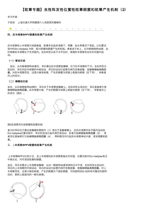
【眩晕专题】良性阵发性位置性眩晕眼震和眩晕产⽣机制(2)本⽂作者:于栋祯上海交通⼤学附属第六⼈民医院⽿⿐喉科四、后半规管BPPV眼震和眩晕产⽣机制后半规管和上半规管与地⾯垂直,眩晕多在起床或躺下、弯腰、抬头等情况下诱发。
以位置试验中的Dix-Hallpike 为例,探讨眩晕和眼震产⽣的机制。
患者坐于床上,头向患侧旋转45度,此时患侧后半规管处于⽮状⾯内。
在后仰⾄头低于⽔平位时,患侧后半规管完全在⽮状⾯内运动。
(⼀)管结⽯症坐位,头向患侧旋转45度时,⽿⽯靠近后半规管壶腹嵴,位于后半规管的下⽅。
在后仰⾄头低位时,⽿⽯向后半规管的中部运动,⽿⽯的运动引起管内淋巴流离壶腹,壶腹嵴帽偏离椭圆囊。
对后半规管⽽⾔,这是兴奋性刺激,产⽣的眼震为双眼上极旋向患侧(在下⽿),伴垂直向上的成分。
(⼆)嵴帽结⽯症坐位,头向患侧旋转45度时,⽿⽯位于半规管壶腹嵴上。
在后仰⾄头低位时,⽿⽯直接牵引壶腹嵴帽偏离椭圆囊。
后半规管兴奋,产⽣的眼震为双眼上极旋向患侧(在下⽿),伴垂直向上的成分(图5)。
图5后规管⽿⽯症眼震和眩晕机制直⽴时⽿⽯位于靠近壶腹嵴的管腔内(1)或位于壶腹嵴帽上,在后半规管所在平⾯内运动如Dix-Hallpike位置试验中,⽿⽯的运动引起内淋巴液运动,后者引起嵴帽偏离椭圆囊(2),或者⽿⽯直接牵引引起嵴帽偏离椭圆囊(4),两种情况均引起后半规管神经兴奋,诱发眼震和眩晕。
半规管BPPV眼震和眩晕产⽣机制五、上半规管上半规管BPPV⽐较少见,且上半规管较后半规管更接近⽮状⾯,位置试验中Dix-Hallpike和正中悬头位,均可诱发眩晕和眼震。
坐位,⽿⽯多靠近上半规管壶腹嵴,头向⼀侧旋转45度或保持正中不变,在后仰⾄头低位时,⽿⽯向上半规管的中部运动,⽿⽯的运动引起管内淋巴流离壶腹,壶腹嵴帽偏离椭圆囊。
对上半规管⽽⾔,这是兴奋性刺激,产⽣的眼震为下跳性眼震,可伴旋转成份.如伴有可确定的旋转成份,眼球上极旋向的⼀侧为患侧。
良性阵发性位置性眩晕诊断:10张图帮你理清
良性阵发性位置性眩晕诊断:10张图帮你理清良性阵发性位置性眩晕(BPPV)是常见的周围性眩晕性疾病之一,作为眩晕的重要鉴别诊断而为神经内科医师所熟知。
BPPV 也称为耳石症,根据耳石部位分为前半规管 BPPV、后半规管 BPPV 和水平半规管BPPV。
其重要的诊断性手段为Dix-Hallpike 试验和仰卧位滚转试验。
近期 International Journal of Otolaryngology 杂志发表了一篇由希腊学者Balatsouras 等撰写的综述,用图表的方式生动地阐述了三种类型BPPV 发病机制及对应的诊断性试验结果,一起来学习下吧。
后半规管BPPV图1 进行右侧Dix-Hallpike 试验时对后半规管的刺激(c),(a,b)分别显示了左耳和右耳半规管当耳石位于同侧(右侧)后半规管时(b),Dix-Hallpike 试验导致耳石沿管腔移动,诱导出现BPPV。
当耳石位于对侧(左侧)后半规管(a)时,Dix-Hallpike 试验并不会导致耳石的运动,因为头部运动与受累半规管平面垂直,此时Dix-Hallpike 试验为阴性。
前半规管BPPV图2 进行右侧Dix-Hallpike 试验时对左侧前半规管的刺激(c),(a,b)分别显示了左耳和右耳半规管当耳石位于对侧(左侧)前半规管时(a),由于内淋巴液的流动,受累侧的前半规管受到刺激,诱发出现BPPV。
图3 进行右侧Dix-Hallpike 试验时对右侧前半规管的刺激(c),(a,b)分别显示了左耳和右耳半规管当耳石位于同侧(右侧)前半规管时(b),头部旋转的方向与受累半规管平面垂直。
但由于其壶腹嵴部分几乎是垂直的,所以也会导致耳石移位,表现为Dix-Hallpike 试验阳性,但对壶腹嵴顶的压力以及患者表现出的症状不是那么显著。
水平半规管 BPPV图 4 当左耳受累时,水平半规管 BPPV 管结石症的发生机制(受累半规管用黑色标示,左图)(a)患者位于仰卧位,耳石位于左侧半规管的后部;(b)当旋转头部至受累侧时,耳石碎片向壶腹部移动,诱发面向壶腹嵴的流动,产生强烈的向地的水平性眼震;(c)当旋转头部至健康侧时,耳石碎片向相反方向移动,诱发背向壶腹嵴的流动,产生背地性眼震,但强度较弱。
良性阵发性位置性眩晕(BPPV)的诊断与治疗PPT课件

水平半规管BPPV眼震特点
右水平半规管
注意事项
由于耳石消散和中枢适应的原因,会出现 疲劳性
所以要避免重复诱发 对于疗效,要考虑疲劳性。不要误把疲劳
性未引出眼震认为治疗好转
复位原则
腹返耳 嵴回石 直椭只 接圆能 返囊从 回,半 。不规
能管 从总 壶脚
复位手法:后半规管BPPV
Eply复位:针对管石(相对柔和) Semont复位:针对嵴顶结石(复位有
处理:对因治疗+康复训练
并发精神源性头晕:
解释+行为认知+抗焦虑
复位后注意
复位后走路不稳感或轻度头晕者
复位后24小时采用高枕卧位(头抬高 30°)或者健侧卧位睡眠
避免头部剧烈活动 保持充足睡眠 避免情绪波动
复位无效
分析原因:手法、角度、速度、复位 中眼震观察、复位后的体位
去除诱因 无效时,习服疗法 对于诊断明确、复位及康复训练均无
嵴顶结石:目的让耳石从嵴顶变到半规管,注意 甩头过程要快。
Barbecue复位
Gufoni复位:管石
Gufoni复位:嵴顶结石
复位手法:上半规管BPPV
反向Eply复位:疗效不确切 Semont复位: 海军总院李进让:李氏复位法 Yacovino复位:不用判断左右侧
Yacovino复位
临床分类
特发性BPPV:原因不明(50%-97%) 继发性BPPV:继发于耳科或全身系统性疾病
(25%) 头外伤(7%-17%)-双侧 病毒性迷路炎(15%) 梅尼埃病(15%) 偏头痛(﹤5%) 内耳手术(﹤1%)
骨迷路
椭圆囊 (es) 球囊
内淋巴囊
前庭迷路解剖
壶腹
半规管空间位置
良性阵发性位置性眩晕诊断和治疗

组织学上,椭圆囊和球囊的囊斑上面存在着耳石膜
的结构,由一层粘多糖类的物质和碳酸钙结晶颗粒
组成,覆盖在毛细胞的纤毛顶部
囊斑(macules)
扫描电镜
扫 描 电 镜 ( 耳 石 )
总结(summary)
人体的平衡需要视觉、前庭和本体感觉加以维系 外周的前庭器官包括三管两囊,即三个半规管、一个
球囊和一个椭圆囊
半规管的感受部位在壶腹嵴,球囊和椭圆囊则是囊斑
耳石位于囊斑,脱落后游离在内耳液中,随着体位变
化能够刺激前庭感受器,形成耳石症
耳石症 = 囊斑的耳石脱落 + 体位变化 + 内淋巴液的流动 - 耳石撞击壶腹嵴 或囊斑本身 - 前庭器官兴奋 - 双侧前
庭系统不对称 - 眩晕
BPPV临床的界定
流程图
Dix-Hallpike 体位诱发试验
受试耳侧旋转45 度
仰卧垂头30度
眩晕感
眼 震
复位时
恢复坐位
再次眩晕 眼震重现
朝向对侧方向
Dix-Hallpike检查法
前庭中枢性眩晕与周围性眩晕眼震的鉴别
眼震 潜伏期 持续时间 疲劳性 位置 方向 眩晕
周围性
2~10S
中枢性
无 30S以内 持续1min以上 重复试验,眼震消失 眼震不消失 仅见于一种头位 见于多种头位 固定向一侧 方向随头位改变 常伴 无或轻度
壶腹嵴
壶腹嵴位于半规管的
壶腹端,主要有毛细 胞和支持细胞构成,
毛细胞的顶部有动和
静两种纤毛,而纤毛 的上覆盖着一层如同
僧帽一样的胶质,即
嵴帽
壶腹嵴与半规管长轴
垂直,形成半规管和
良性阵发性位置性眩晕患者裸眼及视频眼震图下眼震特征及定位诊断分析
良性阵发性位置性眩晕患者裸眼及视频眼震图下眼震特征及定位诊断分析张波;孙敬武【摘要】Objective The VNG has played an important role in the diagnosis and treatment of the benign positional paroxysmal vertigo (BPPV). This article intends to compare the differences of detection rate of nystagmus between naked eyes and VNG. Methods 108 BPPV cases from July 2010 to June 2011 were reviewed and the findings of Dix-Hallpikc and Roll test with Frcnzcl were compared to those obtained with naked eyes simply. Results 75(69.44%) cases of patients with PSC-BPPV and 18(16. 67%) cases with ASC-BPPV were clinically diagnosed through observation and VNG. By comparison,observation with naked eyes and VNG yielded 66. 67% and 93. 33% for PSC -BPPV and 72.22% and 100% for ASC -BPPV, respectively. There were statistical differences between the two methods (P<0. 05). Conclusion VNG can objectively record the nystagmus of BPPV patients, thus allowing more accurate diagnosis with the ability to keep the results for further analysis.%目的探讨裸眼及视频眼震图(videonystagmograph,VNG)下观察良性阵发性位置性眩晕(benign positional paroxysmal vertigo,BPPV)患者眼震及诊断结果的差异.方法 108例BPPV患者分别进行Dix-Hallpike和滚转试验(Roll),应用红外视频眼震电图仪观察眼震特点,进行定位诊断,并与裸眼下观察其眼震特征及诊断结果进行比较.结果 108例患者中共诊断后半规管BPPV(PSC-BPPV)75例(69.44%,75/108),裸眼及VNG辅助下典型眼震检出率分别为66.67%(50/75)和93.33%(70/75);前半规管BPPV(ASC-BPPV)18例(16.67%,18/108),裸眼及VNG下典型眼震检出率分别为72.22%(13/18)和100%(18/18),两者差异均有统计学意义(P<0.05);水平半规管BPPV(HSC-BPPV)9例(8.33%),裸眼及VNG辅助下分别正确诊断7例和9例;混合型BPPV6例(5.56%),裸眼及VNG辅助下分别正确诊断3例和6例.结论 VNG能够客观地记录BPPV患者的眼震情况,有助于提高眼震的检出率及诊断的准确性.【期刊名称】《听力学及言语疾病杂志》【年(卷),期】2012(020)003【总页数】3页(P235-237)【关键词】良性阵发性位置性眩晕;视频眼震图;半规管;眼震【作者】张波;孙敬武【作者单位】安徽省立医院耳鼻咽喉头颈外科,合肥,230001;安徽省立医院耳鼻咽喉头颈外科,合肥,230001【正文语种】中文【中图分类】R764.3良性阵发性位置性眩晕(benign positional paroxysmal vertigo,BPPV)是头部运动到某一特定位置时诱发的短暂眩晕,是一种具有自限性的周围性前庭疾病[1]。
【眩晕专题】良性阵发性位置性眩晕眼震和眩晕产生机制(1)

【眩晕专题】良性阵发性位置性眩晕眼震和眩晕产生机制(1)本文作者:于栋祯上海交通大学附属第六人民医院耳鼻喉科眼球震颤,简称眼震,是眼球的一种不随意的节律性运动,前庭性眼震由交替出现的慢相和快相运动组成。
慢相为眼球向某一方向的缓慢运动,由前庭刺激所引起;快相则为眼球的快速回位运动,为中枢矫正性运动。
眼球运动的慢相朝向前庭兴奋性较低的一侧,快相朝向前庭兴奋性较高的一侧。
便于观察,通常将快相所指方向作为眼震方向。
一、半规管刺激诱发眼球的运动和眼震二、BPPV眼震方向的定义眼震以快相方向而定,可分水平性、垂直性和旋转性等。
在BPPV的诊断中,眼震的方向有特别的界定。
在滚转试验中,病人左转头和右转头或左侧卧位和右侧卧位时,诱发的水平性眼震与地面垂直,快相可以向地,亦可背地,水平性眼震的快相如向地,称之为向地性眼震;如背离地,则称之为背地性眼震,或向天性眼震。
在Dix-Hallpike试验中,以眼球的上极为标志描述旋转方向,记录眼球的上极转向左侧或右侧,或记录为顺时针或逆时针旋转,同时描述垂直方向,上跳性或下跳性。
三、水平半规管BPPV眼震和眩晕产生机制水平半规管BPPV患者多在平躺左侧和/或右侧翻身时,诱发眩晕发作,可伴有恶心、呕吐。
平卧时,水平半规管所处的平面大致与地面垂直,在重力的作用下耳石最容易在半规管内运动,引起内淋巴液的流动,刺激壶腹嵴,引起眩晕发作和眼震。
以位置试验中的翻滚试验为例,讲述眩晕和眼震产生的机制。
(一)管结石症(后臂型)1. 平卧时,双侧水平半规管与地面垂直,壶腹位于上内侧,非壶腹位于下内侧,耳石多数位于非壶腹端即后臂。
2. 头向患侧旋转时,耳石在重力作用下,克服各种阻力,向壶腹端运动,引起内淋巴液流向壶腹,壶腹嵴帽向椭圆囊偏斜,患侧兴奋,产生快相向患侧的水平性眼震,患侧眼在下,即向地性眼震,客观体征为眼震,主观感觉为眩晕(图2)。
3. 头向健侧旋转时,耳石在重力作用下,向非壶腹端运动,引起内淋巴液流离壶腹,壶腹嵴帽向偏离椭圆囊,患侧抑制,产生快相向健侧的水平性眼震,健侧眼在下,即向地性眼震,伴眩晕(图3)。
- 1、下载文档前请自行甄别文档内容的完整性,平台不提供额外的编辑、内容补充、找答案等附加服务。
- 2、"仅部分预览"的文档,不可在线预览部分如存在完整性等问题,可反馈申请退款(可完整预览的文档不适用该条件!)。
- 3、如文档侵犯您的权益,请联系客服反馈,我们会尽快为您处理(人工客服工作时间:9:00-18:30)。
良性阵发性位置性眩晕的眼震图定位诊断王娜1太生2林鹏2宋伟2董红21医科大学一中心临床学院2市第一中心医院耳鼻咽喉头颈外科;市耳鼻喉科研究所(300192)【摘要】目的:探讨眼震图(Videonystagmograph,VNG)对良性阵发性位置性眩晕(BPPV)的定位诊断价值。
方法:回顾126例BPPV患者的眼震图资料,分析总结各型BPPV 在Dix- Hallpike 和滚转试验中眼震图上的眼震特点。
结果:126例BPPV患者中,后半规管(PSC)BPPV98例(77.8%)、水平半规管(HSC)BPPV17例(13.5%)、前半规管(ASC)BPPV 仅5例(3.9%)、混合型BPPV6例(4.8%),28例PSC-BPPV记录到反转相眼震。
眼震图上显示PSC和ASC管石症Dix- Hallpike悬头位垂直相眼震分别向上、向下,水平相眼震均向对侧,回到坐位时眼震反向。
HSC-BPPV滚转试验向两侧转头均可诱发出眼震,眼震与转头方向相同时,可判断为水平半规管管石症,以能够诱发较强眼震的转头侧为患侧;眼震与转头方向相反时,则为水平半规管嵴顶结石症,以能够诱发较弱眼震的转头侧为患侧。
结论:眼震图能够客观的记录BPPV患者眼震情况,准确判断耳石所在的半规管,并且保存了眼震数据资料,可以进一步指导临床实践,值得广泛推广。
【关键词】视频眼震图;良性阵发性位置性眩晕;半规管;耳石通讯作者:太生(cts501sina.)Positioning diagnosis of benign positional paroxysmal vertigo by VNG Wang Na1, Lin Peng2, Chen Taisheng2 , Song Wei2, Dong Hong2(1The first center clinic college of Tianjin Medical University, Tianjin, 300192,China; 2Otolaryngology-Head & Neck Surgery, Tianjin First Center Hospital, Tianjin, 300192, China)【Abstract】Objective To analyze the value of positioning diagnosis of VNG (Videonystagmograph) in patients with benign paroxysmal positional vertigo(BPPV). Methods 126 patients with BPPV were enrolled in this retrospective study. Their positional nystagmus recorded by VNG in Dix-Hallpike and roll tests were analyzed to summarize the characteristics of nystagmus on nystagmograph of various BPPV. Results Of 126 patients with BPPV diagnosed in our center, the posterior semicircular canals(PSC) were involved in 98 patients(77.8%),whereas the horizontal semicircular canal(HSC) and anterior semicircular canal(ASC) were involved in 17(13.5%) and 5(3.9%), respectively.6 patients(4.8%) confirmed combined-BPPV had HSC-BPPV and ipsilateral PSC-BPPV. 28 patients with PSC-BPPV had reversal phase on nystagmograph. The nystagmus of patients with P/ASC-canalithiasis showed upward /downwardon the vertical phase of nystagmograph and orientated the opposite side on horizontal phase in the head hangging position, and the nystagmus reversed when returned to sit. Nystagmus on horizontal phase can be provoked when the head turned to both sides of the roll tests in patients with HSC-BPPV. If the nystagmus and the head-turning shared the same direction, then HSC-canalithiasis was confirmed, and the direction of the head-turning which provoked the stronger nystagmus indicates the lesion side. If the nystagmus and the head-turning had the opposite direction, then HSC-cupulolithiasis was confirmed, and the direction of the head-turning which provoked the weaker nystagmus indicates the lesion side. Conclusion Positional nystagmus can be recorded objectively using VNG. Positioning the semicircular canal involved will be easier and more accurate according to it. The recording conserved also be helpful for clinical diagnosis and repositioning of BPPV.【Key words】Videonystagmograph (VNG);benign positional paroxysmal vertigo(BPPV);semicircular canal;otolithCorresponding author: Chen Taisheng (cts501sina.)良性阵发性位置性眩晕(benign positional paroxysmal vertigo,BPPV)是头部运动到某一特定位置时诱发的短暂眩晕,是一种具有自限性的周围性前庭疾病[1]。
该病发病率较高,约占所有周围性眩晕的17%~20 %[2]。
BPPV耳石复位治疗技术已经成熟,成功复位治疗的基础是对耳石累及半规管的准确定位。
目前BPPV的定位诊断和复位都基于医者目测患者的眼震,但旋转性眼震的方向常常不易辨别,加之一些细小眼震容易被肉眼所忽略,影响了BPPV 的定位诊断和后续复位治疗。
近年来,视频眼震电图(Videonystagmograph,VNG)的普及应用使眼震方向的判断更加客观、方便。
我们将其与BPPV诊治相结合,更直观、客观地记录和分析患者在各个体位的诱发眼震,利用眼震图对BPPV进行准确定位诊断和疗效评估,并且保存了眼震数据资料,也有利于进一步指导临床实践和教学和科研工作。
1.材料与方法1.1 临床资料2006年1月-2008 年4月在市第一中心医院耳鼻咽喉头颈外科眩晕诊疗中心诊断为BPPV的患者126 例,其中女性85例(67.5%),男性41例(32.5%),平均年龄为54岁(17~78 岁)。
患者从发病到就诊的时间为1 天~20 年,平均59 周。
1.2方法1.2.1常规检查所有患者均详细询问并记录病史,包括眩晕发作情况、既往史(尤其是相关的耳科疾病史、耳毒性药物应用史、外伤史)、家族史,并进行常规耳科检查。
应用VNG(法国Synapsys)进行常规眼震图检查及变位试验、冷热试验等,VNG图上红色为水平相,蓝色为垂直相。
对可疑中枢病变患者行相关检查除外中枢性眩晕。
1.2.2变位试验⑴ Dix-Hallpike试验:患者坐于检查床上,头戴视频眼罩,检查者于病人后方,双手扶其头部向患侧转45°,并协助其迅速变为仰卧位,使头向后悬于床沿外,与水平面呈30°,头位始终保持该侧转45°不变,询问患者有无眩晕,并通过眼动监视窗口及眼动描记曲线图观察有无眼震及其方向,至少观察1分钟或者待其眼震消失为止。
然后迅速使患者坐起,继续观察眼震情况。
再以同法检查对侧。
⑵滚转试验(roll test):患者戴视频眼罩,仰卧头屈曲30°,头快速向一侧转动90°并保持此头位2分钟,然后头回转正中位,保持此头位2分钟,再快速向对侧转90°并保持此头位2分钟,在这三个头位均询问患者有无眩晕并记录、观察眼震出现情况。
1.2.3诊断依据依据中华医学会耳鼻咽喉科学分会制定的“良性阵发性位置性眩晕的诊断依据与疗效评估” (2006年,) [1],结合患者病史及Dix-Hallpike和滚转试验两项体位检查所诱发的眼震情况做出诊断,并排除中枢性病变。
BPPV分为四种类型:⑴后半规管BPPV(PSC-BPPV);⑵前半规管BPPV(ASC-BPPV);⑶水平半规管BPPV(HSC-BPPV);⑷混合型BPPV。
2.结果2.1本组资料中PSC-BPPV 98例(78%),HSC-BPPV 17例(13.5%),其中水平半规管管石症(HSC-can)13例,水平半规管嵴顶结石症(HSC-cup) 4例,ASC-BPPV仅5例,另有6例为混合型BPPV,均为HSC合并同侧PSC-BPPV(表1)。
表1. 126例BPPV临床分型临床类型例数百分比(%)PSC-BPPV 98 77.8HSC-BPPV 17 13.5ASC-BPPV 5 3.9混合BPPV 6 4.82.2 28例PSC-BPPV在悬头位观察到反转相眼震,该眼震与回到坐位时诱发的眼震同方向,但其时程和强度均低于Dix-Hallpike悬头位和坐位诱发的眼震。