VIG病毒诱导基因沉默技术PPT课件
vigs原理

vigs原理VIGS原理。
VIGS(病毒诱导基因沉默)是一种基因沉默技术,通过利用病毒来诱导植物基因的沉默,从而研究基因功能和调控机制。
VIGS技术是一种快速、高效的基因沉默方法,被广泛应用于植物分子生物学研究中。
本文将介绍VIGS的原理及其在植物科学研究中的应用。
VIGS的原理主要是利用病毒载体来携带目标基因片段,并通过病毒的侵染和复制过程,将目标基因片段转录成siRNA(小干扰RNA),siRNA能够诱导RNA干扰(RNAi)途径,从而导致目标基因的沉默。
VIGS技术的关键在于选择合适的病毒载体和目标基因片段,以及优化转染条件和病毒复制过程,从而实现对目标基因的特异性沉默。
VIGS技术在植物科学研究中有着广泛的应用。
首先,VIGS可以用来研究基因功能。
通过沉默目标基因,可以观察到植物表型的变化,从而推断目标基因在生物学过程中的作用。
其次,VIGS还可以用来研究基因调控网络。
通过沉默一个基因,可以观察到其他相关基因的表达变化,从而揭示基因之间的相互作用关系。
此外,VIGS还可以用来筛选抗病基因。
通过沉默潜在的抗病基因,可以评估其对病原体的抗性作用,为植物抗病育种提供理论基础。
总之,VIGS技术是一种重要的基因沉默方法,具有快速、高效、特异性的优点,被广泛应用于植物科学研究中。
随着分子生物学技术的不断发展,VIGS技术在植物基因功能和调控研究中将发挥越来越重要的作用。
希望本文的介绍能够帮助读者更好地理解VIGS的原理及其在植物科学研究中的应用,为相关领域的研究工作提供参考和借鉴。
vigs基因沉默原理

1997年,VIGS这一术语最早被Van Kammen用于描述植物受病毒侵染后的症状恢复现象。
1999年,Baulcombe认识到,由于VIGS允许通过讲解特定基因的转录本来有针对性地下调该基因,VIGS将被作为基因功能研究的潜力巨大。
2004年,Burch-Smith通过使用重组病毒来沉默植物内源基因,此后,VIGS作为一种反向遗传学技术被研究者广泛使用。
病毒诱导的基因沉默(virus induced gene silencing,VIGS)是指携带目的基因片段的病毒侵染植物后,随着病毒的复制和转录而特异性的诱导序列同源基因mRNA降解或被甲基化等修饰,从而引起植物内源基因沉默、引起表型或生理指标变化,进而根据表型变异研究目标基因的功能。
VIGS是根据植物对RNA病毒防御机制发展起来的一种用以表征植物基因功能的基因转录技术,其内在的分子基础可能是转录后基因沉默(post-transcript gene silence)。
与传统的基因功能分析方法相比,VIGS能够在侵染植物当代对目标基因进行沉默和功能分析;无需开发稳定的转化子,并且具有沉默单个或多个基因家族成员的潜力。
因此,VIGS一经建立,即被视为研究植物基因功能的强有力工具,得到了深入的研究和广泛应用,已用于烟草、番茄、小麦、水稻等植物的抗病反应、生长发育以及代谢调控的功能基因研究。
沉默机制基因沉默在生物中普遍存在,表现在抵御病毒、转座子等外来核酸的入侵,识别并抑制外源基因表达,维持生物基因组稳定性等。
VIGS 作为基因沉默的特殊形式,是植物抗病毒侵染的一种自然机制。
当病毒或携带cDNA 的病毒载体侵染植物后,在复制与表达过程中通常会形成双链RNA (Double stranded RNA,dsRNA)形式的中间体。
dsRNA 作为基因沉默关键激发子,首先在细胞中被类似RNase Ⅲ家族特异性核酸内切酶Dicer 类似物(如DCL4)切割成21 ~ 24 nt 的小分子干扰RNA(Small interfering RNA,siRNA)。
第一实验:以烟草脆裂病毒为载体通过基因沉默分析植物基因功能(VIGS
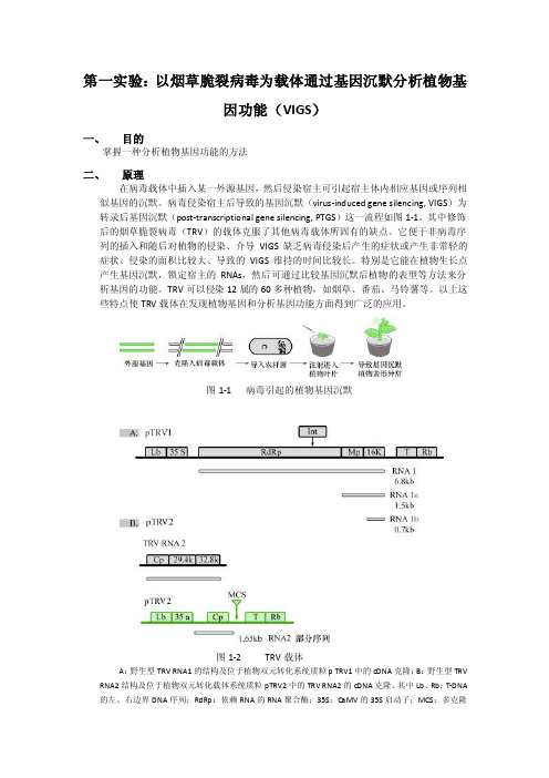
第一实验:以烟草脆裂病毒为载体通过基因沉默分析植物基因功能(VIGS)一、目的掌握一种分析植物基因功能的方法二、原理在病毒载体中插入某一外源基因,然后侵染宿主可引起宿主体内相应基因或序列相似基因的沉默。
病毒侵染宿主后导致的基因沉默(virus-induced gene silencing, VIGS)为转录后基因沉默(post-transcriptional gene silencing, PTGS)这一流程如图1-1。
其中修饰后的烟草脆裂病毒(TRV)的载体克服了其他病毒载体所固有的缺点。
它便于非病毒序列的插入和随后对植物的侵染、介导VIGS缺乏病毒侵染后产生的症状或产生非常轻的症状、侵染的面积比较大、导致的VIGS维持的时间比较长。
特别是它能在植物生长点产生基因沉默,锁定宿主的RNAs,然后可通过比较基因沉默后植物的表型等方法来分析基因的功能。
TRV可以侵染12属的60多种植物,如烟草、番茄、马铃薯等。
以上这些特点使TRV载体在发现植物基因和分析基因功能方面得到广泛的应用。
图1-1 病毒引起的植物基因沉默图1-2 TRV载体A:野生型TRV RNA1的结构及位于植物双元转化系统质粒p TRV1中的cDNA克隆;B:野生型TRV RNA2结构及位于植物双元转化载体系统质粒pTRV2中的TRV RNA2的cDNA克隆。
其中Lb、Rb:T-DNA 的左、右边界DNA序列;RdRp:依赖RNA的RNA聚合酶;35S:CaMV的35S启动子;MCS:多克隆位点;T:转录终止点;16K、29.4K、32.8K分别为病毒编码的蛋白序列;CP:病毒编码的外壳蛋白序列;Int:插入病毒基因的内含子;MPbd的运动蛋白TRV是正链RNA病毒,包括两个基因组分,其中RNA1编码病毒在植物中的复制和运动所需的蛋白,RNA2编码病毒组装和介体传播所需的蛋白。
在构建载体时把RNA1和RNA2的cDNA分别克隆在植物转化所用的农杆菌的双元载体系统中的两个质粒上,具体结构如图1-2所示。
vigs基因沉默原理和rnai沉默

vigs基因沉默原理和rnai沉默VIGS基因沉默原理和RNAi沉默是两种常用的分子生物学技术,被广泛应用于研究植物基因功能。
它们有些相似之处,同时也存在一些差异。
本文将对这两种技术的原理、应用和优缺点进行详细探讨。
1. VIGS基因沉默原理VIGS(Virus-induced gene silencing)是通过病毒侵染植物细胞,诱导宿主植物基因的沉默。
该技术利用了病毒的复制和传播机制,将目标基因序列整合到病毒基因组中,并且在感染的植物细胞中产生可复制的RNA介导的沉默效应。
具体步骤包括:(1)构建VIGS载体:将目标基因的部分序列插入病毒载体,形成VIGS载体。
(2)VIGS载体侵染植物细胞:将构建好的VIGS载体导入病毒感受性植物中,让病毒基因组中的目标基因RNA与宿主植物基因的mRNA互补杂交,导致宿主基因沉默。
(3)基因沉默效应:病毒RNA会在植物细胞中被RNA依赖性RNA 聚合酶复制成多个复制体,随后通过系统性运输到植物全身,触发整个植物的基因沉默效应。
2. RNAi基因沉默原理RNA干扰(RNA interference)是一种保守的基因沉默机制,通过切割目标mRNA分子而将其降解,从而实现对基因表达的调控。
RNAi主要通过siRNA和miRNA两条途径实现。
具体步骤包括:(1)siRNA的产生:长双链RNA被核酸酶Dicer切割成短双链siRNA。
(2)siRNA的介导:siRNA将RISC(RNA诱导的沉默复合体)导入到靶基因mRNA上,使其降解。
(3)miRNA的产生:由miRNA基因转录出的pri-miRNA在细胞核中被核酸酶Drosha切割为pre-miRNA,经过转运到细胞质后被Dicer 进一步切割成短miRNA。
(4)miRNA的介导:miRNA与RISC结合后能够通过完全或不完全互补靶序列的mRNA上结合,从而通过诱导转录后调控蛋白质的翻译或降解。
3. VIGS和RNAi的异同点(1)机制差异:VIGS是依赖于病毒的复制和传播来诱导植物基因沉默,而RNAi是通过siRNA和miRNA介导的沉默机制来调控基因表达。
vigs原理
vigs原理VIGS原理。
VIGS(Virus-Induced Gene Silencing)是一种通过病毒诱导基因沉默的技术,被广泛应用于植物基因功能研究中。
VIGS技术利用植物病毒的RNA干扰机制,通过病毒载体将目标基因的片段导入植物细胞,从而抑制目标基因的表达。
本文将详细介绍VIGS原理及其在植物基因研究中的应用。
VIGS原理。
VIGS技术的原理是利用病毒诱导植物基因沉默。
病毒载体中携带了目标基因的部分序列,一旦进入植物细胞,这些序列将被转录成双链RNA。
这些双链RNA 能够被植物细胞内的Dicer酶切割成小的siRNA(small interfering RNA),siRNA 与靶标基因的mRNA互补结合,导致靶标mRNA降解,从而抑制了目标基因的表达。
这一过程与RNA干扰(RNA interference, RNAi)的机制相似。
VIGS技术的优势。
相比于传统的转基因和突变体筛选技术,VIGS技术具有以下优势:1. 快速,VIGS技术可以在短时间内沉默目标基因,研究者可以快速获得目标基因沉默后的表型变化。
2. 高效,VIGS技术可以在整个植物体内实现目标基因的沉默,而不仅限于特定组织或细胞。
3. 灵活,VIGS技术可以用于多种不同类型的植物,而不需要针对每种植物进行特定的转基因操作。
VIGS技术的应用。
VIGS技术在植物基因功能研究中有着广泛的应用,包括:1. 基因功能研究,VIGS技术可以帮助研究者快速验证目标基因的功能,特别是对于那些没有已知突变体的基因。
2. 代谢途径研究,通过沉默代谢途径中的关键基因,可以帮助研究者了解这些基因在植物代谢中的作用。
3. 抗病性研究,利用VIGS技术可以验证植物中与抗病相关的基因,有助于揭示植物抗病机制。
总结。
VIGS技术作为一种快速、高效、灵活的基因沉默技术,在植物基因功能研究中发挥着重要作用。
通过利用VIGS技术,研究者可以快速验证目标基因的功能,探究植物代谢途径和抗病机制,为植物育种和生产提供重要的科学依据。
vigs基因沉默原理和rnai沉默

vigs基因沉默原理和rnai沉默VIGS(Virus-induced gene silencing)基因沉默和RNAi(RNA interference)沉默都是生物学研究中常用的方法,用于研究基因的功能和调控机制。
它们共同的原理是通过引入相应的外源RNA分子来诱导靶基因(target gene)的沉默,从而研究基因在细胞与整个生物体中的功能。
虽然二者的原理有所不同,但它们在生物学研究中都发挥着重要作用。
VIGS(Virus-induced gene silencing)基因沉默是通过利用病毒来传递RNA分子,并引发对应的基因沉默的一种方法。
这种方法最早是在植物领域中发现的,后来也在许多其他生物中得到应用。
VIGS 的基本原理是通过将目标基因片段插入表达病毒载体中,然后通过病毒感染植物或动物细胞,利用病毒的复制过程来产生RNA分子,从而导致目标基因的沉默。
这种沉默通常是由于RNA分子通过切割或RNA-DNA相互作用等机制,导致目标基因的mRNA降解,或抑制其翻译成蛋白质,从而实现基因沉默的目的。
相比之下,RNAi是一种借助内源的RNA分子来诱导基因沉默的方法。
它是由一种特殊的RNA分子,即小干扰RNA(siRNA)和微小内源RNA(miRNA)介导的细胞内调控机制。
这些RNA分子通常由一类酶称为Dicer切割出来,该酶能够将双链RNA分子切割成约20-25个碱基对(bp)长度的小分子RNA。
这些小RNA分子与靶基因的mRNA结合,并通过RNA诱导的合成酶(RISC)复合物的作用,将靶基因的mRNA切割成短片段,从而抑制目标基因的翻译过程,实现基因沉默。
不同于VIGS基因沉默方式,RNAi机制更加普遍,并广泛存在于真核生物细胞中。
VIGS和RNAi基因沉默技术在生物学研究中的应用非常广泛。
通过利用这些技术,研究人员可以选择特定的靶基因,并通过适当设计的RNA分子来诱导其沉默。
这些研究通常通过研究目标基因沉默后的表型变化来揭示基因的功能和调控机制。
vigs原理

vigs原理VIGS原理。
VIGS(病毒诱导基因沉默)是一种利用病毒来诱导植物基因沉默的技术,它可以通过病毒载体将特定基因的RNA序列引入植物细胞内,从而抑制目标基因的表达。
VIGS技术因其简单、高效、快速的特点,被广泛应用于植物基因功能研究、抗病性育种等领域。
本文将对VIGS原理进行详细介绍。
VIGS的实现依赖于RNA干扰(RNAi)机制。
RNAi是一种由双链RNA介导的基因沉默过程,通过RNAi,细胞内的特定mRNA被降解,从而导致目标基因的沉默。
VIGS技术利用这一机制,通过病毒载体将特定基因的RNA序列引入植物细胞内,从而诱导目标基因的沉默。
VIGS技术的实现过程可以分为以下几个步骤,首先,选择合适的病毒载体。
常用的病毒载体包括烟草花叶病毒(Tobacco Rattle Virus,TRV)和番茄花叶病毒(Tomato Bushy Stunt Virus,TBSV)等。
其次,构建含有目标基因RNA序列的病毒载体。
这一步通常通过分子克隆技术来实现,将目标基因RNA序列插入病毒载体的适当位置。
然后,将构建好的病毒载体导入植物细胞内。
最后,观察目标基因的表达情况,验证VIGS效果。
VIGS技术的优势在于其简单、高效、快速。
相比于传统的基因敲除技术,VIGS不需要构建转基因植物,也不需要等待多代植物的繁殖,能够在短时间内实现目标基因的沉默。
此外,VIGS技术还可以用于多种植物物种,具有较广泛的适用性。
然而,VIGS技术也存在一些局限性。
首先,VIGS诱导的基因沉默是暂时的,通常只能持续数周至数月。
其次,不同植物物种对VIGS的敏感性不同,有些植物物种对VIGS的效果较差。
此外,由于VIGS是利用病毒来传递RNA序列,存在一定的生物安全风险。
总的来说,VIGS技术作为一种快速、高效的植物基因沉默技术,为植物基因功能研究、抗病性育种等领域提供了重要工具。
随着对VIGS原理的深入研究和技术的不断改进,相信VIGS技术在未来会有更广泛的应用前景。
VIGS 病毒诱导的基因沉默
14Analysis of Gene Function in Rice ThroughV irus-Induced Gene SilencingXin Shun Ding, C. Srinivasa Rao, and Richard S. NelsonSummaryVirus-induced gene silencing (VIGS) is a powerful RNA-silencing based technology adapted for the study of host-gene function. VIGS functions through the expression of a host gene from a virus vector. Both the virus-encoded host sequence and the homologous host target messenger RNA are destroyed or made inactive through a host surveillance system. Here, we describe procedures for the use of a new virus vector for VIGS in mono-cotyledonous hosts and, in particular, in rice (Oryza sativa), a species for which no VIGS vector was previously available.Key Words: Virus-induced gene silencing; rice; monocotyledons; Brome mosaic virus;functional genomics; gene knockout.1. IntroductionIn the past few years, virus-induced gene silencing (VIGS) has been used to determine gene function in plants (1–4). The mechanism of VIGS is not fully understood; however, it is known to target RNA molecules in a sequence-specific manner. Although VIGS originally was a term used to describe the recovery of the host from virus infection, it now is used most often to describe the downregulation of host gene expression via a virus vector (2). Double-stranded RNA (dsRNA), produced by fold back of the single-stranded viral RNA or during virus replication through the activity of a viral RNA-dependent RNA polymerase, triggers silencing. The dsRNA is then cleaved into small interfering RNAs (siRNA; 21 to 25 nucleotides in length) by an RNase III-type dicer-like endonuclease. The resulting siRNAs are believed to provide a single strand of RNA for incorporation into the RNA-induced silencing complex. The RNA-induced silencing complex then identifies and cleaves additional single-From:Methods in Molecular Biology, vol. 354: Plant–Pathogen Interactions: Methods and ProtocolsEdited by: P. C. Ronald © Humana Press Inc., Totowa, NJ145stranded target RNA sequences complementary to the “captured” siRNA sequence(2,5,6). Of interest, cleavage of additional target RNA through this pathway may not be the only method of controlling virus accumulation. It is possible that small RNAs derived from viral single-stranded RNA stem-loops, similar in structure to the microRNAs derived from host RNAs, inhibit virus accumulation through translational repression (4). With time, the mechanism of VIGS will be determined, but this lack of basic information does not prevent its practical use to study gene function. To date, multiple RNA plant virus vectors (e.g. Tobacco mosaic virus,Potato virus X, and Tobacco rattle virus) are available for VIGS in dicotyledonous plant species and one RNA virus vector,Barley stripe mosaic virus, is available for VIGS in two monocotyle-donous species (barley and wheat [7–14]). Here, we review the use of a plant virus vector newly created from a strain of Brome mosaic virus (BMV), to silence genes in various monocotyledons, including rice, through VIGS.To construct this BMV vector, we isolated a virus from an infected Tall Fescue plant (Festuca arundinacea Schreb.) and purified viral RNA from the virus capsid (15,16). Previous analysis of the capsid, through electron microscopy, and viral RNA, through Northern blot analysis, indicated that this virus was a strain of BMV, which we named the fescue strain of BMV (F-BMV[15]; Ding, X. S. and Nelson, R. S., to be submitted). Full-length reverse transcription polymerase chain reaction (RT-PCR) products of the three viral genomic RNAs were synthesized, gel purified, and ligated individually into the pGEM T-Easy vector (Promega, Madison, WI). The ligation products were transformed individually into Escherichia coli JM109-competent cells. The plasmids containing the viral complementary DNAs (cDNAs) were named pF1-11, pF2-2, and pF3-5 (for F-BMV cDNA representing BMV genomic RNAs 1, 2, and 3, respectively [15a]). Three RNAs from the Russian strain of BMV (R-BMV [16]) also were cloned (pB1-26 for RNA1, pB2-4 for RNA2, and pB3-3 for RNA3) using the same technique as described for the F-BMV. We determined that the viral sequence necessary for infecting rice did not reside in RNA 3 of BMV [15a]). This information allowed us to use the clone repre-senting RNA 3 from R-BMV, which has a more convenient restriction site for inserting host sequences than does the cDNA representing F-BMV RNA 3, in our VIGS vector. Co-inoculation of rice plants with transcripts from pF1-11, pF2-2, and pB3-3 harboring a fragment of phytoene desaturase from maize (Zea mays) caused silencing of this gene in rice (15).VIGS has several advantages over other functional genomics approaches, such as no transformation of the host plant is required and the function of indi-vidual gene family members can be studied (2). An additional major advantage is the rapidity with which a researcher can observe a silencing phenotype during VIGS. A silencing phenotype can be observed within 3 wk after inserting ahost sequence into the BMV vector and inoculating plants. For important monocotyledonous crops such as rice and maize, for which no rapid gene knockout technology exists, VIGS with the F-BMV vector was effective in silencing genes (15a).This chapter provides procedures to execute the following:1.Insert plant sequences into bacterial plasmids and transfer them to the BMV vector.2.Synthesize the BMV vector transcripts.3.Inoculate plants with transcripts.4.Analyze virus infection and gene silencing in the plants.It is important that all plants inoculated with BMV or BMV harboring a foreign insert be kept inside a greenhouse or growth chambers. Infected plants and materials used to grow these plants (e.g., soil, pots, and trays) should be autoclaved before disposal to prevent release of the virus into the environ-ment, as per regulations from the US Department of Agriculture governing the use of pathogens and transgenic viral sequences.2. Materials2.1. Insertion of Foreign Sequences Into the BM V V ector1.pB3-3 (plasmid containing cDNA of R-BMV RNA 3; Fig. 1).2.pGEM–T Easy vector (Promega; Madison, WI).3.JM109 competent cells (108 cfu; Promega)., 4.SOC medium: tryptone 2% (w/v), 8.6 m M NaCl, 2.5 m M KCl, 20 m M MgSO420 m M glucose (Sigma; St. Louis, MO).5.2% X-gal (5-bromo-4-chloro-3-indolyl-E-D-galactoside): 20 mg in 1 mL ofdimethylformamide (Promega)O 6.20% Isopropyl-E-D-thio-galactopyranoside (IPTG), 200 mg in 1 mL of H2 (Promega)7.Luria broth (LB) medium: 25 LB medium capsules per liter of distilled HO2 (Q-BIOgene; Irvine, CA).O 8.LB agar medium: 16 LB agar medium capsules per 400 mL of distilled H2 (Q-BIOgene).9.LB liquid medium containing ampicillin (Sigma): autoclave LB liquid mediumat 121q C for 20 min in 1-L autoclavable bottle. Remove the bottle from the auto-clave after the pressure is fully released. Cool the medium to approx 50q C and add ampicillin (100 P g/mL medium; see Note 1). Mix well and store the medium at 4q C before use.11.LB medium plate containing ampicillin (Sigma): autoclave LB agar medium at121q C for 20 min. Remove the bottle from the autoclave after the pressure is fully released. Cool the medium to approx 50q C and add ampicillin (100 P g/mL medium). Mix well and pour the medium into 20 Petri dishes (100 u 15 mm; see Note 1). Store plates at 4q C before use.Fig. 1. Schematic drawing of pB3-3 showing restriction sites for inserting foreign deoxyribonucleic acid sequence or linearizing the plasmid before in vitro transcription.12.LB medium plate containing ampicillin, X-gal and IPTG: Mix 20 P L of 2%O with 50 P L of 20% IPTG. Spread the X-gal solution and 30 P L sterilized H2mix onto the surface of an LB medium plate containing ampicillin. Incubate the plate at 37q C for 1 h and then store the plate at 4q C before use (see Note 1).13.T4 DNA ligase with 10X buffer (NEB; Ipswich, MA).14.Nuclease-free water.15.1-kb DNA ladder (NEB).16.Hin dIII restriction enzyme with 10X reaction buffer (NEB).17.QIAquick PCR Purification kit (Qiagen; Valencia, CA).18.QIAquick Gel Extraction kit (Qiagen).19.Buffer P1 (Qiagen).20.Buffer P2 (Qiagen).2.2. Preparation of BM V RNA Transcripts and Inoculation of Plant1.BMV clones (pF1-11, pF2-2, pB3-3, and pB3-3 containing a plant gene sequence).2.Phenol/chloroform/IAA solution (Ambion; Austin, TX).3. 3 M Sodium acetate, pH 5.2.4.mMESSAGE mMACHINE T3 kit (Ambion).5.Spe I restriction enzyme with 10X buffer (NEB).6.PshA I restriction enzyme with 10X buffer (NEB).2.3. Analysis of V irus Infection and Gene Silencing in Plants1.Polyclonal antibody against BMV coat protein (see Note 2).2.Primers to allow specific detection of host insert sequence in virus genome, virusgenome, and host transcript targeted for silencing.3.SuperScript reverse transcriptase with 5X first strand buffer and 0.1 M dithiothrei-tol (DTT; Invitrogen, Carlsbad, CA).4.Taq DNA polymerase in buffer B with 10X PC R buffer and 25 m M MgC l2 (Promega).5.SUPERase In™ (Ambion).6.10 m M dNTP Mix: Mix 10 P L of 100 m M dATP, 10 P L of 100 m M dCTP, 10 P LO.of 100 m M dGTP, and 10 P L of 100 m M dTTP with 60 P L of nuclease-free H27.Recombinant RNasin (40 U/P L; Promega).8.TRIzol Reagent (Invitrogen).9.DNaseI (RNase free; Ambion).10.Chloroform.11.Isopropanol.12.Ethanol.13.Agarose (Shelton Scientific, Shelton, CA).14.Mortars and pestles: bake mortars and pestles at 180q C for 3 h before use.15.0.6-mL Microfuge tube (ISC Bioexpress, Kaysville, UT).3. Methods3.1. Insertion of Foreign Sequences Into the BM V V ectorTo silence a plant gene of interest through VIGS, DNA representing a portion of the gene should be inserted into the Hin dIII restriction site within the pB3-3 construct (Fig. 1). Inserts can be 150 to 250 nucleotides in length for this vector.A gene fragment is amplified from total cellular RNA isolated from plant tissue through RT-PCR using primers specific for the gene. The amplified gene frag-ment is then ligated into the T-Easy vector. After purifying the T-Easy vector containing the inserted host-gene fragment, the fragment is released through digestion with Hin dIII and gel purified. The gene fragment then is ligated into the pB3-3 vector predigested with Hin dIII.3.1.1. Synthesis and Ligation o f a Target Host-G ene FragmentInto T-Easy V ector1.Amplify a fragment (150 to 250 nucleotides in length) from a host mRNA represent-ing a gene of interest using specific primers under predetermined RT-PCR condi-tions (see Note 3). An approx 100-P L PCR reaction mix is needed for each fragment.2.Begin purifying the PCR product by mixing it with 500 P L of PB buffer (Qiagen;see Note 4).3.Load the mixed solution onto a QIAquick spin column inserted into a 2-mL col-lection tube. Spin the column-collection tube unit at 15,000g for 1 min and dis-card the flow-through.4.Add 0.7 mL of PE buffer (Qiagen) to the column and spin the column with acollection tube at 15,000g for 1 min. Discard the flow-through and spin the column with collection tube again at 15,000g for 1 min.5.Place the column into a clean 1.7-mL microfuge tube. Add 50 P L of EB buffer(Qiagen) to the column and spin the column-collection tube unit at 15,000g for 1 min.6.Concentrate the PCR product by spinning the tube in a microfuge under vacuumuntil the final volume of the PCR product is approx 5 P L.7.Set up a ligation reaction by mixing 3 P L of PCR product (approx 200 ng of DNA)with 0.5 P L of pGEM-T Easy Vector (50 ng), 5 P L of 2X ligation buffer, 1 P L ofO, and 1 P L of T4 DNA ligase (Promega) in a microfuge tube.nuclease-free H28.Mix the contents by flicking the tube several times. Spin the tube briefly tocollect the ligation mixture at the bottom of the tube and incubate the tube overnight at 4q C.3.1.2. Trans f ormation o f Competent E. coli Cells With Plasmid Containing the Host Se q uence1.Place JM 109 competent cells from –80q C freezer on ice until just thawed(5 to 10 min).2.Transfer 25 P L of just-thawed competent cells into a prechilled 14-mL Falcontube (Becton Dickinson, Franklin Lakes, NJ). Add 2 P L of ligation mixture into the tube and mix gently by flicking the tube several times (see Note 5).3.Incubate the tube on ice for 20 min and then subject the tube to a heat-shocktreatment by holding it in a 42q C water bath for 40 s followed by a 2-min incuba-tion on ice.4.Add 250 P L of SOC medium into the tube and incubate the tube in a 37q C shakerset at 150 rpm for 1 h.5.Plate 100 P L of transformed cell culture onto a LB medium plate containingampicillin, X-gal and IPTG (see Note 1). If a greater number of colonies are desired, spin the transformed competent cell culture at 1000g for 2 min, resus-pend the pellet in 100 P L of fresh LB liquid medium, and plate the cells on one LB medium plate containing ampicillin, X-gal, and IPTG.6.Incubate the plate overnight in a 37q C incubator. After incubation, pick two tothree white colonies from the plate with toothpicks and inoculate each colony to individual Falcon tubes containing 2.5 mL of LB liquid medium with ampicillin.7.Incubate the Falcon tubes in a 37q C shaker set at 250 rpm overnight.3.1.3. Isolation o f T-Easy V ector With Plant-G ene Insert1.Before isolating plasmid DNA from the overnight cell culture, take 0.5 mL of theovernight culture and mix with 0.5 mL of sterile 50% glycerol in a sterile 1.7-mL microfuge tube. Store the tube at –70q C for future use.2.Transfer 1.7 mL of the remaining overnight cell culture into a microfuge tube.Pellet cells by centrifuging the microfuge tube at 15,000g in a microfuge at room temperature (RT) for 5 min (see Note 6).3.Discard the supernatant. Resuspend the pellet in 0.3 mL of buffer P1 with RNaseA (Qiagen) by pipetting the cells up and down until the pellet is fully dissolved.Incubate the microfuge tube at RT for 5 min.4.Add 0.3 mL of lysis buffer P2 (Qiagen) into the microfuge tube and mix by inver-sion five or six times. Incubate the tube on ice for 5 min.5.Add 0.3 mL of 3 M potassium acetate, pH 5.5, to the microfuge tube and mix byinversion two to three times. Incubate the microfuge tube on ice for 10 min. 6.At RT, centrifuge the mixture at 15,000g for 10 min in a microfuge. Pour the super-natant into a clean microfuge tube and centrifuge again at 15,000g for 5 min.7.Pour the supernatant (approx 0.9 mL) into a clean microfuge tube. Add 0.65 mLof isopropanol into the microfuge tube, mix and incubate the mixture on ice for 20 min.8.At RT, centrifuge the mixture at 15,000g for 15 min to pellet plasmid DNA.Discard the supernatant. To wash the DNA, carefully add 0.7 mL of 75% ethanol to the tube, flicking the tube several times, and centrifuge the tube at 15,000g for 5 min.9.Carefully remove the supernatant without dislodging the DNA pellet. Dry theremaining pellet by centrifugation in a vacuum centrifuge (e.g., Savant SC110A, Cambridge Scientific Products, Cambridge, MA) for 5 min.10.Resuspend the pellet in 20 P L of nuclease-free H2O. Estimate the concentrationof the plasmid DNA preparation by electrophoresis in a 1% agarose gel contain-ing 0.1 P g of ethidium bromide per milliliter of agarose solution (see Note 7) and comparison with differing known concentrations of 1-kb DNA ladder loaded in adjacent lanes. Alternatively, determine the plasmid concentration with a spec-trophotometer.3.1.4. Trans f er o f Plant-G ene Se q uence From p G EM T-Easy to pB3-3 1.To release the plant-gene sequence from the T-Easy plasmid, mix 5 P L of thepurified T-Easy plasmid containing the plant sequence (approx 2 P g of DNA)with 3 P L of 10X Hin dIII reaction buffer (NEB), 21 P L of nuclease-free H2O and1.5P L of Hin dIII restriction enzyme (NEB) in a microfuge tube. Incubate thetube at 37q C for 2 h.2.After digestion, mix the DNA sample with a loading dye and load the sampleonto a 1% agarose gel containing 0.1 P g of ethidium bromide per milliliter of agarose solution. Electrophorese to separate the DNA fragment containing the plant gene sequence from the T-Easy vector DNA. Also load a DNA ladder as a size marker.3.Visualize DNA bands with an ultraviolet transilluminator. Excise the gel frag-ment containing the plant gene sequence using a clean razor blade and transfer the gel slice into a microfuge tube (approx 300 P g of gel slice per tube).4.Add 1 mL of QG buffer (Qiagen; see Note 8) to the microfuge tube and incubateat 50q C for 10 min. Invert the tube several times during the incubation. The gel slice should be completely dissolved before moving to the next step.5.Place a QIAquick spin column into a 2-mL collection tube. Add 0.75 mL of dis-solved gel solution into the column. Centrifuge the column and collection tube unit in a microfuge at 15,000g and RT for 1 min.6.Discard the flow-through and place the column back onto the same collectiontube. Add the remaining gel solution onto the column and then centrifuge the column-collection tube unit at 15,000g and RT for 1 min.7.Discard the flow-through, re-attach the collection tube, and add 0.75 mL of freshQG buffer to the column. Centrifuge the column-collection tube unit at 15,000g and RT for 1 min.8.Wash the column by adding 0.75 mL of PE buffer (Qiagen; see Note 8) and centri-fuge the column and collection tube at 15,000g for 1 min. Discard the flow-through and spin the column-collection tube unit again at 15,000g for an additional minute, discarding the flow-through.9.Place the column above a clean 1.7-mL microfuge tube. Add 50 P L of nucleotide-O or EB Buffer (Qiagen; see Note 8) to the column and centrifuge the free H2column-collection tube unit at 15,000g for 1 min. Concentrate the purified DNA fragment by centrifuging the collection tube under vacuum for 20 min or until the final volume of the DNA sample is approx 5 P L.10.To ligate the purified DNA fragment containing the host gene sequence intopB3-3, set up a ligation reaction by mixing 3 P L of the gel-purified DNA fragment (approx 200 ng), 1 P L of pB3-3 vector linearized with Hin dIII enzyme (approx 100 ng), 5 P L of 2X ligation buffer, and 1 P L of T4 DNA ligase (Promega) in a0.7-mL microfuge tube.11.Mix the reagents by flicking the tube several times. Centrifuge the tube brieflyto collect the reaction mixture in the bottom of the tube and incubate overnight at 4q C.12.Transform JM109 competent cells with the ligation product and isolate pB3-3containing the plant gene sequence from the transformed cells as described in the Subheading 3.1.2.3.2. Synthesis of BM V RNA Transcripts and Plant InoculationBefore in vitro transcription, the three plasmids containing the BMV genome sequences should be linearized using Spe I (pF1-11) or PshA I (pF2-2 and pB3-3) restriction enzymes. RNA transcripts representing the three BMV genomic RNAs are prepared individually from the linearized plasmids in vitro and the products mixed before inoculating host plants. RNA transcripts can be stored at –70q C before use. All reagents and materials used during the in vitro transcription reactions should be nuclease-free.3.2.1. Synthesis o f RNA Transcripts1.Linearize pF1-11, pF2-2, and pB3-3 individually in 50-P L reactions contain-ing 2 P g of template DNA, 5 P L of 10X the restriction enzyme buffer, 0.5 P L of bovine serum albumin (100 mg/mL), and 1.5 P L of Spe I (pF1-11) or 1.5 P L ofPshA I (pF2-2, pB3-3, and pB3-3 with insert) restriction enzymes. Incubate the reactions at 37q C for 1.5 h.2.After incubation, add 50 P L of a phenol/chloroform/IAA solution to eachmicrofuge tube (see Note 9). Mix the content thoroughly by vortexing the microfuge tubes for 20 s and then centrifuge at 15,000g and RT for 5 min.3.Transfer the upper liquid phase (approx 50 P L) from each tube into a cleannuclease-free microfuge tube.4.Add 50 P L of chloroform to each microfuge tube. Mix the contents thoroughly byvortexing the microfuge tubes for 15 s and then centrifuging the tubes at 15,000g for 5 min.5.Transfer the upper liquid phase (approx 50 P L) from each tube into a cleannuclease-free microfuge tube. Add 5 P L of 3 M sodium acetate, pH 5.2, and 100P L of ice-cold ethanol to each tube. Mix the content by flicking the tubes several times. Incubate the tubes at –20q C for 1 h or at –70q C for 20 min.6.Centrifuge the microfuge tubes at 15,000g for 15 min and discard the supernatant.7.Add 0.7 mL of ice-cold 70% ethanol to each tube. Flick the tubes several timesand centrifuge them at 15,000g and RT for 5 min.8.Discard the supernatant from each tube. Dry the pellets in a vacuum centrifugefor 5 min.O. Visualize the efficiency of 9.Resuspend each pellet in 15 P L of nuclease-free H2the linearization and estimate the concentration of each linearized template by electrophoresis through a 1% agarose gel containing 0.1 P g of ethidium bromide per milliliter of agarose solution. An aliquot of 1-kb DNA ladder containing a fragment of known concentration should be loaded on the gel to aid in determin-ing the amount of product produced and the efficiency of the reaction.Transcripts from the individual linearized templates are synthesized by adding 10P L of 2X NTP/CAP solution, 2 P L of 10X reaction buffer (both from mMessage mMachine kit, Ambion) and 6 P L of template DNA (approx 0.5 P g, representing BMV RNA 1, 2, or 3) into a nuclease-free microfuge tube.10.The in vitro transcription reaction is initiated by adding 2 P L of T3 enzyme mixinto each tube, mixing well by flicking the tubes several times and incubating tubes at 37q C for 1.5 h.11.The product RNA transcripts can be used to infect a plant immediately, or storedat –70q C for later use.3.2.2. Inoculation o f Plant With TranscriptBMV RNA transcripts can be used to infect seedlings directly through mechanical inoculation. To achieve more successful infections, the viral transcripts should be inoculated first to Nicotiana benthamiana seedlings; this plant is easily infected and allows high titers of many viruses to accumulate in its leaves. Crude extracts from N. benthamiana leaves are then used to infect rice seedlings.1.Seeds of N. benthamiana are sown in a soil mixture (Metromix 350; SunGroHorticulture, Bellevue, WA) in small pots. At 3 wk after planting, individualseedlings are transplanted into 4.5-in. pots. Seeds of rice are planted directly intoa soil in 4.5-in. pots. Rice is particularly intolerant of suboptimal soil conditions.Greenhouse temperatures for N. benthamiana and rice are 25/20q C (day/night).Seedlings are watered and fertilized as needed.2.Mix in vitro transcripts representing BMV RNAs 1, 2, and RNA 3, harboring ornot harboring a host-gene insert. Select two similar sized N. benthamiana seed-lings at 5 d after transplanting. Dust two leaves of each plant with Carborundum (320 grit; Sigma) through cheesecloth layers and inoculate the leaves by gently rubbing 7.5 P L of mixed transcripts across the surface of each leaf. The inoculated plants are then moved to a growth chamber at 24/20q C (day/night). As controls, leaves of two N. benthamiana seedlings are inoculated with mixed transcripts representing BMV RNAs 1, 2, and 3, without the host-gene insert.3.Monitor virus levels and stability of the plant target sequence in the virus in eachinoculated N. benthamiana plant by immunocapture RT-PCR (IC) using primers specific for BMV RNA3 sequences as described in Heading 3.4.Harvest leaf tissue from infected N. benthamiana plants, grind the tissue in 0.1 Mphosphate buffer, pH 6.5 (1:10, w/v) at 4q C.5.Dust leaves of 2-wk-old rice seedlings with Carborundum and inoculate them withthe crude extracts prepared from the infected N. benthamiana leaves in step 4.The inoculated rice seedlings are grown in a greenhouse at 25/20q C (day/night) and monitored for virus infection and gene silencing through symptom observa-tion, IC RT-PCR and semiquantitative RT-PCR described in Heading 3.3.3. Analysis of V irus Infection and Gene Silencing in PlantThe progress of infection by the BMV vector in the host can be monitored through IC RT-PCR. The stability of the plant-gene insert in the virus vector during systemic infection of the host can also be monitored using this technique.3.3.1. Detection o f V irus Through IC RT-PCRThis procedure involves the capture of BMV virions from crude plant extracts on walls of microfuge tubes precoated with an antibody specific for the BMV coat protein (CP). Reverse transcription reactions can then be performed in the same microfuge tube without the need to release viral RNA from the bound virions. The resulting first strand cDNAs are then amplified via PCR using primers specific to the BMV RNA 3 sequence.1.Dilute 1 P L of BMV CP antibody (15) in2.5 mL of coating buffer (coating buffer:1.59 g Na2CO3and 2.93 g NaHC O3to 1 L distilled H2O. Adjust pH to 9.6with HCl).2.Add 30 P L of diluted antibody solution to each 0.6-mL microfuge tube (ISCBioexpress) and incubate the tubes overnight at 4q C.3.Wash each tube twice using 0.1 M phosphate buffer (PB), pH 7.0. After removingPB from the tubes, store them at –20q C for later use.4.Collect two leaf discs by pressing leaf with the lid of a microfuge tube (approx100 mg total) from each plant and grind them inside the microfuge tube in 100 P L of 0.1 M PB with disposable plastic pestles at RT.5.Add 30 P L of crude leaf extract from each sample to a 0.6-mL tube pre-coatedwith the CP antibody and incubate the tube overnight at 4q C.6.Wash each tube three times with 0.1 M PB.7.Add 5 P L of solution containing a primer complementary to the 3' end of the BMVRNA 3 sequence (0.5 P L of 10 m M reverse primer in 4.5 P L of DEPC H2O) to eachtube and incubate the tubes at 70q C for 10 min followed by 2 min on ice.8.Add 5.0 P L of premixed RT reagents (2 P L of 5X first-strand buffer, 1 P Lof 0.1 M DTT, 0.5 P L of 10 m M dNTP Mix, 0.5 P L of RNasin, 0.5 P L of SuperScript RT, and 0.5 P L of nuclease-free H2O) to each tube and incubate at 42q Cfor 1 h.9.After 1 h, add 1 to 2 P L of RT reaction mix to a PCR tube containing 18.5 P L ofmixed PCR reagents (2 P L of 10X PCR buffer, 2 P L of 25 m M MgCl, 0.5 P L of 10P M forward primer (specific for the BMV CP gene sequence), 0.5 P L of 10 P M reverse primer, 0.4 P L of 10 m M dNTPs, 0.1 P L of Taq polymerase, 14.5 P Lof nuclease-free H2O).10.Perform 30 cycles of PCR under predetermined conditions.11.Analyze PCR products on a 1% agarose gel containing 0.1 P g of ethidium bro-mide per milliliter of agarose solution. Some quantitation of virus accumulation can be achieved by decreasing the number of PCR cycles so the system is not saturated. Products from BMV vectors maintaining the host-gene insert should yield a higher-molecular-weight fragment than that observed from the BMV vector with no host insert (Fig. 2).3.3.2. Analysis o f Target-G ene Silencing Through Semi q uantitative RT-PCRThe level of plant-target-transcript silencing can be monitored through semiquantitative RT-PCR. The procedure described in this section uses prim-ers specific for the target gene, complementary or identical to sequences out-side the region of the gene expressed within the virus vector, and the gene encoding elongation factor-1D, as an internal control (other host mRNAs may serve as internal controls; the best are those whose levels do not fluctuate dur-ing virus infection). Relative transcript levels for the gene of interest between various treatments are estimated by comparing the intensity of PCR product gel bands obtained after multiple PC R cycle numbers and normalizing the intensities according to estimates of the substrate RNA levels based on results from the internal control.3.3.2.1. I SOLATION OF T OTAL RNA F ROM L EAF T ISS U ES1.Harvest leaf tissue from plants infected with the BMV vector containing or notcontaining the plant gene insert at 2 to 3 wk after inoculation. The harvested tissue can be used for RNA isolation immediately or stored at –70q C for future use.。
《基因沉默》课件

2
miRNA和siRNA的区别
miRNA和siRNA是两种不同的RNA分子,它们在RNA干扰机制中扮演着不同的 角色。
3
RNA干扰的作用
RNA干扰可以调节基因表达,对细胞的功能和生理过程产生重要影响。
基因沉默的应用
疾病治疗
基因沉默在疾病治疗中可以 作为一种新的治疗策略,为 患者提供希望。Fra bibliotek农业应用
基因沉默技术可以用于提高 农作物的产量和抗性,改善 农业可持续发展。
《基因沉默》PPT课件
基因沉默是一种重要的生物学现象,本课程将介绍基因沉默的机制、应用以 及未来的发展方向。
引言
基因沉默是指通过不同机制抑制基因表达的过程。了解基因沉默对于疾病治 疗和科学研究具有重要意义。
RNA干扰机制
1
RNA干扰基本原理
通过RNA干扰,细胞可以通过RNA分子的介入,抑制特定基因的表达。
基因沉默未来发展方向
1
未来的趋势和挑战
基因沉默领域仍面临许多挑战,如副作用和技术难题,但也有许多发展机会。
2
基因沉默的新应用
基因沉默技术可能在癌症治疗、基因治疗等领域中发展出新的应用。
结论
基因沉默在生物研究和应用中的重要性
基因沉默为疾病治疗、农业发展和科学研究提供了新的思路和工具。
未来的发展前景
随着技术的进步和新的应用的发现,基因沉默将继续展现其巨大的潜力。
科研应用
基因沉默为科学研究提供了 一种重要工具,有助于我们 更好地理解基因功能。
基因沉默的研究方法
RNAi技术
RNA干扰技术是一种常用的方 法,通过介入RNA的存在来实 现基因沉默。
克隆基因敲除技术
通过将特定基因从细胞中彻底 去除,探索该基因在细胞和生 物体中的功能。
基因沉默课件ppt

基因沉默技术的发展前景与展望
拓展应用领域
提高效率和特异性
随着技术的不断进步,基因沉默技术的应 用领域将不断拓展,包括治疗遗传性疾病 、抗肿瘤、抗病毒感染等领域。
未来基因沉默技术将朝着提高效率和特异 性的方向发展,以更好地靶向特定的基因 。
联合其他技术
法规和监管
基因沉默技术可以与其他技术如基因编辑 、基因激活等技术联合应用,以实现更广 泛和深入的基因调控。
03
基因沉默的应用
疾病治疗
癌症治疗
基因沉默技术可用于沉默癌症细 胞中的致癌基因,抑制肿瘤生长
和扩散。
遗传性疾病治疗
通过基因沉默技术,可以治疗一些 由基因突变引起的遗传性疾病,如 囊性纤维化、镰状细胞贫血等。
病毒感染治疗
针对某些病毒,如丙型肝炎病毒和 艾滋病病毒,基因沉默技术可用于 抑制病毒复制,控制疾病进展。
沉默状态的维持需要DNA甲基化和组蛋白修饰的持续存在, 以确保基因表达的长期抑制。
基因沉默的遗传与进化
遗传性基因沉默
某些基因沉默可以遗传给后代,影响 基因表达模式和表型。
进化中的基因沉默
基因沉默在生物进化过程中发挥重要 作用,影响物种适应性和进化。
基因沉默与其他生物学过程的关系
胚胎发育与基因沉默
随着基因沉默技术的发展和应用,相关的 法规和监管框架也将不断完善,以保障技 术的安全和合理应用。
感谢您的观看
THANKS
基因沉默不持久
目前的基因沉默技术只能在一定时间内抑制基因的表达,而不能永 久性地沉默基因。
基因沉默技术的安全性与伦理问题
潜在的副作用
基因沉默技术可能对其他非目标基因产生不期望的影响,导致潜 在的副作用。
伦理考量
vigs基因沉默实验步骤
vigs基因沉默实验步骤VIGS基因沉默实验步骤引言:基因沉默是生物学研究中常用的一种方法,可以帮助我们了解基因在生物体内的功能和调控机制。
VIGS (Virus-Induced Gene Silencing) 是一种利用病毒介导的方式实现基因沉默的方法。
本文将介绍VIGS基因沉默实验的具体步骤。
一、选取目标基因:在进行VIGS实验之前,首先需要确定要研究的目标基因。
目标基因应具有独特的序列,且在该生物体中具有重要的生理功能。
通过文献调研和基因数据库的查询,可以找到适合作为目标基因的候选基因。
二、构建VIGS载体:VIGS实验需要使用特定的载体来实现基因沉默。
一般来说,VIGS 载体由病毒基因组的一部分和目标基因的片段构成。
目标基因片段通常选择长度约为200-500 bp的序列,这可以通过PCR扩增获得。
将目标基因片段与VIGS载体的适配序列连接后,使用适当的酶进行连接反应,并进行测序验证。
三、病毒载体转化:将构建好的VIGS载体转化到适当的宿主细胞中,如大肠杆菌。
通过菌落PCR或限制酶切鉴定,确认成功转化了含有目标基因片段的VIGS载体。
四、病毒繁殖:将转化得到的VIGS载体提取纯化,并转染到适当的植物细胞中。
在植物细胞内,病毒载体会进行复制和繁殖。
经过一定时间的培养,病毒会大量积累。
五、病毒感染:将繁殖得到的病毒悬浮液涂抹于目标植物的叶片上,或注射到植物体内。
通过这种方式,病毒会进入植物细胞并感染,从而导致目标基因的沉默。
六、观察和分析:在病毒感染后的一段时间内,观察目标植物的表型变化。
如果目标基因的功能受到抑制,那么植物可能会出现一些明显的表型异常,比如颜色变化、形态改变等。
此外,还可以通过检测目标基因的表达水平,如RT-PCR或荧光探针技术,来验证基因沉默的效果。
七、数据分析和结果展示:根据观察和分析的结果,进行数据统计和分析。
可以使用适当的统计学方法,比如t检验或方差分析,来验证观察结果的显著性。
vigs技术原理图
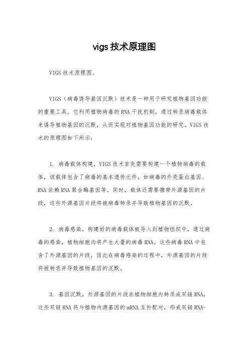
vigs技术原理图VIGS技术原理图。
VIGS(病毒诱导基因沉默)技术是一种用于研究植物基因功能的重要工具。
它利用植物病毒的RNA干扰机制,通过转录病毒载体来诱导植物基因的沉默,从而实现对植物基因功能的研究。
VIGS技术的原理图如下所示:1. 病毒载体构建,VIGS技术首先需要构建一个植物病毒的载体,该载体包含了病毒的基本遗传元件,如病毒的外壳蛋白基因、RNA依赖RNA聚合酶基因等。
同时,载体还需要携带外源基因的片段,这些外源基因片段将被病毒转录并导致植物基因的沉默。
2. 病毒感染,构建好的病毒载体被导入到植物组织中,通过病毒的感染,植物细胞内将产生大量的病毒RNA。
这些病毒RNA中包含了外源基因的片段,因此在病毒感染的过程中,外源基因的片段将被转录并导致植物基因的沉默。
3. 基因沉默,外源基因的片段在植物细胞内转录成双链RNA,这些双链RNA将与植物内源基因的mRNA互补配对,形成双链RNA-mRNA复合物。
这些复合物将被RNA酶切割成小片段,导致植物内源基因的mRNA降解,从而实现基因的沉默。
4. 表型观察,经过一段时间的病毒感染和基因沉默后,研究人员可以观察植物的表型变化,如叶片形态的改变、花期的延迟等。
通过对表型的观察,可以推断出被沉默基因的功能和作用机制。
VIGS技术的原理图清晰地展示了这一技术的工作原理。
通过构建病毒载体、病毒感染、基因沉默和表型观察等步骤,研究人员可以利用VIGS技术对植物基因进行研究,从而揭示植物基因的功能和调控机制。
这一技术在植物分子生物学和植物基因工程研究中具有重要的应用价值,为植物遗传改良和新品种培育提供了有力的工具支持。
VIGS技术的原理图为研究人员提供了一个清晰的思路和方法,帮助他们更好地理解和应用这一技术。
VIGS技术的不断完善和推广将为植物科学研究带来新的突破和进展。
植物生物技术-病毒诱导的基因沉默

设计合理的实验对
照
❖对于每个 VIGS 实验,都必须要有合适的对 照 来监控沉默的效果。阳性对照通常采用八 氢番 茄红素脱氢酶 (phytoene desaturase , PDS) 基因的沉默,因为 PDS 沉默能引起沉默区域发生光漂白,并迅 速产生 可见的表型 (Kumagai 1995) 。实 验中,为 了说明病毒自身是否会引起表型, 还必须用空 病毒载体接种植物作为阴性对照 。另一方面, 阴性对照也可以显示接种技术 对植物生长的影 响。
virus,TMV) 、马铃薯 X 病毒 (potato virus X , PVX) 、 番茄金色花叶病毒 (tomato golden mosaic virus , TGMV) 、烟草脆裂病毒 (tobacco rattle virus , TRV) 、卫星病毒 诱导的沉默 系 统 (satellitevirusinducedsilencing ystem , SVISS) 、大 麦条状花叶病毒 (barley stripe osaic virus , BSMV) 、甘 蓝缩叶病毒 (cabbage leaf
VIGS: virus- induced gene silencing
VIGS : virus- induced gene silencing
是一种转录后基因沉默现像,可引起内源
mRNA 特异性降解。
在 病 毒 载体 中 插入 目 标基 因 片 段 , 侵染寄主后,植 物 会表 现出 目 标基 因 功 能 丧失 或 表 达水 平下降的表型
1 ∶1混合 , 室温下放置 3 h 。选取 4 ~ 5 叶龄 的烟草小苗 , 将混合菌液用不带针头的 2 mL 注射器从背面注射渗入烟草叶片 , 使菌液充 满 整个叶片 , 每棵苗注射 2 片叶子。每个基 因每 次注射 12 株烟草 , 5 次重复。
病毒诱导的植物基因沉默详解
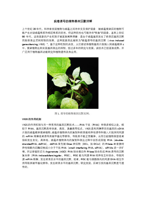
病毒诱导的植物基因沉默详解上个世纪20年代,科学家发现植物与病毒之间存在交叉保护现象:被病毒侵染后的植物可能产生对该病毒株系和相近株系的抗性。
但这种抗性也可能存在“恢复”的现象。
直到上世纪90年代,这些现象的产生机制才被逐渐阐释清楚:是由于病毒基因发生了转录后基因沉默而致使表达受到抑制的结果。
这种现象因此被称为“病毒诱导的基因沉默(virus-induced gene silencing, VIGS)”。
基于这种机制的启发,人们尝试将植物基因片段插入到病毒载体z 中,侵染植物达到实现基因表达的抑制。
经过多年的研究与发展,该技术已经逐渐成熟,并广泛用于植物基因功能研究和植物遗传改良应用。
图1.诱导的植物基因沉默实例。
VIGS的作用机制VIGS的作用机制与另一种常用的基因沉默技术——RNA干扰(RNAi)有很多相似之处。
相较于RNAi,基因沉默具有快速、高效、通量高等优点。
VIGS是利用携带目的基因的cDNA 片段的病毒载体侵染植物,病毒在植物体内的复制和转录能特异性诱导和插入片段序列同源的mRNA降解或者诱导其被甲基化等修饰,导致其不能正常翻译,从而引起植物表型或者指标发生变化。
具体地,病毒在植物体内的复制和表达过程中会形成双链RNA(double-strandedRNA, dsRNA)。
dsRNA首先被Dicer类似物(DCL,如DCL4)的RNase-III家族特异性核酸内切酶切割成小分子干扰RNA(small interfering RNA, siRNA)。
siRNAs进一步扩增,并以单链形式与Argonatute(AGO)RNA结合蛋白和RNase结合形成RNA诱导的沉默复合体(RNA-inducedsilencingplex,RISC)。
RISC能与同源RNA特异性互补结合,导致同源mRNA降解,发生转录后水平的基因沉默。
或者,RISC能与细胞核内的同源DNA相互作用导致其被甲基化修饰,发生转录水平的基因沉默。
VIG病毒诱导基因沉默技术PPT课件
病毒诱导的基因沉默(VIGS)的获得
• 1.设计引物(保守序列,200bp) • 2.构建载体(γ) • 3.体外转录 • (准备质粒,线性化质粒) • 4.体外涂抹 • 5.观察病毒表型(BSMV) • (提取RNA,反转录成cDNA,检测基因表达水平) • 6.观察表型
感谢亲观看此幻灯片,此课件部分内容来源于网络, 如有侵权请及时联系我们删除,谢谢配合!
What are RNAi?
RNA干扰广泛存在于生物界,在不同物种中RNA干扰被 赋予不同的名称:
• 在植物体中被称为基因共抑制(co-suppression) • 在真菌中被称为基因阻抑(qulling) • 在动物体内被称为RNA干扰(RNAi)
现今用于植物RNAi技术按导入方式的不同有粒子轰击、 病毒诱导基因沉默(VIGS)、农杆菌介导等方式。
VIGS的应用
• 品质改良相关基因的功能分析 • 2012,利用VIGS技术发现高分子量谷蛋白亚基HMV-
GS 1Bx14和小麦籽粒中麦谷蛋白聚合体合成有关。 • 这也是首次报道VIGS技术应用于小麦穗子和籽粒相
关的基因功能,同时这个体系的建立将会对研究与 籽粒品质和在小麦渐成的籽粒中有关基因的功能起 到很大帮助作用。
Virus Induced Gene Silencing(VIGS)
• 病 毒 诱 导 的 基 因 沉 默 (virus induced gene silencing,VIGS)
• 是指通过对已知植物病毒的改造,将目标基因片 段整合入病毒载体中,然后侵染植物,诱发RNAi途 径,随着病毒的复制和转录而特异性地诱导序列同 源基因mRNA降解或被甲基化等修饰,从而引起植 物表型或生理指标变化,实现对该功能基因的鉴 定。
年终总结 VIGS

二、VIGS的原理
• 双链RNA ( dsRNA )是基因沉默的起始因子. • 第一阶段包括dsRNA 形成、识别和小干扰 RNA ( siRNAs) 片段的产生, 由RNA 病毒复 制机制、源于转基因的双向克隆形成的发夹 RNA(hpRNA) 和反义RNA(asRNA) 克隆技术 均可产生dsRNA ; dsRNA 被识别并被Dicer 酶从两端降解成21~23 核苷酸的siRNA。
二、VIGS的原理
• VIGS 是利用植物对病毒的天然防御机制发展 的瞬时、快速鉴定植物基因功能的技术,是从 基因序列到功能的反义遗传学方法,也是转录 后基因沉默(Post-transcript gene silencing , PTGS) 、RNA 沉默机制的一种表现,相似于 线虫的RNA 干扰。 • 已在真菌、植物和动物的许多物种中发现了 PTGS 过程的同源基因和酶,因此认为PTGS 机制在整个生物进化过程中是很保守的。目前 关于基因沉默的假说认为,PTGS 包括3 个阶 段:起始、维持和信号放大与传播。
•第三,VIGS 无需构建转基因植株。这对于 那些转基因植株获取非常困难的植物种类来 说至关重要。
三、VIGS的优缺点
• 第四,VIGS 可用于突变致死的基因功能研 究。 • 第五,VIGS 可用于功能冗余基因的功能研 究。 • 第六,VIGS 可用于快速的不同植物种类间 比较基因组学的研究。
三、VIGS的优缺点
四、VIGS技术的应用
由于VIGS的有效性和快速性,近些年将一 些VIGS体系的载体进行改良,在特定的寄主 植物上赋予新的功能,并且成功地用于解析 许多具有重要意义的植物基因功能。其中, 应用最多的就是基于TRV烟草脆裂病毒 Tobacco Rattle Virus)的VIGS体系,并广 泛用于验证重要经济作物的多基因沉默。
- 1、下载文档前请自行甄别文档内容的完整性,平台不提供额外的编辑、内容补充、找答案等附加服务。
- 2、"仅部分预览"的文档,不可在线预览部分如存在完整性等问题,可反馈申请退款(可完整预览的文档不适用该条件!)。
- 3、如文档侵犯您的权益,请联系客服反馈,我们会尽快为您处理(人工客服工作时间:9:00-18:30)。
VIGS原理
dsRNA:双链RNA; Dicer酶:核糖核酸酶Ⅲ (RibonucleaseⅢ, RNase Ⅲ) 家族的成员;
siRNA:小干扰RNA ; RISC:RNA诱导沉默复合体
VIGS应用的载体
利用植物病毒作为载体的优点:
• 病毒能吸附到完整的植物细胞并将它们的核酸导 入到细胞中
• 感染的植物细胞产生大量的病毒,因此重组病毒 载体可以高效表达转入的外源基因。
VIGS应用寄主
• 寄主范围有限,针对不同的寄主植物往往需要开发 不同的病毒载体。
• 2002,大麦抑制PDS(大麦条纹花叶病毒BSMV,进 一步在小麦上应用)
• →2006,水稻、大麦和玉米(雀麦花叶病毒BMV • →2006,大豆(多组分病毒菜豆荚斑驳病毒BPMV) • →2010,大豆(改造后无需体外转录,增加沉默效
What are RNAi?
RNA干扰广泛存在于生物界,在不同物种中RNA干扰被 赋予不同的名称:
• 在植物体中被称为基因共抑制(co-suppression) • 在真菌中被称为基因阻抑(qulling) • 在动物体内被称为RNA干扰(RNAi)
现今用于植物RNAi技术按导入方式的不同有粒子轰击、 病毒诱导基因沉默(VIGS)、农杆菌介导等方式。
病毒诱导的基因沉默(VIGS)的获得
• 1.设计引物(保守序列,200bp) • 2.构建载体(γ) • 3.体外转录 • (准备质粒,线性化质粒) • 4.体外涂抹 • 5.观察病毒表型(BSMV) • (提取RNA,反转录成cDNA,检测基因表达水平) • 6.观察表型
感谢亲观看此幻灯片,此课件部分内容来源于网络, 如有侵权请及时联系我们删除,谢谢配合!
率)
VIGS的应用
• 抗旱研究中相关基因的功能分析 • 2011,沉默小麦上S-腺苷甲硫氨酸合成酶基因
(SAMS)、S-腺苷甲硫氨酸脱羧酶基因(SAMDC)、γ谷氨酰半胱氨酸合成更美基因(γ-ECS)三个基因。 • 沉默植株经干旱胁迫后较正常植株在形态上有明显 的变化,其叶片的卷曲萎蔫程度较为突出,说明这 三个基因在小麦抗干旱胁迫中具有重要功能。
VIGS优点
• 在未获取全长序列甚至事先不知道序列的情况下 即可进行VIGS分析。
• 操作简便,能快速获取表型。 • 无需构建转基因植株。这对于那些转基因植株获
取非常困难的植物种类来说至关重要 • 可用于突变致死的基因功能研究。 • 可用于功能冗余基因的功能研究。 • 可用于快速的不同植物种类间比较基因组学的研
Virus Induced Gene Silencing(VIGS)
• 病 毒 诱 导 的 基 因 沉 默 (virus induced gene silencing,VIGS)
• 是指通过对已知植物病毒的改造,将目标基因片 段整合入病毒载体中,然后侵染植物,诱发RNAi途 径,随着病毒的复制和转录而特异性地诱导序列同 源基因mRNA降解或被甲基化等修饰,从而引起植 物表型或生理指标变化,实现对该功能基因的鉴 定。
VIGSБайду номын сангаас应用
• 品质改良相关基因的功能分析 • 2012,利用VIGS技术发现高分子量谷蛋白亚基HMV-
GS 1Bx14和小麦籽粒中麦谷蛋白聚合体合成有关。 • 这也是首次报道VIGS技术应用于小麦穗子和籽粒相
关的基因功能,同时这个体系的建立将会对研究与 籽粒品质和在小麦渐成的籽粒中有关基因的功能起 到很大帮助作用。
究。
VIGS缺陷
• VIGS引起目标基因的沉默特征不具有遗传性,以 致VIGS技术并不能彻底揭示基因的功能,这是此 种技术自身最大的局限性。 • 如何获得目标基因的系统性沉默仍然是VIGS技术 目前需要解决的问题。 • 沉默持续的时间问题。 • 病毒载体如何有效地接种入植物体内,并产生较 强的沉默效果,是VIGS技术实验操作中的关键性 步骤。
What are RNAi?
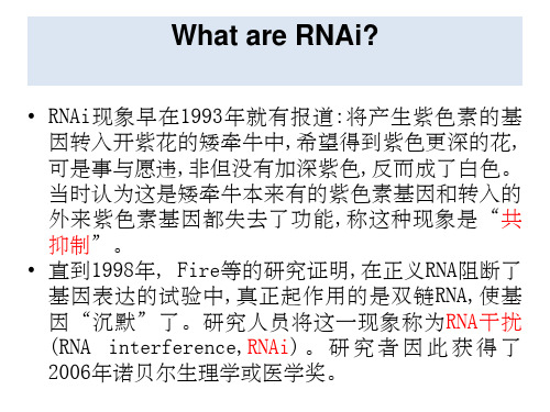
• RNAi现象早在1993年就有报道:将产生紫色素的基 因转入开紫花的矮牵牛中,希望得到紫色更深的花, 可是事与愿违,非但没有加深紫色,反而成了白色。 当时认为这是矮牵牛本来有的紫色素基因和转入的 外来紫色素基因都失去了功能,称这种现象是“共 抑制”。
• 直到1998年, Fire等的研究证明,在正义RNA阻断了 基因表达的试验中,真正起作用的是双链RNA,使基 因“沉默”了。研究人员将这一现象称为RNA干扰 (RNA interference,RNAi) 。 研 究 者 因 此 获 得 了 2006年诺贝尔生理学或医学奖。
• 病毒感染可以蔓延整个植株 • 病毒感染快
VIGS应用的载体
RNA病毒(最早最多)
→1995,首次基于烟草花叶病毒TMV载体构建(携带八 氢番茄红素脱氢酶PDS侵染烟草,上位叶片褪绿白化) →1998,马铃薯X病毒PVX(沉默PDS诱发白化效应) →2001,烟草脆裂病毒TRV(因其具有病毒症状较轻、 沉默效率高、持续时间长、能够侵染分生组织应用, 目前应用最广泛VIGS载体)
PDS即八氢番茄红素脱氢酶,是类胡卜素合成所必需的酶,具保护叶绿 素免受光漂白的作用,而PDS基因发生沉默后被侵染植物就会表现出光 漂白症状。
PDS表型变异易于辨别,因此PDS基因成为VIGS体系评价的参照基因。
VIGS应用的载体
DNA病毒(载体构建简便、无需体外转录、操作难度 低) →1998,基于双生病毒番茄金色花叶病毒TGMV的VIGS 体系成功建立(第一例DNA病毒载体) →2004,非洲木薯花叶病毒ACMV →2008,棉花皱叶病毒CLCrV →2010,水稻东格鲁杆状病毒RTBV(实现农杆菌介导 接种高效沉默水稻內源基因PDS,第1例能够通过农 杆菌介导接种水稻的VIGS载体)