双语影像读片PPT课件
医学影像学英文课件

医学影像学英文课件Medical Imaging Course Slides1. Introduction to Medical ImagingMedical imaging is a broad field that encompasses various techniques used to visualize the internalstructures and functions of the human body. These techniques play a crucial role in the diagnosis, treatment, and monitoring of various medical conditions. The most commonly used medical imaging modalities include X-ray, computed tomography (CT), magnetic resonance imaging (MRI), ultrasound, and nuclear imaging.医学影像学是一个广泛的领域,包括各种用于可视化人体内部结构和功能的技术。
这些技术在诊断、治疗和监测各种医疗状况中发挥着关键作用。
最常用的医学影像模态包括X射线、计算机断层扫描(CT)、磁共振成像(MRI)、超声波和核医学成像。
2. X-ray ImagingX-ray imaging is one of the oldest and most widely used medical imaging techniques. It utilizes high-energy electromagnetic radiation to create images of the body's internal structures. X-rays are able to pass through thebody, and the degree of absorption by different tissues is used to create the image. This technique is particularly useful for visualizing bones, joints, and the chest cavity.X射线成像是最古老和最广泛使用的医学影像技术之一。
影像学读片ppt演示教学

病例选择
选择具有代表性的病例,涵盖不 同系统、部位和疾病类型,以便 学生全面了解影像学在临床诊断 中的应用。
病例介绍
简要介绍病例的临床表现、病史 、实验室检查等相关信息,帮助 学生了解病例背景,为影像学表 现解析做准备。
病例影像学表现解析
影像获取
影像与临床信息结合
介绍病例的影像学检查方法,如X线、 CT、MRI等,说明不同检查方法的适 用范围和优缺点。
做出准确的诊断。
清晰简洁
报告的语言要清晰简洁,避免使用过 多的专业术语和复杂的句子结构。
客观描述
报告中要客观描述影像的特征,避免 主观臆断和猜测。
提供建议
在报告中,可以提供一些建议或意见, 如进一步检查或治疗等。
影像学诊断与临床实践结合
及时反馈
当影像学检查结果出现异常时, 要及时反馈给临床医生,以便医
断的可靠性。
03 常见病变影像学表现
肺部病变
01
02
03
04
肺炎
X线可见肺部纹理增粗,呈斑 片状阴影,常伴有胸腔积液。
肺癌
X线可见肺部结节或肿块,形 态不规则,边缘有毛刺,可能
伴有肺门淋巴结肿大。
肺结核
X线可见肺部斑点状、条索状 或结节状病灶,可能伴有钙化
。
肺气肿
X线可见肺部透亮度增加,肺 纹理稀疏。
。
培训与教育
随着医学影像技术的不 断发展,需要不断加强 影像学读片培训和教育 ,提高专业人员的技能 和水平,为临床诊疗提
供更好的服务。
THANKS FOR WATCHING
感谢您的观看
肝脏病变
肝囊肿
CT可见肝脏内低密度病灶,边界清晰。
脂肪肝
超声可见肝脏回声增强,肝实质光点密集。
医学影像学英文课件:The system of digestion

Case 5
Describe sonographic appearances of diffuse hepatic lesions.
Case 6
A 63 year-old man found a renal lesion in health examination. Physical exam: no positive signs.
Early stage: (inflammatory , no abscess) • Hypoechoic • Irregular border • Abundant CDFI
The ultrasound appearance of an abscess may be variable depending on the internal consistency of the mass.
Liver abscess
Liquefied stage:(abscess lumen)
Complex: with some debris and irregular walls.
Absorbent stage
Hypoechoic area
Liver metastases
The following three specific patterns have been described: ➢ A Well-defined hypoechoic mass. ➢ A Well-defined hyperechoic mass. ➢ Diffuse distortion of the normal homogeneous
医学影像读片双语ppt课件

8
9
• Question :No.2 • Brain MR images demonstrate which of the following? (Check all that apply) 正确选项:A.D 颅脑 MR图像的表现包括下列那种?(选择全部正确选项) A.Sphenoid sinus opacification 蝶窦浑浊 B.Internal carotid artery occlusion颈内动脉闭塞 C.Normal pattern of sphenoid sinus mucosal enhancement 蝶窦粘膜正常强化 D.Unilateral cavernous sinus expansion单侧海绵的膨胀
A. Orbital apex involvement 眶尖部侵犯
B. Osseous
C. Mastoid air
sclerosis 骨质硬化
cell destruction 乳突气房破坏 蝶窦皮质破坏
6
D. Sphenoid sinuscortical disruption
• 注释: • The lesioninvolves the lateral wall of the sphenoid sinus extending into the rightorbital apex, as demonstrated by loss of normal fat attenuation in thislocation. • 此病变累及蝶窦外侧壁,侵入右侧眶尖,表现为此处正常脂肪消失。 • The walls of thebilateral sphenoid sinuses are thickened and sclerotic, as is the intersphenoidalseptum. The sphenoid sinus is opacified. • 双侧蝶窦壁及间隔增厚、硬化,蝶窦浑浊。
影像学读片方法幻灯片课件

⑥邻近器官和组织的改变
周 围 间 隙 消 失 - 胰 腺 炎
39
⑦器官功能的改变
蠕 动 功 能 差 - 肠 梗 阻
40
⑦器官功能的改变
呼 吸 功 能 减 弱 - 胸 膜 肥 厚 粘 连
41
5、特别注意
① X线表现常无特征;
异病同影:肺炎结核 ②不同发展阶段、类型而出现不同表现;同 病异影:肺癌包块、空洞
2、熟悉各部位正常影像解剖和变异 3、熟悉各系统基本病变的影像学表现
4、结合临床资料进行综合推理
5
1、掌握基本检查方法的操作和成像 原理
X线:透视、摄片、造影、断层
6
1、掌握基本检查方法的操作和成像 原理
CT:平扫、增强、重建……
7
1、掌握基本检查方法的操作和成像 原理
MRI:平扫、增强、多序列……
8
1、掌握基本检查方法的操作和成像 原理
MRI:平扫、增强、多序列……
9
1、掌握基本检查方法的操作和成像 原理
DSA:血管性、非血管性……
10
2、熟悉各部位正常影像解剖和变异
11
2、熟悉各部位正常影像解剖和变异
12
2、熟悉各部位正常影像解剖和变异
13
3、熟悉各系统基本病变的影像学表 现
3)可能性诊断:肺内包块,可能结核瘤、脓
肿、良性肿瘤、恶性肿瘤
44
8、不同成像技术的综合应用
各有短长
相互印证 不可替代
结合临床
诊断明晰
兼顾经济
综合应用
45
9、不得不说-申请单书写
1)目的明确 2)资料完整
3)阳性结果
英文影像学PPT

杨绛先生:翻译的技巧
要把西方语文翻成通顺的汉语,就得翻个大跟头才颠倒得过来 汉语和西方语言同样是从第一句开始,一句接一句,一段接一段 ,直到结尾;不同主要在句子内部的结构。西方语言多复句,可 以很长;汉文多单句,往往很短。即使原文是简短的单句,译文 也不能死挨着原文一字字的次序来翻,这已是常识了。所以翻译 得把原文的句子作为单位,一句挨一句翻。
Plain scan of lumbar vertebra CT was performed. 腰椎平扫 Plain scan of lumbar vertebra CT reveals that…..腰椎平扫显示(主 谓) Be displayed/Be showed :显示的
The mediastinum and heart shadow are normal.Bilateral diaphragms are smooth. The ribs and clavicles are normal too.肋骨、锁骨未见异常(肯定与否定交换) Mediastinum (ˌmi:dɪæs'taɪnəm ):纵膈 diaphragm['daɪəfræm] : 隔膜 hemi+diaphragm : 偏侧膈,半隔 Hemi: pref.表示“半; 偏侧;单侧 例:The left hemidiaphragm is blurred.左膈面不清楚
检查部位检查部位examinationpositionexaminationpositionlateralfilmlateralfilm33影像所见影像所见findingsfindings如影像所见如影像所见应将图像内显示的异常变化按病变的主次及左右上下应将图像内显示的异常变化按病变的主次及左右上下upsublowsupersub前后前后anteriorposterioranteriorposterior内外顺序内外顺序alignmentalignment进行描述进行描述记录病变的范围记录病变的范围thelesionlesionrangerange大大小形态轮廓小形态轮廓contourcontour内在结构内在结构intraluminalcerebellarvenousintraluminalcerebellarvenous及其与周围组织的关系或增强及其与周围组织的关系或增强后表现并后表现并描述正常结构
脑炎影像诊断英文版护理课件

In multiple cases of ethics, Vascular changes such as vasculitis or infarcts (areas of issue death due to lake of blood supply) may occur These changes can lead to further brain damage and disability
02 Imaging diagnosis of ethics
Imaging examination methods for ethics
• CT scan: A computed tomography scan uses X-rays to create detailed images of the brain It can detect structural changes such as switching, blending, or masses
Administrator fluids and electrolytes to maintain hydration and electrolyte balance
Rehabilitation care
Assist with physical rehabilitation, including range of motion exercises and strength training
Importance of early diagnosis and treatment
Early diagnosis and treatment of ethics can significantly improve outcomes Timely imaging diagnosis and nursing care can help reduce the impact of the disease on the child's development and quality of life
影像课件-七年制chest2

Bronchial Carcinoma
Bronchial carcinoma is the most common malignancy in our country these days. The strongest risk factor for bronchial carcinoma development is cigarette smoking. Environmental and occupational exposure have been implicated in an estimated 3% to 17% of cases of bronchial carcinoma. Interstitial pulmonary fibrosis and focal scarring have been reported to increase the risk for bronchial carcinoma.
Lung neoplasm
SCLC(small cell lung cancer) Small cell carcinoma is a rapidly growing tumor that
医学影像读片指南-PPT课件

重要的CT图像参数
了解CT扫描的重要参数
了解MRI影像的重要参数
观察系列资料(增强时相)
观察系列资料(增强时相)
观察系列资料(增强时相)
观察系列资料(增强时相)
MRI扫描序列各有作用
MRI主要分析信号特征
谢谢大家!
CTA与MRA的选择
CTA的图像质量略好
三、影像学诊断的原则
(一)、影像诊断原则
(二)、读片的步骤
胸片观察步骤
骨关节的分析步骤
骨骼的观察步骤
上腹部MRI
脾脏MRI
步骤三:发现异常
步骤四:异常征象的观察
病变的位置和分布结核、骨肉瘤、骨巨细胞瘤等;病变的数目多发:转移多;病变的形状斑片影、结节影或者块影;病变的边缘形态锐利、光滑:良性病变的密度:骨?临近器官和组织的改变:诊断、治疗注意器官功能的变化:
影像学诊断三角探索法
仔细构建的鉴别诊断表
准确分析影像表现
结合影像表现、鉴别诊断表及病人的临床和实验室资料寻得最近似的诊断
步骤五:务必结合临床
异病同影;同病异影;
(三)、影像学检查的局限性
(四)、期望值与现实
肯定诊断:否定诊断:可能有假阴性(病程或者检查方法的缺陷)可能性诊断:建议其他检查、随访、治疗性试验等。
CT值
窗口技术 窗宽(Window Width,W): 图像上16个 灰阶内包含的CT值范围 (越大显示的组织结构越丰富) 窗位(Window Level,L):窗的中心位 置(一般以所观察组织的CT值为窗 位)
同样的窗宽,窗位不同,其包括的CT值范围不同。
螺旋CT扫描方式特点
扫描床连续移动,连续扫描和数据采集,无漏扫速度快,减少运动影响,可多期扫描球管旋转,X线束轨迹呈螺旋状容积扫描,利于重建,空间分辨率高扫描参数:KV、MA、层厚、螺距 【Pitch=S(mm)/[D (mm)/N]】MSCT透视定位更准确,提高像测量中用于表示组织密度的统一计量单位,称为亨氏单位(Hounsfield Unit, Hu)。公式: CT值= μM μW μW α α 为分度因数(scaling factor),Hu的分度因数为1000,水的衰减系数为1,空气的衰减系数为0.0013,近似于0,故水的CT值为0,而空气的CT值为-1000。
双语影像读片1

• 注释: • There is noevidence of a destructive process involving the mastoid air cells on theprovided images. • 所示图像并无证据表明乳突气房破坏。 • Focal corticaldisruption of the lateral wall of the right sphenoid sinus is present. • 可见右侧蝶窦外侧壁局部骨皮质破坏。
• This nonenhancedT1 MR image in the coronal plane shows asymmetric T1 isointense signal andexpansion of the right cavernous sinus secondary to an intracavernous mass(arrow). • 此T1WI冠状位平扫MR像示右侧海 绵窦肿块(箭)呈等信号,致海绵 窦膨胀,与左侧海绵窦不对称。
• This T1 coronalpostcontrast MR image shows hyperintense mucosal enhancement of the sphenoidsinus with disruption of the enhancement pattern superolaterally by a nodularmass (arrow) that is less intense than adjacent mucosal. • 此冠状位增强T1 MR像示蝶窦粘膜 强化呈高信号,侧上方可见结节样 肿块(箭),强化信号低于临近粘 膜。
Approximately 50% of patients with malignant lymphoma clinically present with head and neck involvement, with the majority of cases showing nodal disease. Extranodal involvement of the head and neck is present in approximately 10% of cases and most commonly occurs in tonsillar tissue, sinonasal cavities, and the thyroid. Sinonasal lymphoma is found most commonly in the nasal fossa and maxillary sinuses with rare frontal and sphenoid sinus involvement.
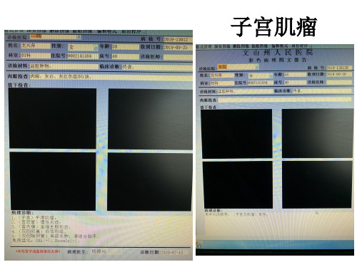
2020-07-15影像读片

CT表现
子宫外形呈分叶状增大或自子宫向外突出的实性肿块, 密度均匀或较均匀,CT值与子宫肌层相当,可见不规则低密 度坏死液化区,少数可见不规则钙化。增强扫描子宫肌瘤与
肌层强化程度相近,变性、液化或坏死部分不强化。
子宫内膜癌
• 平扫示子宫腔扩大,内有菜花或结节状软组织密度肿物,周围可为子宫 腔内积液环绕。增强扫描肿瘤轻微强化或不强化,侵犯肌层时在强化的 正常子宫肌内可见局限或弥漫性低密度。肿瘤向下侵犯子宫颈时可见宫 颈增大,可引起宫腔积水、积血,导致子宫腔扩大,壁薄。肿瘤外侵犯 时,子宫和阴道旁脂肪间隙消失。晚期肿瘤可直接侵犯膀胱、直肠等周 围结构,可出现盆腔、腹膜后淋巴结转移及腹腔等远隔转移。
鉴别诊断
❶子宫黏膜下肌瘤:肿块边界清楚,增强后可见明显强化, 相应区域黏膜正常。 ❷宫颈癌:宫颈增大、强化,有接触性出血史,依靠组织活
检确诊。
浆膜下子宫肌瘤(阔韧带肌瘤)
近日,危地马拉(拉丁美洲国家)的一名医生切除了一个重 达64斤的子宫肌瘤。
子宫肌瘤
概述
子宫肌瘤为最常见的子宫肿瘤,发病与雌激素水平有关, 肿瘤常多发,也可单发,发生在子宫体多见,分为浆膜下、 肌壁间及黏膜下,肌壁间肌瘤最常见,血供不足时,可发生 玻璃样变、液化囊变、红色样变及坏死等,恶变罕见。体积 小时无明显症状,
读影像图谱PPT课件

3D重建与可视化
3D重建
通过计算机技术将一系列二维医学影像转换为三维模型,更直观地展示人体内 部结构。
可视化技术
利用三维模型进行旋转、缩放、切割等操作,多角度展示人体内部结构,辅助 医生进行诊断和治疗方案制定。
03 影像图谱的解读与分析
影像特征提取
影像特征
从影像图中提取出与疾病 相关的特征,如病灶大小、 形态、密度等。
AI算法应用
利用AI算法对影像图谱进行辅助解读, 以提高诊断效率和准确性。
自动化诊断系统
开发自动化诊断系统,通过AI算法对 影像数据进行自动分析,为医生提供 初步诊断结果。
个性化诊疗与精准医疗
个性化诊疗方案
根据患者的个体差异和病情特点,制定个性化的诊疗方案。
精准医疗
利用基因测序、分子诊断等精准医疗技术,为患者提供更精准的治疗方案,提高治疗效果和患者生存 率。
影像图谱的重要性
提高医生诊断准确性
促进医学交流
病情。
影像图谱可以作为医学交流的工具, 方便医生之间分享经验和知识,提高 整个医疗行业的水平。
辅助医生决策
影像图谱可以提供多种影像信息,帮 助医生全面了解患者情况,做出更准 确的诊断和治疗方案。
质量保证
建立严格的质量控制体系,对标注数据进行审核和校验,以确保数据质量可靠。
跨模态影像融合
多模态信息融合
将不同模态的影像信息进行融合,如MRI、CT、X光等,以 提供更全面的医学信息。
跨模态影像比较
通过比较不同模态的影像,医生可以更全面地了解患者的病 情,提高诊断的准确性和可靠性。
AI辅助影像解读
将治疗前后的影像进行对比,评估治疗效果。
量化评估
通过测量病灶大小、形态等指标,对治疗效果进 行量化评估。
胸部影像学读片 SS

快,中心可坏死. • 转移瘤,呈多发状.
空洞及空腔 Cavity and cyst
空洞 肺内病变组织发生坏死,坏死组织经引流
支气管排除而形成.
X线表现:大小,形状不同的透明区
依病理变化可分为三种:
• 虫蚀样空洞----无壁空洞
是大片坏死组织内的空洞,较小,形状不一,多发,洞壁 由坏死组织形成
X线:实变肺野内多发的透明区,轮廓不整,虫蚀状,多见 于干酪性肺炎(caseous pneumonia).
• 薄壁空洞
洞壁薄,在2~3mm以下,由薄层纤维组织及肉芽组织 形成
X线:境界清晰,内壁光滑,圆形透明区,一般无液面,周 围很少实变影.常见于结核.
肺不张 Atelectasis
支气管完全阻塞后,肺内气体多在18~24小时 内被循环的血液吸收,肺叶萎缩,同时肺泡产生 一定量的渗液. • ⑴一侧性atelectasis 肺野均匀致密,隔升高,纵隔移向患侧,肋间隙变 窄,对侧代偿
肺部病变
肺部病变主要有以下几个方面
• 渗出与实变 • 增殖 • 钙化 • 肿块 • 空洞及空腔 • 肺间质性改变
• 肺间质:支气管和血管周围,肺泡间隔及脏层胸 膜下由结缔组织所组成的支架和间隙.
肺叶、肺段
Lung lobe, segment
• 肺叶,右三左二 • 肺段,右10左8
左右肺侧面观
正常胸部横断面解剖
两肺尖 肺野的横断面解剖
两上肺
右上肺
气管 左上肺
右上肺 右主支气管
气胸线
气胸程度预计
X线片 1/4 压缩程度 35%
1/3 1/2 2/3 50 % 70 % 90 %
《CT读片基础篇》课件

无创、无痛、无辐射、操作简便、检查速度快、图像清晰等。
CT技术的局限性
对于某些部位的检查存在盲区,如肺部和胃肠道等。
CT设备原理
CT设备主要由X线发生装置、探测器 、数据采集系统和计算机系统组成。
计算机系统对采集到的数据进行处理 和重建,形成CT图像。
X线发生装置产生X线,探测器接收透 过人体组织的X线,并将其转换为电 信号,数据采集系统将这些信号采集 并传输给计算机系统。
常见病变图像识别
肺部结节
掌握如何识别肺部结节, 了解不同类型结节的特征 及恶性概率。
肝脏占位
熟悉肝脏占位的影像表现 ,掌握良恶性占位的鉴别 要点。
骨肿瘤
了解骨肿瘤的CT表现,掌 握骨肿瘤的分类及恶性程 度评估。
图像误判与纠正
误判原因分析
分析常见的误判原因,如经验不 足、观察不仔细等。
误判纠正方法
综合分析
结合患者的病史、临床表现和其他检查结果,进行综合分析,提高 诊断准确性。
实际操作注意事项
遵循操作规程
严格遵守操作规程,确保扫描质 量和安全性。
保护患者隐私
确保患者隐私得到保护,遵守医 疗伦理规范。
密切关注患者反应
在扫描过程中,密切关注患者反 应,及时处理可能出现的不良反
应。
读片实践与案例分析
掌握如何通过重新观察、请教同行 等方式纠正误判。
案例分享与讨论
分享实际案例,通过讨论提高图像 解读的准确性和可靠性。
03 病例分析
CHAPTER
病例选择与展示
病例选择
选择具有代表性的常见病例,覆 盖不同部位和病变类型,以便学 习者能够全面了解CT读片的实际 应用。
病例展示
- 1、下载文档前请自行甄别文档内容的完整性,平台不提供额外的编辑、内容补充、找答案等附加服务。
- 2、"仅部分预览"的文档,不可在线预览部分如存在完整性等问题,可反馈申请退款(可完整预览的文档不适用该条件!)。
- 3、如文档侵犯您的权益,请联系客服反馈,我们会尽快为您处理(人工客服工作时间:9:00-18:30)。
Question :No.1
Which of the following are present on these images? (Check all that apply.) 正确选项:A.C.D 此组图像的可见下列那种表现?(选择全部正确选项)
• 注释: • Thecontrast-enhanced T1-weighted images show diffuse loss of normal aeration ofthe sphenoid sinus with intrinsic T1 hyperintense signal and peripheral mucosalenhancement. There is some associated left sphenoid sinus expansion, consistentwith early mucocele-like changes. • 对比增强T1WI示蝶窦内气体弥漫缺失,周围粘膜强化,T1像呈高信号,相应左侧 蝶窦膨胀,与黏液囊肿早期改变相符。 • There is noevidence of internal carotid artery occlusion in these images. • 此组图像未见颈内动脉闭塞。
这世界,你最珍贵~
2017.01.11
晴
小房子.
01
简要病史
Question
02
目
录
CONTENTS
03
图片说明 Diagnosis
04
05
Case Point
Discussion
06
复视 !!
??
→诊断
→总结要点
【简要病史】 • A 72-year-old woman presents with rapidly progressive horizontal and vertical diplopia as well as pain in the right forehead and right nasal region
• Question :No.2 • Brain MR images demonstrate which of the following? (Check all that apply) 正确选项:A.D 颅脑 MR图像的表现包括下列那种?(选择全部正确选项) A.Sphenoid sinus opacification 蝶窦浑浊 B.Internal carotid artery occlusion颈内动脉闭塞 C.Normal pattern of sphenoid sinus mucosal enhancement 蝶窦粘膜正常强化 D.Unilateral cavernous sinus expansion单侧海绵的膨胀
• 注释: • At thesuperolateral margins of the bilateral sphenoid sinuses, there is loss ofmucosal enhancement pattern, and nodular soft-tissue thickening is present. • 双侧蝶窦侧上方未见粘膜强化,并见结节样软组织增厚。 • Asymmetricexpansion of the right cavernous sinus with soft-tissue attenuation and outwardconvexity of the lateral wall of the right cavernous sinus are seen. • 右侧海绵窦膨胀并见软组织信号,向右侧海绵窦侧壁外侵犯,与左侧不同。
• This axialcontrast-enhanced CT of the brain in bone window shows cortical disruption ofthe lateral wall of the right sphenoid sinus (arrow) with a permeative patternof bone destruction. • 此骨窗轴位增强颅脑CT示右侧 蝶窦外侧壁骨皮质中断(箭), 呈穿凿样骨质坏。
A. Orbital apex involvement 眶尖部侵犯
B. Osseous
C. Mastoid air
sclerosis 骨质硬化
cell destruction 乳突气房破坏 蝶窦皮质破坏
D. Sphenoid sinuscortical disruption
• 注释: • The lesioninvolves the lateral wall of the sphenoid sinus extending into the rightorbital apex, as demonstrated by loss of normal fat attenuation in thislocation. • 此病变累及蝶窦外侧壁,侵入右侧眶尖,表现为此处正常脂肪消失。 • The walls of thebilateral sphenoid sinuses are thickened and sclerotic, as is the intersphenoidalseptum. The sphenoid sinus is opacified. • 双侧蝶窦壁及间隔增厚、硬化,蝶窦浑浊。
• 注释: • There is noevidence of a destructive process involving the mastoid air cells on theprovided images. • 所示图像并无证据表明乳突气房破坏。 • Focal corticaldisruption of the lateral wall of the right sphenoid sinus is present. • 可见右侧蝶窦外侧壁局部骨皮质破坏。
