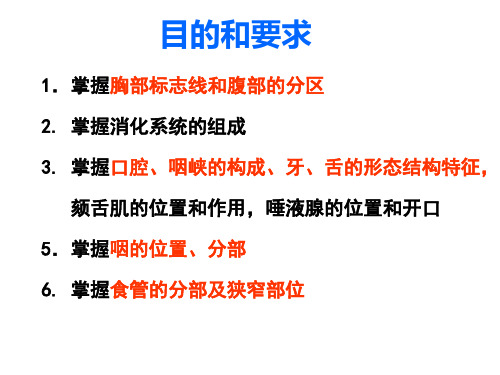
全英-内脏总论与消化系统1(foreign)
合集下载
内脏总论、消化管(一)

口腔 腮腺 舌下腺
下颌下腺 肝
十二指肠 升结肠
盲肠 阑尾 回肠
咽
食管
胃
胰
横结肠 空肠 降结肠 乙状结肠 直肠
第一节 口腔(oral cavity)
口腔的境界
借牙弓、牙龈分为: 口腔前庭 固有口腔
一、口唇(Oral lips)
唇红 人中 鼻唇沟 口角 上、下唇系带
二、颊(cheek )
腮腺管乳头
腮腺 腮腺管
(一)腮腺:耳廓前下方
分部:浅、深两部 腮腺管:开口于腮腺管乳头
腮腺管乳头位于平对上颌 第2磨牙牙冠的颊粘膜处
(二)下颌下腺:下颌下三角内
下颌下腺管:开口于舌下阜
下颌下腺
下颌下腺管
下颌下腺
(三)舌下腺:舌下襞深面
舌下腺大管(1条)开口于舌下阜; 舌下腺小管(10条)开口于舌下襞表面。
舌系带
轮廓乳头
舌下襞:舌下腺小管开口
舌下阜:下颌下腺管及
舌下腺大管开口
(三)舌肌
舌内肌:纵肌、横肌和垂直肌
舌上纵肌
舌横肌
舌垂直肌
舌下纵肌
舌外肌: •颏舌肌
两侧同时收缩,拉舌向前 下方,即伸舌;单侧收缩 可使舌尖伸向对侧。
•茎突舌肌 •舌骨舌肌
六、唾液腺(salibary gland)
小唾液腺:位于口腔粘膜内 大唾液腺:3对
思考题
❖ 口腔检查能看见哪些结构? ❖ 简述牙的形态、构造及牙周组织的构成? ❖ 试述咽的形态、位置、分部和交通? ❖ 简述食管的形态、食管狭窄位置及意义? ❖ 试述胃的形态分部、位置? ❖ 简述十二指肠的形态、位置及各部的构造? ❖ 名词解释:上、下消化道,咽峡、咽淋巴环、
Treitz韧带
内脏学总论及消化系统

12
(一)、 口唇Oral Lips
唇红 人中 鼻唇沟
13
(二)、颊Cheek
14
(三)、腭
palate
硬腭hard palate 软腭soft palate
腭垂uvula 腭舌弓palatoglossal arch 腭咽弓palatopharyngeal arch
咽峡isthmus of fauces
咽上缩肌 咽中缩肌 咽下缩肌
42
[附]吞咽动作 吞咽动作是指食团由背舌背经咽和食管进 入胃的过程。舌背上的食团由于舌肌收缩 贴靠硬腭,将食团经咽峡推向咽腔,此时 软腭抬起,咽后壁向前,阻断口咽部和鼻 咽部的交通,防止食团进入鼻咽部,舌骨 被肌肉收缩而上提并带动喉向前上方移动, 舌根被提向后上方,会厌下落,遮盖喉口, 因而,当食团经过咽腔的一瞬间呼吸停止。 食团进入咽和食管,由于肌肉由上向下依 次收缩推动食团下行,最后通过贲门入胃。 整个吞咽过程包括两个阶段:第一阶段是 舌、腭肌肉有意识地收缩压挤食团经咽峡 入咽腔;第二阶段是食团由咽经食管入胃, 完全是反射性活动。
Sublingual gland
位于舌下襞的深方 大管开口于舌下阜
小管开口于舌下襞表32面
3
下颌下腺 submandibular gland
位于下颌下三角内 开口于舌下阜
33
二、 咽 Pharynx
(一)咽的位置和形态
颅底
鼻咽部
腭帆游离缘 口咽部
会厌软骨上缘
喉咽部
第六颈椎体下缘
34
(二) 咽的分部
界沟terminal sulcus 轮廓乳头vallate papillae 叶状乳头foliate papillae
舌体
菌状乳头fungiform papillae
内脏总论及消化系统(系统解剖学)

(四)舌
舌肌
舌内肌 舌外肌
颏舌肌
第二节口腔
咽
(五)唾液腺 五
• 小唾液腺 • 大唾液腺
腮腺 parotid gland 下颌下腺 submandibular gland 舌下腺 sublingual gland
salivary gland
三大唾液腺
名称 位置 腮腺 外耳道前下方 和下颌后窝内 上颌第二磨牙相 对的颊粘膜处 下颌下腺 下颌下三角 舌下腺 舌下襞深面
开口
舌下阜
大管于舌下阜 小管于舌下襞
唾液腺
消化系统目录
第三节 咽
位置和形态
(1)位置 ) (2)形态 )
pharynx
分部
(1)鼻咽 ) (2)口咽 ) (3)喉咽 )
咽壁肌
第四节食管
第三节咽
鼻咽
nasopharynx
•位置 位置: 位置 •形态: 形态: 形态
咽鼓管咽口 咽鼓管圆枕 咽隐窝 咽鼓管扁桃体 咽扁桃体
第二节口腔
(二)口唇、颊、腭 口唇、
口唇oral lips 颊cheek 腭palate
• 硬腭 • 软腭
腭帆、 腭帆、腭垂 、 腭舌弓、腭咽弓 腭舌弓、 咽峡 • 软腭肌
第二节口腔
(三)牙
种类和排列
• 种类
据出现先后顺序分: 乳牙(20)、恒牙 恒牙(32) 乳牙 恒牙 据形状和功能分: 切牙、尖牙、前磨牙、 切牙、尖牙、前磨牙、磨牙
泌尿系统模式图
内脏总论
生殖系统模式图
内脏总论
胸部标志线
前正中线 胸骨线 锁骨中线 胸骨旁线 腋前线 腋后线 腋中线 肩胛线 后正中线
内脏总论
腹部标志线和分区
两横线两垂线分9区 两横线两垂线分 区
2.9系统解剖学9内脏学总论消化系统1

Alimentary system
四、teeth 牙 2、shape of teeth
Crown 牙冠:露在牙龈外的部分
Neck 牙颈:牙冠和牙根之间的部 分,被牙龈覆盖。
Root 牙根:深藏于牙槽内
每个牙根有apical foramen根尖孔 牙根管 和root canal牙根管。
牙冠有pulp chamber牙冠腔。 牙根管与牙冠腔合称牙腔dental 根尖孔 cavity或 髓腔pulp cavity,contains dental pulp 牙髓。
会厌谷
Alimentary system
tonsillar ring of pharynx咽淋巴环
pharyngeal tonsile 咽扁桃体、 tubal tonsile 咽鼓管扁桃体、 palatine tonsile 腭扁桃体 lingual tonsile 舌扁桃体,
Functuion: defend and protection
Composition:
腮腺
Digestive tube 消化管
口腔 咽 食管 胃 小肠
上消化道
十二指肠 空肠 回肠
舌下腺
下颌下腺 肝
盲肠
大肠
阑尾 结肠
直肠
肛管
下消化道
Digestive glands 消化腺
大消化腺:大唾液腺,肝和胰
十二指肠
升结肠 盲肠
小消化腺:
阑尾
Function: 消化、吸收、味觉,发音 回肠
牙冠腔
Alimentary system
四、teeth 牙
釉质
3、the structure of the teeth: 牙质
Dentine 牙质、
【系解012】内脏总论、消化系统总论

【系解012】内脏总论、消化系统总论
※※打造影像人自己的医学影像平台※※
◎让学习成为一种习惯◎让知识成为一种内涵
◎让专业成为一种交流◎让我们成为一世朋友
※※※※※※※※※※※※※※※※※※※※※
总论
内脏(viscera):包括消化、呼吸、泌尿、生殖系统。
特点:1)大多数存在于胸、腹腔和骨盆腔内。
2)借管道直接或间接与外界相通。
(一)内脏一般结构:
1、中空性器官:粘膜、(粘膜下层)、肌层、外膜
2、实质性器官:一侧凹陷形成门,出入门的结构被结缔组织包被形成蒂或根。
(二)胸、腹部的标志线和腹部的分区:
1、胸部的标志线
1)前正中线,2)胸骨线,3)锁骨中线,4)胸骨旁线,5)腋前、后线,6)腋中线,
7)肩胛线, 8)后正中线
2、腹部的标志线和分区
1)四分法:右上腹,左上腹,右下腹,左下腹(以脐为中心)
2)九分法:腹上区,脐区,腹下区,左右季肋区,左右外侧区,左右髂区
消化系统
消化管:口腔、咽、食管、胃、小肠(十二指肠、空肠、回肠)大肠(盲肠、阑尾、结肠、直肠和肛管)。
其中口腔至十二指肠称为上消化道,空肠以下称为下消化道。
消化腺:大消化腺有口腔腺、肝和胰,小消化腺位于消化管壁内。
版权声明。
内脏总论消化系统

口
腔
上唇 系带
舌系带
下唇 系带
内脏总论消化系统软腮组腺织管乳头
腭舌弓 腭咽弓 腭扁桃体
腭帆游离缘 腭垂 舌根
腭咽弓 腭舌弓
咽峡:腭垂、腭帆游离缘、两侧腭舌弓及舌 根共同围成咽峡,它是口腔和咽之间的狭窄 部,也是两者的分界。
内脏总论消化系统
口腔内结构 牙 舌
内脏总论消化系统
牙
• 是人体最坚硬的器官 • 具有咀嚼食物和辅助发音的功能
喉咽
梨状隐窝
内脏总论消化系统
咽淋巴环
由咽扁桃体、咽鼓管扁桃体、 腭扁桃体、舌扁桃体共同构 成,形成限制炎症扩散的重 要防线
内脏总论消化系统
食管 前后扁平的肌性器官,消化道最狭窄的部分
在第6颈椎高度起于咽,穿过膈后续于 胃贲门。 全长可分三段: 颈段、胸段和腹段。
三个狭窄: 距中切牙的距离分 别是:15、25、 40cm
舌下腺
内脏总论消化系统
咽
•是消化管和呼吸道的共同通道
•上宽下窄、前后略扁的漏斗形肌性管道
•上端起于颅底,下端到第6颈椎下缘
咽的分部
鼻咽 口咽 喉咽
内脏总论消化系统
后面观
鼻咽
咽隐窝:鼻咽癌好发部位 咽扁桃体
咽鼓管咽口 咽鼓管圆枕
内脏总论消化系统
口咽
舌会厌正中襞 会厌谷
腭扁桃体
咽肌:
咽缩肌 咽提肌
消化系统
内脏总论消化系统
主要功能
-摄取、消化食物和吸收营养物质; 排出代谢残渣 -参与呼吸,发音和语言等功能 •物理性消化;化学性消化
内脏总论消化系统
包括消化管和消化腺两大部分 消化管
口腔 咽 食管
胃
上消化道
(系解课件)内脏总论及消化系统

上消化道(从口腔到十二指肠) 下消化道(从空肠到肛管)
消化腺 alimentary gland
大消化腺(大唾液腺、肝和胰) 小消化腺
消化系统目录
第二节 口腔 oral cavity
(一)概述 (二)口唇、颊、腭 (三)牙 (四)舌 (五)唾液腺
第三节咽
第二节口腔
(一)概述
境界
分部
口腔前庭oral vestibule 固有口腔oral cavity proper
(系解课件)内脏总论及消化系统
内脏学 splanchnology
概述
内脏viscera包括消化系统、呼吸系统、泌尿系统、生殖系统
内脏一般结构
中空性器官、实质性器官
胸部标志线和腹部分区
胸部标志线 腹部分区
消化系统
内脏总论
消化系统模式图
内脏总论
呼吸系统模式图
内脏总论
泌
第八节肝
(四)肝的分叶与分段
第八节肝
(五)肝外胆道系统
第九节胰
• 定义
• 组成
胆囊 输胆管道
(肝左右管、肝总管、胆总管)
胆汁排泄途径
肝外胆道系统
胆囊 gallbladder
• 形态
• 位置 • 分部
底、体、颈、管 胆囊底体表投影 Hartmann囊 螺旋襞
胆囊三角(Calot三角)
胆囊
名称
腮腺
下颌下腺
舌下腺
位置
外耳道前下方 和下颌后窝内
下颌下三角 舌下襞深面
开口 上颌第二磨牙相 对的颊粘膜处
舌下阜 大管于舌下阜 小管于舌下襞
唾液腺
消化系统目录
第三节 咽 pharynx
位置和形态
(1)位置 (2)形态
《系统解剖学》教学资料 双语庞磊内脏总论消化一-41页精选文档

D• Migoeustthiv口e腔glands消化腺
• Pharynx咽
• Esophagus食管 • Stomach胃
Superior digestive tube
• Small intestine小肠
Duodenum
Jejunum
Ileum
Inferior digestive tube
•Large intestine大肠
Lymphatic ring咽淋巴环
-consists of pharyngeal tonsil, tubal tonsil,
Palatine tonsil, and lingual tonsil, forming a circular band of lymphoid tissue at oropharyngeal isthmus
Ⅵ. Major salivary glands
Ⅰ) Parotid gland 腮腺
• Superficial part • Deep part • Parotid duct: opposite
the upper second molar tooth
Ⅱ) Submandibular gland 下颌下腺
两条横线
腹上区 右季肋区
左季肋区
两条纵线
右腹外侧区 脐 区 左腹外侧区
右髂区 腹下区 左髂区
2.腹部的分区
Quadrants: 一横线 一垂直
右上腹 左上腹 右下腹 左下腹
Chapter 5
The Alimentary System 消化系统
Composition
Digestive tube 消化管
Ⅱ. Division
Three segments
- 1、下载文档前请自行甄别文档内容的完整性,平台不提供额外的编辑、内容补充、找答案等附加服务。
- 2、"仅部分预览"的文档,不可在线预览部分如存在完整性等问题,可反馈申请退款(可完整预览的文档不适用该条件!)。
- 3、如文档侵犯您的权益,请联系客服反馈,我们会尽快为您处理(人工客服工作时间:9:00-18:30)。
two surface:
2)the parts
cardiac part fundus of stomach body of stomach pyloric antrum pyloric part pyloric canal
greater curvature greater curvature
3)The position
umbilical region
4)The relation of stomach
The anterior surface: The L part is contact with diaphragm The R part is relation with the left and quadrate lobes of live and the anterior abdominal wall. The posterior surface:
right hypochondriac right lumbar
right inguinal (right iliac)
hypogastric/pubic
Chapter 5 Alimentary/digestive system mouth/oral cavity pharynx alimentary canal
esophagus stomach
small intestine large intestine salivary glands
duodenum jejunum ileum
alimentary liver gland Pancreas Small gland The upper/lower alimentary canal
1. The oral cavity
oral vestibule oral cavity proper 1) Lips 2) Cheek 3) Palate: Hard palate Soft palate Palatine glands Isthmus of fauces Tonsillar fossa 4) Palatine tonsile
tongue
5) The teeth/dentes
deciduous teeth (20):
incisors 2,
canine 1, molars 2 permanent teeth (32): incisors 2 canine 1
premolars 2
molars 2~3
The basic structure of teeth The shape : crown, neck and root Dental cavity : cavity of crown, root canal
The muscle: intrinsic muscle extrinsic muscle: styloglossus hyoglossus genioglossus
frenulum of tongue
2. The pharynx
1) Position nasopharynx
the opening of the opening of auditory tube auditory tube
2) Parts
oropharynx
laryngopharynx
3) Communication
nasopharynx nasal cavity
tympanic cavities of the middle ear oropharynx laryngopharynx oral cavity laryngeal cavity The sagittal section The sagittal section of the skull and neck of the skull and neck
2. General structure of viscera
porta hepatis
mucosa (submucosa) tubular organs
renal hilum
muscular coats serosa
Parenchymatous organs
The visceral surface of the limach/gaster
cardia cardia
1)the shape
two orifices: cardia pylorus two curvature:
lesser curvature lesser curvature
angular incisure angular incisure pylorus pylorus
Principal bronchus Second constriction constriction position Thoracic aorta the beginning (commencement) the first Thoracic part or lower border of 6th C or the level of cricoid cartilage Third constriction the intersection with left Abdominal partthe sternalbronchus angle the second or the level of or the lower border of the 4th T Inferior vena cava the esophageal hiatus the third or the lever of the 10th T The esophagus and its relationships the distance from incisor 15cm
esophagus.
The anterior wall of pharynx
First 3. Esophagus constriction Esophagus
Cervical part
1) The part Trachea
Aortic arch 2) The position of the three constriction
In middle full
left hypochondriac region epigastric region,
hypochondriac region epigastric region hypochondriac region
and umbilical region;
cardia on the left of the eleventh thoracic vertebra, pylorus on the right of the first lumbar vertebra. The position of stomach
Sublingual gland Submandibular gland Parotid gland Parotid duct Uvula Palate Palatopharyngeal arch The opening of the parotid duct Palatoglossal arch
Palatine tonsil
It is relation with the spleen, the diaphragm, the left suprarenal gland, the upper part of the left kidney, the splenic blood vessels, the pancreas, the left colic flexure, the transverse colon and its mesocolon. These structures form the stomach bed.
Xulin 徐 professor
林
splanchnology
Chapter 4 Introduction
1. The concept and function
Splanchnology is a subject that studies the shape, structure and position of the organs of viscera. Viscera includes alimentary system, respiratory system, urinary system and genital system. funtion: The main functions of the viscera are to fulfil the metabolism and maintain the life of species.
the parasternal line
the anterior axillary line the midaxillary line the posterior axillary line the scapular line
the posterior median line
The 4 abdominal regions
The main functions of alimentary system are to ingest the food, to secrete enzymes, to absorb the products of the digestive action, and to eliminate the unused residues. The respiratory system is to carry out the gas exchanges -- supply of oxygen for living cells and remove of carbon dioxide resulting from cell metabolism. The primary function of urinary system is to keep the body in homeostasis by removing and restoring selected amount of water and solutes, excreting the various wastes. The functions of genital system are to produce germ cells (ovum and sperm) and to secrete some hormones.
