小肠肿瘤(中英文)
小肠肿瘤疾病研究报告
小肠肿瘤疾病研究报告疾病别名:小肠肿瘤所属部位:腹部就诊科室:肿瘤科,外科病症体征:呕血与黑便,嗳气,柏油便,肠痉挛,低位小肠梗阻疾病介绍:小肠肿瘤是什么?小肠肿瘤是指从十二指肠起到到回盲瓣止的小肠肠管所发生的肿瘤,小肠占胃肠道全长的75%,其粘膜表面积约占胃肠道表面积的90%以上,但是小肠肿瘤的发生率仅占胃肠道肿瘤的5%左右,小肠恶性肿瘤则更为少见,约占胃肠道恶性肿瘤的1%,小肠肿瘤的确切病因目前尚不清楚,小肠肿瘤的临床表现很不典型,一般与肿瘤的类型,部位,大小,性质及是否有梗阻,出血和转移有关症状体征:小肠肿瘤有哪些症状?小肠肿瘤病人以50岁以下为多,平均在35岁左右。
男女性大致相等。
小肠肿瘤的常见临床表现如下:1、腹痛为常见症状,可因肿瘤表面溃烂、刺激肠管引起肠痉挛所引起,也可因肠梗阻或肠套叠所致。
当肿瘤巨大、突入肠腔,可引起肠堵塞;肿瘤侵犯肠壁可引起肠管狭窄、梗阻。
这类梗阻较多见于小肠恶性肿瘤。
肠套叠多半是小肠良性肿瘤所致。
可急性发作,也可反复慢性发作。
2、消化道出血约有1/3~2/3病人因肿瘤表面溃烂而引起出血。
多数为隐性出血,表现为大便隐血试验阳性或黑粪,长时间也可产生缺铁性贫血。
也可出现间断小量出血,甚至大量便血。
最易引起出血的平滑肌瘤和肉瘤、血管瘤和腺瘤,神经纤维瘤。
3、腹块由于小肠活动度大、位置又不固定,所以小肠肿瘤在体检时偶可扪到肿块,但有时又扪不到,时有时无。
可扪及肿块的多半是体积较大的小肠肉瘤。
4、全身症状除肿瘤反复出血导致贫血外,小肠恶性肿瘤尚可引起消瘦,乏力等全身症状。
化验检查:小肠肿瘤的诊断方法小肠肿瘤的诊断主要依靠临床表现和X线钡餐检查,由于小肠肿瘤的临床症状不典型,并又缺少早期体征和有效的诊断方法,因此容易延误诊断。
对具有上述一种或数种表现者.应考虑小肠肿瘤的可能,需作进一步的检查。
1、X线钡餐检查,对疑有十二指肠的肿瘤.采用弛张性十二指肠钡剂造影。
2、纤维十二指肠镜、纤维小肠镜检查及选择性动脉造影术.可提高小肠肿瘤的诊断率。
小肠肿瘤2

【CT表现】
(1)管腔的狭窄和扩张:肿瘤沿着管腔长轴生长, 形成管状圆形或椭圆形肠壁增厚、僵直,管腔变 窄、长短不一。由于黏膜下神经丛的破坏,肠管 瘫软、扩张,管腔呈“动脉瘤样 ”扩张、壁增厚; 瘤内可有残留内容物 。 (2)肿块:呈圆形、分叶状或不规则软组织肿块, 可单发、也可多法,密度均匀或不均匀;伴有局 部或弥漫性肠壁增厚。增强后,肿块轻—中度强 化,远不及腺癌和平滑肌肿瘤。肿块出现坏死, 表现为密度不均匀,或不规则的空腔形成;肿瘤 穿破肠壁,可见瘘管形成,瘤体内出现液气平。
增强:
大多数肿瘤有中度或明显强化;强化密 度均匀,多见于良性肿瘤;有时瘤体表面瘤体周边不规则强化,中央广泛坏死或囊变,则提 示为恶性肿瘤。有时恶性肿瘤呈浸润性生长,无明 确的边界,与淋巴瘤和腺癌相似。
十二指肠间质瘤
a
b
c
d
十二指肠恶性间质瘤、囊变
回 肠 类 癌
各种小肠肿瘤的特点及鉴别诊断
小肠良性肿瘤:(息肉、腺瘤、、脂肪瘤等):一 般体积较小,多为腔内生长,边界清楚。息肉或腺 瘤可带蒂生长。 腺癌:肠管向心性缩窄、肠梗阻、肠系膜浸润、局 部淋巴结肿大。 小肠GIST:肿块常较大,恶性程度较高。多数为 腔外肿块,边缘强化;常见坏死及溃疡,肝脏及腹 膜转移多,很少有淋巴结转移。 淋巴瘤:较长肠管或几处肠管肠壁明显增厚,肠管 动脉瘤样扩张,并有腹腔或腹膜后淋巴结肿大。 类癌:一般体积较小,多为腔内生长,边界清楚, 可伴有类癌综合征。
脂肪瘤是第三位小肠良性肿瘤; 可发生在胃肠道的任何部位,最常见在小 肠的远端; 肿瘤单发多见,大小不一; 大的肿瘤可因发生脂肪坏死而误为脂肪肉 瘤。
【 CT表现】
小肠肿瘤诊疗

小肠肿瘤诊疗什么是小肠肿瘤?小肠肿瘤是小肠内发生的恶性肿瘤,是比较少见的一种肿瘤。
小肠是人体中最长的消化道之一,其主要作用是吸收营养物质。
小肠的长度大约为6-7米,与人体内其他消化器官不同,小肠没有一个固定的位置,它呈现出一种蜿蜒的形态。
因此,小肠肿瘤的位置、大小和形态都具有一定的特殊性。
小肠肿瘤通常分为良性和恶性两种,其中恶性小肠肿瘤又分为原发性和继发性两种。
原发性小肠肿瘤是指肿瘤首次出现于小肠上皮内或肌层内,而继发性小肠肿瘤是指肿瘤来自小肠以外的其他器官(如结直肠、胃、卵巢等)扩散到小肠。
小肠肿瘤的症状小肠肿瘤的症状多样,因为小肠长度很长,而肿瘤发生的位置、大小和形态也多种多样,因此症状是不可预测的。
一般来说,小肠肿瘤的症状比较难以被识别,症状的表现也和疾病的严重程度和病变部位有关。
最常见的小肠肿瘤症状是腹部疼痛和胀气,特别是在肿瘤处。
肿瘤可以导致肠腔狭窄和小肠内腔的堵塞,进而导致腹部胀气和肠绞痛。
同时,还可以出现类似溃疡性结肠炎的腹泻和便血,或是缺铁性贫血等。
小肠肿瘤的诊断小肠肿瘤比较难以诊断,主要是因为它的症状非常多样,而且有时候肿瘤位置比较难以观察到。
一般来说,如果有肠道问题的症状,医生会先考虑结肠镜检查或胃肠钡剂检查等。
如果怀疑是小肠肿瘤,医生会根据症状进行临床评估,并且进行相关的影像学检查,如超声波、CT和MRI。
然而,这些检查有时并不能确定肿瘤的确切位置和性质,所以仍需要手术切除并送检才能确诊。
小肠肿瘤的治疗小肠肿瘤的治疗原则是手术切除。
如果肿瘤切除不完全,或者转移至其他部位,就需要其他治疗手段,如放疗和化疗。
手术切除是小肠肿瘤的最主要治疗方式,根据肿瘤的位置和范围、大小等特点,选择不同的手术方式。
总的来说,小肠肿瘤的手术切除可分为保留最大程度小肠的的根治手术和节段性切除手术两种。
根治手术根治手术是指在保证肿瘤切除彻底的基础上,尽可能地保留小肠,达到保护肠道功能的目的。
这种手术适用于肿瘤位置和范围较分散,且没有出现严重的肠堵塞的患者。
小肠肿瘤
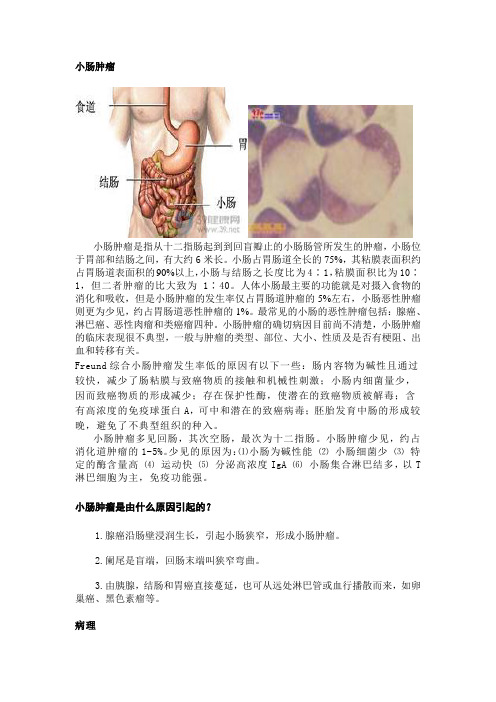
小肠肿瘤小肠肿瘤是指从十二指肠起到到回盲瓣止的小肠肠管所发生的肿瘤,小肠位于胃部和结肠之间,有大约6米长。
小肠占胃肠道全长的75%,其粘膜表面积约占胃肠道表面积的90%以上,小肠与结肠之长度比为4∶1,粘膜面积比为10∶1,但二者肿瘤的比大致为1∶40。
人体小肠最主要的功能就是对摄入食物的消化和吸收,但是小肠肿瘤的发生率仅占胃肠道肿瘤的5%左右,小肠恶性肿瘤则更为少见,约占胃肠道恶性肿瘤的1%。
最常见的小肠的恶性肿瘤包括:腺癌、淋巴癌、恶性肉瘤和类癌瘤四种。
小肠肿瘤的确切病因目前尚不清楚,小肠肿瘤的临床表现很不典型,一般与肿瘤的类型、部位、大小、性质及是否有梗阻、出血和转移有关。
Freund综合小肠肿瘤发生率低的原因有以下一些:肠内容物为碱性且通过较快,减少了肠粘膜与致癌物质的接触和机械性刺激;小肠内细菌量少,因而致癌物质的形成减少;存在保护性酶,使潜在的致癌物质被解毒;含有高浓度的免疫球蛋白A,可中和潜在的致癌病毒;胚胎发育中肠的形成较晚,避免了不典型组织的种入。
小肠肿瘤多见回肠,其次空肠,最次为十二指肠。
小肠肿瘤少见,约占消化道肿瘤的1-5%。
少见的原因为:⑴小肠为碱性能⑵小肠细菌少⑶特定的酶含量高⑷运动快⑸分泌高浓度IgA ⑹小肠集合淋巴结多,以T 淋巴细胞为主,免疫功能强。
小肠肿瘤是由什么原因引起的?1.腺癌沿肠壁浸润生长,引起小肠狭窄,形成小肠肿瘤。
2.阑尾是盲端,回肠末端叫狭窄弯曲。
3.由胰腺,结肠和胃癌直接蔓延,也可从远处淋巴管或血行播散而来,如卵巢癌、黑色素瘤等。
病理恶性大于良性。
恶性:平滑肌肉瘤、腺癌、恶性淋巴瘤、淋巴肉瘤。
良性:平滑肌瘤、脂肪瘤、纤维瘤、腺瘤、淋巴瘤、错钩瘤。
良性肿瘤1、平滑肌瘤:腔内型腔外型壁间型。
2、脂肪瘤:回肠多见、单发,侵犯粘膜下或浆膜下,向肠外突出。
3、腺瘤:回肠多见,空肠、十二指肠少,多见向肠腔内生长,带蒂。
4、纤维瘤:粘膜或浆膜下纤维结缔组织。
5、血管瘤:来源于粘膜下血管丛或浆膜下血管丛,以消化道出血为主要表现。
小肠肿瘤是怎么引起的?

小肠肿瘤是怎么引起的?小肠肿瘤是由什么原因引起的?(一)发病原因小肠肿瘤发病原因目前尚不明确,较为一致的看法有:①小肠腺瘤样息肉,腺癌和某些遗传性家族性息肉病关系密切;②厌氧菌可能在一部分小肠肿瘤中起一定作用;③免疫增生性小肠疾病(immunoproliferativesmallintestinaldisease,IPSID)被认为是淋巴瘤的癌前病变,各方面的证据均提示感染可能在IPSID淋巴瘤的发生发展中起着重要作用;④炎症性肠病具有发展为小肠恶性肿瘤的倾向性;⑤一些疾病如口炎性腹泻,Crohn病,神经纤维瘤病,某些回肠手术后与腺癌的发生有关;另一些疾病如结节性淋巴样增生,AIDS 则与非霍奇金淋巴瘤有关;⑥化学性致癌剂如二甲基肼,氧化偶氮甲烷在小肠肿瘤的发生中可能起一定的作用。
(二)发病机制1.病理分类小肠肿瘤的病理类型较多,国外报道已达35类,国内有人报道为20类,具体可作如下分类。
(1)按分化程度分类:根据肿瘤细胞的分化程度,分为良性肿瘤和恶性肿瘤两类。
①良性肿瘤:A.腺瘤或息肉;B.平滑肌瘤或腺肌瘤;C.纤维瘤;D.脂肪瘤;E.血管瘤;F.神经纤维瘤,神经鞘膜瘤;G.错构瘤,畸胎瘤,淋巴管瘤,黑色素瘤及其他。
良性肿瘤中最常见的是腺瘤,平滑肌瘤,脂肪瘤,纤维瘤,血管瘤五种肿瘤,国内报道空回肠平滑肌瘤较多占38~54%。
②恶性肿瘤:A.癌(腺癌,乳头状癌,黏液腺癌);B.肉瘤(纤维肉瘤,神经纤维肉瘤,平滑肌肉瘤,网状细胞肉瘤,黏液肉瘤);C.类癌或嗜银细胞瘤;D.霍奇金病;E.恶性血管瘤;F.恶性色素瘤;G.恶性神经鞘膜瘤。
恶性肿瘤中以癌居多,其次为各类肉瘤,肉瘤中以各类恶性淋巴瘤居首位,占35~40%,癌,肉瘤比例为1∶5.5。
(2)按组织来源分类:可分为上皮性肿瘤及非上皮性肿瘤。
2.肿瘤分布不同的小肠肿瘤在小肠的不同部位分部,似有一定的倾向性(表1)。
恶性肿瘤在小肠各段的发生率无差异,良性肿瘤则十二指肠肿瘤的发生率明显低于空,回肠,后两者间无差异。
肿瘤常用英文及缩写简介
肿瘤常用英文及缩写简介作为与癌共舞论坛的草根肿瘤患者以及家属,我们也常常需要看一些诊断报告、查找资料或者看看最新的医学期刊杂志的药物及临床试验的结果。
而这些前沿的资料,有相当一部分是英文的,或者是带有英文缩写的数据,为了方便看懂以上资料,特整理些肿瘤常用的中英文对照翻译。
因为本人非英语和医学专业,难免有不对的地方,请多包涵。
第一部分:常用肿瘤中英文对照1、Ca:癌症:cancer的简写即恶性肿瘤;2、chemotherapy:化学治疗;3、Mx:远处转移存在与否不能确定;4、M0:远处转移不存在;5、M1:远处转移存在;6、MST(Median Survival Time):中位生存期;7、MR:好转;8、NSCLC(Non-small-cell carcinoma ):非小细胞肺癌;9、SCLC(Small CellLung Cancer,SCLC):小细胞肺癌;10、SCC(squamous cell carcinoma):鳞状细胞癌;11、SCC:鳞状细胞癌抗原;12、G, Grad 肿瘤细胞分化程度;13、--G1,即高分化,细胞分化程度较好。
一般来说,G1的肿瘤细胞分裂速度较慢;14、--G2,即中分化,细胞分化程度居中;15、--G3,即低分化,细胞分化程度较差。
肿瘤细胞分裂速度较快;16、EGFR表皮生长因子受体:(Epidermal Growth FactorReceptor);17、VEGF(Vascular Endothelial Growth Factor):血管内皮生长因子;18、WBRT(Whole Brain RadiationTherapy):脑放疗;第二部分:肿瘤客观疗效评定标准缩写:1、Qol(Quality of life):生活质量;2、RFS(Recurrence free survival):无复发生存期;3、CR(Complete remission):完全缓解;4、PR(Partial remission):部分缓解;5、SD(stable disease):病情稳定;6、PD(progression disease):病情进展;7、RR(Remissionrate)=CR PR:缓解率;8、TTP(Time-to-time):病情进展时间;9、ORR(Overallremission rate):总缓解率;10、PFS(Progression free survival):无进展生存期;11、MST(Median survival time):中位生存期;12、OS(Overall surviva):总生存期;13、PS(Performance staus ):肿瘤病人生存质量评分;第三部分:常见肿瘤治疗协作组织的英文缩写1、AJCC (American Joint Committe on cancer):美国癌症协会;2、ASCO(American Society of clinical Oncology):美国临床肿瘤学会;3、EORTC(European Organization for Research onTreatment of Cancer):欧洲癌症治疗研究组织;4、FDA(Food and Durg Administration):食品和药品管理局;5、FIGO(Federation Internationale ofGynecologie and Obstetrigue)国际妇产科协会;6、FCOG(Eastern Cooperative Oncology Group):东部癌症协作组;7、IASCT(International Association for thesensitization of Cancer Treatment):国际癌治疗增敏研究协会;8、IAEA(International Atomic Energe Agency):国际原子能委员会组织;9、ICRP(International Commission onRadiological protection):国际辐射防护委员会;10、ICRU(International Commission onRadiological Units and measurement):国际辐射单位和测量委员会;11、MRC(Medical Research Council):英国医学研究委员会;12、NCCTG(North Central Cancer Treatment Group):北方癌症治疗中心;13、NCI(National Cancer Institute):美国癌症研究所;14、RTOG(Radiation Therapy Oncology Group):美国肿瘤放射治疗协作组织;15、UICC(International Union Against Cancer):国际抗癌联盟;第四部分:药物服用次数英文缩写:1、 Qh:小时一次,比如Q2h就是每两小时一次,Q8h就是每八小时一次;2、 Qd:每日一次;3、 Bid:每日两次;4、 Tid:每日三次;5、 Qid:每日四次;6、 Qod:隔日一次;7、 Qw:每周一次;8、 Biw:每周两次;9、 Qm:每晨一次;10、 Qn:每晚一次;11、 ac:饭前;12、 pc:饭后;13、 am:上午;14、 pm:下午;15、 Id:皮内注射;16、 H:皮下注射;17、 IM/iv:静脉注射;18、 IV、ggt:静脉滴注;19、 Po:口服;20、 St:立即;21、 Dc:停止;22、 Hs:临睡前;与癌共舞成立于2010年,是草根抗癌的大本营。
小肠肿瘤的诊断与治疗31例临床报告

小肠肿瘤的诊断与治疗31例临床报告初兆毅;唐镇;李忠民【期刊名称】《基层医学论坛》【年(卷),期】2005(009)005【摘要】目的探讨原发性小肠肿瘤的临床特点及其诊断与治疗.方法对1985年以来诊治的31例原发性小肠肿瘤的临床资料及随访结果进行回顾性分析.结果31例中,良性肿瘤8例,恶性肿瘤23例;肿瘤位于十二指肠者2例,空肠13例,回肠16例.最常见的临床表现为腹痛、腹部包块、消化道出血、肠梗阻、肠穿孔等.31例均经手术治疗,术前诊断率为51.6%(16/31).23例恶性肿瘤根治性切除率为30.4%(7/23),其中15例获随访,死亡10例,平均存活期(32±17.1)个月,存活5例.B 超、CT诊断符合率较低,DSA检查有助于确定病变的部位及性质.结论原发性小肠肿瘤临床表现不典型、恶性肿瘤早期诊断极为困难,当明确诊断时多数恶性肿瘤已经有转移,改善病人预后的关键是加强对本病的认识.【总页数】2页(P390-391)【作者】初兆毅;唐镇;李忠民【作者单位】辽阳市第三人民医院,辽宁,辽阳,111000;辽阳市第三人民医院,辽宁,辽阳,111000;辽阳市第三人民医院,辽宁,辽阳,111000【正文语种】中文【中图分类】R73【相关文献】1.小肠转移性肿瘤临床和病理特点—附4例报告 [J], 廖谦和;夏康;徐丹2.原发性小肠肿瘤的诊断与治疗(附40例报告) [J], 邱云峰;瞿敏;王科峰;涂志远;;;;3.误诊为急性阑尾炎的小儿小肠肿瘤一例临床报告 [J], 李静涛;马新生;刘伟;郎兴;魏建新4.原发性小肠肿瘤的诊断与治疗(附21例报告) [J], 梅虹;孙莉5.原发性小肠肿瘤的诊断与治疗:附58例报告 [J], 邱云峰;许世吾;陈晓军;范晓峰;周远航;杨维良因版权原因,仅展示原文概要,查看原文内容请购买。
小肠肿瘤课件ppt

❖小肠癌的症状出现要早得多 50%小肠肿瘤患者 表现为梗阻或穿孔等急腹症 且随肿瘤增大而出现 比例增高
❖ 小肠腺癌与其他恶性类型相比更易出现疼痛和梗 阻 肉瘤经常表现为急性消化道出血 而淋巴瘤更 常见的是肠穿孔
❖不同亚型好发部位不同 腺癌主要位于十二指肠 ❖神经内分泌肿瘤多位于回肠 肉瘤和淋巴瘤可发生
Through t his pu blicit y camp aign, furthe r impr ove th e sens e of b usines s inte grity of t为ou深ri入sm学e习nt习er近pr平is新es时t代o 中he国lp特t色ou社ri会sm主c义on思su想me和rs党a的nd十o九pe大ra精to神rs,贯to彻s全af国eg教u a育rd大t会h e精l神eg,i充ti分ma发te挥r中ig小ht学s 图an书d 室in育te人re功st能s, t o ensu re tha t our City's touri sm sus tainab le, he al thy and c oordin ated D evelop ment.
十二指肠
❖ 十二指肠长约2530厘米 位于胃幽门与空肠 之间 呈马蹄铁形状 包饶胰头
十二指肠分为上部 降部 横部和升部
❖ 空肠 回肠为腹膜 内位器官
Through t his pu blicit y camp aign, furthe r impr ove th e sens e of b usines s inte grity of t为ou深ri入sm学e习nt习er近pr平is新es时t代o 中he国lp特t色ou社ri会sm主c义on思su想me和rs党a的nd十o九pe大ra精to神rs,贯to彻s全af国eg教u a育rd大t会h e精l神eg,i充ti分ma发te挥r中ig小ht学s 图an书d 室in育te人re功st能s, t o ensu re tha t our City's touri sm sus tainab le, he al thy and c oordin ated D evelop ment.
第六节 小肠肿瘤

第六节小肠肿瘤小肠肿瘤(small intestinal tumor)的发病率较胃肠道其他部位为低,约占胃肠道肿瘤的2%左右,恶性肿瘤占3/4左右。
由于小肠肿瘤诊断比较困难,容易延误治疗。
小肠肿瘤有良性及恶性两类。
良性肿瘤较常见的有腺瘤、平滑肌瘤,其他如脂肪瘤、纤维瘤、血管瘤等。
恶性肿瘤以恶性淋巴瘤、腺癌、平滑肌肉瘤、类癌等比较多见。
腺癌可突向肠腔内生长,呈息肉样,也可沿肠壁浸润生长,引起肠腔狭窄,一般腺瘤和癌常见于十二指肠。
其他则多见于回肠和空肠。
类癌常发生于胃肠道,45%位于阑尾,28%位于回肠末端,直肠占16%,源于中肠者(胃、十二指肠、空回肠及右半结肠)多分泌五羟色胺(serotonin),源于后肠者(左半结肠、乙状结肠)分泌生长抑素(somatostain)为主。
类癌中75%小于1 cm,约2%可有转移,1-2 cm者50%可有转移,大于2 cm者80~ 90%可出现转移,如肝转移。
此外,小肠还有转移性肿瘤,可由胰、结肠和胃癌直接蔓延,也可从远处经淋巴管或血行播散而来,如卵巢癌、黑色素瘤等。
【临床表现】很不典型,常表现下列一种或几种症状。
1.腹痛是最常见的症状,可为隐痛、胀痛乃至剧烈绞痛,当并发肠梗阻时,疼痛尤为剧烈。
并可伴有腹泻、食欲不振等。
2.肠道出血常为间断发生的柏油样便或血便,或大出血。
有的因长期反复小量出血未被察觉,而表现为慢性贫血。
3.肠梗阻引起急性肠梗阻最常见的原因是肠套叠,但极大多数为慢性复发性。
肿瘤引起的肠腔狭窄和压迫邻近肠管也是发生肠梗阻的原因,亦可诱发肠扭转。
4.腹内肿块一般肿块活动度较大,位置多不固定。
5.肠穿孔多见于小肠恶性肿瘤,急性穿孔导致腹膜炎,慢性穿孔则形成肠屡。
6.类癌综合征类癌大多无症状,小部分病人出现类癌综合征,由于类癌细胞产生的5-经色胺和血管舒缓素的激活物质缓激肤所引起,主要表现为阵发性面、颈部和上躯体皮肤潮红(毛细血管扩张),腹泻,哮喘和因纤维组织增生而发生心瓣膜病。
医学英语词汇:肿瘤中英互译
医学英语词汇:肿瘤中英互译医学英语词汇:肿瘤中英互译导语:肿瘤(tumor,neoplam)是一种基因病,但并非是遗传的。
它是指细胞在致癌因素作用下,基因发生了改变,失去对其生长的正常调控,导致单克隆性异常增生而形成的新生物。
下面是小编为您收集整理了关于肿瘤中英互译词汇,欢迎阅读!关于肿瘤的医学英语词汇:malignant melanoma神经外胚层:恶性黑色素瘤malignant meningioma脑膜:恶性脑膜瘤malignant mesothelioma间皮:恶性间皮瘤malignant mixed tumor唾液腺:恶性混合瘤malignant peripheral nerve sheath tumor,malignant schwannoma神经鞘:恶性周边神经鞘肿瘤malignant pheochromocytoma肾上腺髓质:恶性嗜铬细胞瘤malignant phyllodes tumor乳房:恶性叶状瘤malignant teratoma恶性畸胎瘤metastasis远处转移Microinvasive carcinoma微侵袭癌mild dysplasia轻度异生moderate dysplasia中度异生moderately differentiated中度分化myeloma浆细胞:骨髓瘤neuroblastoma神经节细胞:神经母细胞瘤neuroblastoma神经母细胞瘤neuron-specific enolase; NSE神经特异性烯醇adenocarcinoma腺管上皮:腺癌adrenal cortical carcinoma肾上腺皮质:肾上腺皮质癌American Joint Committee on Cancer Staging; AJCC美国癌症分期联合委员会angiosarcoma血管内皮:医学教.育网搜集血管肉瘤basal cell carcinoma基底细胞:基底细胞癌calcitonin抑钙素carcinoembryonic antigen; CEA癌胚抗原carcinoma恶性上皮肿瘤Carcinoma in situ原位癌catecholamine儿茶酚胺chondrosarcoma软骨:软骨肉瘤choriocarcinoma胎盘上皮:绒毛膜癌direct extension直接蔓延dysgerminoma恶性胚胎瘤Dysplasia异生fetoprotein; AFP胎蛋白fibrosarcoma纤维组织:纤维肉瘤FIGO:International Federation of Gynecology and Obstetrics国际妇产科学联盟glioma神经胶细胞:神经胶细胞瘤hematogenous metastasis血行转移hepatocellular carcinoma; hepatoma肝细胞:肝细胞癌histopathological grading组织病理分化human chorionic gonadotropin; HCG人类绒毛膜促性腺素immature teratoma全能细胞:未成熟畸胎瘤International Union against Cancer; UICC国际防癌联盟Invasive carcinoma侵袭癌leiomyosarcoma平滑肌:平滑肌肉瘤leukemia造血细胞:白血病liposarcoma脂肪组织:脂肪肉瘤lymphangiosarcoma淋巴管内皮:淋巴管肉瘤lymphatic metastasis淋巴转移lymphoma类淋巴组织:淋巴瘤node淋巴结oma良性肿瘤osteosarcoma硬骨:骨肉瘤poorly differentiated分化不良prostate-specific antigen; PSA前列腺特异性抗原prostatic acid phosphatase; PAP前列腺酸性磷酸renal cell carcinoma肾脏上皮:肾细胞癌rhabdomyosarcoma横纹肌:横纹肌肉瘤sarcoma恶性间叶肿瘤seminoma生殖细胞:精细胞瘤severe dysplasia重度异生squamous cell carcinoma鳞状上皮:鳞状细胞癌stage期别synovial sarcoma滑膜:医.学教.育网搜.集整理滑膜肉瘤thymic carcinoma胸腺上皮:胸腺癌transitional cell carcinoma泌尿道上皮:过渡细胞癌tumor marker临床检验:含肿瘤标记undifferentiated未分化well differentiated分化良好。
临床肿瘤学-小肠肿瘤

第二十一章小肠肿瘤第一节概况小肠占整个消化道长度的75%,包括十二指肠、空肠、回肠,其粘膜面也占消化道面积的90%,但其癌肿的发病率仅占所有胃肠道恶性肿瘤的5%,发病率明显低于胃及大肠,表明其起源及发病具有独特的生物学特性。
小肠的恶性肿瘤发病率较良性为高,国外资料提示约2/3的小肠肿瘤为恶性,国内文献统计原发性小肠肿瘤5905例,其中良性肿瘤1265例,恶性肿瘤4640例,恶性占79%。
良性肿瘤包括腺瘤、平滑肌瘤、错构瘤、神经源性肿瘤等。
而恶性肿瘤包括腺癌、类癌、肉瘤、淋巴瘤等,以上四种恶性病变占小肠恶性肿瘤的95%。
不同恶性肿瘤的发生部位不同。
癌多发生于十二指肠,尤其是壶腹部;而淋巴瘤及类癌易发生于回肠及小肠远端部位;肉瘤则可分布于整个小肠的不同肠段。
根据不同部位易发生的癌肿,可提示诊断的可能性。
小肠肿瘤难以诊断,文献报道术前诊断的正确率仅为21%~56%。
由于肿瘤部位及性质不同,临床症状可无或轻微,腹痛、腹块、出血是最常见临床表现,严重者表现肠梗阻、出血性休克等,因此小肠肿瘤是临床变异最大的肿瘤。
今年来随着影像学及其他诊断措施的增多,诊断率较前有所提高。
小肠肿瘤的治疗包括,手术、化疗、放疗,均适合不同癌肿及不同部位的肿瘤。
如何选择正确治疗手段及提高生存率仍是当今肿瘤专业研究的课题一、流行病学及病因小肠癌肿的发病率呈现缓慢上升的趋势。
据美国报道,每年约4600新病例发病,小肠癌在1983-1993年十年中也从1.2/10万人增加至1.6/10万人。
随着内镜的开展,十二指肠肿瘤发现增多。
1990年代初,我国十二指肠肿瘤已占小肠肿瘤的30%。
小肠肿瘤好发年龄为30-59岁,腺癌的病人年龄常大于淋巴瘤。
小肠肿瘤的男性发病稍高于女性。
小肠肿瘤发病率较低的原因可能与以下有关:①小肠内为碱性,不利于肿瘤生长;②肠内液体流动快,较少了食物中致癌物质的接触;③小肠内菌群少,细菌致癌因素降低;④小肠内有密集的淋巴组织具备高免疫力,其中IgA介导的免疫系统可能防止了癌的发生。
高中生物中英文
一、最主要的区别是消化系统牙齿:草食动物一般牙齿扁平,适于切断食物,并有磨牙,磨碎食物。
肉食动物牙齿尖锐,适于撕裂动物皮肉。
肠:草食动物的肠一般较短,消化快。
肉食动物肠一般很长,消化充分,慢。
二、爪草食动物一般为蹄,适于奔跑跳跃。
肉食动物一般为爪,能抓、撕食物。
食肉动物消化道短.如果你有机会去解剖的话,你会发现吃草的动物的消化道比吃肉的发达。
主要是因为植物所含的纤维多,必须反复消化Digestive systemsexplain the relationship between the length and overall complexity of digestive systems of a vertebrate herbivore and a vertebrate carnivore with respect to:the chemical composition of their dietthe functions of the structures involvedKey TermsDigestion: the physical or chemical process of breaking down complex insoluble food into small soluble substances that are suitable for the body to absorbDigestive system: The system of organs responsible for digestion. The digestive system includes salivary glands, tongue, oesophagus, stomach, small intestine, large intestine, liver, gall bladder, pancreas and reectum.Herbivores: Organisms that consume plants onlyCarnivores: Organisms that consume other animals. They are also known as meat-eaters.Microbial fermentation微生物发酵: the breakdown of food which involves the action of bacteriaFore-gut fermentation: microbial fermentation which occurs in the stomach. This causes cellulose to digest before the food reaches the intestinesHindgut fermentation后肠发酵: microbial fermentation which occurs in the caecum 盲肠. Cellulose纤维素is digested in this organ which is located at the end of the small intestine.Ruminant fore-gut fermenters: animals that regurgitate回流the food in the first part of their stomachs and re-chew it so that food particles may be further reduced in size.Egestion排泄: the removal of undigested food from an organismWhat do herbivores eat?*Plants are the main diet of herbivores.*They are high in fibre and starch which provides the main energy source in their diet.*Since some parts of plant materials such as cellulose and cell wall thickenings are hard to digest and that cellulose does not provide enough energy unless microbes are present, the digestive tract of herbivores are adapted, so that food may bedigested properly.A herbivorous diet would contain more carbohydrate, particularly cellulose.A carnivorous diet would contain more fat and complete protein (ie protein with all 20 amino acids).The Digestive tracts地带of herbivores*Many large herbivores need microbes细菌in their guts to help them in digesting cellulose*They have long and complex digestive tract to allow microbial fermentation to occur *The digestibility of food is closely related to the complexity and the length of the guts.Digestive Tracts are long because...*They provide adequate space to hold and store large amount of food which must be eaten*Allow maximum opportunity for microbial fermentation to occur* Allow time for the absorption of nutrientsDigestive tracts are complex becau se…* The high-fibre diets they eat are difficult to digestSmall herbivores such as fruit eaters and nectar feeders have shorter and simpler digestive tracts because…*Plant tissues in their diets are easier to digest compared to that of large herbivores *Their diets contain large quantities of sugar and little or no fibreFore-gut内脏Fermentation发酵*Microbial fermentation which occurs in the stomach.*Cellulose is digested before the food reaches the intestines.*Microbes provide energy by releasing short chain fatty acids*Some microbes digested by the host as a source of amino acids*The stomachs of ruminant fore-gut fermenters (e.g. cattle) are very complex and are composed of 4 parts-2 chambers where microbial fermentation occurs-1 part for storage-1 part functions as a true stomach*Microbes have plenty of time to digest cellulose as food is stored in the microbial fermentation chambers for a long timeCarnivoresWhat do they eat?*other animals*animal matter is more easy to digest as animals cells do not have cell walls*animal matter is low in fibre but high in protein*has a high energy content (higher than plants)*the muscles, skin and internal organs of the prey provide fat and protein in their diet*the cartilage and bones provide small amount of fibreThe digestive tracts of carnivores*short and unspecialised gut as their diet is easy to digest compared to plant materials*simple stomach*little undigested food is egested as they do not consume much fibre*small caecum or no caecumCompare the digestive systemThe diet of the honey possum is a mix of nectar and pollen which provides the energy rich food neededto maintain its high metabolic rate.Nectar花蜜feeding animalsThe diet of the honey possum is a mix of nectar and pollen which provides the energy rich food neededto maintain its high metabolic rate.Nectar is essentially a sugary solution containing sucrose, glucose, fructose, produced by plants to rewardpollinators. Banksia nectar has high concentrations of these carbohydrates, sometimes in similar proportions,with an average sugar concentration of 25% w/w sucrose equivalents but low concentrations of protein,vitamins and minerals. Pollen, on the other hand, is essentially the male gametes of a plant. The food reservesof pollen cells contain protein, carbohydrate and fat.Banksia pollen is special. It is high in protein (36-42%) and the pollen from only a few “flowers”(inflorescences) would be needed to satisfy the protein requirements of the honey possum. Most pollen grainscontain only about 20% protein, with 37% carbohydrate, 4% lipid and 3% minerals. Pollen is an idealsupplement to the sugar rich nectar, and pollen and nectar always go together. Honey possums visit plants that produce the nectar they require (Banksias, bottlebrushes, Hakeas andDryandras). In testing for feeding preference, honey possums prefer sucrose to fructose, and both sucroseand fructose to glucose, but there is no evidence that they can tell differences in concentrations of thesethree sugars in combinations.The honey possum is a nectar and honey specialist, and unlike its close relatives it almost never eats insects.Several features of the honey possum are correlated with this diet.·The head is sharply tapered, delicately constructed and has only a few, minute teeth. The mandible (lowerjaw) is reduced to a thin flexible rod and is not suitable for the insertion of strong musculature or deeprooted teeth. The incisors and canines are pointed but the cheek teeth are flattened pegs with roundedtips and do not resemble the normal structure of mammalian teeth.·The tongue is long and stiffened by a keratinised keel (keratin is what hair and nails are made of) and overthe surface of the tongue are finger-like projections (long filiform papillae at the tip of the tongue andshorter compound papillae over most of the dorsal surface). The long tongue is used to collect nectar andpollen from flowers, particularly of Banksia spp. As the tongue is withdrawn into the mouth the pollen isscraped from the finger-like projections by a series of transverse ridges on the roof of the mouth and thepollen falls back into the mouth and is swallowed.How a named technological advance?EndoscopeEndoscopy means looking inside and typically refers to looking inside the body for medical reasons using an endoscope, an instrument used to examine the interior of a hollow organ or cavity of the body.Unlike most other medical imaging devices, endoscopes are inserted directly into the organ. Endoscope can also refer to using a borescope in technical situations where direct line of-sight observation is not feasible.一、胃肠道疾病的检查(1)食道:慢性食道炎、食道静脉曲张、食管道孔疝、食道平滑肌瘤、食道癌及贲门癌等。
普外科常见疾病中英文对照表1

Cholangitis stenosis
胆源性肝脓肿
Biliary liver abscesses
胆囊息肉样变
Polypoid lesions of Gallbladder
胆囊癌
Carcinoma of gallbladder
胆管癌
Carcinoma of bile duct
上消化道大出血
肝肿瘤
Tumor of liver
原发性肝癌
Primaryliver cancer
继发性肝癌
Secondaryliver cancer
肝良性肿瘤
Benign tumor of liver
肝囊肿
Cyst of liver
门静脉高压
Portal hypertension
Budd-chiari综合症
Budd-chiari syndrome
急性化脓性腹膜炎
Acutepurulentperitonitis
急性弥漫性腹膜炎
Acute diffuseperitonitis
腹腔脓肿
Abdominal abscess
膈下脓肿
Subphrenic abscess
盆腔脓肿
Pelvic abscess
肠间脓肿
Interloop abscess
胃十二指肠溃疡
肠扭转
volvulus
肠套叠
Intussusception
短肠综合征
Short bowel syndrome
肠息肉
Intestinal polyps
肠息肉病
Intestinal polyposis
小肠肿瘤
Small Intestinal tumor
小肠肿瘤的诊治(附21例报告)

小肠肿瘤的诊治(附21例报告)
陆少波
【期刊名称】《临床和实验医学杂志》
【年(卷),期】2005(4)4
【摘要】小肠肿瘤较为少见,因其无典型的临床症状,故早期诊断较困难,误诊率较高。
我院自1985年9月至2005年5月共收治小肠肿瘤21例,均经手术探查切除及病理检查证实为小肠肿瘤。
【总页数】2页(P225-226)
【作者】陆少波
【作者单位】如皋市人民医院普外科,江苏,如皋,226500
【正文语种】中文
【中图分类】R73
【相关文献】
1.原发性小肠肿瘤的诊治经验(附60例报告) [J], 邱云峰;陈虹;许海民;王为民
2.小肠肿瘤的诊治(附5例报告) [J], 史凤楼
3.原发性小肠肿瘤的诊治(附24例报告) [J], 王旗;孟翔凌;国维克;唐斌
4.原发性小肠肿瘤的诊治个人经验(附32例报告) [J], 蔡圣强
5.原发性小肠肿瘤的诊治(附30例报告) [J], 后强;曹道成;张昌汉
因版权原因,仅展示原文概要,查看原文内容请购买。
【疾病名】小肠血管瘤【英文名】angiomaofsmallintestine【缩写】【..

【疾病名】小肠血管瘤【英文名】angioma of small intestine【缩写】【别名】intestinal angioma;angeioma of small intestine;angioneoplasm of small intestine;haemangioma of small intestine;hemangioma of small intestine;vascular tumor of small intestine 【ICD号】D13.3【概述】小肠血管瘤属错构瘤,多源于黏膜下血管丛,亦可来自浆膜下血管。
分血管瘤和血管畸形。
血管瘤为真性肿瘤,多发生于空肠,其次为回肠,十二指肠非常少见。
小肠良性血管瘤分为毛细血管瘤、海绵状血管瘤、混合型血管瘤3种类型,临床上以消化道出血为主要表现。
血管畸形则是由于肠壁黏膜下层小动脉、小静脉扩张、扭曲变形、毛细血管呈簇状增生并形成沟通。
血管畸形并非真正肿瘤,分为先天性和获得性。
先天性包括多发静脉扩张症、遗传性出血性毛细血管扩张症(Osler Weber-Rendu综合征)、Turner综合征等。
后天性血管畸形好发于老年人,如假性黄色弹力瘤、系统性硬化症伴毛细血管扩张症等。
血管瘤和血管畸形的临床表现特点为反复无痛性、间歇性出血,常为自限性。
小肠血管瘤有恶化潜能,有报道极少数的小肠血管瘤可恶变为血管肉瘤。
【流行病学】小肠血管瘤少见,约占小肠良性肿瘤的10%~15%。
国内2881例小肠良性肿瘤中血管瘤390例(13.5%),次于腺瘤及平滑肌瘤居第三位;国外2977例中,血管瘤262例(8.8%),较脂肪瘤少而居第四位,国内外统计结果一致。
小肠血管可发生于任何年龄,生后即可出现,女性较男性多见。
【病因】血管瘤一般来自黏膜下层的血管或淋巴管,亦可来自浆膜下血管。
【发病机制】1.好发部位 小肠血管瘤的90%以发生于空回肠,其中以空肠最多,约为48.2%,其次为回肠(41.6%),十二指肠血管瘤仅8%~10%。
小肠肿瘤:前瞻性评价MR小肠灌肠检查

小肠肿瘤:前瞻性评价MR小肠灌肠检查G. Masselli; E. Polettini; E. Casciani; L Be【期刊名称】《《国际医学放射学杂志》》【年(卷),期】2009(32)4【摘要】目的前瞻性评价有症状的病人MR小肠灌肠检出小肠肿瘤的准确性。
以传统的内镜、组织标本、胶囊内镜、常规小肠灌肠和随访结果作为参考标准。
方法人类研究委员会批准了本项研究计划,病人签署知情同意书。
150例(男性83例.女性67例;平均年龄42.6岁,年龄范围17~84岁)临床怀疑小肠肿瘤,且既往上、下胃肠道内镜检查结果正常的病人进行了MR小肠灌肠检查。
MR小肠灌肠表现依下述内容进行前瞻性评价:局限性肠壁增厚、小肠肿块和小肠狭窄。
【总页数】2页(P391-392)【作者】G. Masselli; E. Polettini; E. Casciani; L Be【作者单位】Department; of; Radiology; Umberto; 1; Hospital; La; Sapienza; University; Viale; del; Policlinico; 155; 00161; Rome; Italy.; 不详【正文语种】中文【中图分类】R737.14【相关文献】1.多层螺旋CT小肠造影与传统气钡双重造影检查在诊断小肠可疑性肿瘤疾病的价值比较 [J], 杜培南;黄德成2.螺旋CT小肠造影及传统气钡双重对比造影检查在诊断小肠肿瘤性疾病中的价值比较 [J], 张琨;李健丁;张瑞平;乔英;任卓琼3.螺旋CT小肠造影及传统气钡双重对比造影检查在诊断小肠肿瘤性疾病中的价值比较 [J], 张琨;李健丁;张瑞平;乔英;任卓琼4.邻近回肠末端的小肠克罗恩病:磁共振注气小肠灌肠检查[J], Ochsenkühn T.;Herrmann K.;Schoenberg S.O.;程欣5.克罗恩病小肠受累:无线胶囊内镜检查与CT小肠造影的前瞻性比较 [J], Voderholzer W.A.;Beinhoelzl J.;Rogalla P.;陈云茹因版权原因,仅展示原文概要,查看原文内容请购买。
小肠良性肿瘤: 地点 isnt 一切!

小肠良性肿瘤:地点 isnt 一切!Danielle M Hari;Stephanie L Goff;Heidi J Reich;Anna M Leung;Myung-Shin Sim;Ji Hey Lee;Edward Wolin;Farin Amersi【期刊名称】《世界胃肠外科杂志:英文版(电子版)》【年(卷),期】2013()8【摘要】AIM: To investigate the prognostic significance of the primary site of disease for small bowel carcinoid(SBC) using a population-based analysis.METHODS: The Surveillance,Epidemiology and End Results(SEER) database was queried for histologically confirmed SBC between the years 1988 and 2009.Overall survival(OS) and disease-specific survival(DSS) were analyzed using the Kaplan-Meier method and compared using Log rank testing.Log rank and multivariate Cox regression analyses were used to identify predictors of survival using age,year ofdiagnosis,race,gender,tumor histology/size/location,tumor-node-metastasis stage,number of lymph nodes(LNs) examined and percent of LNs with metastases.RESULTS: Of the 3763 patients,51.2% were male with a mean age of 62.13 years.Median follow-up was 50 mo.The 10-year OS and DSS for duodenal primaries were significantly better when compared to jejunal and ileal primaries(P = 0.02 and < 0.0001,respectively).On multivariate Cox regression analysis,after adjusting for multiple factors,primary site location was not a significant predictor of survival(P = 0.752 for OS and P = 0.966 DSS) while age,number of primaries,number ofLNs examined,T-stage and M-stage were independent predictors of survival.CONCLUSION: This 21-year,population-based study of SBC challenges the concept that location of the primary lesion alone is a significant predictor of survival.【总页数】6页(P239-244)【关键词】Small;bowel;carcinoid;Primary;tumor;location;Survival;Prognosis;National;C omprehensive;Cancer;network;guidelines【作者】Danielle M Hari;Stephanie L Goff;Heidi J Reich;Anna M Leung;Myung-Shin Sim;Ji Hey Lee;Edward Wolin;Farin Amersi【作者单位】Gastrointestinal Research Program, John Wayne Cancer Institute at Saint John’s Health Center;Dana-Farber CancerInstitute;Division of Surgical Oncology, Cedars-Sinai MedicalCenter;Department of Biostatistics, John Wayne Cancer Institute at Saint John’s Health Center【正文语种】中文【中图分类】R735.3【相关文献】1.小肠良性肿瘤的临床与X线表现 [J], 王怀娥;张宗欣;韩广秀2.原发性小肠良性肿瘤34例病理分析 [J], 付红霞;任国庆;李艳丽;张新新3.浅谈小肠良性肿瘤患者的诊疗体会 [J], 李晓林;吕金刚;李晓宴;李驰4.小肠良性肿瘤的临床特点及诊治分析 [J], 王琛;黄瀚章;周锋;阮小蛟;韩少良5.小肠黏膜下层预防腮腺良性肿瘤术后味觉出汗综合征的疗效观察 [J], 刘坤;闫广鹏;陈俊文;高鹏;李军因版权原因,仅展示原文概要,查看原文内容请购买。
- 1、下载文档前请自行甄别文档内容的完整性,平台不提供额外的编辑、内容补充、找答案等附加服务。
- 2、"仅部分预览"的文档,不可在线预览部分如存在完整性等问题,可反馈申请退款(可完整预览的文档不适用该条件!)。
- 3、如文档侵犯您的权益,请联系客服反馈,我们会尽快为您处理(人工客服工作时间:9:00-18:30)。
Back ground
• The most common small bowel malignancies: ALymphoma[lɪmˈfoʊmə] Carcinoid['kɑ:səˌnɔɪd] GIST (Gastrointestinal stromal tumors) [ˌgæstroʊɪnˈtestɪnl]
Lymphoma
• Here a typical presentation • There is irregular wall thickening of the terminal ileum with aneurysmatic dilatation
Lymphoma
• Reversed fold pattern indicating celiac disease • Ileal-ileal intussusception (yellow arrow), in a patient with multifocal small bowel lymphoma (not all lesions shown here). • Mesenteric lymphadenopathy (red arrows).
Adenocarcinoma
• Here an adenocarcinoma in the proximal jejunum • The mass is better depicted with MRI than with CT
Adenocarcinoma
• Here a patient with active Crohn's disease, who has a stenotic segment in the terminal ileum • This patient does not have an adenocarcinoma • Diffuse wall thickening in the distal ileum • Comb sign: hypervascularity in the adjacent mesentery
(HNPCC)Hereditary nonpolyposis colorectal cancer遗传性非息肉病性 大肠癌 [həˈredɪteri]遗传的 [nɒn] [ˌpɒlə'poʊsɪs] 非息肉病的[ˌkoʊlə'rektəl] 结肠直肠的 Familial adenomatous polyposis 家族性腺瘤性息肉病 [ædɪ'nɒmətəs]腺瘤(状)的 [ˌpɒlə'poʊsɪs] Peutz-Jeghers syndrome (PJS) 黑斑息肉病 (Diff.Diagn.)differential diagnosis[ˌdɪfəˈrɛnʃəl ˌdaɪəɡˈnosɪs] 鉴别诊断 aneurysmal dilatation [æn'jʊərɪzməl] [ˌdɪləˈteɪʃən, ˌdaɪlə-]动脉瘤样扩张 sclerosing mesenteritis [sklɪə'rəʊsɪŋ] [mesənˈteraɪtɪs] 硬化性肠系膜炎
Adenocarcinoma
• Features that favour adenocarcinoma are fat stranding due to mesenteric fat infiltration and lymph node metastases • In lymphoma fat stranding is uncommon, but lymph node metastases do occur and are usually more bulky • The images show a short obstructing circular mass in the jejunum (yellow arrow) with enlarged lymph node (red arrow)
Lymphoma
• Here another patient with celiac disease • There is an irregular mass in the jejunum with luminal dilatation • There is infiltration of the mesentery • Pathology showed a T-cell lymphoma in celiac disease • This is called enteropathy associated T-cell lymphoma (EATL)
Carcinoid tumor
• Carcinoid tumors are rare neuroendocrine tumors • Neuroendocrine tumors of the small can be divided in welldifferentiated - also known as carcinoid and poorly differentiated small or large cell neuroendocrine carcinoma • Carcinoid tumors constitute 2% of all gastrointestinal tumors • The incidence of carcinoid tumors increased over the last decades, exceeding that of adenocarcinoma, making it the most common small bowel malignancy
Adenocarcinoma
• Stenotic lesion in the duodenum as a result of an adenocarcinoma (yellow arrow) • Not possible to separate from the pancreas (red arrow) • Pre-stenotic dilatation of the duodenum
Adenocarcinoma
• There is pre-stenotic dilatation • Post-contrast T1Wimage with fatsat (left) and T2W-image (right) show an obstructing mass in the jejunum with shouldering (arrow)
Adenocarcinoma
[ˌædnəʊˌkɑ:sə'nəʊmə]
• Adenocarcinoma represents 25-40% of all small bowel neoplasms['ni:oʊplæzəm] • However colon carcinoma [ˈkolən ˌkɑrsəˈnomə] is 50 times more common • 50% of small bowel adenocarcinomas occur in the duodenum [ˌdu:əˈdi:nəm] and most of these are found with endoscopy [enˈdɑ:skəpi] • The jejunum [dʒɪ'dʒu:nəm] is the second most prevalent site
Adenocarcinoma
• A duodenal carcinoma presenting as irregular wall thickening in the distal duodenum (arrows)
Adenocarcinoma
• Adenocarcinomas often show moderate enhancement • Metastases to the liver and peritoneum occur frequently • Show a circumferential mass with shouldering borders
Lymphoma
• Lymphomas make up about 20 % of all small bowel tumors • The distal ileum is the most common site, owing to the large amount of lymphoid tissue that is present in the distal ileum • The typical presentation of a small bowel lymphoma is a thick walled infiltrating mass with aneurysmal dilatation without obstruction • Aneurysmal dilatation is based upon destruction of the bowel wall and the myenteric nerve plexus
Adenocarcinoma
• Large adenocarcinomas can mimic a lymphoma • The images show an irregular mass in the proximal jejunum • Although it is a large mass, the lumen is not obstructed • This proved to be an adenocarcinoma, but these findings could very well represent a lymphoma
Adenocarcinoma
• The typical imaging representation of a small bowel adenocarcinoma is a focal unilocular, circumferential mass with shouldering of the margins and obstruction • Less frequently adenocarcinomas present as an intraluminal polypoid mass, which can lead to intussusception • Ulceration is a quite common feature • Extraluminal infiltration can present as fat stranding
