人 T 细胞抑制因子 B7H4 ELISA试剂盒北京驰明瑞使用说明书
ELISA试剂盒说明书,人骨髓抑制因子1elisa试剂盒

ELISA试剂盒说明书,人骨髓抑制因子1elisa试剂盒ELISA试剂盒说明书,人骨髓抑制因子1elisa试剂盒供应商:上海樊克生物有限公司ELISA试剂盒规格:(1) 规格:96T 可以测90个样,5个标准孔,1个空白孔(2)规格:48T 可以测42个样,5个标准孔,1个空白孔保存条件:2-8℃低温保存保质期:6个月,所有试剂盒均提供最新批次。
试剂盒成分:酶标板,试剂,标准品等。
ELISA试剂盒说明书,人骨髓抑制因子1elisa试剂盒试剂准备试剂盒从冷藏环境中取出应在室温平衡后方可使用。
1. 标准品复溶:试剂盒提供6管标准品,每管已标定浓度,并且冻干。
实验前在每个标准品管中加入0.5mL样本稀释液,盖好后静置10分钟以上,然后反复颠倒/搓动助其溶解,使其恢复为每个标准品管身标注的浓度。
2. 20×洗涤缓冲液的稀释:蒸馏水按1:20稀释,即1份20×洗涤缓冲液加19份蒸馏水。
试剂盒性能:1.样品线性回归与预期浓度相关系数R值为0.990以上。
2.批内与批见应分别小于9%和11%ELISA试剂盒说明书,人骨髓抑制因子1elisa试剂盒试剂的准备:按试剂盒说明书的要求准备实验中需用的试剂。
ELISA中用的蒸馏水或去离子水,包括用于洗涤的,应为新鲜的和高质量的。
自配的缓冲液应用pH计测量较正。
从冰箱中取出的试验用试剂应待温度与室温平衡后使用。
试剂盒中本次试验不需用的部分应及时放回冰箱保存。
ELISA试剂盒的优势:全面——混合 8 种不同的常见抗原,对自身免疫性疾病进行全面筛查。
快速——检测时间短( ~2 小时),确诊时间更早准确——ELISA 检测方法,灵敏度高,特异性强,适用性好。
质优——美国技术开发,高品质及高可靠性的分析性能。
价低——国内自生产,符合国内实情,降低生产成本。
ELISA试剂盒说明书,人骨髓抑制因子1elisa试剂盒Matters needing attention:1 kit from the cold storage environment should be taken in the room temperature balance 15-30 minutes after the use of the enzyme labeled package was not used after the plate Kaifeng, the board should be loaded into the sealed bag.2 washing buffer will crystallization, heated the water solubilization dilution, washing does not affect the results.3 each step sample should be used, and often to check its accuracy, in order to avoid the test error. A sample within 5 mins, ifthenumberofsampleismuch, recommend to use volley like.4 please each simultaneous determination of standard curve, it is best to do multiple holes. Such as samples to be measured matter content is too high (the sample od high standard product the first hole hole OD), please first sample dilution multiples (n times) were measured and calculated please then multiplied by the total dilution ratio (n * * 5).5 closure plate membrane only a one-time use, to avoid cross contamination.The 6 substrate stored.7 in strict accordance with the instructions of the operation, determine the test results to the microtiter plate reader as a standard.8 of all samples, washing liquid and various waste materials are handled should be transmitted.9 different batches of this reagent can not be mixed.10 and English instructions are different, the English specification shall prevai ELISA试剂盒说明书,人骨髓抑制因子1elisa试剂盒ELISA试剂盒说明书,人骨髓抑制因子1elisa试剂盒具有以下优点:1)快速:仅需传统夹心法ELISA 1/3的操作时间,1小时即可完成整个试验流程;2)简单:只需1个洗涤步骤;3)灵活:可单独或同时检测总蛋白及磷酸化蛋白,可在同一板上检测不同的靶蛋白;4)灵敏:达到甚至超过行业标准,且结果一致性良好;5)兼容:常规比色法检测,实验室常规酶标仪读数;ELISA试剂盒说明书,人骨髓抑制因子1elisa试剂盒产品特点:1.专一性强,灵敏度高2.免费提供产品最新报价,实验原理,产品用途及中英文说明书。
碧云天 Human IgE ELISA Kit 产品说明书
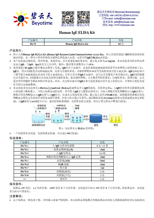
Human IgE ELISA Kit产品编号 产品名称包装 PI479Human IgE ELISA Kit96次产品简介:碧云天的Human IgE ELISA Kit (Human IgE Enzyme-Linked ImmunoSorbent Assay Kit),即人总免疫球蛋白E 酶联免疫吸附检测试剂盒,是一种用于特异性地高灵敏地定量检测人血清、血浆中的IgE 的ELISA 试剂盒。
本产品检测灵敏度高,特异性强,重复性好。
多次重复检测结果表明,最小检出量为14.3ng/ml ,本试剂盒采用特异性抗体识别人IgE ,与IgG 、IgA 没有交叉反应性,板内、板间变异系数均小于10%。
免疫球蛋白E (IgE)只能在哺乳动物身上发现。
IgE 存在于血液中,是免疫系统抵御细菌和病毒等外来物质入侵的重要工具。
IgE 是一类具有δ链的亲同种细胞抗体,是参与过敏性鼻炎、过敏性哮喘和湿疹等发病机制调节的主要抗体。
IgE 是组胺和白三烯等肥大细胞释放抗炎剂水平的主要调剂者。
尽管只有少量IgE 在血液中,但它还负责触发严重过敏反应。
IgE 在I 型超敏反应中起作用,I 型超敏反应表现为各种过敏性疾病,如过敏性哮喘、大多数类型的鼻窦炎、过敏性鼻炎、食物过敏,以及某些类型的慢性荨麻疹和特应性皮炎。
此外,有足够证据证明IgE 还参与免疫系统对寄生虫入侵的反应,并增加与癌症发病有关的白血细胞数量。
本试剂盒采用双抗体夹心ELISA 法(Sandwich ELISA)检测样品中人IgE 的浓度,其原理见图1。
人IgE 特异的单克隆捕获抗体已预包被于酶标板上,当加入标准品或样品时,其中的人IgE 会与捕获抗体结合。
当加入辣根过氧化物酶标记人IgE 抗体后,辣根过氧化物酶标记人IgE 抗体与人IgE 结合,形成夹心的免疫复合物。
最后加入显色剂TMB 溶液,固相捕获的辣根过氧化物酶就会催化无色的显色剂氧化成蓝色物质,在加入终止液后呈黄色。
人凝血酶调节蛋白 ELISA 试剂盒说明书

REV20190712仅供研究,不用于临床诊断。
客服热线: 400-7060-959﹡技术支持邮箱: **************公司官网: 目录简介 ......................................................................................................................................................................... - 3 -检测原理 ................................................................................................................................................................. - 3 -试剂盒组分 ............................................................................................................................................................. - 4 -储存条件 ................................................................................................................................................................. - 5 -其他实验材料 ......................................................................................................................................................... - 5 -注意事项 ................................................................................................................................................................. - 5 -样本收集处理及保存方法 ..................................................................................................................................... - 6 -试剂准备 ................................................................................................................................................................. - 6 -操作步骤 ................................................................................................................................................................. - 8 -操作流程图 ............................................................................................................................................................. - 8 -操作要点提示 ......................................................................................................................................................... - 9 -结果判断 ................................................................................................................................................................. - 9 -结果重复性 ........................................................................................................................................................... - 10 -灵敏度 ................................................................................................................................................................... - 10 -特异性 ................................................................................................................................................................... - 10 -参考文献 ............................................................................................................................................................... - 10 -该产品由北京四正柏生物科技有限公司研制。
酶联免疫分析试剂盒说明书
人chemerin酶联免疫分析试剂盒使用说明书本试剂盒仅供研究使用预期应用ELISA法定量测定人血清、血浆或其它相关液体中chemerin含量。
实验原理本试剂盒应用双抗体夹心酶标免疫分析法测定标本中chemerin水平。
用纯化的抗体包被微孔板,制成固相抗体,往包被单抗的微孔中依次加入chemerin抗原、生物素化的抗人chemerin抗体、HRP标记的亲和素,经过彻底洗涤后用底物TMB显色。
TMB在过氧化物酶的催化下转化成蓝色,并在酸的作用下转化成最终的黄色。
颜色的深浅和样品中的chemerin呈正相关。
用酶标仪在450nm波长下测定吸光度(OD值),计算样品浓度。
试剂盒组成及试剂配制1.酶联板:一块(96孔)2.标准品(冻干品):2瓶,每瓶临用前以样品稀释液稀释至1ml,盖好后静置10分钟以上,然后反复颠倒/搓动以助溶解,其浓度为20,000pg/ml,做系列倍比稀释后,分别稀释10,000pg/ml,5,000pg/ml,2,500pg/ml,1,250pg/ml,625pg/ml,312.5pg/ml,156pg/ml,样品稀释液直接作为标准浓度0pg/ml,临用前15分钟内配制。
如配制10,000pg/ml标准品:取0.5ml20,000pg/ml的上述标准品加入含有0.5ml样品稀释液的Eppendorf管中,混匀即可,其余浓度以此类推。
3.样品稀释液:1×20ml/瓶。
4.检测稀释液A:1×10ml/瓶。
5.检测稀释液B:1×10ml/瓶。
6.检测溶液A:1×120ul/瓶(1:100)临用前以检测稀释液A1:100稀释,稀释前根据预先计算好的每次实验所需的总量配制(每孔100ul),实际配制时应多配制0.1-0.2ml。
如1ul检测溶液A加99ul检测稀释液A的比例配制,轻轻混匀,在使用前一小时内配制。
7.检测溶液B:1×120ul/瓶(1:100)临用前以检测稀释液B1:100稀释。
碧云天生物技术 Human IL-1β ELISA Kit 说明书
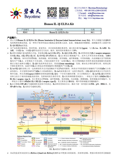
碧云天生物技术/Beyotime Biotechnology 订货热线:400-168-3301或800-8283301 订货e-mail :******************技术咨询:*****************网址:碧云天网站 微信公众号Human IL-1β ELISA Kit产品编号 产品名称包装 PI305Human IL-1β ELISA Kit96次产品简介:碧云天的Human IL-1β ELISA Kit (Human Interleukin-1β Enzyme-Linked ImmunoSorbent Assay Kit),即人白细胞介素1β酶联免疫吸附检测试剂盒,是一种用于特异性地高灵敏地定量检测人血清、血浆、细胞或组织裂解液、或细胞培养上清液中的IL-1β的ELISA 试剂盒。
本产品检测灵敏度高,特异性强,重复性好。
多次重复检测结果表明,最小检出量为2.2pg/ml ,与人IL-1r α、IL-1sRII 、IL-1sRI 、小鼠IL-1α、小鼠IL-1β等均没有交叉反应,板内、板间变异系数均小于10%。
IL-1即白细胞介素-1(简称白介1),其家族由IL-1α(也称IL-1F1)、IL-1β(也称IL-1F2)、IL-1受体拮抗剂(IL-1 receptor antagonist, 简称IL-1RA, 或IL-1F3)及IL-18、IL-33和IL-1F5~F10组成。
IL-1主要由巨噬细胞产生,此外几乎所有的有核细胞,如B 细胞、NK 细胞、体外培养的T 细胞、角质细胞、树突状细胞、星形细胞、成纤维细胞、中性粒细胞、内皮细胞以及平滑肌细胞均可产生IL-1。
正常情况下只有皮肤、汗液及尿液中含有一定量的IL-1,绝大多数细胞在受到外来抗原或细菌内毒素刺激后才能合成和分泌IL-1。
IL-1β在免疫和炎症反应、骨重建(bone remodeling)、发烧、碳水化合物代谢等生理、病理过程中都有重要作用。
四正柏生物 人类TNF-α ELISA试剂盒说明书
REV20190712仅供研究,不用于临床诊断。
客服热线: 400-7060-959﹡技术支持邮箱: **************公司官网: 目录简介 ......................................................................................................................................................................... - 3 -检测原理 ................................................................................................................................................................. - 3 -试剂盒组分 ............................................................................................................................................................. - 4 -储存条件 ................................................................................................................................................................. - 5 -其他实验材料 ......................................................................................................................................................... - 5 -注意事项 ................................................................................................................................................................. - 5 -样本收集处理及保存方法 ..................................................................................................................................... - 6 -试剂准备 ................................................................................................................................................................. - 6 -操作步骤 ................................................................................................................................................................. - 7 -操作流程图 ............................................................................................................................................................. - 8 -操作要点提示 ......................................................................................................................................................... - 8 -结果判断 ................................................................................................................................................................. - 9 -结果重复性 ........................................................................................................................................................... - 10 -灵敏度 ................................................................................................................................................................... - 10 -特异性 ................................................................................................................................................................... - 10 -参考文献 ............................................................................................................................................................... - 10 -该产品由北京四正柏生物科技有限公司研制。
人组织因子(TF)酶联免疫分析试剂盒使用说明书

人组织因子(TF)酶联免疫分析试剂盒使用说明书本试剂盒仅供研究使用检测范围:78pg/ml-5000pg/ml最低检测限:19.5pg/ml特异性:本试剂盒可同时检测天然或重组的人TF,且与其他相关蛋白无交叉反应。
有效期:6个月预期应用:ELISA法定量测定人血清、血浆、细胞培养上清或其它相关生物液体中TF含量。
说明1.试剂盒保存:-20℃(较长时间不用时);2-8℃(频繁使用时)。
2.浓洗涤液低温保存会有盐析出,稀释时可在水浴中加温助溶。
3.中、英文说明书可能会有不一致之处,请以英文说明书为准。
4.刚开启的酶联板孔中可能会含有少许水样物质,此为正常现象,不会对实验结果造成任何影响。
概述TF是一个分子量为47kD,由263个氨基酸残基组成的单链跨膜糖蛋白,其基因定位于染色体1p21~1p22,由6个外显子和5个内含子组成。
TF按细胞表面抗原命名为CD142,属Ⅱ类细胞因子超家族成员,具有信号转导功能。
TF由胞外区、跨膜区和胞浆区三个结构域构成:(1)胞外区由219个氨基酸残基所组成,包括2个二硫键及4个结合FⅦ/FⅦa的关键氨基酸残基,后者是启动外源性凝血级联反应的关键部位。
(2)跨膜区是一个由23个氨基酸残基组成的疏水结构,与磷脂紧密结合,功能尚未明确。
(3)胞内区由21个氨基酸残基组成,其中存在3个与TF功能活性关系密切的丝氨酸残基,能被活化的蛋白激酶C (PKC)磷酸化而激活。
TF又称因子Ⅲ,是丝氨酸蛋白酶凝血因子FⅦ/FⅦa的膜受体,与FⅦ结合促使其转化为FⅦa,并形成TF/Ⅶa复合体。
TF/FⅦa复合体进一步将无活性的FⅩ水解为有活性的FⅩa,FⅩa与FⅤa相结合并在活化血小板磷脂膜表面将凝血酶原酶解为凝血酶而启动外源性凝血过程。
表达TF的单核细胞微粒通过其P选择素糖蛋白配体(PSGL-1)与血小板P选择素(P-selectin)相互作用而与活化血小板结合,在血栓形成过程中起重要作用。
人TH糖蛋白(THP)ELISA试剂盒说明书
Human Protein C ELISA Kit 人蛋白C(Protein C)ELISA试剂盒说明书
Human T-cell acute lymphoblastic leukemia antigen,TALLA-1 ELISA Kit 人T细胞急性淋巴母细胞白血病相关抗原(TALLA-1/CD231)ELISA试剂盒说明书
Human killer cell lectin-like receptor,KLR ELISA Kit 人杀伤细胞凝集素样受体(KLR)ELISA试剂盒说明书
Human Ig-like transcripts Receptor,ILTsR ELISA Kit 人免疫球蛋白样转录体受体(ILTsR/LIR/CD85)ELISA试剂盒说明书
2. 20×洗涤缓冲液的稀释:蒸馏水按1:20稀释,即1份20×洗涤缓冲液加19份蒸馏水。
产品货号:QY-KJ1130人TH糖蛋白(THP)ELISA试剂盒说明书 规格:96T/48T产品别名:人TH糖蛋白(THP)ELISA定量检测试剂盒 人TH糖蛋白(THP)酶联免疫试剂盒 .于测定血清、血浆、组织和相关液体样本中的含量或者活性,规格96T/48T,存储条件:2-8℃,有效期:6个月
Human Cardiac myosin-light chains 1,CMLC-1 ELISA Kit 人心肌肌凝蛋白轻链1(CMLC-1)ELISA试剂盒说明书
Human Fibrin ELISA Kit 人血纤蛋白(Fibrin)ELISA试剂盒说明书
Human fibrinopeptide B,FPB ELISA Kit 人血纤肽/纤维蛋白肽B(FPB)ELISA试剂盒说明书
Elabscience 人抑制素B(INHB)酶联免疫吸附测定试剂盒使用说明书

2022年修订第一版(本试剂盒仅供体外研究使用,不用于临床诊断!)产品货号:E-EL-H0313c产品规格:96T/48T/24T/96T*5Elabscience 人抑制素B(INHB)酶联免疫吸附测定试剂盒使用说明书Human INHB(Inhibin B) ELISA Kit使用前请仔细阅读说明书。
如果有任何问题,请通过以下方式联系我们:销售部电话技术部电话************电子邮箱(销售)********************电子邮箱(技术)**************************网址:具体保质期请见试剂盒外包装标签。
请在保质期内使用试剂盒。
联系时请提供产品批号(见试剂盒标签),以便我们更高效地为您服务。
Copyright ©2021-2022 Elabscience Biotechnology Co.,Ltd. All Rights Reserved目录用途 (3)基本性能 (3)检测原理 (3)试剂盒组成及保存 (4)试验所需自备物品 (5)样品收集方法 (5)注意事项 (6)■ 试剂盒注意事项 (6)■ 样品注意事项 (6)样本稀释方案 (6)检测前准备工作 (7)操作步骤 (8)结果判断 (10)技术资源 (10)典型数据 (10)性能 (11)■ 精密度 (11)■ 回收率 (11)■ 线性 (11)声明 (12)Intended use (13)Character (13)Test principle (13)Kit components & Storage (14)Other supplies required (15)Sample collection (15)Note (16)■ Note for kit (16)■ Note for sample (16)Dilution Method (17)Reagent preparation (17)Assay procedure (18)Calculation of results (20)Technical resources (20)Typical data (20)Performance (21)■ Precision (21)■ Recovery (21)■ Linearity (21)Declaration (22)用途该试剂盒用于体外定量检测人 血清、血浆或其他相关生物液体中INHB浓度。
人白血病抑制因子受体(LIFR)elisa试剂盒使用说明书

人白血病抑制因子受体(LIFR)elisa试剂盒使用说明书Elisa kit规格:48孔配置/96孔配置标准品稀释液:1.5ml×1瓶酶标试剂:3 ml×1瓶(48)/6 ml×1瓶(96)【人白血病抑制因子受体(LIFR)试剂盒】本试剂仅供研究使用计算:以标准物的浓度为横坐标,OD值为纵坐标,在坐标纸上绘出标准曲线,根据样品的OD值由标准曲线查出相应的浓度;再乘以稀释倍数;或用标准物的浓度与OD值计算出标准曲线的直线回归方程式,将样品的OD值代入方程式,计算出样品浓度,再乘以稀释倍数,即为样品的实际浓度。
试剂盒组成:封板膜:2片(48)/2片(96)说明书:1份密封袋:1个标准品:2700ng/L 0.5ml×1瓶0.5ml×1瓶2-8℃保存酶标包被板: 1×48 1×96 2-8℃保存样品稀释液: 3ml×1瓶 6 ml×1瓶2-8℃保存显色剂A液: 3ml×1瓶 6 ml×1瓶2-8℃保存显色剂B液: 3ml×1瓶 6 ml×1瓶2-8℃保存终止液: 3ml×1瓶6ml×1瓶2-8℃保存浓缩洗涤液:(20ml×20倍)×1瓶(20ml×30倍)×1瓶2-8℃保存实验原理:本试剂盒应用双抗体夹心法测定标本中人白血病抑制因子受体(LIFR)水平。
用纯化的人白血病抑制因子受体(LIFR)抗体包被微孔板,制成固相抗体,往包被单抗的微孔中依次加入白血病抑制因子受体(LIFR),再与HRP标记的白血病抑制因子受体(LIFR)抗体结合,形成抗体-抗原-酶标抗体复合物,经过彻底洗涤后加底物TMB显色。
TMB在HRP酶的催化下转化成蓝色,并在酸的作用下转化成最终的黄色。
颜色的深浅和样品中的白血病抑制因子受体(LIFR)呈正相关。
ELISA检测试剂盒使用指南
ELISA检测试剂盒使用指南ELISA(酶联免疫吸附试验)是一种常用的免疫学实验方法,用于检测病原体、抗体或其他分子的存在和浓度。
它具有高灵敏度、高特异性、易操作和较低成本的优势,被广泛应用于生物医学研究、临床诊断和免疫学领域。
本篇文章将介绍ELISA检测试剂盒的使用指南,包括实验准备、试剂的使用步骤和结果解读。
一、实验准备1.阅读检测项目的说明书:在使用试剂盒前,仔细阅读说明书,了解试剂的使用方法、灵敏度和特异性等关键信息。
2.样本准备:根据实验要求,准备样本。
如果需要检测血清中的抗体水平,可以采集血液样本,离心分离血清;如果需要检测细胞培养上清中的分子,将上清收集,并使其清晰,避免细胞或杂质的污染。
3.样本预处理:根据实验需求,对样本进行必要的预处理。
例如,可以通过加热、稀释、酶解等方式来处理样本,以改变样本组分和浓度。
4.准备品质控制样品:制备阳性和阴性控制样品,用于评估试剂盒和实验操作的稳定性和准确性。
5.实验器材准备:准备所需实验器材,如酶标板、移液器、微孔板洗涤仪等。
确保器材的干净和完整,并按照说明书要求对其进行预处理。
二、试剂使用步骤1.试剂准备:根据说明书要求,将试剂从冰箱中取出,并在室温下放置一段时间使其恢复到室温。
确保试剂瓶盖紧闭,并避免暴露在直接阳光下。
2.实验操作:按照说明书的要求,将试剂加入到酶标板中,并根据实验设计进行标准曲线的设置。
标准曲线用于测量未知样品的数量,并计算出浓度。
3.孵育:根据试剂盒的要求,将酶标板放入孵育箱中进行孵育。
孵育温度和时间应根据实验要求进行调整。
4.洗涤:使用洗涤缓冲液对酶标板上的不特异性结合物进行洗涤。
洗涤过程应准确控制洗涤孔板次数和洗涤液的体积。
5.补液:在洗涤完成后,加入辣根过氧化物酶标况稀释液,促进酶标物与特异性结合物的反应。
6.孵育:根据试剂盒的要求,将酶标板放入孵育箱中进行二次孵育。
7.反应停止:根据试剂盒的要求,加入相应的停止液,停止酶反应。
ELISA 检测试剂盒 说明书
本试剂盒只能用于科学研究,不得用于医学诊断人(Human Human))17-17-羟皮质类固醇(羟皮质类固醇(羟皮质类固醇(17-OHCS 17-OHCS 17-OHCS))ELISA 检测试剂盒使用说明书检测原理试剂盒采用双抗体一步夹心法酶联免疫吸附试验(ELISA)。
往预先包被17-羟皮质类固醇(17-OHCS)抗体的包被微孔中,依次加入标本、标准品、HRP标记的检测抗体,经过温育并彻底洗涤。
用底物TMB显色,TMB在过氧化物酶的催化下转化成蓝色,并在酸的作用下转化成最终的黄色。
颜色的深浅和样品中的17-羟皮质类固醇(17-OHCS)呈正相关。
用酶标仪在450nm 波长下测定吸光度(OD 值),计算样品浓度。
样品收集、处理及保存方法1.血清:使用不含热原和内毒素的试管,操作过程中避免任何细胞刺激,收集血液后,3000转离心10分钟将血清和红细胞迅速小心地分离。
2.血浆:EDTA、柠檬酸盐或肝素抗凝。
3000转离心30分钟取上清。
3.细胞上清液:3000转离心10分钟去除颗粒和聚合物。
4.组织匀浆:将组织加入适量生理盐水捣碎。
3000转离心10分钟取上清。
5.保存:如果样本收集后不及时检测,请按一次用量分装,冻存于-20℃,避免反复冻融,在室温下解冻并确保样品均匀地充分解冻。
自备物品1.酶标仪(450nm)2.高精度加样器及枪头:0.5-10uL、2-20uL、20-200uL、200-1000uL3.37℃恒温箱操作注意事项1.试剂盒保存在2-8℃,使用前室温平衡20分钟。
从冰箱取出的浓缩洗涤液会有结晶,这属于正常现象,水浴加热使结晶完全溶解后再使用。
2.实验中不用的板条应立即放回自封袋中,密封(低温干燥)保存。
3.浓度为0的S0号标准品即可视为阴性对照或者空白;按照说明书操作时样本已经稀释5倍,最终结果乘以5才是样本实际浓度。
4.严格按照说明书中标明的时间、加液量及顺序进行温育操作。
5.所有液体组分使用前充分摇匀。
人载脂蛋白 P ApoP ELISA试剂盒北京驰明瑞使用说明书

人载脂蛋白P ApoP使用说明书产品编号:本试剂盒仅供科研使用,不得用于临床及诊断使用!操作步骤1.取出试剂盒,于室温(20-25℃)放置15-30分钟。
实验过程应在室温(20-25℃)内进行。
2.取出酶标板,按照标准品的次序分别加入100μl的标准品溶液于空白微孔中。
3.空白微孔中加入100μl的样品,空白对照加入100μl的蒸馏水;4.在各孔中加入50μl的酶标记溶液;(不含空白对照孔)5.将酶标板用封口胶密封后,37℃孵育反应1小时;(在孵育箱中保持稳定的温度与湿度)6.充分清洗酶标板3-5次,保持各孔有充足的水压;(浓缩洗涤液以1:100的比例与蒸馏水稀释)7.酶标板洗涤后用吸水纸彻底拍干;8.各孔加入显色剂A、B液各50μl;(不含空白对照孔)9.20-25℃下避光反应10分钟;10.各孔加入50μl终止液,终止反应;结果判断1.30分钟内在波长450nm的酶标仪上读取各孔的OD值;2.百分结合率计算:设S0管计数为B0,各标准管或样品管计数为B,非特异管计数为NSB,则百分结合率计算公式如下:B/ B0=(B-NSB)/( B0-NSB)×100%3.logit计算:各标准点或样品管的logit值计算公式如下:logit=ln(B/ B0)/(1-B/ B0)4.将标准品的OD均值与标准品0点的OD均相除,为标准点的百分结合率,在log-logit坐标纸上绘图。
5.Log-logit双对数标准曲线:坐标纸上横轴从左至右第一个1-9表示为第一个10进位,第二个1-9表示为第二个10进位。
第三个1-9表示为第三个10进位。
坐标纸纵轴为百分比(1-99),即各标准吸光值的百分结合率。
取一条通过各点的直线。
要求尽可能多的点在线上,同时剩余的点均匀分布在直线的两边。
样品也同样由吸光值计算百分结合率,再从纵轴上的相应结合率找到直线上的点,此点对应的横坐标浓度即为样品的浓度,无须换算。
6.人工处理:以标准浓度取log值为横坐标,对应的logit值为纵坐标在普通坐标纸上或以标准浓度为横坐标,对应的B/B0为纵坐标在logit-log坐标纸上画出标准曲线(理想化时是一条直线)。
人生长因子(HGH)酶联免疫分析试剂盒使用说明书
人生长因子(HGH)酶联免疫分析试剂盒使用说明书本试剂仅供研究使用目的:本试剂盒用于测定大鼠血清,血浆及相关液体样本中生长因子(HGH)的含量。
实验原理:本试剂盒应用双抗体夹心法测定标本中人生长因子(HGH)水平。
用纯化的人生长因子(HGH)抗体包被微孔板,制成固相抗体,往包被单抗的微孔中依次加入生长因子(HGH),再与HRP 标记的生长因子(HGH)抗体结合,形成抗体-抗原-酶标抗体复合物,经过彻底洗涤后加底物TMB 显色。
TMB 在HRP 酶的催化下转化成蓝色,并在酸的作用下转化成最终的黄色。
颜色的深浅和样品的生长因子(HGH)呈正相关。
用酶标仪在450nm 波长下测定吸光度(OD 值),通过标准曲线计算样品中人生长因子(HGH)浓度。
试剂盒组成:试剂盒组成 48 孔配置 96 孔配置保存说明书 1 份 1 份封板膜 2 片(48) 2 片(96)密封袋 1 个 1 个酶标包被板 1×48 1×96 2-8℃保存标准品:45ng/L 0.5ml×1 瓶 0.5ml×1 瓶 2-8℃保存标准品稀释液 1.5ml×1 瓶 1.5ml×1 瓶 2-8℃保存酶标试剂 3 ml×1 瓶 6 ml×1 瓶 2-8℃保存样品稀释液 3 ml×1 瓶 6 ml×1 瓶 2-8℃保存显色剂A 液 3 ml×1 瓶 6 ml×1 瓶 2-8℃保存显色剂B 液 3 ml×1 瓶 6 ml×1 瓶 2-8℃保存终止液 3ml×1 瓶 6ml×1 瓶 2-8℃保存浓缩洗涤液(20ml×20 倍)×1 瓶(20ml×30 倍)×1 瓶 2-8℃保存样本处理及要求:1. 血清:室温血液自然凝固10-20 分钟,离心20 分钟左右(2000-3000 转/分)。
人组织因子(TF)ELISA分析检测试剂盒使用说明书
人组织因子(TF)ELISA分析检测试剂盒使用说明书人组织因子(TF)ELISA分析检测试剂盒使用说明书本试剂仅供研究使用目的:本试剂盒用于测定小鼠血清,血浆及相关液体样本中转化生长因子(TF)的含量。
实验原理:本试剂盒应用双抗体夹心法测定标本中人组织因子(TF)水平。
用纯化的转化生长因子(TF)抗体包被微孔板,制成固相抗体,往包被单抗的微孔中依次加入转化生长因子(TF),再与HRP标记的羊抗鼠抗体结合,形成抗体-抗原-酶标抗体复合物,经过彻底洗涤后加底物TMB显色。
TMB在HRP酶的催化下转化成蓝色,并在酸的作用下转化成最终的黄色。
颜色的深浅和样品中的转化生长因子(TF)呈正相关。
用酶标仪在450nm波长下测定吸光度(OD值),通过标准曲线计算样品中人组织因子(TF)浓度。
试剂盒内容及其配制:试剂盒成份96孔配置48孔配置96/48人份酶标板1块板(96T)半块板(48T)塑料膜板盖1块半块标准品:100ng/ml 1瓶(1.0ml)1瓶(0.5ml)空白对照1瓶(1.0ml)1瓶(0.5ml)标准品稀释缓冲液1瓶(8.0ml)1瓶(4.0ml)生物素标记的抗OT抗体1瓶(8.0ml)1瓶(4.0ml)亲和链酶素-HRP 1瓶(12ml)1瓶(5ml)洗涤缓冲液1瓶(20ml)1瓶(10ml)底物A 1瓶(6.0ml)1瓶(3.0ml)底物B 1瓶(6.0ml)1瓶(3.0ml)终止液1瓶(6.0ml)1瓶(3.0ml)试剂盒组成:试剂盒组成48孔配置96孔配置保存说明书1份1份封板膜2片(48)2片(96)密封袋1个1个酶标包被板1×48 1×96 2-8℃保存标准品:45μmol/L0.5ml×1瓶0.5ml×1瓶2-8℃保存标准品稀释液1.5ml×1瓶1.5ml×1瓶2-8℃保存酶标试剂3 ml×1瓶6 ml×1瓶2-8℃保存样品稀释液3 ml×1瓶6 ml×1瓶2-8℃保存显色剂A液3 ml×1瓶6 ml×1瓶2-8℃保存显色剂B液3 ml×1瓶6 ml×1瓶2-8℃保存终止液3ml×1瓶6ml×1瓶2-8℃保存浓缩洗涤液(20ml×20倍)×1瓶(20ml×30倍)×1瓶2-8℃保存自备材料:1. 蒸馏水。
人淋巴细胞因子ELISA试剂盒说明书

人淋巴细胞因子ELISA试剂盒说明书
人淋巴细胞因子ELISA定量检测试剂盒等ELISA试剂盒检测类型:双抗体夹心法测抗原、双抗原夹心法测抗体、间接法测抗体、竞争法测抗体、竞争法测抗原、捕获包被法测抗体、ABS-ELISA法。
我司人淋巴细胞因子ELISA定量检测试剂盒等ELISA试剂盒,灵敏性高,高效性,原装进口抗体,吸附均匀、吸附性好,回收利用率高、可靠性强,无效包退包换,公司提供免费代测服务,各种种属ELISA 试剂盒产品齐全,质量可靠。
详情可来电咨询销售人员或咨询我司在线客服。
人淋巴细胞因子ELISA定量检测试剂盒操作步骤:
1、加样:分别设空白孔(空白对照孔不加样品及酶标试剂,其余各步操作相同)、标准孔、待测样品孔。
在酶标包被板上标准品准确加样50µl,待测样品孔中先加样品稀释液40µl,然后再加待测样品10µl (样品最终稀释度为5倍)。
加样将样品加于酶标板孔底部,尽量不触及孔壁,轻轻晃动混匀。
2、温育:用封板膜封板后置 37℃温育30分钟。
3、配液:将30倍浓缩洗涤液用蒸馏水30倍稀释后备用
4、洗涤:小心揭掉封板膜,弃去液体,甩干,每孔加满洗涤液,静置30秒后弃去,如此
重复5次,拍干。
5、加酶:每孔加入酶标试剂50µl,空白孔除外。
6、显色:每孔先加入显色剂A50µl,再加入显色剂B50µl,轻轻震荡混匀,37℃避光显色
10 分钟.
7、终止:每孔加终止液50µl,终止反应(此时蓝色立转黄色)。
8、测定:以空白空调零,450nm波长依序测量各孔的吸光度(OD值)。
测定应在加终止液后15分钟以内进行。
1、ELISA 试剂盒使用说明书(5 孔板格式)
ELISA kitInstruction Manual5-plate formatJuly, 2006For research use only.Not for use in diagnostic or therapeutic procedures.ContentsAbbreviations 2 Introduction 3 Contents of the kit 4 Hazard information 4 Materials and reagents required but not provided 4 Working solutions 4 General procedure 5 Coating antibodies 5 Blocking 5 Test samples and standards 5 Biotinylated detector antibodies 5 SPP conjugate 5 Substrate 5 Cytokine standards 6 Storage kit reagents 6 Directions for washing 7 Trouble shooting 7 References 8AbbreviationsAPC Antigen presenting cellsBSA Bovine serum albuminCD Cluster of differentiationCSB Cytokine stabilization bufferDMSO Dimethyl sulfoxideELISA Enzyme linked immunosorbent assayGM-CSF Granulocyte-Macrophage Colony Stimulating Factor IFN InterferonIL InterleukinMHC Major histocompatibility complexOD Optical densityPB Phosphate bufferPBS Phosphate buffered salinePBST PBS containing 0.05% Tween-20PBST-B PBST containing 0.5% bovine serum albuminSPP Streptavidin-HRP polymerT h T helper subsetTMB TetramethylbenzidineTNF Tumor necrosis factorIntroductionCytokines are a group of regulatory proteins critically involved in many physiological processes such as immune recognition, cell differentiation and cell proliferation. They have been identified in many vertebrate species and are produced by a variety of different cell types. Cytokines are usually produced transiently and locally, acting in a paracrine or autocrine manner. They interact with high affinity cell surface receptors specific for each cytokine or cytokine group and are active at very low concentrations mostly in the picogram range.It is well known now that the type of an antigen-specific immune response largely depends on the selection or preferential activation of defined CD4+T cell subsets (i.e. T h1 and T h2). Activation of these subsets is characterized by the secretion of distinct patterns of cytokines. T h1, but not T h2 cells, primarily secrete IL-2 and IFN-γ while T h2, but not T h1 cells, produceIL-4, IL-5, IL-6, IL-10 and IL-13. Other cytokines, such as TNF-α and GM-CSF are produced by both T h subsets. In addition, the production of IL-12 and IL-10, produced by antigen presenting cells (APC) such as macrophages and dendritic cells, critically contributes to the preferential expansion of T h1- or T h2-type of cells. For instance, early production of IL-12 is considered essential for the development of T h1 cells. On the other hand, the absence or low concentrations of IL-12 and IFN-γ in the early phase of an immune response and concomitant production of IL-4 by cells of the mastcell/basophil lineage or T cells themselves is known to favor the development of T h2 cells. In addition to their regulatory effects on T h subset differentiation, the cytokines released by the two types of T h cells also produce distinct effector functions. For instance, IL-4 and IFN-γhave differential or antagonistic activities on immunoglobulin isotype selection or MHC class II expression. Therefore, the properties of an immune response can be best studied by determining the amounts of cytokines produced by the responding T cells and APC.Contents of the kitItemsQuantity(5-plate format)StorageconditionsCoating antibodies 1 vial 4ºC (39ºF)Cytokine standard 5 vials 4ºC (39ºF)Biotinylated detector antibodies 1 vial 4ºC (39ºF)SPP conjugate (Streptavidin-HRP polymer) 1 vial ≤ -20ºC (-4°F)TMB substrate tablets 5 4ºC (39ºF)Substrate buffer capsules 5 Rt*BSA stock solution (10%) 2 vials (24 ml) 4ºC (39ºF)Cytokine stabilization buffer (CSB)** 1 vial (5 ml) 4ºC (39ºF)Tween-20 1 vial (5 ml) Rt*ELISA plates8 Rt*Adhesive cover slips 10 Rt** Room temperature** For serum and plasma samples only; see under “Test samples and standards”Materials and reagents required but not provided•PB stock: dissolve 96.0 g Na2HPO4.2H2O plus 17.5 g KH2PO4in 1.0 L distilled water and adjust pH to 7.4•Sterile distilled water•H2SO4•Dimethyl sulfoxide (DMSO)•Pipetting devices for the accurate delivery of volume required for the assay performance •Plate washer: automated or manual (squirt bottle, manifold dispenser, etc)•Reading device for microtiter-plate set to 370, 450 and/or 655 nmWorking solutions•PBS: add 10 ml PB stock and 8.8 g NaCl to 1 L distilled water. Adjust pH to 7.4.Alternatively, use commercially available liquid PBS from Invitrogen or other suppliers.Do not use commercially available PBS tablets for the preparation of the coating solution (the filler in the tablets interferes with the coating process).•PBST: 0.5 ml Tween-20 dissolved in 1 L PBS.•PBST-B: 2 ml BSA stock solution (10%) added to 38 ml PBST.•Blocking buffer: 2 ml BSA stock solution (10%) added to 18 ml PBS (for 1 ELISA plate). •Substrate buffer: the contents of one capsule is dissolved in 100 ml distilled water (takes approximately 5 minutes). For optimal performance, the buffer solution should be used within60 minutes.•Stopping solution: 2 M H2SO4TMB (tetramethylbenzidine) and sodium perborate (in substrate buffer)General procedureCoating antibodies•Reconstitute the lyophilized antibodies by injecting 250 µl of sterile distilled water into the vial. Mix the solution gently for approximately 15 seconds and allow it to stand for 2 minutes at room temperature. Avoid vigorous shaking. To coat 96 wells of an ELISA plate 50 µl is pipetted out of the vial (or use a frozen aliquot of 50 µl; see "Storage kit reagents") and added to 5 ml PBS. Mix gently.•Add 50 µl of diluted antibody solution to each well of the ELISA plate and fill up to 100 µl with PBS.•Seal the plate to prevent evaporation.Incubate overnight at 4ºC or alternatively 1 to 2 hours at 37ºC.Blocking•Remove the coating antibody solution and wash the wells at least six times with PBST. •Add 200 µl of blocking buffer.•Seal the plate and incubate at 37ºC for 1 hour.Test samples and standards•Remove the blocking buffer but do not wash.•Add 1/20 volume of CSB to serum or plasma samples but not to other samples such as cell culture supernatants; CSB inhibits the degradation of cytokines in pure serum or plasma. •Dilute standards and test samples in an appropriate diluent (see “Cytokine standards”). •Add 100 µl to each well.•Seal the plate and incubate at 37ºC for 2 hours or overnight at 4ºC.Biotinylated detector antibodies•Remove test samples/standards and wash the wells at least six times with PBST. •Reconstitute the lyophilized antibodies by injecting 0.5 ml of sterile distilled water into the vial. Mix the solution gently for approximately 15 seconds and allow it to stand for 2 minutes at room temperature. Avoid vigorous shaking. Hundred microliter is pipetted out of the vial (or use a frozen aliquot of 100 µl; see "Storage kit reagents") and added to 10 ml PBST-B.Mix gently.•Add 100 µl of diluted antibody solution to each well.•Seal the plate and incubate at 37ºC for 1 hour.SPP conjugate•Remove detector antibody solution and wash the wells at least six times with PBST. •Reconstitute the contents of the vial by injecting 0.5 ml of sterile distilled water into the vial.Mix the solution gently for approximately 15 seconds and allow it to stand for 1 minute at room temperature. Avoid vigorous shaking. Hundred microliter is pipetted out of the vial (or use a frozen aliquot of 100 µl; see "Storage kit reagents") and added to 10 ml PBST-B. Mix gently.•Add 100 µl to each well.•Seal the plate and incubate at 37ºC for 1 hour.Substrate•Remove SPP conjugate and wash the wells at least six times with PBST.•Dissolve one TMB tablet in 1.0 ml DMSO (vortex at high speed for 5 minutes for complete dissolution)and than add 10 ml substrate buffer.•Mix thoroughly and immediately dispense 100 µl into each well. Leave the plate on the laboratory bench at room temperature (color development between 10 and 30 minutes).The substrate produces a soluble end-product that is blue in color and can be read spectrophotometrically at 370 or 655 nm. The reaction can be stopped by adding 50 µl of2 M H2SO4 (resulting in a yellow solution which can be read at 450 nm).Cytokine standardsFor maximum recovery, the vial with lyophilized cytokine standard should be reconstituted in 0.5 ml distilled water and allowed to stand for 1 minute at room temperature. Thereafter, the reconstituted cytokine standard (stock solution) is placed on melting ice and is immediately diluted as indicated below (preferentially within one hour). Use vials with cytokine standards only once.Please note that temperature of buffers and standard solution(s) should now be kept at 0-4ºC until use in the ELISA.The total amount of cytokine standard is indicated on the label of the vial (ng/vial). After reconstitution in 0.5 ml water, the concentration (ng/ml) will become twice the amount on the label [e.g. amount on label is 4.8 ng/vial; after reconstitution, the concentration becomes9.6 ng/ml = 9600 pg/ml].The standard stock solution is diluted to 320 pg/ml in PBST-B (highest concentration cytokine to be used in the standard range).The linear region of the cytokine standard curve is now obtainable in a series of two-fold dilutions in PBST-B ranging from 320 to 5 pg/ml. Always include a blank control (PBST-B only) in the standard range.Before establishing the standard curve, the OD value of the blank control (OD.bl) is subtracted from the measured OD values of the different standard solutions. The standard curve is now plotted as the standard cytokine concentration versus the corresponding (measured) OD value minus OD.bl. In addition, the actual OD values of the test samples are determined by subtracting OD.bl from the measured OD values.The concentration of the cytokine in the test sample can then be interpolated from the standard curve. It is useful to prepare a series of dilutions of the unknown test sample to assure that the OD will fall in the linear portion of the standard curve.Note 1: The OD value measured for the blank control (OD.bl) must be below 0.2.Note 2: for measuring cytokines in cell culture supernatant, samples should be diluted inPBST-B. However, when measuring cytokines in pure serum or plasma, the diluent for the standard and blank control should preferentially be control serum or plasma originating from the same species.Storage kit reagentsThe vials with lyophilized coating antibodies and biotinylated detector antibodies can be safely stored in a refrigerator for a defined length of time (expiry date indicated on the vial). After reconstitution, the antibodies remain fully active for minimal 6 months at 4ºC (39ºF) when kept sterile. However, it is strongly recommended to divide the reconstituted antibody solutions into small aliquots for single use. These aliquots should be stored at ≤-20ºC. Under these conditions the antibodies are stable for at least one year.Upon arrival, the vial with lyophilized SPP conjugate should be stored at ≤ -20°C. Storage of the vial at room temperature or at 4ºC for several months may lead to lower OD readings in the ELISA. After reconstitution, the SPP solution is stable for 2 months at 4°C but rapidly looses activity when stored at room temperature. It is strongly recommended that after reconstitution, the solution is immediately divided into small aliquots for single use and stored at ≤-20°C. Under these conditions SPP is stable for minimal 12 months.Directions for washing•Incomplete washing will adversely affect the assay. All washing must be performed with wash buffer (PBST).•Washing can be performed manually as follows: completely aspirate the liquid from all wells by gently lowering an aspiration tip (aspiration device) into each well. After aspiration, fill the wells with at least 300 µl wash buffer. Let soak for 10 to 20 seconds, then aspirate the liquid. Repeat as directed under "General procedure". After washing, the plate is inverted and tapped dry on absorbent paper.•Alternatively, the wash buffer may be put into a squirt bottle. If a squirt bottle is used, flood the plate with wash buffer, completely filling all wells. After washing, the plate is inverted and tapped dry on absorbent paper.•If using an automated washing device, the operating instructions should carefully be followed.Trouble shooting•Poor consistency of replicates can be overcome by increasing the stringency of washes particularly after the incubation step with detector antibody.•High values of the blank control (optical density > 0.2) can be overcome by shortening the incubation time with the substrate solution or is caused by improper washing procedures. •Inconsistent replicates may be due to cross-contamination of wells by improper pipetting procedures.•If no signal is observed in the wells with the standards•try a new vial with cytokine standard•check the pH of the substrate solution (between 5.0 and 5.5)•verify whether the antibody, SPP conjugate and standardpreparations were properly diluted•Avoid sodium azide in wash buffers and diluents, as this is an inhibitor of peroxidase activity.•Storage of reconstituted SPP at room temperature for several days can lead to a significant loss of SPP activity and consequently low OD readings.ReferencesBooks:•Practice and theory of enzyme immunoassays 1985In: Laboratory techniques in biochemistry and molecular biology, Vol.15 (eds R.H.Burdon and P.H. van Knippenberg)Science Publishers bv, Amsterdam, The Netherlands•ELISA and other Solid Phase Immunoassays.Theoretical and Practical Aspects 1988(eds D.M.Kemeny and S.J.Challacombe)John Wiley & Sons Ltd, Chichester, UK• A practical guide to ELISA 1991(ed D.M.Kemeny) Pergamon Press, Oxford, UKReview of U-CyTech ELISA references:Human cytokines:•Arend, S.M. et al. 2000 J. Infect. Diseases 181: 1850-1854 •Demirkiran, A. et al. 2006 Liver Transpl. 12: 277-284 •Hoogendoorn, M. et al. 2005 Clin. Cancer Res. 11: 5310-5318 •Tang, Y-M. et al. 2006 World J. Gastroenterol. 11: 4575-4578•de Waal, L. et al. 2004 J. Virol. 78: 1775-1781Monkey cytokines:•Fallon, P.G. et al. 2003 J. Infect. Dis. 187: 939-945•Hartman, G. et al. 2005 Vaccine 23: 3310-3317•Kornfeld, C. et al. 2005 J. Clin. Invest. 115: 1082-1091 •Mascarell, L. et al. 2006 Vaccine 24: 3490-3499•Polakos, N.K. et al. 2001 J. Immunol. 166: 3589-3598•de Swart, R.L. et al. 2002 J. Virol. 76: 11561-11569Mouse cytokines:•Eijkelkamp, N. et al. 2004 J. Neuroimmun. 150: 3-9•Kavelaars, A. et al. 2005 J. Neuroimmun. 161: 162-168•Vroon, A. et al. 2005 J. Immunol. 174: 4400-4406Rat cytokines:•Dieleman, J.M. et al. 2006 Life Sci. 79: 551-558•Pacheco-López, G. et al. 2005 J. Neurosci. 25: 2330-2337•Sajti, E. et al. 2004 Brain Behav. Immun. 18: 505-514•Teunis, M.A.T. et al. 2002 J. Neuroimmun. 13: 30-38。
★人内皮抑素(ES)酶联免疫分析
人内皮抑素(ES)酶联免疫分析试剂盒使用说明书本试剂盒仅供研究使用产品编号:CSB-E07973h检测范围:0.78 ng/ml - 50 ng/ml最低检测限:0.195 ng/ml特异性:本试剂盒可同时检测天然或重组的人ES,且与其他相关蛋白无交叉反应。
有效期:6个月预期应用:ELISA法定量测定人血清、血浆、细胞培养上清或其它相关生物液体中ES含量。
说明1.浓洗涤液低温保存会有盐析出,稀释时可在水浴中加温助溶。
2.中、英文说明书可能会有不一致之处,请以英文说明书为准。
3.试剂盒保存:-20℃(较长时间不用时);2-8℃(频繁使用时)。
4.刚开启的酶联板孔中可能会含有少许水样物质,此为正常现象,不会对实验结果造成任何影响。
概述内皮抑素(Endostatin, ES)为内源性抗血管生成物质,是迄今为止已知的活性最强的肿瘤血管形成抑制因子。
ES通过特异性地作用于内皮细胞,尤其是微血管的内皮细胞,抑制其迁移,诱导其凋亡,从而抑制新生血管形成、阻断肿瘤组织血液供应,最终特异地抑制肿瘤生长、诱导肿瘤细胞凋亡。
实验原理用纯化的抗体包被微孔板,制成固相载体,往包被抗ES抗体的微孔中依次加入标本或标准品、生物素化的抗ES抗体、HRP标记的亲和素,经过彻底洗涤后用底物TMB显色。
TMB在过氧化物酶的催化下转化成蓝色,并在酸的作用下转化成最终的黄色。
颜色的深浅和样品中的ES呈正相关。
用酶标仪在450nm波长下测定吸光度(OD值),计算样品浓度。
试剂盒组成及试剂配制1.酶联板(Assay plate ):一块(96孔)。
2.标准品(Standard):2瓶(冻干品)。
3.样品稀释液(Sample Diluent):1×20ml/瓶。
4.生物素标记抗体稀释液(Biotin-antibody Diluent):1×10ml/瓶。
5.辣根过氧化物酶标记亲和素稀释液(HRP-avidin Diluent):1×10ml/瓶。
人转移因子TF酶联免疫分析ELISA

人转移因子(TF)酶联免疫分析(ELISA)试剂盒使用说明书本试剂仅供研究使用目的:本试剂盒用于测人血清,血浆及相关液体样本中转移因子(TF)的含量。
实验原理:本试剂盒应用双抗体夹心法测定标本中人转移因子(TF)水平。
用纯化的人转移因子(TF)抗体包被微孔板,制成固相抗体,往包被单抗的微孔中依次加入转移因子(TF),再与HRP 标记的转移因子(TF)抗体结合,形成抗体-抗原-酶标抗体复合物,经过彻底洗涤后加底物TMB显色。
TMB在HRP酶的催化下转化成蓝色,并在酸的作用下转化成最终的黄色。
颜色的深浅和样品中的转移因子(TF)呈正相关。
用酶标仪在450nm波长下测定吸光度(OD值),通过标准曲线计算样品中人转移因子(TF)浓度。
试剂盒组成:1.试剂盒从冷藏环境中取出应在室温平衡15-30分钟后方可使用,酶标包被板开封后如未用完,板条应装入密封袋中保存。
2.浓洗涤液可能会有结晶析出,稀释时可在水浴中加温助溶,洗涤时不影响结果。
3.各步加样均应使用加样器,并经常校对其准确性,以避免试验误差。
一次加样时间最好控制在5分钟内,如标本数量多,推荐使用排枪加样。
4.请每次测定的同时做标准曲线,最好做复孔。
如标本中待测物质含量过高(样本OD值大于标准品孔第一孔的OD值),请先用样品稀释液稀释一定倍数(n倍)后再测定,计算时请最后乘以总稀释倍数(×n×5)。
5.封板膜只限一次性使用,以避免交叉污染。
6.底物请避光保存。
7.严格按照说明书的操作进行,试验结果判定必须以酶标仪读数为准.8.所有样品,洗涤液和各种废弃物都应按传染物处理。
9.本试剂不同批号组分不得混用。
10. 如与英文说明书有异,以英文说明书为准。
操作步骤1.标准品的稀释与加样:在酶标包被板上设标准品孔10孔,在第一、第二孔中分别加标准品100μl,然后在第一、第二孔中加标准品稀释液50μl,混匀;然后从第一孔、第二孔中各取100μl分别加到第三孔和第四孔,再在第三、第四孔分别加标准品稀释液50μl,混匀;然后在第三孔和第四孔中先各取50μl弃掉,再各取50μl分别加到第五、第六孔中,再在第五、第六孔中分别加标准品稀释液50ul,混匀;混匀后从第五、第六孔中各取50μl分别加到第七、第八孔中,再在第七、第八孔中分别加标准品稀释液50μl,混匀后从第七、第八孔中分别取50μl加到第九、第十孔中,再在第九第十孔分别加标准品稀释液50μl,混匀后从第九第十孔中各取50μl弃掉。
E-EL-H0013

人骨成型蛋白7 (BMP-7)酶联免疫吸附测定试剂盒使用说明书产品编号:E-EL-H0013(本试剂盒仅供体外研究使用、不用于临床诊断!)声明:尊敬的客户,感谢您选用本公司的产品。
本产品选用世界著名生产厂家的原料,采用专业ELISA kit 生产技术制造。
适用于体外定量检测人血清、血浆或细胞培养上清液中天然和重组BMP-7浓度。
使用前请仔细阅读说明书并检查试剂组分!如有疑问,请及时联系伊莱瑞特生物科技有限公司。
*: [96T/48T](打开包装后请及时检查所有物品是否齐全完整)检测原理:本试剂盒采用双抗体夹心ELISA 法。
用抗人BMP-7抗体包被于酶标板上,实验时标本或标准品中的BMP-7会与包被抗体结合,游离的成分被洗去。
依次加入生物素化的抗人BMP-7抗体和辣根过氧化物酶标记的亲和素。
抗人BMP-7抗体与结合在包被抗体上的人BMP-7结合、生物素与亲和素特异性结合而形成免疫复合物,游离的成分被洗去。
加入显色底物,TMB 在辣根过氧化物酶的催化下现蓝色,加终止液后变黄。
用酶标仪在450nm 波长处测OD 值,BMP-7浓度与OD 450值之间w ww .e l a bs ci e n ce标本收集:1. 血清:全血标本于室温放置2小时或4℃过夜后于1000×g 离心20分钟,取上清即可检测,收集血液的试管应为一次性的无热原,无内毒素试管。
2. 血浆:抗凝剂推荐使用EDTA ,标本采集后30分钟内于1000×g 离心15分钟,取上清即可检测。
避免使用溶血,高血脂标本。
3. 其它生物标本:1000×g 离心20分钟,取上清即可检测4. 标本应清澈透明,悬浮物应离心去除。
5. 标本收集后若不及时检测,请按一次使用量分装,冻存于-20℃/-80℃电冰箱内,避免反复冻融,1-6月内检测,4℃保存的应在1周内进行检测。
6. 如果您的样本中检测物浓度高于标准品最高值,请根据实际情况,做适当倍数稀释(建议先做预实验,以确定稀释倍数)。
- 1、下载文档前请自行甄别文档内容的完整性,平台不提供额外的编辑、内容补充、找答案等附加服务。
- 2、"仅部分预览"的文档,不可在线预览部分如存在完整性等问题,可反馈申请退款(可完整预览的文档不适用该条件!)。
- 3、如文档侵犯您的权益,请联系客服反馈,我们会尽快为您处理(人工客服工作时间:9:00-18:30)。
人T细胞抑制因子B7H4使用说明书产品编号:本试剂盒仅供科研使用,不得用于临床及诊断使用!操作步骤1.取出试剂盒,于室温(20-25℃)放置15-30分钟。
实验过程应在室温(20-25℃)内进行。
2.取出酶标板,按照标准品的次序分别加入100μl的标准品溶液于空白微孔中。
3.空白微孔中加入100μl的样品,空白对照加入100μl的蒸馏水;4.在各孔中加入50μl的酶标记溶液;(不含空白对照孔)5.将酶标板用封口胶密封后,37℃孵育反应1小时;(在孵育箱中保持稳定的温度与湿度)6.充分清洗酶标板3-5次,保持各孔有充足的水压;(浓缩洗涤液以1:100的比例与蒸馏水稀释)7.酶标板洗涤后用吸水纸彻底拍干;8.各孔加入显色剂A、B液各50μl;(不含空白对照孔)9.20-25℃下避光反应15分钟;10.各孔加入50μl终止液,终止反应;结果判断1.30分钟内在波长450nm的酶标仪上读取各孔的OD值;2.百分结合率计算:设S0管计数为B0,各标准管或样品管计数为B,非特异管计数为NSB,则百分结合率计算公式如下:B/ B0=(B-NSB)/( B0-NSB)×100%3.logit计算:各标准点或样品管的logit值计算公式如下:logit=ln(B/ B0)/(1-B/ B0)4.将标准品的OD均值与标准品0点的OD均相除,为标准点的百分结合率,在log-logit坐标纸上绘图。
5.Log-logit双对数标准曲线:坐标纸上横轴从左至右第一个1-9表示为第一个10进位,第二个1-9表示为第二个10进位。
第三个1-9表示为第三个10进位。
坐标纸纵轴为百分比(1-99),即各标准吸光值的百分结合率。
取一条通过各点的直线。
要求尽可能多的点在线上,同时剩余的点均匀分布在直线的两边。
样品也同样由吸光值计算百分结合率,再从纵轴上的相应结合率找到直线上的点,此点对应的横坐标浓度即为样品的浓度,无须换算。
6.人工处理:以标准浓度取log值为横坐标,对应的logit值为纵坐标在普通坐标纸上或以标准浓度为横坐标,对应的B/B0为纵坐标在logit-log坐标纸上画出标准曲线(理想化时是一条直线)。
根据待测样品的B/B0可以从坐标纸上查出样品的浓度值。
如果使用普通坐标纸,查出的数值应取反对数才是最后的浓度值。
7.自动处理:使用logit-log或四参数数据处理模式,由电脑自动计算得出结果。
8.敏感度:1.0ng/ml;9.图例Human B7H4 ELISA Kit96 TestsCatalogue Number:Store all reagents at 2-8°CFOR LABORATORY RESEARCH USE ONLY. NOT FOR THERAPEUTIC OR DIAGNOSTIC APPLICATIONS!PLEASE READ THROUGH ENTIRE PROCEDURE BEFORE BEGINNING!INTENDED USEThis BOGOO B7H4 ELISA kit is intended Laboratory for Research use only and is not for use in diagnostic or therapeutic procedures.The Stop Solution changes the color from blue to yellow and the intensity of the color is measured at 450 nm using a spectrophotometer. In order to measure the concentration of B7H4 in the sample, this B7H4 ELISA Kit includes a set of calibration standards. The calibration standards are assayed at the same time as the samples and allow the operator to produce a standard curve of Optical Density versus B7H4 concentration. The concentration of B7H4 in the samples is then determined by comparing the O.D. of the samples to the standard curve.PRINCIPLE OF THE ASSAYThe coated well immunoenzymatic assay for the quantitative measurement of serum B7H4 utilizes a monoclonal anti-B7H4 and a B7H4-HRP conjugate. The assay asample and buffer are incubated together with anti-B7H4 antibody coated plate for sixty and washed. The diluted B7H4-HRP conjugate is then added to each well and incubated. After the incubation period, the wells are decanted and washed three times. The wells are then incubated with a substrate for the enzyme. The product of the enzyme-substrate reaction forms a blue colored complex. Finally, a stopping solution is added to stop the reaction, which will then turn the solution yellow.The intensity of color is measured spectrophotometrically at 450nm in a microplate reader. The intensity of the color is inversely proportional to the B7H4 concentration since B7H4 from samples and B7H4-HRP conjugate compete for the anti-B7H4 antibody binding site. Since the number of sites is limited, as more sites are occupied by B7H4 from the sample, fewer sites are left to bind B7H4-HRP conjugate.Standards of known B7H4 concentrations are run concurrently with the samples being assayed and a standard curve is plotted relating the intensity of the color (Optical Density) to the concentration of B7H4. The unknown B7H4 concentration in each sample is interpolated from this curve.REAGENTS PROVIDEDAll reagents provided are stored at 2-8° C. Refer to the expiration date on the label.1.MICROTITER PLATE 96 wells2.ENZYME CONJUGATE 6.0 mL 1 vial3.STANDARD.1 0ng/ml 1 vial4.STANDARD.2 50ng/ml 1 vial5.STANDARD.3 100ng/ml 1 vial6.STANDARD.4 250ng/ml 1 vial7.STANDARD.5 500ng/ml 1 vial8.STANDARD.6 1000ng/ml 1 vial9.SUBSTRATE A 6.0 mL 1 vial10.SUBSTRATE B 6.0 mL 1 vial11.STOP SOLUTION 6.0 mL 1 vial12.WASH SOLUTION x100 10 mL 1 vial13.Instruction 1MATERIALS REQUIRED BUT NOT SUPPLIED1.Microplate reader capable of measuring absorbance at 450 nm.2.Precision pipettes to deliver 2 ml to 1 ml volumes.3.Adjustable 10ml -100ml pipettes for reagent preparation.4.Adjustable 10ml -100ml pipettes for reagent preparation.5.100 ml and 1 liter graduated cylinders.6.Calibrated adjustable precision pipettes, preferably with disposable plastic tips. (A manifold multi-channelpipette is desirable for large assays.)7.Absorbent paper.8.37°C incubator.9.Distilled or deionized water.10.Data analysis and graphing software. Graph paper: linear (Cartesian), log-log or semi-log, or log-logit asdesired.11.Tubes to prepare standard or sample dilutions.ASSAY PROCEDURE1. 1 .Prepare all Standards before starting assay procedure (see Preparation Reagents). It is recommended thatall Standards and Samples be added in duplicate to the Microtiter Plate.Standards or Samples to2.First, secure the desired number of coated wells in the holder, then add 100 μL ofthe appropriate well of the antibody pre-coated Microtiter Plate.3.Add 50 μL of Conjugate to each well. Mix well. Complete mixing in this step is important. Cover andincubate for 1 hours at 37°C.4.Prepare Substrate Solution no more than 15 minutes before end of incubation (see Preparation of Reagents).5.Wash the Microtiter Plate using one of the specified methods indicated below:6.Manual Washing: Remove incubation mixture by aspirating contents of the plate into a sink or proper wastecontainer. Using a squirt bottle, fill each well completely with distilled or de-ionized water, then aspiratecontents of the plate into a sink or proper waste container. Repeat this procedure four more times for a totalof FIVE washes. After final wash, invert plate, and blot dry by hitting plate onto absorbent paper or papertowels until no moisture appears. Note: Hold the sides of the plate frame firmly hen washing the plate toassure that all strips remain securely in frame.7.Automated Washing: Aspirate all wells, then wash plate FIVE times using distilled or de-ionized water.Always adjust your washer to aspirate as much liquid as possible and set fill volume at 350 μL/well/wash(range: 350-400 μL). After final wash, invert plate, and blot dry by hitting plate onto absorbent paper paper towels until no moisture appears. It is recommended that the washer be set for a soaking time of 10seconds or shaking time of 5 seconds between washes.8.Add 50 μL Substrate A&B to each well. Cover and incubate for 15 minutes at 20-25°C.9.Add 50 μL of Stop Solution to each well. Mix well.10.Read the Optical Density (O.D.) at 450 nm using a microtiter plate reader within 30 minutes.BOGOO CALCULATION OF RESULTS1.Calculate the mean absorbance value A450 for each set of reference standards and samples.2.Divide the average A450 value for each standard, control and test sample by the average A450 of standard0and multiply by 100 to obtain %B/B0 for each sample.3.Prepare a standard curve by plotting the average absorbance of each standard versus the correspondingconcentrations of the standards on linear-log graph paper or the %B/B0 value for each standard versus the corresponding concentration of the standard on linear-log or logit-log graph paper. logit=ln(B/ B0)/(1-B/ B0)4.Any values obtained for diluted samples must be further converted by applying the appropriate dilution factorin the calculations.5.The standard density is a X, the B/ B0 is a Y, sitting to mark the paper in the logit- log up draw a standardcurve.According to the B/ B0 that need to be measured the sample can from sit to mark the density value that the paper looks up the sample up.6.The sensitivity by this assay is 1.0ng/ml7.The standard curve presented here is an example of the data typically produced with this kit; however, yourresults will not be identical to these.8.Standard curve。
