Valence and spin states in perovskites LaCo0.95M0.05O3 (M = Mg, Ga, Ti)
Evalueserve Circle of Experts - Primerus易唯思界专家primerus

Evalueserve Value Proposition
Quality
→ Attention to detail in every element of client interaction
→ Fraction of traditional cost → 100% customized projects → Access to experts in your field → Multi-industry experience
(10,000+ completed projects) → Highly scalable/rapid response → Flexible working relationship
Investment Research
© Validation of investment ideas © Corporate finance © Equity research support © Industry landscape and company
profiles © Fixed income research © Target identification © Quantitative Analysis © Financial modeling and valuation
→ Managed Worldwide Supply Chain Sales Group
Nand Gangwani → CFO
→ MBA, Bentley College in Boston
→ BTech, IIT Delhi
人工智能及发展的作文英语

Artificial Intelligence AI has been a rapidly evolving field with profound implications for society,economy,and technology.Heres an essay on AI and its development:Introduction to Artificial IntelligenceArtificial Intelligence refers to the simulation of human intelligence in machines that are programmed to think like humans and mimic their actions.The term was coined in1956 at a conference at Dartmouth College,and since then,AI has been a subject of fascination and research.Historical DevelopmentThe development of AI can be traced back to the1950s with the advent of the first AI program,the Logic Theorist,developed by Allen Newell and Herbert A.Simon.This was followed by the development of the General Problem Solver and the creation of the first AI laboratory at MIT.However,the field faced a period of stagnation in the1970s, known as the AI winter,due to a lack of funding and overestimation of AI capabilities.Renaissance of AIThe field saw a resurgence in the1990s with the introduction of machine learning,a subset of AI that focuses on the development of algorithms that can learn from and make predictions or decisions based on data.The availability of big data,advancements in computational power,and the development of new algorithms have all contributed to this renaissance.Current State of AIToday,AI is pervasive in various sectors,from healthcare,where it assists in diagnosing diseases,to finance,where it is used for fraud detection and algorithmic trading.In the consumer market,AI is evident in virtual assistants like Siri and Alexa,which can perform tasks and answer questions through natural language processing.Machine Learning and Deep LearningMachine learning,a core component of AI,has further evolved with the advent of deep learning,which uses neural networks with many layers to analyze complex patterns in data.This has led to significant advancements in image and speech recognition,as well as natural language processing.Ethical Considerations and ChallengesDespite the benefits,AI development has raised ethical concerns,such as privacy issues, the potential for job displacement,and the need for transparency in AI decisionmaking processes.There is also a debate on the potential risks of AI becoming too powerful andthe need for regulation to ensure its safe and beneficial use.Future ProspectsThe future of AI is promising,with ongoing research into areas such as autonomous vehicles,advanced robotics,and personalized AI assistants.However,it is crucial to address the ethical and societal implications to ensure that AI development aligns with human values and contributes positively to society.ConclusionArtificial Intelligence is a transformative technology that continues to push the boundaries of what machines can do.As it develops,it is essential to foster a multidisciplinary approach that includes technologists,ethicists,and policymakers to guide its responsible and beneficial integration into all aspects of life.。
PZT的半导体特性
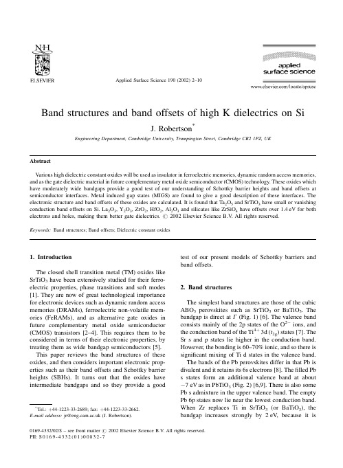
Band structures and band offsets of high K dielectrics on SiJ.Robertson *Engineering Department,Cambridge University,Trumpington Street,Cambridge CB21PZ,UKAbstractVarious high dielectric constant oxides will be used as insulator in ferroelectric memories,dynamic random access memories,and as the gate dielectric material in future complementary metal oxide semiconductor (CMOS)technology.These oxides which have moderately wide bandgaps provide a good test of our understanding of Schottky barrier heights and band offsets at semiconductor interfaces.Metal induced gap states (MIGS)are found to give a good description of these interfaces.The electronic structure and band offsets of these oxides are calculated.It is found that Ta 2O 5and SrTiO 3have small or vanishing conduction band offsets on 2O 3,Y 2O 3,ZrO 2,HfO 2,Al 2O 3and silicates like ZrSiO 4have offsets over 1.4eV for both electrons and holes,making them better gate dielectrics.#2002Elsevier Science B.V .All rights reserved.Keywords:Band structures;Band offsets;Dielectric constant oxides1.IntroductionThe closed shell transition metal (TM)oxides like SrTiO 3have been extensively studied for their ferro-electric properties,phase transitions and soft modes [1].They are now of great technological importance for electronic devices such as dynamic random access memories (DRAMs),ferroelectric non-volatile mem-ories (FeRAMs),and as alternative gate oxides in future complementary metal oxide semiconductor (CMOS)transistors [2±4].This requires them to be considered in terms of their electronic properties,by treating them as wide bandgap semiconductors [5].This paper reviews the band structures of these oxides,and then considers important electronic prop-erties such as their band offsets and Schottky barrier heights (SBHs).It turns out that the oxides have intermediate bandgaps and so they provide a goodtest of our present models of Schottky barriers and band offsets.2.Band structuresThe simplest band structures are those of the cubic ABO 3perovskites such as SrTiO 3or BaTiO 3.The bandgap is direct at G (Fig.1)[6].The valence band consists mainly of the 2p states of the O 2Àions,and the conduction band of the Ti 4 3d (t 2g )states [7].The Sr s and p states lie higher in the conduction band.However,the bonding is 60±70%ionic,and so there is signi®cant mixing of Ti d states in the valence band.The bands of the Pb perovskites differ in that Pb is divalent and it retains its 6s electrons [8].The ®lled Pb s states form an additional valence band at about À7eV as in PbTiO 3(Fig.2)[6,9].There is also some Pb s admixture in the upper valence band.The empty Pb 6p states now lie near the lowest conduction band.When Zr replaces Ti in SrTiO 3(or BaTiO 3),the bandgap increases strongly by 2eV,because itisApplied Surface Science 190(2002)2±10*Tel.: 44-1223-33-2689;fax: 44-1223-33-2662.E-mail address:jr@ (J.Robertson).0169-4332/02/$±see front matter #2002Elsevier Science B.V .All rights reserved.PII:S 0169-4332(01)00832-7controlled by the energy of the Zr d states.In contrast,in PZT,the Pb 6p states form the conduction band minimum,so the gap barely increases from 3.3to 3.7eV [10].It is recognised that the resonant covalence of Ti-d/O-p states is the origin of ferroelectricity in SrTiO 3type perovskites [11].In Pb perovskites,there is additional resonant covalence between Pb s and O p states which increases the ferroelectric polarity.SrBi 2Ta 2O 9is a layered crystal built from perovs-kite blocks separated by Bi 2O 2layers.It turns out that the Bi s and p states form the highest valence band and lowest conduction bands,respectively,while the ferro-electric response originates mainly from the TaO 3perovskite blocks [12].There is therefore an interest-ing separation of the functionality onto the Ta and Bi sub-lattices.Cubic ZrO 2has the ¯uorite structure.It has a simple band structure,as shown in Fig.3.The O p states form the valence band with a maximum at X [13].The conduction band minimum is at G ,and consists of Zr d states.The Zr d x 2Ày 2and d z 2states lie below the d xy states.The Zr s state lies midway between these at G ,but it disperses rapidly upwards.2.1.Models of Schottky barriers and semiconductor heterojunctionsThe band line-up of two semiconductors is deter-mined,like the SBH of a semiconductor on a metal,by charge transfer across the interface and the presence of any dipole layer at the interface.The charge transfer is that between the metal and the interface states of the semiconductor (Fig.4)[14].The charge transfertendsFig.1.Band structure of BaTiO 3calculated by pseudo-potential method [6].J.Robertson /Applied Surface Science 190(2002)2±103to align the Fermi level E F of the metal to the energy level of the interface states.The SBH for electrons f n between a semiconductor S and a metal M is f n S F M ÀF S F S Àw S(1)Here,F M is the work function of the metal,F S the energy of the semiconductor interface states,w S the semiconductor's electron af®nity (EA)and S the Schottky pinning parameter.S is given by [15]S11 e 2N d =ee 0(2)where e is the electronic charge,e 0the permittivity of free space,N the areal density of the interface states and d their decay length in the semiconductor.The dimensionless pinning factor S describes if the barrieris `pinned'or not.S varies between the limits S 1for unpinned Schottky barriers,and S 0for `Bardeen'barriers pinned by a high density of interface states in which the SBH is f n F S Àw S .There are numerous models of the origins of inter-face states,both intrinsic and extrinsic.In the intrinsic model originating from Bardeen and Heine,a semi-in®nite semiconductor in contact with a metal pos-sesses intrinsic states which are now called metal-induced gap states (MIGS)by Tersoff [14].F S is then the charge neutrality level (CNL)of the interface states,de®ned as the energy above which the states are empty for a neutral surface [16±18].On the other hand,the extrinsic models stress that the metal can react with the semiconductor [19].Brillson correlated the heat of reaction with S .This reaction maycreateFig.2.Band structure of PbTiO 3calculated by pseudo-potential method [6].4J.Robertson /Applied Surface Science 190(2002)2±10interface defects such as vacancies,whose gap states can pin the metal Fermi level,as noted by Spicer [20]and Dow [21].These models were supported by theobservation that pinning occurs even for monolayer coverage of metal,before the MIGS could be estab-lished.It is now believed that,overall,the intrinsic model gives a better description of Schottky barriers,because intrinsic states have a larger pinning dipole,N d ,than surface defects.The pinning parameter S has been in¯uential in our empirical understanding of Schottky barriers.Some years ago,Kurtin et al.[22]noted that S seemed to vary sharply with the ionicity of semi-conductor (Fig.5),from near 0for low ionicity semiconductors like Si and GaAs to 1for higher ionicity solids like SiO 2,SrTiO 3and KTaO 3.S is a dimensionless slope of barrier height to metal work function,S@f n @F M(3)Fig.3.Band structure of ZrO 2calculated by pseudo-potential method[6].Fig.4.Schematic diagram of SBHs.J.Robertson /Applied Surface Science 190(2002)2±105However,Louie [23]and Schluter [24]noted that Kurtin [22]had actually correlated the barrier heights to S H :S H@f n @X(4)which is the slope of barrier height to the Pauling electronegativity of the metal,and not the dimension-less S in (4).The work function and electronegativity vary roughly as [25,26]:F M 2:27X M 0:34(5)Thus,S H 2:27S ,and the Schottky limit should be S H 2:27.The data rarely reach this limit and Schluter [24]observed that S had a better correlation with the dielectric constant of the semiconductor e 0.Empiri-cally,Mo Ènch [14,27]found that S varied with e I as S11 0:1 e I À1 2(6)Certain materials are key tests of Schottky barriermodels.Diamond and xenon [14,28]have zero ioni-city but small e I ,and so their large S values show that S depends on e not on ionicity.This is tested by plotting log 1= S À1 against log e I À1 as in Fig.6.The wide gap oxides provide another key test,because they have intermediate e I values.SrTiO 3and KTaO 3were taken as high ionicity solids in the original Kurtin plot,with S H $1.However,this wasbefore data was actually known.When data [29]became available for SrTiO 3,showing S lying between 0.25and 0.4(Fig.6),it was clear that S is much lower.SrTiO 3falls well on the trend in Fig.3.The reason for this is that the SBHs depend on e I .e I is controlled by the states closest to the bandgap [5].In SrTiO 3,these are the moderately ionic Ti±O states of Ti±O bonds,not the highly ionic Sr±O states which lie well away from the gap and provide a much smaller contribution to e I .This can be seen in the partial density of states (DOS)of SrTiO 3in Fig.6.Thus,SrTiO 3and KTaO 3were misplaced in Fig.5as highly ionic solids.A lesser point is that the moderate value of S of SrTiO 3clearly correlates with e I ,and not with the low frequency dielectric constant e 0,which has a very large value for ferroelectrics and would give S %0from (6).SrTiO 3also serves as an evidence against the defect model,in that the barrier lies some way into the gap,not at the conduction band edge where the O vacancy states lie and would cause pinning.In sum-mary,the MIGS model of Schottky barriers holds for a wide range of solids of various ionicity and dielectric constants [5].The band alignment between two semiconductors is controlled by charge transfer and interface dipoles,just as Schottky barriers [30].For no dipoles,the Schottky limit,the conduction band offset isgivenFig.5.Schottky barrier pinning factor S H in the (incorrect)model of Kurtin etal.Fig. 6.Log±log plot of 1= S À1 vs.e I À1for various semiconductors and insulators to verify the MIGS model of Schottky barrier pinning factor S .6J.Robertson /Applied Surface Science 190(2002)2±10by the difference in their electron af®nities,the `elec-tron af®nity rule'.A similar idea was that for no charge transfer,the band line-ups are derived by placing each semiconductor's band on an absolute energy scale such as those of the free atom energy levels [31].Tersoff [16]showed that the band offset between two semiconductors a and b is controlled by interface dipoles as in the Schottky barrier,and so the conduc-tion band offset is given by f n w a ÀF CNL ;a À w b ÀF CNL ;bS F CNL ;a ÀF CNL ;b(7)The offsets are now described by aligning the CNLs of each semiconductor,modi®ed by the S factor.For simple semiconductors like Si,e I is large,and so S is small and the third term was negligible in the original formulation,but it is retained here for wide gap oxides.For strong pinning,the alignment is just given by the alignment of the two CNLs.The CNL energy below the vacuum level is a measure of the mean electronegativity of the semiconductor,in the same way that the work function of a metal is propor-tional to the metal's electronegativity.Thus,Eq.(7)says that the band alignment is the difference in electronegativity screened by the S factor.A wide ranging quantitative comparison found that the CNL models gives a good description of the band offsets [30].The CNL is the branch point of the semiconductor interface states.It is the integral of the Green's func-tion of the band structure,taken over the Brillouin zone [17],G E ZBZ N E H d H EE ÀE H0(8)Cardona and Christensen later provided a quicker method using a sum over special points of the Bril-louin zone [5,32].G E X i 1E ÀE i (9)2.2.Application to oxidesThe band alignments for the various wide gapoxides in contact with metal or silicon are found by calculating their CNLs and S parameters.The S factors are found from (6)using the experimental values of e Iand are shown in Table 1.The CNLs were found by calculating the oxide band structures by the tight-binding method [5,6,8,33].The tight-binding para-meters are found by ®tting to existing band structures [9,10,34],photoemission spectra and optical data [2,35±37].The CNLs for the various oxides are given in Table 1,together with the experimental values of their bandgaps and electron af®nities [2,38].SrTiO 3is an important oxide for future DRAM capacitor dielectrics.SrTiO 3is also the most studied system and the best test of our calculations.Fig.7compares the predicted SBHs of SrTiO 3on various metals with the experimental values [30,39±43].The experimental data are quite scattered but are quite consistent with S !1and our calculated value of 0.28.This shows that SrTiO 3is a key oxide in the tests of Schottky barrier models.The calculated barrier height for SrTiO 3on Pt is 0.9eV ,which is close to the 0.8eV found by photoemission by Copel et al.[43].However we cannot account for the much larger S value found by Shimizu et al.[42].BaTiO 3has similar band offsets to SrTiO 3.PbTi x Zr 1Àx O 3or PZT is an important ferroelectric for non-volatile memories,optical memories and other applications.The predicted barrier height for Pt onTable 1Calculated values for various oxides of their CNL and conduction band (CB)offset with Si aGap (eV)EA (eV)CNL (eV)e I S CB offset (eV)SiO 290.9 2.250.86 3.5b Si 3N 4 5.3 2.1 4.10.51 2.4b Ta 2O 5 4.4 3.3 3.3 4.840.40.3BaTiO 3 3.3 3.9 2.6 6.10.28À0.1BaZrO 3 5.3 2.6 3.740.530.8TiO 2 3.05 3.9 2.27.80.180.05ZrO 2 5.8 2.5 3.6 4.80.41 1.4HfO 26 2.5 3.740.53 1.5Al 2O 38.81c 5.5 3.40.63 2.8Y 2O 362c 2.4 4.40.46 2.3La 2O 36c 2c 2.440.53 2.3ZrSiO 46.5 2.4 3.6 3.80.56 1.5SrBi 2Ta 2O 94.13.53.35.30.4aExperimental values [36,37]of the bandgap,EA [2,38],dielectric constant e I [37]are also given.In Eqs.(2)and (5),F S is the energy of the CNL below the vacuum level,in this table,it is its energy above the valence band.bExperimental values.cEstimated values.J.Robertson /Applied Surface Science 190(2002)2±107PZT (Pb 0.55Zr 0.45O 3)is 1.45eV ,which is close to the 1.5eV measured by Dey et al.[44].The electron barrier of Pt on PZT is larger than that on BST because its CNL lies lower in the gap.This is because of the different band structure of PZT,in which the Pb 6s and 6p states form the band edges and this tends to lower the CNL.The larger value of the hole barrier than the electron barrier means that PZT thin ®lms can have predominantly electron injection,even though bulk PZT tends to be p-type.SrBi 2Ta 2O 9(SBT)is an important ferroelectric for non-volatile memories [2,45].It does not suffer from the loss of switchable polarisation (fatigue)when used with Pt electrodes,which is a problem for PZT.Note that more recent optical data ®nd that the bandgap of SBT is 4.1eV [2].The Schottky barrier of Pt is predicted to be 1.2eV ,which is essentially the same as that found by photoemission [46].There is an important need for high dielectric constant oxides to act as gate oxides instead of silicon dioxide [3,4].This is because the SiO 2layer is now so thin (2nm),that it no longer acts as a good insulator because of direct tunnelling across it.The solution is to replace SiO 2with a thicker layer of a medium k oxide,with the same equivalent capacitance or `equivalence oxide thickness't ox .The oxides must also satisfy certain other conditions,including chemi-cal stability in contact with Si [47].This rules out Ti and Ta which both react with Si to form SiO 2.The other key requirement is that they act as barriers toboth electrons and holes [5,32].This requires that both their valence and conduction band offsets be over 1eV .There is presently considerable effort to identify the most effective oxide,from a choice of ZrO 2,HfO 2,La 2O 3,Y 2O 3,Al 2O 3and the silicates ZrSiO 4and HfSiO 4.The calculated CB band offsets with Si are given in Table 1and summarised in Fig.8.They are compared in Table 2with recent experimental values [48±53],which is seen to be in good agreement.The important feature of Ta 2O 5and SrTiO 3is that both of them have CB offsets on Si under 1eV ,in fact 0in the case of SrTiO 3.This prediction was recently con®rmed by photoemission data of Chambers et al.[48].This means that SrTiO 3or BST cannot be a good gate oxide.The calculated CB offset for Ta 2O 5is only 0.36eV for Ta 2O 5on Si.This is consistent with recent photoemission data of Miyazaki and Hirose [49].Data for Ta 2O 5gate FETS also showed only a small elec-tron barrier [50].The CB offsets for BST and Ta 2O 5and BST are small or negligible because the bandgap is quite small and the band offsets are so asymmetric.To increasetheparison of calculated and observed SBHs of SrTiO 3on variousmetals.Fig.8.Predicted band offsets of various oxides on Si.Table 2Comparison of calculated and experimental values [48±53]of conduction band offsets on SiCalculatedExperiment References Ta 2O 50.350Miyazaki SrTiO 3À0.1<0.1Chambers ZrO 2 1.4 1.4Miyazaki 2.0Houssa Al 2O 32.82.8Ludeke8J.Robertson /Applied Surface Science 190(2002)2±10CB offset,we must either increase the bandgap or lower the CNL.The gap can be increased by raising the TM d levels,by using4d or5d metals instead of3d metals or using group IIIB metals instead of group IV. We should use zirconates,not titanates.The gap of BaZrO3is2eV wider than BaTiO3.Its offset is0.8eV.A better strategy is to lower the CNL.The CNL is lowered if the metal valence is lowered from4to3. Indeed,in Y2O3and La2O3,the CNL is much lower in the bandgap.Y2O3and La2O3are the oxides with largest CB offsets for reasonable dielectric constants. ZrO2has a bandgap of5.8eV,which is slightly wider than BaZrO3,and it also has a lower metal/ oxygen stoichiometry.This gives a larger CB offset for ZrO2(1.4eV)than BaZrO3,and indeed one which is just high enough.HfO2is similar.The calculated CB offset of1.4eV for ZrO2compares with an experi-mental value of1.4eV from photoemission[51]and a value of2eV by internal photoemission[52].This CB offset is large enough for devices.Zirconium silicate ZrSiO4and hafnium silicate HfSiO4are glassy oxides with bandgaps of $6.5eV.ZrSiO4consists of chains of alternate edge-sharing ZrO4and SiO2tetrahedra,with addi-tional Zr±O bonds between the chains,leading to an overall six-fold Zr coordination.We estimate the bandgap of ZrSiO4to be6.5eV.The calculated CB offsets are1.5eV,slightly more than ZrO2.Al2O3has a bandgap of8eV close to SiO2but with a higher k($9).Its calculated CB offset is2.8eV, which compares exactly with that measured by Ludeke et al.[53].Overall,the agreement between the calculated and subsequent experimental values for CB offsets in Table2is surprisingly good.References[1]M.E.Lines,X.Glass,Ferroelectrics,Oxford UniversityPress,Oxford,1990.[2]J.F.Scott,Ferroelectrics Rev.1(1998)1.[3]G.D.Wilk,R.M.Wallace,J.M.Anthony,J.Appl.Phys.89(2001)5243.[4]A.I.Kingon,J.P.Maria,S.K.Streiffer,Nature406(2000)1032.[5]J.Robertson,J.Vac.Sci.Technol.B18(2000)1785.[6]P.W.Peacock,J.Robertson,Unpublished work.[7]L.F.Mattheis,Phys.Rev.B6(1972)4718.[8]J.Robertson,W.L.Warren,B.A.Tuttle,D.Dimos,D.M.Smyth,Appl.Phys.Lett.63(1993)1519.[9]R.D.King-Smith,D.Vanderbilt,Phys.Rev.B49(1994)5828.[10]J.Robertson,W.L.Warren,B.A.Tuttle,J.Appl.Phys.77(1995)3975.[11]R.E.Cohen,Nature358(1992)136.[12]J.Robertson,C.W.Chen,W.L.Warren,C.D.Gutleben,Appl.Phys.Lett.69(1996)1704.[13]R.H.French,S.J.Glass,F.S.Ohuchi,Y.N.Xu,W.Y.Ching,Phys.Rev.B49(1994)5133.[14]W.MoÈnch,Phys.Rev.Lett.58(1987)1260.[15]W.MoÈnch,Surf.Sci.300(1994)928.[16]A.W.Cowley,S.M.Sze,J.Appl.Phys.36(1965)3212.[17]C.Tejedor,F.Flores,E.Louis,J.Phys.C10(1977)2163.[18]J.Tersoff,Phys.Rev.Lett.52(1984)465.[19]J.Tersoff,Phys.Rev.B30(1984)4874;J.Tersoff,Phys.Rev.B32(1985)6989.[20]L.J.Brillson,Surf.Sci.300(1994)909.[21]W.E.Spicer,T.Kendelewicz,N.Newman,K.K.Chin,I.Lindau,Surf.Sci.168(1986)240.[22]R.E.Allen,O.F.Sankey,J.D.Dow,Surf.Sci.168(1986)376.[23]S.Kurtin,T.C.McGill,C.A.Mead,Phys.Rev.Lett.30(1969)1433.[24]S.G.Louie,J.R.Chelikowsky,M.L.Cohen,Phys.Rev.B15(1977)2154.[25]M.Schluter,Phys.Rev.B17(1978)5044;M.Schluter,Thin Solid Films93(1982)3.[26]W.Gordy,W.J.O.Thomas,Phys.Rev.24(1956)439.[27]H.B.Michaelson,J.Appl.Phys.48(1977)4729.[28]W.MoÈnch,Phys.Rev.Lett.58(1986)1260.[29]W.MoÈnch,Europhys.Lett.27(1994)479.[30]R.C.Neville,C.A.Mead,J.Appl.Phys.43(1972)4657.[31]W.A.Harrison,J.Vac.Sci.Technol.14(1977)1016.[32]M.Cardona,N.E.Christensen,Phys.Rev.B35(1987)6182.[33]E.T.Yu,J.O.McCaldin,T.C.McGill,Solid State Phys.46(1992)1.[34]J.Robertson,C.W.Chen,Appl.Phys.Lett.74(1999)1168.[35]G.M.Rignanese,X.Gonze,A.Pasquarello,Phys.Rev.B63(2001)104305.[36]R.H.French,J.Am.Ceram.Soc.73(1990)477.[37]E.D.Palik,Handbook of Optical Properties of Solids,V ol.1±3,Academic Press,New York,1985.[38]W.Schmickler,J.W.Schultze,in:J.M.O'Bockris(Ed.),Modern Aspects of Electrochemistry,V ol.17,Plenum Press, London,1986.[39]G.W.Dietz,W.Antpohler,M.Klee,R.Waser,J.Appl.Phys.78(1995)6113.[40]H.Hasegawa,T.Nishino,J.Appl.Phys.69(1991)1501.[41]K.Abe,S.Komatsu,Jpn.J.Appl.Phys.31(1992)2985.[42]T.Shimizu,N.Gotoh,N.Shinozaki,H.Okushi,App.Surf.Sci.117(1997)400;()T.Shimizu,N.Gotoh,N.Shinozaki,H.Okushi,Mat.Res.Soc.Symp.Proc.(2000).[43]M.Copel,P.R.Duncombe,D.A.Neumayer,T.M.Shaw,R.M.Tromp,Appl.Phys.Lett.70(1997)3227.[44]S.K.Dey,J.J.Lee,P.Alluri,Jpn.J.Appl.Phys.34(1995)3134.[45]C.A.Paz de Araujo,J.D.Cuchiaro,L.D.McMillan,M.C.Scott,J.F.Scott,Nature374(1995)627.[46]C.D.Gutleben,Appl.Phys.Lett.71(1997)3444.[47]H.J.Hubbard,D.G.Schlom,J.Mater.Res.11(1996)2757.J.Robertson/Applied Surface Science190(2002)2±109[48]S.A.Chambers,Y.Liang,Z.Yu,R.Dropad,J.Ramdani,K.Eisenbeiser,Appl.Phys.Lett.77(2000)1662.[49]S.Miyazaki,Appl.Surface Science(2002)``these proceed-ings''.[50]S.Miyazaki,M.Narasaki,M.Ogasawara,M.Hirose,Microelec.Eng.59(2001)373.[51]A.Chatterjee,et al.,IEDM Tech Digest,1998,p.777.[52]M.Houssa,M.Tuominen,M.Nailli,V.Afansev, A.Stesmans,J.Appl.Phys.87(2000)8615.[53]R.Ludeke,M.T.Cuberes,E.Cartier,Appl.Phys.Lett.76(2000)2886;D.J.Maria,J.Appl.Phys.45(1974)5454.10J.Robertson/Applied Surface Science190(2002)2±10。
钛酸铅基化合物晶体结构及其负热膨胀性

论文题目:钛酸铅基化合物晶体结构及其负热膨胀性作者简介:陈骏,男,1979年8月出生,2001年9月师从于北京科技大学邢献然教授,于2007年3月获博士学位。
中文摘要钛酸铅(PbTiO3)是一种重要的钙钛矿结构的铁电体,在介电、压电、铁电、热释电等方面具有重要的研究与应用价值;同时,它在室温至居里温度范围内还表现出奇特的热缩冷胀行为,即负热膨胀性(NTE),这种负热膨胀行为是其它钙钛矿结构化合物所不具有的,如CaTiO3、BaTiO3、KNbO3、BiFeO3等。
研究PbTiO3的负热膨胀性将有利于开发出负热膨胀性可控以及零膨胀材料,拓展负热膨胀材料在实际中的应用,PbTiO3负热膨胀机理的研究可指导新型负热膨胀材料的开发。
本论文主要以钙钛矿结构的铁电材料Pb1-x A x Ti1-y B y O3(A=La、Sr、Cd、Bi、(La1/2K1/2)等;B=Fe、Zn等不同价态金属原子)为中心,研究A 位与B位替代对其负热膨胀性、晶体结构、点阵动力学的影响,实现负热膨胀性能可控,开发零膨胀材料,并研究PbTiO3负热膨胀机理。
本文研究了Pb1-x A x TiO3(A=La、Sr、(La1/2K1/2)、Cd)体系的固溶体特性、晶体结构以及负热膨胀性能受掺杂的影响。
La、Sr、(La1/2K1/2)的掺杂都使PbTiO3的轴比(c/a)及居里温度(T C)不同程度地线性下降,La的掺杂大幅度地降低了PbTiO3的负热膨胀性能,在0.15 ≤ x La ≤ 0.20范围内,Pb1-x La x TiO3表现出零膨胀性能。
PbTiO3-CdTiO3体系中,Cd的A位替代不仅提高了PbTiO3的轴比(c/a),而且增强了其负热膨胀效应,这是目前所发现的唯一能使PbTiO3负热膨胀性得到增强的A位掺杂体系。
Pb1-x La x TiO3晶体结构研究发现,La的掺杂使四方相点阵晶格中Pb/La与Ti原子的自发极化位移(c轴方向)非线性降低,然而位移比值(δPb/La/δTi)呈线性降低趋势,氧八面体具有刚性特征不受La掺杂的影响;Pb1-x Cd x TiO3体系X射线与中子衍射联合晶体结构研究发现,Pb1-x Cd x TiO3体现反常的晶体结构特征,虽然轴比(c/a)增加,但是Pb/Cd与Ti原子自发极化位移反常下降,从而导致居里温度的略微降低。
Evalueserve Circle of Experts - Primerus易唯思界专家primerus 共16页

Evalueserve Circle of Experts © 2019 Privileged and Confidential
Evalueserve – Knowledge Process Outsourcing Leader
© Evalueserve is the oldest and largest Knowledge Process Outsourcing (KPO) company that provides custom research services to leading-edge companies in the US and worldwide.
For Institutional Investors
© Fast access to industry experts with unique opinions on markets, companies products
© Enhance channel-check surveys with insights from experts in the field
→ Fraction of traditional cost → 100% customized projects → Access to experts in your field → Multi-industry experience
(10,000+ completed projects) → Highly scalable/rapid response → Flexible working relationship
© Evalueserve has six synergistic competencies—Investment Research, Business Research, Intellectual Property and Legal Support Services, Data and Financial Analytics, Market Research, and Circle of Experts.
马铃薯周皮中木栓形成层细胞和超微结构特点

The Importance of Phellogen Cells and their Structural Characteristics in Susceptibility and Resistance to Excoriation in Immature and MaturePotato Tuber (Solanum tuberosum L.)PeridermEDWARD C.LULAI*{and THOMAS P.FREEMAN {{USDA-ARS,Northern Crop Science Laboratory,P.O.Box 5677,University Station,Fargo,ND 58105-5677,USA and {North Dakota State University,Northern Crop Science Laboratory,P.O.Box 5677,University Station,Fargo,ND 58105-5677,USAReceived:10April 2001Returned for revision:13May 2001Accepted:13June 2001Published electronically:17August 2001Potato tuber (Solanum tuberosum L.)periderm maturation is an important physiological process that directly a ects the susceptibility and development of resistance to costly excoriation (skinning-type wounds)at harvest.The objectives of this research were to identify the speci®c types of cells and the cellular changes associated with susceptibility and resistance to tuber excoriation in immature and mature tubers respectively.Epi¯uorescent microscopic examination of immature tuber periderm (phellem,phellogen and phelloderm cells)from several genetically diverse cultivars has shown that the cellular damage resulting from excoriation occurs within the phellogen (cork cambium),a meristematic layer of cells that gives rise to neighbouring phellem and phelloderm cells.Tuber excoriation is the result of the fracture of radial phellogen cell walls linking the skin (phellem)to the phelloderm.As the tuber periderm matures,phellogen cells become inactive and the radial walls of these cells become more resistant to fracture;resistance to excoriation develops.Ultrastructural studies of immature tuber periderm show that radial walls of active phellogen cells are thin and fragile.During periderm maturation,both radial and tangential phellogen cell walls thicken as they strengthen and become resistant to fracture,thereby providing resistance to excoriation.These results refute previous theories of the physiological changes responsible for the onset of resistance to tuber skinning injury.The combined results establish a paradigm whereby the thickening and strengthening of tuber phellogen cell walls upon periderm maturation are the determinant for resistance to tuber excoriation.Key words:Cambium,meristematic,periderm,phellem,phelloderm,phellogen,potato,skinning,Solanum tuberosum L.,0tuber.INTRODUCTIONPotato tuber periderm is of great agricultural importance and scienti®c interest because of the protection a orded by the intact periderm to the tuber against pathogens and dehydration (Soliday et al .,1979;Peterson et al .,1985;Lulai and Orr,1994,1995;Lulai and Corsini,1998).The periderm covering immature tubers is very fragile and is susceptible to wounding by excoriation (idiom skinning);this type of wounding is a serious and costly problem at harvest (Lulai and Orr,1993).The term skinning is poorly and anecdotally de®ned and has been used to imply that the entire periderm is physically detached from the underlying tuber cells.The term excoriation is interchangeable with the term skinning in describing this type of wound,but without the anecdotally derived implications and de®nitions.Matura-tion of the periderm,during and after potato vine desiccation,is an important physiological process that is directly related to the development of resistance to excoria-tion (Lulai and Orr,1993).Although slow or hindereddevelopment of resistance to excoriation during periderm maturation constitutes a serious problem,the anatomical and physiological basis for susceptibility and resistance of the potato tuber to skinning injury has not been addressed scienti®cally.Confusion and uncertainty concerning tuber periderm physiology remains.Reeve et al .(1969)indicated that potato tuber periderm is composed of:(1)phellem (suberized cells);(2)phellogen (cork cambium);and (3)phelloderm tissues.Lyshede's (1977)analysis of mature tuber periderm did not produce de®nitive evidence for the presence of a phellogen or phelloderm.The suberization processes involved in phellem development are only partially characterized (Kolattukudy,1984;Lulai and Morgan,1992;Thomson et al .,1995;Bernards and Lewis,1998;Lulai and Corsini,1998).There are no published data identifying or relating maturational changes in phellem,phellogen or phelloderm to susceptibility and resistance to tuber excoriation.Perhaps more importantly,there is no information on the changes that occur in phellogen cell walls when the cork cambium becomes inactive.The lack of information on the type of periderm cells and cellular changes that are responsible for susceptibility and resistance to excoriation has hampered the development of e ective,rational approaches to describeAnnals of Botany 88:555±561,2001doi:10.1006/anbo.2001.1497,available online at on0305-7364/01/100555+07$35.00/00Mention of a proprietary product name is for identi®cation only and does not imply a warranty or an endorsement to the exclusion of other products that may be similar.*For correspondence.Fax 0017012391349,e-mail lulaie@periderm maturation and to solve the costly problem of tuber skinning injuries that occur during harvest.The skin of the potato tuber is the protective part of the tuber surface tissue that is easily excoriated from immature tubers.Although the skin is an important part of the periderm,it has not been clearly identi®ed or de®ned scien-ti®cally and it is often mistakenly referred to as constituting the total tuber periderm.The confusion surrounding excoriation and use of the terms`skin'and`skinning'has been exacerbated because skin thickness and suberization have been proposed to be responsible for susceptibility and resistance to tuber excoriation in immature and mature tubers(Hiller et al.,1985;de Haan,1987;Hiller and Thornton,1993).However,these theories arose anecdotally and have not been researched or con®rmed scienti®cally. Controlled environment studies have shown that low relative humidity hastens periderm maturation in freshly harvested tubers(Lulai and Orr,1993)and that periderm maturation is more rapid in tubers from cultivars with characteristically higher water vapour loss(Lulai and Orr, 1994).Consequently,the®rst layer of fully hydrated cells within the periderm,i.e.the phellogen,should play an important role in tuber periderm maturation and excoria-tion.However,there is no published information on the changes that occur within the cork cambium/phellogen of potato tuber periderm as growth ceases and as the periderm matures.Extensive studies have been conducted on the structure,ultrastructure,cytology and biochemistry of the vascular cambium of woody plants and taproots as plants cycle through dormancy and growth.These are periods when the vascular cambium correspondingly cycles from inactive to active meristematic activity(Catesson,1994; Catesson et al.,1994;Cha ey et al.,1997,1998;Lachaud et al.,1999).Similar information is not available for cell walls of meristematically active and inactive cork cambium/ phellogen from potato tuber or other plants.However, changes in cell wall architecture of the vascular cambium from perennial plants may only serve as a partial model for the cork cambium/phellogen from periderm tissues of annual plants such as potato tubers.We conducted a series of experiments with several potato cultivars raised during three growing seasons to identify the type of periderm cells and cellular characteristics that are responsible for susceptibility to tuber excoriation,or skin-ning injury,and the changes responsible for the develop-ment of resistance to skinning injury.The terms`skin'and `skinning'are rede®ned and a structural model for skinning and resistance to skinning(skin-set)is presented.These results are requisites to the identi®cation of the biochemical processes and mechanisms that regulate the development of resistance to tuber excoriation.MATERIALS AND METHODSPotato cultivarsPotato tubers from cultivars with diverse genetic back-grounds were sampled from plants grown using standard cultural practices in non-irrigated®elds during three growing seasons.The results presented were obtained from cultivars with rapid(`Goldrush'and`Russet Norkotah')and slow (`Red Ruby'and`Kennebec')periderm maturation(Lulai and Orr,1993,1994).Tubers that were immature(suscept-ible to excoriation/skinning),maturing or approaching periderm maturity(developing resistance to excoriation/ skinning)were sampled from growing plants,senescing plants and plants whose vines were dead and desiccated, respectively.Harvested tubers were stored in controlled environment chambers(95%relative humidity and98C)to allow postharvest sampling when the periderm was fully mature(resistant to skinning).The periderms of the sampled tubers were examined microscopically to identify the cellular layers damaged during skinning and to determine the structural characteristics of these cells before and after development of resistance to skinning.The periderm properties observed in this study were similar for all growing seasons.Determination of resistance to excoriation(skinning injury) in relation to phellogen activityThe time course for development of resistance to skinning injury in comparison to phellogen activity was determined objectively using a mechanical skin-set(resist-ance to skinning/excoriation)measuring device described by Lulai and Orr(1993).The device measured the amount of torsional force(milliNewton meters)required to produce skinning injury.Phellogen activity was determined in a manner similar to that described previously for vascular cambium activity,as reviewed and described by Larson(1994b).Brie¯y,this activity value was determined by counting the number of active phellogen cells and nascent phellogen derivatives in a segment of the cork cambial layer and dividing by the total number of columns of adjoining phellem cells produced from this phellogen.These calculations provided the number of cell layers associated with active phellogen that gave rise to a single column of phellem cells.The measured changes in resistance to skinning injury and phellogen activity occurred over the time course of periderm matu-ration during and after potato vine desiccation and autumn harvest.Tissue sampling,preparation and histochemical analysis for ¯uorescence microscopyAt each sampling time six tubers from each cultivar were rinsed in distilled water.Tissue blocks(2mmÂ3mmÂ4mm deep)of periderm and neighbouring cortical tissues were excised from the equatorial region of each tuber surface and®xed in a solution of formalin:acetic acid:95% ethanol:water(3:1:10:7,v/v/v/v).Excoriated samples were obtained from tubers whose periderm had separated naturally and from undamaged portions of the same tubers which had the layer of skin gently pulled from the tuber with a pair of forceps;these approaches produced identical tissue injury.Fixed tissues were dehydrated in a tertiary-butyl alcohol series,embedded in para n(Paraplast Plus,Sigma, St.Louis,MO,USA),sectioned and depara nized as described previously(Lulai and Morgan,1992).The556Lulai and FreemanÐPotato Periderm Maturationfollowing histochemical treatments were employed as out-lined previously(Lulai and Morgan,1992):berberine and ruthenium red were used to identify suberin polyphenolic accumulations;neutral red and toluidine blue O were used to identify suberin polyaliphatic accumulations;and Sudan III/IV to stain polyaliphatic accumulations a faint pink while maintaining pale yellow auto¯uorescence of the polyphenolic accumulations.Light microscopy was per-formed as outlined previously(Lulai and Morgan,1992) using a Zeiss Axioskop50microscope con®gured for epi¯uorescent illumination using the model100illuminator equipped with an HBO50W/L2mercury lamp.Blue-violet excitation was employed for neutral red and berberine induced¯uorescence,and Sudan III/IV colouration and auto¯uorescence(exciter®lter BP436/10,dichromatic beam-splitter FT460,barrier®lter LP470).Tissue sampling and preparation for electron microscopy Sampling for ultrastructural studies included:(1)imma-ture tubers that were freshly harvested and susceptible to skinning injury;and(2)mature tubers that had aged and were resistant to skinning injury.For transmission electron microscopy(TEM),samples from six tubers of each cultivar were prepared by removing a small block (8mmÂ4mmÂ4mm deep)of the periderm and adjoin-ing cortical tissue.Each of these samples was then placed in 0.1M phosphate bu er(pH7.4)and was subdivided into approx.2mm cubes and®xed for20h in bu ered2.5% glutaraldehyde,washed in phosphate bu er,and post®xed for6h at228C in phosphate-bu ered2%osmium tetroxide.Samples were dehydrated in a graded acetone series and stained en bloc with saturated uranyl acetate in 70%acetone prior to being embedded in Spurr's resin. Ultrathin sections(600to8008A)were cut with a diamond knife.Obliquely cut sections do not give rise to the appearance of symmetrical cell walls.Consequently,the tissue blocks were aligned as closely as possible for sectioning at right angles to the tuber surface.However, slight obliqueness is unavoidable and these sections can change the apparent cell wall symmetry.The sections were stained with lead citrate and examined on a JEOL100CX transmission electron microscope.For scanning electron microscopy(SEM),samples of each cultivar containing the periderm were cut from the tuber and placed in acidi®ed2-2dimethoxypropane(DMP) for®xation and rapid dehydration.Samples were removed from DMP and were washed with several changes of absolute ethanol and critical point dried in a Tousimis810 critical point drier using CO2as the transitional¯uid.Dried specimens were coated with Au/Pd in a Balzer SCD030 sputter coater and examined and photographed using a JEOL6300scanning electron microscope.RESULTSFluorescence microscopy of tuber periderm Fluorescence microscopy of immature potato tuber periderm shows that periderm damage incurred during excoriation involves the separation of the phellem from the underlying tissue,and that the phellem constitutes what has loosely been referred to as the skin(Fig.1A).At higher magni®ca-tion(Fig.1B),it is apparent that the separation occurred within the phellogen and that there was no fracturing or separation within the layer of suberized cells(phellem).The same results were obtained using berberine/ruthenium red and neutral red/toluidine blue O(micrographs not shown) to visualize the suberin polymers and thereby identify the phellem cells.The phellogen interfaces the phellem and phelloderm.The phelloderm,consisting of a loosely organized rectangular matrix of cells,can be discerned in Fig.1D.The radial walls of the phellogen were susceptible to fracture(Fig.1A and B).Careful analysis of the micrographs of the active phellogen at higher magni®cation (Fig.1B and C)shows that the radial and tangential cell walls appear frail and wispy.The fragility of radial and tangential cell walls of the active phellogen was also evidenced by the ease with which they were damaged during embedding and sectioning,particularly for TEM.Mature tuber periderm(Fig.1D)was resistant to skinning injury. Phellogen cell walls from mature tuber periderm were not fragile and were indistinguishable from the cell walls of the adjoining phelloderm which were strong and resistant to mechanical fracture.Higher magni®cation showed that the phellogen cell walls of immature tuber periderm were weakly auto¯uorescent(Fig.1C).Phellogen cell walls did not ¯uoresce after tandem treatment with suberin¯uoro-chromes and auto¯uorescent quenching agents(berberine/ ruthenium red or neutral red/toluidine blue O),indicating that the walls did not contain suberin lignin-like polyphe-nolic or polyaliphatic material.Since no¯uorescent signals were obtained,these micrographs are not shown.Electron microscopy of tuber peridermResults from SEM analysis(Fig.2A)show the contrast-ing morphology and integration of the three types of cells that constitute the periderm:phellem,phellogen,and phelloderm.The TEM micrographs in Fig.2B±G reveal more ultrastructural details relevant to the susceptibility or resistance of phellogen cell walls to fracture and tuber skinning injury.In immature periderm,the radial cell walls of the phellogen were thin compared to the adjoining tangential phellem cell walls(Fig.2B).Phellogen radial cell walls were sometimes folded(Fig.2C),unlike the rigid radial walls of the phellem or phelloderm.Folding of these ¯exible walls may have been due to changes in turgor in situ or may have occurred upon dehydration for tissue embedding.The fragility of radial phellogen cell walls of immature periderm was evidenced by their ease of fracture upon tuber excoriation and during embedding and section-ing(Fig.2D).Two months after the tubers were harvested and the periderm was no longer susceptible to excoriation, i.e.after the periderm had matured,the radial walls of the inactive phellogen were noticeably thickened and were no longer prone to fracture(Fig.2E).Similarly,phellogen tangential cell walls from mature tubers(Fig.2F)were thicker than those from tubers with immature periderm (Fig.2G).Plasmodesmata were observed in the cell walls ofLulai and FreemanÐPotato Periderm Maturation557inactive phellogen (mature periderm),but were not detected in the walls of the active phellogen cells (immature periderm)that we examined.Resistance to excoriation in relation to phellogen activity In addition to the changes noted above,our results show that an inverse relationship exists between the development of resistance to tuber skinning injury,and the combined number of layers of active phellogen and immediate/nascent phellogen derivatives (Fig.3).As phellogen activity decreased,the resistance to skinning injury increased.Results for all experiments were similar for all growing seasons.DISCUSSIONIn immature tuber periderm,the phellogen is the speci®c layer of cells that is labile and prone to fracture and allows separation to occur within the periderm causing excoria-tion/skinning injury (Fig.1A and B ).Although potato tuber excoriation during harvest has been a costly and persistent problem (Murphy,1968;Hiller et al .,1985;Lulai and Orr,1993),this study is the ®rst to identify and describe the type of tuber periderm cells and cell wall changes responsible for susceptibility and resistance to tuber skinning injury.Previous theories incorrectly held that periderm thickening,skin thickening,or suberization cause the development of resistance to excoriation during periderm maturation (Hiller et al .,1985;de Haan,1987;Hiller and Thornton,1993).Our results show that the phellem constitutes what has loosely been referred to as the skin,and that periderm or skin thickening and suberization are not involved in resistance to excoriation because there is no separation within the phellem upon skinning injury.Excoriation results in tissue separation that is speci®c to the layer of phellogen cells.These microscopical studies of excoriation and periderm architecture also show the presence of a loosely structured phelloderm which may be slightly easier to identify in periderm possessing a meristematically active phellogen,but can also be detected in mature periderm (Fig.1AandF IG .1.Periderm tissues:phellem (PM),phellogen (PG)and phelloderm (PD)and neighbouring cortical (C)cells from `Kennebec'tubers.Tissues were treated with Sudan III/IV to stain phellem cells for identi®cation.The tuber surface is oriented to the top of each micrograph resulting in tangential walls running horizontally and radial walls running vertically.The Sudan treatment retains intense auto¯uorescence of phellem cell walls and a low intensity auto¯uorescence of non-suberized cell walls,which facilitates detection of phellogen and phelloderm cell walls.Figure 1A±C shows immature periderm with active phellogen.Note the separation of the phellem from neighbouring tissues and the fracturing of phellogen cell walls in Fig.1A and B (arrowheads).The fragile appearance and the dim auto¯uorescence of the nascent phellogen cell walls is noticeable in Fig.1C (arrows).The enhanced contrast of the black and white inset in Fig.1B helps in the detection of phellogen cell walls and their fracture (arrowhead).Figure 1D shows cell walls from mature periderm.The cell walls of inactive phellogen in mature periderm (Fig.1D )appear more clearly de®ned than the characteristically fragile cell walls of active phellogen (Fig.1A±C ).558Lulai and FreemanÐPotato Periderm MaturationD ).The few cell layers which constitute the phelloderm show typical phelloderm architecture in that they are characterized by loosely organized layers of starch-depleted cells between the phellogen and cortex (Fig.2A ).Earlier results,which indicated that there was no easily discernable phelloderm present in potato tuber (Lyshede,1977),wereF IG .2.SEM and TEM micrographs of potato tuber periderm showing phellem (PM),phellogen (PG)and phelloderm (PD)and neighbouring cortical (C)cells.Tangential walls are arranged horizontally and radial walls vertically.Figure 2A illustrates the contrasting morphology of the periderm cells and neighbouring cortical cells.The TEM micrographs illustrate the following:Fig.2B ,a radial (R)phellogen cell wall connecting to a lower tangential (LT)phellem cell wall in immature tuber periderm;Fig.2C ,a radial (R)phellogen cell wall connecting to a lower tangential (LT)phellem cell wall in maturing tuber periderm;Fig.2D ,a fractured radial (R)phellogen cell wall connecting to an intact lower tangential (LT)phellogen cell wall in immature periderm;Fig.2E ,a thickened phellogen radial (R)cell wall from mature periderm;Fig.2F ,a thickened lower tangential (LT)phellogen cell wall from mature periderm;and Fig.2G ,a thin lower tangential (LT)phellogen cell wall from immature periderm.Lulai and FreemanÐPotato Periderm Maturation 559obtained with mature tubers,and consequently there was no active phellogen present for easy anatomical reference.The fragile,nascent walls of active phellogen of immature periderm can be distinguished from the walls of inactive phellogen in mature periderm.Walls of inactive phellogen were not easily distinguished from walls of adjoining phelloderm and cortical cells.The walls of inactive phellogen,adjoining phelloderm and cortical cells were commonly observed to possess plasmodesmata,but these were not apparent in the sections of active phellogen that we examined,suggesting that they are di cult to detect or are rare in the cell walls of active phellogen.These results may be considered consistent with the concept of secondary formation of plasmodesmata proposed for vascular cambial cells (Larson,1994a ).Phellogen cell walls change morphologically as the periderm matures and the phellogen becomes inactive.The thicker cell walls present in inactive phellogen of mature periderm (Fig.2E and F )are apparently stronger and more resistant to fracture than the noticeably thinner cell walls of active phellogen cells from immature periderm (Fig.2B ,D and G ).Susceptibility to skinning decreases during phellogen inactivation.Skinning does not occur in mature periderm (Lulai and Orr,1993)which we have found to be characterized by thickened phellogen cell walls.The thickening of phellogen walls,especially radial walls,upon periderm maturation,appears to be a primary physiological and structural basis for the development of resistance to tuber skinning.Like the walls of tuber parenchyma cells,the strong,thickened walls of the inactive phellogen do not accumulate lignin or lignin-like material upon periderm maturation.The nascent radial and tangential cell walls of active phellogen,unlike the thickened cell walls of inactive phellogen which resemble phelloderm,did not display a readily recognizable middle lamella or other morphological characteristics found in neighbouring phelloderm cell walls which are thickened and resistant to fracture.A portion of a middle lamella and stained,electron-dense suberin from a fully formed neighbouring phellem cell could sometimes be detected extending a short distance into the thin,non-lamellar adjoining radial wall of meristematically active phellogen (Fig.2B ).The walls of meristematic cells are not rigid,but must be elastic to withstand changes in turgor pressures associated with growth (Iiyama et al .,1994).Previous results showing a relationship between rapid tuber water vapour transpiration and rapid periderm maturation (Lulai and Orr,1994)are consistent with the completion of cell growth and may play a role in cell wall sti ening or thickening.Cell wall asymmetry was commonly observed in non-meristematic cells (Fig 2E and F ).These di erences may be due to the angle of section,physiological activity or stage of maturation.The absence of a recognizable middle lamella in the cell walls of meristematically active phellogen is consistent with that of tangential walls of meristematically active vascular cambium (Roland,1978;Catesson and Roland,1981).However,unlike cell walls in the active cambial zone of hardwood taproots,and shoots which have radial walls that are noticeably thicker than the adjoining tangential cell walls (Cha ey et al .,1997;Lachaud et al .,1999),we found that radial and tangential cell walls from active tuber phellogen are equally thin.These di erences in radial wall thicknesses are consistent with the patterns of growth for trees,where the annual growth in height is much greater2.22.01.81.61.41.20.00.80.60.40.20.08/314504003503002502001509/79/149/2110/510/1210/2611/2*10/199/28Calendar date(T o r q u e m N .M )R e s i s t a n c e t o s k i n n i n gP h e l l o g e n a c t i v i t y (l a y e r s o f a c t i v e p h e l l o g e n a n d d e r i v a t i v e s )F IG .3.Relationship between phellogen activity (number of layers of phellogen cells and immediate derivatives)and resistance to excoriation/skinning injury (torque in mN m À1)for tubers of `Norkotah'and `Kennebec'during the ®nal stages of growth and periderm maturation.Note that as phellogen activity decreases during periderm maturation,the resistance to skinning injury increases.The arrows pointing diagonally upward indicate that the resistance to skinning had increased beyond the measurable range of the technique and that the tubers were becoming very resistant to skinning injury.*,Tubers were harvested late,28September,to allow for periderm maturation in the ®eld and were then placed into a controlled environment chamber (95%RH and 98C)to allow the periderm to mature further until sampled for analysis on 2November.560Lulai and FreemanÐPotato Periderm Maturationthan that in girth,vs.potato tuber,where vigorous growth is expressed laterally and longitudinally.Another major di erence is that the meristematic activity of the vascular cambium of these woody plants cycles annually from dormancy to growth with the vascular cambial cell walls thickening during dormancy and then becoming thin as meristematic activity resumes during the growing season (Lachaud et al.,1999).This reactivation and cycling of meristematic activity does not occur in the phellogen/cork cambium of tuber periderm because of di erences in biology,including the fact that potato is not a perennial plant.The biological changes and biochemical processes associated with phellogen cell wall thickening during phellogen inactivation are currently being investigated.In summary,these results de®ne the tuber skin as the phellem layer of the periderm.The phellogen is clearly identi®ed as the speci®c layer of periderm tissues directly involved in tuber excoriation/skinning injury.The cell wall characteristics that are responsible for susceptibility and the onset of resistance to tuber excoriation upon potato tuber periderm maturation are synonymous with active and inactive phellogen.Excoriation is related to a prototypal description of the ultrastructural di erences between active and inactive tuber phellogen cells.The results provide a useful model that is the®rst description of the structural physiology of immature vs.mature periderm in relation to potato tuber skinning injury.ACKNOWLEDGEMENTSWe thank Thomas J.Wirta,Kathy L.Iverson,Scott A. Payne and Je rey ler for their technical assistance.#2001US GovernmentLITERATURE CITEDBernards MA,Lewis NG.1998.The macromolecular aromatic domain in suberized tissue:a changing paradigm.Phytochemistry47: 915±933.Catesson AM.1994.Cambial ultrastructure and biochemistry:changes in relation to vascular tissue di erentiation and the seasonal cycle.International Journal of Plant Science155:251±261. Catesson AM,Roland JC.1981.Sequential changes associated with cell wall formation and fusion in the vascular cambium.IAWA Bulletin2:151±162.Catesson AM,Funada R,Robert-Baby D,Quinet-Szely M,Chu-Ba J, Goldberg R.1994.Biochemical and cytochemical cell wall changes across the cambial zone.IAWA Journal15:91±101.Cha ey N,Barnett J,Barlow P.1997.Endomembranes,cytoskeleton, and cell walls:aspects of the ultrastructure of the vascular cambium of taproots of Aesculus hippocastanum L.(Hippo-castanaceae).International Journal of Plant Science158:97±109. Cha ey NJ,Barlow PW,Barnett JR.1998.A seasonal cycle of cell wall structure is accompanied by a cyclical rearrangement of cortical microtubules in fusiform cambial cells within taproots of Aesculushippocastanum(Hippocastanaceae).New Phytologist139: 623±635.de Haan PH.1987.Damage to potatoes.In:Rastovski A,van Es A et al,eds.Storage of potatoes:post-harvest behavior,store design, storage practice,handling.Wageningen,The Netherlands:Pudoc, 371±380.Hiller LK,Thornton RE.1993.Management of physiological disorders.In:Rowe RC,ed.Potato health management.St.Paul,Minnesota, USA:APS Press,87±94.Hiller LK,Koller DC,Thornton RE.1985.Physiological disorders of potato tubers.In:Li PH,ed.Potato physiology.New York: Academic Press Inc,389±455.Iiyama K,Lam TBT,Stone BA.1994.Covalent cross-links in the cell wall.Plant Physiology104:315±320.Kolattukudy PE.1984.Biochemistry and function of cutin and suberin.Canadian Journal of Botany62:2918±2933.Lachaud S,Catesson AM,Bonnemain JL.1999.Structure and functions of the vascular ptes Rendus de l'Academie des Sciences,Paris322:633±650.Larson PR.1994a.Anticlinal cambial divisions.In:Larson PR,ed.The vascular cambium,development and structure.Berlin,Heidelberg: Springer-Verlag,155±318.Larson PR.1994b.Cambial zone characteristics.In:Larson PR,ed.The vascular cambium,development and structure.Berlin,Heidel-berg:Springer-Verlag,587±637.Lulai EC,Corsini DL.1998.Di erential deposition of suberin phenolic and aliphatic domains and their roles in resistance to infection during potato tuber(Solanum tuberosum L.)wound-healing.Physiological and Molecular Plant Pathology53:209±222. Lulai EC,Morgan WC.1992.Histochemical probing of potato periderm with neutral red:a sensitive cyto¯uorochrome for the hydrophobic domain of suberin.Biotechnic and Histochemistry67: 185±195.Lulai EC,Orr PH.1993.Determining the feasibility of measuring genotypic di erences in skin-set.American Potato Journal70: 599±609.Lulai EC,Orr PH.1994.Techniques for detecting and measuring developmental and maturational changes in tuber native periderm.American Potato Journal71:489±505.Lulai EC,Orr PH.1995.Porometric measurements indicate wound severity and tuber maturity a ect the early stages of wound-healing.American Potato Journal72:225±241.Lyshede OB.1977.Studies on the periderm and epidermis of the potato tuber Solanum tuberosum L.cv.Bintje.In:Yearbook of the Royal Veterinary and Agricultural University.Copenhagen, Denmark,68±74.Murphy HJ.1968.Potato vine killing.American Potato Journal45: 472±477.Peterson RL,Barker WG,Howarth MJ.1985.Development and structure of tubers.In:Li PH,ed.Potato physiology.New York: Academic Press Inc,123±152.Reeve RM,Hautala E,Weaver ML.1969.Anatomy and compositional variation with potatoes.I.Developmental histology of the tuber.American Potato Journal46:361±373.Roland JC.1978.Early di erences between radial walls and tangential walls of actively growing cambial zone.IAWA Bulletin1:7±10. Soliday CL,Kolattukudy PE,Davis RW.1979.Chemical evidence that waxes associated with the suberin polymer constitute the major di usion barrier to water vapor.Planta146:607±614. Thomson N,Ray FE,Kelman A.1995.Wound healing in whole potato tubers:a cytochemical,¯uorescence,and ultrastructural analysis of cut and bruise wounds.Canadian Journal of Botany73: 1436±1450.Lulai and FreemanÐPotato Periderm Maturation561。
基于自转一阶非连续式微球双平盘研磨的运动学分析与实验研究
第53卷第8期表面技术2024年4月SURFACE TECHNOLOGY·133·基于自转一阶非连续式微球双平盘研磨的运动学分析与实验研究吕迅1,2*,李媛媛1,欧阳洋1,焦荣辉1,王君1,杨雨泽1(1.浙江工业大学 机械工程学院,杭州 310023;2.新昌浙江工业大学科学技术研究院,浙江 绍兴 312500)摘要:目的分析不同研磨压力、下研磨盘转速、保持架偏心距和固着磨料粒度对微球精度的影响,确定自转一阶非连续式双平面研磨方式在加工GCr15轴承钢球时的最优研磨参数,提高微球的形状精度和表面质量。
方法首先对自转一阶非连续式双平盘研磨方式微球进行运动学分析,引入滑动比衡量微球在不同摩擦因数区域的运动状态,建立自转一阶非连续式双平盘研磨方式下的微球轨迹仿真模型,利用MATLAB对研磨轨迹进行仿真,分析滑动比对研磨轨迹包络情况的影响。
搭建自转一阶非连续式微球双平面研磨方式的实验平台,采用单因素实验分析主要研磨参数对微球精度的影响,得到考虑圆度和表面粗糙度的最优参数组合。
结果实验结果表明,在研磨压力为0.10 N、下研磨盘转速为20 r/min、保持架偏心距为90 mm、固着磨料粒度为3000目时,微球圆度由研磨前的1.14 μm下降至0.25 μm,表面粗糙度由0.129 1 μm下降至0.029 0 μm。
结论在自转一阶非连续式微球双平盘研磨方式下,微球自转轴方位角发生突变,使研磨轨迹全覆盖在球坯表面。
随着研磨压力、下研磨盘转速、保持架偏心距的增大,微球圆度和表面粗糙度呈现先降低后升高的趋势。
随着研磨压力与下研磨盘转速的增大,材料去除速率不断增大,随着保持架偏心距的增大,材料去除速率降低。
随着固着磨料粒度的减小,微球的圆度和表面粗糙度降低,材料去除速率降低。
关键词:自转一阶非连续;双平盘研磨;微球;运动学分析;研磨轨迹;研磨参数中图分类号:TG356.28 文献标志码:A 文章编号:1001-3660(2024)08-0133-12DOI:10.16490/ki.issn.1001-3660.2024.08.012Kinematic Analysis and Experimental Study of Microsphere Double-plane Lapping Based on Rotation Function First-order DiscontinuityLYU Xun1,2*, LI Yuanyuan1, OU Yangyang1, JIAO Ronghui1, WANG Jun1, YANG Yuze1(1. College of Mechanical Engineering, Zhejiang University of Technology, Hangzhou 310023, China;2. Xinchang Research Institute of Zhejiang University of Technology, Zhejiang Shaoxing 312500, China)ABSTRACT: Microspheres are critical components of precision machinery such as miniature bearings and lead screws. Their surface quality, roundness, and batch consistency have a crucial impact on the quality and lifespan of mechanical parts. Due to收稿日期:2023-07-28;修订日期:2023-09-26Received:2023-07-28;Revised:2023-09-26基金项目:国家自然科学基金(51975531)Fund:National Natural Science Foundation of China (51975531)引文格式:吕迅, 李媛媛, 欧阳洋, 等. 基于自转一阶非连续式微球双平盘研磨的运动学分析与实验研究[J]. 表面技术, 2024, 53(8): 133-144.LYU Xun, LI Yuanyuan, OU Yangyang, et al. Kinematic Analysis and Experimental Study of Microsphere Double-plane Lapping Based on Rotation Function First-order Discontinuity[J]. Surface Technology, 2024, 53(8): 133-144.*通信作者(Corresponding author)·134·表面技术 2024年4月their small size and light weight, existing ball processing methods are used to achieve high-precision machining of microspheres. Traditional concentric spherical lapping methods, with three sets of circular ring trajectories, result in poor lapping accuracy. To achieve efficient and high-precision processing of microspheres, the work aims to propose a method based on the first-order discontinuity of rotation for double-plane lapping of microspheres. Firstly, the principle of the first-order discontinuity of rotation for double-plane lapping of microspheres was analyzed, and it was found that the movement of the microsphere changed when it was in different regions of the upper variable friction plate, resulting in a sudden change in the microsphere's rotational axis azimuth and expanding the lapping trajectory. Next, the movement of the microsphere in the first-order discontinuity of rotation for double-plane lapping method was analyzed, and the sliding ratio was introduced to measure the motion state of the microsphere in different friction coefficient regions. It was observed that the sliding ratio of the microsphere varied in different friction coefficient regions. As a result, when the microsphere passed through the transition area between the large and small friction regions of the upper variable friction plate, the sliding ratio changed, causing a sudden change in the microsphere's rotational axis azimuth and expanding the lapping trajectory. The lapping trajectory under different sliding ratios was simulated by MATLAB, and the results showed that with the increase in simulation time, the first-order discontinuity of rotation for double-plane lapping method could achieve full coverage of the microsphere's lapping trajectory, making it more suitable for precision machining of microspheres. Finally, based on the above research, an experimental platform for the first-order discontinuity of rotation for double-plane lapping of microsphere was constructed. With 1 mm diameter bearing steel balls as the processing object, single-factor experiments were conducted to study the effects of lapping pressure, lower plate speed, eccentricity of the holding frame, and grit size of fixed abrasives on microsphere roundness, surface roughness, and material removal rate. The experimental results showed that under the first-order discontinuity of rotation for double-plane lapping, the microsphere's rotational axis azimuth underwent a sudden change, leading to full coverage of the lapping trajectory on the microsphere's surface. Under the lapping pressure of 0.10 N, the lower plate speed of 20 r/min, the eccentricity of the holder of 90 mm, and the grit size of fixed abrasives of 3000 meshes, the roundness of the microsphere decreased from 1.14 μm before lapping to 0.25 μm, and the surface roughness decreased from 0.129 1 μm to 0.029 0 μm. As the lapping pressure and lower plate speed increased, the microsphere roundness and surface roughness were firstly improved and then deteriorated, while the material removal rate continuously increased. As the eccentricity of the holding frame increased, the roundness was firstly improved and then deteriorated, while the material removal rate decreased. As the grit size of fixed abrasives decreased, the microsphere's roundness and surface roughness were improved, and the material removal rate decreased. Through the experiments, the optimal parameter combination considering roundness and surface roughness is obtained: lapping pressure of 0.10 N/ball, lower plate speed of 20 r/min, eccentricity of the holder of 90 mm, and grit size of fixed abrasives of 3000 meshes.KEY WORDS: rotation function first-order discontinuity; double-plane lapping; microsphere; kinematic analysis; lapping trajectory; lapping parameters随着机械产品朝着轻量化、微型化的方向发展,微型电机、仪器仪表等多种工业产品对微型轴承的需求大量增加。
光电转化效率英文
光电转化效率英文Photovoltaic Efficiency: Harnessing the Power of LightThe quest for renewable and sustainable energy sources has been a driving force in the scientific community for decades. Among the various technologies that have emerged, photovoltaic (PV) systems have garnered significant attention due to their ability to directly convert sunlight into electrical energy. The efficiency of this conversion process, known as photovoltaic efficiency, is a crucial factor in determining the overall performance and viitability of solar energy as a viable alternative to traditional fossil fuels.At its core, the photovoltaic effect is a phenomenon in which the absorption of light by a semiconductor material generates electron-hole pairs that can be separated and collected to produce an electric current. The efficiency of this process is determined by a complex interplay of various factors, including the properties of the semiconductor material, the design of the PV cell, and the conditions under which the system operates.One of the primary factors influencing photovoltaic efficiency is the ability of the semiconductor material to absorb a wide range of thesolar spectrum. Ideally, the semiconductor should be able to absorb as much of the incident solar radiation as possible, converting it into usable electrical energy. This is where the concept of bandgap energy comes into play. The bandgap is the energy difference between the valence band and the conduction band of the semiconductor material, and it determines the range of wavelengths that the material can effectively absorb.Researchers have dedicated significant efforts to developing semiconductor materials with optimized bandgap energies to maximize the absorption of solar radiation. Silicon, the most widely used semiconductor in PV systems, has a bandgap energy of around 1.1 eV, which allows it to absorb a significant portion of the visible and near-infrared regions of the solar spectrum. However, there are other semiconductor materials, such as gallium arsenide (GaAs) and cadmium telluride (CdTe), that have bandgap energies more closely matched to the solar spectrum, potentially offering higher photovoltaic efficiencies.Another crucial factor in photovoltaic efficiency is the ability of the PV cell to effectively separate and collect the generated electron-hole pairs. This process is influenced by the design and structure of the PV cell, including the choice of electrode materials, the quality of the semiconductor-electrode interface, and the presence of any recombination centers or defects within the cell. Researchers haveexplored various device architectures, such as heterojunction and tandem designs, to optimize the charge separation and collection processes, ultimately improving the overall photovoltaic efficiency.The performance of a PV system is also heavily dependent on the operating conditions, such as temperature and irradiance levels. Increased temperatures can lead to a decrease in the bandgap energy of the semiconductor material, which can result in a lower open-circuit voltage and a reduction in photovoltaic efficiency. Conversely, higher irradiance levels can enhance the generation of electron-hole pairs, potentially increasing the current output and overall efficiency.To address these challenges, researchers have developed various strategies to improve photovoltaic efficiency. One approach is the use of advanced materials, such as perovskites and organic semiconductors, which have shown promising results in terms of efficiency and cost-effectiveness. Perovskite solar cells, for example, have demonstrated rapid advancements in efficiency, reaching over 25% in laboratory settings, making them a compelling alternative to traditional silicon-based PV technologies.Another approach is the development of tandem or multi-junction solar cells, which combine multiple semiconductor materials with different bandgap energies. By stacking these materials in a strategicmanner, the system can effectively capture a broader range of the solar spectrum, leading to higher overall photovoltaic efficiencies. These tandem designs have the potential to surpass the theoretical efficiency limits of single-junction solar cells, paving the way for even more efficient PV systems.In addition to material and device innovations, researchers have also explored techniques to optimize the system-level performance of PV installations. This includes the development of advanced tracking systems, which can adjust the orientation of the solar panels to follow the sun's path, maximizing the amount of incident solar radiation. Furthermore, the integration of energy storage solutions, such as batteries or thermal storage, can help overcome the intermittency of solar energy, enabling a more reliable and consistent power supply.The quest for higher photovoltaic efficiency is not just a scientific pursuit but also a critical step towards the widespread adoption of solar energy as a viable alternative to traditional fossil fuels. As the world grapples with the pressing challenges of climate change and the need for sustainable energy solutions, the continued advancement of photovoltaic technology holds the promise of a future powered by the abundant and renewable energy of the sun.。
量子纠缠 双缝干涉 英语 范例

量子纠缠双缝干涉英语范例Engaging with the perplexing world of quantum entanglement and the double-slit interference phenomenon in the realm of English provides a fascinating journey into the depths of physics and language. Let's embark on this exploration, delving into these intricate concepts without the crutchesof conventional transition words.Quantum entanglement, a phenomenon Albert Einstein famously referred to as "spooky action at a distance," challengesour conventional understanding of reality. At its core, it entails the entwining of particles in such a way that the state of one particle instantaneously influences the stateof another, regardless of the distance separating them.This peculiar connection, seemingly defying the constraints of space and time, forms the bedrock of quantum mechanics.Moving onto the enigmatic realm of double-slit interference, we encounter another perplexing aspect of quantum physics. Imagine a scenario where particles, such as photons or electrons, are fired one by one towards a barrier with twonarrow slits. Classical intuition would suggest that each particle would pass through one of the slits and create a pattern on the screen behind the barrier corresponding tothe two slits. However, the reality is far more bewildering.When observed, particles behave as discrete entities, creating a pattern on the screen that aligns with the positions of the slits. However, when left unobserved, they exhibit wave-like behavior, producing an interferencepattern consistent with waves passing through both slits simultaneously. This duality of particle and wave behavior perplexed physicists for decades and remains a cornerstoneof quantum mechanics.Now, let's intertwine these concepts with the intricate fabric of the English language. Just as particles become entangled in the quantum realm, words and phrases entwineto convey meaning and evoke understanding. The delicate dance of syntax and semantics mirrors the interconnectedness observed in quantum systems.Consider the act of communication itself. When wearticulate thoughts and ideas, we send linguistic particles into the ether, where they interact with the minds of others, shaping perceptions and influencing understanding. In this linguistic entanglement, the state of one mind can indeed influence the state of another, echoing the eerie connectivity of entangled particles.Furthermore, language, like quantum particles, exhibits a duality of behavior. It can serve as a discrete tool for conveying specific information, much like particles behaving as individual entities when observed. Yet, it also possesses a wave-like quality, capable of conveying nuanced emotions, cultural nuances, and abstract concepts that transcend mere words on a page.Consider the phrase "I love you." In its discrete form, it conveys a specific sentiment, a declaration of affection towards another individual. However, its wave-like nature allows it to resonate with profound emotional depth, evoking a myriad of feelings and memories unique to each recipient.In a similar vein, the act of reading mirrors the double-slit experiment in its ability to collapse linguistic wave functions into discrete meanings. When we read a text, we observe its words and phrases, collapsing the wave of potential interpretations into a singular understanding based on our individual perceptions and experiences.Yet, just as the act of observation alters the behavior of quantum particles, our interpretation of language is inherently subjective, influenced by our cultural background, personal biases, and cognitive predispositions. Thus, the same text can elicit vastly different interpretations from different readers, much like the varied outcomes observed in the double-slit experiment.In conclusion, the parallels between quantum entanglement, double-slit interference, and the intricacies of the English language highlight the profound interconnectedness of the physical and linguistic worlds. Just as physicists grapple with the mysteries of the quantum realm, linguists navigate the complexities of communication, both realmsoffering endless opportunities for exploration and discovery.。
vasp 赝势 说明

for the PBE potentials, you do not need to specify VOSKOWN=1 in the INCAR file, since this is the default
J.P. Perdew et al., Phys. Rev. B 46, 6671 (1992).
download location: pot GGA/potcar.date.tar spin polarized PW91 calculations, set VOSKOWN=1 in the INCAR file
¡
improved accuracy for:
magnetic materials alkali and alkali earth elements, early 3d elements to left of periodic table lathanides and actinides
¡ ¡
wave-function
3p : E= -2.056 R c=2.3
1
4s : E= -0.284 R c=2.3
0
0
1
2 R (a.u.)
3
4
in PAW, this is no problem at all, and the energy cutoffs remain modest (for Ca 200 eV)
3 wave-function s 2 p1/2: E= -0.094 R c=2.5 1 d3/2: E= -0.219 R c=2.0 0 0 1 2 R (a.u.) 3 4 : E= -0.297 R c=2.2
Problem-solvingb...

Neurocomputing44–46(2002)735–742/locate/neucomProblem-solving behavior in a system modelof the primate neocortexAlan H.BondCalifornia Institute of Technology,Mailstop136-93,Pasadena,CA91125,USAAbstractWe show how our previously described system model of the primate neocortex can be extended to allow the modeling of problem-solving behaviors.Speciÿcally,we model di erent cognitive strategies that have been observed for human subjects solving the Tower of Hanoi problem. These strategies can be given a naturally distributed form on the primate neocortex.Further, the goal stacking used in some strategies can be achieved using an episodic memory module corresponding to the hippocampus.We can give explicit falsiÿable predictions for the time sequence of activations of di erent brain areas for each strategy.c 2002Published by Elsevier Science B.V.Keywords:Neocortex;Modular architecture;Perception–action hierarchy;Tower of Hanoi;Problem solving;Episodic memory1.Our system model of the primate neocortexOur model[4–6]consists of a set of processing modules,each representing a corti-cal area.The overall architecture is a perception–action hierarchy.Data stored in each module is represented by logical expressions we call descriptions,processing within each module is represented by sets of rules which are executed in parallel and which construct new descriptions,and communication among modules consists of the trans-mission of descriptions.Modules are executed in parallel on a discrete time scale, corresponding to20ms.During one cycle,all rules are executed once and all inter-module transmission of descriptions occurs.Fig.1depicts our model,as a set of cor-tical modules and as a perception–action hierarchy system diagram.The action of theE-mail address:***************.edu(A.H.Bond).0925-2312/02/$-see front matter c 2002Published by Elsevier Science B.V.PII:S0925-2312(02)00466-6736 A.H.Bond/Neurocomputing44–46(2002)735–742Fig.1.Our system model shown in correspondence with the neocortex,and as a perception–action hierarchy.system is to continuously create goals,prioritize goals,and elaborate the highest priority goals into plans,then detailed actions by propagating descriptions down the action hierarchy,resulting in a stream of motor commands.(At the same time,perception of the environment occurs in a ow of descriptions up the perception hierarchy.Perceived descriptions condition plan elaboration,and action descriptions condition perception.) This simple elaboration of stored plans was su cient to allow is to demonstrate simple socially interactive behaviors using a computer realization of our model.A.H.Bond/Neurocomputing44–46(2002)735–7427372.Extending our model to allow solution of the Tower of Hanoi problem2.1.Tower of Hanoi strategiesThe Tower of Hanoi problem is the most studied,and strategies used by human subjects have been captured as production rule systems[9,1].We will consider the two most frequently observed strategies—the perceptual strategy and the goal recursion strategy.In the general case,reported by Anzai and Simon[3],naive subjects start with an initial strategy and learn a sequence of strategies which improve their performance. Our two strategies were observed by Anzai and Simon as part of this learning sequence. Starting from Simon’s formulation[8],we were able to represent these two strategies in our model,as follows:2.2.Working goalsSince goals are created dynamically by the planning activity,we needed to extend our plan module to allow working goals as a description type.This mechanism was much better than trying to use the main goal module.We can limit the number of working goals.This would correspond to using aÿxed size store,corresponding to working memory.The module can thus create working goals and use the current working goals as input to rules.Working goals would be held in dorsal prefrontal areas,either as part of or close to the plan module.Main motivating topgoals are held in the main goal module corresponding to anterior cingulate.2.3.Perceptual tests and mental imageryThe perceptual tests on the external state,i.e.the state of the Tower of Hanoi apparatus,were naturally placed in a separate perception module.This corresponds to Kosslyn’s[7]image store.The main perceptual test needed is to determine whether a proposed move is legal.This involves(a)making a change to a stored perceived representation corresponding to making the proposed move,and(b)making a spatial comparison in this image store to determine whether the disk has been placed on a smaller or a larger one.With these two extensions,we were able to develop a representation of the perceptual strategy,depicted in Fig.2.3.Episodic memory and its use in goal stackingIn order to represent the goal recursion strategy,we need to deal with goal stacking, which is represented by push and pop operations in existing production rule represen-tations.Since we did not believe that a stack with push and pop operations within a module is biologically plausible,we found an equivalent approach using an episodic memory module.738 A.H.Bond/Neurocomputing44–46(2002)735–742Fig.2.Representation of the perceptual strategy on our brain model.This module creates associations among whatever inputs it receives at any given time, and it sends these associations as descriptions to be stored in contributing modules. In general,it will create episodic representations from events occurring in extended temporal intervals;however,in the current case we only needed simple association. In the Tower of Hanoi case,the episode was simply taken to be an association between the current working goal and the previous,parent,working goal.We assume that these two working goals are always stored in working memory and are available to the plan module.The parent forms a context for the working goal.The episode description is formed in the episodic memory module and transmitted to the plan module where it is stored.The creation of episodic representations can proceed in parallel with the problem solving process,and it can occur automatically or be requested by the plan module.Rules in the plan module can retrieve episodic descriptions usingA.H.Bond/Neurocomputing44–46(2002)735–742739the current parent working goal,and can replace the current goal with the current parent,and the current parent with its retrieved parent.Thus the working goal context can be popped.This representation is more general than a stack,since any stored episode could be retrieved,including working goals from episodes further in the past. Such e ects have,in fact,been reported by Van Lehn et al.[10]for human subjects. With this additional extension,we were able to develop a representation of the goal recursion strategy,depicted in Fig.3.Descriptions of episodes are of the form con-text(goal(G),goal context(C)).goal(G)being the current working goal and goal context(C)the current parent working goal.Theÿgure shows a slightly more general version,where episodes are stored both in the episodic memory module and the plan module.This allows episodes that have not yet been transferred to the cortex to be used.We are currently working on extending our model to allow the learning a sequence of strategies as observed by Anzai and Simon.This may result in a di erent representation of these strategies,and di erent performance.740 A.H.Bond/Neurocomputing44–46(2002)735–742during perceptual analysis during movementP MFig.4.Predictions of brain area activation during Tower of Hanoi solving.4.Falsiÿable predictions of brain area activationFor the two strategies,we can now generate detailed predictions of brain area acti-vation sequences that should be observed during the solution of the Tower of Hanoi ing our computer realization,we can generate detailed predictions of activa-tion levels for each time step.Since there are many adjustable parameters and detailed assumptions in the model,it is di cult toÿnd clearly falsiÿable predictions.However, we can also make a simpliÿed and more practical form of prediction by classifying brain states into four types,shown in Fig.4.Let us call these types of states G,E,P and M,respectively.Then,for example,the predicted temporal sequences of brain state types for3disks are:A.H.Bond/Neurocomputing44–46(2002)735–742741For the perceptual strategy:G0;G;E;P;G;E;P;G;E;P;E;M;P;G;E;P;G;E;P;E;M;P;G;E;P;G;E;P;E;M;P;G;E;P;E;M;P;G;E;P;G;E;P;E;M;P;G;E;P;E;M;P;G;E;P;E;M;P;G0:and for the goal recursion strategy:G0;G;E;P;G+;E;P;G+;E;P;E;M;P;G∗;E;P;E;M;P;G∗;E;P;G+;E;P;E;M;P;G∗;E;P;E;M;P;G;E;P;G+;E;P;E;M;P;G∗;E;P;E;M;E;G;E;P;E;M;P;G0: We can generate similar sequences for di erent numbers of disks and di erent strate-gies.The physical moves of disks occur during M steps.The timing is usually about 3:5s per physical move,but the physical move steps probably take longer than the average cognitive step.If a physical move takes1:5s,this would leave about300ms per cognitive step.The perceptual strategy used is an expert strategy where the largest disk is always selected.We assume perfect performance;when wrong moves are made,we need a theory of how mistakes are made,and then predictions can be generated.In the goal recursion strategy,we assume the subject is using perceptual tests for proposed moves, and is not working totally from memory.G indicates the creation of a goal,G+a goal creation and storing an existing goal(push),and G∗the retrieval of a goal(pop). Anderson et al.[2]have shown that pushing a goal takes about2s,although we have taken creation of a goal to not necessarily involve pushing.For us,pushing only occurs when a new goal is created and an existing goal has to be stored.G0is activity relating to the top goal.It should be noted that there is some redundancy in the model,so that,if a mismatch to experiment is found,it would be possible to make some changes to the model to bring it into better correspondence with the data.For example,the assignment of modules to particular brain areas is tentative and may need to be changed.However, there is a limit to the changes that can be made,and mismatches with data could falsify the model in its present form.AcknowledgementsThis work has been partially supported by the National Science Foundation,Informa-tion Technology and Organizations Program managed by Dr.Les Gasser.The author would like to thank Professor Pietro Perona for his support,and Professor Steven Mayo for providing invaluable computer resources.References[1]J.R.Anderson,Rules of the Mind,Lawrence Erlbaum Associates,Hillsdale,NJ,1993.[2]J.R.Anderson,N.Kushmerick,C.Lebiere,The Tower of Hanoi and Goal structures,in:J.R.Anderson(Ed.),Rules of the Mind,Lawrence Erlbaum Associates,Hillsdale,New Jersey,1993,pp.121–142.742 A.H.Bond/Neurocomputing44–46(2002)735–742[3]Y.Anzai,H.A.Simon,The theory of learning by doing,Psychol.Rev.86(1979)124–140.[4]A.H.Bond,A computational architecture for social agents,Proceedings of Intelligent Systems:ASemiotic Perspective,An International Multidisciplinary Conference,National Institute of Standards and Technology,Gaithersburg,Maryland,USA,October20–23,1996.[5]A.H.Bond,A system model of the primate neocortex,Neurocomputing26–27(1999)617–623.[6]A.H.Bond,Describing behavioral states using a system model of the primate brain,Am.J.Primatol.49(1999)315–388.[7]S.Kosslyn,Image and Brain,MIT Press,Cambridge,MA,1994.[8]H.A.Simon,The functional equivalence of problem solving skills,Cognitive Psychol.7(1975)268–288.[9]K.VanLehn,Rule acquisition events in the discovery of problem-solving strategies,Cognitive Sci.15(1991)1–47.[10]K.VanLehn,W.Ball,B.Kowalski,Non-LIFO execution of cognitive procedures,Cognitive Sci.13(1989)415–465.Alan H.Bond was born in England and received a Ph.D.degree in theoretical physics in1966from Imperial College of Science and Technology,University of London.During the period1969–1984,he was on the faculty of the Computer Science Department at Queen Mary College,London University,where he founded and directed the Artiÿcial Intelligence and Robotics Laboratory.Since1996,he has been a Senior Scientist and Lecturer at California Institute of Technology.His main research interest concerns the system modeling of the primate brain.。
脑机接口概述

美国Smith-Kettlewell视觉科学研究所 Sutter等人设计的脑反应接口以对视觉刺激
反应中所产生的视觉诱发电位作为BCI信号 输入,通过诱发电位选择计算机显示屏上 某一特定部分,进而可以实现选择的功能 。
28
我国清华大学 高上凯等人深入分析了稳态视觉诱发电位 (SSVEP)的特征和提取方法,设计了具有
6
BCI分类
基于视觉诱发电位的BCI 基于P300信号的BCI 基于皮层慢电位的BCI 基于感知运动节律的BCI
7受到一个固定频率的视觉刺 激的时候,人的大脑视觉皮层会产生一个 连续的与刺激频率有关( 刺激频率的基频或 倍频处) 的响应。这个响应被称为稳态视觉 诱发电位( Steady-State Visual Evoked Potentials,SSVEP),它可以可靠的应用于脑 -机接口系统( BCIs) 。
脑-机接口概述
研究背景
肌萎缩性脊髓 侧索 硬化症
脑中风 脑或脊髓损伤 脑瘫 其他疾病
2
脑-机接口的定义
脑机接口(英语:brain-computer interface,简称 BCI;有时也称作direct neural interface或者brainmachine interface),是在人或动物脑与外部设备 间创建的直接连接通路。
31
脑电信息的解析
信号的实时在线处理 个体参数优化的问题 脑-机交互适应学习的问题 异步的BCI系统工作模式
32
实用化的系统设计
系统工作稳定可靠 用户在使用中方便舒适 系统可便携且价格便宜
33
脑-机接口产品
34
35
36
37
38
我们BCI所作的工作
脑疲劳 基于ALPHA波的BCI 基于运动想象的BCI 基于视觉稳态刺激的BCI
Instructional_design

Instructional designFrom Wikipedia, the free encyclopediaInstructional Design(also called Instructional Systems Design (ISD)) is the practice of maximizing the effectiveness, efficiency and appeal of instruction and other learning experiences. The process consists broadly of determining the current state and needs of the learner, defining the end goal of instruction, and creating some "intervention" to assist in the transition. Ideally the process is informed by pedagogically(process of teaching) and andragogically(adult learning) tested theories of learning and may take place in student-only, teacher-led or community-based settings. The outcome of this instruction may be directly observable and scientifically measured or completely hidden and assumed. There are many instructional design models but many are based on the ADDIE model with the five phases: 1) analysis, 2) design, 3) development, 4) implementation, and 5) evaluation. As a field, instructional design is historically and traditionally rooted in cognitive and behavioral psychology.HistoryMuch of the foundations of the field of instructional design was laid in World War II, when the U.S. military faced the need to rapidly train large numbers of people to perform complex technical tasks, fromfield-stripping a carbine to navigating across the ocean to building a bomber—see "Training Within Industry(TWI)". Drawing on the research and theories of B.F. Skinner on operant conditioning, training programs focused on observable behaviors. Tasks were broken down into subtasks, and each subtask treated as a separate learning goal. Training was designed to reward correct performance and remediate incorrect performance. Mastery was assumed to be possible for every learner, given enough repetition and feedback. After the war, the success of the wartime training model was replicated in business and industrial training, and to a lesser extent in the primary and secondary classroom. The approach is still common in the U.S. military.[1]In 1956, a committee led by Benjamin Bloom published an influential taxonomy of what he termed the three domains of learning: Cognitive(what one knows or thinks), Psychomotor (what one does, physically) and Affective (what one feels, or what attitudes one has). These taxonomies still influence the design of instruction.[2]During the latter half of the 20th century, learning theories began to be influenced by the growth of digital computers.In the 1970s, many instructional design theorists began to adopt an information-processing-based approach to the design of instruction. David Merrill for instance developed Component Display Theory (CDT), which concentrates on the means of presenting instructional materials (presentation techniques).[3]Later in the 1980s and throughout the 1990s cognitive load theory began to find empirical support for a variety of presentation techniques.[4]Cognitive load theory and the design of instructionCognitive load theory developed out of several empirical studies of learners, as they interacted with instructional materials.[5]Sweller and his associates began to measure the effects of working memory load, and found that the format of instructional materials has a direct effect on the performance of the learners using those materials.[6][7][8]While the media debates of the 1990s focused on the influences of media on learning, cognitive load effects were being documented in several journals. Rather than attempting to substantiate the use of media, these cognitive load learning effects provided an empirical basis for the use of instructional strategies. Mayer asked the instructional design community to reassess the media debate, to refocus their attention on what was most important: learning.[9]By the mid- to late-1990s, Sweller and his associates had discovered several learning effects related to cognitive load and the design of instruction (e.g. the split attention effect, redundancy effect, and the worked-example effect). Later, other researchers like Richard Mayer began to attribute learning effects to cognitive load.[9] Mayer and his associates soon developed a Cognitive Theory of MultimediaLearning.[10][11][12]In the past decade, cognitive load theory has begun to be internationally accepted[13]and begun to revolutionize how practitioners of instructional design view instruction. Recently, human performance experts have even taken notice of cognitive load theory, and have begun to promote this theory base as the science of instruction, with instructional designers as the practitioners of this field.[14]Finally Clark, Nguyen and Sweller[15]published a textbook describing how Instructional Designers can promote efficient learning using evidence-based guidelines of cognitive load theory.Instructional Designers use various instructional strategies to reduce cognitive load. For example, they think that the onscreen text should not be more than 150 words or the text should be presented in small meaningful chunks.[citation needed] The designers also use auditory and visual methods to communicate information to the learner.Learning designThe concept of learning design arrived in the literature of technology for education in the late nineties and early 2000s [16] with the idea that "designers and instructors need to choose for themselves the best mixture of behaviourist and constructivist learning experiences for their online courses" [17]. But the concept of learning design is probably as old as the concept of teaching. Learning design might be defined as "the description of the teaching-learning process that takes place in a unit of learning (eg, a course, a lesson or any other designed learning event)" [18].As summarized by Britain[19], learning design may be associated with:∙The concept of learning design∙The implementation of the concept made by learning design specifications like PALO, IMS Learning Design[20], LDL, SLD 2.0, etc... ∙The technical realisations around the implementation of the concept like TELOS, RELOAD LD-Author, etc...Instructional design modelsADDIE processPerhaps the most common model used for creating instructional materials is the ADDIE Process. This acronym stands for the 5 phases contained in the model:∙Analyze– analyze learner characteristics, task to be learned, etc.Identify Instructional Goals, Conduct Instructional Analysis, Analyze Learners and Contexts∙Design– develop learning objectives, choose an instructional approachWrite Performance Objectives, Develop Assessment Instruments, Develop Instructional Strategy∙Develop– create instructional or training materialsDesign and selection of materials appropriate for learning activity, Design and Conduct Formative Evaluation∙Implement– deliver or distribute the instructional materials ∙Evaluate– make sure the materials achieved the desired goals Design and Conduct Summative EvaluationMost of the current instructional design models are variations of the ADDIE process.[21] Dick,W.O,.Carey, L.,&Carey, J.O.(2004)Systematic Design of Instruction. Boston,MA:Allyn&Bacon.Rapid prototypingA sometimes utilized adaptation to the ADDIE model is in a practice known as rapid prototyping.Proponents suggest that through an iterative process the verification of the design documents saves time and money by catching problems while they are still easy to fix. This approach is not novel to the design of instruction, but appears in many design-related domains including software design, architecture, transportation planning, product development, message design, user experience design, etc.[21][22][23]In fact, some proponents of design prototyping assert that a sophisticated understanding of a problem is incomplete without creating and evaluating some type of prototype, regardless of the analysis rigor that may have been applied up front.[24] In other words, up-front analysis is rarely sufficient to allow one to confidently select an instructional model. For this reason many traditional methods of instructional design are beginning to be seen as incomplete, naive, and even counter-productive.[25]However, some consider rapid prototyping to be a somewhat simplistic type of model. As this argument goes, at the heart of Instructional Design is the analysis phase. After you thoroughly conduct the analysis—you can then choose a model based on your findings. That is the area where mostpeople get snagged—they simply do not do a thorough-enough analysis. (Part of Article By Chris Bressi on LinkedIn)Dick and CareyAnother well-known instructional design model is The Dick and Carey Systems Approach Model.[26] The model was originally published in 1978 by Walter Dick and Lou Carey in their book entitled The Systematic Design of Instruction.Dick and Carey made a significant contribution to the instructional design field by championing a systems view of instruction as opposed to viewing instruction as a sum of isolated parts. The model addresses instruction as an entire system, focusing on the interrelationship between context, content, learning and instruction. According to Dick and Carey, "Components such as the instructor, learners, materials, instructional activities, delivery system, and learning and performance environments interact with each other and work together to bring about the desired student learning outcomes".[26] The components of the Systems Approach Model, also known as the Dick and Carey Model, are as follows:∙Identify Instructional Goal(s): goal statement describes a skill, knowledge or attitude(SKA) that a learner will be expected to acquire ∙Conduct Instructional Analysis: Identify what a learner must recall and identify what learner must be able to do to perform particular task ∙Analyze Learners and Contexts: General characteristic of the target audience, Characteristic directly related to the skill to be taught, Analysis of Performance Setting, Analysis of Learning Setting∙Write Performance Objectives: Objectives consists of a description of the behavior, the condition and criteria. The component of anobjective that describes the criteria that will be used to judge the learner's performance.∙Develop Assessment Instruments: Purpose of entry behavior testing, purpose of pretesting, purpose of posttesting, purpose of practive items/practive problems∙Develop Instructional Strategy: Pre-instructional activities, content presentation, Learner participation, assessment∙Develop and Select Instructional Materials∙Design and Conduct Formative Evaluation of Instruction: Designer try to identify areas of the instructional materials that are in need to improvement.∙Revise Instruction: To identify poor test items and to identify poor instruction∙Design and Conduct Summative EvaluationWith this model, components are executed iteratively and in parallel rather than linearly.[26]/akteacher/dick-cary-instructional-design-mo delInstructional Development Learning System (IDLS)Another instructional design model is the Instructional Development Learning System (IDLS).[27] The model was originally published in 1970 by Peter J. Esseff, PhD and Mary Sullivan Esseff, PhD in their book entitled IDLS—Pro Trainer 1: How to Design, Develop, and Validate Instructional Materials.[28]Peter (1968) & Mary (1972) Esseff both received their doctorates in Educational Technology from the Catholic University of America under the mentorship of Dr. Gabriel Ofiesh, a Founding Father of the Military Model mentioned above. Esseff and Esseff contributed synthesized existing theories to develop their approach to systematic design, "Instructional Development Learning System" (IDLS).The components of the IDLS Model are:∙Design a Task Analysis∙Develop Criterion Tests and Performance Measures∙Develop Interactive Instructional Materials∙Validate the Interactive Instructional MaterialsOther modelsSome other useful models of instructional design include: the Smith/Ragan Model, the Morrison/Ross/Kemp Model and the OAR model , as well as, Wiggins theory of backward design .Learning theories also play an important role in the design ofinstructional materials. Theories such as behaviorism , constructivism , social learning and cognitivism help shape and define the outcome of instructional materials.Influential researchers and theoristsThe lists in this article may contain items that are not notable , not encyclopedic , or not helpful . Please help out by removing such elements and incorporating appropriate items into the main body of the article. (December 2010)Alphabetic by last name∙ Bloom, Benjamin – Taxonomies of the cognitive, affective, and psychomotor domains – 1955 ∙Bonk, Curtis – Blended learning – 2000s ∙ Bransford, John D. – How People Learn: Bridging Research and Practice – 1999 ∙ Bruner, Jerome – Constructivism ∙Carr-Chellman, Alison – Instructional Design for Teachers ID4T -2010 ∙Carey, L. – "The Systematic Design of Instruction" ∙Clark, Richard – Clark-Kosma "Media vs Methods debate", "Guidance" debate . ∙Clark, Ruth – Efficiency in Learning: Evidence-Based Guidelines to Manage Cognitive Load / Guided Instruction / Cognitive Load Theory ∙Dick, W. – "The Systematic Design of Instruction" ∙ Gagné, Robert M. – Nine Events of Instruction (Gagné and Merrill Video Seminar) ∙Heinich, Robert – Instructional Media and the new technologies of instruction 3rd ed. – Educational Technology – 1989 ∙Jonassen, David – problem-solving strategies – 1990s ∙Langdon, Danny G - The Instructional Designs Library: 40 Instructional Designs, Educational Tech. Publications ∙Mager, Robert F. – ABCD model for instructional objectives – 1962 ∙Merrill, M. David - Component Display Theory / Knowledge Objects ∙ Papert, Seymour – Constructionism, LOGO – 1970s ∙ Piaget, Jean – Cognitive development – 1960s∙Piskurich, George – Rapid Instructional Design – 2006∙Simonson, Michael –Instructional Systems and Design via Distance Education – 1980s∙Schank, Roger– Constructivist simulations – 1990s∙Sweller, John - Cognitive load, Worked-example effect, Split-attention effect∙Roberts, Clifton Lee - From Analysis to Design, Practical Applications of ADDIE within the Enterprise - 2011∙Reigeluth, Charles –Elaboration Theory, "Green Books" I, II, and III - 1999-2010∙Skinner, B.F.– Radical Behaviorism, Programed Instruction∙Vygotsky, Lev– Learning as a social activity – 1930s∙Wiley, David– Learning Objects, Open Learning – 2000sSee alsoSince instructional design deals with creating useful instruction and instructional materials, there are many other areas that are related to the field of instructional design.∙educational assessment∙confidence-based learning∙educational animation∙educational psychology∙educational technology∙e-learning∙electronic portfolio∙evaluation∙human–computer interaction∙instructional design context∙instructional technology∙instructional theory∙interaction design∙learning object∙learning science∙m-learning∙multimedia learning∙online education∙instructional design coordinator∙storyboarding∙training∙interdisciplinary teaching∙rapid prototyping∙lesson study∙Understanding by DesignReferences1.^MIL-HDBK-29612/2A Instructional Systems Development/SystemsApproach to Training and Education2.^Bloom's Taxonomy3.^TIP: Theories4.^Lawrence Erlbaum Associates, Inc. - Educational Psychologist -38(1):1 - Citation5.^ Sweller, J. (1988). "Cognitive load during problem solving:Effects on learning". Cognitive Science12 (1): 257–285.doi:10.1016/0364-0213(88)90023-7.6.^ Chandler, P. & Sweller, J. (1991). "Cognitive Load Theory andthe Format of Instruction". Cognition and Instruction8 (4): 293–332.doi:10.1207/s1532690xci0804_2.7.^ Sweller, J., & Cooper, G.A. (1985). "The use of worked examplesas a substitute for problem solving in learning algebra". Cognition and Instruction2 (1): 59–89. doi:10.1207/s1532690xci0201_3.8.^Cooper, G., & Sweller, J. (1987). "Effects of schema acquisitionand rule automation on mathematical problem-solving transfer". Journal of Educational Psychology79 (4): 347–362.doi:10.1037/0022-0663.79.4.347.9.^ a b Mayer, R.E. (1997). "Multimedia Learning: Are We Asking theRight Questions?". Educational Psychologist32 (41): 1–19.doi:10.1207/s1*******ep3201_1.10.^ Mayer, R.E. (2001). Multimedia Learning. Cambridge: CambridgeUniversity Press. ISBN0-521-78239-2.11.^Mayer, R.E., Bove, W. Bryman, A. Mars, R. & Tapangco, L. (1996)."When Less Is More: Meaningful Learning From Visual and Verbal Summaries of Science Textbook Lessons". Journal of Educational Psychology88 (1): 64–73. doi:10.1037/0022-0663.88.1.64.12.^ Mayer, R.E., Steinhoff, K., Bower, G. and Mars, R. (1995). "Agenerative theory of textbook design: Using annotated illustrations to foster meaningful learning of science text". Educational TechnologyResearch and Development43 (1): 31–41. doi:10.1007/BF02300480.13.^Paas, F., Renkl, A. & Sweller, J. (2004). "Cognitive Load Theory:Instructional Implications of the Interaction between InformationStructures and Cognitive Architecture". Instructional Science32: 1–8.doi:10.1023/B:TRUC.0000021806.17516.d0.14.^ Clark, R.C., Mayer, R.E. (2002). e-Learning and the Science ofInstruction: Proven Guidelines for Consumers and Designers of Multimedia Learning. San Francisco: Pfeiffer. ISBN0-7879-6051-9.15.^ Clark, R.C., Nguyen, F., and Sweller, J. (2006). Efficiency inLearning: Evidence-Based Guidelines to Manage Cognitive Load. SanFrancisco: Pfeiffer. ISBN0-7879-7728-4.16.^Conole G., and Fill K., “A learning design toolkit to createpedagogically effective learning activities”. Journal of Interactive Media in Education, 2005 (08).17.^Carr-Chellman A. and Duchastel P., “The ideal online course,”British Journal of Educational Technology, 31(3), 229-241, July 2000.18.^Koper R., “Current Research in Learning Design,” EducationalTechnology & Society, 9 (1), 13-22, 2006.19.^Britain S., “A Review of Learning Design: Concept,Specifications and Tools” A report for the JISC E-learning Pedagogy Programme, May 2004.20.^IMS Learning Design webpage21.^ a b Piskurich, G.M. (2006). Rapid Instructional Design: LearningID fast and right.22.^ Saettler, P. (1990). The evolution of American educationaltechnology.23.^ Stolovitch, H.D., & Keeps, E. (1999). Handbook of humanperformance technology.24.^ Kelley, T., & Littman, J. (2005). The ten faces of innovation:IDEO's strategies for beating the devil's advocate & driving creativity throughout your organization. New York: Doubleday.25.^ Hokanson, B., & Miller, C. (2009). Role-based design: Acontemporary framework for innovation and creativity in instructional design. Educational Technology, 49(2), 21–28.26.^ a b c Dick, Walter, Lou Carey, and James O. Carey (2005) [1978].The Systematic Design of Instruction(6th ed.). Allyn & Bacon. pp. 1–12.ISBN020*******./?id=sYQCAAAACAAJ&dq=the+systematic+design+of+instruction.27.^ Esseff, Peter J. and Esseff, Mary Sullivan (1998) [1970].Instructional Development Learning System (IDLS) (8th ed.). ESF Press.pp. 1–12. ISBN1582830371. /Materials.html.28.^/Materials.htmlExternal links∙Instructional Design - An overview of Instructional Design∙ISD Handbook∙Edutech wiki: Instructional design model [1]∙Debby Kalk, Real World Instructional Design InterviewRetrieved from "/wiki/Instructional_design" Categories: Educational technology | Educational psychology | Learning | Pedagogy | Communication design | Curricula。
Fe-based(copper)

Cu- and Pd-substituted nanoscale Fe-based perovskites for selective catalytic reduction of NO by propene
Runduo Zhang a , Adrian Villanueva a , Houshang Alamdari b , Serge Kaliaguine a,∗
a Department of Chemical Engineering, Laval University, Ste Foy (Quebec), PQ G1K 7P4, Canada b Nanox Inc., 4975 rue Rideau, Local 100, Ste Foy (Quebec), PQ G2E 5H5, Canada
by C3 H6 in the presence of O2 over perovskites, aiming to satisfy the requirements for their practical application. It is well known that the catalytic characteristics of perovskites for various reactions and their redox properties depend primarily on the preparation procedure. The conventional method of perovskite preparation, the so-called “ceramic method,” involves a calcination step with a temperature of at least 800 ◦ C. As a result, large grain size and low specific surface area are usually obtained with a value of several m2 /g or even less [6]. Some other methods for preparing perovskites, including microemulsion [7], spray-drying [8], freeze-drying [9], citrate complexation [10], co-precipitation [11], and the sol–gel process [12], were developed to increase the specific surface area of perovskites by means of synthesis and calcination at relatively low temperatures (500–600 ◦ C). The specific surface areas of perovskites generated by the foregoing methods can reach 10–30 m2 /g. Recently, a new method for perovskite preparation—reactive grinding—has been proposed
2013英语二text4第37题

2013英语二text4第37题In the 2013 English Language Proficiency Test, the second text, question 37, focuses on the topic of nature conservation and the preservation of natural resources. The passage discusses the importance of protecting the environment and highlights the role of governments and individuals in achieving sustainable development.The passage begins by emphasizing the urgent need for action to address the environmental issues plaguing our planet. It points out that the rapid depletion of natural resources and the destruction of ecosystems have led to devastating consequences such as climate change and the loss of biodiversity. To combat these challenges, the author suggests that both governments and individuals must take responsibility for conservation efforts.Governments play a crucial role in implementing policies and regulations that promote environmental protection. The passage argues that they should focus on sustainable development and prioritize the preservation of natural resources. This can be achieved through measures such as enforcing strict laws against deforestation and illegal hunting, promoting the use of renewable energy sources, and investing in eco-friendly technologies. The passage also suggests that governments should collaborate with international organizations and neighboring countries to address global environmental issues effectively.Furthermore, the passage emphasizes the importance of individual actions in conserving the environment. It states that everyone has a responsibility to reduce their ecological footprint and make sustainable choices in their daily lives. This can be done through simple actions such as recycling, conserving energy and water, and reducing the use of single-use plastics. The passage argues that individual efforts, when combined, can have a significant impact on the overall conservation of natural resources.In addition, the passage highlights the need for education and awareness about the importance of nature conservation. It suggests that governments should prioritize environmental education in schools and universities to instill the values of sustainabilityand environmental stewardship in the younger generation. The passage also calls for the involvement of the media and influential figures in promoting the message of environmental conservation to a wider audience.The passage concludes by reiterating the urgency of the situation and the need for immediate action. It emphasizes that preserving the environment is not just a responsibility but a necessity for the survival of future generations. The passage urges individuals and governments alike to prioritize sustainable development and work together to protect the planet for future generations.In conclusion, the 2013 English Language Proficiency Test, second text, question 37, emphasizes the importance of nature conservation and the preservation of natural resources. It discusses the role of governments in implementing policies and regulations for environmental protection, as well as the responsibility of individuals to make sustainable choices. The passage highlights the need for education and awareness to create a collective effort in conserving the environment. It concludes by emphasizing the urgency of the situation and the necessity of immediate action for the survival of future generations.。
低维光电材料缺陷与界面增强拉曼散射

第1期
侯翔宇, 等: 低维光电材料缺陷与界面增强拉曼散射
171
1 引 言
表面主要指物体与外界接触的最外层。在科 学研究中,从自然界物质常见相态出发,表面被定 义为气相(或真空)与凝聚相(包括固相、液相)之 间的分界面。以气固相中周期性排列的晶格为 例,由于界面处体系的边界条件发生改变,周期性 势场被中断,不同体态的晶格排列或电子电荷密 度分布将导致表面层的形成。表面层原子为了满 足能量最低原则,常发生弛豫和再构,其化学组分 也会不同于体态发生偏析或分凝,这都使得固体 表面具有不同于固体内部的物理和化学性质。对 表面物化性质和微观结构的研究逐渐形成表面科 学这一研究领域,成为涉及化学、材料、物理、生 物等多学科工程的交叉领域,对各学科的发展有 着重要影响。材料物理学领域的领先科学家厄尔• 沃 德 • 普 拉 默 教 授 在 其 课 题 组 主 页 上 就 用 “ Surfaces are the playground of Solid State Physics” 来 形容表面对于固体物理学的重要性。随着科学技 术的发展和进步,表面科学的研究在不断深入,对 于表面层尺度的定义和认识也在不断更新和发展 中。起初表面是指晶体三维周期结构与气相(或 真空)之间的过渡区,是不具备三维结构特征的最 外原子层,Honig 将其定义为“键合在固体最外面 的原子层”。随着研究深入,表面层尺度的量化定 义被提出,Vickerman 将表面指定为固体外表约 一至十个单原子层。目前广泛接受的观点认为表 面是指固体最外层的一个或几个原子层,厚度约 为十分之几至几纳米。
低维光电材料缺陷与界面增强拉曼散射 侯翔宇 邱腾 Defects- and interface-enhanced Raman scattering in low-dimensional optoelectronic materials HOU Xiang-yu, QIU Teng
PhysRevB.84.035406

PHYSICAL REVIEW B84,035406(2011)Energetics and electronic structure of semiconducting single-walledcarbon nanotubes adsorbed on metal surfacesYoshiteru Takagi1and Susumu Okada21Graduate School of Pure and Applied Sciences,University of Tsukuba,1-1-1Tennodai,Tsukuba305-8571,Japan 2Japan Science and Technology Agency,CREST,5Sanbancho,Chiyoda-ku,Tokyo102-0075,Japan (Received11April2011;revised manuscript received30May2011;published19July2011) We investigated the electronic structure of semiconducting single-walled carbon nanotubes(CNTs)adsorbed on the(111)surfaces of Au,Ag,Pt,and Pd and on the(0001)surfaces of Mg byfirst-principles calculations. Our calculations show that the electronic structure of the CNTs adsorbed on the metal surfaces strongly depends on the metal species.We found that on Pd surfaces,the characteristic one-dimensional electronic structure of the CNTs is totally disrupted by the strong hybridization between theπstate of the CNTs and the d state of the Pd surfaces.In sharp contrast,on the Au surfaces,the CNTs retain the one-dimensional properties of their electronic structure.The distribution of the total valence charge of the CNTs on the Pd surfaces also shows a strong covalent nature between the CNTs and the surfaces.Our calculations show the importance of metal electrodes in designing CNT electronic devices.DOI:10.1103/PhysRevB.84.035406PACS number(s):73.20.At,73.40.Ns,61.48.DeI.INTRODUCTIONMiniaturization in semiconductor technology requiresfind-ing and predicting nanometer scale materials that incorpo-rate or substitute for conventional materials in silicon-based electronic devices.Among them,carbon nanotubes(CNTs) (Refs.1and2)remain important as they have various intere-sting electronic properties that depend on tiny differences in atomic arrangements.3,4Their peculiar electronic properties allow for the possible fabrication of superior nanometer scale electronic devices that consist of nanotube and conventional material hybrids.For instance,it has been demonstrated that individual semiconducting nanotubes can function as field-effect transistors(FETs)(Refs.5–16)in which nanotubes can be placed on the insulating substrates and thus form contacts with various metal surfaces such as Pt,Au,Ca,Al, and Pd.5,6It has been reported that the FETs have different properties depending on the contact metal species;e.g.,they exhibit n-type and p-type properties for Ca and Pd electrodes, respectively.13,14This experimental evidence indicates that nanotubes and other conventional material hybrids are essen-tial in these devices and they play a crucial role in determining their fundamental properties.However,little is known about the fundamental properties of the hybrid structures compared with current semiconductor technology.17–21In particular,the stability and properties of the interface between the nanotubes and the metals are most important for the next generation of semiconductor technology.The purpose of this work was to unravel the interplay between the tube-origin and the surface-origin electronic states in determining the stability and properties of the nanotubes attached to metal surfaces.We used single-walled carbon nanotubes(CNTs)that were adsorbed onto metal surfaces and this is considered to be a structural model of the contact between CNT and the metal electrodes. Ourfirst-principles total energy calculation was based on density functional theory and we determined the geometric structures and properties of the nanotube-metal contact,which strongly depended on the metal species.The Fermi level of metal/CNT hybrid system is proportional to the work function of the metal species.This is in sharp contrast to conventional semiconductor-metal contacts in which the Fermi level is virtually pinned.22–24A detailed analysis of the local density of states(LDOS)of the CNTs revealed that the LDOS on the C atoms at the interface region loses the characteristics of semiconducting CNTs for all the metals. At the opposite side of the CNTs for Mg,Ag,and Au,the CNTs repair their inherent density of states(DOS)near the Fermi level.In sharp contrast,on the Pd and Pt surfaces, the LDOS of the CNTs still exhibit substantial hybridization between the CNTs and the metal surfaces.II.CALCULATION METHODSIn this work,we used the TAPP(Refs.25and26)code to study the geometric and electronic structures of(10,0) nanotube adsorbed on metal surfaces.All the calculations were performed using density functional theory.27,28To express the exchange-correlation energy between electrons,we used a functional formfitted to the Monte-Carlo results for a homoge-neous electron gas.29,30Ultrasoft pseudopotentials were used to describe the electron-ion interaction.31The valence wave functions were expanded in terms of the plane-wave basis set with a cutoff energy of30Ry.The conjugate-gradient minimization scheme was used for the electronic structure calculation and for geometry optimization.The lattice param-eters werefixed during the structural optimization.For the optimized geometry,the atoms were subjected to a force of less than0.002hartree/a.u.32Integration over the two-dimensional Brillouin zone was carried out using eight k points.For this calculation,we assumed the structure shown in Fig.1to simulate the contact between the CNTs and the metal electrode.We chose the(111)surfaces of Pd,Pt,Ag,and Au as substrates for CNTs.We also chose the(0001)surfaces of the Mg substrate.The surfaces were simulated by repeating slab models withfive atomic layers.Each slab was separated by 22˚A vacuum regions to determine the optimized geometries, electronic states,and energetics of the CNTs adsorbed on the surface.We therefore placed semiconducting(10,0)nanotubesYOSHITERU TAKAGI AND SUSUMU OKADA PHYSICAL REVIEW B84,035406(2011)(34 634 6FIG.1.Structural model of the CNTs on a metal surface.(a)Top view and(b)side view of a CNT on the(111)surface of fcc structure.(c)Top view and(d)side view of a CNT on the(0001)surface of HCP structure.The open circles and the closed circles represent carbon atoms and metal atoms,respectively.The indexes in(b)and(d)denote the atomic position in terms of the metal surface.on the metal surfaces.Each nanotube was separated in the lateral direction by7˚A or more to simulate the characteristics of individual nanotubes on metal surfaces.We imposed a commensurability condition between the one-dimensional periodicity of the nanotube and the lateral periodicity of the metal surface.Consequently,the unit cell along the tube axis parallel to the surfaces contains double periodicity because of the zigzag nanotube.The commensurability results in the lattice constant of the nanotube on the metal surface becoming elongate by around3%.III.RESULTS AND DISCUSSIONA.Energetics of CNTs on the metal surfacesFigure2shows the optimized structures of the CNTs adsorbed on metal surfaces.As shown in Fig.2(b),substantial structural relaxation was found for both the CNT and the topmost subsurface of the Pd substrate.The CNT is slightly deformed along the direction normal to the Pd surface.The Pd atoms at the topmost layer are shifted slightly downward. The equilibrium distance between the wall of the CNT and the Pd surfaces is2.2˚A.Substantial structural relaxation was also found to occur for the CNT on the Pt surface.In this case,the CNT is elongated along the direction normal to the Pt surface.On the other hand,the topmost layer of the Pt surface is protruded,resulting in a decrease in interunit spacing.The optimum spacing between the wall of the CNTs and the surfaces was found to be2.2˚A and this is the same as for the CNTs on Pd surfaces.In sharp contrast to Pd and Pt surfaces,the CNT and the metal atoms on the substrate do not exhibit structural relaxation on Au,Ag,and Mg surfaces. The CNTs adsorbed on the Au,Ag,and Mg surfaces retain their cylindrical shape.Furthermore,these metal surfaces also retain their planar structure.In this case,the optimum distance between the walls of the CNTs and the surfaces are2.9,2.5, and2.9˚A for the Mg,Ag,and Au surfaces,respectively.The local potential of the CNT/metal hybrid structure averaged along the normal to the surfaces is shown in Fig.2. Among the metal surfaces studied,Pd and Pt seem to be the preferred electrodes for CNTs that possess good contact properties because there is no potential barrier between the CNTs and the Pd/Pt surfaces.Therefore,the electrons are injected smoothly from the metal electrode into the CNT. These experiments show that Pd is a good electrode for carbon-based electronic devices.7,8Besides the Pd surface,no potential barrier exists between the CNTs and the Ag surface. As shown later,however,the electronic structure of the CNTs on the Ag surface is very different from that of CNTs on Pd/Pt surfaces.In sharp contrast,Au and Mg seem to be unfavorable candidates for metal electrodes which form ohmic contact with CNTs.Substantial barriers exist between the CNTs and the metal surfaces.These potential barriers may scatter the electrons that are transferred from the metal to the CNT.It should be noted that the local potential profiles in the metal region depend on the metal species.This dependency comes from the configuration of valence electrons in each metal atoms.Configurations of valence electrons are(3s)2,(4d)10, (4d)10(5s)1,(5d)9(6s)1,and(5d)10(6s)1for Mg,Pd,Ag,Pt,and Au,respectively.The local potential profile of Mg comprising only of s orbital is the most simple and has local minimum values at atomic layers.In contrast,the local potential profile of Pd comprising only of d orbital is bumpy and has some local minima due to the characteristic distribution of d orbital.The local minima are not only at atomic layers but also at middle points of atomic layers.For the other metal surface possessing d orbitals,the slopes of the local potential profile are more easier than that of Pd,because of the presence of s orbital.B.Electronic structure of the CNTs on the metal surfacesFigure3shows the total DOS of CNT/metal hybrid structures.Substantial DOS emerge near the Fermi level, which conceals the CNT spectrum from the total DOS. However,we can assign thefirst van Hove singularity of the CNTs on Mg,Ag,and Au,by comparing with LDOS for these systems.As will be mentioned later,the E11gap clearly exits in LDOS for the CNTs on Mg,Ag,and Au.It is clear that electrons are injected into the CNTs from the Mg surface since the lowest unoccupied state of the CNT is located below the Fermi level.In sharp contrast,for the Ag and Au surfaces, a charge transfer does not occur.On the Ag surfaces,the Fermi level is located just below the lowest unoccupied state of the CNTs.These facts indicate that the CNTs on the Ag surfaces can potentially be an n-type channel as for the Mg electrode. On the Au surfaces,the Fermi level is located just above theENERGETICS AND ELECTRONIC STRUCTURE OF ...PHYSICAL REVIEW B 84,035406(2011)(b)(d)z (Angstrom)z (Angstrom)z (Angstrom)z (Angstrom)z (Angstrom)av. Vlocal(z) (eV)av. Vlocal(z) (eV)av. Vlocal(z) (eV)av. Vlocal(z) (eV)av. Vlocal(z) (eV)FIG.2.Optimized structure (left panel)and plane-averaged local potential (right panel)for the CNTs on (a)Mg,(b)Pd,(c)Pt,(d)Ag,and (e)Au substrates.In the left panels,the open circles and the closed circles represent carbon atoms and metal atoms,respectively.In the right panels,the black dots represent the position of carbon atoms or metal atoms.Equilibrium distance separation between the wall of the CNT andthe Mg,Pd,Pt,Ag,and Au surfaces is 2.9,2.2,2.2,2.5,and 2.9˚A,respectively.highest occupied state of the CNT.Therefore,the CNTs withAu electrodes may exhibit a p -type character.In sharp contrast to the Ag,Au,and Mg surfaces,it is difficult to assign the peaks in the spectra that originate from the CNTs on the Pt and Pd surfaces.This fact indicates that the πstates of the CNT are substantially hybridized with the d states of surface Pd and Pt atoms.It is thus important to investigate how the local electronic structure of a CNT is modulated by the adsorption of CNTs onto the metal surfaces.In particular,it is important to determine if the semiconducting CNTs on the metal surfaces retain their semiconducting properties on the surfaces.To obtain theoretical insight into this issue,we investigated the LDOS on each C atom along the circumference of a CNT.Figures 4,5,and 6show the LDOS of the CNTs adsorbed on the Mg surface,the Ag surface,and the Au surface,respectively.As shown in these figures,all the C atoms in the CNTs retain the characteristic nature of a CNT.For each atomic site,the peaks that originate from the first van Hove singularities are present in the spectra.In particular,at the atomic sites facing the vacuum region,the two peaks associatedYOSHITERU TAKAGI AND SUSUMU OKADA PHYSICAL REVIEW B 84,035406(2011)-4-3-2-101e n e r g y (e V )DOS (states/eV)FIG.3.Density of states for the CNTs on Mg,Pd,Pt,Ag,and Au substrates.The Fermi level is at 0eV .The arrows represent the E 11band gap of the CNTs,respectively.-0.8-0.400.40.81.2LDOS (states/eV)e n e r g y (e V )FIG.4.Local density of states (LDOS)around C 1,C 2,C 3,C 4,C 5,and C 6,which are shown in Fig.1,for the CNTs on a Mg (0001)surface.The Fermi energy is at 0eV .The arrow represents the E 11band gap of the CNTs,respectively.-0.8-0.40.40.81.2LDOS (states/eV)e n e r g y (e V )FIG.5.Local density of states (LDOS)around C 1,C 2,C 3,C 4,C 5,and C 6,which are shown in Fig.1,for the CNTs on a Ag (111)surface.The Fermi energy is at 0eV .The arrow represents the E 11band gap of the CNTs,respectively.-0.8-0.40.40.81.2LDOS (states/eV)e n e r g y (e V )FIG.6.Local density of states (LDOS)around C 1,C 2,C 3,C 4,C 5,and C 6,which are shown in Fig.1,for the CNTs on an Au (111)surface.The Fermi energy is at 0eV .The arrow represents the E 11band gap of the CNTs,respectively.-0.8-0.400.40.81.2C 1C 2C 3C 4C 5C 6LDOS (states/eV)e n e r g y (e V )FIG.7.Local density of states (LDOS)around C 1,C 2,C 3,C 4,C 5,and C 6,which are shown in Fig.1,for the CNTs on a Pd (111)surface.The Fermi energy is at 0eV .with the E 11gap are clear in the spectra and the LDOS are absent between these two peaks.Upon approaching the metal surfaces,metal-induced gap states appear 23,24and give small but substantial DOS in the band gap of the CNTs.It should be noted that the peak position weakly depends on the atomic site.This shift should correspond with band bending in the semi-conductor at the interfaces with the metal surfaces.The CNTs on the Ag,Au,and Mg surfaces mostly retain their electronic structures except for the above-mentioned differences.In contrast to the Ag,Au,and Mg surfaces,the electronic structure of the CNTs is drastically modulated upon their adsorption onto Pd and Pt surfaces.The LDOS of a CNT on Pd and Pt surfaces is shown in Figs.7and 8,respectively.As shown in these figures,the characteristic feature of the DOS of CNT is absent and a finite DOS emerges around the Fermi level.For the C 6atoms facing the Pd/Pt surfaces,a large DOS without significant structure emerges at the Fermi level.Furthermore,a small but finite DOS emerges around the Fermi level even on C atoms facing the vacuum region.This modulation is due to strong hybridization between the πstate of the C atoms and the d states of the Pd/Pt surfaces.The LDOS of the CNTs on the Pd/Pt surfaces indicates that the CNTs lose their original character,at least in the nanometer range.Finally,we focused on the Fermi level of our CNT/metal hybrid system as a function of the work function of the metal species.It is well known that the Fermi level of conventional semiconducting materials such as Si and GaAs is pinned at a certain energy.However,the Fermi level is not pinned in the CNT/metal hybrid system.As shown in Fig.9,the Fermi level E f of the CNT/metal hybrid system measured from the vacuum level is almost proportional to the work function of the metal species.For CNTs on Mg whose work function is the-0.8-0.400.40.81.2C 1C 2C 3C 4C 5C 6LDOS (states/eV)e n e r g y (e V )FIG.8.Local density of states (LDOS)around C 1,C 2,C 3,C 4,C 5,and C 6,which are shown in Fig.1,for the CNTs on a Pt (111)surface.The Fermi energy is at 0eV .ENERGETICS AND ELECTRONIC STRUCTURE OF ...PHYSICAL REVIEW B 84,035406(2011)3456−E fwork function (eV)(e V )FIG.9.Fermi level of CNT/metal system as a function of the work function of the metal substrate.E f is the Fermi level measured from the vacuum level.Here,the work functions of the metal species are calculated values.smallest among the metals studied here,−E f is the shallowest.With increasing the work function,−E f also monotonically increases.Therefore,the Fermi level of the CNT/metal hybrid system strongly depends on the work function of metal surface.The fact indicates the possibility that the height of the Schottky barrier between CNT and metal surface is tunable by selecting metal species.This agrees well with experimental results,which showed that CNTs can function as p -type and n -type channels depending on the metal electrode species.14IV .CONCLUSIONWe investigated the electronic structures of CNTs on (111)surfaces of Ag,Au,Pd,and Pt,and the (0001)surfaces of Mg by performing first-principles total-energy calculations.Our calculations show that the electronic structure of the CNTs adsorbed on metal surfaces strongly depends on themetal species.For Ag,Au,and Mg surfaces,the CNTs mostly retain their pristine electronic structures upon adsorption.In particular,for the C atoms far from the metal surfaces,the characteristic DOS for the CNTs are found to emerge around the Fermi level.On the other hand,for the C atoms that face the metal surfaces,metal-induced gap states appear and give small but substantial DOS in the band gap of the CNTs.In sharp contrast,the characteristic electronic feature of the CNTs is totally disrupted after adsorption onto Pd and Pt surfaces due to the hybridization between the πstate of the CNTs and the d orbital of the metal surfaces.Our analysis of the total valence charge clearly shows that CNTs form covalent bonds with the Pd surface and the Pt surface.Finally,our calculations indicate that the metal-induced gap states and the interface states caused by hybridization between the πstate of the CNTs and the d orbital of the metal surfaces do not pin the Fermi level in the CNT/metal hybrid system.The Fermi level at the CNT/metal interface is proportional to the magnitude of the work function of the metal species.ACKNOWLEDGMENTSThis work was supported by CREST,Japan Science and Technology Agency,and a Grant-in-Aid for Scientific Research from the Ministry of Education,Culture,Sports,Sci-ence and Technology of putations were performed on an NEC SX-8/4B at the University of Tsukuba,on an NEC SX-9at the Institute for Solid State Physics,The University of Tokyo,on an NEC SX-9at the Information Synergy Center,Tohoku University,and on an NEC SX-9at the Cybermedia Center,Osaka University.1S.Iijima,Nature 354,56(1991).2M.S.Dresselhaus,G.Dresselhaus,and P.C.Eklund,Science of Fullerenes and Carbon Nanotubes (Academic Press,San Diego,1996).3N.Hamada,S.I.Sawada,and A.Oshiyama,Phys.Rev.Lett.68,1579(1992).4R.Saito,M.Fujita,M.S.Dresselhaus,and G.Dresselhaus,Appl.Phys.Lett.60,2204(1992).5S.J.Tans,A.R.M.Verschueren,and C.Dekker,Nature 393,49(1998).6R.Martel,T.Schmidt,H.R.Shea,T.Hartel,and Ph.Avouris,Appl.Phys.Lett.73,2447(1998).7D.Mann,A.Javey,J.Kong,Q.Wang,and H.Dai,Nano Lett.3,1541(2003).8A.Javey,J.Guo,Q.Wang,M.Lundstrom,and H.Dai,Nature 424,654(2003).9A.Javey,J.Guo, D.B.Farmer,Q.Wang,R.G.Gordon,M.Lundstrom,and H.Dai,Nano Lett.4,447(2004).10M.H.Yang,K.B.K.Teo,ne,and D.G.Hasko,Appl.Phys.Lett.87,253116(2005).11R.V .Seidel,A.P.Graham,J.Kretz,B.Rajasekharan,G.S.Duesberg,M.Liebau,E.Unger,F.Kreupl,and W.Hoenlein,Nano Lett.5,147(2005).12Z.Chen,J.Appenzeller,J.Knoch,Y .Lin,and Ph.Avouris,Nano Lett.5,1497(2005).13Y .Nosho,Y .Ohno,S.Kishimoto,and T.Mizutani,Appl.Phys.Lett.86,073105(2006).14Y .Nosho,Y .Ohno,S.Kishimoto,and T.Mizutani,Nanotechnology 17,3412(2006).15X.Song,X.Han,Q.Fu,J.Xu,N.Wang,and D.P.Yu,Nanotechnology 20,195202(2009).16A.D.Franklin and Z.Chen,Nat.Nanotechnol.5,858(2010).17F.L´e onard and J.Tersoff,Phys.Rev.Lett.84,4693(2000).18B.Shan and K.Cho,Phys.Rev.B 70,233405(2004).19S.Okada and A.Oshiyama,Phys.Rev.Lett.95,206804(2005).20T.Meng,C.Wang,and S.Wang,J.Appl.Phys.102,013709(2007).21Y .Matsuda,W.Deng,and W.A.Goddard III,J.Phys.Chem.C 111,11113(2007).22A.M.Cowley and S.M.Sze,J.Appl.Phys.36,3212(1965).23V .Heine,Phys.Rev.138,A1689(1965).24S.G.Louie and M.L.Cohen,Phys.Rev.B 13,2461(1976).25M.Tsukada,Computer Program Package TAPP (University of Tokyo,Tokyo,Japan,1983–2010).26J.Yamauchi,M.Tsukada,S.Watanabe,and O.Sugino,Phys.Rev.B 54,5586(1996).27P.Hohenberg and W.Kohn,Phys.Rev.136,B864(1964).28W.Kohn and L.J.Sham,Phys.Rev.140,A1133(1965).29J.P.Perdew and A.Zunger,Phys.Rev.B 23,5048(1981).30D.M.Ceperley and B.J.Alder,Phys.Rev.Lett.45,566(1980).31D.Vanderbilt,Phys.Rev.B 41,7892(1990).32O.Sugino and A.Oshiyama,Phys.Rev.Lett.68,1858(1992).。
Lewis Structure

Shared Electrons are considered to contribute to the electron requirements of both atoms involved; thus, the electron pairs shared by H and O in the water molecule are counted toward both the 8-electron requirement of oxygen and 2-electron requirement of hydrogen. Double Bonds: 4 electrons contained(Carbon Dioxide);
Examples:
Cyanic acid, Isocyanic acid and fulminic acid
Applications of Lewis structure(Lewis structure的 应用).
1. Judge the bonding stability of atoms in molecules.(判断分子中原子连接方式的 稳定性)。 N2O:
计算分子中原子之间的键级进而可以判断键能的大小和键长的长短
Yuwen Cui
CONTENTS
How to show Lewis structure(Lewis structure的表 示方法)? Determination of the numbers of bonds in covalent molecules(共价分子中成键数的确定). Drawing Lewis structure(Lewis structure的画法). Applications of Lewis structure(Lewis structure的 应用). Special Cases(特殊情况)
- 1、下载文档前请自行甄别文档内容的完整性,平台不提供额外的编辑、内容补充、找答案等附加服务。
- 2、"仅部分预览"的文档,不可在线预览部分如存在完整性等问题,可反馈申请退款(可完整预览的文档不适用该条件!)。
- 3、如文档侵犯您的权益,请联系客服反馈,我们会尽快为您处理(人工客服工作时间:9:00-18:30)。
Valence and spin states in perovskites LaCo 0.95M 0.05O 3 (M=Mg, Ga, Ti)J. Hejtmánek a , Z. Jirák a,*, K. Knížek a , M. Maryško a , M. Veverka a , and C. Autret baInstitute of Physics ASCR, Cukrovarnická 10, Prague 6, Czech republic bUniversité François Rabelais, Parc de Grandmont, 37200 Tours, FranceAbstractThe samples LaCoO 3 with dilute substitutions on cobalt sites have been studied using the resistivity, thermopower and magnetic susceptibility measurements over the temperature range up to ~900 K. The Co-site substitution does not affect the magnetic transition at ~100 K and the onset of massive population of hole carriers at ~500 K, characteristic for undoped LaCoO 3. On the other hand, the low-temperature transport and magnetism is markedly distinct for samples with extra charge on cobalt ions introduced by the heterovalent dopants (Mg 2+, Ti 4+) compared to samples with minor non-stoichiometry (LaCoO 3, Ga 3+ doped sample). Magnetic properties suggest that these extra charges create thermally stable magnetic polarons of total S ~ 2-3. Common features of Co-site doped and La-site doped samples (La 1-x Sr x CoO 3) are discussed.PACS: 74.25.Ha; 75.30.Wx; 72.20.-i;Keywords: Transition metal oxide; spin-state transition; electrical resistivity; thermopower;* Corresponding author. E-mail address : jirak@fzu.cz.1. IntroductionThe interest in LaCoO 3 and related perovskite cobaltites is associated with an occurrence of thermally induced transitions from low-spin (LS) to high-spin (HS) or intermediate-spin (IS) state for the trivalent, octahedrally coordinated cobalt atoms, as well as with a possibility to vary the transport and magnetic properties by heterovalent substitution on the La and/or Co sites. In this way a mixed Co 3+/Co 4+or Co 3+/Co 2+ valence states can be introduced, generating extra holes or electrons in the cobalt sublattice.The parent compound LaCoO 3 shows an insulating ground state based on the diamagnetic LS (t 2g 6e g 0, S=0) state of Co III ions. Starting at about 50 K, it undergoes a magnetic transition, which is a consequence of gradual population of high-spin HS (t 2g 4e g 2, S=2) Co 3+ states. The transition is saturated at about 150 K where a new phase of insulating character and Curie-Weiss susceptibility with pronounced AFM interactions is stabilized. The phase is interpreted as a dynamic mixture of LS Co III -HS Co 3+ pairs while an alternative model of homogeneous IS (t 2g 5e g 1, S=1) Co iii anticipates a metallic conductivity and FM interactions, and can be discarded also on the theoretical ground [1]. At about 500 K, the compound undergoes an electronic transition that is associated with an increase of magnetic susceptibility and decrease of electricresistivity to the quasimetallic values ρ~ 1 m Ωcm.The present work reports on the effects of dilute substitutions of the cobalt ions in LaCoO 3 by isovalent (Ga 3+) and heterovalent (Mg 2+, Ti 4+) nonmagnetic ions. 2. ExperimentalThe LaCo 0.95M 0.05O 3 (M= Mg 2+, Ga 3+ and Ti 4+) samples have been prepared by solid state reaction at high temperatures. The mixtures of respective oxides in stoichiometric proportions were calcined at 800-900°C. The powder was then homogenized, pressed into the form of pellets and sintered at 1300°C (for Mg doped compound at 1200°C) for 40-60 hours in air. All the samples were of single phase of rhombohedral c R 3 symmetry. The structural parameters obtained by the Rietveld fit of the X-ray diffraction spectra are summarized in Table I. The magnetic susceptibility was measured on a SQUID magnetometer up to 400 K using a DC field of 0.01 to 1 Tesla without significant difference, while the high temperature data up to 800 K were obtained using a compensated pendulum system MANICS in a field of 1.8 Tesla. The details of the electrical resistivity and thermopower measurements can be found elsewhere [2].2The electric transport data are presented in Fig.1. For comparison, the data on LaCoO 3 and La 1-x Sr x CoO 3 with x=0.02 and 0.05 are also included. The largest resistivity and thermopower are observed for the Ga 3+ doped and pure LaCoO 3 samples that are nominally single-valent compounds (Co III ). The concentration of p-type carriers, present due to weak non-stoichimetry, can be estimated to n h ~0.0005/Co ion (~1019cm -3) when the Heikes formula is applied to observed Seebeck coefficient α300K =700 µV/K. Conduction of these carriers is characterized by the activation energy that decreases over the temperature range 100-300 K. This means that the carriers are first trapped due to local accommodation of lattice and, at higher temperature, they are released and the hopping barriers decrease.The Sr 2+ and Mg 2+ doped samples are analogous in the sense that substitutions on both the La and Co sites generate extra p-type carriers in the Co III sublattice. The specific feature of the Mg doped compound is weakly dependent resistivity and constant thermopower in the range 100-350 K. If Heikes formula is applied to the value of Seebeck coefficient α300K =400 µV/K, the mobile hole concentration is estimated to n h =0.01 for the 0.05 doping of Mg 2+. This means that that main part of the generated holes screen the charged Mg 2+ impurity in the Co sublattice and only minor part become itinerant.The Ti 4+ doping in LaCoO 3 generates theoretically n-type carriers in the Co III sublattice. This is indeed manifested by the negative sign of the low-temperature thermopower. With increasing temperature, these extra electrons seem to recombine with thermally populated holes and, as a result, the resistivity at room temperature is as high as in pure LaCoO 3. The thermopower turns to be positive above 450 K.For all the samples, the resistivity decreases steeply in the 400-600 K range, which is associated with massive population of intrinsic hole carriers due to collaps of charge transfer gap, as demonstrated by Hall experiments in La 1-x Sr x CoO 3 [3]. The conduction at the highest temperatures is not strictly metallic, but the observed activation energy is well below the thermal one. The lowest resistivity is achieved for pure LaCoO 3. Seebeck coefficient is small and positive for all compounds, α800K ~30 µV/K.Magnetic properties are illustrated by the susceptibility data in Fig.2. The marked feature is the Curie-Weiss behaviour at 150-350 K, which suggests that the paramagnetic state, based on LS Co III -HS Co 3+ pairs with minor contribution of Co II or Co IV , is saturated in that temperature range.The evolution of the Ga doped and pure LaCoO 3 compounds towards the low-temperature diamagnetic phase is seen in Fig.2 by a decrease of susceptibility below 150 K. At much lower temperatures, the diamagnetic ground state of these two compounds is obscured by certain paramagnetic Curie-type component χC =C /T where molar Curie constant C =0.125c .g 2S (S +1) is given by spin S and concentration c of the paramagnetic species. The analysis gives comparable C =0.030 for LaCoO 3 and C =0.045 for the Ga 3+ doped samples. In the Ti 4+ and Mg 2+ doped samples, this Curie term is increased considerably, C =0.15 and 0.30, respectively. These latter values are too large to be explained by impurity phase or surface Co ions. We suggest that the paramagnetic moments are thus intrinsic. Considering the dopant concentration c =0.05, theLaCoO 3 LaCo 0.95Ga 0.05O 3 LaCo 0.95Mg 0.05O 3 LaCo 0.95Ti 0.05O 3 a (Å) 5.4422(7) 5.4431(3) 5.4446(4) 5.4460(8) c (Å) 13.093(2) 13.096(1) 13.099(1) 13.101(2) V (Å3) 335.83(8) 336.01(4) 336.29(6) 336.52(8) x (O1) 0.448(2) 0.456(3) 0.452(2) 0.448(2) Co-O (Å) 1.934(7) 1.928(7) 1.931(6) 1.935(6) [0,0,0], O in [x,0,3/4].1010101010101010R e s i s t i v i t y (m Ω.c m )T h e r m o e l e c t r i c p o w e r (µV .K -1)Temperature (K)Fig.1. Electric resistivity and thermopower for LaCo 0.95M 0.05O 3 compared to La 1-x Sr x CoO 3 (x = 0, 0.02 and 0.05). Inset shows an activation energy in the low temperature region.J.Hejtmánek / Journal of Magnetism and Magnetic Materials 00 (2007) 000–000 3 observed values C show that each extra electron or holeintroduced to LaCoO3reverts several diamagnetic Co III statesinto paramagnetic ones, forming a magnetic polaron of totalspin S=2-3 (cobalt dimer or trimer). Supposing the samepolarons for the Ga doped sample and pure LaCoO3, theobserved Curie contribution leads to concentration of extraholes of about c=0.001, in agreement with the value deducedfrom the Seebeck coefficient. Presence of magnetic polarons inthe diamagnetic matrix was suggested earlier for weakly dopedLa1-x Sr x CoO3 on the basis of neutron scattering experiments [4].Most interestingly, when Curie contribution due to polaronsis subtracted from observed data, the magnetic susceptibilitycurves for all doped compounds merge together (see the solidsymbols in the lower panel of Fig.2) and, moreover, theybehave analogously to pure LaCoO3. The linear part of theinverse susceptibility in the Curie-Weiss region 150-350 Kgives µeff ~ 3.2 µB and Θ~-350 K (Θ~-200 K for LaCoO3). Thedata show further that the electronic transition at 400-600 K isaccompanied with similar increase of magnetic susceptibilityfor all the samples. When the transition is accomplished,another region with linear Curie-Weiss dependence of inversesusceptibility begins and, at least for LaCoO3, it spreads up to1000-1100 K (see e.g.[2,5]), pointing to an increase ofeffective moment in this high-temperature phase and/or to achange of AFM interactions towards FM ones.3. DiscussionThe low-temperature transition in LaCoO3originates inthermal population of HS Co3+. The stable paramagnetic phaseover 150-350 K is interpreted as a dynamic equilibrium ofLS/HS states in 1:1 ratio. Such equilibrium presumes a kind of real electron exchange between Co neighbours, in which t2g electron transfer between LS-HS neighbours, LS Co III (t2g6e g0) + HS Co3+ (t2g4e g2) → LS Co IV (t2g5e g0) + HS Co2+ (t2g5e g2), is an initial step (charge transfer gap ~ 0.7 eV [1]). The reverse process may occur through both the t2g and e g channels, where the latter one results in a population of new ionic states due to reaction LS Co IV(t2g5e g0) + HS Co2+(t2g5e g2) →2 IS Co iii (t2g5e g1). Above 500 K, the IS Co iii regions grow cooperatively, leading finally to a stabilization of the metallic phase. This picture of the high-temperature transition is consistent with the optical conductivity data that evidence a collapse of the e g charge transfer gap (originally ~ 2 eV) and demonstrate that the temperature effect in LaCoO3is analogous to effects of heavy doping in La1-x Sr x CoO3 [3]. The IS Co iii character of the high-temperature phase is confirmed by an emission Mössbauer spectroscopy [6].In the low-temperature region, the electrical conduction is associated with extra charge carriers that are present due to small non-stoichiometry (in LaCoO3 and Ga-doped compounds) or due to heterovalent substitutions. The results on the Ga doped compound show that the static disorder on Co sites has only marginal effect on the electrical transport. The p-type character in the nominally single-valent compounds and in the Mg2+ doped sample should be ascribed to the hopping between LS Co III(t2g6e g0) and LS Co IV(t2g5e g0) that are present in the ground state. A more complex situation occurs in the Ti4+ doped sample. The n-type character at low-temperatures points to a presence of LS Co II states in the LS Co III sublattice, so that the conduction channel is of the (t2g6e g1) →(t2g6e g0) type. With increasing temperature the doped n-type carriers are gradually localized, presumably due to formation of stable HS Co2+ (t2g5e g2), and the conduction is realized through the thermally induced p-type carriers.Acknowledgements. The work was supported by grant A100100611 of Grant Agency of ASCR.References[1] K. Knizek et al., J. Phys.:Condens. Matter 18 (2006) 3285.[2] C. Autret et al., J. Phys.:Condens. Matter 17 (2005) 1601.[3] Y. Tokura et al., Phys. Rev. B 58 (1998) R1699.[4] A. Podlesnyak et al., J. Magn. Magn. Materials 310 (2007) 1552.[5] R. R. Heikes, R. C. Miller and R. Mazelsky, Physica 30 (1964) 1600.[6] V. G. Bhide et al., Phys. Rev. B 6 (1972) 1021.InversemolarsusceptibilityMolarsusceptibilityTemperature (K)Fig.2. Upper panel: Inverse susceptibility for LaCo0.95M0.05O3 andLa1-x Sr x CoO3 (x = 0, 0.01 and 0.05). Lower panel: Measuredsusceptibility (open symbols) and the data after subtraction Curie typecomponent χC=C/T (full symbols).。
