Mixed epithelial and stromal tumor of the kidney
乳腺上皮细胞单细胞亚群

乳腺上皮细胞单细胞亚群乳腺癌单细胞数据集也有十多个了,拿到表达量矩阵后的第一层次降维聚类分群通常是:•immune (CD45+,PTPRC),•epithelial/cancer (EpCAM+,EPCAM),•stromal (CD10+,MME,fibo or CD31+,PECAM1,endo)参考我前面介绍过CNS图表复现08—肿瘤单细胞数据第一次分群通用规则,这3大单细胞亚群构成了肿瘤免疫微环境的复杂。
比如的文章《 A single-cell map of intratumoral changes during anti-PD1 treatment of patients with breast cancer 》,就是如此:第一层次降维聚类分群可以看到癌细胞非常散乱,因为每个病人的癌特征都不一样,所以才需要精准医疗以及个性化医疗。
绝大部分文章都是抓住免疫细胞亚群进行细分,包括淋巴系(T,B,NK细胞)和髓系(单核,树突,巨噬,粒细胞)的两大类作为第二次细分亚群。
但是也有不少文章是抓住stromal 里面的fibo 和endo进行细分,并且编造生物学故事的。
反而是上皮细胞,大家很少涉及到,但是乳腺癌既然是来源于乳腺这样的组织,它的上皮细胞就不可能是一个纯粹的上皮,理论上是可以细分的。
但是上面的文章并没有针对乳腺上皮细胞进行细分,如果要分,首先得通过inferCNV等算法从上面的上皮细胞里面挑选到少量比例的正常细胞。
虽然绝大部分乳腺癌单细胞研究都并不会涉及到正常上皮细胞的细分亚群,因为有一些研究本来就是仅仅是关心肿瘤微环境所以在测序的时候就有目的过滤了非免疫细胞后进行单细胞建库测序。
但是文章:《Stromal cell diversity associated with immune evasion in human triple-negative breast cancer》做了一个还算是比较好的例子,如下所示:乳腺上皮的basal和lum一般来说,在肿瘤样品单细胞测序能区分出来normal luminal 和myoepithelial 就挺好的了,上面的文章给出来的luminal上皮细胞亚群标记基因是ESR1这样的激素相关基因,而myoepithelial是KRT5+, KRT14+ and ACTA2+).如果我们想比较好的知道乳腺上皮细胞的戏份亚群其实需要看正常乳腺的单细胞取样建库测序数据了,比如2018的文章:《Profiling human breast epithelial cells using single cell RNA sequencing identifies cell diversity》正常乳腺的单细胞可以看到,这个时候的 an inner layer of secretory luminal cells 可以细分成为“Basal” or “Myoepithelial” ,但是界限非常的模糊。
甲状腺样滤泡性肾细胞癌的临床病理特征分析

【第一作者】许 静,女,主治医师,主要研究方向:肿瘤病理。E-mail:xujingviky@
【通讯作者】许 静
4·
JOURNAL OF RARE AND UNCOMMON DISEASES, AUG. 2021,Vol.28, No.4, Total No.147
2结 果 2.1 临床特征 4例患者包括男性3例,女性1例,男女比 3∶1,年龄39~57岁,平均年龄48岁,肿瘤最大径6.5cm, 平均最大径4.8cm。左肾3例,右肾1例。其中3例伴血尿、腰
【关键词】肾细胞癌;甲状腺样;滤泡癌;免疫组织化学 【中图分类号】R737.2 【文献标识码】A 【基金项目】深圳市科技创新委员会基础研究面上项目(JCYJ20190806154610953) DOI:10.3969/j.issn.1009-3257.2021.04.002
Clinicopathological Analysis of Thyroid-Like Follicular Renal Cell Carcinoma*
临床症状
影像学特征
肿瘤最大径(cm)
无
实性
4
血尿
囊实性
3.5
腰酸
实性
6.5
血尿
囊实性
5
手术方式
肾门淋巴结
部分肾切除
无转移
部分肾切除
无转移
根治性肾切除 无转移
根治性肾切除 无转移
随访(月) 24 36 96 60
2.2 大体特征 切面灰黄暗红色,界限清楚,质地柔软,可见 出血及坏死,其中2例见微囊性改变,内含灰褐粘稠样物。 2.3 组织学特征 肿瘤被覆纤维性包膜与周围肾组织界清, 由似甲状腺滤泡癌的大滤泡和微滤泡组成,间质富于纤细血 管。管腔充满均质嗜酸性物质,无吸收空泡。高倍镜观察, 胞质嗜酸,核卵圆形,核分裂约1~2个/10HPF,可见核重叠 及磨玻璃样核,核仁可见,偶见核沟,部分病例周围伴出血
临床病理——卵巢肿瘤病理.描述
黄体的功能
分泌孕酮 分泌雌激素
卵巢肿瘤组织学分类
体腔上皮来源的肿瘤 50-70% 性索间质肿瘤 5% 生殖细胞肿瘤 20-30% 转移性肿瘤 5-10% 脂质细胞瘤 性腺母细胞瘤 非卵巢特异性软组织肿瘤 未分类肿瘤 瘤样病变
体腔上皮来源的肿瘤
浆液性肿瘤 粘液性肿瘤 子宫内膜样肿瘤 透明细胞瘤 勃勒纳瘤 混合性上皮瘤 未分化癌
并发症
蒂扭转(torsion) 破裂(rupture) 感染(infection) 恶变(malignant
change)
卵巢肿瘤蒂扭转
•好发于瘤蒂长、中等大
活动度良好、重心偏于
一侧的肿瘤 •蒂由骨盆漏斗韧带、卵 巢固有韧带和输卵管 组成 •静脉回流受阻,瘤体急 剧增大;动脉血流受阻 发生坏死,易破裂和继
卵丘:卵母细胞+透明带+放射冠+部分卵泡 细胞 卵泡腔形成 卵泡膜:内层细胞多、富含毛细血管,后期细 胞增大,胞质内含类脂质并分泌性激素;外层 血管少,与周围结缔组织无明显界限。
14
卵泡膜外层 卵泡膜内层
透明带 细胞核 卵细胞
卵泡腔 卵泡细胞 毛细血管
卵泡生长过程
成熟卵泡
人类卵泡12-14天发育成熟并向卵巢表面 凸出
典型男性性索肿瘤+典型女性性索肿瘤=两性母 细胞肿瘤
生殖细胞肿瘤
无性细胞瘤 内胚窦瘤 胚胎癌 多胚瘤 绒毛膜癌 畸胎瘤 混合型
病理
卵巢上皮性肿瘤 卵巢生殖细胞肿瘤 卵巢性索间质肿瘤 卵巢转移性肿瘤
卵巢上皮性肿瘤
epithelial ovarian tumor
肾囊性病变CT诊断

21
肾结核 (renal tuberculosis)
肾结核早期在乳头部或髓质锥体的深部形成结核结节 或肉芽肿,中心发生干酪坏死,随着病变扩展与肾盏相通, 坏死物质经肾盏排出,形成空洞,病变进一步扩展可自一 个肾盏至一组肾盏,成为肾盂结核。
根据病情的发展不一样,形态学可分为:结节型,囊 肿型,积水型,积脓型,萎缩型,钙化型,混合型。
增强扫描可表现为边缘环形强化并内部分隔 明显强化。静止期或寄生虫死亡时可出现钙化。 治疗及预后:
手术治疗是最有效的治疗方法。
精选ppt课件
29
(a) 平扫右肾复杂性囊性肿块 (b) 增强CT扫描病变环形强化伴分隔明显强化
精选ppt课件
30
精选ppt课件
31
肾小球囊肿病 (glomerulocystic kidney disease, GCKD)
精选ppt课件
19
精选ppt课件
20
肾脓肿 ( renal abscesses )
肾实质感染所致广泛的化脓性病变,或输尿管梗阻后 肾盂肾盏积水、感染而形成的一个集聚脓液的囊腔。
早期肾实质内略低密度影,轻度不 规则强化。
成熟期为类圆形均匀低密度影,增 强呈低密度影周围伴环形强化。
成熟期脓肿
精选ppt课件
男性单侧发病较多;女性则以双侧发病较多,且往往 左肾受累更明显。
精选ppt课件
38
患肾呈不规则分 叶的多发囊肿或 呈葡萄状,几乎 看不到肾实质。
精选ppt课件
39
肾脏多发葡萄串样囊变,囊肿间有互不相通的分隔, 分隔或少量实体部分可强化。
精选ppt课件
40
获得性肾囊性疾病 (acquired cystic kidney disease,ACKD)
2020年第五版WHO卵巢肿瘤组织学分类
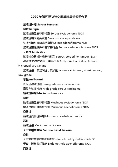
2020年第五版WHO卵巢肿瘤组织学分类浆液性肿瘤Serous tumours良性benign浆液性囊腺瘤非特指型Serous cystadenoma NOS浆液性表面乳头状瘤Serous surface papilloma浆液性腺纤维瘤非特指型Serous adenofibroma NOS浆液性囊性腺纤维瘤非特指型Serous cystadenofibroma NOS交界性borderline浆液性交界性肿瘤非特指型Serous borderline tumour NOS浆液性交界性肿瘤,微乳头亚型Serous borderline tumour,Micropapillary variant浆液性癌,非浸润性,低级别serous carcinoma,non-invasive,Low-grade恶性malignant低级别浆液性癌Low-grade serous carcinoma高级别浆液性癌High-grade serous carcinoma粘液性肿瘤Mucinous tumours良性黏液性囊腺瘤非特指型Mucinous cystadenoma NOS黏液性腺纤维瘤特指型Mucinous adenofibroma NOS交界性黏液性交界性肿瘤Mucinous borderline tumour恶性黏液性癌Mucinous carcinoma子宫内膜样肿瘤Endometrioid tunours良性子宫内膜样囊腺瘤非特指Endometrioid cystadenoma NOS子宫内膜样腺纤维瘤Endometrioid adenofibroma NOS交界性子宫内膜样交界性肿瘤Endemetrioid borderline tumour恶性子宫内膜样癌非特指型Endometrioid carcinoma NOS浆液黏液性癌Seromucinous carcinoma透明细胞肿瘤Clear cell tumours良性透明细胞囊腺瘤Clear cell cystadenoma透明细胞腺纤维瘤Clear cell adenofibroma交界性透明细胞交界性肿瘤Clear cell borderline tumourr恶性透明细胞癌非特指Clear cell carcinoma NOS浆液粘液性肿瘤Seromucinous tumours良性浆液粘液性囊腺瘤Seromucinous cystadenoma浆液粘液性腺纤维瘤Seromucinous adenofibroma交界性浆液粘液性交界性肿瘤Seromucinous borderline tumourBrenner肿瘤Brenner tunours良性Brenner瘤非特指型Brenner tumour NOS交界性Brenner肿瘤,交界恶性Brenner tumour,borderline malignancy恶性Brenner瘤,恶性Brenner tumour,malignant其它类型癌中肾样腺癌 mesonephric-like adenocarcinoma癌,未分化,非特指carcinoma,undifferentiated,NOS去分化癌 dedifferentiated carcinoma癌肉瘤非特指型carcinosarcoma NOS混合细胞腺癌 mixed cell carcinoma间叶性肿瘤Mesenchymal tumours子宫内膜间质肉瘤,低级别endemetrioid stromal sarcoma,low-grade子宫内膜间质肉瘤,高级别endemetrioid stromal sarcoma,high-grade平滑肌瘤非特指型leiomyoma NOS平滑肌肉瘤非特指型leiomyosarcoma NOS恶性潜能未定平滑肌肿瘤smooth muscle tumour of uncertain malignant potential黏液瘤Myxoma混合性上皮和间叶肿瘤Mixed epithelial and mesenchymal tumours腺肉瘤Adenosarcoma性索-间质肿瘤(sex cord-stromal tumours)纯间质肿瘤纤维瘤非特指型Fibroma NOS富细胞纤维瘤Cellular fibroma卵泡膜细胞瘤非特指型Thecoma NOS黄素化卵泡膜细胞瘤Luteinized thecoma硬化性间质瘤sclerosing stromal tumour微囊性间质瘤Microcystic stromal tumour印戒细胞型间质瘤signet-ring stromal tumour卵巢莱迪细胞瘤非特指型Leydig cell tumour of the ovary NOS 类固醇细胞瘤非特指型Steroid cell tumour NOS类固醇细胞瘤,恶性Steroid cell tumour, malignant纤维肉瘤Fibrosarcoma纯性索肿瘤成年型颗粒细胞瘤Adult granulosa cell tumour幼年型颗粒细胞瘤granulosa cell tumour,juvenile支持细胞瘤非特指型Sertoli cell tumour NOS环小管性索瘤Sex cord tumour with annular tubules混合性性索-间质肿瘤Mixed sex cord-stromal tumours支持-莱迪细胞肿瘤非特指型Sertoli-Leydig cell tumours NOS 支持-莱迪细胞肿瘤,高分化型Sertoli-Leydig cell tumours,Well differentiated支持-莱迪细胞肿瘤,中分化型Sertoli-Leydig cell tumours,Moderately differentiated支持-莱迪细胞肿瘤,低分化型Sertoli-Leydig cell tumours,Poorly differentiated支持-莱迪细胞肿瘤,网状型Sertoli-Leydig cell tumours,Retiform性索肿瘤非特指型sex cord tumour NOS性母细胞瘤 Gynandroblastoma生殖细胞肿瘤Germ cell tumours畸胎瘤,成熟teratoma,mature未成熟性畸胎瘤非特指型immature teratoma NOS无性细胞瘤Dysgerminoma卵黄囊瘤非特指型Yolk sac tumour NOS胚胎性癌非特指型Embryonal carcinoma NOS绒毛膜癌非特指型choriocarcinoma NOS混合性生殖细胞肿瘤Mixed germ cell tumour单胚层畸胎瘤和起源于皮样囊肿的体细胞型肿瘤Monodermal teratoma and somatic-type tumours arising from a dermoid cyst甲状腺肿非特指型Struma ovaryi NOS恶性甲状腺肿Struma ovarii,malignant甲状腺肿类癌Strumal carcinoid畸胎瘤伴恶性转化teratoma with malignant transformation囊性畸胎瘤非特指cystic teratoma NOS生殖细胞-性索-间质肿瘤Germ cell-sex cord-stromal tumours性腺母细胞瘤GonadoblastomaDissecting gonablastoma未分化性腺组织undifferentiated gonadal tissue混合性生殖细胞-性索-间质肿瘤非特指Mixed germ cell-sex cord-stromal tumour NOS杂类肿瘤Miscellaneous tumours卵巢网腺瘤Adenoma of rete ovarii卵巢网腺癌Adenocarcinoma of rete ovarii午菲管肿瘤Wolffian tumour实性假乳头状肿瘤Solid pseudopapillary neoplasm小细胞癌,高钙血症型Small cell carcinoma,hypercalcaemic type小细胞癌,大细胞型 Small cell carcinoma, large cell variant肾母细胞瘤Wilms tumour瘤样病变Tumour-like lesions滤泡囊肿Follicle cyst黄体囊肿Corpus luteum cyst巨大孤立性黄素化滤泡囊肿Large solitary luteinized follicle cyst过度黄素化反应Hyperreactio luteinalis妊娠黄体瘤Pregnancy luteoma间质增生Stromal hyperplasia间质卵泡膜细胞增生Stromal hyperthecosis纤维瘤病Fibromatosis卵巢巨型水肿Massive oedema莱迪细胞增生Leydig cell hyperplasia转移到卵巢metastasis to ovary。
泌尿系统疾病英文单词

二、后肾肿瘤metanephric tumors ['metənɪfrɪk] 后肾腺瘤Metanephric adenoma 后肾腺纤维瘤metanephric adenofibroma [ædənəʊfaɪ'brəʊmə] 后肾间质肿瘤metanephric interstitial tumor [ˌɪntəˈstɪʃl] 三、肾母细胞肿瘤Wilms tumor 肾源性残件Nephrogenic remnants ['remnənts] 肾母细胞瘤embryoma of kidney; nephroblastoma; Whilms tumor; Wilm tumor [embrɪ'əʊmə] [nefrɒb'lɑ:stəmə] 囊性、部分分化性肾母细胞瘤Cystic part, differentiation of Wilms tumor
骨肉瘤osteosarcoma [ɒstɪəʊsɑ:'kəʊmə] 血管肌肉脂肪瘤 上皮样血管肌肉脂肪瘤Epithelioid angiomyolipoma [ænɡɪəma'ɪɒlɪpəmə] 平滑肌瘤leiomyoma [laɪəʊmaɪ'əʊmə] 脂肪瘤lipoma 血管瘤angioneoplasm;hemangioma;vascular tumor [ændʒəni:əʊp'læzəm] [hi:ˌmændʒi:'əʊmə] 淋巴管瘤lymphangioma[lɪmˌfændʒɪ'əʊmə] 球旁细胞瘤juxtaglomerular cell tumor;reninoma [dʒʌkstəglɒ'merʊlə] [ri:nɪ'nəʊmə] 肾髓质间质细胞瘤Renal medullary interstitial cell tumor [ˌɪntəˈstɪʃl] 神经鞘瘤neurilemmoma;neurinoma [nɜ:raɪ'leməʊmə] [njʊəraɪ'nəʊmə] 孤立性纤维性肿瘤Solitary fibrous tumor
肺腺癌罕见部位转移3例并文献复习

高临床和病理医师对该肿瘤的认识,也有助于评估该肿瘤的 生物学行为及预后。
参考文献:
[1] MichalM,SyrucekM.Benignmixedepithelialandstromaltumor ofthekidney[J].PatholResPract,1998,194(6):445.
[2] MochH,HumphreyP,UlbrghtT,etal.WHO classificationof tumoursoftheurinarysystem andmalegenitalorgan[M].Lyon: IARCPress,2016:5-8.
1 材料与方法
1.1 临床资料 收集河南省人民医院病理科 2010~2017 年诊断的 3例肺腺癌转移至卵巢、十二指肠、前列腺病例,临 床及影像学资料均查自电子病历系统。例 1女性,47岁,无 吸烟史,1年 前 外 院 细 胞 学 检 查 诊 断 肺 腺 癌,多 基 因 检 测: EGFR、KRAS、BRAF、ROS1基 因 为 野 生 型,ALK基 因 为 融 合 型。全身检查未提示转移,CT示肿块最大径 4cm,胸膜无凹 陷。化疗 1年后逐渐出现下腹部增大。盆腔 MRI示盆腔囊 实性占位,考虑左侧附件交界性囊腺瘤或囊腺癌可能 (图 1)。例 2女性,70岁,因黑便 3天急诊入院,无明确病史。 胃镜示十二指肠多发新生物。临床诊断十二指肠占位。例 3男性,73岁,因骶尾部疼痛半年余,尿频、尿痛半个月就诊。 彩超示前列腺增大伴异常回声。全身骨显像骶骨及左侧髋 关节摄取放射性增高灶,可疑骨转移,无明确病史,临床诊断 前列腺癌骨转移。 1.2 方法 标本分别为手术肿瘤完整切除、内镜下活检及 穿 刺活检标本。均经10%中性福尔马林固定,脱水、石蜡包
儿童肾母细胞瘤诊疗规范(2019年版)

附件9儿童肾母细胞瘤诊疗规范(2019年版)一、概述肾母细胞瘤是儿童时期最常见的肾脏恶性肿瘤,在所有儿童恶性肿瘤中约占6%;15岁以下儿童中,发病率约为7.1/100,000(美国),但亚洲人群中发病率略低;单侧肾母细胞瘤患儿中,男:女=0.92:1,平均诊断年龄为44个月,大约10%的肾母细胞瘤伴有先天发育畸形。
随着化疗、手术、放疗等多种治疗手段的综合应用,总体生存率已经达到85%以上。
目前,世界范围内肾母细胞瘤的治疗可分为两个类别,一是以北美地区为首的COG,推荐直接手术治疗,根据术后病理和分期采取进一步治疗;二是以欧洲地区为首的SIOP,推荐术前化疗,待肿瘤缩小后再行手术切除,根据不同危险度来治疗;此两种治疗方式同样普遍存在于国内,故本规范将两者分别予以罗列,根据采用的初始治疗方式不同,各自选择使用治疗方案。
二、适用范围根据病史、体格检查、影像学检查和/或组织病理学检查确定的肾脏占位,考虑为肾母细胞瘤(不包含中胚叶肾瘤,肾脏透明细胞肉瘤,恶性横纹肌样瘤)。
三、诊断(一)临床表现常见临床表现为无症状的腹部包块、腹痛和腹胀,约40%患儿伴有腹痛表现;肾母细胞瘤患儿中,约有18%表现为肉眼血尿,24%为镜下血尿;大约有25%患儿有高血压表现。
在10%的患儿中可能会伴有发热、厌食、体重减轻;肺转移患儿可出现呼吸系统症状、肝转移可引起上腹部疼痛,下腔静脉梗阻可表现为腹壁静脉曲张或精索静脉曲张。
肺栓塞罕见。
(二)辅助检查。
1.血、尿常规、生化检查:包括肝肾功、电解质、乳酸脱氢酶等。
2.肿瘤标记物肾母细胞瘤目前缺乏特异性瘤标,一些指标如NSE可以用于鉴别肿瘤破裂/肾脏神经母细胞瘤。
AFP可予以鉴别畸胎瘤型肾母。
3.腹部影像学检查(1)腹部超声:初步判断肿瘤位置,大小,与周围组织关系,血管内有无瘤栓等;(2)腹部增强CT(有肾功能不全时禁用造影剂)或MRI:。
肾母细胞瘤中大约4%的患儿伴有下腔静脉或心房瘤栓,11%患儿伴有肾静脉瘤栓,肺栓塞十分罕见,但常常是致命的;因此,术前增强CT和MRI不仅可以确定肾脏肿瘤的起源,确定对侧肾脏有无病变,观察腹部脏器有无转移,同样可以确定腔静脉瘤栓;肾母细胞瘤中部分患儿伴有肾母细胞瘤病,增强MRI可以较好的予以区分鉴别;(3)胸部CT:肝脏和肺部是肾母细胞瘤最常见的转移部位,其中大约15%的患儿伴有肺转移,胸部CT检查可以很好的判断肺内转移病灶情况;(4)PET-CT:并不推荐作为常规检查,但如果患儿高度怀疑多发转移或复发,可予以检查;(5)活检:如果临床上考虑为肾母细胞瘤1期或2期,根据北美地区COG经验,术前穿刺活检会提升肿瘤分期,因此并不推荐进行常规穿刺活检,可直接手术切除;以欧洲为主的SIOP推荐进行术前化疗,虽然认为穿刺活检并不会影响肿瘤分期,但也不作为常规操作。
复方苁蓉益智胶囊治疗血管性痴呆的研究进展
血管性痴呆(vascular dementia,VD)是一种由脑血管病变导致的疾病,其临床症状包括引起记忆和执行功能障碍等。
它被认为是继阿尔茨海默病之后的第二大常见痴呆类型[1]。
目前,在亚洲和发展中国家的痴呆病例中,VD 约占30%,高于北美和欧洲(15%~20%)[2-3]。
据研究资料显示,我国60岁及以上人群的血管性痴呆发病率为每年每千人中有2.42例[4-5]。
研究表明,我国约有1507万人60岁以上的痴呆患者,其中约有392万人为VD 患者[6]。
VD 会造成日常生活质量不断下降,而且不能扭转,给家庭和社会带来极大的冲击和负担。
复方苁蓉益智胶囊是由王永炎院士多年临床实践研制的具有益智养肝,化浊活血和增智健脑等功效的中成药[7],主料何首乌、肉苁蓉、荷叶、地龙、漏芦等。
Progress of compound ciYizhi capsule in the treatment of vascular dementia Di Shuai, Zhang Jiapeng, Liu Yixuan, LiYanan, Zhang Jiang, Zhou Fuling. The Affiliated Hospital of North China University of Science and Technology, Tangshan 063000, China【Abstract 】Compound ciYizhi capsule has the effect of nourishing liver,promoting turbidity and activating blood, and increasing wisdom and brain. It is suitable for mild to moderate vascular dementia with liver and kidney deficiency and phlegm stasis blocking collateral syndrome. Recently, it has been widely used in the long-term and synergistic treatment of vascular dementia with remarkable efficacy.To summarizes the clinical and experimental studies of compound ciYizhi capsule. It is found that compound ciYizhi capsule can treat vascular dementia by reducing the expression of MARKS mRNA in hippocampus, inhibiting oxidative stress in brain tissue, protecting mitochondria, reducing the range of cerebral infarction, protecting cerebral ischemic injury and pound ciYizhi capsule combined with other anti-dementia drugs can significantly improve the clinical symptoms of patients with vascular dementia and improve the self-care ability and quality of life.In order to provide some reference for the subsequent study of compound cistanche qianyi capsule.【Key words 】Vascular dementia; Compound ciYizhi capsule; Dementia; Clinical application 复方苁蓉益智胶囊治疗血管性痴呆的研究进展邸帅 张佳朋 刘乙璇 李亚楠 张江* 周福玲作者单位:063000 河北省唐山市,华北理工大学附属医院神内二、四病区*通讯作者【摘要】 复方苁蓉益智胶囊具有益智养肝,化浊活血和增智健脑的功效,适用于肝肾亏虚兼痰瘀阻络证的轻中度血管性痴呆。
妇产科英语词汇

妇产科英语词汇外阴色素减退及外阴搔痒chronic vulvardystrophy 慢性外阴营养不良pruritus vulvae 外阴搔痒外阴炎、阴道炎、宫颈炎vulvitis 外阴炎bartholins gland abscess 前庭大腺脓肿bartholins gland cyst前庭大腺囊肿trichomonas vaginitis 滴虫性阴道炎monilial vaginitis 念珠菌性阴道炎senile vaginitis老年性阴道炎cervicitis宫颈炎cervical erosion宫颈糜烂cervical polyp 宫颈息肉Nabothian cyst 纳氏囊肿盆腔炎pelvic inflammatory disease <PID> 盆腔炎ascending infection 上行性感染salpingitis输卵管炎frozen pelvis 冰冻骨盆pelvic tuberculosis 盆腔结核妊娠合并性传播性疾病gonorrhea 淋病syphilis 梅毒condyloma acuminata尖锐湿疣sexually transmitted disease (STD)性传播性疾病human papilloma virus (HPV)人乳头状病毒genital herpes 生殖器疱疹herpes simplex virus <HSV〉单纯疱疹病毒chlamydia 衣原体子宫颈癌cervical squamous cancer 子宫颈鳞癌cervical adenocarcinoma 子宫颈腺癌cervical carcinoma in situ 〈CIS〉子宫颈原位癌cervied intraepithelial carcinoma 子宫上皮内癌cervical intraepithelial neoplasm (CIN) 子宫颈上皮内瘤样病变cervical squamous atypical hyperplasia 鳞状上皮不典型增生squamous metaplasia 鳞状上皮化生micro—invasive carcinoma 镜下早期浸润癌herpes simplex virus (HSV) 单纯疱疹病毒human papilloma virus 人类乳头瘤病毒human cytomegalovirus 〈CMV〉人类巨细胞病毒pap smear 巴氏涂片检查colposcopy 阴道镜检查biopsy 活检cervical conization 子宫颈锥形切除子宫肌瘤myoma of uterus 子宫肌瘤intramural myoma 肌壁间肌瘤subserous myoma 浆膜下肌瘤submucous myoma 粘膜下肌瘤total hysterectomy 全子宫切除术subtotal hysterectomy 次全子宫切除术myomectomy 肌瘤切除术hyaline degeneration 玻璃样变性cystic degeneration 囊性变性red degeneration 红色变性sarcomatous change 肉瘤变性子宫内膜癌carcinoma of endometrium 子宫内膜癌irregular vaginal bleeding 阴道不规则流血adenocarcinoma 腺癌adenoacarthorna 腺角化癌adeno—squamous carcinoma 鳞腺癌chemotherapy 化疗radiotherapy 放疗fractional curettage 分段诊刮卵巢肿瘤ovarian tumour 卵巢肿瘤epithelial tumour of ovary 卵巢上皮性肿瘤serous cystadenoma 浆液性囊腺瘤borderline serous cystadenoma 交界性浆液性囊腺病serous cystadenocarcinoma 浆液性囊腺癌mucinous cystadenoma 粘液性肿瘤mucinous cystadenocarcinoma 粘液性囊腺癌endometrioid tumour 子宫内膜样肿瘤endometrioid carcinoma 子宫内膜样癌clear cell tumour 透明细胞瘤teratoma 畸胎瘤dysgerminoma 无性细胞瘤endodermal sinus tumour 内胚窦瘤granulosa—stromal cell tumor 颗粒间质细胞瘤theca cell tumor 卵泡膜细胞瘤fibroma 纤维瘤sertoli—leydig cell tumour 支持一间质细胞瘤tumour—like condition 卵巢瘤样病变solitary follicle cyst 单发性滤泡囊肿corpus luteum cyst黄体囊肿germ cell tumor生殖细胞瘤sex cord stromal tumor 性索间质细胞瘤benign 良性的malignant 恶性的laparotomy 剖腹探查tumor marker 肿瘤标志物torsion of ovarian tumor 卵巢肿瘤蒂扭转Polycystic ovarian syndrome(PCOS) 多囊卵巢综合征Insulin resistance 胰岛素抵抗Cyproterone acetate (CPA)醋酸环丙孕酮子宫脱垂、尿瘘uterine prolapse 子宫脱垂stress incontinence 张力性尿失禁urinary fistula 尿瘘cystocele 膀胱膨出enterocele 直肠膨出女性生殖器官发育异常imperforate hymen 处女膜闭锁(无孔处女膜)congenital absence of vagina 先天性无阴道atresia of vagina 阴道闭锁transverse vaginal septum 阴道横膈longitudinal vaginal septum 阴道纵膈congenital absence of uterus 先天性无子宫primordial uterus 始基子宫hypoplasia of uterus 子宫发育不良infantile uterus 幼稚子宫uterus didelphys 双子宫不孕症infertility 不孕症hysterosalpingography<HSG〉子宫输卵管造影术in vitro fertilization (IVF) 体外授精妇女保健计划生育family planning 计划生育contraception 避孕intrauterine device (IUD)宫内节育器condom 阴茎套conrtaceptive 避孕药oral conrtaceptive pill(ocp) 口服避孕药artificial abortion 人工流产medical abortion 药物流产sterilization 绝育indication 适应证contraindication 禁忌证complication 并发症womens healthcare 妇女保健女性生殖系统生理menstruation月经steroid hormone 甾体激素estrogen 雌激素progesterone 孕激素androgen 雄激素proliferative phase 增生期secretive phase 分泌期ovary 卵巢follicle 卵泡ovulation 排卵corpus lutein 黄体feedback 反馈prolactin (PRL)泌乳素follicle stimulating hormone (FSH)卵泡刺激素luteinizing hormone (LH) 黄体生成激素妊娠生理(包括妊娠期母体变化)pregnancy 妊娠placenta 胎盘fetus 胎儿umbilical cord 脐带amniotic membrane 羊膜amniotic fluid 羊水villus 绒毛human chorionic gonadotropin (hCG) 绒毛膜促性腺激素human placental lactogen(HPL)胎盘生乳素妊娠诊断amenorrrhea 停经morning sickness 早孕反应fetal lie 胎产式longitudinal lie 纵产式transverse lie 横产式fetal presentation 胎先露fetal position 胎位孕期监护及保健expected date of confinement, EDC 预产期last menstrual period,LMP 末次月经four maneuver of Leopold 四步触诊法external pelvimetry 骨盆外测量internal pelvimetry 骨盆内测量acceleration 加速early deceleration 早期减速variable deceleration 变异减速late deceleration 晚期减速non-stress tess,NST 无应激试验oxytocin challenge test, OCT 缩宫素激惹试验contraction stress tess, CST 宫缩应激试验fetal maturity 胎儿成熟度lecithin/sphingomyelin,L/S 卵磷脂/鞘磷脂正常分娩delivery 分娩premature delivery 早产term delivery 足月产postterm delivery 过期产pelvic inlet 骨盆入口pelvic outlet 骨盆出口effacement of cervix 宫颈管消失dilatation of cervix 宫口扩张biparietal diameter (BPD)双顶径mechanism of labor 分娩机制engagement 衔接descent 下降flexion 俯屈internal rotation 内旋转extension 仰伸restitution 复位external rotation 外旋转show 见红partogram 产程图crowning of head 胎头着冠push 向下屏气episiotomy (EP)会阴切开术正常产褥puerperium 产褥期involution of uterus 子宫复旧colostrums 初乳after pains 产后宫缩痛lochia 恶露bottle feeding 人工喂养breast feeding 母乳喂养early suckling 早吸吮流产abortion 流产threatened abortion 先兆流产inevitable abortion 难免流产incomplete abortion 不完全流产complete abortion 完全流产missed abortion 过期流产habitual abortion 习惯性流产异位妊娠ectopic pregnancy 异位妊娠tubal pregnancy 输卵管妊娠abdominal pregnancy 腹腔妊娠ovarian pregnancy 卵管妊娠cervical pregnancy 宫颈妊娠culdocentesis 后穹窿穿刺laparoscopy 腹腔镜检查conservative surgery 保守性手术rupture of tubal pregnancy 输卵管妊娠破裂妊娠高血压综合征edema 水肿proteinuria 蛋白尿hypertension 高血压hemolysis,elevated liver enzyme,low platelets syndrome HELLP 综合征antihypertensive 抗高血压药物anti convulsant 抗抽搐药物diuretics 利尿药sedative 镇静药物pregnancy induced hypentension syndrome,PIH 妊娠高血压综合征preeclampsia 先兆子痫eclampsia 子痫mean arterial blood pressure,(mABP)平均动脉压前置胎盘placenta previa 前置胎盘complete previa 完全性(中央性)前置partial previa 部分性前置marginal previa 边缘性前置胎盘早期剥离placental abruption 胎盘早剥disseminated intravascular coagulation (DIC)弥散性血管内凝血uteroplacental apoplexy 子宫卒中revealed abruption 显形剥落concealed abruption 隐性剥落mixed hemorrhage 混合性出血妊娠合并心脏病perinatal cardiomyopathy 围产期心肌病heart failure 心力衰竭cardiac function 心功能rheumatic heart disease 风湿性心脏病congenital heart disease 先天性心脏病妊娠合并乙型肝炎intrahepatic cholestasis of pregnancy 妊娠期肝内胆汁郁积症acute fatty liver of pregnancy 妊娠急性脂肪肝妊娠合并糖尿病gestational diabetes mellitus,GDM 妊娠期糖尿病macrosomia 巨大儿shoulder dystocia 肩难产congenital malformation 先天性畸形oral glucose tolerance test 口服葡萄糖耐量试验高危妊娠high risk pregnancy 高危妊娠fetal monitoring 胎儿监护non—stress test ,(NST)无应激试验contraction stress test (CST)宫缩应激试验oxytocin challenge test (OCT)催产素激惹试验amniocentesis 羊膜腔穿刺lecithin/sphingomyelin (L/S)卵磷脂/鞘磷脂perinatal mortality rate围产儿死亡率deceleration 减速acceleration 加速biophysical profile score (BPS)B超生物物理评分fetal movement 胎动fetal tone 胎儿张力fetal breath movement 胎儿呼吸样运动amniotic fluid index (AFI) 羊水指数异常分娩dystocia 异常分娩primipara 初产妇multipara 经产妇hypotonic uterine contraction 低张性子宫收缩hypertonic uterine contraction 高张性子宫收缩oxytocin 催产素prostaglandin 前列腺素simple flat pelvis 单纯扁平骨盆funnel shaped pelvis 漏斗骨盆generally contracted pelvis 均小骨盆persistent occipitoposterior position 持续性枕后位persistent occipitotransverse position 持续性忧横位sincipital presentation高直位anterior asynclitism 前不均倾位face presentation 面先露breech presentation 臀先露shoulder presentation 肩先露shoulder dystocia 肩难产hydrocephalus 脑积水spina bifida 脊柱裂conjoined twins 联体双胎产后出血postpartum hemorrhage 〈PPH> 产后出血uterine atony 宫缩乏力trauma of genital tract 生殖道创伤placental implantation 胎盘植入子宫破裂rupture of uterus 子宫破裂pathologic retraction ring 病理缩过期妊娠postterm pregnancy 过期妊娠meconium 胎粪induction of labor 引产placental function 胎盘功能胎儿窘迫fetal distress 胎儿窘迫perinatal medicine 围产医学产褥感染puerperal infection 产褥感染puerperal morbidity 产褥病率late postpartum hemorrhage 晚期产后出血。
子宫内膜息肉给予宫腔镜息肉电切术辅以左炔诺孕酮宫内缓释系统的疗效分析

临床医学China &Foreign Medical Treatment 中外医疗子宫内膜息肉给予宫腔镜息肉电切术辅以左炔诺孕酮宫内缓释系统的疗效分析张丽艳,孙爽长春中医药大学附属医院妇产诊疗中心,吉林长春 130021[摘要] 目的 观察在子宫内膜息肉的治疗当中在给予宫腔镜息肉电切术之后辅助使用左炔诺孕酮宫内缓释系统治的效果。
方法 便利选取2022年1月—2023年2月长春中医药大学附属医院收治的98例子宫内膜息肉患者。
按随机数表法分为两组,各49例。
对照组采用宫腔镜息肉电切术治疗,观察组在对照组基础上辅以左炔诺孕酮宫内缓释系统。
对比治疗后腺上皮及间质雌激素受体、腺上皮及间质孕激素受体、血清指标、复发率等。
结果 观察组术后1个月月经量更少、血红蛋白水平上升,且观察组术后3及6个月其子宫内膜厚度及月经量少于对照组,血红蛋白水平高于对照组,差异有统计学意义(P <0.05);治疗后观察组腺上皮及间质雌激素受体、腺上皮及间质孕激素受体、血清指标水平均低于对照组,差异有统计学意义(P <0.05);观察组的复发率为4.08%,低于对照组的16.33%,差异有统计学意义(χ2=4.455,P <0.05)。
结论 在子宫内膜息肉的患者的宫腔镜息肉电切术之后辅以左炔诺孕酮宫内缓释系统治疗,能够使原本较厚的子宫内膜变薄,并减少原本过多的月经量,恢复血红蛋白及血清糖类抗原125、高敏C 反应蛋白、血管内皮生长因子水平,调节雌激素及孕激素受体表达平衡,降低复发率,且具有良好的治疗安全性。
[关键词] 子宫内膜息肉;宫腔镜;息肉电切术;左炔诺孕酮宫内缓释系统;子宫内膜厚度;月经量[中图分类号] R713 [文献标识码] A [文章编号] 1674-0742(2023)11(b)-0059-04Efficacy of Hysteroscopic Electropolypectomy Combined with Levonorg⁃estrel Intrauterine Sustained Release System for Endometrial PolypsZHANG Liyan, SUN ShuangDepartment of Obstetrics and Gynecology Diagnosis and Treatment Center, the Affiliated Hospital of Changchun Uni⁃versity of Chinese Medicine, Changchun, Jilin Province, 130021 China[Abstract] Objective To observe the effect of levonorgestrel intrauterine sustained release system after hysteroscopic electropolypectomy in the treatment of endometrial polyps. Methods A total of 98 patients with endometrial polyps ad⁃mitted to the Affiliated Hospital of Changchun University of Traditional Chinese Medicine from January 2022 to Febru⁃ary 2023 were conveniently selected. According to the random number table method, they were divided into twogroups, 49 cases in each group. The control group was treated with hysteroscopic polyp resection, and the observationgroup was supplemented with levonorgestrel intrauterine sustained-release system on the basis of the control group. The glandular epithelial and stromal estrogen receptors, glandular epithelial and stromal progesterone receptors, serum indexes, and recurrence rate were compared after treatment. Results The menstrual volume of the observation group was lower and the hemoglobin level was higher 1 month after operation, and the endometrial thickness and menstrualvolume of the observation group were less than those of the control group 3 and 6 months after operation, and the he⁃moglobin level were higher than that of the control group, and the differences were statistically significant (P <0.05) af⁃ter treatment, the levels of estrogen receptor in glandular epithelium and stroma, progesterone receptor in glandularepithelium and stroma, and serum indexes in the observation were all lower than those in the control group, and the differences were statistically significant (P <0.05), the recurrence rate of the observation group was 4.08%, which was DOI :10.16662/ki.1674-0742.2023.32.059[作者简介] 张丽艳(1989-),女,硕士,住院医师,研究方向为妇产科学临床研究。
MRI联合罗马指数在卵巢肿瘤中的应用价值
卵巢肿瘤为女多发性的生殖系统肿瘤,以卵巢癌的危害最大[1]。
卵巢癌为死亡率较高的妇科癌症,有强隐匿性,且良恶性肿瘤间症状无明显差异,易混淆,易延误恶性肿瘤的最佳治疗时机[2],因此早诊断、早治疗对于改善卵巢癌患者预后具有积极意义。
病理学检查由于存在创伤性,对于患者耐受性要求较高,故临床应用存在局限性[3]。
磁共振成像(magnetic resonance imaging,MRI)具有扫描范围多、层面外干扰小以及空间结构显示好等多个优势,能利用多方位扫描定量定性分析卵巢肿瘤,增加诊断准确性[4-5]。
研究指出,罗马指数在卵巢癌的诊断中具有良好敏感度与特异度[6]。
鉴于此,本文将探讨MRI联合罗马指数对卵巢肿瘤的诊断价值,以期为临床完善治疗方案提供依据。
1资料与方法1.1一般资料经我院医学伦理委员会审核同意,选取2020年3月至2023年2月平煤神马集团总医院收治的107例卵巢肿瘤患者作为研究对象。
纳入标准:(1)初筛有性质不明卵巢肿块;(2)接受影像学检查和手术治疗;(3)可耐受病理检查;(4)知情本研究且签署同意协议书。
排除标准:(1)既往有卵巢手术治疗史;(2)伴有失控性出血;(3)原发性卵巢急性与慢性感染;(4)严重内分泌系统病变;(5)伴血液系统病变;(6)伴严重脏器功能损伤。
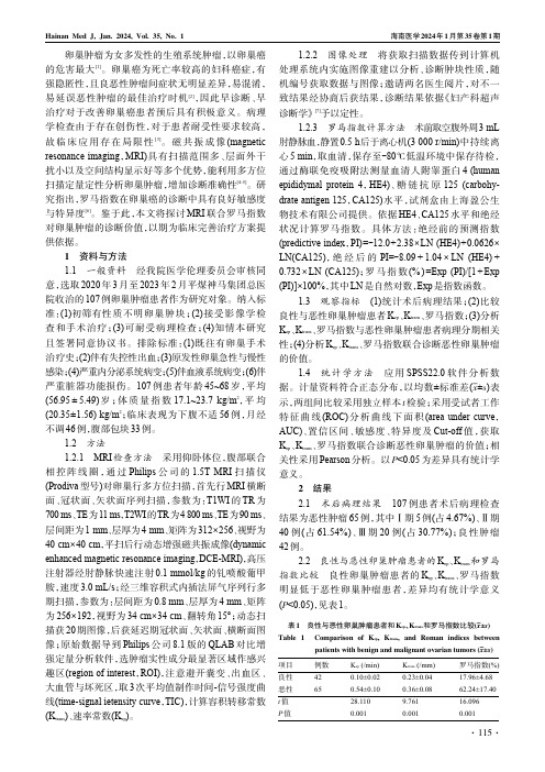
107例患者年龄45~68岁,平均(56.95±5.49)岁;体质量指数17.1~23.7kg/m2,平均(20.35±1.56)kg/m2;临床表现为下腹不适56例,月经不调46例,腹部包块33例。
1.2方法1.2.1MRI检查方法采用仰卧体位,腹部联合相控阵线圈,通过Philips公司的1.5T MRI扫描仪(Prodiva型号)对卵巢行多方位扫描,首先行MRI横断面、冠状面、矢状面序列扫描,参数为:T1WI的TR为700ms、TE为11ms,T2WI的TR为4800ms、TE为90ms、层间距为1mm、层厚为4mm、矩阵为312×256、视野为40cm×40cm,平扫后行动态增强磁共振成像(dynamic enhanced magnetic resonance imaging,DCE-MRI),高压注射器经肘静脉快速注射0.1mmol/kg的钆喷酸葡甲胺,速度3.0mL/s;经三维容积式内插法屏气序列行多期扫描,参数为:层间距为0.8mm、层厚为4mm、矩阵为256×192,视野为34cm×34cm、翻转角15°;动态扫描获20期图像,后获延迟期冠状面、矢状面、横断面图像;原始数据导到Philips公司8.1版的QLAB对比增强定量分析软件,选肿瘤实性成分最显著区域作感兴趣区(region of interest,ROI),注意避开囊变、出血区、大血管与坏死区,取3次平均值制作时间-信号强度曲线(time-signal ietensity curve,TIC),计算容积转移常数(K trans)、速率常数(K ep)。
胰腺囊性病变的影像表现与临床特点(下)

国际医学放射学杂志IntJMedRadiol2020Nov 鸦43穴6雪胰腺囊性病变的影像表现与临床特点(下)徐建国1唐光健2彭泰松1赵丽丽1于萍1任龙飞1许志高1【摘要】胰腺囊性病变(PCL)是胰腺上皮和间质组织发生囊腔病变的一大类疾病,以胰腺内囊性包块为主要特征,具有不同的病因、临床和组织病理学特点。
本文的前两部分介绍了胰腺炎症相关囊性病变(包括胰腺假性囊肿与胰腺包裹性坏死)与胰腺真性囊肿(包括孤立性胰腺上皮囊肿、von Hippel-Lindau 病、多囊肾和囊性纤维化)以及常见的胰腺浆液性囊腺瘤、胰腺黏液性囊腺瘤和胰腺实性假乳头状瘤的影像表现与临床特点。
本篇为最后一部分,就常见的胰腺导管内乳头状黏液瘤与胰腺少见囊性肿瘤予以介绍和分析,以期为临床诊断与治疗提供重要依据。
【关键词】胰腺囊性病变;体层摄影术,X 线计算机;磁共振成像;导管内乳头状黏液瘤;囊性肿瘤中图分类号:R576;R445.2;R445.3文献标志码:APancreatic cystic lesions:imaging findings and clinical features (Part 3)XU Jianguo 1,TANG Guangjian 2,PENGTaisong 1,ZHAO Lili 1,YU Ping 1,REN Longfei 1,XU Zhigao 1.1Department of Radiology,Third People ’s Hospital of Datong City,Datong 037008,China;2Department of Radiology,Peking University First Hospital【Abstract 】Pancreatic cystic lesions (PCLs)are a broad spectrum of diseases with cystic lesions in pancreaticepithelial and interstitial tissues,characterized by cystic mass,with various etiology,clinical and histopathological characteristics.The first two parts of the article introduces the imaging manifestations and clinical features of 1)pancreatitis related cystic lesionincluding pancreatic pseudocyst and pancreatic encapsulated necrosis,2)pancreatic true cysts including isolated pancreatic epithelial cyst,von Hippel Lindau disease,polycystic kidney and cystic fibrosis,and3)the common cystic tumors of the pancreas including serous cystadenoma,mucinous cystadenoma and solid pseudopapilloma of the pancreas.In the last part of this issue,we introduce another common cystic tumor of pancreas (intraductal papillary mucinous neoplasm of the pancreas)and rare cystic tumors of the pancreas,in order to provide an important basis for clinical diagnosis and treatment of pancreatic cystic lesions.【Keywords 】Cystic disease of pancreas;Tomography,X -ray computed;Magnetic resonance imaging;Intraductalpapillary mucinous neoplasm;Cystic tumorIntJMedRadiol,2020,43(6):716-720作者单位:1大同市第三人民医院医学影像科,大同037008;2北京大学第一医院放射科通信作者:唐光健,E-mail:***************.com DOI:10.19300/j.2020.J18513图文讲座3.4胰腺导管内乳头状黏液瘤胰腺导管内乳头状黏液瘤(intraductal papillary mucinous neoplasm,IPMN )实际上并不是囊性肿瘤,由于肿瘤分泌黏液,引起胰腺导管扩张,大体病理与影像表现为伴有囊的病变,故在胰腺囊性病变内一并讨论。
肾脏混合性上皮和间质肿瘤1例报告

※ 通信作者:王健明,Email:wjming68@sina.com肾脏混合性上皮和间质肿瘤1例报告王健明※ 耿庆伟 张正园(临沂市人民医院 泌尿外科,山东 临沂 276000) 肾脏混合性上皮和间质肿瘤是一种很少见的肾脏肿瘤,文献报道较少。
本单位收治1例,整理临床及病理资料,结合文献,现报道如下。
1 病例简介患者,女,24岁。
查体发现右肾占位2年。
患者于2年前查体发现右肾占位,大小约44mm×48mm,呈囊性样,无低热、无盗汗、无尿频、尿急、尿痛、无排尿困难、无腰背酸痛,其后定期复查。
近期复查B超发现右肾占位体积增大,遂来本院就诊。
行双肾增强CT检查示:右肾囊性低密度影,最大截面约6.3cm×4.7cm,局部见线状分隔,增强扫描可见分隔强化(图1)。
患者入院后,完善各项术前检查,排除手术禁忌,于气管插管全麻下行腹腔镜右肾囊肿去顶减压术,术中行快速病理示右肾良性囊肿性病变。
术后病理大体形态:右肾囊壁切除,大小约6cm×4cm,囊内见透明色液体。
病理组织形态:右肾囊肿性病变,诊断混合性上皮和间质肿瘤。
免疫组化:ER(间质+)、Desmin(间质+)、CK(上皮+)(图2)。
本例术后随访6个月,未见复发与转移,现随访中。
2 讨论肾脏混合性上皮和间质肿瘤(mixedepitheIialandstromaltumourofthekidney,MESTK)是由上皮和间质成分混合构成的良性肾肿瘤。
本病由Michal和Syrucek于1998年首先报道并命名,具有一系列独特而多样的上皮和间质细胞组织形态改变。
MESTK是一种很少见的肾肿瘤,国内外文献报道较少,目前文献报道190余例,男女之比为1 8,年龄范围8~92岁,平均51岁,多见于围绝经期女性[1,2]。
图1 CT检查示右肾一囊性低密度影,增强可见分隔强化图2 病理检查示囊肿性病变,倾向于混合性上皮和间质肿瘤。
免疫组化:ER(间质+),Desmin(间质+),CK(上皮+)镜下病理显示的上皮成分常排列呈腺管样散布于间质中,也可聚集成簇或呈囊性扩张。
宠物兔三种典型子宫肿瘤的病理学诊断

2. College ofLife Sciences , Heilongjiang University,Harbin 1500S0,China)
Abstract:In this study,three cases(Oryctolagus cuniculus) of uterine tumors in pet rabbits were analyzed pathologically.Histological identification was performed by H.E.staining and immunohistochemical methods of paraffin sections.For case one,histopathologically,the uterine masses revealed papillary projections and most neoplastic glands were lined by single to multiple layers of cells.Neo plastic glandular epithelia had dark basophilic nucleus,prominent nucleoli,scant cytoplasm,low mito sis,and cellular pleomorphism.According to immunohistochemistry,the tumor cells in uterine masses demonstrated strong positive signals for cytokeratin,but negative for vimentin.Based on the gross, histopathologic and immunohistochemical features,this case was diagnosed as uterine adenocarcinoma; For case two,histopathologically ,the neoplastic mass had invaded the myometrium with poorly demarcat ed.The cell appeared as cribriform/tubular type and solid arrangement,lined by single to multiple lay ers,which had the characteristics of cell aggregation.Neoplastic epithelial cells consisted of col~ umn,polygonal cells,with moderately abundant eosinophilic cytoplasm and distinct cell borders.The hyperchromatic nuclei varied from oval to round,with moderate anisokaryosis and distinct nucleoli.Ac
子宫体部ミュラー氏腺肉肿の1症例 ―特に中胚叶性肿疡の文献的.

of Mullerian
Adenosarcoma
in Uterine
Corpus
-Especially on MesodermalTumor appearedin MedicalLiteratureDepartment of Obstetrics and Gynecology, Fuchiu Hospital Hiroyuki MANDAI Department of Obstetrics and Gynecology, Aizenbashi Hospital Takafumi OGAWAand Kenjiro IBARAGI Second Department of Pathology, Hyogo Medical College Akihiko KURATA
Žq‹{ ‚Ì ’†ãó—t •« Žîᇠ‚É‚¨ ‚¢ ‚Ä ƒ~ƒ… ƒ‰ •[ Ž••¬•‡Žî á‡‚Ì “ÁŽêŒ`‚Æ ‚µ‚ÄClement & Scully(1974)‚Í •ó ‘• •B ‚ðŽ¦ ‚µ, ‘g •DŠw“I ‚É—Ç •« Žîᇠ‚Æ ‚µ‚Ä‚Ì ‘B•ã”ç ‚Æ
(Mixed 1954.
of the uterus) A study of twenty-one 22) Symmonds, R. E. et al.:Sarcoma like proliferation
lesion resembling sarcoma
and sarcoma-
of the endmetrial stroma. Am.
of the body of the uterus following irradiation therapy for carcinoma of the cervix. J. Obstet. Gynecol. Brit. Cwlth., histological Histological 16) Roth, 71:281, 1964. No.13. tract 15) Pulsen, H. E. & Taylor, C. W.:International classification typing of tumors genital of female Geneva, 1975necol., 35:769,
肾混合性上皮间质肿瘤1例及文献复习

肾混合性上皮间质肿瘤1例及文献复习发布时间:2022-10-27T05:43:43.090Z 来源:《健康世界》2022年14期作者:李虎博①胡俊超①贾鸣飞①康绍叁①[导读] 肾混合性上皮间质肿瘤(MESTK)是一种无明显临床特异性表现的罕见疾病,通常见于女性患者。
李虎博①胡俊超①贾鸣飞①康绍叁①华北理工大学 063000 河北唐山;①华北理工大学附属医院泌尿外科 [摘要] 肾混合性上皮间质肿瘤(MESTK)是一种无明显临床特异性表现的罕见疾病,通常见于女性患者。
它的表现最常见为腹部肿块、血尿和腹痛,常用的治疗手段为手术切除,预后较好。
我们报道了一名60岁女性,左肾上皮和间质混合瘤。
肿瘤是通过左肾部分切除术切除的,术后1年半未复发。
[关键词] 成人中胚层肾瘤肾混合性上皮间质肿瘤 MESTK [中图分类号] R 692.9 [文献标识码] A [文章编号] Mixed epithelial stromal tumor of kidney: a case report and review of literature LI Hubo,JIA Mingfei,Kang Shaosan,( North China University of Science and Technology TangShan,063000,China) [ABSTRACT] Renal mixed epithelial-mesenchymal tumor (MESTK) is a rare disease with no obvious clinical specificity and usually occurs in women. Its most common manifestations are abdominal mass, hematuria and abdominal pain, and the commonly used treatment is surgical resection with a good prognosis. We report a 60-year-old woman with mixed epithelial and stromal tumors of the left kidney. The tumor was removed by partial nephrectomy of the left kidney and did not recur one and a half years after surgery. [KEY WORDS] Mesoblastic nephroma in adult. Mixed epithelial stromal tumor of the kidney. MESTK 肾混合性上皮和间质肿瘤(mixed epitheIial and stromaltumour of the kidney,MEsTK)是由上皮和间质成分混合构成的良性肾肿瘤,由于其形态学和病理特征均不具有特异性,故以往报道中也曾称其为成人中胚层肾瘤(adult mesoblastic nephrom)[1]。
肾混合性上皮间质肿瘤1例并文献复习
肾混合性上皮间质肿瘤1例并文献复习
金雨舟;李嘉成;唐晨豪;何康炜;邓刚
【期刊名称】《中国现代医生》
【年(卷),期】2024(62)3
【摘要】肾混合性上皮间质肿瘤(mixed epithelial and stromal tumor of the kidney,MESTK)是一种罕见的肾脏肿瘤,其特点是囊性扩张且不等量增生的腺上皮细胞与排列方式各异的梭形细胞混合。
现报告1例24岁女性MESTK患者,其无明显临床表现,由体检发现右肾占位,给予右肾部分切除术后恢复良好。
【总页数】3页(P140-141)
【作者】金雨舟;李嘉成;唐晨豪;何康炜;邓刚
【作者单位】浙江中医药大学附属第四临床医学院;浙江大学医学院附属杭州市第一人民医院泌尿外科
【正文语种】中文
【中图分类】R737.11
【相关文献】
1.青少年肾混合性上皮间质肿瘤1例并文献复习
2.肾混合性上皮间质肿瘤4例报道及文献复习
3.双肾恶性混合性上皮和间质肿瘤1例并文献复习
4.肾混合性上皮间质肿瘤一例病例报告并文献复习
5.肾恶性混合性上皮间质肿瘤一例报道及文献复习
因版权原因,仅展示原文概要,查看原文内容请购买。
腮腺混合瘤与腺淋巴瘤的磁共振成像表现对比分析

腮腺混合瘤又称为多形性腺瘤,起源于腮腺 上皮组织;腮腺腺淋巴瘤又称Warthin瘤,淋巴 囊腺瘤或淋巴乳头状囊腺瘤,目前研究认为其起 源于腮腺内、外淋巴结内异位的唾液腺导管上皮
组织[1-2]。两者分别居腮腺常见肿瘤的前两位, 虽然两者均为良性肿瘤,但腮腺混合瘤若切除 不彻底,则容易复发,5年后复发率为3.4%,10 年后为6.8%,且有发生恶变的可能,而腺淋巴
高信号,21例呈混杂信号,增强扫描病灶明显 不均匀强化,且有进行性强化的特点(图1)。 42例腺淋巴瘤患者中11例为多发病灶(其中9例 双侧多发,2例为单侧多发);24例病灶伴有囊 变,病灶边界清楚,T1WI上均呈低信号,T2WI 上10例呈均匀稍高信号,32例信号不均匀,其中 24例病灶伴有囊变,囊变部分呈高信号,增强扫 描病灶实性部分早期明显强化,延迟扫描造影 剂迅速廓清,信号明显减低,囊变部分无强化 (图2)。
0.658
0.001
《肿瘤影像学》2020年第29卷第3期
321
2.1 临床特点 本组68例均以触及耳前慢性无痛性包块就 诊,病程长约1个月至3年不等。本组患者中混合 瘤26例,其中男性12例,女性14例,平均发病年 龄约为44岁;腺淋巴瘤42例,其中男性41例,女 性1例,平均发病年龄约为62岁。综合分析混合 瘤多发于中青年女性,而腺淋巴瘤好发于中老年 男性,与文献报道相符[5]。 2.2 MRI影像学特征 26例混合瘤中病变均为单发,病灶边界清 楚,T1WI上均呈低信号,T2WI上5例呈均匀稍
《肿瘤影像学》2020年第29卷第3期
Oncoradiology 2020 Vol.29 No.3
欢迎关注本刊公众号
319
·论 著·
腮腺混合瘤与腺淋巴瘤的磁共振成像表现对比 分析
- 1、下载文档前请自行甄别文档内容的完整性,平台不提供额外的编辑、内容补充、找答案等附加服务。
- 2、"仅部分预览"的文档,不可在线预览部分如存在完整性等问题,可反馈申请退款(可完整预览的文档不适用该条件!)。
- 3、如文档侵犯您的权益,请联系客服反馈,我们会尽快为您处理(人工客服工作时间:9:00-18:30)。
Chao-jun Wang,1Yi-wei Lin,1Hua Xiang,2Dan-bo Fang,1Peng Jiang,1 and Bo-hua Shen1Mixed epithelial and stromal tumor of the kidney (MESTK), is a rare kidney tumor [1]. The tumor was first identified by Michal and Syrucek in 1998 [2] and has been ariously termed ‘cystic hamartoma of the renal pelvis,’ ‘adult mesoblastic nephroma,’ ‘cystic nephroma,’ ‘mature nephroblastic tumor’ or ‘cystic partially differentiated nephroblastoma.’ To date, approximately 100 cases have been reported [3], with most of these reports focusing on the pathological and radiological features of the tumors. In this paper, we report detailed clinicopathological findings and clinical outcomes of a series of MESTK cases, and review the related literature.Go to:During the period 2005 to 2012, eight cases with a diagnosis of MESTK were identified from the surgical pathology files of the urology department at our hospital. The clinical information and pathological data were obtained from the medical records, and demographic information, presenting symptoms, treatment, tumor size, immunohistochemical staining profiles, and scheduled follow-up data were collected.The clinical features and follow-up data are summarized in Table 1. Of the eight patients, six were women and two were men. Mean age at presentation was 38 years. The initial clinical presentation in one patient was flank pain, but all the other cases were discovered incidentally during regular examination. None of these patients had any history of hormonal therapy. In all cases, the computed tomography (CT) scan showed a partially cystic mass in the kidney, which was classified as a Bosniak III or IV lesion, indicating a pre-operative clinical impression of cystic renal cancer (Figure 1). Thus all eight patients underwent either nephrectomy or partial nephrectomy, and the diagnosis of MESTK was made postoperatively.Table 1Clinicopathologic features of 8 patients with mixed epithelial and stromal tumor of the kidneyFigure 1Representative radiological findings of mixed epithelial and stromal tumor of the kidney. (A) Patient 3. Abdominal computed tomography scan showed a left renal tumor with cystic and solid components. (B)Patient 7. T2-weighted coronal magnetic resonance ...On gross examination, the excised specimens were found to be of varying size and consisted of multi-cystic and solid septa. Histological examination showed that all specimens were composed of cysts or dilated tubules of diverse diameter. All specimens presented with the characteristic mixture of epithelial and stromal components (Figure 2A). The tubular glandular epithelium was scattered within abundant spindle cells. Assays showed that the specimens had diverse immunochemical profiles (Table 1).Figure 2Representative pathological findings of mixed epithelial and stromal tumor of the kidney. (A) MESTK showed characteristic biphasic components, including tubules embedded in the spindle cell stroma. (B)The mesenchymal component resembled that of densely ...The patients were followed up for a mean duration of 28 months (4 to 50 months); at the end of which, all eight patients were alive without any evidence of recurrence or metastasis.Go to:MESTK, which was included in the WHO 2004 renal tumor classification, is a rare and distinctive kidney tumor composed of both epithelium and stroma with solid andprognosis, thus the histogenesis and clinical behavior of MESTK warrants further study.Go to:MESTK is a rare clinical entity. It is generally considered to be a benign tumor with good prognosis, but there is malignant potential. MESTK should be considered as a possible diagnosis in cases of cystic renal mass, especially in peri-menopausal women or those who have received hormonal therapy.Go to:Written informed consent was obtained from the patient for publication of this report and any accompanying images.Go to:The authors declare that they have no competing interests.Go to:CW and YL summarized the clinicopathologic data and writed the manuscript; HX provided the pathological information; DF and PJ collected the clinical data, and BS revised and edited the manuscript. All authors read and approved the final manuscript.Go to:4.Eble JN, Sauter G, Epstein JI, Sesterhenn IA. Pathology and Genetics ofTumours of the Urinary System and Male Genital Organs. Lyon: IARC Press;2004.5.Adsay NV, Eble JN, Srigley JR, Jones EC, Grignon DJ. Mixed epithelial andstromal tumor of the kidney. Am J Surg Pathol. 2000;11:958–970. doi:10.1097/00000478-200007000-00007. [PubMed][Cross Ref]6.Michal M, Hes O, Bisceglia M, Simpson RH, Spagnolo DV, Parma A,Boudova L, Hora M, Zachoval R, Suster S. Mixed epithelial and stromaltumors of the kidney. A report of 22 cases. Virchows Arch.2004;11:359–367.doi: 10.1007/s00428-004-1060-y. [PubMed] [Cross Ref]7.Choy B, Gordetsky J, Varghese M, Lloyd GL, Wu G, Miyamoto H. Mixedepithelial and stromal tumor of the kidney in a 14-year-old boy. UrolInt. 2012;11:247–248. doi: 10.1159/000334335. [PubMed][Cross Ref]8.Suzuki T, Hiragata S, Hosaka K, Oyama T, Kuroda N, Hes O, Michal M.Malignant mixed epithelial and stromal tumor of the kidney: Report of the first male case. Int J Urol. 2012;11:448–450. [PubMed]9.Menendez CL, Rodriguez VD, Fernandez-Pello S, Venta Menendez V, PochArenas M, Corrales B, Diaz Mendez B. A new case of malignant mixedepithelial and stromal tumor of the kidney with rhabdomyosarcomatoustransformation. Anal Quant Cytol Histol. 2012;11:331–334. [PubMed]10.Hara N, Kawaguchi M, Murayama S, Maruyama R, Tanikawa T, Takahashi K.Mixed epithelial and stromal tumor of the kidney in a 12-year-old girl. Pathol Int. 2005;11:670–676. doi:10.1111/j.1440-1827.2005.01888.x. [PubMed] [Cross Ref]11.Vergine G, Drudi F, Spreafico F, Barbisan F, Brachi S, Collini P, Federici S,Pericoli R, Marsciani A, Vecchi V. Mixed epithelial and stromal tumor ofkidney: an exceptional renal neoplasm in an 8-year-old prepubertal girl with isolated clitoral hypertrophy. Pediatr Hematol Oncol. 2012;11:89–91. doi:10.3109/08880018.2011.637285. [PubMed] [Cross Ref]12.Wood CG 3rd, Casalino DD. Mixed epithelial and stromal tumor of thekidney. J Urol. 2011;11:677–678. doi:10.1016/j.juro.2011.05.014. [PubMed] [Cross Ref]13.Chu LC, Hruban RH, Horton KM, Fishman EK. Mixed epithelial and stromaltumor of the kidney: radiologic-pathologiccorrelation. Radiographics. 2010;11:1541–1551. doi:10.1148/rg.306105503.[PubMed] [Cross Ref]14.Sahni VA, Mortele KJ, Glickman J, Silverman SG. Mixed epithelial andstromal tumour of the kidney: imaging features. BJU Int. 2010;11:932–939.doi: 10.1111/j.1464-410X.2009.08918.x. [PubMed][Cross Ref]15.Montironi R, Mazzucchelli R, Lopez-Beltran A, Martignoni G, Cheng L,Montorsi F, Scarpelli M. Cystic nephroma and mixed epithelial and stromal tumour of the kidney: opposite ends of the spectrum of the same entity? EurUrol. 2008;11:1237–1246. doi:10.1016/j.eururo.2007.10.040. [PubMed] [Cross Ref]16.Kum JB, Grignon DJ, Wang M, Zhou M, Montironi R, Shen SS, Zhang S,Lopez-Beltran A, Eble JN, Cheng L. Mixed epithelial and stromal tumors of the kidney: evidence for a single cell of origin with capacity for epithelial and stromal differentiation. Am J Surg Pathol. 2011;11:1114–1122. doi:10.1097/PAS.0b013e3182233fb6. [PubMed] [Cross Ref]17.Mohanty SK, Parwani AV. Mixed epithelial and stromal tumors of the kidney:an overview. Arch Pathol Lab Med. 2009;11:1483–1486. [PubMed]18.Jung SJ, Shen SS, Tran T, Jun SY, Truong L, Ayala AG, Ro JY. Mixedepithelial and stromal tumor of kidney with malignant transformation: report of two cases and review of literature. Hum Pathol.2008;11:463–468. doi:10.1016/j.humpath.2007.08.008. [PubMed] [Cross Ref]19.Svec A, Hes O, Michal M, Zachoval R. Malignant mixed epithelial andstromal tumor of the kidney.Virchows Arch. 2001;11:700–702. [PubMed] 20.Sukov WR, Cheville JC, Lager DJ, Lewin JR, Sebo TJ, Lewin M. Malignantmixed epithelial and stromal tumor of the kidney with rhabdoid features:report of a case including immunohistochemical, molecular genetic studies and comparison to morphologically similar renal tumors. HumPathol.2007;11:1432–1437. doi:10.1016/j.humpath.2007.03.022. [PubMed] [Cross Ref]21.Nakagawa T, Kanai Y, Fujimoto H, Kitamura H, Furukawa H, Maeda S,Oyama T, Takesaki T, Hasegawa T. Malignant mixed epithelial and stromal tumours of the kidney: a report of the first two cases with a fatal clinicaloutcome. Histopathology. 2004;11:302–304. doi:10.1111/j.1365-2559.2004.01782.x. [PubMed] [Cross Ref]22.Montironi R, Cheng L, Lopez-Beltran A, Scarpelli M. Editorial comment fromDr Montironi et al. to malignant mixed epithelial and stromal tumor of thekidney: report of the first male case. Int J Urol.2013;11:451–452. doi:10.1111/j.1442-2042.2012.03164.x. [PubMed] [Cross Ref]。
