Chronic interleukin-6 (IL-6) treatment increased IL-6 secretion
白介素6化学发光

白介素6化学发光白介素6(Interleukin-6,简称IL-6)是一种细胞因子,它在免疫系统中发挥着重要的调节作用。
除了免疫系统,IL-6还参与了多个生理和病理过程,包括炎症反应、细胞增殖、分化和存活等。
近年来,研究人员发现IL-6还具有化学发光的特性,这为生物医学研究和临床诊断提供了新的工具和方法。
化学发光是指在某些特定条件下,物质能够发出可见光或紫外光的现象。
通常情况下,化学发光需要通过外界能量激发物质,使其处于激发态,然后在返回基态的过程中释放出光子。
而IL-6的化学发光是通过特定的化学反应来实现的。
IL-6化学发光的原理是利用酶联免疫吸附测定(Enzyme-Linked Immunosorbent Assay,ELISA)的基本原理。
ELISA是一种常用的生化分析方法,用于定量检测特定蛋白质或其他生物分子在样品中的含量。
而IL-6化学发光ELISA则在传统的ELISA基础上进行了改进,使其能够发出可见光信号。
IL-6化学发光ELISA的具体步骤如下:首先,将含有待测IL-6的样品加入特定的试剂盒中,并与盒内的抗体结合。
然后,加入特定的底物,底物在酶的作用下发生化学反应,产生可见光。
最后,使用专用的发光仪器测量发出的光信号的强度,根据光信号的强弱来确定样品中IL-6的含量。
IL-6化学发光ELISA具有许多优点。
首先,它具有高灵敏度和高特异性,能够准确地检测到极低浓度的IL-6。
其次,它的操作简便,不需要复杂的仪器和昂贵的试剂,适用于大规模的样品检测。
此外,IL-6化学发光ELISA还可以同时检测多个样品,提高工作效率。
IL-6化学发光在生物医学研究和临床诊断中有着广泛的应用。
在生物医学研究中,IL-6化学发光可用于研究IL-6在免疫系统中的调节机制,以及其在炎症、肿瘤和自身免疫性疾病等疾病中的作用。
在临床诊断中,IL-6化学发光可用于早期诊断和监测炎症性疾病、肿瘤和感染等疾病的进展。
白介素6(IL—6)测定试剂(免疫荧光层析法)产品技术要求广州华澳生物科技
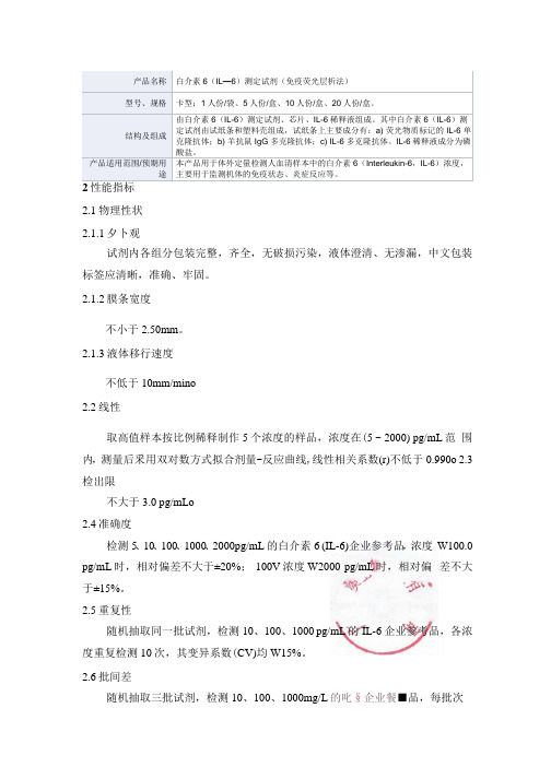
产品名称白介素6(IL—6)测定试剂(免疫荧光层析法)型号、规格卡型:1人份/袋、5人份/盒、10人份/盒、20人份/盒。
结构及组成由白介素6(IL-6)测定试剂、芯片、IL-6稀释液组成。
其中白介素6(IL-6)测定试剂由试纸条和塑料壳组成,试纸条上主要成分有:a) 荧光物质标记的 IL-6 单克隆抗体;b) 羊抗鼠 IgG 多克隆抗体;c) IL-6 多克隆抗体。
IL-6稀释液成分为磷酸盐。
产品适用范围/预期用途本产品用于体外定量检测人血清样本中的白介素 6(Interleukin-6,IL-6)浓度,主要用于监测机体的免疫状态、炎症反应等。
2.1物理性状2.1.1夕卜观试剂内各组分包装完整,齐全,无破损污染,液体澄清、无渗漏,中文包装标签应清晰,准确、牢固。
2.1.2膜条宽度不小于2.50mm。
2.1.3液体移行速度不低于10mm/mino2.2线性取高值样本按比例稀释制作5个浓度的样品,浓度在(5 ~ 2000) pg/mL范围内,测量后釆用双对数方式拟合剂量-反应曲线,线性相关系数(r)不低于0.990o 2.3检出限不大于3.0 pg/mLo2.4准确度检测5、10、100、1000、2000pg/mL的白介素6 (IL-6)企业参考品,浓度W100.0 pg/mL时,相对偏差不大于±20%; 100V浓度W2000 pg/mL时,相对偏差不大于±15%。
2.5重复性随机抽取同一批试剂,检测10、100、1000 pg/mL的IL-6企业参考品,各浓度重复检测10次,其变异系数(CV)均W15%。
2.6批间差随机抽取三批试剂,检测10、100、1000mg/L的叱§企业餐■品,每批次对各浓度重复检测io次,其变异系数(cv)应均亏凶戚A宿環、(1 ★ S)。
il-6蛋白分子量
il-6蛋白分子量IL-6(Interleukin-6),又称白细胞介素-6,是一种细胞因子蛋白,属于白细胞介素家族的一员。
它在免疫调节、炎症反应和组织修复等生理过程中发挥重要作用。
了解IL-6的分子量对于研究其结构和功能具有重要意义。
一、基本概述IL-6由人体内多种细胞产生,包括T淋巴细胞、B淋巴细胞、单核细胞、成纤维细胞等。
它的生物活性通过与细胞表面的IL-6受体结合,并启动下游信号转导通路来实现。
IL-6在免疫系统、代谢调节和神经系统等方面发挥着重要的生理和病理功能。
二、IL-6的结构IL-6是一种多肽蛋白,由184个氨基酸残基组成。
它具有四个α螺旋结构域和两个连接链(linker)区域。
其中,α螺旋结构域负责与受体结合,连接链起到稳定蛋白结构的作用。
IL-6的N端具有信号肽序列,用于定位和转运蛋白。
三、IL-6的分子量IL-6的分子量可以通过多种方法进行测定。
常见的方法包括SDS-PAGE电泳和质谱法。
1.SDS-PAGE电泳:通过将蛋白样品与SDS(十二烷基硫酸钠)和还原剂混合,使蛋白质在凝胶中形成复杂的结构,然后根据电荷大小和分子量进行迁移,最终通过染色或免疫印迹等方法确定目标蛋白的位置。
根据实验条件和所用凝胶的类型,IL-6的预测分子量约为22-26kDa。
2.质谱法:质谱法是一种直接测量蛋白质分子量的方法,常用的包括质谱仪和MALDI-TOF/TOF质谱技术。
这些技术能够以高精度和灵敏度测定蛋白质的分子量,其中IL-6的实际分子量约为21-28kDa。
需要注意的是,由于不同实验条件和试剂的差异,实际测得的IL-6分子量可能会存在一定的变异性。
此外,蛋白质的空间构象和修饰(如糖基化、磷酸化等)也会影响其分子量。
四、应用与意义了解IL-6的分子量对于研究其生物学功能和相互作用具有重要意义。
1.生理功能:IL-6在免疫调节、炎症反应、细胞增殖和分化、组织修复等方面发挥重要作用。
通过研究IL-6的分子量,可以更好地理解其在这些生理过程中的功能和机制。
白细胞介素6中文说明书 (3)
白细胞介素6中文说明书
白细胞介素6(Interleukin-6,简称IL-6)是一种蛋白质
分子,属于细胞因子家族中的一员。
它在人体中起着重要
的免疫调节作用。
IL-6来源于多种细胞,包括T细胞、B细胞、单核细胞等。
它的产生受到多种刺激因素的调控,如细菌感染、病毒感染、组织损伤等,这些刺激因素可以激活免疫细胞产生IL-6。
IL-6可以作用于多种细胞靶点,包括免疫细胞、神经系统
细胞、肠道上皮细胞等。
它在免疫系统中起到促炎症和抗
炎症的双重作用。
一方面,IL-6能够促进免疫细胞的增殖
和分化,增强免疫应答;另一方面,IL-6还能够抑制炎症
反应和细胞凋亡,起到保护作用。
IL-6在疾病过程中也发挥着重要的调节作用。
它参与了多
种炎症性疾病的发生和发展,如类风湿关节炎、系统性红
斑狼疮等。
此外,IL-6还与肿瘤的生长和转移、神经系统
疾病的发生和发展等相关。
在临床上,IL-6已经成为某些炎症性疾病的重要治疗靶点。
一些IL-6抗体药物已经被开发出来,用于治疗类风湿关节炎、系统性红斑狼疮等疾病。
总的来说,IL-6是一个重要的免疫调节因子,它在免疫系
统中起着关键的作用。
对于了解IL-6的功能机制和调控途径,有助于我们进一步理解它在疾病发生和发展中的作用,并提供新的治疗策略。
白细胞介素6第二国际标准
白细胞介素6第二国际标准白细胞介素6(Interleukin-6, IL-6)是一种重要的细胞因子,具有多种生物学功能,包括调节炎症反应、免疫应答、造血和代谢等方面。
IL-6在机体免疫调节中起着关键作用,其异常表达与多种疾病的发生发展密切相关。
为了准确评估IL-6的生物学活性,国际上建立了IL-6标准物质,其中IL-6第二国际标准是最常用的一种。
IL-6标准物质的建立旨在提供一个稳定、准确且可重复的测量方法,以确保在不同实验室和不同实验条件下对IL-6的生物学活性进行比较。
IL-6第二国际标准在全球范围内被广泛接受和应用,成为评估IL-6生物学活性的重要参考。
IL-6第二国际标准的建立经历了严格的实验设计和严格的验证过程。
通过对不同来源的IL-6进行定量比较,并在多个实验室中进行跨实验室验证,确保了IL-6第二国际标准的准确性和可靠性。
IL-6第二国际标准的建立为国际间IL-6生物学活性的比较和疾病研究提供了重要的标准基准。
IL-6在疾病中的调节作用备受关注。
IL-6通过多种途径参与炎症反应、免疫调节和细胞信号传导,对机体的免疫应答和炎症反应起着重要调节作用。
IL-6异常表达与多种疾病的发生和发展密切相关,包括类风湿关节炎、炎症性肠病、白血病和肿瘤等。
IL-6的高表达与疾病的进展和预后相关,因此对IL-6的准确测量和生物学活性评估至关重要。
IL-6第二国际标准的应用对于疾病诊断、治疗和预后评估具有重要意义。
通过对IL-6水平的测量和生物学活性的评估,可以更好地了解疾病的发展机制和预后情况,为临床诊断和治疗提供重要依据。
IL-6第二国际标准的建立和推广将促进IL-6在疾病研究和临床应用中的进展,为疾病的早期诊断和治疗提供重要支持。
梳理一下本文的重点,我们可以发现,IL-6第二国际标准在IL-6生物学活性评估中发挥着重要作用。
IL-6作为一种重要的细胞因子,在免疫调节和炎症反应中起着关键作用,其异常表达与多种疾病的发生发展密切相关。
白细胞介素6中文说明书
白细胞介素6中文说明书1. 产品描述白细胞介素6(Interleukin-6,IL-6)是一种由不同细胞产生的细胞因子,对多个生物学过程起到调节作用。
IL-6是一种蛋白质,在机体内通过与特定受体结合而发挥作用。
2. 产品特性•分子量:约21-28kDa•纯度:>95%•内毒素:低于0.01 EU/μg•保存条件:-20°C保存•表达系统:大肠杆菌重组表达3. 产品应用白细胞介素6在许多生物学过程中发挥作用,包括:1.免疫反应:IL-6是一种重要的免疫调节因子,可以促进B细胞增殖和分化,刺激抗体产生。
它还可以刺激T细胞增殖,并增加巨噬细胞和粒细胞的活性。
2.炎症反应:IL-6在炎症反应中起着重要的调节作用。
它可以增加炎症细胞的聚集和炎症介质的产生,并促进炎症反应的发展。
3.组织修复:IL-6可以促进组织修复和再生过程,它可以刺激细胞增殖和分化,促进损伤组织的修复。
4.肿瘤发生和发展:IL-6在肿瘤发生和发展过程中起到重要的调节作用。
它可以促进肿瘤细胞的增殖和侵袭,并降低免疫监视对肿瘤细胞的杀伤作用。
4. 使用方法1.重组IL-6的浓度和用量因具体实验要求而异,建议根据实验设计进行优化。
2.IL-6在溶液中稳定,但建议在-20°C保存。
3.在实验前,将冻存的IL-6溶解于无菌的缓冲液中,充分混合。
避免反复冻融。
4.IL-6可以通过注射、培养基补充或其他适当的途径应用于细胞或动物实验中。
5. 注意事项1.本产品仅供科研使用,不得用于临床诊断或治疗。
2.本产品可能具有生物危险性,请在安全条件下操作。
3.存放时,请避免长时间暴露在室温下,防止蛋白质的降解和变性。
4.如发现产品有异常,请勿使用,及时与厂家联系。
6. 包装规格•5μg/瓶•50μg/瓶•100μg/瓶7. 订购信息请联系我们的销售团队获取最新的订购信息。
8. 参考文献1.Xing Z, Gauldie J, Cox G, Baumann H, Jordana M, LeiXF, Achong MK. IL-6 is an antiinflammatory cytokinerequired for controlling local or systemic acuteinflammatory responses. J Clin Invest. 1998 Sep1;102(5):956-63.2.Tanaka T, Narazaki M, Kishimoto T.Immunotherapeutic implications of IL-6 blockade forcytokine storm. Immunotherapy. 2016 Oct;8(11):959-970.3.Nowell MA, Richards PJ, Fielding CA, Ognjanovic S,Topley N, Williams AS, Bryant-Greenwood GD, Jones SA.Regulation of pre-B cell colony-enhancing factor by STAT-3-dependent acute phase and cytokine responses inhepatocytes. J Immunol. 2006 Nov 15;177(10):7130-7.以上即为白细胞介素6中文说明书。
白细胞介素-6在青光眼发病过程中的作用
白细胞介素-6在青光眼发病过程中的作用刘攀;孟杰;刘星;王强【摘要】青光眼是全球范围内第二位致盲性眼病,其发病原理目前尚不十分明确,治疗方面多从对症治疗、降低眼压方面进行,以降低青光眼对视神经的不可逆损伤.房水中含有大量的细胞因子,包括促炎性因子、氧化应激因子、生长因子等,这些细胞因子在青光眼发病及预后过程中均起到至关重要的作用.白细胞介素-6(interleukin-6,IL-6)作为最常见的促炎性因子之一,近年来成为众多国内外临床试验监测的对象.本文旨在综述IL-6在青光眼发病过程中的作用,以期为青光眼的诊断和治疗开发新思路.【期刊名称】《国际眼科杂志》【年(卷),期】2018(018)010【总页数】3页(P1803-1805)【关键词】青光眼;小梁网;房水;白细胞介素-6;眼压【作者】刘攀;孟杰;刘星;王强【作者单位】256603 中国山东省滨州市,滨州医学院附属医院眼科;256603 中国山东省滨州市,滨州医学院附属医院眼科;256603 中国山东省滨州市,滨州医学院附属医院眼科;256603 中国山东省滨州市,滨州医学院附属医院眼科【正文语种】中文0引言青光眼是一类以视神经节细胞受到进行性破坏而产生的以视野缺损为特征的不可逆性疾病,常表现为视神经纤维层及视盘萎缩变薄。
目前,青光眼已成为第二位致盲性眼病。
白细胞介素-6(interleukin-6,IL-6)作为一种常见的促炎因子,在多种炎性反应中都有不同程度表达。
多项临床研究发现,青光眼患者房水中IL-6含量明显升高[1-3]。
本文回顾既往文献,从组织损伤角度对IL-6在青光眼发病过程中的作用进行综述,为从分子生物学方面治疗青光眼提供新思路。
1小梁网在青光眼中的作用1.1青光眼损伤学说目前,青光眼的病因及发病机制尚不十分明确,多数学者认为,青光眼是一种强烈的炎症反应[4-5]。
关于青光眼引起的组织损伤,主流学说有以下几点:(1)眼压升高对神经元、筛板及血管产生机械性压迫损伤;(2)机械性阻断和(或)遗传因素引起神经营养因子缺乏,视网膜和脉络膜血管的循环灌注异常、氧运输减少等造成缺血、缺氧;(3)其它自身免疫病理机制及蛋白质合成不足。
白介素6_人白介素6_Human IL-6使用说明书
GMP级重组人白介素6(冻干粉)Recombinant Human IL-6(interleukin-6)作用机理:白介素 6 (IL-6)是由纤维母细胞、单核、巨噬细胞、T 淋巴细胞、B 淋巴细胞、上皮细胞、角质细胞、以及多种瘤细胞所产生。
IL-1、TNF-a, PDGF、病毒感染、双链 RNA 及 c AMP 等,均可诱导正常细胞产生白介素 6。
白介素 6 能够刺激参与免疫反应的细胞增殖、分化并提高其功能。
IL-6R 广泛表达于活化 B 细胞、静止 T 细胞、NK 细胞、骨髓瘤细胞、肝细胞、髓样白血病细胞等表面。
IL- 6 的主要作用为:促进 B 细胞增生分化和分泌抗体,对肝细胞、T 细胞、神经组织、造血系统也具有广泛效应;具有抗瘤效应,也可直接或间接增强 NK 细胞及 CTL 的杀瘤活性。
规格参数:货号:TL-512 规格:50ug/100ug产品信息:表达宿主:HEK 293细胞生物活性:1×10⁷ IU/mg纯度:>95%内毒素:<0.01EU/ug纯化方式:层析纯化性状:白色疏松体保存温度:2-8℃有效期:24 个月生产厂家:同立海源生物使用说明:如需分装,可用注射用水、生理盐水、培养基或 PBS 稀释,稀释后浓度保持在 100ug/mL 以上。
稀释后置于-20℃保存期 6 个月,-80℃保存期 12 个月。
适用范围:细胞培养研究相关产品推荐:Human IL-2、Human IL-15、Human IL-12、Human IL-4、Human IL-18、Human IL-21、Human IFN-γ。
人白细胞介素-6(IL-6)说明书2

准品 100μl,然后在第一、第二孔中加标准品稀释液 50μl,混匀;然后在第一孔和第二
孔中先各取 50μl 弃掉;再各取 50μl 分别加到第三孔和第四孔,再在第三、第四孔分别
加标准品稀释液 50μl,混匀后,再各取 50μl 分别加到第五、第六孔中,再在第五、第
六孔中分别加标准品稀释液 50ul,混匀;混匀后从第五、第六孔中各取 50μl 分别加到
2
第七、第八孔中,再在第七、第八孔中分别加标准品稀释液 50μl,混匀后从第七、第
八孔中分别取 50μl 加到第九、第十孔中,再在第九第十孔分别加标准品稀释液 50μl,
混匀后从第九第十孔中各取 50μl 弃掉(。稀释后各孔加样量都为 50μl,浓度分别为 8ng/L,
4ng/L ,2ng/L,1ng/L,0.5ng/L)。
液后 15 分钟以内进行。
计算 以标准物的浓度为横坐标,OD 值为纵坐标,在坐标纸上绘出标准曲线,根据样品的
OD 值由标准曲线查出相应的浓度;再乘以稀释倍数;或用标准物的浓度与 OD 值计算出标 准曲线的直线回归方程式,将样品的 OD 值代入方程式,计算出样品浓度,再乘以稀释倍数, 即为样品的实际浓度。 注意事项 1.试剂盒从冷藏环境中取出应在室温平衡 15-30 分钟后方可使用,酶标包被板开封后如未
7 终止液
6ml×1 瓶
2 酶标试剂
6ml×1 瓶
8 标准品(12ng/L) 0.5ml×1 瓶
3 酶标包被板
12 孔×8 条
9 标准品稀释液
1.5ml×1 瓶
4 样品稀释液
6ml×1 瓶
10 说明书
1份
5 显色剂 A 液
6ml×1 瓶
11 封板膜
2张
白细胞介素-6在帕金森病发病机制中的作用

白细胞介素-6在帕金森病发病机制中的作用
白细胞介素-6(Interleukin-6,IL-6)是一种细胞因子,它在免疫系统和神经系统中都具有重要作用。
近年来,研究发现IL-6在帕金森病的发病机制中也起着重要作用。
帕金森病是一种神经系统退行性疾病,主要表现为肌肉僵硬、震颤和运动障碍等症状。
目前认为,帕金森病的发病机制涉及多种因素,包括氧化应激、神经元死亡、神经炎症反应等。
IL-6作为一种免疫系统调节因子,可以促进T细胞和B细胞的增殖和分化,并参与调节免疫细胞的活化和分泌。
同时,IL-6也能够通过多种途径影响神经元的功能和存活。
研究发现,IL-6在帕金森病的神经元损伤和死亡中起着重要作用。
IL-6可以通过激活多种信号通路,如JAK/STAT、
PI3K/Akt等,促进神经元的凋亡和炎症反应。
此外,IL-6还可以促进神经元突触的损伤和神经元突触再生的抑制。
除了直接影响神经元的功能和存活外,IL-6还可以通过调节神经元周围的免疫细胞活化和分泌,影响神经元的状态。
例如,IL-6可以促进微胶质细胞的活化和分泌,导致神经元周围的炎症反应加剧,从而加速神经元的损伤和死亡。
此外,IL-6还可以影响帕金森病患者的认知和情绪状态。
研究发现,IL-6可以影响海马区的神经元功能和突触可塑性,从而影响患者的学习记忆能力和情绪稳定性。
总之,IL-6在帕金森病的发病机制中起着重要作用。
未来的研究需要深入探讨IL-6与其他因素之间的相互作用,并寻找针对IL-6的治疗策略,以改善帕金森病患者的生活质量。
白介素6与降钙素原联合检测对脓毒血症的诊断价值

白介素6与降钙素原联合检测对脓毒血症的诊断价值【摘要】目的:研究分析白介素6与降钙素原联合检测对脓毒血症的诊断价值。
方法:选择回顾分析的方法,选取2021年1月-2023年1月100名做脓毒血症检测的患者作为研究对象,根据脓毒症的诊断标准,将患者分成脓毒血症50人和非脓毒血症50人。
分别采用电化学发光法测定白介素6、降钙素原,对患者的指标进行对比分析,总结白介素6与降钙素原联合检测对脓毒血症的诊断价值。
结果:本研究结果非脓毒血症组的PCT水平、IL-6水平、SAA水平均低于脓毒血症组(P<0.05),其中PCT的诊断价值最高。
PCT+IL-6诊断脓毒症的ACU值显著高于其他检测项,且敏感性和特异性最高。
结论:针对脓毒血症的评估,使用白介素6联合降钙素原具有较高的诊断价值,对于脓毒血症诊断的敏感性及特异性较高。
关键词:白介素6 降钙素原脓毒血症价值分析1 引言脓毒症(sepsis)常见于感染性疾病,如肺炎、腹腔感染、泌尿系统感染等。
在感染过程中,病原微生物会释放出一些毒素,如内毒素和外毒素,这些毒素与宿主免疫系统的反应会引起一系列炎症反应,导致全身炎症反应综合征的发生。
白介素6(Interleukin-6,IL-6)是一种由多种细胞产生的免疫细胞因子,它在炎症反应、免疫调节和细胞增殖方面发挥着重要作用[1]。
它在炎症反应中起着重要的作用,可以增加白细胞数量和激活免疫细胞,从而促进机体的防御和修复过程。
许多研究已经表明,白介素6的升高与脓毒血症预后不良相关。
一些研究发现,白介素6和死亡率之间存在正相关关系,即白介素6水平越高,死亡率越高。
降钙素原(PCT)是一种由甲状腺C细胞产生的激素,在感染和炎症反应中起着重要的调节作用。
临床上,降钙素原的水平测量可以用于诊断和评估脓毒血症的严重程度。
一些研究表明,降钙素原与脓毒血症的发生和严重程度密切相关。
例如,一项回顾性研究发现,在脓毒血症患者中,降钙素原的水平明显高于非脓毒血症患者(P<0.01),在预测脓毒血症的早期诊断方面表现良好[2]。
临床检验白介素-6的临床应用

临床检验白介素-6的临床应用临床检验白介素-6的临床应用一、介绍白介素-6(Interleukin-6,IL-6)是一种细胞因子,被广泛应用于临床检验中。
本文将详细介绍白介素-6的临床应用。
二、白介素-6的生理功能1.促进炎症反应:IL-6通过调节免疫细胞的活化作用,促进炎症反应的发生和发展。
2.促进组织修复:IL-6在组织损伤后的修复过程中起重要作用,促进细胞增生和再生。
3.调节代谢:IL-6参与调节能量代谢,影响脂质代谢、糖代谢等。
4.其他作用:IL-6还与肿瘤发生、骨代谢、纤维化等过程相关。
三、临床检验中的白介素-61.临床样本采集:白介素-6的检测常用血液样本,包括全血、血清和血浆。
2.检测方法:目前常用的检测方法有酶联免疫吸附法(ELISA)、放射免疫测定法(RIA)、流式细胞术等。
3.临床应用:a.炎症性疾病的诊断和监测:IL-6水平的变化可以反映炎症反应的程度和预后。
b.免疫功能评估:IL-6水平的检测可以评估机体的免疫功能状态。
c.肿瘤相关疾病的评估:IL-6与某些肿瘤的发生、发展和预后密切相关,可以作为肿瘤相关疾病的评估指标。
d.自身免疫性疾病的诊断和监测:IL-6与自身免疫性疾病的发生和发展有关,可以用于其诊断和监测。
e.其他临床应用:IL-6在心血管疾病、神经系统疾病、肝炎等方面也有一定的应用。
四、附件本文档涉及的附件请参见附件文件。
五、法律名词及注释1.细胞因子:促进或抑制生物体内干细胞或免疫细胞的增生、分化和功能的一类蛋白质。
2.ELISA:酶联免疫吸附测定法,一种通过酶的作用来检测目标物质的方法。
3.RIA:放射免疫测定法,一种通过放射性同位素标记来检测目标物质的方法。
注:本文档仅为示例文本,具体内容和格式请根据实际需求进行调整。
白介素-6在银屑病中的研究进展
白介素-6在银屑病中的研究进展王文明;晋红中【摘要】银屑病为一种常见的多因素参与的慢性炎症性疾病,其发病机制尚未完全清楚.在银屑病皮损中存在多种细胞因子的表达增高,并且血清中细胞因子的升高与疾病的严重度相关.白介素-6为一种多功能的促炎因子,被证实参与到包括银屑病在内的多种自身免疫性及慢性炎症性疾病中.本文主要综述白介素-6在银屑病中的研究进展.%Psoriasis is a common,multifactorial,chronic inflammatory skin disease with an incompletely understood pathogenesis.A substantial number of inflammatory cytokines have been found to be elevated in psoriatic lesions,and the serum levels of a subset of these cytokines also correlate with the severity of psoriasis.Interleukin-6 is a multifunctional pro-inflammatory cytokine.Interleukin-6 is proved to be associated with many chronic inflammatory diseases and autoimmunity diseases such as psoriasis.This article reviews the relationship between interleukin-6 and psoriasis.【期刊名称】《中国医学科学院学报》【年(卷),期】2018(040)002【总页数】5页(P284-288)【关键词】白介素-6;银屑病;Th17细胞;Treg细胞【作者】王文明;晋红中【作者单位】中国医学科学院北京协和医学院北京协和医院皮肤科,北京100730;中国医学科学院北京协和医学院北京协和医院皮肤科,北京100730【正文语种】中文【中图分类】R758.63Acta Acad Med Sin,2018,40(2):284-288白介素- 6 (interleukin- 6,IL- 6)为一种多功能细胞因子[1],在免疫反应、造血、急性期反应及炎症中发挥重要作用,其表达增加在多种炎症性及肿瘤性疾病,包括类风湿性关节炎、银屑病、多发性硬化、克罗恩病、乳腺癌、卵巢癌、黑色素瘤及多发性骨髓瘤等的发生发展中发挥重要作用[2]。
白细胞介素-6与脑卒中

白细胞介素-6与脑卒中研究发现中枢神经系统(central nervous system,CNS)的损伤存在炎症反应,其发生和发展的过程中,细胞因子起着重要的作用。
白细胞介素-6 (interleukin-6, IL-6) 是白介素家族中一种多功能的细胞因子,也是机体内复杂的细胞因子网络中的关键成员,它在免疫调节、参与炎症反应等过程中起着重要作用。
作为一种重要的前炎症细胞因子,IL- 6被认为在中枢神经系统具有神经保护和神经营养作用。
本文将就IL-6的理化性质、生物学活性及其在脑卒中过程中所起的作用作简要综述。
1 IL-6的一般特征IL-6是一种由184个氨基酸残基组成的糖蛋白,相对分子量质量为21~26kDa。
人IL-6基因定位于7p21[1]。
活化的单核细胞是血液中IL-6的主要来源,机体发生炎症时单核细胞和巨噬细胞是最早产生IL-6的反应细胞。
还有多种有核细胞都可产生IL – 6,如B细胞、T细胞、成纤维细胞、内皮细胞等。
颅内IL-6的主要来源常被认为是CNS中的星形胶质细胞。
局部组织的IL-6主要由成纤维细胞或局部巨噬细胞产生。
一些肿瘤细胞也能产生IL-6,如心脏黏液瘤、宫颈癌、骨髓瘤等的细胞。
IL-6的产生受许多刺激物的正调节和负调节。
细胞因子IL-1、IL-2,肿瘤坏死因子(TNF)、干扰素β(IFN-β)、血小板衍生的生长因子(PDGF)、脂多糖(LPS)增强单核细胞、成纤维细胞产生IL-6的能力及某些肿瘤细胞产生IL-6能力。
糖皮质激素、雌激素和环胞霉素A等能抑制产生IL-6;IL-4、IL-10和IL-13也抑制单核细胞产生IL-6。
IL-6通过广泛分布于机体组织细胞上的IL-6受体(IL-6R)介导其生物学活性。
IL-6R由两条多肽链组成:特异性配体结合链——α链(IL-6Rα),分子量为80 KD;和信号传导链——β链,为分子量130 KD的糖蛋白(gp130)。
IL-6首先与IL-6Rα结合,形成IL-6/ IL-6Rα复合物,再与gp130 结合,形成具有信号传导功能的高亲和力复合物。
白介素6化学发光

白介素6化学发光白介素6(Interleukin-6,简称IL-6)是一种细胞因子,它在免疫系统中发挥着重要的调节作用。
而白介素6化学发光则是一种利用化学发光技术来检测和分析IL-6的方法。
本文将从IL-6的生物学功能、化学发光技术的原理和应用以及白介素6化学发光的优势等方面进行介绍。
一、白介素6的生物学功能白介素6是一种由多种细胞产生的蛋白质,包括淋巴细胞、单核细胞、纤维母细胞等。
它在机体的免疫应答中扮演着重要的角色。
首先,白介素6可以激活B细胞,促进抗体的产生。
其次,它可以刺激T细胞的增殖和分化,参与细胞免疫的调节。
此外,白介素6还能够促进巨噬细胞的活化和细胞因子的产生,发挥重要的炎症调节作用。
因此,白介素6在免疫系统的平衡和炎症反应中起着至关重要的作用。
二、化学发光技术的原理和应用化学发光技术是一种常用的分析方法,它主要通过测量样品中化学发光反应产生的光信号来检测目标物质的含量。
对于白介素6的检测,化学发光技术具有许多优势。
首先,化学发光技术具有高灵敏度和高特异性,可以在低浓度下准确检测目标物质。
其次,化学发光技术的操作简便,不需要复杂的仪器和设备,适用于临床实验室等各种场所。
此外,化学发光技术还具有快速、稳定和可重复性好等特点,可以满足高通量检测的需求。
化学发光技术在临床诊断、药物研发和生命科学研究等领域得到了广泛应用。
例如,在感染性疾病的诊断中,通过检测白介素6的水平可以判断炎症反应的程度和严重程度,为临床治疗提供重要依据。
此外,在免疫学研究中,化学发光技术可以用于检测白介素6的产生和释放,深入了解其在免疫调节中的作用机制。
因此,白介素6化学发光技术在医学和生命科学领域具有重要的应用价值。
相比于传统的检测方法,白介素6化学发光具有许多优势。
首先,化学发光技术可以实现高通量检测,同时检测多个样品,提高检测效率。
其次,化学发光技术的信号稳定性好,不受样品浓度和反应时间的影响。
此外,化学发光技术还可以通过选择不同的标记物和底物来实现多种检测模式,满足不同实验需求。
白介素6指导原则

白介素6指导原则
白介素6(Interleukin-6, IL-6)是一种细胞因子,参与调节免疫和炎症反应。
在某些情况下,IL-6的过度产生可能导致炎症性疾病的发展。
以下是一些关于白介素6的指导原则:
1. 了解IL-6的作用:IL-6在免疫系统中起着重要的调节作用,可以促进炎症反应、调节细胞增殖和分化等。
了解IL-6的作用有助于理解其在疾病中的作用机制。
2. 评估IL-6水平:对于某些炎症性疾病,检测血液中的IL-6水平可以作为评估疾病活动性和预后的指标之一。
通过监测IL-6水平的变化,可以指导治疗和判断疾病进展情况。
3. 选择合适的治疗方法:针对IL-6过度产生引起的疾病,可以采取一些治疗方法来干预IL-6的作用。
例如,使用特定的抗体药物来阻断IL-6的受体,或者使用抗炎药物来调节炎症反应。
4. 个体化治疗:IL-6在不同疾病中的作用和表达水平可能存在差异,因此治疗方案需要根据具体情况进行个体化设计。
根据患者的病情、病史和其他相关因素,制定合适的治疗策略。
5. 综合治疗策略:针对IL-6相关疾病,综合治疗策略可能更有效。
除了针对IL-6的干预措施,还可以结合其他治疗手段,如抗炎治疗、免疫调节等,以达到更好的治疗效果。
需要注意的是,针对特定疾病和个体情况,上述指导原则可能会有所不同。
因此,在具体治疗过程中,建议咨询专业医生或遵循医生的指导,以确保获得最佳的治疗效果。
白细胞介素6第二国际标准
白细胞介素6第二国际标准白细胞介素6(Interleukin-6,IL-6)是一种重要的细胞因子,参与调节免疫反应、炎症反应和细胞增殖等生理过程。
IL-6的异常表达和功能异常与多种疾病的发生和发展密切相关。
为了准确、可比较地测量IL-6水平,国际标准化组织(InternationalStandardization Organization,ISO)于2014年制定了白细胞介素6第二国际标准。
本文将对该标准进行详细介绍,并探讨其在临床应用中的意义。
一、白细胞介素6第二国际标准的制定过程1.1 标准化组织的重要性在临床实践中,不同实验室使用不同试剂盒和方法测量IL-6水平,结果存在较大差异。
为了解决这一问题,制定统一的国际标准是必要的。
1.2 标准化组织对于制定国际标准的作用ISO作为一个独立、非盈利性质组织,在全球范围内制定和推广各种标准。
其参与制定IL-6第二国际标准,旨在确保不同实验室之间的结果可比性,提高IL-6的测量准确性。
1.3 制定IL-6第二国际标准的流程制定国际标准需要经过多个步骤,包括制定工作组、收集数据、统计分析、制定标准物质和验证等。
在制定IL-6第二国际标准过程中,专家们通过大量实验和数据分析,最终确定了该标准。
二、白细胞介素6第二国际标准的主要内容2.1 标准物质白细胞介素6第二国际标准通过纯化和浓缩技术获得了高纯度的IL-6蛋白,并将其作为标准物质。
该物质具有稳定性好、纯度高等特点,可作为测量IL-6水平的参考。
2.2 标准曲线根据不同浓度的标准物质制备一系列稀释液,并通过酶联免疫吸附法(enzyme-linked immunosorbent assay, ELISA)进行测量。
将各个稀释液中所得结果与其对应的浓度进行曲线拟合,得到一条线性关系良好的曲线。
2.3 检测方法IL-6第二国际标准规定了测量IL-6水平的方法,主要包括ELISA、免疫荧光法和流式细胞术等。
这些方法经过验证,能够准确、可靠地测量IL-6水平。
白介素6 室间质量评价标准

白介素6 室间质量评价标准-概述说明以及解释1.引言1.1 概述白介素6(IL-6)是一种重要的细胞因子,广泛参与多种生物体的炎症反应和免疫调节过程。
它在免疫细胞、内皮细胞、成纤维细胞等多种细胞类型中表达,并且具有多种生物学功能。
IL-6在疾病发生和发展中发挥重要角色,与肿瘤、感染性疾病、自身免疫疾病等多种疾病密切相关。
随着近年来研究的深入,室间环境对人体健康的影响也越来越受到重视。
人们长期生活在室内,室内空气质量对健康的影响不可忽视。
在室内环境中,IL-6是一种重要的生物标志物,可以反映室内环境的炎症水平和室内污染物的暴露水平。
因此,对室内空气中IL-6的浓度进行评价成为了一项重要任务。
然而,目前还缺乏针对IL-6的室内质量评价标准。
现有的室内质量评价标准主要关注一些化学污染物和微生物污染物,对于IL-6的考量较少,无法全面准确地评估室内环境的质量。
鉴于此,本文旨在建立一套完备的白介素6室间质量评价标准,通过分析室内空气中IL-6的浓度来评估室内环境的炎症水平和健康风险。
该评价标准将有助于提高人们对室内环境的认知和保护,为保障人们的健康提供科学依据。
总而言之,本文将对白介素6室间质量评价标准的建立进行深入探讨,旨在为室内环境的评估提供更全面准确的指导,保障人们的健康。
通过研究IL-6在室内空气中的浓度,我们可以更好地了解室内环境的质量,并采取相应措施改善室内空气质量,提高人们的居住环境和生活质量。
1.2 文章结构本文主要分为引言、正文和结论三个部分。
下面将详细介绍每个部分的内容。
1. 引言部分:在这一部分中,我们将对整篇文章进行概述,并说明文章的结构、目的和总结。
- 1.1 概述:在这一小节中,将简要介绍白介素6和室间质量评价标准的背景和重要性,引发读者的兴趣。
- 1.2 文章结构:这一小节正是本部分的内容,将详细阐述文章的结构,即引言、正文和结论三个部分的分布和概要。
- 1.3 目的:本节将明确本文的目的,即建立白介素6室间质量评价标准的必要性和重要性。
白细胞介素6神经调节机制探究

白细胞介素6神经调节机制探究白细胞介素6(Interleukin-6,IL-6)是一种细胞因子,广泛存在于人体各种组织和细胞中,具有多种生物学功能。
除了免疫调节和炎症介导外,近年来的研究发现,IL-6也参与了神经调节的过程。
这一发现引起了广泛的关注和研究,我们有必要深入了解白细胞介素6在神经调节方面的机制。
首先,IL-6在中枢神经系统中的产生与释放起着重要作用。
中枢神经系统包括大脑、脊髓和神经节等,是整个神经系统的核心。
研究表明,IL-6在脑组织中的表达量较高,并参与了神经元的生长、损伤修复以及突触可塑性等神经调节过程。
此外,一些研究还发现,神经元的活动能够诱导白细胞介素6的释放,从而形成神经-免疫的相互调节机制。
其次,白细胞介素6在神经递质释放中的作用也值得关注。
神经递质是神经元之间传递信息的化学物质,对神经系统的正常功能发挥着至关重要的作用。
研究发现,IL-6能够调节多种神经递质的合成和释放,如儿茶酚胺类递质(如多巴胺、去甲肾上腺素等)和γ-氨基丁酸(GABA)。
这些神经递质在情绪调节、认知功能和运动控制中具有重要作用,而IL-6的调节可能对这些过程产生深远的影响。
此外,白细胞介素6还参与了疼痛感知和处理的神经调节。
疼痛是身体对外界刺激的一种自我保护机制,同时也是神经系统中一项复杂的调控过程。
研究发现,IL-6能够介导疼痛神经传递过程中的炎症反应,从而调控炎症相关的疼痛感受。
此外,IL-6还与胶质细胞的激活和星形胶质细胞的释放有关,这些细胞在疼痛处理中扮演着重要角色。
最后,白细胞介素6在神经系统疾病中的研究也逐渐受到关注。
很多神经系统疾病,如帕金森病、阿尔茨海默病和抑郁症等,与免疫系统的异常活化和炎症反应密切相关。
近年来的研究发现,白细胞介素6在这些疾病中可能发挥重要作用。
例如,IL-6的过度激活与帕金森病的神经退行性变有关,而抑郁症患者体内IL-6含量明显增加。
这些研究结果表明,免疫系统和神经系统之间的相互作用在神经系统疾病的发生和发展中具有重要意义。
白细胞介素6在心脑血管疾病中的临床应用新进展

白细胞介素6在心脑血管疾病中的临床应用新进展心脑血管疾病是指心脏和血管系统的病变,包括冠心病、高血压、脑卒中等。
这些疾病严重影响着全球人口的健康和生活质量。
近年来,研究人员发现白细胞介素6 (Interleukin 6, IL-6) 在心脑血管疾病的发病机制中起着重要的作用,并且探索了其在临床应用中的新进展。
一、白细胞介素6简介白细胞介素6是一种细胞因子,它由多种细胞产生,如T淋巴细胞、B淋巴细胞和单核细胞等。
在机体免疫系统中,白细胞介素6发挥着调节和调控多种免疫反应的作用。
此外,IL-6还具有炎症介导作用,参与炎症反应的调节和维持。
二、IL-6的作用机制IL-6在心脑血管疾病中的作用机制复杂多样。
首先,IL-6是炎症介导物质,当心脑血管受到损伤或炎症刺激时,白细胞介素6的产生会大量增加,进而引发炎症反应。
其次,IL-6可以促进血脂代谢异常和胰岛素抵抗等病理过程,导致血管内皮功能异常和血管收缩。
此外,IL-6还可以影响血小板活性和凝血功能,增加血栓形成的风险。
三、IL-6在心脑血管疾病中的临床研究近年来,研究人员对IL-6在心脑血管疾病中的临床应用进行了深入研究。
一方面,通过抑制IL-6的产生或作用,可以减轻心脑血管疾病的炎症反应,从而改善患者的临床症状和预后。
另一方面,一些研究发现,通过增加IL-6的产生或作用,可以促进心脑血管病变的修复和再生,有望成为心脑血管疾病的新的治疗方法。
四、IL-6在冠心病中的临床应用前景冠心病是心脑血管疾病的一种常见类型,严重威胁着人民的身体健康。
一些研究表明,IL-6水平与冠心病患者的病情严重程度和预后密切相关。
因此,IL-6可能成为重要的冠心病标志物,并且通过调节IL-6的相关信号通路,可以为冠心病的治疗提供新的方向。
五、IL-6在高血压中的临床应用前景高血压是心脑血管疾病中的另一大类,给全球范围内的人民健康带来了巨大的威胁。
研究发现,IL-6可以调节血管收缩和扩张功能,并且与高血压的发病风险密切相关。
- 1、下载文档前请自行甄别文档内容的完整性,平台不提供额外的编辑、内容补充、找答案等附加服务。
- 2、"仅部分预览"的文档,不可在线预览部分如存在完整性等问题,可反馈申请退款(可完整预览的文档不适用该条件!)。
- 3、如文档侵犯您的权益,请联系客服反馈,我们会尽快为您处理(人工客服工作时间:9:00-18:30)。
Chronic interleukin-6(IL-6)treatment increased IL-6secretion and induced insulin resistance in adipocyte:prevention by rosiglitazoneClaire Lagathu,Jean-Philippe Bastard,Martine Auclair,Mustapha Maachi,Jacqueline Capeau,and Martine Caron *INSERM U.402and IFR65Saint-Antoine Faculty of Medicine,Paris,FranceReceived 12September 2003AbstractIL-6has emerged as an important cytokine upregulated in states of insulin resistance such as type 2diabetes.We evaluated the chronic effect of IL-6on insulin signaling in 3T3-F442A and 3T3-L1adipocytes.First,cells responded to a chronic treatment with IL-6by initiating an autoactivation process that increased IL-6secretion.Second,IL-6-treated adipocytes showed a decreased protein expression of IR-b subunit and IRS-1but also an inhibition of the insulin-induced activation of IR-b ,Akt/PKB,and ERK1/2.Moreover,IL-6suppressed the insulin-induced lipogenesis and glucose transport consistent with a diminished expression of GLUT4.IL-6-treated adipocytes failed to maintain their adipocyte phenotype as shown by the downregulation of the adipogenic markers FAS,GAPDH,aP2,PPAR-c ,and C/EBP-a .IL-6also induced the expression of SOCS-3,a potential inhibitor of insulin signaling.Finally,the effects of IL-6could be prevented by rosiglitazone,an insulin-sensitizing agent.Thus,IL-6may play an important role in the set-up of insulin resistance in adipose cell.Ó2003Elsevier Inc.All rights reserved.Keywords:Interleukin-6;Insulin resistance;Adipocyte;Thiazolidinedione;CytokineInsulin resistance is a prominent feature of the met-abolic syndrome and type 2diabetes and constitutes a major risk for cardiovascular disease [1].In recent years,evidence has been gathered that type 2diabetes is as-sociated with a cytokine-related acute-phase reaction,part of an overall inflammatory state.Indeed,insulin resistance is correlated with increased blood concentra-tions of several markers of the acute-phase response,including C-reactive protein,the major cytokine medi-ator interleukin-6(IL-6)[2–4],tumor necrosis factor alpha (TNF-a )[5],and interleukin-1-beta (IL-1-b )[6].The blood concentration of IL-6is elevated in subjects with type 2diabetes [2]and obesity [7,8],and a positive correlation was observed between adipose tissue IL-6content and insulin resistance in vivo [7,8].Furthermore,insulin-resistant patients have a high level of soluble IL-6receptor which potentiates IL-6bioactivity,and signsof innate immunity activation are often present before type 2diabetes [9].Studies have indicated that 10–35%of circulating IL-6is derived from adipose tissue [8,10],a production that increases with adiposity [10].Although immune cells,fibroblasts,endothelial cells,and monocytes have traditionally been regarded as the major sources of cir-culating IL-6[8],it has been clearly established that adipocytes themselves can secrete IL-6[8]together with IL-1-b [11]and TNF a [12].TNF a has been demon-strated to negatively interfere with adipocyte metabo-lism at numerous sites including glucose and fatty acid metabolism and insulin receptor signaling [13].Only a few studies have assessed the effect of IL-6on the adi-pocyte metabolism [14].Preliminary studies suggested that a short term treatment with IL-6could affect the adipocyte lipid metabolism [15,16].However,no data are available concerning a long-term treatment with IL-6on insulin signaling in adipocytes.IL-6signal transduction involves the JAK (janus-ac-tivated kinase)/STAT (signal transducers and activators*Corresponding author.Fax:+33-1-40-01-13-52.E-mail address:caron@st-antoine.inserm.fr (M.Caron).0006-291X/$-see front matter Ó2003Elsevier Inc.All rights reserved.doi:10.1016/j.bbrc.2003.10.013Biochemical and Biophysical Research Communications 311(2003)372–379BBRC/locate/ybbrcof transcription)signaling pathway[17]that could cross-react in many points with the insulin signaling network [18].Suppressor of cytokine signaling(SOCS)proteins were originally described as cytokine-induced molecules involved in negative feedback loop of cytokine[19]and insulin signaling[20].Indeed,SOCS-3can directly inter-act with IR-b and IRS-1and,when overexpressed in adipocytes,inhibit insulin signal transduction[18]. Moreover,SOCS-3is upregulated in adipose tissue of obese mice[20]and IL-6was identified to play a role in insulin resistance in hepatocytes by acting at the insulin signaling pathways through induction of SOCS-3protein expression[21,22].Rosiglitazone belongs to the thiazolidinedione (TZDs)class of antidiabetic drugs that improve meta-bolic control in patients with type2diabetes through the improvement of insulin sensitivity[23].TZDs exert their antidiabetic effects through a mechanism that involves activation of peroxisome proliferator-activated receptor (PPAR cÞ[24].Although PPAR c is predominantly expressed in adipose tissue,the TZDs reduce insulin resistance in adipose tissue,muscle,and the liver.There are still many unknowns about the mechanism of action of TZDs in insulin resistance.Potential signaling factors include free fatty acids and adipocyte-derived cytokines such as TNF a or IL-6,which are overexpressed in obesity and insulin resistance[23].In the current study,we investigated the effect of chronic IL-6treatment(100and200ng/mL,8days)on several functions of cultured3T3-F442A and3T3-L1 adipocytes.We show that adipocytes are target cells for IL-6.IL-6upregulated its own secretion and expression. Furthermore,IL-6induced insulin resistance by inhib-iting the acute phosphorylation of the insulin signaling components(IR-b,IRS-1,ERK1/2,Akt/PKB)and a series of insulin-dependant processes(lipogenesis, glucose transport).IL-6-treated adipocytes failed to maintain their adipocyte phenotype as shown by the downregulation of adipogenic markers(FAS,GAPDH, aP2,PPAR-c,and C/EBP-aÞ.IL-6also increased by 6-fold the expression of SOCS-3,a potential negative regulator of insulin signaling pathway[18].Finally we found that the insulin-sensitizing agent rosiglitazone decreased IL-6secretion and SOCS-3hyperexpression and prevented IL-6adverse effects on insulin signaling. Materials and methodsMaterials.Recombinant human IL-6(rhIL-6)(specific activity, 1Â107U/mg)was provided by PeproTech(NJ,USA)and rosiglitaz-one(BRL49653)was provided by SmithKline Beecham Pharmaceu-ticals.Cell culture and treatment.3T3-F442A preadipocytes were cultured and induced to differentiate as previously described[25].In brief,the preadipocytes were maintained in DMEM containing25mM glucose and10%new born calf serum(NCS)until they reached confluence (day0).They were then grown in DMEM containing10%FCS and 1l M insulin for six days,when more than92%of the cells showed fat accumulation.3T3-L1preadipocytes were subcultured in DMEM containing10%FCS.At confluence(day0),preadipocytes were grown for two days in DMEM containing10%FCS,1l M insulin,0.5mM isobutylmethylxanthine,and1l M dexamethasone and then cultured in DMEM containing10%FCS and1l M insulin.After seven days, more than90%of the cells showed fat accumulation.In some exper-iments,1l M rosiglitazone was added at day0.The effect of IL-6(100 and200ng/mL)was tested on differentiating3T3-F442A cells(from days0–8of differentiation)and on mature3T3-L1adipocytes(from days7–15of differentiation).IL-6measurements.Murine IL-6(mIL-6)concentration was de-termined at day8of differentiation in24h-secretion medium of3T3-F442A cells by the sandwich ELISA Quantikine Mouse test(R&D Systems,USA,MN).This test specifically identifies murine IL-6(de-tection limit:3.1pg/mL)without cross-reaction with the rhIL-6used for cell treatment.Western blotting.Cells were solubilized in a Laemmli buffer with 100mM dithiothreitol.Cell lysates(104cells)were subjected to SDS–PAGE and Western blotting with the indicated antibody:SREBP-1 (K-10),C/EBP a(C-19),PPAR c(H-100),C/EBP b(C-19),and GLUT4 (H-61)(Santa Cruz Biotechnology).Immune complexes were visual-ized with a chemiluminescence method(Enhanced Chemiluminescent kit,Amersham Biosciences).Protein expression on Western blot was normalized on the basis of cell number.RNA preparation and real-time RT-PCR.Total RNA was isolated with the RNeasy kit(Qiagen,USA,CA).Complementary-DNA was synthetized using random hexamers and AMV reverse transcriptase (Promega Biosciences,USA,CA).Real-time PCR was performed with the LightCycler system(Roche Molecular Biochemicals).PCRs were performed using the LightCycler SYBR greenfluorophore.The fol-lowing primers were used:aP2,50-AAC ACC GAG ATT TCC TT-30 and50-ACA CAT TCC ACC ACC AG-30;FAS,50-TGC TCC CAG CTG CAG GC-30and50-GCC CGG TAG CTC TGG GTG TA-30; GAPDH,50-CAA GGT CAT CCA TGA CAA CTT-30and50-GGC CAT CCA CAG TCT TCT GG-30;IL-6,50-CTG CAA GAG ACT TCC ATC CAG TT-30and50-GAA GTA GGG AAG GCC TGG-30; SOCS3,50-ACC AGC GCC ACT TCT TCA CG-30and50-GTG GAG CAT CAT ACT GAT CC-30;18S ribosomal RNA,and50-GAG CGA AAG CAT TTG CCA AG-30and50-GGC ATC GTT TAT GGT CGG AA-30.Each sample was normalized on the basis of its RNA18S content.Insulin signaling.The effect of insulin was evaluated on tyrosine phosphorylation of the insulin receptor b-subunit(IR-bÞand IR sub-strate-1(IRS-1),and on the activation of extracellular signal-regulated kinase1/2(ERK-1/2)and protein kinase B(Akt/PKB).At day8,the cells were starved for18h and stimulated for10min by insulin (100nM).Aliquots of cell lysates were immunoblotted with an anti-phosphotyrosine antibody(PY-99,Santa Cruz Biotechnology)or antibodies directed against the activated forms of ERK-1/2(Phospho-ERK(E-4),Santa Cruz Biotechnology)or Akt/PKB(phospho-Akt-ser473,#9271,Cell Signaling Technology).Protein expression was checked by using antibodies directed against IR-b(C-19),IRS-1(C-20),ERK-1/2(C-16)(Santa Cruz Biotechnology)or Akt/PKB(#9272, Cell Signaling Technology).Lipogenesis.Lipogenesis was studied at day8of differentiation in 3T3-F442A adipocytes cultured for24h in serum-free medium.Insulin (100nM)was added for30min.After a further30-min incubation with 0.5l Ci/well of[U-14C]glucose(302mCi/mmol,Amersham Bio-sciences),the cells were beled lipids were extracted with chloroform/methanol(1/2).After30min on ice,the inferior phase was collected,dried by evaporation,and counted in scintillationfluid.The results were expressed as percentÆSEM of above basal value.Glucose transport.Glucose transport was studied on day8of dif-ferentiation in3T3-F442A adipocytes starved for24h.Prior to ex-periment cells were incubated for2h in500l l of glucose transportgathu et al./Biochemical and Biophysical Research Communications311(2003)372–379373solution(pH7.6)containing HEPES12.5mM,NaCl120mM,KCL 5mM,MgSO41.2mM,CaCl21mM,NaHPO41mM,sodium pyru-vate2mM,and BSA2%.Insulin(100nM)and2-DOG mix(0.25l Ci 2-deoxy-D-[1-14C]glucose(60mCi/mmol,Amersham Biosciences)and 0.25mM2-DOG)were successively added for30and5min.To stop transport,cells were washed with PBS,lysed for30min with0.1% SDS,and counted.The results were expressed as percentÆSEM of above basal value.Statistical analysis.Results are meansÆSEM of the indicated number of independently performed experiments.Statistical signifi-cance was determined using the nonparametric Mann–Whitney U test results.The threshold for significance was at P¼0:05.ResultsIL-6activated a regulatory loop in cultured adipocytes resulting in its autocrine/paracrine secretionInitial studies focused on the ability of hrIL-6-treated adipocytes(8days)to secrete murine IL-6.We observed that at concentrations(100and200ng/mL)close to those found in the serum of obese patients[8,10],hrIL-6 induced secretion of IL-6(Fig.1A).IL-6effect was dose-dependent and observed also on IL-6mRNA expression (Fig.1B).IL-6altered insulin signaling in3T3-F442A cells We evaluated the chronic effect of IL-6(8days:from day0to day8)on the insulin signal transduction.As shown in Fig.2,IL-6decreased the protein expression of IRS-1and IR-b(Fig.2A),without modification of the protein expression of ERK1/2and Akt/PKB(Fig.2B). IL-6blunted the ability of insulin to increase the tyro-sine phosphorylation of its receptor b-subunit(Fig.2A) and decreased the insulin-induced activation of ERK1/2 and Akt/PKB,three key enzymes of the insulinsignaling Fig.2.IL-6alters insulin signaling in3T3-F442A cells.3T3-F442A preadipocytes were differentiated in the presence of the indicated concentration of hrIL-6.At day8of differentiation,IL-6-treated or untreated cells were stimulated by insulin(100nM).Aliquots were immunoblotted with antibodies that recognized the b subunit of the insulin receptor(IR-b),its substrate IRS-1,the phosphorylated tyro-sines of IR-b(A),the downstream insulin signaling enzymes ERK1/2 and Akt/PKB,and their phosphorylated forms(B).A representative immunoblot from3to5separate experiments isshown.Fig.3.IL-6alters insulin-stimulated lipogenesis and glucose transport. 3T3-F442A preadipocytes were differentiated in the presence of the indicated concentration of IL-6.(A)At day8of differentiation, IL-6-treated or untreated cells were stimulated by insulin(100nM)and lipogenesis was evaluated by incorporation of[U-14C]glucose and ex-pressed as percentage above basal(control:105Æ15pmol/h/106cells). Results are meansÆSEM of3–6experiments performed in duplicate.(B)IL-6-treated or untreated cells were stimulated by insulin(100nM) and glucose transport was evaluated by incorporation of2-deoxy-D-[1-14C]glucose and expressed as percentage above basal(control: 0.44Æ0.05nmol/min/106cells).Results are meansÆSEM of3–4 experiments performed in duplicate.(C)IL-6-treated or untreated cells were solubilized and aliquots were immunoblotted with anti-GLUT4 antibody.A representative immunoblot from3separate experiments is shown.***P<0:001.374 gathu et al./Biochemical and Biophysical Research Communications311(2003)372–379pathways (Fig.2B).Although a chronic IL-6treatment did not modify the range of lipolysis (data not shown),it totally suppressed the insulin-induced lipogenesis (Fig.3A)and glucose transport (Fig.3B),consistent with the decreased GLUT4protein expression (Fig.3C).Thus,IL-6could directly decrease adipose cell response to insulin.IL-6alters the adipocyte phenotypeIn 3T3-F442A cells,IL-6(8days:from day 0to day 8)did not modify the early steps of the differentiation program.This was indicated by the lack of IL-6effect on clonal expansion (not shown)and C/EBP b and SREBP-1protein expression measured at days 2and 8of dif-ferentiation,respectively (Fig.4A).By contrast,at day 8,IL-6decreased protein expression of PPAR c and C/EBP a (Fig.4A)and the mRNA expression of FAS,GAPDH,and aP2(Fig.4B),all markers of the adipo-cyte phenotype and insulin sensitivity.The defective expression of FAS and GAPDH,two enzymes of lipo-genesis,may explain the lack of insulin activation of this pathway (Fig.3A).The decreased expression of adipo-genic markers was observed without significant decrease of the number of differentiated adipocytes evaluated by counting the cells with lipid droplets (88.5%Æ1.3%and 92.5%Æ2.2%of total cells in IL-6-treated and untreated cells,respectively,at day 8of differentiation).In fully differentiated 3T3-L1adipocytes,IL-6(8days:from day 7to day 15)also altered insulin response and the ex-pression of adipogenic markers.We observed a de-creased expression of IR-b and IRS-1protein,a blunted insulin-stimulated phosphorylation of ERK1/2and Akt/PKB,and an altered expression of FAS,GAPDH,and aP2(mRNA)and of C/EBP a and PPAR c (protein)(Data not shown).These results indicated that IL-6had an effect on adipocytes whether the treatment occurred during (3T3-F442A)or after completion of differentia-tion (3T3-L1).Rosiglitazone reverted all the adverse effects of IL-6on adipose cellCo-treatment of 3T3-F442A cells with IL-6(100ng/ml)and rosiglitazone (1l M)prevented IL-6from in-creasing its own secretion (Fig.5A).Interestingly,ros-iglitazone also decreased the basal IL-6secretion,suggesting that one possible mechanism forinsulinFig.4.IL-6alters the adipocyte phenotype.3T3-F442A preadipocytes were differentiated in the presence of the indicated concentration of IL-6.(A)At day 2(C/EBP b )or 8(C/EBP a ,SREBP-1and PPAR c )of differentiation,IL-6-treated or untreated cells were solubilized and aliquots were immunoblotted with the antibodies against the indicated transcription factor.A representative immunoblot from three separate experiments is shown.(B)At day 8of differentiation,total RNA was extracted and subjected to real-time PCR.FAS,GAPDH,and aP2mRNA levels normalized to 18S expression were determined relative to untreated cells (1.0).Results are means ÆSEM of 4–8experiments performed in triplicate.**P <0:01,***P <0:001.sensitization may be mediated by an inhibition of IL-6secretion and mRNA expression (Fig.5B).Rosiglitaz-one (1l M)prevented IL-6from decreasing insulin re-sponse:the insulin-dependent phosphorylation of ERK1/2and Akt/PKB was reestablished (Fig.6A)as well as insulin-induced lipogenesis (Fig.6B).Rosiglit-azone also impaired the downregulation by IL-6of mRNA expression of FAS,GAPDH,and aP2(Fig.6C).Rosiglitazone inhibited SOCS-3expression and its induc-tion by IL-6We then tested whether a chronic IL-6treatment could increase the expression of SOCS-3,a potentialinhibitor of insulin signaling.As shown in Fig.7A,IL-6upregulated the mRNA expression of SOCS-3up to 6-fold at 200ng/ml.Rosiglitazone exerted a negative effect on the basal and IL-6-induced SOCS-3expression (Fig.7B).In accordance with a decreased basal IL-6secretion,rosiglitazone also decreased the basal SOCS-3expression,suggesting that it can prevent IL-6effect through a direct or indirect effect on IL-6signaling.DiscussionThe adipose tissue can play an important role in modulating whole body insulin sensitivity through its endocrine functions,such as cytokine and adiponectin release.Each of the proinflammatory cytokines,TNF a [5],IL-1-b [6],and IL-6[2–4]has been recently impli-cated in the development of insulin resistance and type 2diabetes.In insulin-resistant patients the degree of cor-relation between the IL-6concentration and the severity of insulin resistance was actually found to be higher than that found with TNF a [2,26].The adipose tissue plays a key role in IL-6production [14]and is a major target of the cytokine.The objective of the present study was to evaluate the chronic effect of IL-6(8days)on adipocytes during differentiation (3T3-F442A)or on fully mature adipocytes (3T3-L1)and to approach the mechanism whereby IL-6induced insulin resistance.We initially demonstrated that adipose cells re-sponded to IL-6by increasing the production and se-cretion of IL-6.This autocrine stimulation loop,that has been observed in vascular smooth muscle cells [27],probably reinforces the adverse effects of IL-6on adi-pose cell.This autoactivation by IL-6is almost totally prevented by the antidiabetic agent,rosiglitazone [23].Interestingly,rosiglitazone decreased basal secretion of IL-6(Fig.5)and TNF a [28]which reinforcestheFig.6.Rosiglitazone prevented the IL-6-induced insulin resistance.3T3-F442A preadipocytes were differentiated in the presence of 100ng/ml IL-6(open bars)and/or 1l M of rosiglitazone (dark bars).At day 8of differentiation,IL-6-treated or untreated cells were stimulated by insulin (100nM).Aliquots were immunoblotted with antibodies that recognized ERK1/2and Akt/PKB and their phosphorylated forms.A representa-tive immunoblot from three separate experiments is shown.(A)Lipo-genesis was evaluated as described in Fig.3.Results are means ÆSEM of 3–6experiments performed in duplicate.(B)Total RNA was extracted and subjected to real-time PCR.FAS,GAPDH,and aP2mRNA levels normalized to 18S were determined relative to untreated cells (1.0).(C)Results are means ÆSEM of 4–8experiments performed in triplicate.**P <0:01,***P <0:001,and NS,notsignificant.potential role of these cytokines as mediators of insulin resistance.We next observed that IL-6altered insulin signaling after a chronic exposure(8days)to differentiating (3T3-F442A)or fully differentiated(3T3-L1)adipo-cytes.In fact IL-6had dual effects on insulin action.It acutely(30–180min)mimicked insulin action on ERK 1/2and Akt activation(not shown)and chronically induced insulin resistance.Indeed,the inhibition of ERK1/2and Akt/PKB phosphorylation appeared after a24-h exposure and aggravated after4days of expo-sure.IL-6had adverse effects at both the receptor (IR-b and IRS-1tyrosine phosphorylation)and post-receptor levels(ERK1/2,Akt/PKB,glucose transport, and lipogenesis).Finally,we observed that IL-6interfered with the last step of adipocyte differentiation.Indeed,IL-6did not affect the transcription factors SREBP-1and C/EBP b that are expressed early in the differentiation program [29]but decreased the expression of C/EBP a and PPAR c,which play a role in both terminal differentia-tion,maintenance of the adipocyte phenotype and in-sulin sensitivity[29,30].This was confirmed by the adverse effects of IL-6on the markers of the mature adipocyte involved in glucose and lipid metabolism (FAS,GAPDH,GLUT4,and aP2)and insulin sensi-tivity(IRS-1and IR-bÞ.Surprisingly,when compared to the other adipogenic markers,aP2is weakly decreased by IL-6(by15–20%).This could be explained by the fact that unlike FAS,GAPDH,and GLUT4,aP2expression is not controlled by insulin[31,32].The regulation of aP2expression remains unclear[33],even if PPAR c and fatty acids were shown to be responsible for the high expression level of aP2in adipocytes in vitro[34,35].In our study,the low incidence of IL-6on aP2expression may also be explained by the fact that IL-6poorly af-fected the differentiation program of3T3-F442A cells (88.5%Æ1.3%and92.5%Æ2.2%of IL-6-treated and untreated cells contained lipid droplets,respectively) and did not induce dedifferentiation of3T3-L1adipo-cytes(89–95%of cells contained lipid droplets).Thus, we conclude that IL-6preferentially altered the set-up and maintenance of insulin response(C/EBP a, GAPDH,FAS,GLUT4,IR-b,and IRS-1)rather than the adipocyte differentiation program(SREBP-1,C/ EBP b,and aP2).Interestingly,rosiglitazone prevented all the adverse effects of IL-6on both differentiating(3T3-F442A)and fully differentiated(3T3-L1)adipocytes.Rosiglitazone increases whole body insulin sensitivity[23]and de-creases insulin resistance in muscle and liver probably via endocrine signaling from adipocytes.This hypothesis is supported by the fact that rosiglitazone decreased IL-6secretion.Suppressor of cytokine signaling(SOCS)proteins have been recently implicated in insulin signaling network[18].Indeed,SOCS-3can bind to phosphory-lated tyrosine of IR-b and inhibit insulin signaling[36]. Here,we show that the negative effect of IL-6on insulin signaling is linked to the upregulation of SOCS-3 mRNA expression.Rosiglitazone-treated cells had a decreased SOCS-3and IL-6mRNA expression that could result from the decreased IL-6secretion or from a direct effect of rosiglitazone on IL-6signaling.The group of Spiegelman[37]provided evidence that TNF a can directly blunt insulin action in vitro and in animal studies via an increased serine/threonine-phos-phorylation of IRS-1.In the present study we failed to detect increased IRS-1serine phosphorylation in re-sponse to IL-6treatment(not shown).Alternatively, recent evidence suggests that some of the effects of TNF a may be mediated by its ability to induce IL-6and IL-6receptor expression in tissues such as the liver and muscle[38,39].Interestingly,in accordance with in vivo studies[40]we have found that a chronic treatment with TNF a(20ng/mL,8days)of3T3-F442A adipocytes in-duced upregulation of IL-6mRNA level(4.5-fold)(not shown).The upregulation of IL-6gene expression might contribute to the impairment of insulin sensitivity found in states of increased TNF a levels.Other mechanisms such as the downregulation of adiponectin expression by IL-6[41]probably also con-tribute to insulin resistance induced by IL-6.We con-clude that IL-6may act in concert with other cytokines which are upregulated or downregulated in adipocytes in insulin resistance.In conclusion,these data provide clear evidence that IL-6is not only produced by the fat cells but it is also capable of inducing insulin resistance in these cells.A chronic exposure to IL-6inhibits insulin signal trans-duction in both differentiating(3T3-F442A)and differ-entiated(3T3-L1)adipocytes.In accordance with other studies[14–16],IL-6altered the adipocyte phenotype. Rosiglitazone had a powerful preventive effect on the ability of IL-6to alter insulin signaling.Whether this effect of rosiglitazone operated through an improvement of the adipocyte phenotype is still unclear,but the effect of rosiglitazone on basal IL-6secretion and on SOCS-3 expression suggests that one mechanism for insulin-sensitizing by rosiglitazone may be through a direct ef-fect on IL-6signaling.While these data showed that one site of action of IL-6is IR-b autophosphorylation,the precise mechanism for IL-6-dependent insulin resistance in adipocytes has to be pursued.AcknowledgmentsThis work was supported by grants from INSERM.Claire Lagathu is a recipient of a fellowship from the French Ministere de la Recherche.The authors are grateful to Laurent Yvan-Charvet and Dr.Claude Forest for providing the3T3-L1and3T3-F442A cell lines.gathu et al./Biochemical and Biophysical Research Communications311(2003)372–379377References[1]J.S.Yudkin,M.Kumari,S.E.Humphries,V.Mohamed-Ali,Inflammation,obesity,stress and coronary heart disease:is interleukin-6the link?Atherosclerosis148(2000)209–214. [2]J.P.Bastard,M.Maachi,J.Tran Van Nhieu, C.Jardel, E.Bruckert, A.Grimaldi,J.J.Robert,J.Capeau, B.Hainque, Adipose tissue IL-6content correlates with resistance to insulin activation of glucose uptake both in vivo and in vitro,J.Clin.Endocrinol.Metab.87(2002)2084–2089.[3]J.M.Fernandez-Real,M.Vayreda,C.Richart,C.Gutierrez,M.Broch,J.Vendrell,W.Ricart,Circulating interleukin6levels, blood pressure,and insulin sensitivity in apparently healthy men and women,J.Clin.Endocrinol.Metab.86(2001)1154–1159. [4]B.Vozarova,C.Weyer,K.Hanson,P.Tataranni,C.Bogardus,R.Pratley,Circulating interleukin-6in relation to adiposity, insulin action,and insulin secretion,Obes.Res.9(2001)414–417.[5]G.Hotamisligil,N.Shargill,B.Spiegelman,Adipose expression oftumor necrosis factor-alpha:direct role in obesity-linked insulin resistance,Science259(1993)87–91.[6]H.O.Besedovsky,A.Del Rey,Metabolic and endocrine actions ofinterleukin-1.Effects on insulin-resistant animals,Ann.N.Y.Acad.Sci.594(1990)214–221.[7]J.P.Bastard,C.Jardel,E.Bruckert,P.Blondy,J.Capeau,M.Laville,H.Vidal,B.Hainque,Elevated levels of interleukin6are reduced in serum and subcutaneous adipose tissue of obese women after weight loss,J.Clin.Endocrinol.Metab.85(2000) 3338–3342.[8]S.K.Fried,D.A.Bunkin,A.S.Greenberg,Omental and subcu-taneous adipose tissues of obese subjects release interleukin-6: depot difference and regulation by glucocorticoid,J.Clin.Endo-crinol.Metab.83(1998)847–850.[9]S.Muller,S.Martin,W.Koenig,P.Hanifi-Moghaddam,W.Rathmann,B.Haastert,G.Giani,T.Illig,B.Thorand,H.Kolb, Impaired glucose tolerance is associated with increased serum concentrations of interleukin6and co-regulated acute-phase proteins but not TNF-alpha or its receptors,Diabetologia45 (2002)805–812.[10]V.Mohamed-Ali,S.Goodrick, A.Rawesh, D.R.Katz,J.M.Miles,J.S.Yudkin,S.Klein,S.W.Coppack,Subcutaneous adipose tissue releases interleukin-6,but not tumor necrosis factor-alpha,in vivo,J.Clin.Endocrinol.Metab.82(1997)4196–4200.[11]L.Flower,R.Gray,J.Pinkney,V.Mohamed-Ali,Stimulation ofinterleukin-6release by interleukin-1beta from isolated human adipocytes,Cytokine21(2003)32–37.[12]G.S.Hotamisligil,P.Arner,J.F.Caro,R.L.Atkinson, B.M.Spiegelman,Increased adipose tissue expression of tumor necrosis factor-alpha in human obesity and insulin resistance,J.Clin.Invest.95(1995)2409–2415.[13]J.Sethi,G.Hotamisligil,The role of TNF alpha in adipocytemetabolism,Semin.Cell.Dev.Biol.10(1999)19–29.[14]G.Path,S.R.Bornstein,M.Gurniak,G.P.Chrousos,W.A.Scherbaum,H.Hauner,Human breast adipocytes express inter-leukin-6(IL-6)and its receptor system:increased IL-6production by beta-adrenergic activation and effects of IL-6on adipocyte function,J.Clin.Endocrinol.Metab.86(2001)2281–2288. [15]A.S.Greenberg,R.P.Nordan,J.McIntosh,J.C.Calvo,R.O.Scow,D.Jablons,Interleukin6reduces lipoprotein lipase activity in adipose tissue of mice in vivo and in3T3-L1adipocytes:a possible role for interleukin6in cancer cachexia,Cancer Res.52 (1992)4113–4116.[16]T.Tanaka,H.Itoh,K.Doi,Y.Fukunaga,K.Hosoda,M.Shintani,J.Yamashita,T.H.Chun,M.Inoue,K.Masatsugu,N.Sawada,T.Saito,G.Inoue,H.Nishimura,Y.Yoshimasa,K.Nakao,Down regulation of peroxisome proliferator-activatedreceptorgamma expression by inflammatory cytokines and its reversal by thiazolidinediones,Diabetologia42(1999)702–710.[17]T.Taga,M.Hibi,Y.Hirata,K.Yamasaki,K.Yasukawa,T.Matsuda,T.Hirano,T.Kishimoto,Interleukin-6triggers the association of its receptor with a possible signal transducer,gp130, Cell58(1989)573–581.[18]B.Emanuelli,P.Peraldi, C.Filloux, D.Sawka-Verhelle, D.Hilton,E.Van Obberghen,SOCS-3is an insulin-induced negative regulator of insulin signaling,J.Biol.Chem.275(2000)15985–15991.[19]R.Starr,T.Willson,E.Viney,L.Murray,J.Rayner,B.Jenkins,T.Gonda,W.Alexander,D.Metcalf,N.Nicola,D.Hilton,A family of cytokine-inducible inhibitors of signalling,Nature387 (1997)917–921.[20]B.Emanuelli,P.Peraldi,C.Filloux,C.Chavey,K.Freidinger,D.J.Hilton,G.S.Hotamisligil,E.Van Obberghen,SOCS-3inhibits insulin signaling and is up-regulated in response to tumor necrosis factor-alpha in the adipose tissue of obese mice,J.Biol.Chem.276(2001)47944–47949.[21]J.Senn,P.Klover,I.Nowak,T.Zimmers,L.Koniaris,R.Furlanetto,R.Mooney,Suppressor of cytokine signaling-3 (SOCS-3),a potential mediator of interleukin-6-dependent insulin resistance in hepatocytes,J.Biol.Chem.278(2003)13740–13746.[22]J.J.Senn,P.J.Klover,I.A.Nowak,R.A.Mooney,Interleukin-6induces cellular insulin resistance in hepatocytes,Diabetes51 (2002)3391–3399.[23]H.Hauner,The mode of action of thiazolidinediones,DiabetesMetab.Res.Rev.18(2002)S10-5.[24]B.M.Spiegelman,PPAR-gamma:adipogenic regulator and thia-zolidinedione receptor,Diabetes47(1998)507–514.[25]M.Caron,M.Auclair,C.Vigouroux,M.Glorian,C.Forest,J.Capeau,The HIV protease inhibitor indinavir impairs sterol regulatory element-binding protein-1intranuclear localization, inhibits preadipocyte differentiation,and induces insulin resis-tance,Diabetes50(2001)1378–1388.[26]P.A.Kern,S.Ranganathan,C.Li,L.Wood,G.Ranganathan,Adipose tissue tumor necrosis factor and interleukin-6expression in human obesity and insulin resistance,Am.J.Physiol.Endo-crinol.Metab.280(2001)E745–E751.[27]M.Klouche,S.Bhakdi,M.Hemmes,S.Rose-John,Novel path toactivation of vascular smooth muscle cells:up-regulation of gp130 creates an autocrine activation loop by IL-6and its soluble receptor,J.Immunol.163(1999)4583–4589.[28]P.G.McTernan, A.L.Harte,L.A.Anderson, A.Green,S.A.Smith,J.C.Holder,A.H.Barnett,M.C.Eggo,S.Kumar,Insulin and rosiglitazone regulation of lipolysis and lipogenesis in human adipose tissue in vitro,Diabetes51(2002)1493–1498.[29]R.M.Cowherd,R.E.Lyle,R.E.McGehee Jr.,Molecular regulationof adipocyte differentiation,Semin.Cell Dev.Biol.10(1999)3–10.[30]Z.Wu,E.D.Rosen,R.Brun,S.Hauser,G.Adelmant,A.E.Troy,C.McKeon,G.J.Darlington,B.M.Spiegelman,Cross-regulationof C/EBP alpha and PPAR gamma controls the transcriptional pathway of adipogenesis and insulin sensitivity,Mol.Cell3(1999) 151–158.[31]K.H.Kaestner,R.J.Christy,ne,Mouse insulin-respon-sive glucose transporter gene:characterization of the gene and trans-activation by the CCAAT/enhancer binding protein,Proc.A87(1990)251–255.[32]S.Le Lay,I.Lefrere,C.Trautwein,I.Dugail,S.Krief,Insulin andsterol-regulatory element-binding protein-1c(SREBP-1C)regula-tion of gene expression in3T3-L1adipocytes.Identification of CCAAT/enhancer-binding protein beta as an SREBP-1C target, J.Biol.Chem.277(2002)35625–35634.[33]A.Soukas,N.D.Socci,B.D.Saatkamp,S.Novelli,J.M.Fried-man,Distinct transcriptional profiles of adipogenesis in vivo and in vitro,J.Biol.Chem.276(2001)34167–34174.378 gathu et al./Biochemical and Biophysical Research Communications311(2003)372–379。
