DMSO对鸡蛋白溶菌酶溶液变性的影响
鸡蛋清中溶菌酶的提取与抑菌作用_黄敏 (1)

图 4 保温时间对溶菌酶精制率的影响
试验研究 中国食品添加剂 ChinaFoodAdditives
2.6 溶菌酶对大肠杆菌与金黄色葡萄球菌的抑制 图 5清楚地显示蛋清溶菌酶对大肠杆菌与金
HUANG Min1 , KONGMing-hang2 , WANG Li-ming1
(1.CollegeofLifeScience, YangtzeUniversity, Jingzhou434025; 2.StateFarmofDayuan, Hubei, Dayuan433300)
Abstract:TheextractionandantibacterialeffectofLysozymefrom Hen-eggWhite(HEW) werestudied.Theextractionprocessoflysozymefrom HEW wereasbelow:HEW weregotandfiltrated, dissolvedwithdistilledwater, NaClwasadded, thepHofthesolutionwasadjustedto5.0, thenlysozymeseedwasadded, thecrystallinewasaccomplishedat4℃, andcrudeLysozymewasgot.Thecrudelysozymewasrefinedbydissolutionandrecrystallization. TheantibacterialeffectoflysozymeonEscherichiacoliandStaphylococcusaureuswasestimatedbyfilterpapermethod. TheresultsshowedthatthebetterprocessingparameterforHEW extractionwerebelow:thepHofHEW solutionwas 10.0, HEW wasdilutedby2 timesvolumedistilledwaterand5% (w/vNaClHEW) wasadded.Theyieldofrefiningwashighestwhentempetaturekept5 minat70℃. HEW hadevidentantibacterialeffectonEscherichiacoliand Staphylococcusaureus.Processingat80℃ for30 minhadnearlynoeffectonantibacterialeffectofHEW. Keywords:lysozyme;hen-eggwhite(HEW);extraction;antibacterialeffect
蛋清溶菌酶提取技术的研究共3篇

蛋清溶菌酶提取技术的研究共3篇蛋清溶菌酶提取技术的研究1蛋清溶菌酶提取技术的研究概述在医学、食品、制药等行业中,蛋清溶菌酶(ovomucin)是一种重要的蛋白质,在不同领域中有不同的应用价值。
目前,提取蛋清溶菌酶的方法有多种,根据对目标产物的要求,选择合适的提取工艺是提高提取效率、降低成本,同时保证产品品质的关键。
传统的蛋清溶菌酶提取方法包括酸碱处理、物理打碎等方法,虽然能得到目标产物,但这种方法存在不少问题,例如操作复杂,产物纯度不高等。
近年来,一些新技术如超声波提取、微波提取等逐渐被应用到蛋清溶菌酶提取中,为蛋清溶菌酶提取研究带来了新思路和新突破。
酸碱处理法酸碱处理法是传统的蛋清溶菌酶提取方法,主要是通过对蛋清的酸碱性调节使得蛋清溶菌酶在溶液中释放。
酸碱处理法操作简单,但是由于酸碱处理对蛋清溶菌酶的结构影响较大,使其产物的纯度较低。
物理打碎法物理打碎法是另一种常用的蛋清溶菌酶提取方法,主要是通过机械力的作用使蛋清溶菌酶从细胞中释放。
其优点是成本低,但存在操作复杂、易受外界因素影响,同时产物纯度低的缺陷。
超声波提取法超声波提取法是一种新兴的蛋清溶菌酶提取技术,该方法通过超声波的力量使细胞壁破裂,从而使蛋清溶菌酶易于释放。
相比传统的提取方法,该方法具有操作简单,提取效率高,提取速度快,产物纯度优等明显优点。
微波提取法微波提取法是另一种新兴的提取技术,该方法主要是通过微波辐射使蛋清细胞壁被加热;随着温度升高,蛋清溶菌酶会逐渐释放。
该方法具有提取速度快、提取效率高等优点,但是同时由于微波剩余辐射等原因,该方法的应用还存在一些安全隐患。
总结综上所述,提取蛋清溶菌酶是目前相关行业中的重要问题之一,选择适合的提取方法具有重要的实际应用价值。
传统的方法虽然操作简单,但存在许多问题,因此越来越多的新技术被应用到该领域。
超声波提取、微波提取是比较新兴的技术,在提取效率,产物纯度等方面都具有很大的优势。
我们需要深入研究新技术在蛋清溶菌酶提取中的应用,不断探索新的提取方案,为相关行业的发展贡献力量提取蛋清溶菌酶是一个重要的问题,不仅在生命科学研究中有广泛应用,而且在食品工业、医药等领域也有着重要的应用价值。
“万能溶剂”DMSO的使用误区,你中招了吗?
“万能溶剂”DMSO的使用误区,你中招了吗?导读之前我们为大家介绍了药物溶解的各种配方及策略,其中不被推荐的DMSO也作为助溶剂占有一席之地,然而却颇受争议。
这个被FDA限制使用,广传毒性很大的溶剂,究竟毒性有多大,我们在实验中如何使用?今天我们就为大家揭晓,“万能溶剂”的DMSO,在细胞和动物实验中的正确使用方法。
DMSO简介二甲基亚砜(Dimethyl sulfoxide, DMSO)是一种含硫有机化合物,结构如下图所示。
物理性质:常温下为无色无臭的透明液体,具有强烈的吸湿性,凝固点为18.55℃,密度与水接近为1.100 g/mL;化学性质:分子式为(CH3)2SO,因为结构中具有一个亲水的亚硫酰基和两个疏水的甲基,它既可与水溶液也可与除石油醚外的大多数有机溶剂互溶;可以溶解约80%的化合物,大部分的水溶性化合物和脂溶性化合物都可被其溶解,所以DMSO又被誉为“万能溶剂”。
DMSO的发展史DMSO在刚发现阶段因其广泛的用途被称为“神药”,科学家进行了大量研究,主要包括消炎止痛,利尿,镇静等作用,在早期医药工业中,DMSO可直接用作某些药物的原料及载体,也可作为一种渗透性保护剂,血小板冷冻保存剂等。
然而,1965年,美国对DMSO的研究突然被叫停,因为FDA和一些制药公司参加的关于DMSO的研究讨论会指出,发现DMSO影响了许多哺乳动物的晶状体结构,但在人类和灵长类动物中并未发现有此改变。
如下图为DMSO的发展历程:可以看到自1965年后,科学家对DMSO毒性进行了广泛研究,不过并未发现其对健康实验动物和人体有严重毒副作用,但是却对细胞毒性较大,因此DMSO被FDA限制在非特殊非不可替代情况下不能使用。
DMSO近几十年的发展史DMSO的毒性到底有多大?1. DMSO的细胞毒性:需要控制<0.1%(v/v)•2008年,Qi Weidong等人的研究发现培养液中含0.1 %-0.25 %(v/v)的DMSO时,24h内对大鼠茸毛细胞没有损伤和影响,但是培养液中DMSO含量到达0.5 %-6 %时,茸毛细胞出现了损伤,以及剂量依赖性的细胞死亡[2]。
响应面法优化蛋清溶菌酶提取工艺参数

tionships between the factors and response value,and is feasible in practice.
Key words:egg white;lysozyme;extraction;response surface methodology;optimization
关键词:鸡蛋清;溶茵酶;提取;响应面法;优化
中图分类号:TS201.2+5 文献标识码:A
Optimization of the technology for
extracting
lysozyme from
egg white by response surface methodology
Yu Haifen,Ma Meihu
Cor-
糖球菌(S£口加yZofocc越s aureus),另外溶菌酶对于 一些革兰氏阴性菌,如大肠杆菌(Escherichia
coli)、
poration);SCR20BC高速冷冻离心机(日立);SHZ —D(III)型循环水真空泵(上海东玺制冷仪器设备 有限公司);pHS一3C pH计(上海精科雷磁有限公 司);DYY一12型电泳仪(北京市六一仪器厂)。
RSREG
Procedure软件对表(2)的
色测定~。。。,此时为零时读数。然后加入酶液
0.2ml(10弘g酶),迅速摇匀,从加入酶起计时,每隔
实验数据进行方差分析、参数估计及显著性检测,具 体结果分析见表(3)、表(4),得到回归模型见式(3):
Y=0.339667+0.041 Xl+0.038375 Xz一
0.021375 0.0105 Xz
30s测1次~。嘶,共测三次(90s)。每分钟~50。下
南师大蛋清中提取溶菌酶实验

蛋清中提取溶菌酶南京师范大学生命科学学院姓名:穆旭学号:09130333摘要:本实验通过离子交换层析提取蛋清中的溶菌酶,掌握静态和动态离子交换的方法;和从生物材料中提取活性蛋白质方法,并用超滤法分离溶菌酶和盐析浓缩溶菌酶,掌握超滤分离技术的原理和操作。
并且用SDS-PAGE检测溶菌酶的纯度和含量,从而掌握SDS-PAGE的原理和操作。
关键词:溶菌酶,柱层析,离子交换树脂,超滤,盐析,SDS-PAGE研究材料与实验方法1.柱层析前的准备工作实验材料:树脂:724型阳离子交换树脂、层析柱:φ1.6cm×30cm、溶液:0.1M NaOH,0.1M HCl、其他材料:布氏漏斗,抽滤瓶,铁架台,恒流泵,核酸蛋白检测仪等。
1.1预处理724型阳离子交换树脂碱-酸-碱的方法(Na型),每次用0.1N NaOH或者0.1N HCl溶液(体积约为树脂的2—3倍)浸泡树脂10—15min后,都要用蒸馏水将碱液、酸液冲洗掉。
1.2装填离子交换层析柱(重力沉降法)固定层析柱,保持层析柱垂直;将蒸馏水倒入层析柱中,以排出管道中的空气,当蒸馏水高度约为层析柱高的一半时,将层析柱下端的塑料管夹紧;用玻璃棒将树脂搅拌均匀,倒入层析柱内,松开下端的塑料管,树脂自然沉降,最后保持蒸馏水面高于树脂表面约2cm。
1.3配制0.1M磷酸钠缓冲液pH7.0先配制 1M Na2HPO4 57.7mL,1M NaH2PO4 42.3mL将两者混合后稀释至1000mL,装于试剂瓶中。
1.4平衡离子交换树脂0.1M磷酸钠缓冲液恒速缓慢流经树脂,直至流出液的pH值与缓冲液相同,平衡结束。
2.柱层析法提取溶菌酶实验材料:起始缓冲液:0.1M 磷酸钠缓冲液(pH7.0)洗脱液:50mM NaCl溶液200mL(溶剂:起始缓冲液)、500mM NaCl溶液150mL (溶剂:起始缓冲液)再生溶液:0.5M NaOH溶液仪器:磁力搅拌器等其他材料:新鲜鸡蛋2.1样品的预处理取2个新鲜鸡蛋,在其一端敲一个小洞,收集蛋清,加入约其体积1.5倍的起始缓冲液,搅拌均匀,用4层纱布过滤除去不溶性物质,检查其pH值是否为7.0,否则用0.5M酸、碱调节。
鸡蛋中溶菌酶的提取

鸡蛋中溶菌酶的提取,纯化及纯度鉴定李莹姝蒋华云*(南京师范大学生命科学学院,南京 210046)摘要:从蛋清中用724弱酸性阳离子交换树脂提取溶菌酶,然后用硫酸铵溶液洗脱,盐析得到溶菌酶。
用葡聚糖凝胶层析(SephadexG-75)纯化溶菌酶,收集峰值处样品。
用SDS-聚丙烯酰胺凝胶(SDS-PAGE)电泳鉴定个步骤留样及所得溶菌酶样品的纯度。
溶菌酶得率为0.0474mg/100ml(0.03g/kg);葡聚糖凝胶层析过程纯化样品得到了洗脱峰曲线,但未能收集到峰值处样品;电泳结果显示在纯化过程中溶菌酶纯度不断升高。
本实验溶菌酶得率较低,提取工艺有待改进,需进一步减少样品损失;层析过程需进一步熟练掌握,以更好达到纯化效果。
关键词:溶菌酶;离子交换树脂,凝胶层析;SDS-PAGEExtraction, purification and purity of Egg lysozymeYings Li Huay Jiang*(College of Life Science, Nanjing Normal University, Nanjing 210046, China)Abstract:From egg white, with 724 weak acid cation exchange resin extraction of lysozyme, and then eluted with ammonium sulfate, salting to be lysozyme. With dextran gel chromatography (SephadexG-75) purified lysozyme, the peak of the sample collection. By SDS-polyacrylamide gel (SDS-PAGE) electrophoresis and identified steps to stay kind of purity of the samples from lysozyme. Lysozyme yield 0.0474mg/100ml (0.03g/kg); dextran gel chromatography purified samples obtained during elution peak curve, but failed to collect samples at the peak; electrophoresis showed that the purification process of lysozyme enzyme purity rising. In this study, lysozyme was the low rate of extraction process could be improved, the need to further reduce sample losses; chromatography process needs further proficiency in order to better achieve purification.Key words:lysozyme, Ion exchange resin, Gel chromatography, SDS-PAGE溶菌酶( Lysozyme , 1,4-β-N-溶菌酶) 是一种专门作用于革兰氏阳性菌细胞壁中肽聚糖的β-1,4- 糖苷键的水解酶, 因而又称为胞壁质酶或者N- 胞壁质聚糖水解酶。
蛋清溶菌酶的提取分离与活性研究
据文献报导,蛋清溶菌酶在较宽的温度范围 内都可以保持活性,鸡蛋白溶菌酶的最适pH值 比其它植物溶菌酶都略高在pH6左右。本实验 所得的最适pH值基本接近。(见图5) 2.6温度对酶比活性的影响
在20~90℃的温度范围内,溶菌酶均有一 定的活性。以60℃时活性最高,约为20cc时的
由图2及图3分析可得,在盐浓度为4%,
12
中国食品学报
pH值为10.5时酶的提取率可以达到最高。 据文献参考,蛋清提取率约为4.63%0, 本
实验的酶提取率有一些偏低。
3.6倍;在35—85。C的范围内,活性为60℃时的 50%以上,可见蛋清溶菌酶在很宽的温度范围内 均能起作用(见图6)。
3·55
一!竺里生堕鱼堕里旦望堕!生坐堡堕!旦鲤墅!塑旦塑堕墅!地塑竺墅
!1
10mL加入NaCl 0.59,滴加1M NaOH溶液调pH 值至9.5—10.0,置40C冰箱中,结晶析出即 成)。在4℃下静置,溶菌酶结晶将慢慢析出, 不加晶种两周左右结晶率最高,反之72—96h最 高。数天后,结晶完全时倾去上清液并离心即可 得到粗酶液,干燥后即得溶菌酶酶粉。 1.2.3溶菌酶的活力测定 将溶壁微球菌菌体 置入无菌水中,轻轻振荡使其溶解呈悬浮状。菌 体悬浮液移植于适宜的琼脂斜面,备用[6J6。将溶 壁微球菌接种到液体培养基上(牛肉膏O.5%、 蛋白胨1.0%、NaCl 0.5%,调pH值至7.5)。摇 床温度37。C,摇床转速200rpm,培养时间20~ 40h。将培养的溶壁微球菌接种至固体培养基上 (液体培养基中加琼脂2.O%,调pH值至7.5), 在28℃下培养48h后用去离子水将菌体洗下,离 心(4 000 rpm,20 min)收集菌体,反复洗涤, 离心数次,将菌体干燥后至低温下保存待用[7|。
蛋清溶菌酶提取工艺研究

蛋清溶菌酶提取工艺研究摘要从新鲜鸡蛋的蛋清中提取溶菌酶,通过控制温度、调酸和加盐,使溶菌酶得到分离和纯化,最后采用自然干燥的方法制成产品。
关键词溶菌酶;蛋清;提取溶菌酶是一种专门作用于微生物细胞壁的水解酶,又称细胞壁溶解酶[1],它作用于N-乙酰氨基葡萄糖胺和N-乙酰胞壁酸之间的β-1,4糖苷键[2],能使某些细菌细胞壁中粘多糖成分分解,因而具有溶菌能力。
溶菌酶具有破坏细菌细胞壁结构的功能,处理革兰氏阳性细菌可得到原生质体。
因此,溶菌酶是基因工程、细胞工程中细胞融合操作必不可少的工具酶,同时也是食品工业的防腐剂。
因此,开发溶菌酶具有良好的经济价值。
本文就蛋清溶菌酶的生产工艺进行了研究,现将有关结果报告如下。
1材料与方法1.1原料与试剂鲜鸡蛋(市售)、磷酸氢二钾、磷酸二氢钾、硫酸铵、氢氧化钠、盐酸、氯化钠、丙酮、724型阳离子交换树脂等,以上药品均为分析纯。
1.2主要仪器分光光度计(7230G型)、搅拌器(200~300rpm)、离心机(4 000rpm)。
1.3菌种藤黄微球菌(Micrococcus luteus),由本室保存。
1.4培养基1.4.1液体培养基。
牛肉膏0.5g,葡萄糖0.1g,氯化钠0.5g,蛋白胨1g,溶于1L水中,分装于三角瓶中,121℃灭菌15min。
1.4.2固体培养基。
牛肉膏5g,葡萄糖1g,蛋白胨10g,琼脂20g,置于1L 水中,加热溶解,分装于三角瓶中,121℃灭菌15min。
1.5酶活力测定方法[3]1.5.1底物的制备。
将微球菌接种于液体培养基扩大培养(28℃,24h),再接种于固体培养基培养(28℃,48h),用无菌水将菌体洗涤,4 000rpm离心10min,弃上清,再洗菌体数次,最后用少量无菌水制成悬液,冷冻干燥即得干菌粉。
取干菌粉5g,加入少量0.1mol/L的pH6.2磷酸缓冲液置于匀浆器或研钵中研磨2min,倾出并稀释至20~25mL,悬液的光密度OD450在0.5~0.7范围为宜。
二甲基亚砜
二甲基亚砜二甲基亚砜编辑二甲基亚砜(DMSO)是一种含硫有机化合物,常温下为无色无臭的透明液体,是一种吸湿性的可燃液体。
具有高极性、高沸点、热稳定性好、非质子、与水混溶的特性,能溶于乙醇、丙醇、苯和氯仿等大多数有机物,被誉为“万能溶剂”。
在酸存在时加热会产生少量甲基硫醇、甲醛、二甲基硫、甲磺酸等化合物。
在高温下有分解现象,遇氯能发生剧烈反应,在空气中燃烧发出淡蓝色火焰。
可作有机溶剂、反应介质和有机合成中间体。
也可用作合成纤维的染色溶剂、去染剂、染色载体,以及回收乙炔、二氧化硫的吸收剂。
中文名二甲基亚砜英文名Dimethyl sulfoxide(DMSO)别称二甲亚砜化学式C2H6OS分子量78.13CAS登录号67-68-5EINECS登录号200-664-3熔点18.4 °C沸点189°C水溶性能溶密度1.100g/mL外观无色液体闪点95°C应用有机高分子合成稳定溶剂安全性描述避光低温稳定危险性符号36/37/38危险性描述不可食用,接触就医相对分子量78.078物理性质编辑无色粘稠液体。
有吸湿性。
除石油醚外,可溶解一般有机溶剂。
有强烈的吸湿性。
[1]能与水、乙醇、丙酮、乙醛、吡啶、乙酸乙酯、苯二甲酸二丁酯、二恶烷和芳烃化合物等任意互溶,不溶于乙炔以外的脂肪烃类化合物。
[2]1.无色液体,可燃,几乎无臭,带有苦味。
该品是极性高的有机溶剂,可与水以任意比例混合,除石油醚外,可溶解一般有机溶剂。
在20℃时能吸收氯化氢30%(重量);二氧化氮30%(重量);二氧化硫65%(重量)。
有强烈吸湿性,在20℃,当相对湿度为60%时,可从空气吸收相当于自身重量70%的水分。
该品是弱氧化剂,不含水的二甲基亚砜对金属无腐蚀性。
含水时对铁;铜等金属有腐蚀性,但对铝不腐蚀。
对碱稳定。
在酸存在时加热会产生少量的甲基硫醇;甲醛;二甲基硫;甲磺酸等化合物。
在高温下有分解现象,遇氯能发生激烈反应,在空气中燃烧发出淡蓝色火焰。
不同体积分数二甲基亚砜对DNA 变性的影响

山东科学SHANDONGSCIENCE第35卷第3期2022年6月出版Vol.35No.3Jun.2022收稿日期:2021 ̄05 ̄16基金项目:国家自然科学基金(12074289㊁11574232)作者简介:徐敏(1996 )ꎬ女ꎬ硕士研究生ꎬ研究方向为生命现象中的凝聚态物理ꎮTel:137****8019ꎬE ̄mail:1062608851@qq.com∗通信作者ꎬ杨光参ꎬ男ꎬ教授ꎮE ̄mail:yanggc@wzu.edu.cn不同体积分数二甲基亚砜对DNA变性的影响徐敏ꎬ王艳伟ꎬ杨光参∗(温州大学数理学院ꎬ浙江温州325035)摘要:利用原子力显微镜和动态光散射研究不同体积分数二甲基亚砜(DMSO)对质粒及线性DNA变性的影响ꎮ结果表明ꎬ体积分数1%的DMSO也能导致DNA发生局部变性ꎬ这个结果可以用原子力显微镜直接观测ꎮ同时ꎬ由于质粒DNA连接数的守恒以及DNA单链的产生ꎬ通过原子力显微镜和动态光散射发现ꎬDNA随DMSO体积分数的增加逐渐形成超螺旋结构ꎬDNA聚拢程度增加ꎻDMSO体积分数从0增加到10%时ꎬpBR322DNA㊁5000bpDNA及λ ̄DNA的平均粒径分别从(474ʃ10)nm㊁(554ʃ11)nm和(871ʃ17)nm降低到(257ʃ8)nm㊁(282ʃ18)nm和(449ʃ21)nmꎮ关键词:DNAꎻ变性ꎻDNA拓扑结构ꎻ有机变性试剂ꎻ粒径中图分类号:Q71㊀㊀㊀文献标志码:A㊀㊀㊀文章编号:1002 ̄4026(2022)03 ̄0131 ̄07开放科学(资源服务)标志码(OSID):EffectsofdifferentvolumefractionsofdimethylsulfoxideonDNAdenaturationXUMinꎬWANGYan ̄weiꎬYANGGuang ̄can∗(SchoolofMathematicsandPhysicsꎬWenzhouUniversityꎬWenzhou325035ꎬChina)AbstractʒTheeffectsofdifferentvolumefractionsofdimethylsulfoxide(DMSO)onplasmidandlinearDNAdenaturationwereinvestigatedusingatomicforcemicroscopyanddynamiclightscattering.Itwasfoundthatalowvolumefractionof1%DMSOsolutioncanalsoinducelocalDNAdenaturationꎬwhichcanbedirectlyobservedbyatomicforcemicroscopy.SimultaneouslyꎬduetothelimitationsofplasmidDNAlinkingnumberandsingle ̄strandedDNAgenerationꎬatomicforcemicroscopyanddynamiclightscatteringshowedthatDNAgraduallyformsasuperhelixstructurewithanincreaseinthevolumefractionofDMSO.TheaverageparticlesizeofpBR322DNAꎬ5000basepairDNAꎬandλ ̄DNAdecreasedfromabout(474ʃ10)nmꎬ(554ʃ11)nmꎬand(871ʃ17)nmꎬrespectivelyꎬin0DMSOsolutionto(257ʃ8)nmꎬ(282ʃ18)nmꎬand(449ʃ21)nmꎬrespectivelyꎬin10%DMSOsolution.KeywordsʒDNAꎻdenaturationꎻDNAtopologystructureꎻorganicdenaturationreagentꎻparticlesize㊀㊀DNA是由核苷酸组成的长聚合物ꎬ其对遗传信息进行编码ꎬ在许多生物过程中发挥着重要作用ꎮDNA的转录和复制是维持生命延续的两个核心且重要的过程ꎬ涉及到DNA分子的动态变化ꎬ研究DNA变性是了图1㊀DMSO的分子结构Fig.1㊀ThemolecularstructureofDMSO解这两个过程的有效途径ꎮDNA变性是指当DNA配对碱基之间的氢键发生断裂时ꎬDNA双链分离成两个单链ꎬDNA的二级结构遭到破坏ꎮ使DNA发生变性的方法主要有酸和碱[1 ̄2]㊁离子[3 ̄4]㊁有机试剂[5 ̄6]㊁高温[7 ̄8]等ꎬ一般来说ꎬDNA可以通过加热㊁化学处理(如某些有机溶剂)或其组合来变性ꎬ化学处理更便于DNA变性研究ꎮ有机溶剂二甲基亚砜(DMSO)最早于1867年由二甲硫醚氧化合成[9]ꎬ其结构式如图1所示ꎬDMSO分子含有硫㊁氧和碳原子ꎬ具有与金字塔形状类似的结构ꎬ硫原子在中心ꎬ而两个甲基和氧原子则位于角落ꎬ具有一个亲水亚砜基团和两个疏水甲基基团ꎬ这种结构使其同时具有亲水性和疏水性ꎮDMSO在分子生物学中具有广泛应用ꎬ也是DNA变性的常用化学试剂ꎮ先前有实验表明ꎬ至少需要体积分数为10%的DMSO溶液才能获得在室温下检测到DNA变性[10 ̄11]ꎮ然而ꎬ近年来利用更完善的实验及模拟手段研究DMSO对生物大分子影响的结果表明ꎬ即使在低浓度下ꎬDMSO也能够对一些生物大分子产生显著影响[12 ̄14]ꎬ模拟发现DNA甚至能在特定条件下形成Z ̄DNA[15]ꎮ因此ꎬ研究DMSO对DNA的影响ꎬ特别是在低浓度范围内的影响ꎬ有助于更好地理解DNA的变性过程ꎮ传统检测DNA变性的方法缺点是会隐藏DNA变性的拓扑结构转变过程ꎮ单分子技术如原子力显微镜㊁磁镊㊁光镊等已被广泛应用于研究DNA的个体分子行为[16 ̄18]㊁DNA与其他金属离子或蛋白质等的相互作用[19 ̄21]以及DNA的力学性质[22]等ꎬ但有关DNA变性的研究鲜有报道ꎬ因此ꎬ利用单分子手段对DNA的变性行为进行研究能为DNA局部变性过程以及DNA拓扑结构在DNA代谢过程中的生理作用提供新的视角ꎮ为研究低体积分数DMSO对DNA变性的影响ꎬ本文结合单分子原子力显微镜(AFM)技术与传统动态光散射方法ꎬ研究了不同体积分数DMSO致质粒及线性DNA变性过程中的形貌及粒径变化ꎮ1㊀仪器与材料1.1㊀实验仪器NanoWizardⅢ原子力显微镜(德国JPKinstrumentAG公司)ꎬNCHR ̄50原子力探针(瑞士NanoWorld公司)ꎬZetasizernanoZS90动态光散射装置(英国Malvern公司)ꎬ实验清洗仪器和Milli ̄Q纯水机(美国Millipore公司)ꎮ1.2㊀实验材料pBR322DNA(4361bp)及5000bpDNA购自美国ThermoFisherScientific公司ꎬλ ̄phageDNA(48502bp)㊁Tris(hydroxymethyl)aminomethane(TRIS)㊁(3 ̄amino ̄propyl)Triethoxysilane(APTESꎬ纯度ȡ99%)及DMSO(纯度ȡ99.8%)购自美国sigma公司ꎬ实验用的缓冲液为1mmol/LTRIS ̄HCl(pH=7.5)缓冲液ꎮ2㊀实验原理与方法2.1㊀原子力显微镜原子力显微镜是扫描探针显微镜中的一种ꎬ广泛地应用于生命科学领域ꎬ图2是原子力显微镜的工作原理图ꎬ原子力显微镜使用一根具有弹性的探针来测量针尖与样品间的相互作用力ꎮ实验中我们用到的是轻敲模式ꎬ轻敲模式具有相对接触模式侧向力更小㊁对样品影响更小等优点ꎬ所以非常适合对生物大分子㊁聚合物等软物质样品的成像ꎮ原子力探针在320kHz的共振频率下驱动ꎬ成像过程中ꎬ云母表面以1.0Hz的速率扫描ꎮ硅烷化云母片的主要优点是在待测溶液pH高达10的大范围内仍保持质子化(带正电)ꎬ且不带二价阳离子ꎬ极大地提高了使用的灵活性ꎬ对样品产生的影响也非常小ꎬ其次硅烷化处理的云母片制备的样品能够在几个月内保持稳定ꎮ图2㊀原子力显微镜的工作原理图Fig.2㊀Thefundamentaldiagramofatomicforcemicroscopy图3㊀动态光散射系统Fig.3㊀Dynamiclightscatteringsystem2.2㊀动态光散射测量纳米粒子直径的技术中ꎬ动态光散射具有快速㊁准确㊁可重复性好等优点ꎬ是最受欢迎的技术之一ꎬ本实验用到的动态光散射系统如图3所示ꎮ测量的基本原理是粒子在溶液中的布朗运动会引起光强的波动ꎬ且布朗运动的速度受粒子的大小和溶液黏度的影响ꎬ因此测量光强的波动随时间的变化ꎬ然后利用斯托克斯-爱因斯坦关系ꎬ由扩散系数d计算出粒径及其分布ꎮd=kBT3πηDꎬ(1)其中ꎬkB是玻尔兹曼常数ꎬT是温度ꎬη是溶液的黏度ꎬD是分子在溶液中的粒径ꎮ2.3㊀样品制备2.3.1㊀原子力显微镜样品制备硅烷化云母片的主要优点是在待测溶液pH高达10的大范围内仍保持质子化(带正电)ꎬ且不带二价阳离子ꎬ极大地提高了使用的灵活性ꎬ对样品产生的影响也非常小ꎬ其次硅烷化处理的云母片制备的样品能够在几个月内保持稳定ꎬ因此用经3 ̄氨丙基三乙氧基硅烷(APTES)改性的云母作为基底制备原子力显微镜成像样品ꎮ样品的制备过程如下(图4):将云母切成约1cm2的方形片并贴在载玻片上ꎻ在通风柜中通过蒸发体积分数4%的APTES溶液4hꎬ对新解离的云母表面进行APTES修饰ꎻ将处理过的云母片放入烘干箱中ꎬ在120ħ下钝化云母表面30minꎻ使用Tris ̄HCl缓冲液(pH=7.5)配置不同体积分数的DMSO溶液ꎬ使用时取200μLꎬ加入DNAꎬ最终待测溶液的DNA质量浓度为1ng/μLꎬ室温下在微型旋转仪上旋转30minꎬ使DNA与DMSO的反应充分且均匀ꎻ40μL待测溶液沉积在经APTES处理的云母上ꎬ室温孵育3min后除去多余溶液ꎬ目的是使DNA能够很好地固定在云母片上以保持形态不再发生变化ꎬ并避免其余步骤对成像结果的影响ꎻ最后ꎬ用约200μL的去离子水冲洗样品ꎬ并用氮气吹干ꎮ为保证成像结果的准确性ꎬ每组实验至少进行了3次成像ꎮ图4㊀原子力显微镜样品制备步骤示意图Fig.4㊀Samplepreparationprocedureforatomicforcemicroscope2.3.2㊀动态光散射样品制备使用的待测液制备方法与原子力显微镜相同ꎬ25ħ条件下进行待测液的测量ꎬ每次样品池中滴入40μL的待测液ꎬ同一浓度的样品至少测量3次ꎬ每次3组以保证实验数据的准确性ꎮ3㊀实验结果3.1㊀原子力显微镜实验通过原子力显微镜能够直接观察到DNA等生物大分子ꎬ因此为研究不同DMSO体积分数对DNA变性的形态及结构影响ꎬ首先利用原子力显微镜观察了不同DMSO体积分数对3种不同类型DNA的影响ꎮ图5显示了云母表面环状质粒pBR322DNA的原子力显微镜图像ꎬ作为对照组ꎬ我们首先在与DMSO相同的缓冲条件下单独成像DNAꎬ图5(a)显示了DMSO体积分数为0时DNA的典型环状构象ꎻ图5(b)显示了DMSO体积分数为1%时的图像ꎬDNA形态发生明显改变ꎬ首先ꎬDNA双链更加柔软ꎬ其次ꎬ可以观察到由于变性产生的单链DNA坍塌而导致的局部小泡或扭结ꎬ此外ꎬ部分DNA形成超螺旋结构ꎻ当DMSO体积分数进一步增加至10%时ꎬ如图5(c)所示ꎬ可以观察到DNA分子产生了紧密的超螺旋结构ꎮ图5㊀pBR322DNA在不同DMSO体积分数下扫描的原子力显微镜图像Fig.5㊀AFMimagesofpBR322plasmidsatdifferentvolumefractionsofDMSO图6显示了5000bp线性DNA的原子力显微镜图像ꎬ其长度与质粒DNA相近ꎮ图6(a)显示了在DMSO体积分数为0时ꎬ可以观察到自然舒展的线状DNA形态ꎻ当DMSO体积分数增加至1%时ꎬ如图6(b)所示ꎬDNA链交叉及重叠ꎬ形成小环及扭结ꎻ当DMSO体积分数进一步增加至10%时ꎬ如图6(c)所示ꎬDNA分子结构更加紧凑ꎬ无法清晰地观察出其线状结构ꎮ图6㊀5000bpDNA在不同DMSO体积分数下扫描的原子力显微镜图像Fig.6㊀AFMimagesof5000bpDNAatdifferentvolumefractionsofDMSO图7显示了λ ̄DNA的原子力显微镜图像ꎬ图7(a)显示了DMSO体积分数0时DNA自由松散的形态ꎻ图7(b)显示了加入DMSO体积分数1%时ꎬ与5000bp线性DNA相似ꎬDNA出现部分小环及扭结ꎻ当DMSO体积分数进一步增加至10%时ꎬ如图7(c)所示ꎬDNA扭结更加明显ꎬ由于DNA较长ꎬ因此仍可观察出其线状结构ꎮ图7㊀λ ̄DNA在不同DMSO体积分数下扫描的原子力显微镜图像Fig.7㊀AFMimagesofλ ̄DNAatdifferentvolumefractionsofDMSO3.2㊀动态光散射实验由3.1节可见ꎬDMSO还会使DNA的聚拢程度发生明显变化ꎬ而粒径大小能够更加准确且定量地反映DNA的聚拢程度ꎬ因此为验证并定量分析这一现象ꎬ使用动态光散射测量了不同DMSO浓度下三种类型DNA粒径的变化ꎬ实验结果如图8和图9所示ꎮ图8为DNA的粒径分布图ꎬ从图8中可以看出ꎬ随DMSO体积分数的增加ꎬ粒径的峰值逐渐左移ꎻ图9为DNA的平均粒径随DMSO体积分数的变化曲线ꎬpBR322DNA的平均粒径从未添加DMSO时的(474ʃ10)nm变化至添加DMSO体积分数为10%时的(257ʃ8)nmꎬ5000bpDNA的粒径从(554ʃ11)nm变化至(282ʃ18)nmꎬλ ̄DNA的粒径从(871ʃ17)nm变化至(449ʃ21)nmꎮ根据此实验结果可得ꎬ三种类型DNA的粒径都随DMSO体积分数的增加而减小ꎬ这与原子力显微镜观察到的结果对应ꎬ两种实验得出的结论有较好的一致性ꎻ三种类型DNA平均粒径的变化趋势大致相同ꎬ而由于λ ̄DNA的长度远大于pBR322DNA及5000bpDNA长度ꎬ因此粒径也较大ꎻpBR322DNA与5000bpDNA长度相似ꎬ但环状DNA受到连接数的限制ꎬ更易收缩并形成超螺旋结构ꎬ因此粒径较5000bpDNA偏小ꎮ图8㊀不同DMSO体积分数下三种类型DNA的粒径分布Fig.8㊀ThesizedistributionofDNAatdifferentDMSOvolumefractions图9㊀pBR322DNA㊁5000bpDNA及λ ̄DNA在不同DMSO体积分数下的平均粒径Fig.9㊀TheaverageparticlesizeofpBR322DNAꎬ5000bpDNAꎬandλ ̄DNAatdifferentvolumefractionsofDMSO4㊀讨论与结论4.1㊀结果讨论利用原子力显微镜和动态光散射实验ꎬ首次观测到低体积分数DMSO对DNA的显著影响ꎮ首先ꎬ通过直接观察原子力显微镜图像可知低体积分数DMSO(1%)也能使DNA发生变性ꎬ且DMSO的变性能力会随体积分数增加而增强ꎮDMSO的变性能力可以归因于DMSO亚砜官能团的强氢键接受性质和甲基的疏水性质:DMSO与鸟嘌呤碱基的胺基通过DMSO的氧基团与NH2的氢原子之间的氢键相互作用ꎬ破坏了DNA碱基配对的稳定性ꎬ使更多单链区域产生ꎮ其次ꎬ观察到随DMSO体积分数的增加ꎬDNA的构象发生显著变化ꎬ即逐渐产生超螺旋或扭结结构ꎮ这些现象可以根据以下原因得到合理地解释:一方面由于变性的单链会发生卷曲ꎬ使DNA看上去更加柔软ꎻ另一方面根据C lug reanu ̄White ̄Fuller定理[23 ̄24]:Lk=Wr+Twꎮ(2)对于一个封闭的环状DNA分子ꎬ两条螺旋链连接在一起的次数ꎬ即DNA的连接数(Lk)是一个常数ꎬ只有通过破坏DNA骨架才能改变它ꎮWr为缠绕数或超螺旋数ꎬ是衡量DNA轴缠绕程度的指标ꎬ而Tw是扭转数ꎬ反映了DNA链相互之间的螺旋缠绕ꎮ因此ꎬDMSO变性DNA产生的单链导致了DNA扭转数的减少ꎬ为保持连接数不变ꎬDNA产生的扭转应力增强了超螺旋的形成ꎬ即增加了螺旋数ꎬ形成超螺旋结构ꎮ随着DMSO体积分数的增加ꎬ变性区域逐渐增多ꎬDNA扭转数不断减小ꎬ螺旋数不断增加ꎬ因此使质粒DNA形成越来越紧密的超螺旋结构ꎮ而对于线性DNAꎬ链的两端不能被认为是完全自由的ꎬ因此会产生小环及扭结结构ꎮ最后ꎬ通过原子力显微镜的图像观察和动态光散射的测量结果ꎬ发现DNA的聚拢程度也随DMSO体积分数的增加而增加ꎬ这是由于质粒及线性DNA超螺旋及扭结结构的逐步形成ꎬ以及DNA单链区域的逐渐增加ꎬ且单链DNA的持久长度远小于双链DNA的持久长度ꎬ会使DNA的聚拢程度发生变化ꎮ4.2㊀结论本文通过单分子技术原子力显微镜和传统动态光散射结合ꎬ观测了低体积分数的DMSO致DNA变性引起的形貌结构及粒径的变化ꎬ较为系统地研究了低体积分数的DMSO致DNA变性的过程ꎮ结果表明即使DMSO体积分数1%也能诱导DNA发生局部变性ꎻ由于连接数的限制ꎬDNA会形成DNA超螺旋及扭结结构ꎻ同时ꎬ由于超螺旋结构及单链的产生ꎬDNA的聚拢程度会随DMSO体积分数的增加而增加ꎮ本研究可以为DNA变性的过程㊁DNA超螺旋结构在DNA代谢过程中的生理作用以及低体积分数的DMSO对DNA的影响等方面的研究提供新的视角及理论指导ꎮ参考文献:[1]TEMPESTINIAꎬCASSINAVꎬBROGIOLIDꎬetal.MagnetictweezersmeasurementsofthenanomechanicalstabilityofDNAagainstdenaturationatvariousconditionsofpHandionicstrength[J].NucleicAcidsResearchꎬ2013ꎬ41(3):2009 ̄2019.DOI:10.1093/nar/gks1206.[2]STRIDERWꎬCAMIENMNꎬWARNERRC.RenaturationofdenaturedꎬcovalentlyclosedcircularDNA[J].JournalofBiologicalChemistryꎬ1981ꎬ256(15):7820 ̄7829.DOI:10.1016/S0021 ̄9258(18)43352 ̄2.[3]HAMMOUDABꎬWORCESTERD.ThedenaturationtransitionofDNAinmixedsolvents[J].BiophysicalJournalꎬ2006ꎬ91(6):2237 ̄2242.DOI:10.1529/biophysj.106.083691.[4]REHMANSUꎬSARWARTꎬHUSAINMAꎬetal.Studyingnon ̄covalentdrug ̄DNAinteractions[J].ArchivesofBiochemistryandBiophysicsꎬ2015ꎬ576:49 ̄60.DOI:10.1016/j.abb.2015.03.024.[5]NAKANOSIꎬSUGIMOTON.ThestructuralstabilityandcatalyticactivityofDNAandRNAoligonucleotidesinthepresenceoforganicsolvents[J].BiophysicalReviewsꎬ2016ꎬ8(1):11 ̄23.DOI:10.1007/s12551 ̄015 ̄0188 ̄0.[6]HAMMOUDAB.InsightintothedenaturationtransitionofDNA[J].InternationalJournalofBiologicalMacromoleculesꎬ2009ꎬ45(5):532 ̄534.DOI:10.1016/j.ijbiomac.2009.09.002.[7]YANLFꎬIWASAKIH.ThermaldenaturationofplasmidDNAobservedbyatomicforcemicroscopy[J].JapaneseJournalofAppliedPhysicsꎬ2002ꎬ41(12):7556 ̄7559.DOI:10.1143/jjap.41.7556.[8]THEODORAKOPOULOSNꎬDAUXOISTꎬPEYRARDM.OrderofthephasetransitioninmodelsofDNAthermaldenaturation[J].PhysicalReviewLettersꎬ2000ꎬ85(1):6 ̄9.DOI:10.1103/PhysRevLett.85.6.[9]YUZWꎬQUINNPJ.Dimethylsulphoxide:Areviewofitsapplicationsincellbiology[J].BioscienceReportsꎬ1994ꎬ14(6):259 ̄281.DOI:10.1007/BF01199051.[10]WANGXFꎬLIMHJꎬSONA.CharacterizationofdenaturationandrenaturationofDNAforDNAhybridization[J].EnvironmentalHealthandToxicologyꎬ2014ꎬ29:e2014007.DOI:10.5620/eht.2014.29.e2014007.[11]WANGXFꎬSONA.EffectsofpretreatmentonthedenaturationandfragmentationofgenomicDNAforDNAhybridization[J].EnvironmentalScienceProcesses&Impactsꎬ2013ꎬ15(12):2204 ̄2212.DOI:10.1039/c3em00457k.[12]KASHINOGꎬLIUYꎬSUZUKIMꎬetal.AnalternativemechanismforradioprotectionbydimethylsulfoxideꎻpossiblefacilitationofDNAdouble ̄strandbreakrepair[J].JournalofRadiationResearchꎬ2010ꎬ51(6):733 ̄740.DOI:10.1269/jrr.09106. [13]NODAMꎬMAYꎬYOSHIKAWAYꎬetal.Asingle ̄moleculeassessmentoftheprotectiveeffectofDMSOagainstDNAdouble ̄strandbreaksinducedbyphoto ̄andγ ̄ray ̄irradiationꎬandfreezing[J].ScientificReportsꎬ2017ꎬ7:8557.DOI:10.1038/s41598 ̄017 ̄08894 ̄y.[14]JIAZQꎬZHUHꎬLIYBꎬetal.Potentinhibitionofperoxynitrite ̄inducedDNAstrandbreakageandhydroxylradicalformationbydimethylsulfoxideatverylowconcentrations[J].ExperimentalBiologyandMedicine(MaywoodꎬNJ)ꎬ2010ꎬ235(5):614 ̄622.DOI:10.1258/ebm.2010.009368.[15]TUNÇERSꎬGURBANOVRꎬSHERAJIꎬetal.Lowdosedimethylsulfoxidedrivengrossmolecularchangeshavethepotentialtointerferewithvariouscellularprocesses[J].ScientificReportsꎬ2018ꎬ8:14828.DOI:10.1038/s41598 ̄018 ̄33234 ̄z. [16]QIUSXꎬWANGYWꎬCAOBZꎬetal.ThesuppressionandpromotionofDNAchargeinversionbymixingcounterions[J].SoftMatterꎬ2015ꎬ11(20):4099 ̄4105.DOI:10.1039/c5sm00326a.[17]GAOTYꎬZHANGWꎬWANGYWꎬetal.DNAcompactionandchargeneutralizationregulatedbydivalentionsinverylowpHsolution[J].Polymersꎬ2019ꎬ11(2):337.DOI:10.3390/polym11020337.[18]LIUYFꎬRANSY.DivalentmetalionsandintermolecularinteractionsfacilitateDNAnetworkformation[J].ColloidsandSurfacesB:Biointerfacesꎬ2020ꎬ194:111117.DOI:10.1016/j.colsurfb.2020.111117.[19]MAFQꎬWANGYWꎬYANGGC.Themodulationofchitosan ̄DNAinteractionbyconcentrationandpHinsolution[J].Polymersꎬ2019ꎬ11(4):646.DOI:10.3390/polym11040646.[20]郭子龙ꎬ王艳伟ꎬ杨光参.铁离子引起的DNA电荷逆转和凝聚[J].山东科学ꎬ2016ꎬ29(4):93 ̄98.DOI:10.3976/j.issn.1002 ̄4026.2016.04.018.[21]李聪聪ꎬ杨光参ꎬ王艳伟.不同价态的阴离子在DNA凝聚效应中的抑制作用[J].山东科学ꎬ2020ꎬ33(5):119 ̄126. [22]CHENBTꎬWANGYWꎬYANGGC.ThepromotionandsuppressionofDNAchargeneutralizationbythecosoluteectoine[J].RSCAdvancesꎬ2019ꎬ9(70):41050 ̄41057.DOI:10.1039/c9ra09355a.[23]BAUERWRꎬLUNDRAꎬWHITEJH.TwistandwritheofaDNAloopcontainingintrinsicbends[J].PNASꎬ1993ꎬ90(3):833 ̄837.DOI:10.1073/pnas.90.3.833.[24]CRICKFH.Linkingnumbersandnucleosomes[J].PNASꎬ1976ꎬ73(8):2639 ̄2643.DOI:10.1073/pnas.73.8.2639.。
蛋清溶菌酶的重组合成及生化特性研究
蛋清溶菌酶的重组合成及生化特性研究蛋清溶菌酶的重组合成及生化特性研究引言:蛋清溶菌酶(lysozyme,EC 3.2.1.17)是一种广泛存在于动植物体内的抗菌酶类蛋白质,能够降解细菌的细胞壁,是一种重要的天然防御分子。
由于其广谱抗菌活性和较低毒性,蛋清溶菌酶在医药、食品工业和农业等领域具有广阔的潜力。
为了进一步理解蛋清溶菌酶的生化特性和提高其生产效率,重组合成蛋清溶菌酶的研究变得尤为重要。
本文将探讨蛋清溶菌酶的重组合成过程及其生化特性的研究进展。
一、蛋清溶菌酶的基本结构和功能蛋清溶菌酶是由129个氨基酸残基组成的小分子蛋白质,具有单一的多肽链结构。
其主要作用是通过水解作用降解细菌细胞壁的β-(1,4)糖苷键,进而导致细菌细胞的破裂。
这种抗菌作用广泛应用于食品保鲜、医药领域及植物保护等。
蛋清溶菌酶还具有抗病毒、抗真菌等其他生物学功能,因此其研究领域也逐渐扩展。
二、蛋清溶菌酶的重组合成方法目前,蛋清溶菌酶的重组合成方法主要有两种,即基因工程技术和化学合成方法。
基因工程技术是在真核或原核表达系统中,通过大肠杆菌或酵母等主机细胞来重组表达蛋清溶菌酶。
该方法可以实现大规模高效生产,降低成本。
化学合成方法则是通过化学合成技术合成蛋清溶菌酶的氨基酸序列。
这种方法适用于小规模实验室制备研究,但在大规模应用方面存在一定的局限性。
三、蛋清溶菌酶的重组合成技术改进为了提高蛋清溶菌酶的生产效率和质量,研究人员进行了一系列的技术改进。
首先,通过优化基因序列和启动子的选择,可以提高蛋清溶菌酶的表达水平。
其次,通过调节培养条件、优化培养基成分,可以提高蛋清溶菌酶的产量。
此外,利用蛋清溶菌酶的浓缩纯化技术和离子交换层析技术,可以提高蛋清溶菌酶的纯度和活性。
四、蛋清溶菌酶的生化特性研究蛋清溶菌酶的生化特性研究对于揭示其抗菌机制和改进高效生产技术具有重要意义。
研究发现,蛋清溶菌酶的抗菌活性受到多种因素的影响,包括pH值、离子强度、结构和温度等。
酵母转化dmso作用
酵母转化dmso作用
酵母细胞在与DMSO (二甲基亚砜)接触时可能会发生转化。
DMSO是一种常用的有机溶剂,具有许多生物化学和生物学应用。
下面是一些酵母细胞经历DMSO处理时可能出现的作用:
1. 细胞膜通透性增加:DMSO具有良好的渗透性,可以通过细胞膜迅速进入细胞内。
因此,当酵母细胞与DMSO接触时,DMSO可能导致细胞膜通透性增加。
2. DNA转染:DMSO可以在某些情况下促进DNA的转染。
当酵母细胞处于DMSO存在的环境中时,DMSO可能有助于DNA的穿透和进入细胞内,从而完成转染。
3. 胞内环境改变:DMSO可以改变细胞内的氧化还原状态和电解质平衡,从而影响酵母细胞的生理状态。
这可能会对细胞生长、代谢以及某些生物学过程产生影响。
需要注意的是,酵母细胞对DMSO的响应取决于多种因素,包括酵母菌株、浓度、处理时间等。
因此,在使用DMSO进行实验或应用时,需要进行适当的实验设计和控制。
DMSO对鸡蛋白溶菌酶溶液变性的影响

物理化学学报(Wuli Huaxue Xuebao )Acta Phys.鄄Chim.Sin .,2007,23(7):1025-1031Received:December 21,2006;Revised:March 9,2007;Published on Web:April 27,2007.∗Corresponding author.Email:hxgwzu@;Tel:+86577⁃88373111;Fax:+86577⁃88373113.国家自然科学基金(20673077),山东省自然科学基金(Q2003B01),浙江省教育厅科研经费(20031104)和胶体与界面化学教育部重点实验室(山东大学)高级访问学者科研经费(200506)资助项目ⒸEditorial office of Acta Physico ⁃Chimica Sinica[Article]July DMSO 对鸡蛋白溶菌酶溶液变性的影响方盈盈1胡新根1,∗于丽2李文兵1朱玉青1余生1(1温州大学化学与材料科学学院,浙江温州325027;2山东大学胶体与界面化学教育部重点实验室,济南250100)摘要:利用差示扫描量热(DSC)和温度调制差示扫描量热(MDSC)研究了鸡蛋白溶菌酶在纯水及二甲基亚砜(DMSO)/水混合溶剂中的热变性过程,探讨了酶的浓度、扫描速率和共溶剂的含量对热变性行为的影响规律.在纯水溶液中,溶菌酶的变性焓(ΔH m )随酶浓度的增大而增大.而在DMSO /水混合溶剂中,变性温度(T m )随DMSO 体积分数的增大向低温方向移动,变性峰变低变宽;当DMSO 体积分数达到70%后,热变性曲线变成了一条光滑的直线.另外,在纯水溶液中溶菌酶的MDSC 图除了出现DSC 中可观察到的主吸热峰(I)外,在峰(I)的前面还出现一个小而对称的吸热峰(II),并且当体系中有DMSO 存在时也未能观察到此峰.当溶菌酶浓度增大时,T m (II)移向低温,ΔH m (II)减小,T m (I)与T m (II)之间的距离变长.吸热峰(II)的出现被认为是由于水溶液中溶菌酶二聚体的可逆离解造成的.关键词:DSC;MDSC;DMSO;鸡蛋白溶菌酶;热变性中图分类号:O642Effect of DMSO on Denaturation of Hen Egg 鄄white Lysozyme in Aqueous SolutionFANG Ying ⁃Ying 1HU Xin ⁃Gen 1,∗YU Li 2LI Wen ⁃Bing 1ZHU Yu ⁃Qing 1YU Sheng 1(1College of Chemistry and Materials Engineering,Wenzhou University,Wenzhou 325027,Zhejiang Province,P.R.China ;2Key Laboratory for Colloid and Interface Chemistry of the Ministry of Education,Shandong University,Jinan 250100,P.R.China )Abstract :A study on thermal denaturation of hen egg ⁃white lysozyme (HEWL)was carried out by differential scanning calorimetry (DSC)and modulated temperature differential scanning calorimetry (MDSC).It was found that DSC profiles of HEWL were dependent on heating rates and concentrations of enzyme as well as cosolvent.The enthalpy of denaturation (ΔH m )increased with increasing enzyme concentration in aqueous solution.In aqueous solution containing dimethylsolfoxide (DMSO),denaturation temperature (T m )shifted toward lower temperature,and the transition peak became shallower and broader with increasing volume proportion of DMSO.When the content of DMSO was more than 70%,only a declining smooth line could be observed instead of a single endothermic transition curve.Interestingly,from MDSC curves of HEWL in aqueous solution without DMSO,a small and symmetrical peak in front of the main transition (I)was observed marginally,which was regarded as a pre ⁃transition (II).Specially,this small and marginal peak could not be found in aqueous solution containing DMSO.In addition,with increasing amount of protein,T m (II)shifted toward lower temperature,and ΔH m (II)decreased along with the temperature gap between two transitions lengthened.The results were considered to be due to the reversible dissociation of HEWL dimers in aqueous solution.Key Words :DSC;MDSC;DMSO;Hen egg ⁃white lysozyme;Thermal denaturationThe stability of proteins is strongly dependent on the state of water affected by coexisting additive as a solute.Some addi-tives stabilize proteins while others destabilize them.Solute ad-ditives such as urea,guanidinium hydrochloride,and some salts1025Acta Phys.鄄Chim.Sin.,2007Vol.23such as NaSCN and MgCl2,destabilize proteins by binding which weakens the hydrophobic interactions between nonpolar residues of proteins[1-8].The solute additives such as sugars,low-er homologues of amino acids,and methylamine stabilize pro-teins by preferential hydration which strengthens the hydropho bic interactions between the nonpolar residues of proteins[9-14]. However,“solvent engineering”has been proposed as a poten-tial approach to modulate protein structures and properties[15]. Short⁃chain alcohols such as methanol,ethanol,and trifluo-roethanol(TFE)have been used as denaturants,in particular, TFE[16-19].Such solvents not only stabilize nativelike secondary structures[16,20]but also transform other structural elements to nonnative structures[21-24].Solvent environment plays an essential role in folding and unfolding of proteins.Dimethylsulfoxide(DMSO)is known to be a strong structure⁃perturbing reagent for proteins and peptides[25,26]. The denaturant property of DMSO could be attributed to the strong H⁃bond accepting ability of the sulfoxide group,whereas two methyl groups presumably interact with the hydrophobic residues of proteins[27,28].DMSO could be a choice as denaturant, since many proteins are completely soluble in pure DMSO[25,29].Hen egg⁃white lysozyme(HEWL),an enzyme with a molec-ular weight of14,400Da and a pI(isoelectric point)of10.7,has been studied as a model protein for a long time because of its suitable properties for folding study.It is one of the most thor-oughly studied proteins containing129residues.Its tertiary structure has been solved by X⁃ray diffraction and NMR.HEWL contains two domains namedαandβdomains.Theαdomain has fourαhelices and one310helix.Theβdomain has one three⁃stranded antiparallel sheet,one310helix,and one loop region. There are four disulfide bonds.HEWL is a small multi⁃domain protein with high content of secondary structure.A highly unfolded state of HEWL had been reported in 100%DMSO[30,31].A low⁃resolution infrared(IR)and optical ro-tatory dispersion(ORD)studies had indicated a structured species of HEWL in DMSO/H2O mixtures[32].A detailed structural charac-terization of a highly ordered intermediate state of HEWL in 50%DMSO/H2O has been carried out by circular dichroism (CD),fluorescence,NMR,and H/D exchange techniques[33].By using both wide⁃angle neutron and small⁃angle X⁃ray scattering (SAXS)techniques,the denaturation processes of HEWL in DMSO/H2O mixtures have been studied in comparison with bovineα⁃lactalbumin[34],from which DMSO⁃induced denaturation was suggested to relate to the collapse of hydration shell sur-rounding the protein surface.Differential scanning calorimetry(DSC),which is the most direct experimental technique to resolve the energetics of con-formational transitions of biological macromolecule[35,36],has ex-tensively been applied to the lysozyme system which is well known to exhibit reversibility upon unfolding[37-39].Modulated temperature differential scanning calorimetry(MDSC)[40]is an ad-vanced extension of conventional DSC,in which a sinusoidal wave modulation is applied to the standard linear temperature program.A discrete Fourier Transform algorit is then applied to the resultant data to deconvolve the sample response to the un-derlying(linear)and modulated temperature programs.The re-sponse to the underlying temperature program is similar to that obtained by a conventional DSC.MDSC is thus,a software de-velopment rather than a change in the basic DSC equipment. The use of the modulated temperature program improves the quality and quantity of information that may be obtained by con-ventional DSC.In the case of conventional DSC,the heat flow signal is a combination of“kinetic”and“heat capacity”re-sponses.During a linear heating program,in the absence of any physical or chemical reaction,the heat flow signal is governed by the heat capacity of the sample.When temperature attains a certain value such that a kinetically controlled event occurs,the resultant heat flow signal is a combination of the heat capacity of the sample and that associated with the kinetically controlled event.MDSC is capable of separating these two processes,while conventional DSC heat flow signal represents the sum of the two types of processes.The basis of the separation of the two types of heat flow signals is their difference in response to the underly-ing and modulated temperature programs.MDSC thus,repre-sents a significant advancement over conventional DSC[40,41].In this paper,we tried to evaluate the effect of DMSO on the thermal stability of HEWL by using both DSC and MDSC techniques in comparison.1Materials and methodsLyophilized HEWL(ultrapure grade,Amresco0663)was purchased from Amresco and used without further purification. Bis⁃Tris and dimethylsulfoxide(DMSO),purity>99.9%,were obtained from Sigma and Amresco,respectively.All other mate-rials and reagents were of analytical lipore ultrapure water was used throughout the experiment.Bis⁃Tris solution of concentration0.05mol·L-1with0.1mol·L-1NaCl,pH=6.45, was used as a buffer solution.HEWL stock solution of100g·L-1 was prepared by dissolving enzyme in the buffer solution and deposited at4℃.The other studied concentrations of HEWL were obtained by adding appropriate volumes of DMSO and buffer solution mixtures to certain amount of the stock solutions.All DSC and MDSC scans were performed on a TA Q1000 differential scanning calorimeter equipped with a mechanical cooling accessory and TA Universal Analysis2000software. The instrument was calibrated for temperature and enthalpy us-ing pure indium(m.p.=156.6℃)before experiment.Ultrapure nitrogen was used as a carrier gas at the flux of50mL·min-1to prevent condensation of water vapor on the measuring surface. Sample solution(25μL)was placed in aluminum pan and sealed hermetically.Vacant sealed aluminium pan of the same mass was used as a reference.DSC scanning was carried out from20to100℃at a programmed heating rate of3℃·min-1. MDSC scanning was performed at an underlying heating rate of 3℃·min-1and a modulation amplitude of±1℃every60s.For each enzyme solution at least three experiments were performed.1026No.7FANG Ying⁃Ying et al.:Effect of DMSO on Denaturation of Hen Egg⁃white Lysozyme in Aqueous SolutionTransition enthalpy(ΔH m,estimated as the area of the peak),de-naturation temperature(T m,determined by the baseline extrapo-lation method using the peak sigmoidal horizontal option),half⁃hill width(ΔT1/2),and symmetry parameter(A s,defined as the ra-tio of the slopes of the increasing and decreasing part of the peak)were computed from each thermal transition curve.2Results and discussion2.1Overall characteristics of thermal transition curvesIn Fig.1,curves depict typical apparent heat flux profiles(at a heating rate of3℃·min-1)for the thermal denaturation of HEWL in two different solutions at the concentration of40g·L-1 lysozyme.In the case of curve(1),the denaturation temperature (T m)is72.68℃,the transition enthalpy(ΔH m)is1.309J·g-1,and the half⁃hill width(ΔT1/2)is6.61℃.The transition is considered to be cooperative because of the shape of DSC profile and the remarkable symmetry parameter(A s)which is close to1.In the case of curve(2),which exhibits the DSC profile of HEWL in aqueous solution with30%(φ)of DMSO as solvent additive,the denaturation temperature(T m),the transition enthalpy (ΔH m),the half⁃hill width(ΔT1/2),and the symmetry parameter (A s)were found to be68.33℃,1.483J·g-1,6.17℃,and0.86, respectively.The transition peak shifts to lower temperature and the transition enthalpy becomes larger compared with curve(1). The smaller value of A s gives the evidence of less cooperative transition in the presence of DMSO.The single endothermic transition observed for HEWL in all conditions studied is explained from the point of view of the breaking of the H⁃bonded structure and the loss of hydrophobic interactions,in which water successfully competes with back-bone and side⁃chain groups in the protein molecule[41].However, the dipolar DMSO molecule with a sulfoxide group and two methyl groups is more favorable to the hydrogen bonding and hydrophobic interactions.It could strip away water molecules from the surface of a protein and then enter the hydrophobic center to disturb the interaction of inner molecules.Two curves in Fig.1are resembling,but the lower value of T m in curve(2)just indicates less stability of lysozyme in the presence of DMSO.2.2Concentration dependenceIt is important to check whether the calorimetric profiles of proteins are concentration⁃dependent,because the effect of pro-tein or additive concentration on the position of T m gives some information about the changes in the molecularity occurring dur-ing thermal denaturation process[42].From Fig.2and Table1,it can be seen that T m decreases slightly with increasing enzyme concentration(c)whileΔH m in-creases fleetly.Such concentration dependence can be ascribed to the equilibrium between the aggregation of polypeptide chains and the dissociation of aggregated protein to monomers. At lower concentration of HEWL,each protein molecule is sur-rounded in the cage of water molecules,and enzyme molecules are almost separated from each other.The smallerΔH m is clearly observed for little aggregation.On the contrary,at higher con-centrations,the steric space of enzyme molecule and the density of water are obviously diminished.It is the good chance for en-zyme molecules to aggregate together.So,larger heat⁃absorption peaks can be seen at the higher enzyme concentrations.Many studies of thermal denaturation of proteins using DSC have clarified the thermodynamic basis of stability of the conformational states of proteins,and that at equilibrium unfold-ing transitions of single⁃domain proteins are usually two⁃state where only the fully folded and unfolded states arepopulated. Fig.1Typic DSC curves of lysozyme in two differentsolutions at a programmed heating rate of3℃·min-1concentration of HEWL:40.0g·L-1;(1)in aqueous solution;(2)in DMSO(30%,φ)aqueoussolutionFig.2Typic DSC curves of HEWL in aqueous solution ofvarious enzyme concentrations at a programmedheating rate of3℃·min-1concentration(g·L-1)of enzyme:(1)100.0,(2)80.0,(3)60.0,(4)40.0,(5)20.0Table1The thermodynamic parameters of thermaldenaturation of HEWL in aqueous solutions of variousenzyme concentrations by DSC at a programmedheating rate of3℃·min-1T e:the onset temperature,T m:the denaturation temperaturec/(g·L-1)T e/℃T m/℃ΔH m/(J·g-1)ΔH1/2/℃A s20.065.9772.730.8247.47 1.3040.066.7172.68 1.3097.40 1.0060.066.6972.62 1.965 6.790.9380.067.0572.60 2.358 6.610.86100.066.7072.54 3.508 6.810.981027Acta Phys.鄄Chim.Sin.,2007Vol.23φ(DMSO)(%)T e /℃T m /℃ΔH m /(J ·g -1)ΔT 1/2/℃A s 5.066.4672.64 1.450 6.630.8110.066.1271.99 1.526 6.730.8430.062.3868.33 1.483 6.170.8640.059.6565.76 1.485 6.610.9950.054.6461.84 1.4857.110.8660.046.3555.011.2968.120.84Fig.3Typic DSC curves of HEWL in aqueous solutionwith different volume proportions of DMSO at a programmed heating rate of 3℃·min -1concentration of enzyme:40.0g ·L -1;φ(DMSO)(%):(1)5.0;(2)10.0;(3)30.0;(4)40.0;(5)50.0;(6)60.0;(7)70.0;(8)80.0;(9)90.0;(10)100.0Table 2The thermodynamic parameters of thermaldenaturation of HEWL in aqueous solution with different volume proportions of DMSO by DSC at a programmedheating rate of 3℃·min -1Under equilibrium conditions the folding ⁃unfolding transition of HEWL is shown to be a highly cooperative two ⁃state process [43-46].H/D exchange experiments using 2⁃dimensional NMR show that HEWL consists of two structural domains which differ signifi-cantly in the folding pathway.These structural domains are ex-pected to be stabilized with different kinetics as distinct folding ⁃domains involving parallel alternative pathways.SAXS mea-surements [47]on thermal denaturation of HEWL of different en-zyme concentrations at pH=5under a constant heating rate which is comparably used for DSC measurements,elucidated that thermal structural transition of HEWL depends on confor-mational hierarchy and concentration,namely upon heating structural fluctuation occurs at first in the polypeptide chain ar-rangement and next in the intramolecular domain correlation,and finally induces the significant collapse of the tertiary struc-ture with such as the change of the radius of gyration and the surface roughness of enzyme molecule.Such a consecutive in-tramolecular structural fluctuation can not be described by a first ⁃order transition but by a higher ⁃order transition.A lattice Monte Carlo simulation study of protein folding [48]characterized the folding ability of the polypeptide chain in terms of two intrinsic characteristic temperatures corresponding to transitions of a collapsed structure and a native conformation.This would relate to the evidences of thermal structural transi-tion of HEWL characterized in terms of the tertiary and in-tramolecular structures.With increasing lysozyme concentration the above intramolecular fluctuation is suppressed to lead to the abrupt collapse of the tertiary structure at a narrow denaturation temperature in such a way of two ⁃state transition,suggesting the increase of the protein concentration would stabilize the tertiary and intramolecular structural fluctuations under the presence of strong repulsive intramolecular interaction.In other word,at high concentration the presence of the proteins can work as macro ions to stabilize effectively the native conformation,which might be the case for native cells since the concentration of proteins in cells is around 0.15-0.18g ·L -1[49].Recently,the olig-omerization of lysozyme in aqueous solution was investigated by Monte Carlo simulation as a function of protein concentra-tion,pH,and electrolyte screening [50].It was observed that increas-ing protein concentration,or decreasing the electrostatic repul-sion between protein molecules by either reducing the protein charge or increasing the ionic strength,promotes the formation of cluster.DSC curves for HEWL in solutions containing various vol-ume proportions of DMSO are shown in Fig.3.It is apparent that the more DMSO,the lower T m and ΔH m .The single peak on thermal transition curve becomes smaller and boarder,and dis-appears macroscopically,that is to say only a declining smooth line can be observed when the volume fraction of DMSO>70%.The related thermodynamic parameters are listed in Table 2.In general terms,various effects on protein transition have been studied,hydrogen ⁃bonding,hydrophobic intraction,and static interaction are usually taken into account to explain thechanges of calorimetric property observed in protein when a for-eign substance is present.Zheng and Ornstein [51]have studied theeffect of DMSO on enzyme (subtilisin)structure and dynamics using molecular dynamic and quantum mechanic methods.The simulation results indicate that DMSO is capable of stripping water molecules and metal ions away from the protein surface,and that the total number of intra ⁃protein hydrogen bonds is in-creased in DMSO compared to that in water.It is well known that DMSO ⁃water interaction is stronger compared to water ⁃wa-ter interaction [52].The calculated interaction energy at the 6⁃31G level for the DMSO ⁃water complex [53]is also found to be stronger than that for the hydrogen bonding interactions present in protein ⁃water system,namely (1)between a backbone carbonyl oxygen and amide hydrogen,(2)between a carbonyl oxygen and water,(3)between an amide hydrogen and water.So it is not surprising that some water molecules leave the surface of protein into DMSO solution.In addition,DMSO ⁃amide hydrogen inter-action at the 6⁃31G(D)level is about the same strength as the DMSO ⁃water hydrogen bond,but is stronger at the MP2/6⁃31G (D)level [49].DMSO ⁃amide hydrogen interactions also seem to be stronger than hydrogen ⁃bonding interactions between a back-bone carbonyl and an amide hydrogen [53].Furthermore,due to the presence of a polar hydrophilic S ‗O group and two hydropho-bic alkyl groups in DMSO molecule,it is easy to approach the hydrophobic core,and disturb the globular structure of protein.1028No.7FANG Ying ⁃Ying et al .:Effect of DMSO on Denaturation of Hen Egg ⁃white Lysozyme in AqueousSolution Fig.4Heat capacity curves of aqueous HEWL solutionsat different heating ratesconcentration of enzyme:40.0g ·L -1;heating rate (℃·min -1):(1)1.5,(2)3.0,(3)5.0,(4)7.5,(5)10.01.561.7667.49 1.74 6.230.613.062.2368.33 1.62 6.130.935.063.4669.33 1.447.050.867.563.5969.28 1.417.54 1.1310.063.0169.331.667.751.36β/(℃·min -1)T e /℃T m /℃ΔH m /(J ·g -1)ΔT 1/2/℃A s Table 3The thermodynamic parameters of thermaldenaturation of HEWL at a constant enzyme concentrationof 40.0g ·L -1by DSC at different heatingratesFig.5MDSC curves of HEWL in different aqueous solutions at an underlying heating rate of 3℃·min -1and a modulationamplitude of ±1℃every 60s(1)and (2):without DMSO,concentration of enzyme:(1)40.0g ·L -1,(2)80.0g ·L -1;(3)and (4):concentration of enzyme:40.0g·L -1,φ(DMSO):(3)20%,(4)30%Table 4The thermodynamic parameters of thermal denaturation of HEWL in various aqueous DMSO solutions with a constant enzyme concentration of 40.0g ·L -1by MDSC at an underlying heating rate of 3℃·min -1anda modulation amplitude of ±1℃every 60s2065.2971.47 1.1867.860.723058.7364.771.5577.390.93φ(DMSO)(%)T e /℃T m /℃ΔH m /(J ·g -1)ΔT 1/2/℃A s c /(g ·L -1)T e /℃T m /℃ΔH m /(J ·g -1)ΔT 1/2/℃A s 40.0Trans.(I)65.5472.66 1.1267.57 1.21Trans.(II)57.0559.420.075 2.920.7180.0Trans.(I)65.7072.62 2.1007.540.83Trans.(II)30.4232.800.1103.070.94Table 5The thermodynamic parameters of thermaldenaturation of HEWL in aqueous solutions without DMSO and with different concentrations of enzyme by MDSC at an underlying heating rate of 3℃·min -1and a modulationamplitude of ±1℃every 60sSo the additive DMSO could strip away water molecules fromthe protein surface and then enter the intra ⁃protein to disturb the stability and unfold the globular protein,which is demonstrated by experimental parameters T m and ΔH m ,the values of which de-crease with increasing volume fraction of DMSO before 70%(Fig.3).However,with the increasing of DMSO proportion,when it reaches 70%,nearly all water molecules are to be pulled out,more DMSO molecules access and enter into the hydropho-bic core to make the globular protein be completely unfolded rapidly.Therefore,a degressive line was observed instead of sin-gle peak on the DSC curve when the volume fraction of DMSO >70%.2.3Heating rate dependenceHEWL denaturation is also dependent on heating rate (β).In Fig.4and Table 3,the denaturation temperature T m and the onset temperature T e shift toward higher temperatures and the peaks of heat capacity become smoother with increasing v .The shift of T m due to the heating rate change from 1.5℃·min -1to 10℃·min -1is about 2℃.This lag phenomenon is relat-ed to the possibility that the rate of thermal denaturation can not catch up with the heat flow compensation when the heating rate increases.2.4MDSC analysisFig.5shows MDSC curves at a constant heating rate of 3℃·min -1for HEWL in aqueous solution with various DMSO con-centrations (curves 1,2)and in aqueous solution without DMSO(curves 3,4).The values of T m are 71.47℃and 64.77℃when the contents of DMSO are 20%and 30%(φ),respectively (Table 4).The shapes of curves are almost identical to the DSC curves discussed before.Curves (3,4)in Fig.5are the MDSC curves for HEWL in aqueous solution without DMSO.In the studied temperature range,besides a strong and sharp main ⁃transition (I)peak exhibit-ing near 72.6℃which is corresponding to the one determined by traditional DSC,another smaller and symmetrical peak in front of the main transition (I)was observed,which is regarded as a pre ⁃transition (II)event and has not been reported as yet for lysozyme.The small marginal endothermic pre ⁃transition (II)is also of dependence on enzyme concentration.With increasing amount of enzyme,T m (II)shifts toward lower temperatures,and ΔH m (II)decreases along with the temperature gap between two transitions lengthened (Table 5).1029Acta Phys.鄄Chim.Sin.,2007Vol.23The small pre⁃transition might be due to the reversible dis-sociation of lysozyme dimers in aqueous solution.It has been proved that in aqueous solution lysozyme always exists in monomolecular or dimolecular form[54-57].The pre⁃transition is a slow process,its small effect is averagely distributed into large temperature range,and therefore no obvious trace can be found on a traditional DSC curve.However,MDSC is a developed method that enables the separation of overlapping thermal tran-sitions and the separation of reversible and non⁃reversible heat flow signals.All heat flow associated with processes capable of following the temperature modulation is captured in the revers-ing heat flow signal,while any remaining heat flow attributable to processes not capable of following the temperature modula-tion is captured in the non⁃reversing heat flow signal[58].It resem-bles to prolong the lingering time right around a thermal event. Therefore the energy of weak dissociation could be detected eas-ily by MDSC.Specially,the pre⁃transition peak can not be found in aque-ous solution containing DMSO.The reason is considered to be that the stronger DMSO⁃protein interaction leads to the collapse of dimeric structure of lysozyme in advance,and only the ter-tiary structure and the intramolecular structural fluctuations ex-ist,which are responsible for the main⁃transition(II)of lysozyme.3ConclusionsThe present results show that DMSO strongly affects the thermal behavior of HEWL in aqueous solution.With the pro-portion of DMSO increasing,T m shifts toward lower tempera-tures and the peak becomes shallower and broader.When the volume fraction of DMSO>70%,only a declining smooth line could be observed instead of the single endothermic transition. These are due to the DMSO⁃water interaction which is stronger than water⁃water interaction,and the DMSO⁃amide interaction which is more than the same strength as the DMSO⁃water hy-drogen bond,so when DMSO molecules are added,water molecules are pulled out,and more and more DMSO molecules access and enter into the hydrophobic core to make the globular protein be completely unfolded rapidly.The extension of our study was involved in the using of MDSC.Ascribed to the reversible dissociation of lysozyme dimers,another small and symmetrical peak in front of the main transition in MDSC determination of lysozyme in aqueous solu-tion was observed,which is specially invisible in aqueous DMSO solution.The newly found pre⁃transition event is considered to be due to the reversible dissociation of lysozyme dimmers in aqueous solution.References1Greene,R.F.;Pace,Jr.C.N.J.Biol.Chem.,1974,249:53882Schellman,J.A.Biopolymers,1978,17:13053Schellman,J.A.Biopolymers,1987,26:5494Schellman,J.A.Annu.Rev.Biophys.Biophys.Chem.,1987,16:115 5Velicelebi,G.;Sturtevant,J.M.Biochemistry,1979,18:11806Arakawa,T.;Timasheff,S.N.Biochemistry,1984,23:59127Creighton,T.E.Curr.Opin.Struct.Biol.,1991,1:58Makhatadze,G.I.;Privalov,P.L.J.Mol.Biol.,1992,226:491 9Kauzmann,W.Adv.Protein Chem.,1959,14:110Gerlsma,G.Y.J.Biol.Chem.,1968,243:95711Uedaira,H.;Uedaira,H.Bull.Chem.Soc.Jpn.,1980,53:2451 12Arakawa,T.;Timasheff,S.N.Biochemistry,1982,21:653613Gopal,S.;Ahluwalia,J.C.J.Chem.Soc.Faraday Trans.,1993, 89:276914Gopal,S.;Ahluwalia,J.C.Pure Appl.Chem.,1994,66:47315Klibanov,A.M.Trends Biochem.Sci.,1989,14:14116Nelson,J.W.;Kallenbach,N.R.Biochemistry,1989,28:5256 17Bruch,M.D.;Dhingra,M.M.;Gierasch,L.M.Proteins:Struct.Funct.Genet.,1992,10:13018Dyson,H.J.;Merutka,G.;Waltho,J.P.;Lerner,R.A.;Wright,P.E.J.Mol.Biol.,1992,226:79519Sonnichsen,F.D.;van Eyk,J.E.;Hodges,R.S.;Sykes,B.D.Biochemistry,1992,31:879020Fan,P.;Bracken,C.;Baum,J.Biochemistry,1993,32:157321Dufour,E.;Haertl,T.Protein Eng.,1990,4:18522Jackson,M.;Mantsch,H.H.Biochim.Biophys.Acta,1992,1118: 13923Wang,J.M.;Takeda,A.;Yang,J.T.;Wu,C.S.J.Protein Chem., 1992,11:15724Shiraki,K.;Nishikawa,K.;Goto,Y.J.Mol.Biol.,1995,245:180 25Singer,S.J.Adv.Protein Chem.,1962,17:126Karle,I.L.;Flippen⁃Anderson,L.J.;Uma,K.;Balaram,P.Biopolymers,1993,33:82727Rosemann,M.;Jencks,W.J.Am.Chem.Soc.,1975,97:63128LeHmann,S.M.;Stansfield,R.Biochemistry,1989,28:7028 29Hoven,G.Acta Chim.Scand.,1996,50:6830Evans,P.A.;Topping,K.D.;Woolfson,D.N.;Dobson,C.M.Proteins,1991,9:24831Jackson,M.;Mantsch,H.H.Biochim.Biophys.Acta,1991,1078: 23132Hamaguchi,K.Biochem.J.,1964,56:44133Surajit,B.;Balaram,P.Proteins,1997,29:49234Iwase,H.;Hirai,M.;Arai,S.;Mitsuya,S.;Shimizu,S.;Otomo,T.;Furusaka,M.J.Phys.Chem.Solids,1999,60:137935Jelesarov,I.;Bosshard,H.R.J.Mol.Recognit.,1999,12:336Drzazga,Z.;Michnik,A.;Bartoszek,M.;Beck,E.J.Therm.Anal.Cal.,2001,65:57537Sturtevant,J.M.;Velicelebi,G.Biochemistry,1981,20:3091 38Takano,K.;Yamagata,Y.;Kubota,M.;Funahashi,J.;Fujii,S.;Yutani,K.Biochemistry,1999,38:662339Branchu,S.;Forbes,R.T.;York,P.;Nyqvist,H.Pharm.Res., 1999,16:70240Badkar,A.;Yohannes,P.;Banga,A.Inter.J.Pharm.,2006,309: 14641Madaisy,C.;Jesús Dorta,M.;Obdulia,M.Inter.J.Pharm.,2003, 252:1591030。
三种非水溶性供试品溶剂对抑菌试验效果的影响
三种非水溶性供试品溶剂对抑菌试验效果的影响
胡君茹;姜华;李喜香
【期刊名称】《西部中医药》
【年(卷),期】2013(000)011
【摘要】目的:考察吐温-80、二甲基亚砜(DMSO)、1,2-丙二醇3种非水溶性物质溶剂,在抑菌试验中溶解非水溶性供试品时对抑菌效果的影响。
方法:通过体外抑菌试验,用琼脂扩散法测定3种溶剂的抑菌作用。
结果:DMSO与水混合的不同浓度对金黄色葡萄球菌和大肠杆菌均无抑菌作用,在抑菌试验中可用于溶解非水溶性供试品,而吐温-80和1,2-丙二醇对抑菌效果有干扰。
结论:在考察非水溶性供试品的抑菌活性时,DMSO对抑菌结果无干扰,可作为适宜的溶剂溶解非水溶性供试品。
【总页数】2页(P30-31)
【作者】胡君茹;姜华;李喜香
【作者单位】甘肃省中医药研究院,甘肃兰州 730050;甘肃省中医药研究院,甘肃兰州 730050;甘肃省中医院
【正文语种】中文
【中图分类】R927
【相关文献】
1.三种非水溶性供试品溶剂对抑菌试验效果的影响 [J], 胡君茹;姜华;李喜香;
2.非水溶性软膏药品供试液两种制备方法的探讨 [J], 王志红
3.供试品的储存和搅拌对内毒素测定的影响 [J], 侯庆源;赵雁鸿;聂志广
4.供试品溶液的pH对细菌内毒素检查法结果的影响 [J], 张毓梅;代晓静;郑长春;李守申;张秀芳
5.非水溶性红敏光致聚合物溶剂的选择及对全息性能的影响 [J], 邵继宝;章鹤龄;李展华;徐向敏;石磊
因版权原因,仅展示原文概要,查看原文内容请购买。
圆二色谱中加入DMSO:对蛋白质结构有何影响?
圆二色谱中加入DMSO:对蛋白质结构有何影响?在圆二色谱分析中,溶剂选择对于保持蛋白质的结构完整性非常关键。
其中,二甲基亚砜(DMSO)作为常用的溶剂之一,对蛋白质结构有一定影响。
本文将详细介绍在圆二色谱分析中加入DMSO溶剂的作用机制、其对蛋白质结构的影响以及实验中使用DMSO的注意事项,以帮助读者全面了解DMSO在圆二色谱分析中的应用,并理解其对蛋白质结构研究的重要性。
1.DMSO的作用机制。
DMSO是一种极性有机溶剂,具有较强的溶解能力和脱水特性。
在圆二色谱分析中,DMSO可用于溶解蛋白质样品,帮助获得更好的光学信号。
此外,DMSO还可通过与蛋白质相互作用,影响蛋白质的二级结构和折叠状态。
2.DMSO对蛋白质结构的影响。
加入DMSO溶剂可能对蛋白质结构产生影响。
DMSO的脱水特性可能导致蛋白质的结构变化,如减少溶剂包膜和蛋白质溶液中的水分子,从而引起蛋白质的折叠状态改变。
此外,DMSO还可以与蛋白质中的氢键和疏水相互作用,进一步影响蛋白质的稳定性和结构。
3.使用DMSO的注意事项。
在使用DMSO进行圆二色谱分析时,需要注意以下几点。
首先,DMSO的浓度应适当控制,以避免对蛋白质结构产生过大的影响。
其次,不同蛋白质可能对DMSO的响应不同,需要进行优化实验条件。
此外,应注意选择合适的参比物来校正DMSO引起的信号变化。
4.DMSO在蛋白质结构研究中的重要性。
虽然DMSO对蛋白质结构有一定影响,但合理使用DMSO可以提供有价值的信息。
它可以帮助研究蛋白质的折叠状态、稳定性和构象变化。
同时,DMSO的加入可以模拟生物体内的环境条件,增加对蛋白质行为的理解。
5.结论。
在圆二色谱分析中加入DMSO溶剂对于蛋白质结构研究具有重要意义。
通过了解DMSO的作用机制、对蛋白质结构的影响以及实验中的注意事项,可以正确应用DMSO,并准确解读圆二色谱分析结果。
图1。
纯化鸡蛋清溶菌酶的热稳定性分析
实验四 纯化鸡蛋清溶菌酶的热稳定性分析一、实验目的1,了解各环境因素(如:温度、pH 值等)对酶稳定性的影响。
2,了解衡量酶稳定性的指标:半衰期T 1/2。
二、实验原理酶的稳定性常用半衰期(T 1/2::一定条件下酶活力丧失50%所需的时间)来衡量。
一般来说半衰期越长表明酶在特定条件下的稳定性越好。
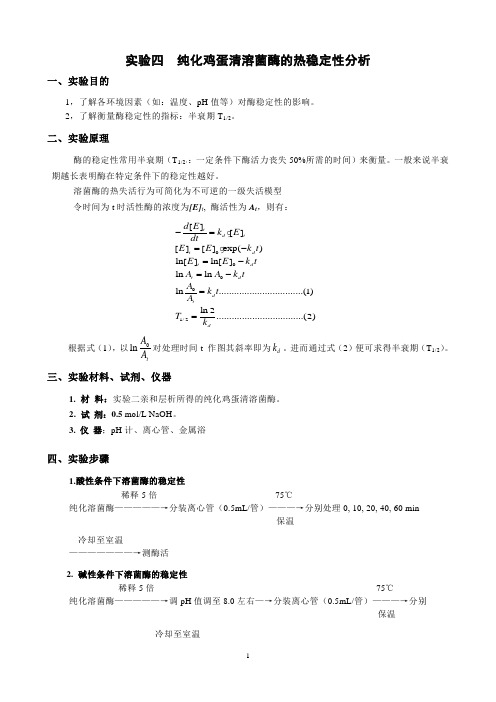
溶菌酶的热失活行为可简化为不可逆的一级失活模型 令时间为t 时活性酶的浓度为[E]t , 酶活性为A t ,则有:根据式(1),以0lntA A 对处理时间t 作图其斜率即为d k 。
进而通过式(2)便可求得半衰期(T 1/2)。
三、实验材料、试剂、仪器1. 材 料:实验二亲和层析所得的纯化鸡蛋清溶菌酶。
2. 试 剂:0.5 mol/L NaOH 。
3. 仪 器;pH 计、离心管、金属浴四、实验步骤1.酸性条件下溶菌酶的稳定性稀释5倍 75℃纯化溶菌酶—————→分装离心管(0.5mL/管)———→分别处理0, 10, 20, 40, 60 min 保温 冷却至室温———————→测酶活 2. 碱性条件下溶菌酶的稳定性稀释5倍 75℃纯化溶菌酶—————→调pH 值调至8.0左右—→分装离心管(0.5mL/管)———→分别保温冷却至室温00001/2[][][][]exp()ln[]ln[]ln ln ln.................................(1)ln 2..................................(2)td tt d t d t d d tdd E k E dtE E k t E E k t A A k t A k t A T k -==-=-=-==处理0, 10, 20, 40, 60 min———————→测酶活3. 酶活的测定及计算方法方法:玻璃比色杯快速混匀底物悬浮液2.0mL—————→加入0.05mL酶液————→测A450nm值1min内的变化(15s记录一次)计算:采用自定义酶活性单位(U):当前测定条件下,测定体系在450 nm波长下吸光度每分钟下降0.01所需的酶量为1个酶活力单位(U)。
鸡蛋清溶菌酶提取工艺的改进

( 5) 8
维普资讯
搅 0 mi( ,
次, 最后 1 次滤除树脂水分。
12 14 洗脱溶 菌酶 ...
树脂 用等体 积 1 %的 ( 42O 0 NH )S 4浸泡, 4 下过夜, 日搅拌 3 n 洗脱液经尼龙布 2 1 次 0 , mi
纯 化液。 12 18 浓缩与 干燥 ...
纯化液在(0 2 ℃真空旋转蒸发, 4± ) 变浊时 倒出、 冷冻, 室温真空干燥。
1 2 2 溶 菌酶产品的纯度 鉴定 ..
A一 菌酶 标准 样 品 ; 一第 1 样品 ; 溶 B 批 C一第 2批 样品 ;
D一第 3批样品; E一第 4批样品
树 脂、H 1 N O C、 a H、 N 2 P 4 a 2o 、 aH O 、N i P 4 l K 2O 、 N 420 、 H P 4 ( H ) 4 甘氨酸、 8 十二烷基硫 酸
将冷蛋清加入处理好的 7 4 2 树脂 ( 每千克 蛋清需要未处理树脂 10 )大力搅拌 ( 6 , g 冰浴
集蛋清,"放置 1 4 C h以上。 ()2 274树脂的处理 : l o/ C 浸泡 用 ml H I L
新鲜冷藏的鸡蛋清。
1 12 试 剂 .。
树脂 2 , h 用去离子水洗至近中性, 再用 1 o L l m / NO a H浸泡 2 , 去离子水洗至近中性( h 用过的 树脂直接用 1 o LN O l a H浸泡, m / 用去离子水 洗至近中性 )再用 0 1 m l H . , .5 o Lp 65的磷酸 / 盐缓冲液浸泡过夜, 滤后备用。
少于第 3批旧法 D的, 溶菌酶浓度也相对提
( H ) O 等废液可回收造肥料, N 4, 4 S 所有这些都
鸡蛋白溶菌酶结晶学研究的开题报告
鸡蛋白溶菌酶结晶学研究的开题报告一、研究背景鸡蛋白溶菌酶是一种由禽蛋种在卵清中产生的酶类分子,具有广泛的生物学和医学应用价值。
在生物医学领域,鸡蛋白溶菌酶可以作为治疗和抗生素的候选药物,同时还可以被用作制剂中的生物成分。
然而,鸡蛋白溶菌酶的结晶性质却一直是一个难题,这限制了它在这些应用领域中的进一步应用。
二、研究内容本研究旨在通过结晶学技术,研究鸡蛋白溶菌酶的结晶性质和晶体结构,为进一步深入研究鸡蛋白溶菌酶的生物学性质和应用价值提供基础。
具体来说,研究内容包括:通过生化分离和提纯技术,获得高纯度的鸡蛋白溶菌酶样品;通过不同的结晶试验和条件,寻找适合鸡蛋白溶菌酶结晶的最佳条件,包括结晶剂的筛选、结晶试剂的调配、结晶温度和pH等;采用X射线晶体学技术,解析并测定鸡蛋白溶菌酶的晶体结构,分析其生物学性质和应用价值。
三、研究意义通过本研究的开展,可以揭示鸡蛋白溶菌酶的结晶性质和晶体结构,为进一步深入研究鸡蛋白溶菌酶在生物学和医学领域中的应用价值奠定基础。
同时,通过调制和优化结晶试验和条件,可以为鸡蛋白溶菌酶在制剂中的应用提供技术支持,为其产业化应用打下基础。
四、研究方法本研究采用生化分离和提纯、结晶试验和X射线晶体学技术,进行鸡蛋白溶菌酶的结晶学研究。
具体步骤包括:1. 鸡蛋白溶菌酶的生化分离和提纯,采用亲和层析、离子交换层析等技术,获得高纯度的鸡蛋白溶菌酶样品。
2. 结晶剂的筛选和结晶试剂的调配,根据鸡蛋白溶菌酶的结晶性特点,评估不同试剂的适用性,选择最适合结晶的条件,并进行性能分析。
3. 结晶试验的开展,根据不同结晶条件的要求,进行样品处理、结晶试剂调配、结晶试验设备设置等工作。
结晶成像和培养过程监测,并针对结果进行分析和修正。
4. X射线晶体学技术的应用,对鸡蛋白溶菌酶晶体进行X射线衍射分析,测定分子结构和晶体形状等信息。
五、研究进度及预期结果目前,研究已完成鸡蛋白溶菌酶的生化分离和提纯工作,并对结晶条件进行了前期探索和优化。
蛋清溶菌酶部分酶学性质及酶活性的影响因素研究

蛋清溶菌酶部分酶学性质及酶活性的影响因素研究刘慧;王凤山;楚杰【期刊名称】《中国生化药物杂志》【年(卷),期】2008(29)6【摘要】目的探讨将蛋清溶菌酶制成液体制剂的可行性.方法通过改变不同影响因素测定蛋清溶菌酶的活性,观察几种常用辅料对蛋清溶菌酶活性的影响.结果酶活性在pH 6.0~6.5最强,且在pH 5~7范围内较稳定;在25~65℃范围内随着作用温度的升高酶的活性增强,但温度太高则变性失活;Na+、K+对其活性有轻微激活作用 ,Mn2+、Mg2+对溶菌酶活性无明显影响,Co2+、Ca2+、Cu2+ 、Fe2+、Zn2+使溶菌酶的活性下降;吐温20、吐温80、甘油溶液对酶活有抑制作用;EDTA-2Na在0.000 5%~0.300 0%浓度范围内对溶菌酶活性具有激活作用,浓度继续增加反而有抑制活性作用.结论初步试验结果表明溶菌酶可以制成合适的液体制剂.【总页数】4页(P385-387,391)【作者】刘慧;王凤山;楚杰【作者单位】山东大学药学院生化与生物技术药物研究所,山东,济南,250012;济南三源药业有限公司,山东,济南,250014;山东大学药学院生化与生物技术药物研究所,山东,济南,250012;山东省科学院生物研究所,山东,济南,250014【正文语种】中文【中图分类】Q556【相关文献】1.牛肾溶菌酶的分离纯化及部分酶学性质 [J], 傅婷;万骥;王丹;唐云明2.鸡蛋储存时间对蛋清溶菌酶活性和蛋品质影响研究 [J], 李红;易建中;王家豪;涂盈盈;刘成倩;严华祥3.玉米蛋白粉替代鱼粉对暗纹东方鲀溶菌酶活性及c型溶菌酶mRNA表达的影响[J], 钟国防;钱曦;华雪铭;周洪琪4.不同pH对蛋清溶菌酶形成淀粉样纤维的影响 [J], 李颖;白瑜;冯自立5.蛋清溶菌酶的提取及其酶学性质探究 [J], 周钦育;黄燕燕;赵珊;黄泳尧;林钟培;刘冬梅因版权原因,仅展示原文概要,查看原文内容请购买。
- 1、下载文档前请自行甄别文档内容的完整性,平台不提供额外的编辑、内容补充、找答案等附加服务。
- 2、"仅部分预览"的文档,不可在线预览部分如存在完整性等问题,可反馈申请退款(可完整预览的文档不适用该条件!)。
- 3、如文档侵犯您的权益,请联系客服反馈,我们会尽快为您处理(人工客服工作时间:9:00-18:30)。
维普资讯
物 理化 学学 报( lH au ub o Wui ux e ea ) X
J l uy
A t S - hm. i. 0 7 2 ()1 2 — 0 1 caP .C i Sn, 0 , 3 7 :0 5 1 3 2
12 05
[ t l] Ar ce i
b c me s a lwe n r a e t c a i g v l me p o o t n o e a h l o r a d b o d rwi i r s n o u r p r o fDM S W h n t e c n e to h n e i O. e o t n fDM S Wa r h O s mo e hn 7 t a 0% ,o l e l ig s o i e c u d b b e v d i se f a i g e e d t em i r s o u v . n y a d c i n mo t l o l e o s r e n ta o s l n o h r c ta i n c r e n h n d n n t i
Ab ta t A td ntema e a rt n o e g — i y o y sr c : s yo r ld n t a o f n e g wh t ls z me( u h u i h e HEWL)wa are u ydfee t l sc rid o tb i rn a i s a nn aoi t DS c n igc r l mer y( C)a d mo u ae mp rtr fe n a cn igc oi t MDS . t sfu d ta n d ltdt ea ed fr t s a nn a rmer e u i e i l l y( C) I wa o n t h
o d n trt n( ic ae t ce ige z me o c nrt ni q e u lt n I q e u lt nc na ig fe a a o △ u i n r s dwi i ran n y n e ta o a u o s ou o . a u o s oui o ti n e hn s c i n s i n s o n
dme y ofxd DMS , eaua o mprtr sie w r we t ea r, dtet nio ek i t l l ie( hs o O)d ntrt nt eaue( i e hf dt adl r e rt e a as npa t o o mp u n h r t i
WWW. x p u.d c wh b.k e u.n
DM S O对 鸡 蛋 白溶茵 酶 溶液 变 性 的影 响
方盈盈 胡新根 于 丽 李文兵 朱玉青 余 生
200) 5 10
(温州大学化学与材料科学学 院, - 浙江 温州 3 52 ; 山东大学胶体与界面化学教育部重点实验室, 207 济南
Ly o y e i Aqu o sS l io sz m n e u o ut n
F G n - ig AN YigY n H i- n・ U X nGe Y L U i L e - ig I nB n W Z uQig HU Y — n Y Seg U h n
Q ol e fC e ir n C lg h ms yadMaeilE gn eig Wezo nvri , nh u 3 52 , hj n rv c. . C i ; 2 e e o t tr s n ier , nh uU i sy Wezo 2 0 7Z ei gP oi e P R. hn a n e t a n a Ky L b rtrfr olia Itfc C e ir o te ns o E ua o , h n ogU i r t Jn n 2 0 0 , . . hn ) a oao o C l d n ne a e h ms f h Miir f d c ̄ n S a d n nv s , i 5 1 0 P R C i y o d r t y t y ei y a a
可逆 离 解 造 成 的 .
关键词 : D C MDS ; DMS S; C O; 鸡蛋 白溶菌酶; 热变性
中图分类号 : 0 4 62
Ef e to f c fDM S o n t a o o e g whie O n De a ur t n fH n Eg - i t
摘要 : 利用差示扫 描量热( S ) D C 和温度 调制 差示扫描量热( MDS ) C 研究 了鸡蛋 白溶菌酶 在纯水及二 甲基亚砜
( DMS / ̄ O)J混合溶 剂中的热变性过程, g 探讨 了酶 的浓度 、 扫描速率和 共溶 剂的含量对热变性行为的影响规律. 在
纯水溶液 中, 溶菌酶的变性焓( J △ 随酶浓度的增大而增大. 而在 DMS / O水混合 溶剂 中, 变性温度( 随 D O 7 MS
体积分数 的增大 向低温方 向移动, 变性峰变低变宽; D O体积分数达到 7 %后, 变性曲线变成 了一条光 当 MS 0 热
滑的直线. 另外, 在纯水溶液中溶菌酶的 M S D C图除了出现 D C中可观察到的主吸热峰( , 在峰(的前面还 S Ib Y, D
出现一个小而对称 的吸热峰( )并且 当体系中有 D O存在 时也未能观察到此峰 . Ⅱ, MS 当溶菌酶浓度增 大时, Ⅱ ) 移 向低温, nd ) a n 减小, f3 rl 与 Ⅱ 之间的距离变长 菌酶二聚体的
