2016年诺贝尔生理医学奖(细胞自噬)相关生物试题
【理科综合模拟】2017高考押题金卷(全国卷Ⅲ)理科综合
绝密★启封前
2017高考押题金卷(全国卷Ⅲ)
理科综合
本试卷分第Ⅰ卷(选择题)和第Ⅱ卷(非选择题)两部分。
全卷满分300分,考试时间150分钟。
注意事项:
1.答题前,考生务必将自己的姓名、班级、考号用0.5毫米的黑色墨水签字笔填写在答题卡上。
并
检查条形码粘贴是否正确。
2.选择题使用2B铅笔填涂在答题卡对应题目标号的位置上,非选择题用0.5毫米黑色墨水签字笔书写在答题卡对应框内,超出答题区域书写的答案无效;在草稿纸、试题卷上答题无效。
3.考试结束后,将答题卡收回。
可能用到的相对原子质量:H-1 C-12 O-16 Cl-35.5 Na-23 Al-27 Cu-64 Zn-65
第Ⅰ卷(选择题,共126分)
一、选择题(本题共13小题,每小题6分。
在每小题给出的四个选项中,只有一项符合
题目要求)
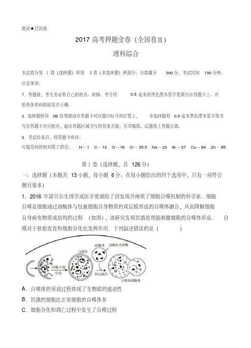
1.2016年诺贝尔生理学或医学奖颁给了因发现并阐明了细胞自噬机制的科学家.细胞
自噬是细胞通过溶酶体与包裹细胞自身物质的双层膜形成的自噬体融合,从而降解细胞
自身病变物质或结构的过程(如图),该研究发现饥饿处理能刺激细胞的自噬体形成.自噬对于胚胎发育和细胞分化也发挥作用.下列叙述错误的是()
A.自噬体的形成过程体现了生物膜的流动性
B.饥饿的细胞比正常细胞的自噬体多
C.细胞分化和凋亡过程中发生了自噬过程。
新教材高中生物第四章细胞的生命历程素养检测卷含解析浙科版必修第一册(含答案)

新教材高中生物浙科版必修第一册:第四章细胞的生命历程[时间:90分钟满分:100分]一、选择题(本题包括25小题,每小题2分,共50分)1.动、植物细胞有丝分裂,在以下哪个时期有明显不同( D )A.间期 B.中期C.后期 D.末期【解析】动、植物细胞有丝分裂过程的不同之处主要发生在前期和末期。
(1)前期纺锤体的形成不同:动物细胞由中心体发出纺锤丝形成纺锤体,高等植物细胞由细胞两极发出纺锤丝形成纺锤体。
(2)末期细胞质的分裂方式不同:动物细胞中部出现细胞内陷,把细胞质缢裂为二,形成两个子细胞;植物细胞中央出现细胞板,扩展形成新细胞壁,并把细胞分为两个。
2.在细胞有丝分裂过程中,DNA和染色体的数目比体细胞各增加一倍,分别发生在细胞周期的( D )①间期②前期③中期④后期⑤末期A.①② B.③④C.④① D.①④【解析】分裂间期进行DNA复制和有关蛋白质的合成,结果DNA增倍,染色体数目不变,一条染色体含2条姐妹染色单体;前期和中期DNA已经复制增倍,染色体数目不变;后期着丝粒分裂,染色单体分离成为染色体,染色体数目增倍,DNA量不变;末期染色体数目恢复原来数目,DNA量下降。
综上所述,DNA复制增倍发生在间期,染色体增倍发生在后期,故D正确。
3.根据下图判断,能正确表达有丝分裂细胞周期概念的一段是( D )A.a~b B.a~cC.b~c D.b~d【解析】从细胞周期的概念可知,一个细胞周期包括分裂间期和分裂期,一次分裂完成开始进入分裂间期,因此一个细胞周期应是分裂间期+分裂期,不能颠倒,题图中能正确表示一个细胞周期的线段是bc+cd,即b~d。
4.有丝分裂保持了细胞在亲代与子代之间遗传性状的稳定性,其原因是( B ) A.有纺锤体出现B.染色体复制和平均分配C.有染色体出现D.核膜消失又重新形成【解析】由于亲代细胞的染色体经过复制以后,精确地平均分配到两个子细胞中去,从而保证亲子代间遗传性状的稳定。
细胞自噬机制--2016年诺贝尔生理或医学奖
Scientific Background Discoveries of Mechanisms for AutophagyThe 2016 Nobel Prize in Physiology or Medicine is awarded to Yoshinori Ohsumi for his discoveries of mechanisms for autophagy. Macroautophagy (“self-eating”, hereafter referred to as autophagy) isan evolutionarily conserved process whereby the eukaryotic cell can recycle part of its own contentby sequestering a portion of the cytoplasm in a double-membrane vesicle that is delivered to the lysosome for digestion. Unlike other cellular degradation machineries, autophagy removes long-lived proteins, large macro-molecular complexes and organelles that have become obsolete or damaged. Autophagy mediates the digestion and recycling of non-essential parts of the cell during starvation and participates in a varietyof physiological processes where cellular components must be removed to leave space for new ones. In addition, autophagy is a key cellular process capable of clearing invading microorganisms and toxic protein aggregates, and therefore plays an important role during infection,in ageing and in the pathogenesis of many human diseases. Although autophagy was recognized already in the 1960’s, the mechanism and physiological relevance remained poorly understood for decades. The work of Yoshinori Ohsumi dramatically transformed the understanding of this vital cellular process. In 1993, Ohsumi published his seminal discovery of 15 genes of key importance for autophagy in budding yeast. In a series of elegant subsequent studies, he cloned several of these genes in yeast and mammalian cells and elucidated the function of the encoded proteins. Based on Yoshinori Ohsumi’s seminal discoveries, the importance of autophagyin human physiology and disease is now appreciated.The mystery of autophagyIn the early 1950’s, Christian de Duve was interested in the action of insulin and studied the intracellular localization of glucose-6-phosphatase using cell fractionation methods developed by Albert Claude. In a control experiment, he also followed the distribution of acid phosphatase, but failed to detect any enzymatic activity in freshly isolated liver fractions. Remarkably, the enzymatic activity reappeared if the fractions were stored for five days in a refrigerator1. It soon became clear that proteolytic enzymes were sequestered within a previously unknown membrane structure that de Duve named the lysosome1,2. Comparative electron microscopy of purified lysosome-rich liver fractions and sectioned liver identified the lysosome as a distinct cellular organelle3. Christian de Duve and Albert Claude, together with George Palade, were awarded the 1974 Nobel Prize in Physiology or Medicine for their discoveries concerning the structure and functional organization of the cell.Soon after the discovery of the lysosome, researchers found that portions of the cytoplasm are sequestered into membranous structures during normal kidney development in the mouse4. Similar structures containing a small amount of cytoplasm and mitochondria were observed in the proximal tubule cells of rat kidney during hydronephrosis5. The vacuoles were found to co-localize with acid-phosphatase-containing granules during the early stages of degeneration and the structures were shown to increase as degeneration progressed5. Membrane structures containing degenerating cytoplasm were also present in normal rat liver cells and their abundance increased dramatically following glucagon perfusion6 or exposure to toxic agents7. Recognizing that the structures had the capacity to digest parts of the intracellular content, Christian de Duve coined the term autophagy in 1963, and extensively discussed this concept in a review article published a few years later8. At that time, a compelling case for the existence of autophagy in mammalian cells was made based on results from electron microscopy studies8. Autophagy was known to occur at a low basal level, and to increase during differentiation and remodeling in a variety of tissues, including brain, intestine, kidney, lung, liver, prostate, skin and thyroid gland4,7-13. It was speculated that autophagy might be a mechanism for coping with metabolic stress in response to starvation6and that it might have roles in the pathogenesis of disease5. Furthermore, autophagy was shown to occur in a wide range of single cell eukaryotes and metazoa, e.g. amoeba, Euglena gracilis, Tetrahymena, insects and frogs8,14, pointing to a function conserved throughout evolution.During the following decades, advances in the field were limited. Nutrients and hormones were reported to influence autophagy; amino acid deprivation induced15, and insulin-stimulation suppressed16 autophagy in mammalian tissues. A small molecule, 3-methyladenine, was shown to inhibit autophagy17. One study using a combination of cell fractionation, autoradiography and electron microscopy provided evidence that the early stage of autophagy included the formation of a double-membrane structure, the phagophore,that extended around a portion of the cytoplasm and closed into a vesicle lacking hydrolytic enzymes, the autophagosome18 (Figure 1).Despite many indications that autophagy could be an important cellular process, its mechanism and regulation were not understood. Only a handful of laboratories were working on the problem, mainly using correlative or descriptive approaches and focusing on the late stages of autophagy, i.e. the steps just before or after fusion with the lysosome. We now know that the autophagosome is transient and only exists for ~10-20 minutes before fusing with the lysosome, making morphological and biochemical studies very difficult.Figure 1. Formation of the autophagosome. The phagophore extends to form a double-membrane autophagosome that engulfs cytoplasmic material. The autophagosome fuses with the lysosome, where the content is degraded.In the early 1990’s, almost 30 years after de Duve coined the term autophagy, the process remained a biological enigma. Molecular markers were not available and components of the autophagy machinery were elusive. Many fundamental questions remained unanswered: How was the autophagy process initiated? How was the autophagosome formed? How important was autophagy for cellular and organismal survival? Did autophagy have any role in human disease? Discovery of the autophagy machineryIn the early 1990’s Yoshinori Ohsumi, then an Assistant Professor at Tokyo University, decided to study autophagy using the budding yeast Saccharomyces cerevisae as a model system. The first question he addressed was whether autophagy exists in this unicellular organism. The yeast vacuole is the functional equivalent of the mammalian lysosome. Ohsumi reasoned that, if autophagy existed in yeast, inhibition of vacuolar enzymes would result in the accumulation of engulfed cytoplasmic components in the vacuole. To test this hypothesis, he developed yeast strains that lacked the vacuolar proteases proteinase A, proteinase B and carboxy-peptidase19. He found that autophagic bodies accumulated in the vacuole when the engineered yeast were grown in nutrient-deprived medium19, producing an abnormal vacuole that was visible under a light microscope. He had now identified a unique phenotype that could be used to discover genes that control the induction of autophagy. By inducing random mutations in yeast cells lacking vacuolar proteases, Ohsumi identified the first mutant that could not accumulate autophagic bodies in the vacuole20; he named this gene autophagy 1 (APG1). He then found that the APG1 mutant lost viability much quicker than wild-type yeast cells in nitrogen-deprived medium. As a second screen he used this more convenient phenotype and additional characterization to identify 75 recessive mutants that could be categorized into different complementation groups. In an article published in FEBS Letters in 1993, Ohsumi reported his discovery of as many as 15 genes that are essential for the activation of autophagy in eukaryotic cells20. He named the genes APG1-15. As new autophagy genes were identified in yeast and other species, a unified system of gene nomenclature using the ATG abbreviation was adopted21. This nomenclature will be used henceforth in the text.During the following years, Ohsumi cloned several ATG genes22-24and characterized the function of their protein products. Cloning of the ATG1gene revealed that it encodes a serine/threonine kinase, demonstrating a role for protein phosphorylation in autophagy24. Additional studies showed that Atg1 forms a complex with the product of the ATG13 gene, and that this interaction is regulated by the target of rapamycin (TOR) kinase23,25. TOR is active in cells grown under nutrient-rich conditions and hyper-phosphorylates Atg13, which prevents the formation of the Atg13:Atg1 complex. Conversely, when TOR is inactivated by starvation, dephosphorylated Atg13 binds Atg1 and autophagy is activated25. Subsequently, the active kinase was shown to be a pentameric complex26 that includes, in addition to Atg1 and Atg13, Atg17, Atg29 and Atg31. The assembly of this complex is a first step in a cascade of events needed for formation of the autophagosome.Figure 2. Regulation of autophagosome formation. Ohsumi studied the function of the proteins encoded by key autophagy genes. He delineated how stress signals initiate autophagy and the mechanism by which protein complexes promote distinct stages of autophagosome formation.The formation of the autophagosome involves the integral membrane protein Atg9, as well as a phosphatidylinositol-3 kinase (PI3K) complex26 composed of vacuolar protein sorting-associated protein 34 (Vps34), Vps15, Atg6, and Atg14. This complex generates phosphatidylinositol-3 phosphate and additional Atg proteins are recruitedto the membrane of the phagophore. Extension of the phagophore to form the mature autophagosome involves two ubiquitin-like protein conjugation cascades (Figure 2).Studies on the localization of Atg8 showed that, while the protein was evenly distributed throughout the cytoplasm of growing yeast cells, in starved cells, Atg8 formed large aggregates that co-localized with autophagosomes and autophagic bodies27. Ohsumi made the surprising discovery that the membrane localization of Atg8 is dependent on two ubiquitin-like conjugation systems that act sequentially to promote the covalent binding of Atg8 to the membrane lipid phosphatidylethanolamine. The two systems share the same activating enzyme, Atg7. In the first conjugation event, Atg12 is activated by forming a thioester bond with a cysteine residue of Atg7, and then transferred to the conjugating enzyme Atg10 that catalyzes its covalent binding to the Atg5 protein26,28,29. Further work showed that the Atg12:Atg5 conjugate recruits Atg16 to form a tri-molecular complex that plays an essential role in autophagy by acting as the ligase of the second ubiquitin-like conjugation system30. In this second unique reaction, the C-terminal arginine of Atg8 is removed by Atg4, and mature Atg8 is subsequently activated by Atg7 for transfer to the Atg3 conjugating enzyme31. Finally, the two conjugation systems converge as the Atg12:Atg5:Atg16 ligase promotes the conjugation of Atg8 to phosphatidylethanolamine26,32.Lipidated Atg8 is a key driver of autophagosome elongation and fusion33,34. The two conjugation systems are highly conserved between yeast and mammals. A fluorescently tagged version of the mammalian homologue of yeast Atg8, called light chain 3 (LC3), is extensively used as a marker of autophagosome formation in mammalian systems35, 36.Ohsumi and colleagues were the first to identify mammalian homologues of the yeast ATG genes, which allowed studies on the function of autophagyin higher eukaryotes. Soon after, genetic studies revealed that mice lacking the Atg5gene are apparently normal at birth, but die during the first day of life due to inability to cope with the starvation that precedes feeding37. Studies of knockout mouse models lacking different components of the autophagy machinery have confirmed the importance of the process in a variety of mammalian tissues26,38.The pioneering studies by Ohsumi generated an enormous interest in autophagy. The field has become one of the most intensely studied areas of biomedical research, with a remarkable increase in the number of publications since the early 2000’s.Different types of autophagyFollowing the seminal discoveries of Ohsumi, different subtypes of autophagy can now be distinguished depending on the cargo that is degraded. The most extensively studied form of autophagy, macroautophagy, degrades large portions of the cytoplasm and cellular organelles. Non-selective autophagy occurs continuously, andis efficiently induced in response to stress, e.g.starvation. In addition, the selective autophagy of specific classes of substrates - protein aggregates, cytoplasmic organelles or invading viruses and bacteria - involves specific adaptors that recognize the cargo and targets it to Atg8/LC3 on the autophagosomal membrane39. Other forms of autophagy include microautophagy40, which involves the direct engulfment of cytoplasmic material via inward folding of the lysosomal membrane, and chaperone-mediated autophagy (CMA). In CMA, proteins with specific recognition signals are directly translocated into the lysosome via binding to a chaperone complex41.Autophagy in health and diseaseInsights provided by the molecular characterizationof autophagy have been instrumental in advancing the understanding of this process and its involvement in cell physiology and a variety of pathological states (Figure 3). Autophagy was initially recognized as a cellular response to stress, but we now know that the system operates continuously at basal levels. Unlike the ubiquitin-proteasome system that preferentially degrades short-lived proteins, autophagy removes long-lived proteins and is the only process capable of destroying whole organelles, such as mitochondria, peroxisomes and the endoplasmic reticulum. Thus, autophagy plays an essential rolein the maintenance of cellular homeostasis. Moreover, autophagy participates in a variety of physiological processes, such as cell differentiation and embryogenesis that require the disposal of large portions of the cytoplasm. The rapid inductionof autophagy in response to different types of stress underlies its cytoprotective function and the capacity to counteract cell injury and many diseases associated with ageing.Because the deregulation of the autophagic flux is directly or indirectly involved in a broad spectrum of human diseases, autophagy is a particularly interesting target for therapeutic intervention. An important first insight into the role of autophagy in disease came from the observation that Beclin-1, the product of the BECN1gene, is mutated in a large proportion of human breast and ovarian cancers. BECN1 is a homolog of yeast ATG6 that regulates steps in the initiation of autophagy42. This finding generated substantial interest in the role of autophagy in cancer43.Misfolded proteins tend to form insoluble aggregates that are toxic to cells. To cope with this problem the cell depends on autophagy44. In fly and mouse models of neurodegenerative diseases, the activation of autophagy by inhibition of TOR kinase reduces the toxicity of protein aggregates45. Moreover, loss of autophagy in the mouse brain by the tissue-specific disruption of Atg5and Atg7 causes neurodegeneration46,47. Several autosomal recessive human diseases with impaired autophagy are characterized by brain malformations, developmental delay, intellectual disability, epilepsy, movement disorders and neurodegeneration48.Figure 3. Autophagy in health and disease. Autophagy is linked to physiological processes including embryogenesis and cell differentiation, adaptation to starvation and other types of stress, as well as pathological conditions including neurodegenerative diseases, cancer and infections.The capacity of autophagy to eliminate invading microorganisms, a phenomenon called xenophagy, underlies its key role in the activationof immune responses and the control of infectious diseases49,50. Viruses and intracellular bacteria have developed sophisticated strategies to circumvent this cellular defense. Additionally, microorganisms can exploit autophagy to sustain their own growth.ConclusionThe discovery of autophagy genes, and the elucidation of the molecular machinery for autophagy by Yoshinori Ohsumi have led to a new paradigm in the understanding of how the cell recycles its contents. Because of his pioneering work, autophagy is recognized as a fundamental process in cell physiology with major implicationsfor human health and disease.Nils-Göran Larsson and Maria G. Masucci Karolinska InstitutetReferences1. de Duve, C. (2005). The lysosome turns fifty.Nat Cell Biol 7, 847–849.2. de Duve, C., Pressman, B.C., Gianetto, R.,Wattiaux, R., and Appelmans, F. (1955)Tissue fractionation studies. 6. Intracellulardistribution patterns of enzymes in rat-livertissue. Biochem J 60, 604–617.3. Novikoff, A.B, Beaufay, H., and de Duve, C.(1956) Electron microscopy of lysosome-richfractions from rat liver. Journal BiophysBiochem Cytol. 2, 179–190.4. Clark, S.L. (1957) Cellular differentiation in thekidneys of newborn mice studied with theelectron microscope. J Biophys BiochemCytol 3, 349–376.5. Novikoff, A.B. (1959) The proximal tubule cellin experimental hydronephrosis. J BiophysBiochem Cytol 6, 136–138.6. Ashford, T.P., and Porter, K.R. (1962)Cytoplasmic components in hepatic celllysosomes. J Cell Biol 12, 198–202.7. Novikoff, A.B., and Essner, E. (1962)Cytolysomes and mitochondrial degeneration.J Cell Biol 15, 140–146.8. de Duve, C., and Wattiaux, R. (1966)Functions of lysosomes. Annu Rev Physiol 28,435–492.9. Behnke, O. (1963) Demonstration of acidphosphatase-containing granules and cytoplasmic bodies in the epithelium of foetalrat duodenum during certain stages ofdifferentiation. J Cell Biol18, 251–265. 10. Bruni, C., and Porter, K.R. (1965) The finestructure of the parenchymal cell of the normalrat liver: I. General observations. Am J Pathol46, 691–755.11. Hruban, Z., Spargo, B., Swift, H., Wissler,R.W., and Kleinfeld, R.G. (1963) Focalcytoplasmic degradation. Am J Pathol 42,657–683.12. Moe, H., and Behnke, O. (1962) Cytoplasmicbodies containing mitochondria, ribosomes,and rough surfaced endoplasmic membranesin the epithelium of the small intestine ofnewborn rats. J Cell Biol 13, 168–171.13. Napolitano, L. (1963) Cytolysomes inmetabolically active cells. J Cell Biol 18, 478–481.14. Bonneville, M.A. (1963) Fine structuralchanges in the intestinal epithelium of thebullfrog during metamorphosis. J Cell Biol 18,579–597.15. Mortimore, G.E., and Schworer, C.M. (1977)Induction of autophagy by amino-aciddeprivation in perfused rat liver. Nature 270,174–176.16. Pfeifer, U., and Warmuth-Metz, M. (1983)Inhibition by insulin of cellular autophagy inproximal tubular cells of rat kidney. Am JPhysiol 244, E109-114.17. Seglen, P.O., and Gordon, P.B. (1982) 3-Methyladenine: specific inhibitor of autophagic/lysosomal protein degradation inisolated rat hepatocytes. Proc Natl Acad SciUSA 79, 1889–1892.18. Arstila, A.U., and Trump, B.F. (1968) Studieson cellular autophagocytosis. The formation ofautophagic vacuoles in the liver after glucagonadministration. Am J Pathol 53, 687–733.19. Takeshige, K., Baba, M., Tsuboi, S., Noda, T.,and Ohsumi, Y. (1992) Autophagy in yeastdemonstrated with proteinase-deficientmutants and conditions for its induction. J CellBiol 119, 301–311.20. Tsukada, M., and Ohsumi, Y. (1993) Isolationand characterization of autophagy-defectivemutants of Saccharomyces cerevisiae. FEBSLett 333, 169–174.21. Klionsky, D.J., Cregg, J.M. Dunn, W.A. Jr.,Emr, S.D., Sakia, J., Sandoval, I.V., Sibirnya,Y.A., Subramani, S., Thumm, M., Veenhuis,M., and Ohsumi, Y. (2003) A unifiednomenclature for yeast autophagy-relatedgenes. Dev Cell 5, 539-545.22. Kametaka, S., Matsuura, A., Wada Y., andOhsumi, Y. (1996) Structural and functionalanalyses of APG5, a gene involved inautophagy in yeast. Gene 178, 139-43.23. Funakoshi, T., Matsuura, A., Noda, T.,Ohsumi Y. (1997) Analyses of APG13 geneinvolved in autophagy in yeast,Saccharomyces cerevisiae.Gene. 192, 207-213.24. Matsuura, A., Tsukada, M., Wada, Y., andOhsumi, Y. (1997) Apg1p, a novel proteinkinase required for the autophagic process inSaccharomyces cerevisiae. Gene 192, 245–250.25. Kamada, Y., Funakoshi, T., Shintani, T.,Nagano, K., Ohsumi, M., and Ohsumi, Y.(2000) Tor-mediated induction of autophagyvia an Apg1 protein kinase complex. J CellBiol 150, 1507–1513.26. Ohsumi, Y. (2014) Historical landmarks ofautophagy research. Cell Res 24, 9–23.27. Kirisako, T., Baba, M., Ishihara, N., Miyazawa,K., Ohsumi, M., Yoshimori, T., Noda, T., andOhsumi, Y. (1999) Formation process ofautophagosome is traced with Apg8/Aut7p inyeast. J Cell Biol 147, 435–446.28. Mizushima, N., Noda, T., Yoshimori, T.,Tanaka, Y., Ishii, T., George, M.D., Klionsky,D.J., Ohsumi, M., and Ohsumi, Y. (1998) Aprotein conjugation system essential forautophagy. Nature 395, 395–398.29. Shintani, T., Mizushima, N., Ogawa, Y.,Matsuura, A., Noda, T., and Ohsumi, Y. (1999)Apg10p, a novel protein-conjugating enzymeessential for autophagy in yeast. EMBO J 18,5234–5241.30. Mizushima, N., Noda, T., and Ohsumi, Y.(1999) Apg16p is required for the function ofthe Apg12p-Apg5p conjugate in the yeastautophagy pathway. EMBO J 18, 3888–3896. 31. Ichimura, Y., Kirisako, T., Takao, T., Satomi,Y., Shimonishi, Y., Ishihara, N., Mizushima,N., Tanida, I., Kominami, E., Ohsumi, M., et al.(2000) A ubiquitin-like system mediatesprotein lipidation. Nature 408, 488–492.32. Hanada, T., Noda, N.N., Satomi, Y., Ichimura,Y., Fujioka, Y., Takao, T., Inagaki, F., andOhsumi, Y. (2007) The Atg12-Atg5 conjugatehas a novel E3-like activity for proteinlipidation in autophagy. J Biol Chem 282,37298–37302.33. Nakatogawa, H., Ichimura, Y., and Ohsumi, Y.(2007) Atg8, a ubiquitin-like protein requiredfor autophagosome formation, mediates membrane tethering and hemifusion. Cell 130,165–178.34. Xie Z., Nair U., Klionsky D.J. (2008) ATG8controls phagophore expansion during autophagosome formation. Mol Cell Biol 19,3290-3298.35. Kabeya, Y., Mizushima, N., Ueno, T.,Yamamoto, A., Kirisako, T., Noda, T.,Kominami, E., Ohsumi, Y., and Yoshimori, T.(2000) LC3, a mammalian homologue of yeastApg8p, is localized in autophagosome membranes after processing. EMBO J 19,5720–5728. 36. Mizushima, N., Yamamoto, A., Matsui, M.,Yoshimori, T., and Ohsumi, Y. (2004) In vivoanalysis of autophagy in response to nutrientstarvation using transgenic mice expressing afluorescent autophagosome marker. Mol BiolCell 15, 1101–1111.37. Kuma, A., Hatano, M., Matsui, M., Yamamoto,A., Nakaya, H., Yoshimori, T., Ohsumi, Y.,Tokuhisa, T., and Mizushima, N. (2004) Therole of autophagy during the early neonatalstarvation period. Nature 432, 1032–1036. 38. Mizushima, N., and Komatsu, M. (2011)Autophagy: Renovation of cells and tissues.Cell 147, 728-741.39. Liu, L., Sakakibara, K., Chen, Q., Okamoto, K.(2014) Receptor-mediated mitophagy in yeastand mammalian systems. Cell Res 24, 787-795.40. Li, W.W., Li, J., Bao, J.K. (2012)Microautophagy: lesser-known self-eating.Cell Mol Life Sci 69, 1125-1136.41. Cuervo, A.M., and Wong, E. (2014)Chaperone-mediated autophagy: roles in disease and aging. Cell Res 24, 92–104.42. Liang, X.H., Jackson, S., Seaman, M., Brown,K., Kempkes, B., Hibshoosh, H., and Levine,B. (1999) Induction of autophagy andinhibition of tumorigenesis by beclin 1. Nature402, 672–676.43. Choi, A.M.K., Ryter, S.W., and Levine, B.(2013) Autophagy in human health anddisease. N Engl J Med 368, 651–662.44. Ravikumar, B., Vacher, C., Berger, Z., Davies,J.E., Luo, S., Oroz, L.G., Scaravilli, F., Easton,D.F., Duden, R., O'Kane, C.J., et al. (2004)Inhibition of mTOR induces autophagy andreduces toxicity of polyglutamine expansionsin fly and mouse models of Huntingtondisease. Nat Genet 36, 585–595.45. Ravikumar, B., Duden, R., and Rubinsztein,D.C. (2002) Aggregate-prone proteins withpolyglutamine and polyalanine expansionsare degraded by autophagy. Hum Mol Genet11, 1107–1117.46. Komatsu, M., Waguri, S., Chiba, T., Murata,S., Iwata, J.-I., Tanida, I., Ueno, T., Koike, M.,Uchiyama, Y., Kominami, E., et al. (2006)Loss of autophagy in the central nervoussystem causes neurodegeneration in mice.Nature 441, 880–884.47. Hara, T., Nakamura, K., Matsui, M.,Yamamoto, A., Nakahara, Y., Suzuki-Migishima, R., Yokoyama, M., Mishima, K.,Saito, I., Okano, H., et al. (2006) Suppressionof basal autophagy in neural cells causesneurodegenerative disease in mice. Nature441, 885–889.48. Ebrahimi-Fakhari, D., Saffari, A., Wahlster, L.,Lu, J., Byrne, S., Hoffmann, G.F., Jungbluth,H., and Sahin, M. (2016) Congenital disordersof autophagy: an emerging novel class of inborn errors of neuro-metabolism. Brain 139,317–337.49. Nakagawa, I., Amano, A., Mizushima, N.,Yamamoto, A., Yamaguchi, H., Kamimoto, T.,Nara, A., Funao, J., Nakata, M., Tsuda, K., etal. (2004) Autophagy defends cells against invading group A Streptococcus. Science 306,1037–1040. 50. Gutierrez, M.G., Master, S.S., Singh, S.B.,Taylor, G.A., Colombo, M.I., and Deretic, V.(2004) Autophagy is a defense mechanisminhibiting BCG and Mycobacterium tuberculosis survival in infected macrophages.Cell 119, 753–766.Nils-Göran Larsson, MD, PhDProfessor of Mitochondrial Genetics, Karolinska InstitutetAdjunct Member of the Nobel CommitteeMember of the Nobel AssemblyMaria G. Masucci, MD, PhDProfessor of Virology, Karolinska InstitutetAdjunct Member of the Nobel CommitteeMember of the Nobel AssemblyIllustration: Mattias Karlén*FootnotesAdditional information on previous Nobel Prize Laureates mentioned in this text can be found at/The Nobel Prize in Physiology or Medicine 1974 to Albert Claude, Christian de Duve and George E. Palade “for their discoveries concerning the structural and functional organization of the cell”/nobel_prizes/medicine/laureates/1974/claude-facts.html/nobel_prizes/medicine/laureates/1974/duve-facts.html/nobel_prizes/medicine/laureates/1974/palade-facts.htmlGlossary of Terms:Lysosome:an organelle in the cytoplasm of eukaryotic cells containing degradative enzymes enclosed in a membrane.Phagophore: a vesicle that is formed during the initial phases of macroautophagy. The phagophore is extended by the autophagy machinery to engulf cytoplasmiccomponents.Autophagosome:an organelle that encloses parts of the cytoplasm into a double membrane that fuses to the lysosome where its content is degraded. The autophagosome is thekey structure in macroautophagy.Selective autophagy: a type of macroautophagy that mediates the degradation of specific cytoplasmic components. Different forms of selective autophagy are called mitophagy(degrades mitochondria), ribophagy (degrades ribosomes), lipophagy (degradeslipid droplets) xenophagy (degrades invading microorganisms) etc.。
细胞自噬的机制而获得诺贝尔生理学或医学奖。肿瘤细

1.2016年日本细胞生物学家大隅良典因发现“细胞自噬的机制”而获得诺贝尔生理学或医学奖。
肿瘤细胞可通过自噬作用降解自身成分,以保护癌细胞免受化疗药物等不利环境的影响,并延缓肿瘤细胞的凋亡。
下列相关的说法中,错误的是()A.细胞自噬过程中起关键作用的细胞器是溶酶体B.化疗过程中辅以药物促进细胞自噬有利于癌症治疗C.肿瘤增殖造成局部营养匮乏时可能通过自噬作用补充营养D.细胞自噬的实质是控制自噬的基因选择性表达的过程2.下列关于植物叶绿体中的色素以及光合作用过程的描述正确的是()A.低温会抑制叶绿素的合成B.植物呈绿色是由于叶绿素能有效地吸收绿光C.某植株叶肉细胞的光合作用速率等于呼吸作用速率时,植株的净光合作用速率为零D.可以用纸层析法提取叶绿体中的色素3.去甲肾上腺素(NE)在调节糖代谢方面与胰岛素有拮抗作用。
NE既可以由肾上腺分泌,也可以由支配肾脏的肾交感神经细胞分泌,肾交感神经兴奋时其末梢释放的NE可增加Na+、Cl-和水的重吸收。
下列相关说法中,错误的是()A.NE可参与体液调节和神经调节,但肾交感神经细胞分泌的NE的作用范围有限B.肾交感神经是反射弧结构中的传入神经,兴奋部位膜两侧的电位表现为内正外负C.肾上腺分泌的NE通过体液运输,与胰高血糖素通过协同作用共同调节血糖的平衡D.如果适度增加肾交感神经元周围的K+浓度,则肾交感神经元的静息电位峰值降低4.下列关于植物激素及其作用的叙述中,正确的是()A.植物激素是由生长旺盛的部位产生的B.植物激素的作用都具有两重性的特点C.植物激素的合成受基因组控制,与环境无关D.植物几乎所有生命活动都受到植物激素的调节5.下列关于遗传病及其预防的叙述中,正确的是()A.禁止近亲结婚只能减少隐性遗传病的发生B.红绿色盲女患者的致病基因都来自于父亲C.若某遗传病的发病率在男女中相同,则为显性遗传病D.男性患者的母亲和女儿都是患者,则该病一定是伴X显性遗传病6.生态护坡是利用植被对斜坡进行保护的一项综合护坡技术。
高三生物一轮复习 第6讲 细胞器与生物膜系统3专题练
细胞器与生物膜系统一、选择题1.菟丝子细胞中没有叶绿体,是一种营寄生生活的高等植物。
下列有关分析中正确的是A. 在生态系统中菟丝子属于生产者B。
菟丝子的细胞内无膜结构的细胞器有核糖体和中心体C. 菟丝子细胞间的信息交流离不开细胞膜上的受体D. 菟丝子具有补肾益精之功效,体现了生物多样性的直接价值2.下列关于线粒体和叶绿体的叙述,错误的是A. 都含有DNAB。
都能产生ATPC. 都能产生[H]D. 都存在于洋葱鳞片叶表皮细胞中3.下列有关真核生物核糖体的叙述,正确的是A。
遗传信息转录和翻译的场所B。
组成成分中含mRNA和蛋白质C. 可以游离在细胞质基质中D。
核糖体是光学显微镜下可见的最小细胞器4.下列关于真核生物的细胞器叙述中,正确..的一组是( )A。
与分泌蛋白形成有关的细胞器只有高尔基体和内质网B. 核糖体和中心体属于无膜的细胞器C. 含RNA的细胞器只有线粒体和叶绿体D. 线粒体是进行有氧呼吸的主要场所,叶绿体是进行光合作用的主要场所5.关于细胞结构、功能及其化合物的说法正确的是( )A。
磷脂是所有原核细胞和真核细胞必不可少的脂质B. 同一生物的不同细胞的细胞器在数量上有区别,但种类是相同的C. 蓝藻与绿藻都能进行光合作用,但二者在细胞结构上的主要区别是叶绿体的有无D。
蛋白质的合成不一定要在核糖体上进行,例如哺乳动物的成熟红细胞6.将某植物细胞各部分结构用差速离心法分离后,取三种细胞器测定其有机物的含量如图所示。
以下有关说法正确的是()A. 若甲是线粒体,则能完成下列反应的全过程:C6H12O6+6O2+6H2O→6CO2+12H2O+能量B。
乙只含有蛋白质和脂质,说明其具有膜结构,肯定与分泌蛋白的加工和分泌有关C. 细胞器丙中进行的生理过程产生水,产生的水中的氢来自于羧基和氨基D。
蓝藻细胞与此细胞共有的细胞器可能有甲和丙7.叶绿体与线粒体在结构和功能上的相同点是()。
①具有双层膜②分解有机物,合成ATP③利用气体和产生气体④消耗水并生成水⑤含有DNA⑥内部含有多种酶⑦含有丰富的膜面积A. ①②③④⑤⑥B. ①③④⑤⑥C。
2016年诺贝尔医学生理学奖

2016年诺贝尔医学生理学奖2016年,诺贝尔医学奖授予了三位科学家Yoshinori Ohsumi、Takaki Kajita和Arthur B. McDonald,以表彰他们在医学生理学领域取得的杰出贡献。
他们的研究成果在深入理解细胞自噬和中微子振荡现象方面起到了重要作用,为医学和物理学领域的未来发展提供了新的思路和方向。
以下将分别介绍他们的研究成果和对医学与物理学领域的影响。
一、Yoshinori Ohsumi的细胞自噬研究1. 细胞自噬的概念和意义细胞自噬是一种被细胞内部自行调控的生理过程,通过此过程,细胞可以将自己内部的损坏蛋白质和细胞器包裹成囊泡,然后通过溶酶体降解和再利用这些物质,在饥饿、压力和感染等情况下保证细胞的稳定运行。
细胞自噬在疾病的发生发展中起到了重要作用,如肿瘤、神经退行性疾病和心血管疾病等。
Yoshinori Ohsumi通过对酵母菌进行的研究,最终揭示了细胞自噬的分子机制和调控原理,这一发现为细胞生物学领域的研究提供了全新的理论和实验依据。
2. 奥崇久的研究成果对医学的影响奥崇久的研究成果为医学领域提供了对自噬途径的深刻理解,为相关疾病的治疗提供了新的思路。
基于奥崇久研究成果,科学家们可以更好地了解自噬在疾病发生发展中的作用机制,进一步开发针对自噬途径的治疗方法,为疾病治疗提供新的方向和希望。
二、Takaki Kajita和Arthur B. McDonald的中微子振荡研究1. 中微子的基本特性中微子是一种基本粒子,质量极小、不带电荷,几乎不与其他物质发生相互作用。
由于这些特性,中微子一直以来被认为对我们的影响非常小,很难被科学家们观测到。
Takaki Kajita和Arthur B. McDonald的研究成果改变了这一观念,为中微子物理学的发展带来了重要的突破。
2. 中微子振荡的发现Takaki Kajita和Arthur B. McDonald在不同的实验设施中独立进行了中微子振荡的观测实验,并最终得出了相同的结论:中微子在传播过程中会发生振荡现象,不同种类的中微子之间可以相互转换。
高中生物-细胞结构(1)

【解析】选C。分隔膜属于生物膜结构,主要成分包括脂质,A正确;图示 自噬过程是对衰老或损伤细胞结构的包裹,整个过程中是有选择,有识别 的,B正确;图中看出,自噬溶酶体中只具有单层膜结构,C错误;溶酶体内含 有丰富的水解酶,若溶酶体膜破裂,水解酶释放出来有可能损伤正常的细 胞结构,D正确。
2. 解析:原核细胞与真核细胞都含有核糖体,核糖体是合成蛋白质的细胞器,A正确;溶酶体含有
多种水解酶,B正确;动植物和真菌细胞含有内质网,内质网与脂质合成、蛋白质合成及加工有关,
加工、包装和发送蛋白质是高尔基体的功能,D错误。
3.下列关于细胞器的叙述,错误的是( ) A.水绵的中心体由两个互相垂直的中心粒及周围物质组成 B.溶酶体只能分解外来的病毒和病菌 C.唾液腺细胞中有较多的内质网和高尔基体 D.花瓣细胞液泡中色素种类和含量可影响花色
9.如图所示为线粒体和叶绿体的“内共生起源”学说图解。下列所述不支持“内共生 起源”学说的是( )
A.线粒体和叶绿体的内膜与细菌的细胞膜相似 B.线粒体和叶绿体都存在于绿色植物的叶肉细胞内 C.线粒体和叶绿体中脱氧核糖核酸分子均呈环状 D.线粒体和叶绿体内都有能合成蛋白质的细胞器
【解析】选B。在膜的化学成分上,线粒体和叶绿体内膜的蛋白质与脂质远大于外膜,接近于细菌细胞膜 的成分,这些特性都暗示线粒体和叶绿体的内膜起源于最初的共生体(需氧细菌和蓝细菌)的细胞膜,支持 了“内共生起源”学说,A项不符合题意。线粒体和叶绿体在植物中的分布情况,不能说明它们与细菌的 关系,故不支持“内共生起源”学说,B符合题意。线粒体和叶绿体中脱氧核糖核酸分子均呈环状,细菌 也含有环状的DNA分子,支持了“内共生起源”学说,C项不符合题意。线粒体和叶绿体中均含有核糖 体,可独立合成蛋白质,支持“内共生起源”学说,D项不符合题意。
近五年诺贝尔生理学奖
近五年诺贝尔生理学奖2015年: 发现治疗丝虫寄生虫新疗法,发现治疗疟疾的新疗法(屠呦呦)[例题]2015年10月5日,中国科学家屠呦呦获得诺贝尔生理学奖,实现了中国自然科学诺奖零的突破,其率领的科研团队提取出的青蒿素可以有效地治疗疟疾,请回答下列问题:(1)在青蒿素被提取之前,奎宁广泛被用来治疗疟疾,治疗机理是,奎宁可以和疟原虫DNA结合,形成复合物,从而直接抑制DNA复制和转录,导致疟原虫生命活动出现异常。
(2)青蒿素是从植物黄花蒿的组织细胞中所提取的一种代谢产物,其作用方式目前尚不明确,推测可能是疟原虫是单细胞动物,青蒿素作用于疟原虫的食物泡膜,从而阻断了营养摄取的最早阶段,使疟原虫较快出现氨基酸饥饿,迅速形成自噬泡,并不断排出虫体外,使疟原虫损失大量胞浆而死亡.疟原虫对外界食物的摄取方式主要是胞吞,该过程体现了细胞膜具有一定的流动性的特点,参与该过程相关的细胞器是线粒体.(3)氨基酸是组成疟原虫的营养物质,其在细胞中形成各种蛋白质.氨基酸进入疟原虫体内以脱水缩合方式形成蛋白质的肽链.2016年:发现了细胞自噬的机制[例题]2016年诺贝尔生理学或医学奖授予“发现了细胞自噬机制”的科学家,当自噬体形成时,它会包裹住某些细胞成分,比如那些被破坏的蛋白质和细胞器。
最终,它与溶酶体融合,这些细胞成分便会降解为更小成分。
这一过程为细胞的更新提供了养分和构建基础。
细胞自噬有着广泛的生理和病理意义,如饥饿和组织缺血时,细胞通过自噬提供营养,维持细胞存活。
细胞自噬存在图中A微自噬、B巨自噬和C分子伴侣介导的三种自噬方式。
(1)清除受损的线粒体是通过B过程来实现的,吞噬过程体现了生物膜在结构上具有___一定的流动性_____的特点。
若线粒体被吞噬,有氧呼吸的第_二、三_阶段会受影响。
(2)细胞内变性的蛋白质的降解通过C过程完成,该过程中的分子伴侣热休克蛋白与变性的蛋白质结合,然后与溶酶体膜上的_膜蛋白_结合,在其作用下转运入溶酶体体腔。
高考语文《新闻阅读》专题复习

课时规范练(三十一)新闻阅读一、阅读下面的文字,完成1~3题。
材料一备受关注的2016年诺贝尔奖拉开了帷幕,在273位被提名的科学家中,日本科学家大隅良典最后折桂,将今年的诺贝尔生理或医学奖独自收入囊中。
其获奖理由是在细胞自噬领域的杰出贡献。
现年71岁的大隅良典是日本知名分子细胞生物学家,目前担任日本东京工业大学名誉教授。
10月3日接到诺贝尔生理学或医学奖获奖通知时,他说:“我很惊讶,我在我的实验室。
”之所以能走上这条科研之路,和他一直信奉的科研准则有直接关系——“做别人没做过的工作”。
2012年获得“京都奖”后,大隅良典曾寄语年轻科学家:“做其他人没有在做的事,并且做你发现真正有兴趣的事。
做研究并不容易。
然而,如果你真的被一个课题吸引,并且对它感兴趣,那么你肯定会克服所有障碍,即便你的工作一时未获得赏识。
人只能活一次。
在所有事情都说完了做好了之后,你终将会品尝到成功的喜悦。
”(取材于2016年10月4日《中国科学报》) 材料二2016年10月3日,大隅良典因为自噬机制的开创性研究,获得了诺贝尔生理或医学奖。
21世纪获得诺贝尔自然科学奖的日本科学家(含日裔)上升到17位。
日本科学虽然取得“井喷”式成就,但大隅良典却未雨绸缪,他认为,不能因为近年来日本诺贝尔奖获得者人数增多就认为日本很棒,并对日本科学研究的空心化等潜在问题表现出忧虑。
中科院生物物理所的张宏研究员与大隅良典熟识,两人合办过4期“中日细胞自噬研讨会”。
张宏在接受科技日报记者采访时说:“先生做了很多极其重要的工作,但从来不追求CNS(Cell、Nature和Science三种最重要的期刊)。
他批评过度追逐CNS的评估体系,认为CNS也有不少文章是错的;文章发出来就可以,重要的是工作扎实。
”今年7月,大隅良典还在《发育细胞》杂志上发了一篇自噬机制的重要论文。
张宏说,大隅良典经常跟他强调基础科学对转化医学的重要性,非常重视基础研究。
大隅良典4日在记者会上也对科学研究的急功近利深表忧虑。
细胞自噬诺贝尔奖
历史进程比利时科学家Duve在上世纪50年代通过电镜观察到自噬体(autophagosome)结构Duve1963年溶酶体国际会议上首先提出了“自噬”这种说法,Duve因发现溶酶体1974年获得了诺贝尔奖。
直到20世纪90年代,日本的大隅良典成功克隆了第一个酵母自噬基因Atg1以及自噬特征蛋白LC3,今年他带领的研究团队探明了细胞自噬的启动机制,这些成就让他获得了2016年诺贝尔生理学或医学奖。
自噬作用(autophagy)是一个非常简单的细胞活动,字面上也很好理解:自己吃自己。
总体上看,动物细胞是一个三层结构:最外面是细胞膜,中间是细胞质,细胞核被包裹在最里面。
大部分功能性细胞器和生物分子都悬浮在细胞质中,因此,很多细胞活动都在细胞质中进行。
由于生理生化反应多而复杂,经常产生大量残渣,致使细胞活动受到影响甚至停滞,在这种情况下,自噬作用就非常重要:将淤积在细胞质中的蛋白质等代谢残渣清除掉,恢复正常的细胞活动。
清理细胞质能让细胞重获新生,对于神经细胞这类不可替换的细胞来说,这个过程尤为重要。
神经细胞一旦分化成熟,就会保持当前状态,直到母体生物死去,它们没有其他方式来恢复和维护自身功能。
细胞生物学家还发现,自噬作用还能抵御病毒和细菌的侵袭。
任何躲过细胞外免疫系统,通过细胞膜进入细胞质的异物或微生物,都可能成为自噬系统的攻击目标。
不论自噬过程启动过慢还是过快,或者出现功能障碍,都将导致可怕的后果。
数百万克罗恩病(Crohn’s disease,一种炎症性肠病)患者的患病原因,可能就是因为他们的自噬系统出现缺陷,无法抑制肠道微生物的过度生长;大脑神经细胞自噬系统的崩溃,则与阿尔茨海默病(Alzheimer's disease)和细胞衰老有关。
即使自噬系统运作良好,它仍可能对人体不利。
当癌症病人接受了放疗及化疗后,自噬系统可能救活奄奄一息的癌细胞,使癌症无法根治。
有时,自噬系统会为了生物体的整体利益,将病变细胞去除,但它偶尔又会热心过度,去除一些重要细胞,完全不理会这样做是否符合生物体的整体利益。
2016年诺贝尔生理学或医学奖:细胞自噬机理

2016年诺贝尔生理学或医学奖:细胞自噬机理作者:王晓冰来源:《百科知识》2016年第22期2016年10月3日,瑞典卡罗林斯卡医学院诺贝尔生理学或医学奖委员会宣布,2016年诺贝尔生理学或医学奖授予日本科学家大隅良典,因为他发现了细胞自噬的机理。
大隅良典独自获得800万瑞典克朗(约合人民币625万元)奖金。
细胞自噬是什么?自噬是细胞内的一种“自食”现象。
细胞自噬已经被研究人员研究了60多年,目前的定义是,细胞自噬是指生物膜(大部分表现为双层膜,有时多层或单层)包裹部分细胞质和细胞内需要降解的细胞器、蛋白质等形成自噬体,并与内涵体形成自噬内涵体,最后与溶酶体融合形成自噬溶酶体,降解其所包裹的内容物,以实现细胞稳态和细胞器的更新。
通俗地讲,细胞自噬就是细胞的自我吞噬,通常发生在细胞或者机体缺乏能量、或受到环境胁迫,如缺乏氨基酸、缺氧的情况下,细胞里会产生双层膜结构,包裹自己的一部分细胞器,运送到溶酶体进行降解。
自噬的出现是因为细胞在新陈代谢过程中会不断产生受损伤的细胞器,如受损的线粒体、蛋白质聚合体等,这就需要细胞清理它们。
通过自噬作用,组织和细胞对自身不断地清理,以保持细胞的稳态平衡。
这个作用有点像人用吸尘器清洁卫生,用以吸收室内各种脏东西,从而保持室内清洁和干净。
自噬这个单词源于希腊语,希腊语单词前缀auto意为“自我”,另一个希腊语单词phagein 意为“吞食”,二者组合成一个词就是自我吞噬。
最早研究细胞自噬并提出这一概念的并非大隅良典,而是比利时科学家杜夫。
他在20世纪50年代通过电镜观察细胞的内部情况时,发现了溶酶体,是细胞内的一种细胞器,其功能是处理细胞摄入的营养物质并分解较大的颗粒。
与此同时,他也发现了自噬现象,并且在1963年溶酶体国际会议上首先提出了“自噬”的概念。
因此,他和他的同事、电子显微镜专家克洛德和帕拉迪分享了1974年诺贝尔生理学或医学奖。
细胞中有三种类型的自噬:大自噬、小自噬和伴侣介导的自噬。
2016年诺贝尔生理医学奖(细胞自噬)相关生物试题汇总
2016年诺贝尔生理医学奖(细胞自噬)相关生物试题日本科学家大隅良典(Yoshinori Ohsumi)凭借“细胞自噬作用的发现与机制探索”获得今年的诺贝尔生理学或医学奖。
细胞自噬是近些年细胞研究的一大热门方向,在高中生物考题中也频频出现,我们在第一时间采编了10道相关试题与大家分享!1.细胞自噬是继细胞凋亡后,当前生命科学最热的研究领域。
细胞自噬是将细胞内受损、变性、衰老的蛋白质或细胞器运输到溶酶体内并降解的过程(如下图),下列相关说法不正确的是( )A. 细胞自噬和细胞凋亡对于细胞具有相同意义B.细胞自噬过程可体现溶酶体的消化营养功能C.细胞自噬被维持在一定水平,能确保细胞内的稳态D.细胞自噬可发生在细胞生长、分化、衰老、凋亡的全过程【答案】A 【解析】细胞凋亡与细胞自噬是两个概念,肯定有不同的生命意义。
细胞凋亡是基因控制的程序性死亡,而细胞自噬是将细胞内受损、变性、衰老的蛋白质或细胞器运输到溶酶体内并降解的过程,产生的部分物质可以再加以利用,细胞没有死亡,A错。
2.细胞自噬是细胞通过溶酶体与包裹细胞自身物质的双层膜融合,从而降解细胞自身病变物质或结构的过程(如图)。
下列有关叙述中,正确的是( )A.图中自噬体的膜由双层磷脂分子组成B.图中溶酶体与自噬体融合过程体现了细胞膜的选择透过性C.图中的水解酶是在自噬溶酶体中合成的D.溶酶体所参与的细胞自动结束生命的过程是由基因决定的【答案】D 【解析】自噬体由两层膜构成,所以,自噬体的膜由四层磷脂分子组成,A项错误;溶酶体与自噬体融合过程体现了细胞膜的流动性,B项错误。
3.细胞自噬是指细胞内受损、变性、衰老的蛋白质或细胞器运输到溶酶体内并进行降解的过程。
下图中A、B、C分别表示细胞自噬的三种方式,相关说法正确的是( )①细胞通过C方式减少有害蛋白在细胞内的积累,从而达到延长细胞的寿命②图中能体现膜结构具有流动性的有:自吞小泡与溶酶体融合、蛋白质与膜蛋白结合③若人工破坏溶酶体膜可阻断细胞自噬进程,受损的物质和细胞器会在细胞中积累④细胞自噬被维持在一定水平,能确保细胞内的稳态⑤细胞自噬贯穿于正常细胞生长、分化、衰老、死亡的全过程。
关于细胞自噬的相关研究成果
关于细胞⾃噬的相关研究成果关于细胞⾃噬的相关研究成果姓名:陶宗学院:化学化⼯学院专业:化学学号:160106010012016年度诺贝尔⽣理学与医学奖刚揭晓不久,获奖者为⽇本科学家⼤隅良典(Yoshinori Ohsumi),以奖励他在“细胞⾃噬机制⽅⾯的发现”。
⼀、概述细胞⾃噬这是细胞组分降解与再利⽤的基本过程。
“⾃噬”(autophagy)⼀词源于希腊语前缀“auto-”,意为“⾃我”,以及另⼀个希腊语单词“phagein”,意为“吞⾷”。
因此,⾃噬作⽤的意思⾮常明确,那就是“⾃我吞噬”。
“⾃噬”的概念由⽐利时科学家Christian de Duve 在1963年溶酶体国际会议上⾸先提出,是指⼀些需降解的蛋⽩质和细胞器等胞浆成分被包裹,并最终运送⾄溶酶体降解的过程,⾃噬性降解产⽣的氨基酸和其他⼀些⼩分⼦物质可被再利⽤或产⽣能量。
现已明确,⾃噬的主要功能之⼀实际上是在细胞受到应激性的死亡威胁时保持细胞的存活,这是真核细胞维持稳态、实现更新的⼀种重要的进化保守机制。
虽然⼴义上的⾃噬包括巨⾃噬(macroautophagy)、微⾃噬(microautophagy)和分⼦伴侣介导的⾃噬(chapeon-mediated autophagy)三种类型,通常所说的⾃噬即指巨⾃噬,也是⽬前研究最多的。
对这⼀过程开展研究⾮常困难,这也就意味着我们对其知之甚少。
直到上世纪1990年代,在经过⼀系列出⾊的实验之后,⽇本科学家⼤隅良典利⽤⾯包酵母找到了与⾃噬作⽤有关的关键基因。
随后他开始致⼒于阐明酵母菌体内⾃噬作⽤的背后机制,并发现与之相似的复杂过程也同样存在于我们⼈类的细胞内。
⼤隅良典的研究更新了我们关于细胞物质循环的旧有观点,他的研究开启了理解⾃噬作⽤在许多⽣理过程中关键作⽤的崭新道路,如⽣物体对于饥饿的适应或者机体对于感染的反应。
⾃噬基因的突变会导致疾病的发⽣,⾃噬作⽤机制在⼀些类型的疾病,如癌症和神经疾病等病症中也发挥了作⽤。
以诺贝尔奖为背景的高中生物试题
以诺贝尔奖为背景的高中生物试题信息给予题是生物常见题型之一,由题干(信息部分)与问题两部分构成,题干是向学生提供解题信息,多以文字叙述为主,有的再辅以图示与数据等信息,内容覆盖生命科学领域的各方面识,题干的特点往往具有隐蔽性、启发性与迁移性;问题部分是围绕题干给出的信息主题展开的。
信息给予题的典型特征是:交代取材背景,情境新、知识活,起点高、落点低,现学现用。
本文就以获诺贝尔奖的生物学知识为背景材料汇编如下的生物试题。
诺贝尔生理学或医学奖由瑞典皇家卡罗林医学院评定。
生物学中与获奖成果有关的内容包括:青霉素的发现、DNA的结构与功能、胚胎发育、蛋白质在细胞定位控制中的内部信号、调控细胞周期的分子机制、单克隆抗体的研究及其作用、神经细胞的信息传递、新型生物的发现、程序性细胞死亡等。
1 题例领悟例1.瑞典生物化学家泰奥雷尔通过对酶的研究,发现有机体在有氧条件下利用营养素以产生有机体可利用能量的方式,并因此而获1955年诺贝尔生理学或医学奖。
下列有关叙述中,不正确的是()A.生物体中能供给能量的营养素主要包括糖类、脂肪与蛋白质B.直接供给生物体生命活动的能源物质是ATPC.很多生物在无氧条件下也可在酶的作用下产生能量D.所有生物都能在有氧条件下独立的通过酶的作用分解营养物质,释放能量分析:由生物体新陈代谢可知,组成生物体的主要物质糖类、脂质、蛋白质都可以氧化分解产生能量供生命活动所用,生物机体的直接能源物质为ATP,生物体新陈代谢类型也有差别,严格厌氧型生物在有氧条件下代谢会停止或受抑制,本题选D。
例2.2001年诺贝尔生理学或医学奖授予三位科学家。
这三位科学家发现了调控细胞周期的一系列基因,以及相关的酶与蛋白质。
这项土作对肿瘤研究等领域产生了重大影响。
请回答下列各小题:(1)同种生物不同类型细胞之间的细胞周期持续时间有异。
卵裂期蛙胚的动物半球细胞的细胞持续时间比植物半球细胞的。
(2)测定某种细胞的细胞持续时间长短时,通常要考虑温度因素。
- 1、下载文档前请自行甄别文档内容的完整性,平台不提供额外的编辑、内容补充、找答案等附加服务。
- 2、"仅部分预览"的文档,不可在线预览部分如存在完整性等问题,可反馈申请退款(可完整预览的文档不适用该条件!)。
- 3、如文档侵犯您的权益,请联系客服反馈,我们会尽快为您处理(人工客服工作时间:9:00-18:30)。
2016年诺贝尔生理医学奖(细胞自噬)相关生物试题日本科学家大隅良典(Yoshinori Ohsumi)凭借“细胞自噬作用的发现与机制探索”获得今年的诺贝尔生理学或医学奖。
细胞自噬是近些年细胞研究的一大热门方向,在高中生物考题中也频频出现,我们在第一时间采编了10道相关试题与大家分享!
1.细胞自噬是继细胞凋亡后,当前生命科学最热的研究领域。
细胞自噬是将细胞内受损、变性、衰老的蛋白质或细胞器运输到溶酶体内并降解的过程(如下图),下列相关说法不正确的是()
A.细胞自噬和细胞凋亡对于细胞具有相同意义
B.细胞自噬过程可体现溶酶体的消化营养功能
C.细胞自噬被维持在一定水平,能确保细胞内的稳态
D.细胞自噬可发生在细胞生长、分化、衰老、凋亡的全过程
【答案】A
【解析】细胞凋亡与细胞自噬是两个概念,肯定有不同的生命意义。
细胞凋亡是基因控制的程序性死亡,而细胞自噬是将细胞内受损、变性、衰老的蛋白质或细胞器运输到溶酶体内并降解的过程,产生的部分物质可以再加以利用,细胞没有死亡,A错。
2.细胞自噬是细胞通过溶酶体与包裹细胞自身物质的双层膜融合,从而降解细胞自身病变物质或结构的过程(如图)。
下列有关叙述中,正确的是()
A.图中自噬体的膜由双层磷脂分子组成
B.图中溶酶体与自噬体融合过程体现了细胞膜的选择透过性
C.图中的水解酶是在自噬溶酶体中合成的
D.溶酶体所参与的细胞自动结束生命的过程是由基因决定的
【答案】D
【解析】自噬体由两层膜构成,所以,自噬体的膜由四层磷脂分子组成,A项错误;溶酶体与自噬体融合过程体现了细胞膜的流动性,B项错误。
3.细胞自噬是指细胞内受损、变性、衰老的蛋白质或细胞器运输到溶酶体内并进行降解的过程。
下图中A、B、C分别表示细胞自噬的三种方式,相关说法正确的是()
①细胞通过C方式减少有害蛋白在细胞内的积累,从而达到延长细胞的寿命
②图中能体现膜结构具有流动性的有:自吞小泡与溶酶体融合、蛋白质与膜蛋白结合
③若人工破坏溶酶体膜可阻断细胞自噬进程,受损的物质和细胞器会在细胞中积累
④细胞自噬被维持在一定水平,能确保细胞内的稳态
⑤细胞自噬贯穿于正常细胞生长、分化、衰老、死亡的全过程。
A.①②③B.①④⑤
C.②③⑤D.③④⑤
【答案】B
【解析】蛋白质与膜蛋白结合未体现膜的流动性,到是图中自吞小泡形成可以体现膜流动性,②错误;若人工破坏溶酶体膜则溶酶体内的水解酶能直接发挥作用,加速细胞自溶,③错误。
4.产生一种外膜蛋白,导致高尔基体片层结构包裹线粒体形成“自噬体”,与溶解体结合形成“自噬体酶体”如下图所示,下列说法不正确的是()
A若线粒体均遭“损伤”酵母菌将无法产生ATP
B.内容物降解后形成的产物,可以为细胞提供营养
C.线粒体产生的“外膜蛋白”是一种特异性受体
D.“自噬溶酶体”的形成依赖生物膜的流动性
【答案】A
5.细胞自噬是将细胞内受损、变性、衰老的蛋白质或细胞器运输到溶酶体内并降解的过程。
下图中甲、乙、丙表示细胞自噬的三种方式,相关说法正确的是()
A.细胞通过丙减少有害蛋白在细胞内的积累,从而缩短细胞寿命
B.自吞小泡与溶酶体融合能体现细胞膜的功能具有选择透性
C.正常生理状态下溶酶体对自身机体的细胞结构无分解作用
D.细胞自噬贯穿于正常细胞的生长、分化、衰老和凋亡的全过程
【答案】D
【解析】自吞小泡与溶酶体融合能体现细胞膜的结构特点,即具有一定的流动性,B错误;溶酶体可以吞噬自身细胞中衰老的细胞器,C错误。
6.(多选)中国女科学家屠呦呦获2015年诺贝尔生理医学奖,她研制的抗疟药青蒿素挽救了数百万人的生命。
青蒿素是从植物黄花蒿的组织细胞中提取的一种代谢产物,其作用方式目前尚不明确,推测可能是作用于疟原虫的食物泡膜,从而阻断了营养摄取的最早阶段,使疟原虫较快出现氨基酸饥饿,迅速形成自噬泡,并不断排出虫体外,使疟原虫损失大量胞浆而死亡。
以下说法不正确的是()
A.疟原虫对外界食物的获取方式主要是胞吞,体现了细胞膜的选择透过性
B.疟原虫细胞中只有一种细胞器-----核糖体
C.自噬泡排出疟原虫体外过程中,需要消耗能量,但不需要载体蛋白的协助
D.氨基酸作为蛋白质的基本组成单位,其数目、种类和排列顺序均能影响蛋白质的结构和功能
【答案】AB
【解析】疟原虫通过胞吞获取食物,胞吞现象体现了膜的流动性,A错误;由题意知,疟原虫是原生动物,属于真核生物,含有除核糖体之外的多种细胞器,B错误。
7.溶酶体的主要功能是吞噬消化作用。
有两种吞噬作用:一种是自体吞噬,另一种是异体吞噬,如下图所示。
请据此判断下列叙述不正确的是()
A.消除细胞内衰老的细胞器是通过自噬作用实现的
B.溶酶体参与抗原的加工处理过程
C.溶酶体与吞噬体的融合体现了生物膜具有流动性
D.吞噬细胞通过吞噬作用特异性识别抗原
【答案】D
【解析】一般认为吞噬细胞不能特异性识别抗原,它起一个处理作用,进而可被T细胞识别。
8.酵母菌是一种单细胞真菌,其结构简单,是研究生命科学的理想微生物。
下图表示酵母菌中线粒体发生的“自噬”现象,请回答下列问题:
(1)线粒体增大膜面积的方式是______,从代谢的角度分析,其现实意义是_______。
(2)由图甲可知,线粒体在_____的条件下会损伤,由线粒体产生的一种外膜蛋白可以引起其发生特异性的“自噬”现象。
“自噬”后的线粒体形成“自噬体”,并与溶酶体结合形成“自噬溶酶体”。
该过程体现了生物膜具有_____。
(3)若该酵母菌的线粒体均遭此“损伤”,则在有氧的条件下,葡萄糖氧化分解的最终产物是_____,对其进行检测所需的试剂是_____。
【答案】
(1)内膜向内折叠形成嵴增大内膜表面积,为催化有氧呼吸的酶提供更大的附着面积(2)饥饿和光照一定的流动性
(3)酒精和二氧化碳橙色的重铬酸钾溶液和澄清的石灰水
9.细胞内受损后的线粒体释放的信号蛋白,会引发细胞非正常死亡。
下图表示细胞通过“自噬作用”及时清除受损线粒体及其释放的信号蛋白的过程,请据图回答:。
