Microscopic interface phonon modes in structures of GaAs quantum dots embedded in AlAs shel
微孔英语单词
微孔英语单词Sure, I understand your requirements. The English word for "微孔" is "micropore."Micropores are tiny pores or openings in a material that are smaller than 2 nanometers in diameter. These tiny pores are crucial for various applications in fields such as material science, environmental engineering, and biotechnology.In material science, micropores play a significant role in the adsorption and storage of gases, such as in the case of activated carbon. The high surface area and numerous micropores of activated carbon make it an excellent material for gas storage and purification.In environmental engineering, micropores are essential for the removal of contaminants from water and air. For example, microporous membranes are used in water filtration systems to remove bacteria, viruses, and other impurities from drinking water.In biotechnology, micropores are utilized in various applications such as drug delivery systems and tissueengineering. Microporous scaffolds can provide a suitable environment for cell growth and tissue regeneration.Now, let me provide the Chinese translation for "micropore." "微孔"的英文单词是"micropore"。
南农生化考研-核酸习题

二、习题(一)名词解释1.单核苷酸(mononucleotide)2.磷酸二酯键(phosphodiester bonds)3.不对称比率(dissymmetry ratio)4.碱基互补规律(complementary base pairing)5.反密码子(anticodon)6.顺反子(cistron)7.核酸的变性与复性(denaturation、renaturation)8.退火(annealing)9.增色效应(hyper chromic effect)10.减色效应(hypo chromic effect)11.噬菌体(phage)12.发夹结构(hairpin structure)13.DNA 的熔解温度(melting temperature T m)14.分子杂交(molecular hybridization)(二)填空题1.DNA 双螺旋结构模型是_________于____年提出的。
2.核酸的基本结构单位是_____。
3.脱氧核糖核酸在糖环______位置不带羟基。
4.两类核酸在细胞中的分布不同,DNA 主要位于____中,RNA 主要位于____中。
5.核酸分子中的糖苷键均为_____型糖苷键。
糖环与碱基之间的连键为_____键。
核苷与核苷之间通过_____键连接成多聚体。
6.核酸的特征元素____。
7.碱基与戊糖间是C-C 连接的是______核苷。
8.DNA 中的____嘧啶碱与RNA 中的_____嘧啶碱的氢键结合性质是相似的。
10.DNA 双螺旋的两股链的顺序是______关系。
11.给动物食用3H 标记的_______,可使DNA 带有放射性,而RNA 不带放射性。
12.B 型DNA 双螺旋的螺距为___,每匝螺旋有___对碱基,每对碱基的转角是___。
13.在DNA 分子中,一般来说G-C 含量高时,比重___,T m(熔解温度)则___,分子比较稳定。
14.在_ __条件下,互补的单股核苷酸序列将缔结成双链分子。
细胞内叠氮化物反应探针的英文
细胞内叠氮化物反应探针的英文Intracellular Nitrogenase Reaction Probes: Applications and Advancements.Intracellular nitrogenase reaction probes have emerged as crucial tools in modern biochemistry, enabling researchers to monitor and understand the intricate nitrogen metabolism within cells. These probes, often fluorescently labeled, allow for real-time visualization of nitrogen fixation and associated processes, thereby providing insights into the dynamic nature of nitrogen metabolism.Background and Importance.Nitrogen is an essential element for all known forms of life, playing a pivotal role in amino acid synthesis, nucleic acid structure, and various other biological processes. However, nitrogen in its free form (N2) is unavailable for direct biological utilization due to itsinertness. Therefore, organisms rely on nitrogenases, a class of enzymes that catalyze the conversion of N2 into ammonia (NH3), a biologically usable form of nitrogen.Within cells, nitrogenase enzymes are often embedded within complex systems, involving multiple cofactors and electron transport chains. Monitoring these reactionswithin the cellular milieu is challenging due to the dynamic nature of the cellular environment and the often-subtle changes in substrate concentration. Intracellular nitrogenase reaction probes have been developed to overcome these challenges, providing a window into the intracellular world of nitrogen metabolism.Types of Intracellular Nitrogenase Reaction Probes.1. Fluorescent Probes: These probes are labeled with fluorescent molecules that change their emission properties upon interacting with nitrogenase or its intermediates. For example, fluorophores such as fluorescein or rhodamine can be conjugated to specific substrates or inhibitors of nitrogenase, allowing for the detection of enzymaticactivity through fluorescence microscopy or flow cytometry.2. Bioluminescent Probes: These probes emit light through a chemical reaction triggered by nitrogenase activity. Bioluminescent probes offer the advantage of being self-luminous, eliminating the need for external excitation sources.3. Radiolabeled Probes: Radiolabeled probes incorporate radioactive atoms (e.g., carbon-14 or tritium) into substrates or inhibitors of nitrogenase. The subsequent detection of radiolabeled products provides quantitative information about enzyme activity. However, the use of radiolabeled probes is limited due to safety concerns and the need for specialized equipment.Applications of Intracellular Nitrogenase Reaction Probes.1. Studying Nitrogen Fixation Pathways: By monitoring the activity of nitrogenase within cells, probes can reveal the preferred nitrogen fixation pathway utilized bydifferent organisms. This information is crucial for understanding the adaptability of microorganisms to varying nitrogen sources and environmental conditions.2. Analyzing Nitrogen Metabolism in Response to External Stimuli: Intracellular probes can be used to study how nitrogen metabolism is affected by external factors such as changes in nutrient availability, pH, or temperature. Such studies can provide insights into the mechanisms underlying cellular responses to environmental perturbations.3. Drug Discovery and Therapeutics: Nitrogenase inhibitors have been explored as potential therapeutics for treating diseases associated with abnormal nitrogen metabolism, such as cancer or certain infectious diseases. Intracellular probes can aid in the identification of effective inhibitors by allowing for high-throughput screening of candidate compounds.Future Directions.With the continuous advancement of biotechnology and imaging techniques, intracellular nitrogenase reaction probes are poised to make significant contributions to our understanding of nitrogen metabolism. Future research may focus on developing probes with improved sensitivity and specificity, enabling the detection of nitrogenase activity in single cells or even subcellular compartments. Additionally, the integration of probes with other omics technologies (e.g., genomics, proteomics, or metabolomics) could provide a comprehensive picture of nitrogen metabolism within cells, leading to new insights and potential therapeutic targets.In conclusion, intracellular nitrogenase reaction probes have emerged as invaluable tools for studying nitrogen metabolism within cells. Their ability to monitor enzymatic activity in real-time, combined with their versatility and sensitivity, makes them critical for advancing our understanding of nitrogen metabolism and its role in health and disease. As technology continues to evolve, these probes will play increasingly important roles in fundamental and applied research.。
微波消解ICP—AES法测定蔬菜中的多种微量元素

乙烯消解罐 中, 再加 4 m L硝酸和 2 m L双氧水消解 液, 拧 紧盖 子 , 过夜 。 第二 天 , 6 0  ̄ C恒 温水 浴振 荡 3 0 mi n后 , 于微波 消 解 系 统进行 消解 [ 2 ] 。第一 阶段压 力 为 I MP a , 温度 为
I O 0 " C, 火力 为 3档 , 进行 消解 3 ai r n, 第 二 阶段压力 为 2 MP a , 温度 为 1 7 5 ℃, 火力 为 7 档, 进行 消解 6 ai r n 。 消 解完 毕 , 冷却 后开 罐 , 将 消 解好 的样 品溶液过 滤倒入
次蒸馏 水 。 香菜 、 菠菜 、 生菜、 油菜、 芹菜 、 莜麦菜 、 牛心莱
待测元素标准溶液 ( 1 . O 0 0 0 mg / mL ) , 临用时配 制成应用液 。用移液管准确量取待测元素标准溶液 于5 0 0 mL 容 量 瓶 中 , 用 二 次 蒸 馏 水 稀 释 定 容 至 5 0 0 mL, 待 测 。元 素及 质量浓 度 见表 1 。
5 0 mL容 量瓶 中 , 用 二 次水 洗 涤消 解罐 , 洗 涤液倒 入
1 实验部 分
容量 瓶 , 且 用 二次 水稀 释定 容至 5 0 mL, 待测 。 1 . 2 . 2 标 准 溶液 的配 制
1 . 1 仪 器、 试 剂和 原料
I C P Q一1 0 0 0 型高频 电感耦合等离子体原子发 射光谱仪 ( F I 本岛津) 、 WX -4 0 0 0型微波快速 消解 系统( 上 海 屹尧分 析 仪器 有 限 公 司 ) 、 F A2 0 0 4 N 型 电 子分析天平( i - 海精密仪器有 限公 司) 、 电热鼓风干 燥箱 ( 江苏省 东 台县 电器 厂 ) 、 玛 瑙研 钵 等 。 待 测 元 素 标 准溶 液 ( 1 . O 0 0 0 mg / mL、 天 津傲 然 精 细化工 研究 所 ) ; 6 5 一6 8 %硝 酸 ( 北京 化工 厂 、 优 级纯) I 3 0 双氧水 ( 天津市东方化工厂、 分析纯) ; 二
中国科学院大学2013年《生物化学与分子生物学》考研专业课真题试卷
中国科学院大学
2013年招收攻读硕士学位研究生入学统一考试试题科目名称:生物化学与分子生物学
考生须知:
1.本试卷满分为150分,全部考试时间总计180分钟。
2.所有答案必须写在答题纸上,写在试题纸上或草稿纸上一律无效。
一、名词解释(每题4分,共20分)
1.核小体(nucleosome)
2.米氏常数(Michaelis constant)
3.酮体
4.逆转录酶(reverse transcriptase)
5.Gene family
二、单项选择题(每题1分,共20分)
1.除了哪个氨基酸外,α-氨基酸都有旋光性:()
A.赖氨酸
B.谷氨酸
C.半胱氨酸
D.甘氨酸
2.核酸对紫外线的最大吸收峰在哪一波长附近:()
A.280nm
B.260nm
C.220nm
D.340nm
3.下列哪个糖不是还原糖:()
A.D-果糖
B.蔗糖
C.乳糖
D.麦芽糖
科目名称:生物化学与分子生物学第1页共6页。
2022电子陶瓷第二章
第二章 电子陶瓷的结构
陶瓷的结构比纯金属的晶体结构复杂。 陶瓷中离子的堆积方式:
1)与正、负离子的电荷有关
如 Ca 2 F21 一个Ca2+必须对应两个F—
2)与正、负离子的相对大小有关 一般正离子的半径rC小于负离子rA的半径
第二章 电子陶瓷的结构
3)与离子的替代方式有关
如B3+的离子半径0.26Ǻ,只能替代SiC中的1 %的Si4+(离子半径0.40Ǻ)
第二章 电子陶瓷的结构
For each category there is a characteristic type of stimulus capable of provoking different responses.
Mechanical properties relate deformation to an applied load or force (including elastic modulus and strength).
第二章 电子陶瓷的结构
。 Linus Pauling (1901~1994) 电子陶瓷的结构
为了解释甲烷的正四面体结构,说明碳原子四 个键的等价性,鲍林在1928一1931年,提出了 杂化轨道的理论。还创造性地提出了许多新的 概念:共价半径、金属半径、电负性标度等。
Magnetic properties demonstrate the response of a material to the application of magnetic field.
第二章 电子陶瓷的结构
For optical properties, the stimulus is electromagnetic or light radiation. (index of refraction and reflectivity)
A SIMPLE AND AFFORDABLE METHOD FOR IMMUOPHENOTYPIN
专利名称:A SIMPLE AND AFFORDABLE METHOD FOR IMMUOPHENOTYPING USING AMICROFLUIDIC CHIP SAMPLE PREPARATIONWITH IMAGE CYTOMETRY发明人:BECK, Markus,TERSTAPPEN, Leon, L.W.M.M.申请号:US2011/035667申请日:20110508公开号:WO2011/143075A3公开日:20111229专利内容由知识产权出版社提供专利附图:摘要:The enumeration of cells in fluids by flow cytometry includes a simple systemfor CD4 and CD8 counting in point-of-care HIV staging within resource poor countries. No sample preparation is required with the sample added directly to a chip containing dried reagents by capillary flow. A large area image cytometer consisting of an LED module is used to excite the fluorochromes PerCP and APC labeled targets and a monochrome CCD camera with a combination of two macro lenses captures images of 40 square milimeters of blood (approximately 1 microliter). CD4 and CD8-T-lymphocyte counts correlate well with those obtained by flow cytometry. The cytometer system described in the present invention provides an affordable and easy-to-use technique for use in remote locations. The novel device, methods and system disclosed herein largely overcome limitations of cost, size and complexity of the instruments for wider usage.申请人:VERIDEX, LLC,BECK, Markus,TERSTAPPEN, Leon, L.W.M.M.地址:1001 US Route 202 Raritan, NJ 08869 US,University of Twente, Medical Cell Biophysics Drienerlolaan 5, PO Box 217 NL-7500 AE Enschede NL,THK Van Lohuizenlaan Z84 NL-1095DW Amsterdam NL国籍:US,NL,NL代理人:HOPE, Ruby, T更多信息请下载全文后查看。
一种检测个体抗氧化遗传易感性的试剂盒[发明专利]
![一种检测个体抗氧化遗传易感性的试剂盒[发明专利]](https://img.taocdn.com/s3/m/3e81019902768e9950e73873.png)
专利名称:一种检测个体抗氧化遗传易感性的试剂盒专利类型:发明专利
发明人:冯哲民,邹祖烨
申请号:CN200710037167.5
申请日:20070206
公开号:CN101241066A
公开日:
20080813
专利内容由知识产权出版社提供
摘要:本发明公开了一种检测个体抗氧化遗传易感性的试剂盒。
该试剂盒包括同时检测过氧化氢酶基因(CAT)上rs769214号SNP位点、还原型辅酶叶绿醇基因(CYBA)上rs4673号SNP位点、内皮型一氧化氮合成酶基因(NOS3)上rs1799983号SNP位点、胞外过氧化物岐化酶基因(SOD3)上
rs1799895号SNP位点、对氧磷酯酶1基因(PON1)上rs662号SNP位点的特异性引物对及特异性荧光探针对、用于荧光定量PCR检测的常规组件等。
本发明的试剂盒通过同时检测与抗氧化能力密切相关的CAT、CYBA、NOS3、SOD3、PON1的单核苷酸多态性位点基因型来评估个体抗氧化遗传易感性。
申请人:上海主健生物工程有限公司
地址:200433 上海市国定路335号2号楼6楼
国籍:CN
更多信息请下载全文后查看。
微纳成像检测原理
微纳成像检测原理Microscopic imaging detection is a crucial technique in various fields such as biology, chemistry, and material science. It allows researchers and scientists to observe and analyze samples at the microscopic level, providing valuable insights into the structure and characteristics of the materials being studied. With the advancement of technology, microscopy has evolved from traditional optical microscopy to more advanced techniques such as electron microscopy and atomic force microscopy. These techniques offer higher resolution and sensitivity, allowing for the visualization of nanostructures and individual atoms.微小成像检测在生物学、化学和材料科学等领域是一项至关重要的技术。
它使研究人员和科学家能够观察和分析样品在微观水平上的结构和特性,为所研究材料的特性提供宝贵的见解。
随着技术的进步,显微镜学已经从传统的光学显微镜学发展到更先进的技术,如电子显微镜和原子力显微镜。
这些技术提供了更高的分辨率和灵敏度,允许可视化纳米结构和单个原子。
One of the key principles behind microscopic imaging detection is the use of different types of radiation or particles to interact with thesample being studied. This interaction allows for the generation of signals or images that can be captured and analyzed to reveal information about the sample's characteristics. For example, in electron microscopy, a focused beam of electrons is used to illuminate the sample, leading to the generation of high-resolution images. Similarly, in atomic force microscopy, a tiny probe is used to scan the surface of the sample, detecting interactions between the probe and the sample to create images with atomic-level resolution.微小成像检测的一个关键原则是利用不同类型的辐射或粒子与所研究的样品进行相互作用。
生物大分子功能化上转换纳米探针的制备及其成像研究
论文摘要掺杂镧系离子(Ln3+)的上转换纳米粒子(UCNPs)因为具有丰富的电子能级、高能带发射、长荧光寿命、出色的光稳定性和从紫外(UV)到红外(IR)的多个发射带等优势而受到了广泛关注,已经在无背景生物传感和生物成像、光触发药物传输、太阳能电池和超分辨生物成像等领域获得了广泛的应用。
然而,UCNPs本身不能识别目标分析物,无法对分析物进行高选择性的传感。
所以,在本论文中,我们选择了不同的生物大分子作为UCNPs的能量受体,同时具备对目标分析物的识别能力,设计了基于UCNPs的新型近红外(NIR)荧光探针,利用上转换荧光(UCL)实现生物成像研究。
具体内容如下:第一章绪论本章首先介绍了上转换荧光(UCL)、上转换纳米晶体的性质,并对其合成方法进行了概述。
重点对基于上转换纳米材料的表面功能化、能量受体的选择,以及在生物传感与成像领域的应用进行了阐述。
最后对本论文研究内容以及意义进行了论述。
第二章藻蓝蛋白修饰上转换纳米探针用于炎症过程中髓过氧化物酶的检测和生物成像研究在本章中,我们合成了PEI-UCNPs,构建了一种基于UCNPs和藻蓝蛋白(PC)共价结合的新型荧光纳米探针(PC-UCNPs),并将该探针用于髓过氧化物酶(MPO)的检测与生物成像研究。
我们通过酰胺化反应将PC偶联到UCNPs的表面,UCL 被PC吸收后淬灭。
由于PC存在特定的识别位点,该纳米探针可用于灵敏评估MPO的生物活性。
利用该探针的光学优势,PC-UCNPs已成功用于活细胞和小鼠急性炎症过程中MPO的生物成像。
第三章缩醛化葡聚糖修饰上转换纳米探针用于细胞自噬过程中溶酶体pH的传感与成像研究在本章中,我们开发了一种基于UCNPs的新型NIR溶酶体pH探针(Ac-DEX@UCNPs)。
首先,我们合成了核壳结构的UCNPs,并对葡聚糖(DEX)进行修饰得到了缩醛化葡聚糖(Ac-DEX)。
Ac-DEX与UCNPs通过配体交换策略结合,基于聚集诱导淬灭效应(ACQ),Ac-DEX@UCNPs的UCL淬灭。
光电催化苯酚特征峰指认_解释说明
光电催化苯酚特征峰指认解释说明引言1.1 概述在当今环境保护和可持续发展的背景下,寻找高效、环境友好的催化剂成为研究的热点。
光电催化技术由于其能将光能直接转化为电能,并可以促进各种有机废水和废气的降解,受到了广泛的关注。
本文主要研究了光电催化苯酚降解过程中产生的特征峰,并通过特征峰指认方法对其进行解释说明。
苯酚作为一种常见的有机废物,具有毒性较大且不易降解等特点,因此发展一种有效去除苯酚污染的方法具有重要意义。
1.2 文章结构本文共分为五个部分进行论述。
除引言外,第二部分将深入介绍光电催化苯酚特征峰指认相关知识。
接着,在第三部分中将详细描述实验设计与方法。
第四部分将呈现研究结果并进行讨论,包括特征峰指认结果展示、对结果可能性及影响因素的讨论解释以及与其他研究结果的对比分析。
最后,在第五部分对整个研究进行总结与展望。
1.3 目的本文的目的在于通过对光电催化苯酚特征峰的指认,并解释说明其产生机制,为进一步了解光电催化技术在有机废水处理中的应用奠定基础。
同时,本研究也有助于为环境保护和废物处理提供理论依据和实践指导。
注:此内容仅供参考,具体文章内容撰写建议根据研究背景和实验结果进行拓展。
2. 光电催化苯酚特征峰指认:2.1 光电催化原理:光电催化是一种将光能转化为化学能的过程。
在光电催化反应中,光能激发了催化剂的电子,使其产生离子或激发态。
这些激发态的催化剂通过与反应物相互作用,促进了反应的进行。
对于苯酚分子而言,在光电催化过程中,其分子中的特定键或基团会参与反应,并在反应过程中形成特征性的振动频率,即特征峰。
2.2 苯酚的特征峰:苯酚分子在红外光谱中表现出多个特征峰。
这些特征峰对应了苯酚分子内部不同键和基团的振动模式。
以常见结构为例,苯环上的C-O键会引起1370-1290 cm^-1范围内出现的C-O 伸缩振动;O-H键则产生3650-3550 cm^-1范围内出现的O-H伸缩振动和1350-1200 cm^-1范围内出现的O-H弯曲振动。
Bio-radMicroPulser电穿孔仪中文说明书
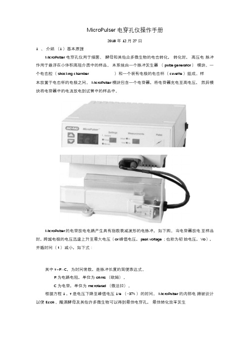
MicroPulser 电穿孔仪操作手册2018 年12 月27 日1、介绍(1 )基本原理MicroPulser 电穿孔仪用于细菌、酵母和其他众多微生物的电击转化,转化时,高压电脉冲作用于悬浮在小体积高阻介质中的样品。
本系统由一个脉冲发生器(pulse generator )模块、一个电击腔(shocking chamber )和一个装有电极的电击杯(cuvette )组成。
样本放置于电击杯的电极之间。
MicroPulser 模块包含一个电容器,将电容器充电至高电压,然后模块将电容器中的电流放电到试管中的样品中。
MicroPulser 的电容放电电路产生具有指数衰减波形的电脉冲,如下图。
当电容器放电至样品时,跨越电极的电压迅速上升至最大电压(or 峰值电压,peak voltage ;也称为初始电压,Vo ),并随时间(t )减小,如下式:其中τ=R · C,为时间常数,是脉冲长度的简便表达式。
R 为电路电阻,单位为ohms (欧姆)。
C 为电容,单位为microfarad (微法拉)。
根据方程1 ,τ是电压下降至峰值电压1/e (~37% )的时间。
MicroPulser 的内部电路被设计以使E.coli 、酿酒酵母及其他许多微生物可以得到最佳电穿孔,最佳转化效率发生在大约5ms 的时间常数内。
这些电穿孔条件是通过使用10 微法拉电容器和将600 欧姆电阻与样品池并联以及将30 欧姆电阻与样品池串联来实现的。
除时间常数外,电场强度是另一个决定转化效率的重要参数。
电场强度E,是施加于电极间的电压,公式为:其中,V 为施加的电压,d 为电极间的距离,单位为cm 。
电场强度和细胞的尺寸(size )决定了横贯每个细胞的电压降,正是电压降可能是电穿孔中电压效应的重要表现。
30 欧姆串联电阻的目的是在发生电弧的情况下保护设备电路。
在正常操作条件下,当样本在高电阻介质中,电阻不会影响施加在样本上的电压。
微区瞬态吸收 钙钛矿
微区瞬态吸收钙钛矿
(原创版)
目录
1.微区瞬态吸收的概述
2.钙钛矿的简介
3.微区瞬态吸收在钙钛矿研究中的应用
4.微区瞬态吸收技术的发展前景
正文
1.微区瞬态吸收的概述
微区瞬态吸收是一种在微小区域内研究材料光吸收特性的技术,能够有效地揭示材料的光物理性质和光化学反应过程。
在众多光吸收材料中,钙钛矿因其独特的光电性能而备受关注。
2.钙钛矿的简介
钙钛矿是一类具有特殊晶体结构的材料,其化学式为 ABX3,其中 A、B、X 分别为阳离子、阴离子和卤素离子。
钙钛矿材料在太阳能电池、发光二极管和激光等领域具有广泛的应用前景。
3.微区瞬态吸收在钙钛矿研究中的应用
微区瞬态吸收技术在钙钛矿研究中的应用主要体现在以下几个方面:(1)钙钛矿的光吸收特性:通过微区瞬态吸收技术,可以研究钙钛矿在不同波长下的光吸收特性,从而优化钙钛矿材料的能带结构,提高光电转换效率。
(2)钙钛矿的光激发态寿命:微区瞬态吸收技术可以测量钙钛矿材料的光激发态寿命,为研究钙钛矿材料的光稳定性提供重要依据。
(3)钙钛矿的光生电荷载流子动力学:通过微区瞬态吸收技术,可
以揭示钙钛矿材料中光生电荷载流子的产生、传输和复合过程,从而优化钙钛矿器件的性能。
4.微区瞬态吸收技术的发展前景
随着科学技术的进步,微区瞬态吸收技术在钙钛矿研究中的应用将更加广泛。
未来,微区瞬态吸收技术有望在钙钛矿材料的设计、制备和器件优化等方面发挥更大的作用,推动钙钛矿材料在光电领域的广泛应用。
zif-67的电导率
zif-67的电导率
ZIF-67(Co)是一种金属有机框架(MOF)材料,由钴离子(Co2+)和2-甲基咪唑酸根(Hmim)组成,具有立方晶系的结构,单胞参数为16.9589 Å,是典型的多孔材料。
ZIF-67(Co)的孔径、比表面积和孔容等参数可以通过调节其合成条件进行控制。
关于ZIF-67的电导率,现有文献表明其具有较高的电导率。
这主要归功于其具有高度可调的孔径、比表面积和多孔性质。
ZIF-67的高比表面积使其能够提供更多的活性位点,从而提高电导率。
此外,ZIF-67的孔径和孔容也可以通过调节其合成条件进行优化,进一步改善其电导率性能。
然而,对于ZIF-67的具体电导率数值,不同文献报道的结果可能存在差异。
这可能是由于合成条件、实验方法等因素的影响。
因此,为了获得更准确和可靠的电导率数据,建议查阅最新的研究文献或权威的科技报告。
总之,ZIF-67是一种具有优异电导率的金属有机框架材料。
其高比表面积、可调的孔径和多孔性质使其在电化学领域具有广泛的应用前景。
通过进一步优化合成条件和实验方法,可以进一步改善ZIF-67的电导率性能,为其在电化学领域的应用提供更好的基础。
- 1、下载文档前请自行甄别文档内容的完整性,平台不提供额外的编辑、内容补充、找答案等附加服务。
- 2、"仅部分预览"的文档,不可在线预览部分如存在完整性等问题,可反馈申请退款(可完整预览的文档不适用该条件!)。
- 3、如文档侵犯您的权益,请联系客服反馈,我们会尽快为您处理(人工客服工作时间:9:00-18:30)。
arX iv:c ond-ma t/16436v1[c ond-m at.m trl-sci ]21J u n 2001Microscopic interface phonon modes in structures of GaAsquantum dots embedded in AlAs shellsShang-Fen Ren and G.Qin †Department of Physics,Illinois State University,Normal,IL 61790-4560(Received)pacs 63.22.+m,81.05.Ys,81.05.Ea,By means of a microscopic valence forcefield model,a series of novel microscopic interface phonon modes are identified in shell quantum dots(SQDs)composed of a GaAs quantum dot of nanoscale embedded in an AlAs shell of a few atomic layers in thickness.In SQDs with such thin shells,the basic principle of the continuum dielectric model and the macroscopic dielectric function are not valid any more.The frequencies of these microscopic interface modes lie inside the gap between the bulk GaAs band and the bulk AlAs band,contrary to the macroscopic interface phonon modes.The average vibrational energies and amplitudes of each atomic shell show peaks at the interface between GaAs and AlAs.These peaks decay fast as their penetrating depths from the interface increase.Keywords:A.Nanostructures;A.Semiconductors;C.Crystal structure and symme-try;D.Phonons1.IntroductionInteresting results have been reported on the surface and interface modes of phonons and polaritons in layered or spherical structures by means of macroscopic contin-uum model.By using the Rosenzweig model,G.Armand et al.[1]have found that there are surface phonon modes in the gap of the bulk phonon modes and below the lowest bulk band.Maradudin et al.[2]have predicted that there are sur-face phonon waves propagating in the gap between the bulk phonon bands and below the lowest bulk phonon band in structures that a semi-infinite GaAs/AlAs superlattice is in contact with a thinfilm of GaAs or AlAs.The amplitudes of some surface modes show strong decaying or variation as their penetrating depths from the surface increase.At about the same time,Quinn et al.predicted that there are surface plasmon modes without suffering Landau damping surviving be-low,above or between the bands of bulk plasmon frequencies in both type-I and type-II semi-infinite semiconductor superlattices[3,4].Interface and surface modes in shell quantum dots(SQDs)composed of a spherical core of one material(core ma-terial)embedded in a matrix of another materiel(shell material)have been studied by many authors[5,6,7,8].However,most of the theoretical treatments on shell QDs are performed in the framework of mechanical continuum model[2],continuum dielectric model[4,7],or continuum model coupling both the mechanical vibrational amplitudes and the electrostatic potential[5].Their regions of validity is limited to modes whose effective wavelength is large compared to the interatomic spacing.In the case of surface plasmons the continuum dielectric approach is limited to dots whose size is large enough that the plasma frequency greatly exceeds the differences between the single-particle energies[8].After surveying systematically the validity of the dielectric approximation in describing electron-energy-loss spectra of surface and interface phonons in thinfilms of ionic crystals,Lambin et al.[9]demonstratedthat the dielectric approximation reproduces the essential features of the phonon response when the layer thickness exceeds20-30˚A.approximation can no longer be applied to thinnerfilms as the concept of bulk dielectric function,the only input required in this approach,breaks down.Even forfilms in above thickness range, small contributions of microscopic surface phonons survive and they may not be neglected.The eigenvectors of the microscopic surface phonons are found to be large at thefirst surface layer and rapidly decreasing as the distance from the sur-face increases.Therefore to describe phonon modes in semiconductor SQDs with small dots size and thin shells,an anisotropic microscopic model is especially needed.2.The theoretical formalismWe have developed a microscopic valence forcefield model(VFFM)in recent years to investigate phonon modes in QDs[10,11,12].In this model,the change of the total energy due to the lattice vibration is considered as two parts,∆E=∆E s+∆E c, where the energy changing due to short-range interactions describes the covalent bonding,and the long range part approximates the Coulomb interactions[17,14,15]. For the short range part,we employed a VFFM as[16],∆E s= i1d i)2+ j1T2,with the sizes of1592,1368,2960,4335,and4560respectively.This approach further allows us to investigate phonon modes with different symmetries in QDs in detail.By employing this model,we have investigated phonon modes in GaAs/AlAs SQDs composed of a spherical GaAs core of radius d S embedded in a AlAs shell with external radius d L[12].Our theoretical formalism has considered every details in the SQDs.it is suitable to deal with SQDs that the the core material and the shell material have totally different parameters.But for calculations of GaAs/AlAs SQDs,the parameters C0,C1,and e∗for both GaAs and AlAs are taken to be the same for simplicity,and only the mass difference is considered.The C0,C1,and e∗are taken as38.80,0.858,and0.6581,and the masses of Ga,As and Al are taken as 69.723,74.922and26.982respectively in atom unit.When considering the interaction between atoms,special attention is paid to atoms near the surfaces and interface of the shell QDs.More specifically,for the short range interaction,when an atom is located near the surface,interaction from its nearest neighboring atom is considered only if that specific nearest atom is within the QD,and interaction from its second neighboring atom is considered only if that specific second neighbor atom is in the QD as well as the nearest neighboring atom that makes the link between them.3.Results and discussionBy employing this model,the frequencies and vibrational strengths of phonon modes of GaAs/AlAs SQDs are calculated as functions of the size of internal dot and the thickness of the external shell of SQDs in each offive representations of A1,A2,E, T1and T2[12].The results of our model shows that the entire optical frequency range of SQDs is divided into two nonoverlaping bands,which are originated frombulk AlAs band and bulk GaAs band respectively,so named as AlAs-like band and GaAs-like band respectively.The lowest and highest frequency of the bulk AlAs (GaAs)optical band is318.48cm−1and396.00cm−1(268.80cm−1and292.13 cm−1)respectively evaluated from the VFFM.We have also noticed that there are many phonon modes inside the gap between bulk GaAs band and the bulk AlAs band in SQDs,especially in SQDs covered by thin shells to which the concept of bulk dielectric function is no longer available.In this letter,we concentrate our attention on the study of phonon modes in SQDs with shells of a few atomic layers by using VFFM.Fig.1is a plot of density of states of phonon modes for10selected10symmetries and scales of SQDs with thin shells.It is plotted by taking half width of the Gaussian broadeningσ=0.4cm−1and the frequency step∆ω=0.2cm−1.Two thin vertical line is used to show the upper edge of the bulk GaAs band and the lower edge of the bulk AlAs band.According to the dielectric model,all the interface modes corresponding to different angular quantum number l fall within the bulk AlAs band or bulk GaAs band respectively,no matter the shell thickness of the GaAs/AlAs SQD is infinite orfinite(for the latter case,we suppose that the SQD is enclosed by vacuum).Therefore,all the modes with frequencies inside the gap between GaAs and Alas bulk bands(with292.13cm−1≤ω≤318.48cm−1)should be categorized into microscopic interface modes,since they are novel modes that could be revealed only by microscopic models.Indeed these modes have obvious characteristics of the interface modes as we will show below.To distinguish with these microscopic interface modes,we call other modes with frequencies within the bulk AlAs band or the bulk GaAs band internal modes.It is seen from Fig.1that for QDs with d S=21.70˚A and d L=26.95˚A(or a21.70˚A/26.95˚A SQD),there are two T2modes with frequencies of299.13cm−1and304.64 cm−1and a single A1mode with frequency of305.71cm−1in the gap between the GaAs-like phonon band and the AlAs-like phonon band.Similarly in that frequency range,for a31.34˚A/34.26˚A SQD,there are two modes of A1and T2symmetries,nomode of A2symmetry,and one single mode of T1and E symmetries,respectively. These are microscopic interface modes that are caused by the microscopic bonding condition at the interface that completely fail to survive in macroscopic continuum dielectric model.In this letter main attention is paid to exam the characteristics of these microscopic interface modes.Wefirst exam the average vibrational amplitudes(AVA)of these atomic shells for the microscopic interface phonon modes.The AVA of the l-th atomic shell is defined as A i l=1of the GaAs-like modes decreases and the lowest frequency of the AlAs-like modes increases as the core size and the shell thickness decrease,which makes the gap between the top of the GaAs-like band and the AlAs-like band much wider than the gap between two bulk materials.Therefore,some modes appear outside the gap but near the gap may still be microscopic interface modes.The AVA of the130-th mode (with frequency of286.43cm−1)of A1symmetry for the31.34˚A/34.26˚A SQD is certainly this case,because it has also a highest peak located exactly at the interface.To reveal more characteristics of the microscopic interface modes in SQDs, we have further calculated a few other physical properties of these interface modes. These include the average vibrational energy,the total vibrational energy,the to-tal vibrational amplitudes,and the radial projection of the vibrational amplitude of the l-th shell.These properties are defined as the following:for the i-th mode, the average vibrational energy of the l-th shell,E i l,is define as E i l=m l A i l=m l 12;where a i lk and n are the same as defined above.The total vibra-tional energy of each shell,E i l,T,is defined as E i l,T=E i l·n;The total vibrational amplitude of each shell in the i-th mode is defined as A i l,T=A i l·n,where A i l is the AVA defined before.The radial projection of the vibrational amplitude in the l−th shell is defined by A i L/A i=1as functions of the shell diameter.Thesefigures are useful to value how important the corresponding mode is in experiments.It is seen that thefirst four quantities all have peaks at the interface of GaAs/AlAs and the peaks decay with oscillatory manner as the distance from the interface increases.Thefifth subfigure show that for this A1microscopic interface modes,the radial projections of the inner shells are almost equal to1.In this44.76˚A/48.85˚A SQD,this is true for shells with diameter less than30˚A.The radial projections of shells close to the interface and surface arefluctuating,indicating that the vibrational motion of atoms in these shells are disturbed by the influence of the interface and surface.The average radial projection of the entire SQD for this A1mode,<A L/A>,is equal to0.7829.To distinguish the interface modes from other modes in SQDs,we have introduced another useful physical quantity:the k-space projection strength of SQDs.Sup-pose that the eigen states of a phonon mode in the SQDs can be written asφα,j, whereα=A1,A2,E,T1,or T2,is the symmetry index,and j is the serial number of this mode in theα−th representation.On the other hand,the eigen states of bulk phonon modes can be written asχi,k where k is the wave vector in k-space,and i=1to6corresponding to six bulk phonon states(three optical modes and three acoustic modes).Then,the k-space projection strength is defined as Pα,j i,k=| χi,k,φα,j |2)∗·Cα,j l,c exp.In Fig.4(a),we plot the k-space projection strenth of the microscopic interface mode with frequencyωT2=299.13cm−1,that is a mode with T2symmetry in a21.70˚A/26.95˚A QD.It is seen that the projection strength of this mode covers a wide range fromΓ-X-Γ-L,and it has significant components for bulk LO,TO2,and LA modes and a slightly weaker components for bulk TO1, TA1,and TA2modes.As a comparison,we have also plotted the k-space projection strength of an internal mode of the same QD with frequencyω=286.92cm−1that falls inside the bulk GaAs band.We can see that even for this small SQD,the char-acteristics of a bulk mode is still shown as a sharp peak in the k-space.In general our results show that for internal modes of SQDs with larger scale,the k-space pro-jections strength always show narrow and sharp peak on one or a few bulk modes.Furthermore,the SQD modes with A1symmetry usually has dominant projection in bulk longitudinal mode,while the SQD modes with T1symmetry usually has dominant projection in bulk transverse mode.In summary,it is pointed out that by employing a microscopic valence force field model,many microscopic interface phonon modes with frequencies in the gap between the bulk AlAs band and the bulk GaAs band can be identified in SQDs with thin shells.These interface modes are unable to be described by means of the continuum dielectric model.The average vibrational amplitude and the average vibrational energy of each atomic shell show peaks at the GaAs/AlAs interface,and the peak value decay in oscillatory manner as the its distance from the interface increases.The k-space projection of the microscopic interface modes shows diverse distributions for all six bulk modes,which are a typical characteristics of an interface mode.AcknowledgementsThis research is supported by the National Science Foundation(DMR9803005and INT0001313)and the Research Corporation(CC4381).†On leave from the Department of Physics,Nanjing University,Nanjing210093,P. R.China.References[1]G.Armand,L.Dobrzysnski,and P.Masri,J.Vacuum Sci.Technol.9,705(1971).[2]R.E.Camley,B.Djafari-Rouhani,L.Dobrzynski,and A.A.Maradudin,Phys.Rev.B27,7318(1983).[3]G.F.Giuliani and J.J.Quinn,Phys.Rev.Lett.27,7318(1983).[4]G.Qin,G.F.Giuliani,and J.J.Quinn,Phys.Rev.B28,6144(1983).[5]E.Roca,C.Trallero-Giner and M.Cardona,Phys.Rev.B49,13704(1994).[6]R.Ruppin,Phys.Rev.B26,3440(1982).[7]R.Ruppin,Physica A178,195(1991).[8]P.A.Knipp and T.L.Reinecke,Phys.Rev.B46,10310(1992).[9]mbin,P.Senet,and A.A.Lucas,Phys.Rev.B44,6416(1991).[10]S.F.Ren,Z.Q.Gu and D.Y.Lu.Solid State Communication,113,273-277(2000).[11]S.F.Ren,D.Y.Lu,and G.Qin,Phys.Rev.B63(19),195315(2001).[12]G.Qin and S.F.Ren,J.of Appl.Phys.,(in print,2001).[13]K.Kunc,M.Naalkanski,and M.Nusimovici,Phys.Stat.Sol.(b)72,229(1975);K.Kunc,Ann.Phys.(France)8,319(1973-1974).[14]S.F.Ren,H.Y.Chu,and Y.C.Chang,Phys.Rev.Lett.59(16),1841(1987).[15]Optical phonons in GaAs/AlAs quantum wires,S.F.Ren and Y.C.Chang,Phys.Rev.B43,11857(1991).[16]Harrison,Electronic Structure and the Properties of Soilds,Freeman,San Fran-cisico,1980.[17]K.Kunc,M.Naalkanski,and M.Nusimovici,Phys.Stat.Sol.(b)72,229(1975);K.Kunc,Ann.Phys.(France)8,319(1973-1974).[18]S.Y.Ren,Phys.Rev.B55,4665(1997).[19]S.Y.Ren,Solid State Comm.102,479(1997).[20]S.Y.Ren,Jpn.J.Appl.Phys,36,3941(1997).Figure1:The density of states of phonon modes of selected ten kinds of SQDs with thin shellsFigure2:The average vibrational amplitudes as function of the radius of atomic shells for phonon modes of two kinds of SQDs with the frequencies covering the whole range from the upper region of bulk GaAs band to the lower region of bulk AlAs bandFigure3:The vibrational energy,the total vibrational energy,the average vibra-tional amplitude,the total vibrational amplitude,and the radial projection of the vibration amplitude as function of the radius of atomic shells for microscopic in-terface mode with frequency294.20cm−1of A1symmetry in SQDs with d s=44.76˚A and d=48.85˚ALFigure4:The k-space projection of vibrational amplitudes for modes of T2symme-try in SQDs with d s=21.70˚A and d L=26.95˚A.A 1A 175.54 A/79.66 A51044.76 A/48.85 A44.76 A/48.85 A T 20510301510 521.70 A/26.95 A1530T 2A 121.70 A/26.95 A 153031.34 A/34.26 A E51031.34 A/34.26 A A 2510T 131.34 A/34.26 A510T 231.34 A/34.26 A2702802903003103203303403503603701530A 1Frequency (cm -1)D e n s i t y o f S t a t e s (a r b . u n i t s )31.34 A/34.26 A0.0080.016NPM=163ω=286.92 cm-1V i b r a t i o n A m p l i t u d e (a r b . u n i t )GaAs/AlAs d s =21.70 A d L =26.95 ADiameter (A)0.0080.016oNPM=166ω=331.92 cm-10.0080.016NPM=165ω=304.64 cm-15101520250.0000.0080.016NPM=164ω=299.13 cm-10.0050.010A 1NPM=130ω=286.43 cm-1T 20.0050.010NPM=131ω=294.92 cm-10.0050.010NPM=132ω=301.21 cm-10510152025300.0000.0050.010NPM=133ω=330.44 cm-1GaAs/AlAs d s =31.34 A d L =34.26 AA L /AE O ,TE OA 1A O ,TA OA 1FIG. 3GaAs/AlAs d s =44.76 A d L =48.85 ANPM=363ω=294.20 cm-1Diameter (A)05101520253035404512<A L /A>=0.7829o(a r b i t r a r y u n i t s )(a r b i t r a r y u n i t s )A 110/7/00 12:22:070.000000.000010.00002MPΓ X Γ LUP2UP3UB2)c*)b*0.000000.000010.00002ω=286.92 cm-1NPM=163nksp=1UB3MB0.000000.000010.00002MPΓ X Γ LUP2UP3UB221.70 A/26.95 A T 2ω=299.13 cm-1NPM=164nksp=10.000000.000010.00002UB3MB。
