(Hum Rep Update 2013)Genetic variants and the risk of GDM
一例染色体微缺失和微重复患儿的细胞遗传学及分子生物学分析

一例染色体微缺失和微重复患儿的细胞遗传学及分子生物学分析邱惠国;胡斌;洪国粦【摘要】目的确定1例畸形儿的核型,探讨二代测序(NGS)技术在分子细胞遗传学中的应用.方法对1例畸形儿、其父母及祖父三代人行外周血染色体G显带核型分析、NGS检测.结果 G显带染色体分析显示患儿、其父母及祖父核型均未见异常;NGS结果显示患儿46,dup(X)(q27.2q28):dup(Y)(p11.2p11.31);del(Y)(q11.223q11.23);del(9)(p23p 24.3),其父母及祖父结果均未见异常.结论通过NGS确定染色体微缺失及微重复是导致患儿多发畸形的主要原因.【期刊名称】《现代检验医学杂志》【年(卷),期】2019(034)003【总页数】3页(P34-36)【关键词】新一代测序(NGS);微缺失;微重复【作者】邱惠国;胡斌;洪国粦【作者单位】厦门大学附属第一医院检验科福建厦门 361000;厦门大学附属第一医院检验科福建厦门 361000;厦门大学附属第一医院检验科福建厦门 361000【正文语种】中文【中图分类】R446.7大片段(>10Mb)的染色体异常通常采用显带技术(如G显带)检测,而小片段(<10Mb)的染色体异常则不易通过显带技术检出[1]。
因此,常规的外周血染色体核型分析往往会漏检小片断平衡易位的携带者,或小片断的不平衡改变(微重复、微缺失)。
该研究应用新一代测序(next generation sequencing,NGS)技术对1例多发性畸形儿进行检测,以探讨NGS在核型诊断中的应用价值。
1 材料与方法1.1 研究对象患儿,男,出生4 h,以“生后吐沫,哭声低”为主诉入院。
患儿系第1胎,足月顺产。
患儿父母健康,非近亲婚配,否认家族遗传病病史。
体格检查:下颌畸形,哭声弱。
心脏彩超:右房右室增大,室间隔扁平(并发肺高压),三尖瓣中度返流,动脉导管未闭(2.0 mm),卵圆孔未闭,左室整体收缩功能早产低限。
新生大鼠缺氧缺血性脑损伤NF-κBp65和TLR4表达变化探讨重点

于缺氧缺血后6hNF.IcBp65(0.2193±o.0247,0.2157 s0.0304)和TLR4(0.3271±0.∞3 3,0.3039s0.0379)表 达增加,24hNF-KBp65(0.3564±0.023 达达到峰值,72
0.022 h 5,0.3365 s0.023
2)和a阿_a4(0.434 2
lifts:report of Eur J Clin
ten cases
and comparison with viral encephalitis[J]
syndromes and Ann
anti・N—methyl—D-aspartate
receptor
encephalitis[J].
Microbiol Infect Dis,2009,28(12):1421-1429.12夜,加入生物素标记的山羊
染色,DAB显色(sP染色试剂盒、DAB显色试剂盒均
由北京中杉金桥生物技术有限公司提供)。 1.2动物分组和模型的制备80只SD新生大鼠按 照随机数字表法分为缺氧缺血模型组(以下简称实 验组)40只和假手术组(以下简称对照组)40只, 缺氧缺血组再根据不同时间点分为缺氧缺血后6
作者单位:450053郑州人民医院新生儿科(梁桂娟、刘艳红、 关海山);450042郑州,解放军第一五三医院检验中心(王迎涛); 453003新乡医学院第一附属医院新生儿科(唐成和) 通信作者:唐成和,Email:tchexer@126.corn [13]
DiI (细胞膜红色荧光探针)说明书

DiI (细胞膜红色荧光探针)产品编号 产品名称包装 C1036DiI (细胞膜红色荧光探针)10mg产品简介:DiI 即DiIC 18(3),全称为1,1'-dioctadecyl-3,3,3',3'-tetramethylindocarbocyanine perchlorate ,是最常用的细胞膜荧光探针之一,呈现橙红色荧光。
DiI 是一种亲脂性膜染料,进入细胞膜后可以侧向扩散逐渐使整个细胞的细胞膜被染色。
DiI 在进入细胞膜之前荧光非常弱,仅当进入到细胞膜后才可以被激发出很强的荧光。
DiI 被激发后可以发出橙红色的荧光,DiI 和磷酯双层膜结合后的激发光谱和发射光谱参考下图。
其中,最大激发波长为549nm ,最大发射波长为565nm 。
DiI 的分子式为C 59H 97ClN 2O 4,分子量为933.88,CAS number 为41085-99-8。
DiI 可以溶解于无水乙醇、DMSO 和DMF ,其中在DMSO 中的溶解度大于10mg/ml 。
发现较难溶解时可以适当加热,并用超声处理以促进溶解。
DiI 被广泛用于正向或逆向的,活的或固定的神经等细胞或组织的示踪剂或长期示踪剂(long-term tracer)。
DiI 通常不会影响细胞的生存力(viability)。
被DiI 标记的神经细胞在体外培养的条件下可以存活长达4周,在体内可以长达一年。
DiI 在经过固定的神经元细胞膜上的迁移速率为0.2-0.6mm/day ,在活的神经元细胞膜上的的迁移速率为6mm/day 。
DiI 除了最简单的细胞膜荧光标记外,还可以用于检测细胞的融合和粘附,检测发育或移植过程中细胞迁移,通过FRAP(Fluorescence Recovery After Photobleaching)检测脂在细胞膜上的扩散,检测细胞毒性和标记脂蛋白等。
用于细胞膜荧光标记时,DiI 的常用浓度为1-25µM ,最常用的浓度为5-10µM 。
子宫内膜异位症合并不孕患者血清内分泌激素、VEGF、IGF-I水平及自身免疫抗体变化及其意义
子宫内膜异位症合并不孕患者血清内分泌激素、VEGF、IGF-I水平及自身免疫抗体变化及其意义王鑫;黄豪光;万美兰;李海林;孙成虎【摘要】目的探讨血清内分泌激素、血管内皮生长因子(VEGF)、胰岛素样生长因子-1(IGF-1)及自身免疫抗体在子宫内膜异位症(EMS)合并不孕症患者中的变化及其意义.方法选取2015年1月至2016年3月上海计生所医院确诊的EMS合并不孕症女性47例(不孕症组)、单纯EMS患者40例(EMS组)、健康育龄期妇女40例(健康组)为研究对象,检测对比各组的血清内分泌激素、VEGF、IGF-1及自身免疫抗体.结果不孕症组患者的血清睾酮(T)、雌二醇(E2)、泌乳素(PRL)均高于健康组和EMS组,差异有统计学意义(P<0.017);EMS组患者的血清T、PRL均高于健康组,差异有统计学意义(P<0.017);不孕症组患者的血清VEGF、IGF-1均高于健康组和EMS组,差异有统计学意义(P<0.017);EMS组患者的血清VEGF、IGF-1均高于健康组,差异有统计学意义(P<0.017);不孕症组患者的血清抗卵巢抗体(AOAb)、抗子宫抗体(EMAb)、抗绒毛膜促激素抗体(HCGAb)阳性率均高于健康组和EMS 组,差异有统计学意义(P<0.017);不孕症组患者的血清抗精子抗体(ASAb)阳性率均高于健康组,差异有统计学意义(P<0.017);EMS组患者血清AOAb、ASAb、HCGAb阳性率均高于健康组,差异有统计学意义(P<0.017).结论 EMS合并不孕症患者中免疫失衡严重,患者血清内分泌激素水平、VEGF、IGF-1水平紊乱可能是导致不孕症发生的重要原因.【期刊名称】《检验医学与临床》【年(卷),期】2017(014)015【总页数】3页(P2299-2301)【关键词】内分泌激素;血管内皮生长因子;胰岛素样生长因子-1;自身免疫抗体;子宫内膜异位症【作者】王鑫;黄豪光;万美兰;李海林;孙成虎【作者单位】上海计生所医院检验科 200032;上海计生所医院超声科 200032;上海计生所医院妇科 200032;上海计生所医院检验科 200032;上海杨思医院检验科200120【正文语种】中文子宫内膜异位症(EMS)是临床上较为常见的妇科良性增生性疾病,相关研究显示EMS的发病率可达0.007%以上,且近年来呈上升趋势[1-2]。
科内培训-基因变异的表述方法

KG Diagnostics Confidential
序列位置编号-列表举例
KG Diagnostics Confidential
序列位置编号-外显子&内含子举例
c.171 c.246+6
c.246
c.247-4
c.247
c.312
KG Diagnostics Confidential
序列位置编号-5’端举例
KG Diagnostics Confidential
参考序列出处-HGVS有不同看法
HGVS建议使用LRG(Locus Reference Genomic)序列作为首选参考序列。 如果该基因没有LRG序列,则采用RefSeq作为参考序列。
HGNC的 页面
KG Diagnostics Confidential
基因变异的表述方法
武汉分子生物学实验室
陈然
上集回顾
@ DNA的化学结构。 @ 正义链和反义链。 @ 碱基书写顺序。 @ mRNA和前体mRNA。 @ 外显子和内含子。 @ CDS和UTR。 @ 外显子 ≠ Coding DNA。 @ 全基因突变、热点突变和已知突变。 @ 查找序列。
KG Diagnostics Confidential
“X”符号代表终止密码子。例如,p.Gly542X表示542位点的甘氨酸残基被 终止密码子所代替。 ——《肿瘤个体化检测治疗指南(试行)》
此处存在 争议
KG Diagnostics Confidential
变异的表述——替换
替换(substitution):一个碱基/氨基酸被另一个碱基/氨基酸替换。 特征是“一对一”。 如果是一个碱基/氨基酸变异成多个碱基/氨基酸,那是缺失-插入。 如果是多个碱基/氨基酸变异成一个碱基/氨基酸,那是缺失或缺失-插入。 如果是多个碱基/氨基酸变异成多个碱基/氨基酸,那是缺失-插入或转换。 故没有“c.76_77AG>TT”这种写法。 用“>”(英文输入法的大于号)表示某个碱基变成了另一个碱基,不建议使 用“A76T”类似的形式表示。 但是氨基酸替换没有“>”,要写成“p.Trp52Ala”这样的形式。 举例:c.76A>T,p.Glu26Asp。
表观遗传与环境

REVIEW PAPEREnvironmental epigenetics:prospects for studying epigenetic mediation of exposure–response relationshipsVictoria K.Cortessis •Duncan C.Thomas •A.Joan Levine •Carrie V.Breton •Thomas M.Mack •Kimberly D.Siegmund •Robert W.Haile •Peter irdReceived:21February 2012/Accepted:7June 2012/Published online:28June 2012ÓThe Author(s)2012.This article is published with open access at Abstract Changes in epigenetic marks such as DNA methylation and histone acetylation are associated with a broad range of disease traits,including cancer,asthma,metabolic disorders,and various reproductive conditions.It seems plausible that changes in epigenetic state may be induced by environmental exposures such as malnutrition,tobacco smoke,air pollutants,metals,organic chemicals,other sources of oxidative stress,and the microbiome,particularly if the exposure occurs during key periods of development.Thus,epigenetic changes could represent an important pathway by which environmental factors influ-ence disease risks,both within individuals and across generations.We discuss some of the challenges in studyingepigenetic mediation of pathogenesis and describe some unique opportunities for exploring these phenomena.Abbreviations ART Assisted reproductive technologies ASM Allele-specific DNA methylation ChIP Chromatin immunoprecipitation CIMP CpG island methylator phenotype CpG Cytosine-phosphate-guanine dinucleotide CRC Colorectal cancer DES Diethylstilbestrol feNO Fractional exhaled nitric oxide FFPE Formalin-fixed paraffin-embedded HDAC Histone deacetylases iNOS Inducible nitric oxide synthase IUGR Intra-uterine growth restriction IVF In vitro fertilizationPBMCs Peripheral blood mononuclear cells PTS Maternal smoking during pregnancy SNP Single nucleotide polymorphismBackgroundThe field of epigenetics grew from attempts,beginning over 70years ago,to understand mechanisms whereby multiple cellular phenotypes arise from a single genotype during the complex process of developmental morpho-genesis termed epigenesis.The term ‘‘epigenetics’’was initially reserved for mechanisms by which phenotypic state,as determined by differential gene expression,could be stably retained through cell division by non-genetic factors.Various mechanisms have been proposed to have the potential to encode this phenotypic information;these include enzymatic methylation of cytosine bases (DNAV.K.Cortessis and D.C.Thomas contributed equally to this work.V.K.Cortessis ÁA.J.Levine ÁT.M.Mack ÁR.W.HaileDepartment of Preventive Medicine,Keck School of Medicine,University of Southern California,USC Norris Comprehensive Cancer Center,1441Eastlake Avenue,Los Angeles,CA 90089,USA D.C.Thomas (&)Department of Preventive Medicine,Keck School of Medicine,University of Southern California,2001N.Soto St.,SSB-202F,Los Angeles,CA 90089-9234,USA e-mail:dthomas@C.V.Breton ÁK.D.SiegmundDepartment of Preventive Medicine,Keck School of Medicine,University of Southern California,2001N.Soto St.,Los Angeles,CA 90089-9234,USAirdDepartments of Surgery,Biochemistry and Molecular Biology,Keck School of Medicine,University of Southern California,USC Norris Comprehensive Cancer Center,Epigenome Center,1441Eastlake Avenue,Los Angeles,CA 90089-9601,USAHum Genet (2012)131:1565–1589DOI 10.1007/s00439-012-1189-8methylation),post-translational modification of tail domains of histone proteins(histone modifications)and associated nucleosome positioning or chromatin remodel-ing,non-coding RNAs,and transcription factor regulatory networks(Ptashne2007).Epigenetic marks established by each of these processes are often shared within a cell lineage; however,whether all persisting epigenetic marks satisfy requirements for stable transmission through cell division or some are merely reestablished from other information fol-lowing mitosis remains a vigorously debated question.The term epigenetics has more recently been used in the scientific literature to describe various unspecified non-genetic mechanisms influencing phenotype.This broader usage emerged from mouse studies addressing transgen-erational nutritional effects on phenotype,as well as human studies of phenotypic differences between monozygotic twins.In the popular press‘‘epigenetics’’has become almost synonymous with nutritional and environmental influences on gene expression.Thus,while‘‘epigenetics’’initially referred to largely self-contained developmental processes,it has come to describe environmental influences on phenotypic readout of genotypes.This semantic evo-lution has caused confusion and controversy regarding the meaning of‘‘epigenetics’’at a time of intensified interest in the possible role of epigenetic mechanisms in disease.In this review we define as epigenetic processes those that stably affect gene expression through mechanisms not involving the primary nucleotide sequence,and epigenetic state as the configuration of chromatin and DNA marks utilized by these processes.By contrast,genetic state is widely understood to refer to the primary nucleotide sequence itself,while genetic processes maintain or change nucleotide sequence.Epidemiologic research addressing epigenetic mecha-nisms as mediators of environmental exposures on disease risk is constrained by important ethical considerations. These often preclude both experimental exposure to candi-date environmental causes,and invasive collection of cell types of greatest developmental and functional relevance to disease processes.Inquiry has therefore progressed largely by integrating information about biological mechanisms obtained in model systems with observational data provided by humans.To address the current state and future promise of this research,we undertook this review with two goals:to illustrate the potential of epigenetic processes to mediate exposure–phenotype relationships and to discuss study design and statistical analysis methods needed to investigate such mechanisms in relation to origins of human disease.We begin by discussing genetic,developmental,and environ-mental determinants of epigenetic state in human and model systems,then describe some of the diverse data implicating epigenetic mechanisms in various human diseases,both within individuals and across generations.We conclude by discussing technical challenges,suggesting promising opportunities for epidemiologic research in environmental epigenetics,and offering some thoughts about translational significance and future directions of thisfield. Determinants of epigenetic stateEpigenetic mechanisms work in concert to influence the potential for gene expression at myriad locations through-out the genome.The resulting epigenetic state of the gen-ome,termed epigenome,varies by cell type.Considering the tremendous diversity of epigenetic marks,which include dozens of different post-translational histone modifications and more than50million sites of potential DNA methylation in a diploid human genome(and thus [250M possible epigenotypes!),it seems that no two human cells would have identical epigenomes.Indeed,within each individual there are many epigenomes,and these change over time as a consequence of both normal developmental and pathological processes,as well as environmental exposures and random drift.Despite this potential for considerable variability of epigenetic patterns within and between individuals,there can also be remarkable consis-tency.In a study of11tissues in6autopsies,DNA meth-ylation patterns in a highly selected set of loci were found to be highly conserved,with intraclass correlations of0.85 across tissues within individuals and0.83across individ-uals within tissues(Byun et al.2009).The authors inter-preted these patterns as revealing different sets of person-specific and tissue-specific differentially methylated genes, anticipating subsequently observed differential genetic and acquired determination(Waterland et al.2010).DNA methylation has been the epigenetic mark most extensively measured in epidemiologic research for numerous reasons.It is of fundamental biological interest owing to its unambiguously stable transmission during cell division.It also has practical advantages:as a chemically stable covalent change to the DNA itself,DNA methylation is the only epi-genetic mark that survives the DNA extraction and purifica-tion that is routine in molecular sample processing,and it can endure decades of archival sample storage(Kristensen et al. 2009).Genetic influencesNucleotide sequence is a primary determinant of epigenetic state,clearly evident from the distribution of epigenetic marks across the genome,determined in part by direct effects of G:C content and CpG(cytosine-phosphate-guanine dinucleotide) density(Tanay et al.2007;Thomson et al.2010).Additional genetic influences include proximity to repetitive elements such as Alu and LINE1(Estecio et al.2010),nucleararchitecture(Berman et al.2011),and binding sequences for transacting proteins(Bell et al.2011b;Weth and Renkawitz 2011).Motif searches and screening strategies have identified sequence elements that predispose to particular epigenetic states(Feltus et al.2006;Ideraabdullah et al.2011;Keshet et al.2006;Lienert et al.2011).Several lines of evidence indicate that genetic poly-morphisms can affect epigenetic state.Greater differences were observed between dizygotic co-twins than between monozygotic co-twins in two forms of epigenetic state: skewed patterns of X-inactivation,and DNA methylation at differentially methylated regions of the imprinted IGF2/ AH19locus(Wong et al.2011;Heijmans et al.2007; Ollikainen et al.2010).Extensive DNA methylation anal-yses in a multigenerational family revealed that epiallelic similarity was greater amongfirst-degree relatives than among more distantly related family members.In the same study,analyses addressing both genetic variation and DNA methylation identified widespread occurrence of allele-specific DNA methylation(ASM)that was associated with polymorphic nucleotides located near the DNA methyla-tion site,but not parent of origin.Authors of this report concluded that the majority of such ASM events depend on cis-acting DNA sequence(Gertz et al.2011).Such ASM events have yet to be characterized in large population-based studies,but more modest studies addressing hetero-zygous non-imprinted loci have identified widespread ASMs associated with nearby genotypic polymorphisms in DNA from multiple tissue types(Kerkel et al.2008;Tycko 2010;Schalkwyk et al.2010),as well as allele-specific chromatin structure and transcription factor binding in lymphoblastoid cell DNA(McDaniell et al.2010,reviewed in Birney et al.2010).Presumed transgenerational inheri-tance of epigenetic changes(‘‘epimutations’’)in the MLH1 (Suter et al.2004)and MSH2(Chan et al.2006)mismatch repair genes,both associated with colorectal cancer,were also traced to germline genetic variation.In the case of the MSH2 epimutation,deletion of a gene immediately upstream of the MSH2gene causes transcription to run through the MSH2 promoter,causing somatic hypermethylation and gene silencing(Ligtenberg et al.2009).The MLH1epimutation was found to be caused by a polymorphism in the50UTR of the MLH1gene,reducing transcriptional activity,and pre-disposing to aberrant somatic DNA methylation in each generation(Hitchins et al.2011).Developmental programming of the epigenomeIn successful mammalian reproduction,the single-cell zygote gives rise to an organism with hundreds of cell types.These diverse cellular phenotypes arise from the same shared genomic sequence by control of the subset of genes expressed in each cell type.Cellular differentiation is tightly linked to extensive erasure and establishment of lineage-specific epi-genetic marks,a process termed epigenetic reprogramming. Relatively detailed descriptions of DNA methylation in developing tissues have been carried out in the mouse,which serves as a model of epigenetic reprogramming in mammalian development(Trasler2009).At fertilization,reprogramming begins with extensive erasure of methyl marks in DNA of the paternal(sperm-derived)DNA,followed by more general loss of methyl marks in the zygote and embryo during cleavage divisions,while sparing parent-of-origin specific imprints.By the blastocyst stage,de novo DNA methylation distinguishes inner cell mass cells(from which embryonic lineages arise to create fetal structures)from relatively hypomethylated trophectoderm cells(from which extra-embryonic lineages arise to create transient structures,including placenta)(Fig.1).Germ cell lineage specification begins in cells of the proximal epiblast,and involves a second extensive erasure of DNA methylation that removes parental imprint marks (Fig.1).Thereafter,the germ line develops in a sexually dimorphic fashion.New DNA methyl marks are established over many stages,extending through sexual maturity in accordance with the sex of the developing individual.At this time,the sex-specific imprint marks that govern parent-of-origin specific expression of imprinted genes in the sub-sequent generation are established(Faulk and Dolinoy2011).Developmental reprogramming can result in dramatic epigenetic differences between the two alleles.The asso-ciation between mono-allelic gene expression and DNA methylation has long been recognized,both in the context of X-inactivation in females(Boggs et al.2002;Sharp et al. 2011)and in parent-of-origin determined genomic imprinting(Ferguson-Smith2011),but now also in the mono-allelic expression of non-imprinted autosomal loci (Harris et al.2010;Tarutani and Takayama2011).Further resetting of epigenetic marks accompanies differ-entiation of many specialized cell types of the body as well as placenta and other transient structures during pregnancy,and subsequent development of body structures during various postnatal stages of development.Chromatin states that arise during development can affect the propensity to subsequent epigenetic change.An example of this is the predisposition of polycomb-repressive complex occupied genes in stem cells to the acquisition of DNA methylation abnormalities in aging and cancer(Ohm et al.2007;Schlesinger et al.2007; Teschendorff et al.2010;Widschwendter et al.2007).Environmental influencesMultiple differences in gene expression,presumably reflecting intrauterine epigenetic differences,have been identified in several tissues from newborn identical twins (Gordon et al.2011).The global methylation pattern ofindividuals changes with increasing age (Bjornsson et al.2008),as does the difference in global methylation between MZ twin pairs (Fraga et al.2005).Genetically identical MZ twins show some epigenetic discordance at birth,as indi-cated by gene expression discordance (Gordon et al.2011).Even over the first decade of life (Wong et al.2010),and as aging adults (Talens et al.2010),MZ twins acquire addi-tional differences in epigenetic state,which may partly reflect different exposure histories,as would be expected if environmental exposures influence epigenetic state.How-ever,stochastic drift in epigenetic state and related con-sequences such as mono-allelic expression described in the previous section is likely responsible for much of the observed divergence.Therefore,other forms of data (dis-cussed below)are needed to determine what type of exposures may influence epigenetic state and the extent of resulting changes.Experimental studiesThe most direct evidence suggesting that ambient exposures may influence epigenetic state is experimental.In vitro studies have demonstrated associations of DNA methylation with various metals (Dolinoy et al.2007b ;Wright and Baccarelli 2007).In the in vivo setting,prenatal protein restriction is associated with hypomethylation of the gluco-corticoid receptor (GR )and PPAR a gene promoter regions inrat liver (Lillycrop et al.2005),changes that were prevented by folic acid supplementation (Lillycrop et al.2005)and which were transmitted to the F2generation (Burdge et al.2007).Plagemann et al.(2009)found hypermethylation in the promoter of the anorexigenic gene for proopiomelano-cortin in rats overfed as neonates.Whether this change could be transmitted to offspring was not assessed.DNA from sperm of mice exposed to steel plant air was found to be persistently hypermethylated long after exposure ended (Yauk et al.2008).Additionally,maternal nurturing behav-ior has been shown to modify methylation at individual CpG sites in the ngf1a binding region of the GR gene in the hip-pocampus of the offspring (Weaver et al.2004),an epige-netic modification that persisted both into adulthood to modify response to stress,and into the F2generation.Human studiesChristensen and Marsit (2011)and Terry et al.(2011)have provided comprehensive reviews of environmental influ-ences on epigenetic state in humans.Here we note expo-sure periods of particular interest and several examples of environmental exposures reportedly associated with epi-genetic state of specific human cell types.The epigenome may be most vulnerable to environ-mental insults during periods of extensive epigenetic reprogramming,which may in theory be disruptedbyFig.1Reprogramming ofDNA methylation in the zygote,early embryo,and primordial germ cells.Thickness of the outer arrows indicates levels of DNA methylation.Red maternal genome,blue paternal genome,black diploid genome.Embryonic lineages arise from cells of the inner cell mass (ICM),the placenta and extraembryonic membranes from trophectoderm cells,and the germ cell lineage fromprimordial germ cells following their determination fromproximal epiblast.Inner circles indicate developmental stages when key elements ofepigenetic programming are thought to occur (Adapted from Feng et al.2010)exposures that interfere with any process that governs reprogramming.Periods of particular vulnerability may therefore include the early stages of embryonic develop-ment mentioned above.Childhood is also proposed as a period of vulnerability,especially in the germline of females,since oocytes remain in a haploid de-methylated state until puberty,so environmental insults may poten-tially disturb the epigenetic state of the oocyte for many years(Faulk and Dolinoy2011),with potential implica-tions for both fertility and initial epigenetic state of off-spring of an exposed female.Somatic changes to DNA methylation may also result from environmental exposures in adults,as have been observed in aging and disease processes such as cancer described in the next section. Energy and nutrient intakeSignificant epigenetic changes in the IGF2gene have been documented in those prenatally exposed to severe caloric restriction during the Dutch hunger winter of World War II (Heijmans et al.2008).Hughes et al.(2009)additionally found that those most likely to be exposed to this famine during adolescence or young adulthood had a significantly decreased risk of developing colorectal cancers(CRC) characterized by the CpG island methylator phenotype (CIMP),suggesting a role for early life exposures in CIMP-specific CRC pathogenesis.Folates are the major source of the methyl groups used for DNA and histone methylation.One study of folates and other one-carbon nutrients reported a differential effect of folate on the risk of the CIMP CRC subset compared to the non-CIMP subset(Van Guelpen et al.2010),while two other studies did not(Slattery et al.2006;van den Donk et al.2007).Most studies of the microsatellite instability high subset,characterized by hypermethylation of the MLH1gene promoter region and CIMP(Weisenberger et al.2006),have yielded similarly negative results(Eaton et al.2005;Schernhammer et al.2008;Slattery et al.2001; Wark et al.2005).On the other hand,in some studies the association between intake of alcohol(which degrades folates)and CRC risk has been reported to be greater in MSI-H and CIMP tumors(Diergaarde et al.2003;Eaton et al.2005;Slattery et al.2001).Micro-RNAs(miRNAs)are very short non-coding RNA molecules that can downregulate protein-coding genes by destabilizing mRNAs or blocking translation.The possi-bility that exogenous microRNA consumed in food may epigenetically regulate gene expression has emerged from recent studies demonstrating the presence of plant-derived miRNAs in sera of humans and other mammals(Zhang et al.2012).One of these plant microRNAs,MIR168a, which was demonstrated to be only of plant origin in control mice,binds coding sequence of the mammalian LDLRAP1 gene in vitro.Functional consequences in mammalian sys-tems were demonstrated experimentally,as MIR168a administered in vitro and during in vivo feeding studies decreased expression of the protein product of LDLRAP1. This line of research suggests novel epigenetic mechanisms whereby diet may modify risk of human disease.Air pollutionEmerging evidence suggests that air pollutants can influ-ence epigenetic changes,including DNA methylation as well as up-or down-regulation of miRNAs(Jardim2011). In human epidemiologic studies,PM2.5and PM10expo-sures are associated with hypomethylation of Alu and/or LINE1elements in leukocytes and buccal cells(Baccarelli et al.2009;Bollati and Baccarelli2010;Madrigano et al. 2011;Salam et al.2012;Tarantini et al.2009),as well as altered DNA methylation in NOS2A,a gene involved in production of nitric oxide(Salam et al.2012;Tarantini et al.2009).Living in highly polluted cities(high PM and ozone)is also associated with hypermethylation of FOXP3 in regulatory T cells(Nadeau et al.2010),while neonates who were prenatally exposed to polyaromatic hydrocarbon (PAH)had hypermethylated ACSL3in DNA of umbilical cord white blood cells(Perera et al.2009);notably,both of these genes are involved in asthma pathogenesis.PAHs are also associated with hypermethylation of LINE1and Alu (Pavanello et al.2009;Perera et al.2009).More limited evidence is emerging to suggest that air pollution is asso-ciated with changes in miRNA expression(Bollati et al. 2010;Jardim2011),and adverse effects of air pollution constituents may be modified by variant alleles of genes involved in miRNA processing(Wilker et al.2010). Tobacco smokeFetal exposure to maternal smoking during pregnancy (PTS)is associated with reduced methylation of several repeated sequences,including Sat2(Flom et al.2011),Alu, and LINE1among children with the GSTM1null genotype (Breton et al.2009).PTS exposure is also associated with increased DNA methylation in specific genes,such as AXL and PTPRO(Breton et al.2009,2011b)and IGF2(Murphy et al.2011).In adult lung cancer patients,quantity and duration of active smoking as well as second-hand smoke is associated with increased DNA methylation of p16(Kim et al.2001;Scesnaite et al.2012),MGMT,and DAPK (Russo et al.2005).Tobacco smoke is also associated with tumor cell DNA methylation changes in esophageal squa-mous cell carcinoma(Huang et al.2011),significantly higher frequencies of abnormal DNA hypermethylation inprostate(Enokida et al.2006)and gastric cancers tumor cells(Nan et al.2005)and with a higher risk of CIMP?colorectal tumors(Limsui et al.2010;Samowitz et al.2006).Lastly,the F2RL3gene is hypomethylated in smokers and may mediate the detrimental impact of smoking on cardiovascular mortality,since hypomethylat-ed F2RL3was found to be strongly associated with car-diovascular mortality among patients with stable coronary heart disease(Breitling et al.2012).Oxidative stressReactive oxygen species(ROS)are involved in numerous cellular processes including cellular redox alterations, immune response,signaling pathways,chromatin remod-eling and gene expression(Sundar et al.2010).ROS have the potential to influence epigenetic mechanisms(Baccar-elli and Bollati2009),and have been shown to inhibit binding of methyl-CpG binding protein2,a critical epi-genetic regulator that recruits cytosine methyl transferases and histone deacetylases to DNA(Valinluck et al.2004). Numerous environmental exposures,including constituents of air pollution and tobacco smoke,can generate ROS and thus may potentially alter epigenetic processes through oxidative stress mechanisms.MetalsPrenatal lead exposure is associated with decreased meth-ylation of LINE1and Alu in cord blood(Pilsner et al. 2009),and a similar pattern of LINE1methylation was reported in an elderly cohort(Wright et al.2010).Studies in humans have shown that arsenic exposure is associated with either global hypermethylation or hypomethylation in peripheral blood mononuclear cells(PBMCs)depending on dose(Majumdar et al.2010),as well as DNA hyperme-thylation of several genes,including CDKN2A(Chanda et al.2006),RASSF1A and PRSS3(Marsit et al.2006). Exposure to airborne particulates rich in lead,cadmium and chromium are associated with miRNA expression in peripheral blood(Bollati et al.2010)and airborne levels of nickel and arsenic are positively correlated with both his-tone3-lysine4trimethylation(H3K4me3)and histone 3-lysine9acetylation(H3K9ac)in blood leukocytes (Cantone et al.2011).Occupational exposure to nickel is associated with increased H3K4me3and decreased H3K9me2in PBMCs(Arita et al.2011).Lastly,cadmium can induce overexpression of the DNA methyltransferase genes DNMT1and DNMT3a in human embryo lung fibroblasts,and is associated with hypermethylation and silencing of the MSH2,ERCC1,XRCC1and OGG1genes in human bronchial epithelial cells(Jiang et al.2008;Zhou et al.2011).Organic chemicalsGas-station attendants and police officers occupationally exposed to low levels of benzene were found to have significantly lower LINE1and Alu methylation,hyper-methylation of p15,and hypomethylation of MAGE-1in blood(Bollati and Baccarelli2010;Bollati et al.2007).Genetic9epigenetic9environmental interactionsMost work investigating effects of environmental factors on epigenetic state has not considered the potential for genetic susceptibility to modify these associations.However,Salam et al.(2012)recently investigated contributions of both genetic and epigenetic variation in air pollution-mediated levels of fractional exhaled nitric oxide(feNO).Measure-ment of feNO provides an in vivo summary assessment of inducible nitric oxide synthase(iNOS)activity as well as airway inflammation.These investigators found interrelated effects of exposure to the air pollution constituents PM2.5, NOS2A promoter haplotypes,and methylation of the iNOS encoding gene NOS2A and NOS2promotor haplotypes on feNO level.These observations illustrate not only the feasi-bility of assessing interactions between epigenetic,genetic, and environmental factors,but also the importance of doing so in order to delineate complex biological relationships and identify susceptible subpopulations.Epigenetic effects in human diseaseConditions associated with improper parental contributions of imprinted genes are currently the clearest examples of human diseases related to epigenetic state.Even before genomic imprinting was described,experiments in which pronuclei were transplanted into enucleated eggs demon-strated that both maternal and paternal chromosomal con-tributions are required for normal development.Control conceptuses receiving one set(haploid genome)of mater-nal(egg-derived)and one set of paternal(sperm-derived) chromosomes could develop normally.However,abnormal development and early demise occurred in all conceptuses receiving either two maternal sets or two paternal sets of chromosomes(McGrath and Solter1984),which devel-oped into tissues with histologic features of dermoid cysts and hydatiform moles,respectively.Model imprinting disorders such as Beckwith–Wiede-mann,Angelman,Russell–Silver,and Prader–Willi syn-dromes are human conditions that can be caused by aberrant epigenetic state(Ferguson-Smith2011).The specific features,early onset,and rarity of these disorders facilitated recognition of their relation to improper parental contributions of imprinted loci(e.g.two maternal or two。
脂联素基因多态性与冠心病的相关性

脂联素基因多态性与冠心病的相关性阎娇娟;张春艳;王聪霞【摘要】Objective To investigate the relationship between adiponectinrs266729 and rs1501299 gene polymorphism and the risk of coronary heart disease(CHD).Methods The adiponectin rs266729 and rs1501299 single nucleotide polymorphism(SNP) were geno typed in CHD patients(n =105) and non-CHD patients(n =75) using matrix-assisted laser desorption/ionization-time of flight(MALDI-TOF).The genotype and the frequency of each genotype and allele were determined.Results There was statistical difference in rs1501299 genotypes and allele gene between CHD group and control group(both P < 0.05).The frequency distribution of each genotype and allele gene of rs266729 showed no statistical difference between CHD group and control group (both P > 0.05).Logistic regression analysis revealed that adiponectin rs1501299 locus with G allele was independently associated with CHD(OR =1.731,95% CI 1.091-2.747,P < 0.05).Conclusion The rs1501299 site may be associated with the incidence of CHD,and the G allele may be the genetic susceptibility gene for CHD.%目的探讨中国陕西地区汉族人群脂联素基因rs266729和rs1501299两个位点的多态性是否是冠心病的遗传易感因素. 方法应用聚基质辅助激光解吸附电离飞行时间质谱方法(matrix-assisted laser desorption ionization-time of flight,MAL-DI-TOF),检测105例冠心病患者和75例非冠心病患者的脂联素基因2个位点rs266729、rs1501299的单核苷酸多态性(singlenueleotide polymorphism,SNP),判定其基因型并统计各基因型及等位基因的频率. 结果rs1501299的基因型以及等位基因频率在冠心病组和对照组间差异有统计学意义(均P <0.05);rs266729的基因型以及等位基因频率在冠心病组和对照组间差异无统计学意义(均P>0.05);应用Logistic回归分析调整了其他相关因素后显示,rs1501299位点携带G等位基因为冠心病发病的独立危险因素(OR=1.731,95%CI l.091-2.747,P<0.05). 结论 rs1501299位点可能与冠心病发病相关,G等位基因可能是冠心病的遗传易感基因.【期刊名称】《山西医科大学学报》【年(卷),期】2017(048)009【总页数】4页(P879-882)【关键词】冠心病;脂联素;单核苷酸多态性【作者】阎娇娟;张春艳;王聪霞【作者单位】铜川市人民医院心血管内科,铜川727000;西安交通大学第二附属医院心血管内科;西安交通大学第二附属医院心血管内科【正文语种】中文【中图分类】R541.4脂联素(adiponectin,APN)是一种脂肪细胞分泌的特异性细胞因子,研究表明脂联素与胰岛素抵抗、2型糖尿病、肥胖、动脉粥样硬化等密切相关[1],已成为目前研究的热点,但其多位点的基因多态性与冠心病的相关性目前国内外研究结论不一[2]。
高频突变外显子测序发现GJA8突变所致先天性白内障的家系分析

高频突变外显子测序发现GJA8突变所致先天性白内障的家系分析曹宗富;刘丽娟;喻浴飞;朱益华;童绎;马旭;阳菊华【摘要】目的:鉴定一个先天性白内障家系的致病基因及位点.方法:对该先天性白内障家系的先证者进行高频突变外显子直接测序,通过生物信息学分析鉴定的致病基因及位点,利用ACMG标准对突变致病性进行分级.结果:在先证者的GJA8基因上存在1个杂合变异位点,c.569A>G (p.N190S).该位点在家系中符合遗传共分离规律.结论:GJA8基因上的c.569A>G位点是引起该先天性白内障家系临床病变的可能致病位点.该突变位点是引起先天性白内障发生的致病位点为首次报道.【期刊名称】《中国计划生育学杂志》【年(卷),期】2019(027)004【总页数】4页(P518-521)【关键词】先天性白内障;突变谱;高频突变外显子;直接测序;GJA8基因【作者】曹宗富;刘丽娟;喻浴飞;朱益华;童绎;马旭;阳菊华【作者单位】国家卫生计生委科学技术研究所北京,100081;国家人类遗传资源中心;福建省福州东南眼科医院;国家卫生计生委科学技术研究所北京,100081;国家人类遗传资源中心;福建医科大学附属第一医院;福建医科大学附属第一医院;国家卫生计生委科学技术研究所北京,100081;国家人类遗传资源中心;福建医科大学医药生物工程中心【正文语种】中文先天性白内障是一组在出生或儿童早期发生的白内障[1],是一种严重的出生缺陷,是世界儿童期可治疗性致盲的首要原因[2]。
在精准医学时代,可以通过基因检测在植入前、孕期和出生后筛查先天性白内障致病突变,实现出生缺陷的三级预防和干预。
先天性白内障具有明显的遗传异质性[3]。
大量研究证实,先天性白内障致病基因包括α/β/γ晶体蛋白基因[4]、膜蛋白基因[5]、调节眼球发育的基因[6]、细胞骨架蛋白基因[7]等基因。
本研究对一个常染色体显性的先天性白内障家系的先证者,利用Sanger测序技术对先天性白内障突变热点区域进行检测,对发现的变异位点在家系中进行验证,利用美国医学遗传学和基因组学学会(ACMG)的解读规则对发现的变异进行致病性分级。
HCN1基因新生变异致Dravet综合征1例

doi:10.3969/j.issn.1000-3606.2021.05.014HCN1基因新生变异致Dravet综合征1例余小华田茂强李娟郎长会束晓梅遵义医科大学附属医院儿科(贵州遵义 563000)摘要:目的探讨Dravet综合征临床及HCN1基因变异特征。
方法回顾分析1例Dravet综合征患儿的临床资料及基因检测结果。
结果 1岁11个月女性患儿,4月龄起病,表现为反复发热诱发的惊厥发作,发作时呈惊厥持续状态,发作形式多样。
基因检测发现HCN1基因c.1199T>C错义变异,导致第400号亮氨酸变异为脯氨酸(p.Leu400Pro);其父母该位点未见异常,为新生变异,尚未见报道。
结论明确HCN1基因新生变异为致病基因,丰富了Dravet综合征的基因型。
关键词: Dravet综合征;早期婴儿癫痫性脑病;新生变异;HCN1基因Case report of a child with Dravet syndrome caused by a de novo heterozygous mutation in HCN1 gene mutation YU Xiaohua, TIAN Maoqiang, LI Juan, LANG Changhui, SU Xiaomei (Department of Pediatrics, Affiliated Hospital of Zunyi Medical University, Zunyi 563000, Guizhou, China)Abstract: Objective To explore the clinical manifestations and HCN1 gene mutation in Dravet syndrome. Methods The clinical data and gene test results of Dravet syndrome in a child were retrospectively analyzed. Results The proband was a one year and 11 months old female who had recurrent febrile seizure onset from 4 months old, she presented with status epilepticus and various seizure types. Gene sequencing identified a de novo heterozygous mutation of c.1199T>C (p.L400P) in the HCN1 gene. Conclusion The novel mutation of HCN1 gene was classified as pathogenic, which enriched the mutation spectrum of Dravet syndrome.Key Words:Dr avet syndrome; early infantile epileptic encephalopathy; de novo mutation; HCN1 geneDravet综合征(Dravet syndrome,DS)又称婴儿严重肌阵挛性癫痫综合征,是一种严重的早期婴儿癫痫性脑病(early infantile epileptic encephalopathy,EIEE),其特征是在婴儿期起病的热敏感性癫痫,最常见的首发症状为发热诱发的惊厥持续状态。
TD信息元素详解
信息元素功能性定义作者:李欣目录目录 (1)信息元素功能性定义 (11)1 核心网信息元素 (11)1.1 CN Information elements (11)1.2 CN Domain System Information (11)1.3 CN Information info (11)1.4 IMEI (11)1.5 IMSI (GSM-MAP) (11)1.6 Intra Domain NAS Node Selector (11)1.7 Location Area Identification (12)1.8 NAS message (12)1.9 NAS system information (GSM-MAP) (12)1.10 Paging record type identifier (12)1.11 PLMN identity (12)1.12 PLMN Type (12)1.13 P-TMSI (GSM-MAP) (12)1.14 RAB identity (12)1.15 Routing Area Code (12)1.16 Routing Area Identification (13)1.17 TMSI (GSM-MAP) (13)2 UTRAN 移动信息元素 (13)2.1 Cell Access Restriction (13)2.2 Cell identity (13)2.3 Cell selection and re-selection info for SIB3/4 (13)2.4 Cell selection and re-selection info for SIB11/12 (13)2.5 Mapping Info (14)2.6 URA identity (14)3 UE 信息元素 (14)3.1 Activation time (14)3.2 Capability Update Requirement (14)3.3 Cell update cause (15)3.4 Ciphering Algorithm (15)3.5 Ciphering mode info (15)3.6 CN domain specific DRX cycle length coefficient (15)3.7 CPCH Parameters (15)3.8 C-RNTI (15)3.9 DRAC system information (15)3.10 Void (16)3.11 Establishment cause (16)3.12 Expiration Time Factor (16)3.13 Failure cause (16)3.14 Failure cause and error information (16)3.15 Initial UE identity (16)3.16 Integrity check info (16)3.17 Integrity protection activation info (17)3.18 Integrity protection Algorithm (17)3.19 Integrity protection mode info (17)3.20 Maximum bit rate (17)3.21 Measurement capability (17)3.22 Paging cause (17)3.23 Paging record (17)3.24 PDCP capability (17)3.25 Physical channel capability (18)3.26 Protocol error cause (18)3.27 Protocol error indicator (18)3.28 RB timer indicator (18)3.29 Redirection info (18)3.30 Re-establishment timer (18)3.31 Rejection cause (18)3.32 Release cause (18)3.33 RF capability FDD (19)3.34 RLC capability (19)3.35 RLC re-establish indicator (19)3.36 RRC transaction identifier (19)3.37 Security capability (19)3.38 START (19)3.39 Transmission probability (19)3.40 Transport channel capability (20)3.41 UE multi-mode/multi-RAT capability (20)3.42 UE radio access capability (20)3.43 UE Timers and Constants in connected mode (21)3.44 UE Timers and Constants in idle mode (21)3.45 UE positioning capability (21)3.46 URA update cause (21)3.47 U-RNTI (21)3.48 U-RNTI Short (21)3.49 UTRAN DRX cycle length coefficient (21)3.50 Wait time (21)3.51 UE Specific Behavior Information 1 idle (21)3.52 UE Specific Behavior Information 1 interRAT (22)4 无线承载信息元素 (22)4.0 Default configuration identity (22)4.1 Downlink RLC STATUS info (22)4.2 PDCP info (22)4.3 PDCP SN info (22)4.4 Polling info (22)4.5 Predefined configuration identity (23)4.6 Predefined configuration value tag (23)4.7 Predefined RB configuration (23)4.8 RAB info (23)4.9 RAB info Post (23)4.10 RAB information for setup (23)4.11 RAB information to reconfigure (24)4.12 NAS Synchronization indicator (24)4.13 RB activation time info (24)4.14 RB COUNT-C MSB information (24)4.15 RB COUNT-C information (24)4.16 RB identity (24)4.17 RB information to be affected (24)4.18 RB information to reconfigure (25)4.19 RB information to release (25)4.20 RB information to setup (25)4.21 RB mapping info (25)4.22 RB with PDCP information (25)4.23 RLC info (25)4.24 Signaling RB information to setup (26)4.25 Transmission RLC Discard (26)5 传输信道信息元素 (26)5.1 Added or Reconfigured DL TrCH information (26)5.2 Added or Reconfigured UL TrCH information (27)5.3 CPCH set ID (27)5.4 Deleted DL TrCH information (27)5.5 Deleted UL TrCH information (27)5.6 DL Transport channel information common for all transport channels (27)5.7 DRAC Static Information (27)5.8 Power Offset Information (28)5.9 Predefined TrCH configuration (28)5.10 Quality Target (28)5.11 Semi-static Transport Format Information (28)5.12 TFCI Field 2 Information (28)5.13 TFCS Explicit Configuration (28)5.14 TFCS Information for DSCH (TFCI range method) (29)5.15 TFCS Reconfiguration/Addition Information (29)5.16 TFCS Removal Information (29)5.17 Void (29)5.18 Transport channel identity (29)5.19 Transport Format Combination (TFC) (29)5.20 Transport Format Combination Set (29)5.21 Transport Format Combination Set Identity (29)5.22 Transport Format Combination Subset (29)5.23 Transport Format Set (29)5.24 UL Transport channel information common for all transport channels (30)6 物理信道信息元素 (30)6.1 AC-to-ASC mapping (30)6.2 AICH Info (30)6.3 AICH Power offset (30)6.4 Allocation period info (30)6.5 Alpha (30)6.6 ASC Setting (30)6.7 Void (31)6.8 CCTrCH power control info (31)6.9 Cell parameters Id (31)6.10 Common timeslot info (31)6.11 Constant value (31)6.12 CPCH persistence levels (31)6.13 CPCH set info (31)6.14 CPCH Status Indication mode (31)6.15 CSICH Power offset (32)6.16 Default DPCH Offset Value (32)6.17 Downlink channelisation codes (32)6.18 Downlink DPCH info common for all RL (32)6.19 Downlink DPCH info common for all RL Post (32)6.20 Downlink DPCH info common for all RL Pre (32)6.21 Downlink DPCH info for each RL (32)6.22 Downlink DPCH info for each RL Post (33)6.23 Downlink DPCH power control information (33)6.24 Downlink information common for all radio links (33)6.25 Downlink information common for all radio links Post (33)6.26 Downlink information common for all radio links Pre (33)6.27 Downlink information for each radio link (33)6.28 Downlink information for each radio link Post (33)6.29 Void (33)6.30 Downlink PDSCH information (33)6.31 Downlink rate matching restriction information (34)6.32 Downlink Timeslots and Codes (34)6.33 DPCH compressed mode info (34)6.34 DPCH Compressed Mode Status Info (34)6.35 Dynamic persistence level (34)6.36 Frequency info (34)6.37 Individual timeslot info (35)6.38 Individual Timeslot interference (35)6.39 Maximum allowed UL TX power (35)6.40 Void (35)6.41 Midamble shift and burst type (35)6.42 PDSCH Capacity Allocation info (35)6.43 PDSCH code mapping (36)6.44 PDSCH info (36)6.45 PDSCH Power Control info (36)6.46 PDSCH system information (36)6.47 PDSCH with SHO DCH Info (36)6.48 Persistence scaling factors (36)6.49 PICH Info (36)6.50 PICH Power offset (37)6.51 PRACH Channelisation Code List (37)6.52 PRACH info (for RACH) (37)6.53 PRACH partitioning (37)6.54 PRACH power offset (37)6.55 PRACH system information list (37)6.56 Predefined PhyCH configuration (38)6.57 Primary CCPCH info (38)6.58 Primary CCPCH info post (38)6.59 Primary CCPCH TX Power (38)6.60 Primary CPICH info (38)6.61 Primary CPICH Tx power (38)6.62 Primary CPICH usage for channel estimation (38)6.63 PUSCH info (38)6.64 PUSCH Capacity Allocation info (38)6.65 PUSCH power control info (39)6.66 PUSCH system information (39)6.67 RACH transmission parameters (39)6.68 Radio link addition information (39)6.69 Radio link removal information (39)6.70 SCCPCH Information for FACH (39)6.71 Secondary CCPCH info (39)6.72 Secondary CCPCH system information (40)6.73 Secondary CPICH info (40)6.74 Secondary scrambling code (40)6.75 SFN Time info (40)6.76 SSDT cell identity (40)6.77 SSDT information (40)6.78 STTD indicator (40)6.79 TDD open loop power control (41)6.80 TFC Control duration (41)6.81 TFCI Combining Indicator (41)6.82 TGPSI (41)6.83 Time info (41)6.84 Timeslot number (41)6.85 TPC combination index (41)6.86 TSTD indicator (41)6.87 TX Diversity Mode (41)6.88 Uplink DPCH info (41)6.89 Uplink DPCH info Post (42)6.90 Uplink DPCH info Pre (42)6.91 Uplink DPCH power control info (42)6.92 Uplink DPCH power control info Post (42)6.93 Uplink DPCH power control info Pre (42)6.94 Uplink Timeslots and Codes (42)6.95 Uplink Timing Advance (42)6.96 Uplink Timing Advance Control (43)7 测量信息元素 (43)7.1 Additional measurements list (43)7.2 Cell info (43)7.3 Cell measured results (43)7.4 Cell measurement event results (44)7.5 Cell reporting quantities (44)7.6 Cell synchronization information (44)7.7 Event results (44)7.8 FACH measurement occasion info (45)7.9 Filter coefficient (45)7.10 HCS Cell re-selection information (45)7.11 HCS neighboring cell information (45)7.12 HCS Serving cell information (45)7.13 Inter-frequency cell info list (46)7.14 Inter-frequency event identity (46)7.15 Inter-frequency measured results list (46)7.16 Inter-frequency measurement (46)7.17 Inter-frequency measurement event results (47)7.18 Inter-frequency measurement quantity (47)7.19 Inter-frequency measurement reporting criteria (47)7.20 Inter-frequency measurement system information (47)7.21 Inter-frequency reporting quantity (47)7.22 Inter-frequency SET UPDATE (48)7.23 Inter-RAT cell info list (48)7.24 Inter-RAT event identity (48)7.25 Inter-RAT info (48)7.26 Inter-RAT measured results list (48)7.27 Inter-RAT measurement (49)7.28 Inter-RAT measurement event results (49)7.29 Inter-RAT measurement quantity (49)7.30 Inter-RAT measurement reporting criteria (49)7.31 Inter-RAT measurement system information (50)7.32 Inter-RAT reporting quantity (50)7.33 Intra-frequency cell info list (50)7.34 Intra-frequency event identity (50)7.35 Intra-frequency measured results list (50)7.36 Intra-frequency measurement (50)7.37 Intra-frequency measurement event results (51)7.38 Intra-frequency measurement quantity (51)7.39 Intra-frequency measurement reporting criteria (51)7.40 Intra-frequency measurement system information (51)7.41 Intra-frequency reporting quantity (52)7.42 Intra-frequency reporting quantity for RACH reporting (52)7.43 Maximum number of reported cells on RACH (52)7.44 Measured results (52)7.45 Measured results on RACH (52)7.46 Measurement Command (52)7.47 Measurement control system information (53)7.48 Measurement Identity (53)7.49 Measurement reporting mode (53)7.50 Measurement Type (53)7.51 Measurement validity (53)7.52 Observed time difference to GSM cell (53)7.53 Periodical reporting criteria (53)7.54 Primary CCPCH RSCP info (54)7.55 Quality measured results list (54)7.56 Quality measurement (54)7.57 Quality measurement event results (54)7.58 Quality measurement reporting criteria (54)7.59 Quality reporting quantity (54)7.60 Reference time difference to cell (54)7.61 Reporting Cell Status (55)7.62 Reporting information for state CELL_DCH (55)7.63 SFN-SFN observed time difference (55)7.64 Time to trigger (55)7.65 Timeslot ISCP info (55)7.66 Traffic volume event identity (55)7.67 Traffic volume measured results list (55)7.68 Traffic volume measurement (55)7.69 Traffic volume measurement event results (56)7.70 Traffic volume measurement object (56)7.71 Traffic volume measurement quantity (56)7.72 Traffic volume measurement reporting criteria (56)7.73 Traffic volume measurement system information (56)7.74 Traffic volume reporting quantity (56)7.75 UE internal event identity (56)7.76 UE internal measured results (57)7.77 UE internal measurement (57)7.78 UE internal measurement event results (57)7.79 UE internal measurement quantity (57)7.80 UE internal measurement reporting criteria (57)7.81 Void (58)7.82 UE Internal reporting quantity (58)7.83 UE Rx-Tx time difference type 1 (58)7.84 UE Rx-Tx time difference type 2 (58)7.85 UE Transmitted Power info (58)7.86 UE positioning Ciphering info (58)7.87 UE positioning Error (58)7.88 UE positioning GPS acquisition assistance (59)7.89 UE positioning GPS almanac (59)7.90 UE positioning GPS assistance data (59)7.91 UE positioning GPS DGPS corrections (59)7.92 UE positioning GPS ionospheric model (59)7.93 UE positioning GPS measured results (59)7.94 UE positioning GPS navigation model (60)7.95 UE positioning GPS real-time integrity (60)7.96 UE positioning GPS reference time (60)7.97 UE positioning GPS UTC model (61)7.98 UE positioning IPDL parameters (61)7.99 UE positioning measured results (61)7.100 UE positioning measurement (61)7.101 UE positioning measurement event results (61)7.102 Void (62)7.103 UE positioning OTDOA assistance data for UE-assisted (62)7.104 Void (62)7.105 UE positioning OTDOA measured results (62)7.106 UE positioning OTDOA neighbor cell info (62)7.107 UE positioning OTDOA quality (63)7.108 UE positioning OTDOA reference cell info (63)7.109 UE positioning position estimate info (64)7.110 UE positioning reporting criteria (64)7.111 UE positioning reporting quantity (64)7.112 T ADV info (65)8 其它信息元素 (65)8.1 BCCH modification info (65)8.2 BSIC (65)8.3 CBS DRX Level 1 information (65)8.4 Cell Value tag (65)8.5 Inter-RAT change failure (65)8.6 Inter-RAT handover failure (66)8.7 Inter-RAT UE radio access capability (66)8.8 Void (66)8.9 MIB Value tag (66)8.10 PLMN Value tag (66)8.11 Predefined configuration identity and value tag (66)8.12 Protocol error information (66)8.13 References to other system information blocks (66)8.14 References to other system information blocks and scheduling blocks (67)8.15 Rplmn information (67)8.16 Scheduling information (67)8.17 SEG COUNT (67)8.18 Segment index (67)8.19 SIB data fixed (67)8.20 SIB data variable (67)8.21 SIB type (67)8.22 SIB type SIBs only (67)9 ANSI-41 Information elements (68)10 Multiplicity values and type constraint values (68)信息元素功能性定义消息是由多个信息元素组合而成,信息元素根据其功能的不同划分为:核心网域信息元素、UTRAN 移动信息元素、UE 信息元素、无线承载信息元素、传输信道信息元素、物理信道信息元素和测量信息元素。
格陵兰岛因纽特人饮食和气候适应性的遗传特征
格陵兰岛因纽特人饮食和气候适应性的遗传特征贺学;张瑶;康龙丽【摘要】The Inuits have been living in the extreme north pole environment for centuries .The annual average temperature on Greenland is very low .The Inuits' diet contains rich protein and fatty acids ,especially ω‐3 polyunsat‐urated fatty acids .Adaptive scan of Inuits' gnomes showed multi loci ,and the most remarkable signal was on a series of fatty acid desaturases .The selected alleles were correlated with human metabolism and phenotypes ,in particular weight and height ,especially the height of Europeans .Analysis of the membrane lipids showed that the alleles select‐ed could regulate the composition of fatty acids ,and were affected by growth hormone .Therefore ,the Inuits' diet which is rich in polyunsaturated fatty acids has genetic and physiological adaptability .%格陵兰岛的原著居民—因纽特人已经在极端的北极条件下生活了很长时间,包括很低的年平均气温,丰富蛋白质和脂肪酸饮食,特别是ω‐3多不饱和脂肪酸。
基因变异报告技术的术语及解读

基因变异报告技术的术语及解读1. Variant: A variant refers to a specific change or alteration in a gene's DNA sequence. It can be a single nucleotide substitution, an insertion or deletion of nucleotides, or a larger structural rearrangement.中文回答:1. 变异体: 变异体指的是基因的DNA序列中的具体变化或改变。
它可以是单个核苷酸的替代,核苷酸的插入或删除,或更大的结构重排。
英文回答:2. Mutation: A mutation refers to a permanent change in the DNA sequence of a gene. It can be caused by various factors, such as errors during DNA replication, exposure to mutagens, or inherited genetic abnormalities.中文回答:2. 突变: 突变指的是基因的DNA序列的永久性改变。
它可以由多种因素引起,例如DNA复制过程中的错误,暴露于诱变剂,或遗传性基因异常。
英文回答:3. SNP (Single Nucleotide Polymorphism): A SNP is a variation in a single nucleotide at a specific position in the DNA sequence. SNPs are the most common type of genetic variation, and they can be used as markers for studying genetic traits and diseases.中文回答:3. SNP(单核苷酸多态性): SNP是指在DNA序列中特定位置的单个核苷酸的变异。
GWAS基因检测
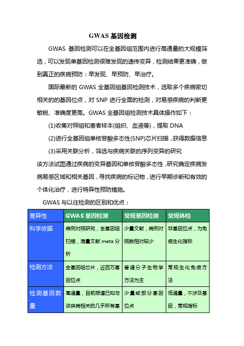
GWAS基因检测GWAS基因检测可以在全基因组范围内进行高通量的大规模筛选,可以发现单基因检测很难发现的遗传变异,检测结果更准确,做到真正的疾病预防:早发现、早预防、早治疗。
国际最新的GWAS全基因组基因检测技术,选取多个疾病密切相关的的基因位点,对SNP进行全面的检测,对易感疾病的判断更敏锐、准确度更高。
GWAS全基因组检测技术具体操作如下:(1)收集对照组和患者样本(组织、血液等),提取DNA(2)进行全基因组单核苷酸多态性(SNP)芯片扫描,获得数据信息(3)采用关联分析,筛选与疾病关联的序列变异的研究该方法试图通过疾病的变异基因和单核苷酸多态性,研究确定疾病发病易感区域和相关基因,寻找疾病的标记物,进行早期诊断和有效的个体化治疗,进行特异性预防措施。
GWAS与以往检测的区别和优点:现在针对我们平台所做的肿瘤套餐介绍如下:我们在GWAS数据库中提取了针对于亚洲人群(主要是日本、韩国、中国、马来西亚等人种)研究的肿瘤相关文献,挑选各个肿瘤的相关疾病位点进行研究。
现针对各个肿瘤介绍其相关位点信息:(1)胃癌基因检测的相关位点:参考文献:1. Zhang H, Jin G, Li H, et al. Genetic variants at 1q22 and 10q23 reproducibly associated with gastric cancer susceptibility in a Chinese population. Carcinogenesis. 2011; 32(6): 848-52.2. Wang N, Zhou R, Wang C, et al. A polymorphism of the interleukin-8 gene and cancer risk: a HuGE review and meta-analysis based on 42 case-control studies. Mol Biol Rep. 2012; 39(3): 2831-41.3. Lu Y, Chen J, Ding Y,et al. Genetic variation of PSCA gene is associated with the risk of both diffuse- and intestinal-type gastric cancer in a Chinese population. Int J Cancer. 2010; 127(9): 2183-9.4. Shi Y, Hu Z, Wu C, et al. A genome-wide association study identifies new susceptibility loci for non-cardia gastric cancer at 3q13.31 and 5p13.1. Nat Genet. 2011; 43(12): 1215-8.(2)、鼻咽癌基因检测的相关位点:(3)、肺癌基因检测的相关位点:参考文献:1. Hu Z, Wu C, Shi Y, et al. A genome-wide association study identifiestwo new lung cancer susceptibility loci at 13q12.12 and 22q12.2 in Han Chinese. Nat Genet. 2011; 43(8): 792-6.2. Ahn MJ, Won HH, Lee J, et al. The 18p11.22 locus is associated with never smoker non-small cell lung cancer susceptibility in Korean populations. Hum Genet. 2012; 131(3): 365-72.3. Yoon KA, Park JH, Han J, et al. A genome-wide association study reveals susceptibility variants for non-small cell lung cancer in the Korean population. Hum Mol Genet.2010; 19(24):4948-54.(4)、甲状腺癌基因检测的相关位点:参考文献:1.Matsuse M, Takahashi M, Mitsutake N, et al. The FOXE1 and NKX2-1 loci are associated with susceptibility to papillary thyroid carcinoma in the Japanese population. J Med Genet. 2011;48(9): 645-8.(5)、前列腺癌基因检测的相关位点:参考文献:1.Takata R, Akamatsu S, Kubo M, Takahashi A, et al. Genome-wide association study identifies five new susceptibility loci for prostate cancer in theJapanese population. Nat Genet. 2010;42(9): 751-4.(6)、子宫内膜癌基因检测的相关位点:参考文献:1. Xu WH, Long JR, Zheng W, et al. Association of the progesterone receptor gene with endometrial cancer risk in a Chinese population. Cancer. 2009; 115(12): 2693-700.2. Long J, Zheng W, Xiang YB et al. Genome-wide association studyidentifies a possible susceptibility locus for endometrial cancer. Cancer Epidemiol Biomarkers Prev. 2012; 21(6): 980-7.(7)、乳腺膜癌基因检测的相关位点:参考文献:1.Long J, Shu XO, Cai Q, et al. Evaluation of breast cancer susceptibility loci in Chinese women. Cancer Epidemiol Biomarkers Prev. 2010; 19(9): 2357-65.2.Kim HC, Lee JY, Sung H, et al. A genome-wide association study identifies a breast cancer risk variant in ERBB4at 2q34: results from the Seoul Breast Cancer Study. Breast Cancer Res.2012; 14(2): R56.3.Long J, Cai Q, Sung H, Shi J, et al. Genome-wide association study in east Asians identifies novel susceptibility loci for breast cancer. PLoS Genet. 2012; 8(2): e1002532.4.Shu XO, Long J, Lu W, et al. Novel genetic markers of breast cancer survival identified by a genome-wide association study. Cancer Res. 2012; 72(5): 1182-9.5.Cai Q, Long J, Lu W, et al. Genome-wide association study identifies breast cancer risk variant at 10q21.2: results from the Asia Breast Cancer Consortium. Hum Mol Genet. 2011; 20(24):4991-9.(8)、结直肠癌基因检测的相关位点:参考文献:1. Xiong F, Wu C, Bi X, et al. Risk of genome-wide association study-identified genetic variants for colorectal cancer in a Chinese population. Cancer Epidemiol Biomarkers Prev 2010 Jul;19(7):1855-61.2. Ho JW, Choi SC, Lee YF, et al. Replication study of SNP associationsfor colorectal cancer in Hong Kong Chinese. Br J Cancer 2011;104: 369-375.以上是我们平台所做的肿瘤套餐基因检测相关位点的信息。
人类遗传基因组变异的研究进展
人类遗传基因组变异的研究进展一、人类遗传基因组变异简介人类基因组包括了20,000-25,000个基因,它们编码了制造蛋白质的指南。
一个基因的变异可能会造成蛋白质的不同形态或缺失,这可能会影响个体的生长发育和健康特征。
人类的遗传基因组变异通常包括DNA单核苷酸变异、DNA复制数变异、染色体结构变异和基因重组等多种类型。
二、DNA单核苷酸变异DNA单核苷酸变异(Single Nucleotide Polymorphisms,SNPs)是人群中常见的变异类型。
SNPs通常被广泛用于大规模遗传关联研究,这种研究方式可以探讨SNPs和个体特征或疾病之间的关系。
SNPs的研究也是研究人类基因组多样性的一个重要方面。
三、DNA复制数变异DNA复制数变异(Copy Number Variations,CNVs)是人类基因组的一个重要方面。
CNVs是指在同一基因区域内,基因拷贝数量的不同。
CNVs在很大程度上可以影响基因的表达。
CNVs可以通过分析微阵列或基因测序数据来鉴定。
CNVs在人类中的研究起源于桥本甲状腺炎的研究,后来发现CNVs可能与许多其他疾病如自闭症和肥胖症有关。
四、染色体结构变异染色体结构变异(Structural Variants,SVs)包括插入、缺少、倒位、转座和交叉互换等不同类型。
SVs通常可以通过对DNA序列进行比对或在基因测序数据中进行计算来鉴定。
SVs对个体的影响可能是抑制基因的表达,也可能会改变基因的功能,促进疾病的发生。
五、基因重组基因重组(Genetic Recombination)是指自然条件下DNA单链断裂、交叉并连和回复,从而改变了个体基因组中的顺序。
基因重组通常是在减数分裂期发生的,每个单倍体基因组都由其父母各一份染色体贡献而来。
基因重组是产生基因多样性的一个重要手段。
六、结论人类基因组中的各种变异类型是一个广泛而复杂的研究领域。
遗传基因组变异研究对于探讨人类进化、基因多样性和疾病发生等方面具有重要意义。
基因检测流程的详细步骤和流程图
基因检测流程的详细步骤和流程图导语:基因检测是一项重要的医学技术,可以用于分析个体基因组中的特定变异,从而提供个性化的医疗和健康指导。
本文将深入探讨基因检测的详细步骤和流程,以帮助读者更全面地了解这一过程。
一、概述基因检测是通过对个体的DNA进行分析,来确定是否存在特定的基因变异或突变。
这些变异可能与遗传病、药物反应性和某些生理特征有关。
1.1 样本收集需要从个体身体的合适部位收集样本。
通常情况下,采用唾液、血液或口腔黏膜细胞等方式来获取DNA样本。
收集样本的方法应确保样本的完整性和纯度,以避免干扰基因检测结果。
1.2 DNA提取收集到的样本中含有DNA,需要经过DNA提取步骤将其纯化。
这一步骤的目的是去除样本中的杂质,并获得纯粹的DNA样本以供后续的分析使用。
常见的DNA提取方法包括盐溶法、酚-氯仿法等。
1.3 DNA扩增经过DNA提取后,接下来需要进行DNA扩增,即制备更多的DNA分子以便于后续的分析。
聚合酶链式反应(PCR)是最常用的DNA扩增方法之一。
通过PCR,可以在短时间内扩增目标DNA序列,从而使得检测更加敏感和准确。
1.4 基因检测方法选择在进行基因检测之前,需要选择适合的检测方法。
根据不同的需求和目的,可以选择不同类型的基因检测方法,包括基因测序、基因芯片等。
1.5 基因检测选择好基因检测方法后,可以开始进行基因检测。
基于所选的方法,可以分析个体基因组中的特定基因或基因组区域。
这些分析可以涉及基因测序、基因表达、基因变异等多个方面。
二、基因检测流程图以下为基因检测的流程图,用于帮助读者更好地理解整个过程。
_________________________| || 样本收集与传送 || (唾液/血液) ||_________________________|||V_________________________| || DNA提取与纯化 || (盐溶法/酚-氯仿法等) ||_________________________|||V_________________________| || DNA扩增(PCR) ||_________________________|||V_________________________| || 基因检测方法选择 || (基因测序/基因芯片等) ||_________________________|||V_________________________| || 基因检测 || (基因序列/表达/变异等) ||_________________________|三、总结与回顾通过上述的详细步骤和流程图,我们可以清晰地了解基因检测的流程和各个环节的重要性。
全基因疾病模式omnigenetic
全基因疾病模式(“Omnigenic” model)1作者包括Stanford大学的Jonathan Pritchard教授,他的研究生Evan Boyle和博后Yang I Li。
其中Jonathan Pritchard是斯坦福大学生物和遗传学部的教授,同时是Howard Hughes Medical Institute的研究者,著名的群体遗传学家,开发了用于研究人群结构的STRUCTURE算法,以人类进化为主要研究兴趣,尤其是在理解遗传变异与人类性状之间的关系方面。
此外,可变剪切的工具LeafCutter也出自Pritchard的课题组博后Yang I Li 的工作。
遗憾的是在我们关注某一或某些基因突变时,却忽略了更重要的基因之间彼此关联因素与疾病的相关性。
基因突变的关联影响或许是诱发疾病的真正成因。
他们认为,“就是一般疾病也可能与成百上千个基因相关联,然而,每个基因所影响疾病的发生和发展微不足道。
在器官组织水平上更无法理解疾病的诱因。
所谓GWAS的“提示”(基因水平上的变化)与疾病几乎毫无关系,也不可能成为开发精准治疗药物的真正靶点(基因变异总是在漂移中)”。
“基因的差异性或许是通过更为复杂的生物调节机制和网络系统最终影响几个“核心基因”而诱发疾病。
我们好像一直在做“丢西瓜,捡芝麻”的事情”。
Pritchard教授的团队也例举了GWAS研究精神分裂症、类风湿性关节炎和克罗恩氏病等与基因的关联性问题。
其基因变化与疾病成因和发展的相关性似乎对不上号。
因为基因突变并不局限于神经细胞或免疫细胞内,几乎所有细胞都有类似改变。
Pritchard教授认为“更有可能是在基因调节网络机制上影响了疾病相关细胞的改变,而非基因突变直接诱发了疾病”2文章指出,发现基因的常规方法是做更大的基因组关联研究,但Pritchard的团队反对这种方法,相反,他建议进行核心基因的深度测序,以寻找可能具有更大影响的罕见突变体。
河北医科大学第四医院2017年度河北省医学科技奖推荐报奖项目公示.doc

河北医科大学第四医院2017年度河北省医学科技奖推荐报奖项目公示根据《河北省卫生计生委办公室关于组织推荐2017年度河北省科学技术奖的通知》(冀卫办科教函【2017】10号)文件精神,现将我院申报2017年度河北省医学科技奖项目的基本情况予以公示。
以上项目公示7天,有异议者请与科研处联系。
联系人:联系电话:邮箱:河北医科大学第四医院科研处二零一七年十二月二十一日项目名称:Raf激酶抑制蛋白介导的信号转导对肝癌细胞乙酰肝素酶表达的影响主要完成单位:河北医科大学第四医院河北北方学院附属第一医院河北大学附属医院全部完成人:王顺祥,吴晓慧,杨永江,高峰,李建坤申报奖种:河北省医学科技奖论文专著专利等知识产权情况:本项目由河北医科大学第四医院承担,为第一完成单位,知识产权归属河北医科大学第四医院。
发表相关论文如下:注意:论文需写明全部作者1. XiaohuiWu(吴晓慧), Yongjiang Yang(杨永江), Zhuo Xu(徐卓), Jiankun Li (李建坤), Baoming Yang (杨宝明), Ningning Feng (冯宁宁),Yueshan Zhang (张越山), ShunxiangWang(王顺祥). Raf kinase inhibitor protein mediated signaling inhibits invasion and metastasis of hepatocellular carcinoma. Biochimica et Biophysica Acta,2016,1860 (2): 384-3912. 吴晓慧,王顺祥,杨永江,李建坤,徐卓,唐瑞峰. Raf激酶抑制蛋白与肝细胞癌侵袭转移的关系. 中华肿瘤杂志, 2011,33(5):358-3623. 吴晓慧,王顺祥,王士杰,杨永江,李建坤,彭利,唐瑞峰,徐卓. 肝癌组织中Raf激酶抑制蛋白的表达及其临床意义. 第三军医大学学报, 2009,31(5):416-4194. 吴晓慧, 张越山, 王顺祥, 李建坤. 肝癌中RKIP与HPA的表达及临床意义. 肿瘤防治研究,2014 ,41(2):166-1675. 王顺祥,高峰,吴晓慧,宋西进,李建坤,TNF-α及IL-1β促进肝癌细胞系SMMC-7721乙酰肝素酶表达. 基础医学与临床,2007,27(12):1352-1355项目名称:E-钙粘蛋白的遗传变异与Wnt/β-catenin信号通路在子宫内膜异位症发生中作用机制的研究主要完成单位:河北医科大学第四医院全部完成人:康山李琰赵喜娃赵健周荣秒王娜申报奖种:河北省医学科技奖论文专著专利等知识产权情况:本项目由河北医科大学第四医院承担,为第一完成单位,知识产权归属河北医科大学第四医院。
与川崎病发病及冠状动脉损伤相关的易感基因

与川崎病发病及冠状动脉损伤相关的易感基因吴镇宇;姚丽萍【摘要】Kawasaki disease (KD) is an acute vasculitis of infancy and childhood that has become the leading cause of ac-quired heart disease in children in the developed world, in whom the resulting coronary artery abnormalities can cause my-ocardial ischaemia, infarction and even death. The etiology and pathogenesis of KD have yet to be known despite more than 5 decades of investigation. Recent advances in research on KD include searches for genetic susceptibility related to KD and research on immunopathogenesis. Genetic factors increase susceptibility to Kawasaki disease, as indicated by its strikingly high incidence rate in children of Asian ethnicity and by an increased incidence in first-degree family members. This re-view summarizes recent advances in understanding of the susceptibility genes related to Kawasaki disease and coronary artery lesion.%川崎病(KD)是婴幼儿时期的急性血管炎,已成为发达国家儿童获得性心脏病最常见的原因,可导致冠状动脉病变而出现心肌缺血﹑梗死甚至死亡。
氨基糖苷类抗生素性耳聋的研究进展_轩贵平

·临床新进展·氨基糖苷类抗生素(aminoglycoside antibiotics ,AmAn )是具有氨基糖与氨基环醇结构的一类抗生素,包括链霉素、新霉素、庆大霉素、阿米卡星等。
AmAn 对铜绿假单胞菌、肺炎杆菌、大肠杆菌等常见革兰阴性杆菌的抗生素后效应较长,目前仍然被用于治疗敏感需氧革兰阴性杆菌所致的严重感染,如脑膜炎和呼吸道、泌尿道、骨关节感染等。
但AmAn 引发的不良反应,尤其是不可逆性的耳聋一直是医学界的难题,大量研究发现,大剂量长时间用药会引起听力损害,而且部分个体低剂量用药也会出现耳聋,甚至“一针致聋”,并且氨基糖苷类抗生素性耳聋(AmAn induceddeafness ,AAID )具有家族聚集和母系遗传现象,因此线粒体DNA (mitochondrial DNA ,mtDNA )极有可能是AmAn 易感性的分子基础。
近年来,已经明确了与AAID 相关的线粒体基因突变位点,其中一些位点的确切致聋机制也得到确定。
同时也开发出了许多筛查相关突变的方法,为临床合理使用AmAn ,预防AAID 的发生提供指导。
1AAID 与线粒体突变1.1AAID 与线粒体12S rRNA A1555G 突变早在1988年,Wallen 等[1]就报道了首例由mtDNA 突变引起的Leber 遗传性视神经眼病,明确了mtDNA 突变可引起人类疾病,首次提出线粒体病的概念。
随后Higash 等[2]整理相关文献后正式提出,对所观察到的母系遗传现象最有可能的解释是存在线粒体DNA 缺陷。
Hutchin 等[3]报道了AmAn 致聋的人类易感性,并用细胞学分析证实mtDNA 12S rRNA 基因A1555G 突变是AmAn 致聋的分子基础。
2011年耶鲁大学Raimundo 研究团队[4]发现A1555G 突变的确切致聋原因是该突变增强了转甲基酶mt-TFB1催化的线粒体12SrRNA 的甲基化,由此激活ROS 依赖的AMP 激酶和促凋亡核转录因子E2F1的活化,引起E2F1依赖性听力损害,即线粒体应激启动E2F1凋亡信号导致耳聋出现。
- 1、下载文档前请自行甄别文档内容的完整性,平台不提供额外的编辑、内容补充、找答案等附加服务。
- 2、"仅部分预览"的文档,不可在线预览部分如存在完整性等问题,可反馈申请退款(可完整预览的文档不适用该条件!)。
- 3、如文档侵犯您的权益,请联系客服反馈,我们会尽快为您处理(人工客服工作时间:9:00-18:30)。
...........................................................................................................................Genetic variants and the risk of gestational diabetes mellitus:a systematic reviewCuilin Zhang 1,*†,Wei Bao 1,*†,Ying Rong 2,Huixia Yang 3,Katherine Bowers 1,Edwina Yeung 1,and Michele Kiely 11Epidemiology Branch,Division of Epidemiology,Statistics and Prevention Research,Eunice Kennedy Shriver National Institute of Child Health and Human Development,National Institutes of Health,6100Executive Blvd,Rockville,MD 20852,USA 2Department of Nutrition and Food Hygiene,Hubei Key Laboratory of Food Nutrition and Safety,School of Public Health,Tongji Medical College,Huazhong University of Science and Technology,Wuhan 430030,China 3Department of Obstetrics and Gynecology,Peking University First Hospital,Beijing 100034,China*Correspondence address.Tel:+1-301-435-6917;Fax:+1-301-435-6917;Email:zhangcu@;wei.bao@Submitted on December 13,2012;resubmitted on February 24,2013;accepted on March 7,2013table of contents†Introduction †Methods †Results †Discussionbackground:Several studies have examined associations between genetic variants and the risk of gestational diabetes mellitus (GDM).However,inferences from these studies were often hindered by limited statistical power and conflicting results.We aimed to systematically review and quantitatively summarize the association of commonly studied single nucleotide polymorphisms (SNPs)with GDM risk and to identify important gaps that remain for consideration in future studies.methods:Genetic association studies of GDM published through 1October 2012were searched using the HuGE Navigator andPubMed databases.A SNP was included if the SNP–GDM associations were assessed in three or more independent studies.Two reviewers independently evaluated the eligibility for inclusion and extracted the data.The allele-specific odds ratios (ORs)and 95%confidence intervals (CIs)were pooled using random effects models accounting for heterogeneity.results:Overall,29eligible articles capturing associations of 12SNPs from 10genes were included for the systematic review.The minoralleles of rs7903146(TCF7L2),rs12255372(TCF7L2),rs1799884(230G/A,GCK ),rs5219(E23K,KCNJ11),rs7754840(CDKAL1),rs4402960(IGF2BP2),rs10830963(MTNR1B ),rs1387153(MTNR1B )and rs1801278(Gly972Arg,IRS1)were significantly associated with a higher risk of GDM.Among them,genetic variants in TCF7L2showed the strongest association with GDM risk,with ORs (95%CIs)of 1.44(1.29–1.60,P ,0.001)per T allele of rs7903146and 1.46(1.15–1.84,P ¼0.002)per T allele of rs12255372.conclusions:In this systematic review,we found significant associations of GDM risk with nine SNPs in seven genes,most of whichhave been related to the regulation of insulin secretion.Key words:gestational diabetes mellitus /single nucleotide polymorphism /gene /genetic factors†The authors contributed equally to this work.&The Author 2013.Published by Oxford University Press on behalf of the European Society of Human Reproduction and Embryology.All rights reserved.For Permissions,please email:journals.permissions@Human Reproduction Update,Vol.19,No.4pp.376–390,2013Advanced Access publication on May 19,2013doi:10.1093/humupd/dmt013IntroductionGestational diabetes mellitus(GDM),defined as glucose intolerance with onset orfirst recognition during pregnancy,is a growing health concern(Reece et al.,2009).The prevalence of GDM varies in different populations or ethnic groups.In the USA, 7%(ranging from1to14%) of all pregnancies are complicated by GDM(American Diabetes Asso-ciation,2004).Native American,Asian,Hispanic and African-American women are at higher risk for GDM than non-Hispanic white women (Ferrara,2007).GDM increases risk of adverse pregnancy outcomes and has substantial long-term adverse health impacts on both mothers and their offspring,including a predisposition to obesity,metabolic syndrome and type2diabetes mellitus(T2DM)in later life(American Diabetes Association,2004;Bellamy et al.,2009;Reece et al.,2009). Well-documented risk factors for GDM include pre-pregnancy overweight and obesity,family history of diabetes and advanced ma-ternal age(Ben-Haroush et al.,2004;Zhang and Ning,2011).In the past decade,accumulating evidence has indicated that poor diet and low physical activity before or during pregnancy may also represent risk factors of GDM(Zhang and Ning,2011).In addition,interesting, though limited,data have shown that a history of subfertility or infer-tility may be related to an elevated risk of GDM(Jaques et al.,2010; Reyes-Munoz et al.,2012).Moreover,polycystic ovarian syndrome,a contributor to ovulatory disorder fertility,has been repeatedly linked to an increased GDM risk(Boomsma et al.,2006;Bals-Pratsch et al., 2011;Reyes-Munoz et al.,2012).There are relatively few published studies of the genetic susceptibility to GDM(Watanabe,2011);although available data suggest that pregnancy complications have a familial tendency(Martin et al.,1985; Solomon et al.,1997).Moreover,GDM recurs in at least30%(range 30–84%)of women with a history of GDM(Kim et al.,2007),potentially suggesting that there is a subgroup of women who may be genetically predisposed to develop GDM.Defects in both insulin secretion and insulin action are crucial in the pathogenesis of GDM(Buchanan and Xiang,2005).A study among Danish twins showed major genetic com-ponents in both traits;more than75%of the variation of the insulin secretion trait and at least53%of peripheral insulin sensitivity can be explained by genetic components(Poulsen et al.,2005).Taken together, the evidence supports a genetic component in the etiology of GDM. Over the past few decades,genetic loci in several genes,responsible for insulin secretion,insulin resistance,lipid and glucose metabolism and other pathways,have been associated with GDM risk.However, inferences have been hindered by inconsistentfindings across studies, partly owing to small sample size,moderate gene effects and insufficient statistical power(Robitaille and Grant,2008).In this study,we aimed to systematically review the current evi-dence regarding the genetic associations of GDM to quantitatively summarize the effect size of replicated single nucleotide polymorph-isms(SNPs)on GDM risk,and to identify important gaps that remain for consideration in future studies.MethodsWe adhered to the Meta-analysis Of Observational Studies in Epidemi-ology(MOOSE)guidelines(Stroup et al.,2000)when undertaking this study.Literature search and data extractionGenetic association studies of GDM published through1October2012 were searched mainly using the HuGE Navigator(Yu et al.,2008),an inte-grated database of genetic associations and human genome epidemiology studies.The HuGE Navigator has been found to be equally sensitive,but more specific than PubMed in a previous validation study(Palomaki et al., 2010).The search term‘gestational diabetes[Text MesH]’was used for the Huge Navigator search.As the HuGE Navigator only retrieves articles published since2001,an additional PubMed search was conducted to identify publications through31December2001.For the PubMed search,the following search terms were used:(‘Diabetes,Gestational/ genetics’[Mesh]or‘Diabetes,Gestational/epidemiology’[Mesh]or‘Gesta-tional diabetes’[tiab])and(‘Polymorphism,Single Nucleotide’[Mesh]or polymorphism*[tiab])not(review[pt]or editorial[pt]).In addition,the references listed in relevant original papers and review articles were screened.No restriction was applied on language or geographical location in the literature search process.Two reviewers(W.B.and Y.R.)independently evaluated the eligibility of inclusion and extracted the data,and disagreements were resolved by con-sensus.Articles were included if they reported original data about testing for SNP main effects on GDM risk.An SNP was included if the SNP–GDM associations were assessed in three or more independent studies. Cross-sectional,case–control and cohort studies were eligible for inclu-sion.Several types of articles were excluded:reviews or editorials,non-human studies(cell culture or animal studies),family-based studies, studies that did not include GDM as the primary outcome,studies that did not evaluate genetic associations of GDM and pharmacogenetics studies for anti-diabetic medication.In addition,other exclusions included studies that did not include a healthy control group,studies that did not report sufficient data for effect estimates of the genetic associations and studies that did not separately report association measures for GDM. The following data were extracted from each published article:thefirst author’s name,year of publication,sample size,number of GDM cases, ethnicity,mean age,study design(case–control,cross-sectional or cohort study),genetic variants,genotyping method,crude genotype and allele distribution by GDM status,odds ratios(ORs)and95%confidence intervals(CIs).If ORs were available but the genotype and allele distribu-tions according to GDM status were not reported in the original article, the corresponding authors were contacted by email.Data synthesis and statistical analysisThe ORs of individual studies were recalculated from the available geno-type distributions according to an allelic model,pooled using random effect models(DerSimonian and Laird,1986)and visualized by forest plots.Hardy–Weinberg equilibrium(HWE)was assessed for each study by use of Fisher’s exact test instead of the x2test reported in the individual studies as it yields increased statistical power(Bauer et al.,2011).HWE was tested in the whole population for cohort studies and in the control group for case–control studies.Heterogeneity across all eligible compar-isons was assessed using the x2-based Cochran’s Q statistic and the I2 metric(I2value of25,50and75%were considered as low,medium, and high heterogeneity,respectively;Higgins et al.,2003).The potential sources of identified heterogeneity among studies were investigated by stratification analyses.A formal meta-regression was not performed because the number of studies for some SNPs was small.Sensitivity analyses were performed by omitting one study at a time and computing the pooled ORs of the remaining studies to evaluate whether the results were affected markedly by a single study.The possibility of publication bias was statistically assessed using Egger regression asymmetry test (Egger et al.,1997).Genetic variants and gestational diabetes risk377All statistical analyses were performed using Stata software version 11.0(Stata Corp,College Station,TX,USA).All P -values presented are two-tailed with a significance level of 0.05,except the Cochran’s Q statistic in heterogeneity test in which the significance level was 0.10(Higgins et al .,2003).ResultsDescription of the included studiesThe initial literature search yielded 89articles from HuGE Navigator (2001–2012)and 23articles from PubMed (1950–2001).After apply-ing the inclusion and exclusion criteria,29articles capturing 12SNPs from 10genes were ultimately included in the systematic review and meta-analysis (Fig.1).Of the 10genes,six were related to insulin secretion,two to insulin resistance,one to energy metabolism and one to an inflammatory pathway (Table I ).The study characteristics and the genotype and allele distributions of SNPs in the included studies are shown in Tables II and III ,respectively.Genes and genetic variants related to insulin secretionTranscription factor 7-like 2(TCF7L2)The rs7903146variant in the TCF7L2gene was the most widely studied variant in association with GDM,and showed a consistent and strong association across different populations.A meta-analysis of nine studies (Shaat et al .,2007;Cho et al .,2009;Lauenborg et al .,2009;Freathy et al .,2010;Pappa et al .,2011;Papadopoulou et al .,2011;Kwak et al .,2012;Vcelak et al .,2012)showed that the T allele of rs7903146was associated with an increased risk of GDM [pooled OR 1.44(95%CI 1.29–1.60),P ,0.001;Table IV ,Fig.2A].The observed heterogeneity across studies for rs7903146resulted from differences in the study populations in a stratification analysis by race/ethnicity;no significant heterogeneity was observed in Asians (I 2¼0.0%;P for the Q statistic ¼0.916),although therewas still a significant heterogeneity among Caucasians (I 2¼68.4%;P for the Q statistic ¼0.007).In addition,a similar association was found in a meta-analysis of four studies (Watanabe et al .,2007;Cho et al .,2009;Papadopoulou et al .,2011;Vcelak et al .,2012)regarding the T allele of rs12255372and GDM risk;the pooled OR was 1.46(95%CI 1.15–1.84,P ¼0.002),without significant heterogeneity across studies (I 2¼48.3%;P for the Q statistic ¼0.122;Table IV ,Fig.2B).The similar effect sizes between associations of GDM risk with rs12255372and rs7903146were expected given the strong correlation between these two variants (r 2¼1in the HapMap CEU population;Povel et al .,2011).No indication of publication bias was observed for either variant (P ¼0.148for rs7903146and P ¼0.259for rs12255372in the Egger’s test).Deviations from the HWE were observed in two studies with rs7903146(Lauenborg et al .,2009;Papadopoulou et al .,2011)and one with rs12255372(Papadopoulou et al .,2011).In sensitivity analyses by omitting these studies,the pooled ORs were not changed materially and remained significant.Glucokinase (GCK)The association between the rs1799884(also known as 230G/A)variant in the GCK gene and GDM risk has been widely investigated,but the results are conflicting (Chiu et al .,1994;Zaidi et al .,1997;Shaat et al .,2006;Freathy et al .,2010;Santos et al .,2010).Although early studies (Chiu et al .,1994;Zaidi et al .,1997)found no significant association between rs1799884and GDM risk,subsequent studies with larger sample sizes found a significant association (Shaat et al .,2006;Freathy et al .,2010).A meta-analysis of these studies showed that the T allele of rs1799884was associated with an increased risk of GDM [pooled OR 1.29(95%CI 1.17–1.42),P ,0.001;Table IV ,Fig.2C].No indication of significant heterogeneity across studies (I 2¼0.0%;P for the Q statistic ¼0.878)or publication bias (P ¼0.467in the Egger’s test)was observed.Potassium inwardly rectifying channel,subfamily J,member 11(KCNJ11)The association between rs5219(also known as E23K)and GDM was modest (ranging from 1.12–1.17)in the included studies (Shaat et al .,2005;Cho et al .,2009;Lauenborg et al .,2009;Pappa et al .,2011).Our meta-analysis showed that the T allele of rs5219was associated with an increased risk of GDM [pooled OR 1.15(95%CI 1.06–1.26),P ¼0.002;Table IV ,Fig.2D].No indication of significant heterogen-eity across studies (I 2¼0.0%;P for the Q statistic ¼0.976)or publi-cation bias (P ¼0.750in the Egger’s test)was observed.CDK5regulatory subunit associated protein 1-like 1(CDKAL1)The association between rs7754840in CDKAL1and GDM risk has been examined in three studies,all of which were conducted among Asian populations (Cho et al .,2009;Wang et al .,2011;Kwak et al .,2012).Our meta-analysis indicated that the C allele of rs7754840was significantly associated with risk of GDM [pooled OR 1.40(95%CI 1.13–1.72),P ¼0.002;Table IV ,Fig.2E].The observed het-erogeneity across these studies resulted from differences in the study populations;two studies in Korean women showed strong associa-tions between rs7754840and GDM risk (Cho et al .,2009;Kwak et al .,2012),whereas a study in Chinese women found nosignificantFigure 1Flow chart for study selection.378Zhang et al .association (Wang et al .,2011).No indication of publication bias (P ¼0.703in the Egger’s test)was observed.Insulin-like growth factor 2mRNA-binding protein 2(IGF2BP2)The association between rs4402960and GDM risk showed similar effect sizes in Asian and Caucasian populations (Cho et al .,2009;Lauenborg et al .,2009;Wang et al .,2011).A meta-analysis of these studies showed that the T allele of rs4402960was significantly asso-ciated with an increased risk of GDM [pooled OR 1.21(95%CI 1.10–1.33),P ,0.001;Table IV ,Fig.2F].No indication of significant heterogeneity across studies (I 2¼0.0%;P for the Q statistic ¼0.842)or publication bias (P ¼0.550in the Egger’s test)was observed.Melatonin receptor 1B (MTNR1B)Kim et al .(2011)first found a significant association of GDM risk with two variants in the MTNR1B locus,rs10830963and rs1387153,which are in tight linkage disequilibrium (LD)with each other (|D ′|¼0.89).The association between rs10830963and GDM risk was replicated in a subsequent study of a Greek population (Vlassi et al .,2012).Our meta-analyses showed that the T allele of rs1387153and G allele of rs10830963were associated with an increased risk of GDM;the pooled ORs were 1.30(95%CI 1.18–1.43,P ,0.001)and 1.28(95%CI 1.05–1.55,P ¼0.016),respectively (Table IV ,Fig.2G and H).There was no indication of significant heterogeneity across studies regarding rs1387153and GDM risk (I 2¼0.0%;P for the Q statistic ¼0.691).The observed heterogeneity across studies for rs10830963resulted from differences in the study populations;the study in Greek women found a strong and significant association (Vlassi et al .,2012),in Korean women,a weak but significant association (Kim et al .,2011),while in Chinese women there was no significant association (Wang et al .,2011).No indication of publi-cation bias was observed for either variant (P ¼0.744for rs1387153and P ¼0.567for rs10830963in the Egger’s test).Genes and genetic variants related to insulin resistancePeroxisome proliferator-activated receptor gamma (PPARG)The association between rs1801282and GDM risk has been exam-ined in eight studies among several populations (Shaat et al.,2004;Tok et al.,2006b ;Shaat et al.,2007;Cho et al.,2009;Lauenborg et al.,2009;Cheng et al.,2010;Heude et al.,2011;Pappa et al.,2011);however,none of these found a significant association.A meta-analysis of these studies showed that the G allele of rs1801282was not significantly associated with GDM risk [pooled OR 0.94(95%CI 0.82–1.07),P ¼0.322;Table IV ,Fig.3A].No indi-cation of significant heterogeneity across studies (I 2¼0.0%;P for the Q statistic ¼0.450)or publication bias (P ¼0.061in the Egger’s test)was observed.Insulin receptor substrate 1(IRS1)The association between rs1801278(also known as Gly972Arg)and GDM has been examined in four studies (Shaat et al .,2005;Fallucca et al .,2006;Tok et al.,2006a ;Pappa et al .,2011),all among Cauca-sians.Our meta-analysis of these studies showed that the T allele of rs1801278was significantly associated with an increased risk of GDM [pooled OR 1.39(95%CI 1.04–1.85),P ¼0.027;Table IV ,Fig.3B).No indication of significant heterogeneity across studies (I 2¼34.5%;P for the Q statistic ¼0.205)or publication bias (P ¼0.602in the Egger’s test)was observed.Genes and genetic variants related to other pathwaysAdrenoceptor beta 3(b 3-adrenergic receptor,ADRB3)Five small studies examined the association between rs4994(also known as Trp64Arg)and GDM with inconsistent results (Festa et al .,1999;Alevizaki et al .,2000;Tsai et al .,2004;Fallucca et al .,2006;Shaat et al .,2007).Festa et al.(1999)found that the A/G geno-type was more frequent in women with GDM (n ¼70)than in those with normal glucose tolerance (n ¼109;26versus 11%;P ¼0.01)..............................................................................................................................................................................................Table I Genes and genetic variants included in the systematic review and their pathwaysGeneChromosome locationDescriptionVariantsInsulin secretionInsulin resistanceOther pathwaysTCF7L210q25.3Transcription factor 7-like 2rs7903146(IVS3C .T);rs12255372Yes GCK 7p15.3–p15.1Glucokinasers1799884(230G/A)Yes KCNJ1111p15.1Potassium inwardly rectifying channel,subfamily J,member 11rs5219(E23K)Yes CDKAL16p22.3CDK5regulatory subunit associated protein 1-like 1rs7754840Yes IGF2BP23q27.2Insulin-like growth factor 2mRNA-binding protein 2rs4402960Yes MTNR1B 11q21–q22Melatonin receptor 1Brs10830963;rs1387153YesPPARG 3p25Peroxisome proliferator-activated receptor gamma rs1801282(Pro12Ala)Yes IRS12q36Insulin receptor substrate 1rs1801278(Gly972Arg)YesADRB38p12Adrenoceptor beta 3rs4994(Trp64Arg)Energy metabolism TNF6p21.3Tumor necrosis factorrs1800629(2308G/A)InflammationGenetic variants and gestational diabetes risk379However,this positive association was not confirmed in subsequent studies (Alevizaki et al .,2000;Tsai et al .,2004;Fallucca et al .,2006;Shaat et al .,2007).Our meta-analysis of these five studies showed no significant association between the G allele of rs4994and GDM risk [pooled OR 1.20(95%CI 0.88–1.65),P ¼0.252;Table IV ,Fig.4A].No indication of significant heterogeneity across studies (I 2¼38.8%;P for the Q statistic ¼0.163)or publication bias (P ¼0.916in the Egger’s test)was observed.Tumor necrosis factor (TNF)A meta-analysis of three studies (Chang et al .,2005;Montazeri et al .,2010;Gueuvoghlanian-Silva et al .,2012)showed no significant associ-ation between rs1800629and GDM [pooled OR 1.64(95%CI 0.73–3.69),P ¼0.228;Table IV ,Fig.4B].The observed heterogeneity across these studies resulted from differences in the study populations;a significant and strong association between rs1800629and GDM was found in a Chinese population (Chang et al .,2005),but the positive genetic association was not replicated in Malaysians (Montazeri et al .,2010)or Brazilians (Gueuvoghlanian-Silva et al .,2012).No indication of significant publication bias (P ¼0.987in the Egger’s test)was observed.It should be noted that the sample size in the included studies was small (in total 224cases and 305controls)and deviations from the HWE were observed in two studies (Chang et al .,2005;Montazeri et al .,2010);therefore the association between rs1800629and GDM needs to be confirmed in more studies.DiscussionIn this study,we investigated relatively frequently studied genetic variants in association with GDM risk.Several previous reviews have mainly focused on the evidence regarding T2DM-associated common variants and GDM susceptibility (Watanabe et al .,2007;Robitaille and Grant,2008;Konig and Shuldiner,2012;Mao et al.,2012).Our systematic review provided a more comprehensive summary of the currently available evidence regarding GDM genetic variants.Overall,we observed significant associations of GDM with SNPs in the TCF7L2,GCK ,KCNJ11,CDKAL1,IGF2BP2,MTNR1B and IRS1genes.Although pregnancy is a condition characterized by progressive insulin resistance (Buchanan and Xiang,2005;Watanabe,2011),GDM develops in only a small proportion of pregnant women (Ameri-can Diabetes Association,2004).The insulin resistance that develops during pregnancy may result from a combination of increased maternal adiposity and the insulin-desensitizing effects of placental products such as human placental lactogen,estrogen and prolactin (Di Cianni et al .,2003).Normally,the increased insulin resistance during preg-nancy is compensated by the increase in insulin secretion by pancreatic islet b cells.As a result,the changes in circulating glucose levels over the course of pregnancy are quite small,compared with the large changes in insulin sensitivity (Buchanan and Xiang,2005).GDM could develop when a genetic predisposition of pancreatic islet b -cell impairment is unmasked by the increased insulin resistance during pregnancy (Lambrinoudaki et al .,2010).Among the most widely studied genes of GDM included in the present systematic review,six genes (TCF7L2,GCK ,KCNJ11,CDKAL1,IGF2BP2and MTNR1B )are thought to modulate pancreatic islet b -cell function (Petrie et al .,2011;Schafer et al .,2011),and all of them were signifi-cantly associated with GDM risk (ORs ranging from 1.15to 1.46).InV l a s s i e t a l .(2012)C a s e –c o n t r o l C a u c a s i a n G r e e c e 779835.45/31.39A D A c r i t e r i aR F L P –P C RaT h e d a t a o f C a u c a s i a n s w e r e u p d a t e d b y S h a a t e t a l .(2007)a n d t h e r e f o r e t h e y w e r e n o t i n c l u d e d h e r e .A D A ,A m e r i c a n D i a b e t e s A s s o c i a t i o n ;E A S D -D P S G ,t h e D i a b e t e s a n d P r e g n a n c y S t u d y G r o u p s o f t h e E u r o p e a n A s s o c i a t i o n f o r t h e S t u d y o f D i a b e t e s ;G D M ,g e s t a t i o n a l d i a b e t e s m e l l i t u s ;H P L C ,h i g h -p e r f o r m a n c e l i q u i d c h r o m a t o g r a p h y ;I A D P S G ,t h e I n t e r n a t i o n a l A s s o c i a t i o n o f D i a b e t e s a n d P r e g n a n c y S t u d y G r o u p s ;I W C G D M ,I n t e r n a t i o n a l W o r k s h o p -C o n f e r e n c e o n G e s t a t i o n a l D i a b e t e s M e l l i t u s ;N D D G ,N a t i o n a l D i a b e t e s D a t a G r o u p ;O G T T ,o r a l g l u c o s e t o l e r a n c e t e s t ;R F L P ,r e s t r i c t i o n f r a g m e n t l e n g t h p o l y m o r p h i s m ;P C R ,p o l y m e r a s e c h a i n r e a c t i o n ;S S C P ,s i n g l e -s t r a n d c o n f o r m a t i o n p o l y m o r p h i s m ;W H O ,W o r l d H e a l t h O r g a n i z a t i o n .Genetic variants and gestational diabetes risk381contrast,only two genes (PPARG and IRS1)are relevant to insulin resistance (Petrie et al .,2011),and only the IRS1variant is significantly associated with GDM risk.These findings suggest that inherited abnormalities of pancreatic islet b -cell function and/or b -cell mass may be implicated in the etiology of GDM.All genetic loci associated with GDM risk (i.e.TCF7L2,GCK ,KCNJ11,CDKAL1,IGF2BP2and MTNR1B )in our systematic review have been previously related to the risk of T2DM (Frayling,2007;McCarthy,2010).The effect size of the associations between these SNPs and GDM was similar to those in the studies of T2DM.More-over,in a recent genome-wide association study of GDM (Kwak et al .,2012),among the 11variants significantly associated with GDM risk,five SNPs were located in or near the known T2DM loci.In addition,two variants that reached the genome-wide signifi-cance level (P ,5×1028),rs7754840in CDKAL1and rs10830962near MTNR1B,were identical or in strong LD with known T2DM var-iants (Kwak et al .,2012).These findings suggest an at least partly shared genetic basis between GDM and T2DM,which is not surprising given that both insulin resistance and defects in insulin secretion play key roles in the etiology of both GDM and T2DM.In addition,women with a history of GDM have a more than 7-fold risk of developing T2DM later in life (Bellamy et al .,2009).It should be noted that not all women who have a history of GDM develop T2DM.Different from T2DM,GDM as a pregnancy compli-cation may be influenced by not only the maternal genome but also the paternal and fetal genomes.Indeed,emerging data suggest both fetal and paternal genotypes may affect glucose metabolism in preg-nancy.For example,Wangler et al.(2005)observed that mothers carrying offspring with Beckwith–Wiedemann syndrome,in which probands have abnormally increased IGF2expression,showed a trend toward an increased risk of GDM.Also,in an animal study by Petry et al.(2010),maternal glucose concentrations in pregnant mice were elevated among women carrying pups with targeted disrup-tion of maternally transmitted fetal H19D 13,which implied that variable fetal IGF2expression could affect risk for GDM.Moreover,in an epidemiological study among 1160mother/partner/offspring trios from the UK,Petry et al.found that polymorphic variations in the paternally transmitted fetal IGF2genotype,but not the maternal or maternally transmitted fetal IGF2genotypes,were associated with increased maternal glucose concentrations in pregnancy,which could potentially alter the risk of maternal GDM (Petry et al .,2011).These studies highlighted a potential role of the paternal and fetal genomes,in addition to the maternal genome itself,in maternal glucose homeostasis during pregnancy.Future genetic studies of GDM considering fetal and/or paternal genome are warranted.Gene–gene and gene–environment interactions may further help illustrate the biological basis for complex diseases and provide import-ant clues for personalized interventions or clinical therapeutics (Collins et al .,2003).These interactions contribute to b -cell function (Nesher et al .,1999;Li et al .,2009),insulin sensitivity (Black et al .,2008)and T2DM risk (Cornelis and Hu,2012).Further,a number of environ-mental factors,such as diet and lifestyle factors,have been significantly associated with GDM risk (Zhang and Ning,2011).However,so far little has been done to investigate gene–environmental interactions in relation to GDM susceptibility.Watanabe et al .(2007)found that the TCF7L2rs12255372variant interacts with adiposity to alter insulin secretion in 132Mexican-American families of a proband with previous GDM.In a recent study of 826GDM cases and 1185healthy controls,Papadopoulou et al.(2011)examined the interaction between TCF7L2and HLA-DQB1*0602variants in association with GDM risk in Swedish women,but observed no interaction between them.Future studies with larger sample sizes are warranted to better understand these complex interactions in the pathogenesis of GDM.The strength of the present study is the systematic way in which we have summarized results of the available studies on SNP–GDM asso-ciations.However,our analysis has several limitations.First,although the pooled sample size for some SNPs (e.g.rs7903146in TCF7L2)was relatively large,for others it was small (e.g.for rs1800629in TNF ,224cases and 305controls).Secondly,we focused on the.............................................................................................................................................................................................Table IV Associations between genetic variants and GDM risk in the systematic review and meta-analysesGeneVariantMinor allele Number of studies Sample size (cases/controls)OR (95%CI)aP -valueHeterogeneity TCF7L2rs7903146T 9b 4406/11445 1.44(1.29–1.60),0.001I 2¼51.3%;P Het ¼0.037TCF7L2rs12255372T 42022/2166 1.46(1.15–1.84)0.002I 2¼48.3%;P Het ¼0.122GCK rs1799884T 6b 1934/6492 1.29(1.17–1.42),0.001I 2¼0.0%;P Het ¼0.878KCNJ11rs5219T 41837/4327 1.15(1.06–1.26)0.002I 2¼0.0%;P Het ¼0.976CDKAL1rs7754840C 32959/3675 1.40(1.13–1.72)0.002I 2¼88.1%;P Het ,0.001IGF2BP2rs4402960T 31836/3986 1.21(1.10–1.33),0.001I 2¼0.0%;P Het ¼0.842MTNR1B rs1387153T 31454/2312 1.30(1.18–1.43),0.001I 2¼0.0%;P Het ¼0.691MTNR1B rs10830963G 31685/2093 1.28(1.05–1.55)0.016I 2¼70.2%;P Het ¼0.035PPARG rs1801282G 82241/63360.94(0.82–1.07)0.322I 2¼0.0%;P Het ¼0.450IRS1rs1801278T 41106/1673 1.39(1.04–1.85)0.027I 2¼34.5%;P Het ¼0.205ADRB3rs4994G 51239/2002 1.20(0.88–1.65)0.252I 2¼38.8%;P Het ¼0.163TNFrs1800629A3224/3051.64(0.73–3.69)0.228I 2¼74.3%;P Het ¼0.020a ORs were calculated based on allelic model.bThe study by Freathy et al .included two independent study populations.384Zhang et al .。
