小鼠Ⅰ型胶原 (Col I)-ELISA试剂盒说明书
小鼠Ⅰ型胶原α2 (COL1a2)-ELISA试剂盒说明书
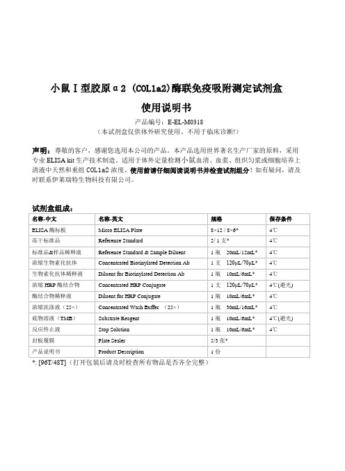
小鼠Ⅰ型胶原α2 (COL1a2)酶联免疫吸附测定试剂盒使用说明书产品编号:E-EL-M0318(本试剂盒仅供体外研究使用、不用于临床诊断!)声明:尊敬的客户,感谢您选用本公司的产品。
本产品选用世界著名生产厂家的原料,采用专业ELISA kit生产技术制造。
适用于体外定量检测小鼠血清、血浆、组织匀浆或细胞培养上清液中天然和重组COL1a2浓度。
使用前请仔细阅读说明书并检查试剂组分!如有疑问,请及时联系伊莱瑞特生物科技有限公司。
*: [96T/48T](打开包装后请及时检查所有物品是否齐全完整)检测原理:本试剂盒采用双抗体夹心ELISA法。
用抗小鼠COL1a2抗体包被于酶标板上,实验时标本或标准品中的COL1a2会与包被抗体结合,游离的成分被洗去。
依次加入生物素化的抗小鼠COL1a2抗体和辣根过氧化物酶标记的亲和素。
抗小鼠COL1a2抗体与结合在包被抗体上的小鼠COL1a2结合、生物素与亲和素特异性结合而形成免疫复合物,游离的成分被洗去。
加入显色底物(TMB),TMB 在辣根过氧化物酶的催化下现蓝色,加终止液后变黄。
用酶标仪在450nm波长处测OD值,COL1a2浓度与OD450值之间呈正比,通过绘制标准曲线求出标本中COL1a2的浓度。
标本收集:1.血清:全血标本于室温放置2小时或4℃过夜后于1000×g离心20分钟,取上清即可检测,收集血液的试管应为一次性的无热原,无内毒素试管。
2.血浆:抗凝剂推荐使用EDTA.Na2,标本采集后30分钟内于1000×g离心15分钟,取上清即可检测。
避免使用溶血,高血脂标本。
3.组织匀浆:用预冷的PBS (0.01M, pH=7.4)冲洗组织,以去除残留血液(匀浆中裂解的红细胞会影响测量结果),称重后将组织剪碎。
将剪碎的组织与对应体积的PBS(一般按1:9的重量体积比,比如1g的组织样本对应9mL的PBS,具体体积可根据实验需要适当调整,并做好记录。
胶原酶Ⅰ使用说明
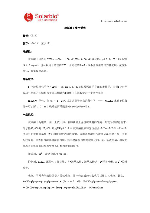
胶原酶Ⅰ使用说明货号:C8140保存:-20°C,至少1年。
溶解性:胶原酶Ⅰ可以用TESCA buffer(50mM TES,0.36mM氯化钙,pH7.4,37°C)配制成1-2mg/ml。
也可以用含钙镁的PBS、含钙镁的hanks或不含血清的培养基配制。
配完后分装,避免反复冻融。
酶活定义:1个胶原消化单位(CDU):在pH7.4,37℃以及钙离子存在的条件下,以每5小时从胶原中释放的多肽相当于茚三酮显色1微摩尔亮氨酸量为一个活性单位。
1FALGPA单位:在pH7.5、25℃以及钙离子存在的条件下,一个FALGPA水解单位每分钟可水解 1.0μmol呋喃基丙烯酰基-Leu-Gly-Pro-Ala。
产品说明:胶原酶Ⅰ为粗品,用于上皮、肺,脂肪和肾上腺组织细胞的分离。
外观为黑棕色粉末。
分子量68,000到125,000,最适PH为6.3-8.8。
胶原酶能够特异性结合-R-Pro-8-X-Gly-Pro-R-序列中的中性氨基酸(X)和甘氨酸之间的肽键。
该粗品是溶组织梭菌分泌的混合酶。
主要为胶原酶、中性蛋白酶和梭菌蛋白酶。
其中梭菌蛋白酶是被氧化的、最不活泼的酶,组织的分离必须依靠胶原酶和中性蛋白酶两者共同作用。
激活剂:Ca2+,最适合浓度为5mM。
抑制剂:EGTA、还原性谷胱甘肽、β-巯基乙醇、巯基乙酸钠、8-羟基喹啉、2,2'-联吡啶等。
底物:不同类型的胶原是其天然底物,而一些合成的多肽也可以作为其底物,比如:N-CBZ-gly-pro-gly-gly-pro-ala(Km=0.71mM)、N-CBZ-gly-pro-leu-gly-pro、N-(3-(2-furyl)acryloyl)-leu-gly-pro-ala(FALGPA)、4-Phenylazobenzyloxycarbonyl-pro-leu-gly-pro-D-arg、N-2,4-Dinitrophenyl-pro-gln-gly-ile-ala-gly-gln-D-arg。
鼠尾胶原蛋白Ⅰ型使用说明
鼠尾胶原蛋白Ⅰ型使用说明货号:C8062保存:4℃保存,切勿冻存,有效期为一年。
规格:10mg,5mg/mL,溶解于0.006mol/L HAc,无菌。
产品简介:本鼠尾胶原蛋白I型(rattailtendon collagen type I)是通过Birkedal-Hansen的方法,通过醋酸抽提、氯化钠沉淀、磷酸氢二钠沉淀等步骤制备的。
鼠尾胶原蛋白可用于包被细胞培养器皿,培养一些在普通细胞培养器皿中不易贴壁的细胞;也可用于制备三维胶,模拟真实的生长环境,使细胞在三维环境中生长。
1,在使用本鼠胶原蛋白I型包被的细胞培养皿中检查到PC-12细胞的贴壁和生长。
2,在1mg/mL浓度以上,pH为7左右时可形成具有一定强度的三维胶,检查到NIH-3T3细胞在三维胶内正常生长、PC-12细胞在三维胶表面正常生长。
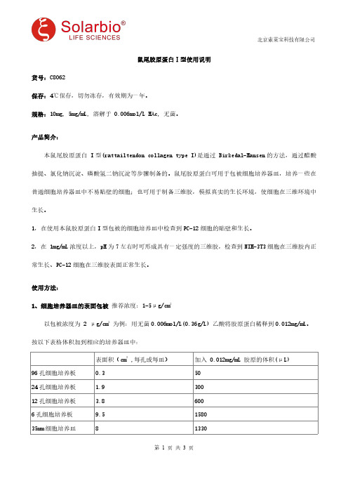
使用方法:1、细胞培养器皿的表面包被推荐浓度:1-5μg/cm2以包被浓度为2μg/cm2为例:用无菌0.006mol/L(0.36g/L)乙酸将胶原蛋白稀释到0.012mg/mL。
按以下表格体积加到相应的培养器皿中:表面积(cm2,每孔或每皿)加入0.012mg/mL胶原的体积(μL)96孔细胞培养板0.35024孔细胞培养板 1.930012孔细胞培养板 3.86006孔细胞培养板9.5158035mm细胞培养皿8133060mm细胞培养皿213500100mm细胞培养皿559170确保胶原蛋白溶液铺满器皿的表面,开盖在超净台上过夜晾干。
也可以在室温放置1小时后,用PBS洗3-4次后直接使用。
包被好的器皿在4-25℃至少可保存3个月以上的时间。
2、三维胶原的制备鼠尾胶原蛋白I型在浓度1mg/mL以上,pH7左右时可形成具有一定强度三维胶,建议成胶浓度1-2mg/mL。
胶原蛋白溶解于0.006mol/L乙酸中,在成胶过程中需要加入0.06×体积的0.1mol/L NaOH来中和。
需要的溶液(均需要无菌、预冷):10×PBS(可含10mg/L的酚红用于pH指示)或10×培养液,0.1mol/L NaOH,0.1mol/L乙酸(一般不用),双蒸水A.不含细胞的三维胶原的制备(以配制1mL,1mg/mL三维胶为例):将200μL鼠尾胶原蛋白I型(5mg/mL)加到置于冰浴的离心管中,加入690μL H2O。
ELISA 检测试剂盒 说明书
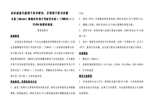
本试剂盒只能用于科学研究,不得用于医学诊断小鼠小鼠((MouseMouse))肿瘤坏死因子可溶性受体肿瘤坏死因子可溶性受体ⅠⅠ(TNFsR-TNFsR-ⅠⅠ)ELISA检测试剂盒使用说明书检测原理试剂盒采用双抗体一步夹心法酶联免疫吸附试验(ELISA)。
往预先包被肿瘤坏死因子可溶性受体Ⅰ(TNFsR-Ⅰ)抗体的包被微孔中,依次加入标本、标准品、HRP标记的检测抗体,经过温育并彻底洗涤。
用底物TMB显色,TMB在过氧化物酶的催化下转化成蓝色,并在酸的作用下转化成最终的黄色。
颜色的深浅和样品中的肿瘤坏死因子可溶性受体Ⅰ(TNFsR-Ⅰ)呈正相关。
用酶标仪在450nm波长下测定吸光度(OD值),计算样品浓度。
样品收集、处理及保存方法1.血清:使用不含热原和内毒素的试管,操作过程中避免任何细胞刺激,收集血液后,3000转离心10分钟将血清和红细胞迅速小心地分离。
2.血浆:EDTA、柠檬酸盐或肝素抗凝。
3000转离心30分钟取上清。
3.细胞上清液:3000转离心10分钟去除颗粒和聚合物。
4.组织匀浆:将组织加入适量生理盐水捣碎。
3000转离心10分钟取上清。
5.保存:如果样本收集后不及时检测,请按一次用量分装,冻存于-20℃,避免反复冻融,在室温下解冻并确保样品均匀地充分解冻。
自备物品1.酶标仪(450nm)2.高精度加样器及枪头:0.5-10uL、2-20uL、20-200uL、200-1000uL3.37℃恒温箱操作注意事项1.试剂盒保存在2-8℃,使用前室温平衡20分钟。
从冰箱取出的浓缩洗涤液会有结晶,这属于正常现象,水浴加热使结晶完全溶解后再使用。
2.实验中不用的板条应立即放回自封袋中,密封(低温干燥)保存。
3.浓度为0的S0号标准品即可视为阴性对照或者空白;按照说明书操作时样本已经稀释5倍,最终结果乘以5才是样本实际浓度。
4.严格按照说明书中标明的时间、加液量及顺序进行温育操作。
5.所有液体组分使用前充分摇匀。
ELISA_kit使用说明书

微囊藻毒素ELISA检测试剂盒使用说明书Microcystin Plate Kit一、基本原理酶联免疫(ELISA)是免疫酶技术的一种,是将抗原抗体反应的特异性与酶反应的敏感性相结合而建立的一种新技术。
ELISA的技术原理是:将酶分子与抗体(或抗原)结合,形成稳定的酶标抗体(或抗原)结合物,当酶标抗体(或抗原)与固相载体上的相应抗原(或抗体)结合时,即可在底物溶液参与下,产生肉眼可见的颜色反应,颜色的深浅与抗原或抗体的量成比例关系,使用ELISA 检测仪,即酶标仪,测定其吸收值可做出定量分析。
此技术具有特异、敏感、结果判断客观、简便和安全等优点,日益受到重视,不仅在微生物学中应用广,而且也被其他学科领域广为采用。
本试剂盒工作原理如下图所示。
二、已配试剂ELISA试剂盒中配备以下条目:1、96孔已包被好的酶标板2、1只阴性对照即0孔溶液3、标准1:0.1ng/mL的MC-LR标准溶液4、标准2:0.2ng/mL的MC-LR标准溶液5、标准3:0.5ng/mL的MC-LR标准溶液6、标准4:1ng/mL的MC-LR标准溶液7、标准5:2.5ng/mL的MC-LR标准溶液8、1瓶溶液1 (单抗)9、1只溶液2(二抗稀释液)10、一只二抗(小管乘装)11、1瓶溶液3(底物溶液)12、1瓶溶液4 (终止液)13、1袋固体PBS(配置洗涤液用)三、需配器材及试剂除试剂盒内的物品外,还需配备以下器材和试剂:1、酶标仪(若可能,可配有洗板机)2、微量移液器(100 L)(若可能,可配8道或12道微量移液器)3、吸管、橡皮吸头(洗板时用,若有洗板机,无需准备此项)4、纯水(用于配置洗涤液)5、玻璃瓶(盛装洗涤液)6、记时器(准确控制每步操作时间)7、封口膜(防止孔内液体挥发)四、酶标二抗的配置将小管中二抗用溶液2按1:3000稀释,充分溶解混匀,现用现配。
五、洗涤液的配置将固体PBS以纯水配置成1L溶液,加1mL Tween 20(PBS-T, pH7.4-7.6),室温保存。
ELISA检测试剂盒使用指南
ELISA检测试剂盒使用指南ELISA(酶联免疫吸附试验)是一种常用的免疫学实验方法,用于检测病原体、抗体或其他分子的存在和浓度。
它具有高灵敏度、高特异性、易操作和较低成本的优势,被广泛应用于生物医学研究、临床诊断和免疫学领域。
本篇文章将介绍ELISA检测试剂盒的使用指南,包括实验准备、试剂的使用步骤和结果解读。
一、实验准备1.阅读检测项目的说明书:在使用试剂盒前,仔细阅读说明书,了解试剂的使用方法、灵敏度和特异性等关键信息。
2.样本准备:根据实验要求,准备样本。
如果需要检测血清中的抗体水平,可以采集血液样本,离心分离血清;如果需要检测细胞培养上清中的分子,将上清收集,并使其清晰,避免细胞或杂质的污染。
3.样本预处理:根据实验需求,对样本进行必要的预处理。
例如,可以通过加热、稀释、酶解等方式来处理样本,以改变样本组分和浓度。
4.准备品质控制样品:制备阳性和阴性控制样品,用于评估试剂盒和实验操作的稳定性和准确性。
5.实验器材准备:准备所需实验器材,如酶标板、移液器、微孔板洗涤仪等。
确保器材的干净和完整,并按照说明书要求对其进行预处理。
二、试剂使用步骤1.试剂准备:根据说明书要求,将试剂从冰箱中取出,并在室温下放置一段时间使其恢复到室温。
确保试剂瓶盖紧闭,并避免暴露在直接阳光下。
2.实验操作:按照说明书的要求,将试剂加入到酶标板中,并根据实验设计进行标准曲线的设置。
标准曲线用于测量未知样品的数量,并计算出浓度。
3.孵育:根据试剂盒的要求,将酶标板放入孵育箱中进行孵育。
孵育温度和时间应根据实验要求进行调整。
4.洗涤:使用洗涤缓冲液对酶标板上的不特异性结合物进行洗涤。
洗涤过程应准确控制洗涤孔板次数和洗涤液的体积。
5.补液:在洗涤完成后,加入辣根过氧化物酶标况稀释液,促进酶标物与特异性结合物的反应。
6.孵育:根据试剂盒的要求,将酶标板放入孵育箱中进行二次孵育。
7.反应停止:根据试剂盒的要求,加入相应的停止液,停止酶反应。
小鼠试剂盒,小鼠细胞间粘附分子1(ICAM-1CD54)酶联免疫检测ELISA试剂盒使用说明书
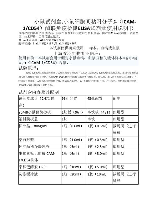
小鼠试剂盒,小鼠细胞间粘附分子1(ICAM-1/CD54)酶联免疫检测ELISA试剂盒使用说明书国内权威的科研试剂供应商,乔羽生物专业经营进口分装和原装、国产的Elisa试剂盒,品质保证,技术严格,无效果退款退货。
Elisa kit规格:48孔配置/96孔配置酶标试剂:3 ml×1瓶(48)/6 ml×1瓶(96)本试剂仅供研究使用标本:血清或血浆ICAM-1/CD54试剂盒是固相夹心法酶联免疫吸附实验(ELISA).已知ICAM-1/CD54浓度的标准品、未知浓度的样品加入微孔酶标板内进行检测。
先将ICAM-1/CD54和生物素标记的抗体同时温育。
洗涤后,加入亲和素标记过的HRP。
再经过温育和洗涤,去除未结合的酶结合物,然后加入底物A、B,和酶结合物同时作用。
产生颜色。
颜色的深浅和样品中ICAM-1/CD54的浓度呈比例关系。
试剂盒内容及其配制96孔配置48孔配置配制试剂盒成份(2-8℃保存)96/48小鼠份酶标板1块板(96T)半块板(48T)即用型塑料膜板盖1块半块即用型标准品:80ng/ml1瓶(0.6ml)1瓶(0.3ml)按说明书进行稀稀空白对照1瓶(1.0ml)1瓶(0.5ml)即用型标准品稀释缓冲液1瓶(5ml)1瓶(2.5ml)即用型生物素标记的抗ICAM-1瓶(6ml)1瓶(3.0ml)即用型1/CD54抗体亲和链酶素-HRP1瓶(10ml)1瓶(5.0ml)即用型洗涤缓冲液1瓶(20ml)1瓶(10ml)按说明书进行稀释底物A1瓶(6.0ml)1瓶(3.0ml)即用型底物B1瓶(6.0ml)1瓶(3.0ml)即用型终止液1瓶(6.0ml)1瓶(3.0ml)即用型标本稀释液1瓶(12ml)1瓶(6.0ml)即用型自备材料1.蒸馏水。
2.加样器:5ul、10ul、50ul、100ul、200ul、500ul、1000ul。
3.振荡器及磁力搅拌器等。
安全性1.避免直接接触终止液和底物A、B。
大鼠PⅠCP,ELISA试剂盒使用说明书

大鼠PⅠCP,ELISA试剂盒使用说明书NID1, nest protein 1 elisa kit instructions大鼠PⅠCP,ELISA试剂盒使用说明书产品组成部分:产品名称大鼠Ⅰ型前胶原羧基端肽(PⅠCP)ELISA试剂盒说明书产品规格48T/96T产品产地上海/美国检测种属人,鼠,马,羊,牛,鸡,猴等库存状态现货供应,款到发货说明书公司网站中英文说明书下载保存要求2-8℃(一个月) 有效期6个月(-20℃)检测目的测定血清,血浆及相关液体等样本适用原则仅供科研使用,不得用于临床大鼠PⅠCP,ELISA试剂盒使用说明书产品优势:.ELISA检测试剂盒是当前使用广泛的一种方法。
.具有直接,间接,夹心,竞争等ELISA方法。
.是敏感性高,高效性,特异性强,重复性好,稳定性的诊断方法。
.产品吸附性好,空白值低,孔底透明高。
.用于检测(血清、血浆和细胞培养上清液,细胞溶解产物,组织匀浆,尿液和脑脊液)。
5,适用于人,大小鼠,猴,牛,猪,马,羊,鸡,狗等多种种属。
6,节约材料与耗材,缩短时间,节省经费准备材料:1,三十倍浓缩洗涤液20ml×1 瓶;终止液6ml×1 瓶2,酶标试剂6ml×1 瓶;标准品(80μmol/L)0.5ml×1 瓶3,酶标包被板12 孔×8 条;标准品稀释液 1.5ml×1 瓶4,样品稀释液6ml×1 瓶;说明书 1 份5,显色剂A 液6ml×1 瓶;封板膜 2 张.显色剂B 液6ml×1/瓶;密封袋 1 个技术原理及自备设备:.ELISA技术原理是抗原或抗体的固相化及抗原或抗体的酶标记。
结合在固相载体表面的抗原或抗体仍保持其免疫学活性,酶标记的抗原或抗体既保留其免疫学活性,又保留酶的活性。
在测定时,受检标本与固相载体表面的抗原或抗体起反应。
.自备设备有,蒸馏水,加样器,振荡器及磁力搅拌器,酶标仪,量筒,烧杯,吸水纸,坐标纸,温育箱,洗瓶,一次性试剂管,微移液及其吸嘴等。
RayBio Mouse MIP-1 beta CCL4 IQELISA Kit 使用说明书
RayBio® Mouse MIP-1 beta/CCL4IQELISA KitCatalog #: IQM-MIP1bUser ManualLast revised April 6, 2023Caution:Extraordinarily useful information enclosedISO 13485 Certified3607 Parkway Lane, Suite 200Peachtree Corners, GA 30092 Tel: 1-888-494-8555 (Toll Free) or 770-729-2992, Fax: 770-206-2393Web: , Email: *******************RayBiotech, Inc.________________________________________ RayBio® Mouse MIP-1 beta/CCL4 IQELISA Kit ProtocolTable of ContentsSection Page # I.Introduction3II.Reagents3III.Storage3IV.Additional Materials Required4V.Reagent Preparation4VI.Assay Procedure5VII.Assay Procedure Summary8VIII.Calculation of ResultsA. Typical DataB. Sensitivity and Recovery 8 9 9IX.Troubleshooting Guide10I. INTRODUCTIONThe RayBio®I mmuno Q uantitative E nzyme L inked I mumuno S orbent A ssay (IQELISA) is an innovative new assay that combines the specificity and ease of use of an ELISA with the sensitivity of real-time PCR. This results in an assay that is simultaneously familiar and cutting edge and enables the use of lower sample volumes while also providing more sensitivity. The RayBio® Mouse MIP-1 beta/CCL4 IQELISA Kit is a modified ELISA assay with high sensitivity qPCR readout for the quantitative measurement of Human MIP-1 beta in serum, plasma, and cell culture supernatants. This assay employs an antibody specific for Human MIP-1 beta coated on a 96-well PCR plate. Standards and samples are pipetted into the wells and MIP-1 beta present in a sample is bound to the wells by the immobilized antibody. The wells are washed and a detection affinity molecule is added to the plates. After washing away unbound detection affinity molecule, primers and a PCR master mix are added to the wells and data is collected using qPCR. C t values obtained from the qPCR are then used to calculate the amount of antigen contained in each sample, where lower C t values indicate a higher concentration of antigen.II. REAGENTS1.MIP-1 beta Microplate (Item A)**: 96 well PCR plate coated with anti-Human MIP-1 beta.2.Wash Buffer I Concentrate (20x) (Item B): 25 ml of 20x concentrated solution.3.Standards (Item C): 2 vials of recombinant Human MIP-1 beta.4.Assay Diluent B (Item E): 15 ml of 5x concentrated buffer.5.Detection Affinity Reagent for MIP-1 beta (Item F): 2 vials of a 4x concentrated solutionof anti-Human MIP-1 beta affinity reagent.6.IQELISA Detection Reagent (Item G): 1 mL of a 10x concentrated stock.7.Primer Solution (Item I): 1.5 mL vial.8.PCR Master Mix (Item J): 1.4 mL vial.9.PCR Preparation buffer (Item K): 1mL vial of 10x concentrated buffer.10.Final Wash Buffer (Item L): 10 mL vial of 10x concentrated buffer.**The PCR plate used is a 0.2 mL, non-skirted 96-well plate (ThermoFisher, cat. no.:AB0600). Please ensure compatibility with your PCR machine prior to purchase. For additional information contact technical support (**************************).III. STORAGEMay be stored for up to 6 months at 2°to 8°C from the date of shipment. Standard (recombinant protein) should be stored at -20°C or -80°C (recommended at -80°C) after reconstitution. Opened PCR plate or reagents may be stored for up to 1 month at 2° to 8°C. Note: the kit can be used within one year if the whole kit is stored at -20°C. Avoid repeated freeze-thaw cycles.IV. ADDITIONAL MATERIALS REQUIRED1.Real-time PCR instrument, Bio-Rad recommended2.Precision pipettes to deliver 2 µl to 1 mL volumes.3.Adjustable 1-25 mL pipettes for reagent preparation.4.100 mL and 1 L graduated cylinders.5.Absorbent paper.6.Distilled or deionized water.7.Log-log graph paper or computer and software for data analysis.8.Tubes to prepare standard or sample dilutions.9.Heating block or water bath capable of 80°CV. REAGENT PREPARATION1.Bring wash buffer, samples, assay diluents, and PCR plate to room temperature (18 -25°C) before use. PCR master mix and Primer solution should be kept at 4°C at alltimes.2.Sample dilution: If your samples need to be diluted, 1x Assay Diluent B should be usedfor dilution of serum/plasma samples.Suggested dilution for normal serum/plasma: 2 fold*.*Please note that levels of the target protein may vary between different specimens.Optimal dilution factors for each sample must be determined by the investigator.3.Assay Diluent B should be diluted 5-fold with deionized water.4.Briefly spin the Detection Antibody vial before use. Add 25 µL of 1x Assay Diluent B intothe vial to prepare a detection antibody concentrate. Pipette up and down to mix gently (the concentrate can be stored at 4°C for 5 days). This concentrate should be diluted 80-fold with 1x Assay Diluent B and used in step 4 of the Assay Procedure.5.PCR preparation buffer should be transferred to a 15 mL tube and diluted with 9 mL ofdeionized or distilled water before use.6.Final Wash Buffer should be transferred to a 15 mL tube and diluted with 9 mL ofdeionized or distilled water for every 1 mL of 10x concentrate used before use.7.Preparation of standard: Preparation of standard: Briefly spin a vial of Standards. Add400 µl 1X Assay Diluent B into Standards vial to prepare a 25 ng/ml standard solution.Dissolve the powder thoroughly by a gentle mix. Add 20 µl of MIP-1beta standardsolution from the vial of Standards, into a tube with 480 µl 1X Assay Diluent B to preparea 1000 pg/ml standard solution. Pipette 200 µl 1X Assay Diluent B into each tube. Use the 1000 pg/ml standard solution to produce a dilution series (shown below). Mix each tube thoroughly before the next transfer. 1X Assay Diluent B serves as the zero standard (0 pg/ml).20.00 µL + 480 µL100 µL+ 200 µL100 µL+ 200 µL100 µL+ 200 µL100 µL+ 200 µL100 µL+ 200 µL100 µL+ 200 µL1000 pg/ml 333.333pg/ml111.111pg/ml37.037pg/ml12.346pg/ml4.115pg/ml1.372pg/mlpg/ml8.If the Wash Buffer Concentrate (20x) contains visible crystals, warm to room temperatureand mix gently until dissolved. Dilute 20 mL of Wash Buffer Concentrate into deionized or distilled water to yield 400 mL of 1x Wash Buffer.9.Prepare the IQELISA detection reagent by calculating how much will be needed. Thismay be accomplished by multiplying the number of wells to be assayed by the volume you plan to use per well. Once the volume of IQELISA detection reagent is known,prepare the reagent by diluting it 1:10 with deionized water and mixing thoroughly.VI. ASSAY PROCEDUREOptional Visual Aid: IQELISA [Good Laboratory Practice Guide]1.Bring all reagents and samples to room temperature (18 - 25°C) before use. It isrecommended that all standards and samples be run in triplicate. Partial plate runs may be accomplished by cutting the PCR plate into the desired number of strips using a pair of sturdy scissors, wire cutters, or shears. The remainder may be saved and used for a later date. If this is done, the PCR Plate Film should also be cut to a suitable size.2.Add 10-25 µL of each standard (see Reagent Preparation step 2) and sample intoappropriate wells. Volumes should be consistent between all wells, samples, andstandards. As little as 10 µL can be used if sample volume is limited, however thisincreases the chance of technical error. Ensure there are no bubbles present at thebottom of the wells. Dislodge any bubbles with gentle tapping or with a pipette tip being careful not to contact the sides or bottom of the well. Cover well and incubate for 1.5 -2.5 hours at room temperature.3.Discard the solution and wash 4 times with 1x Wash Solution. Wash by filling each wellwith Wash Buffer (100 µL) using a multi-channel Pipette or autowasher. Completeremoval of liquid at each step is essential to good performance. After the last wash,remove any remaining Wash Buffer by aspirating or decanting. Invert the plate and blot it against clean paper towels.4.Add 25 µL of prepared Detection Antibody (Reagent Preparation step 4) to each well.Incubate for 1 hour at room temperature with gentle shaking.5.Discard the solution. Repeat the wash as in step 3.6.Add 50 µL of prepared IQELISA detection reagent and incubate 1 hour with rocking(Reagent Preparation step 9)7.Discard the solution. Repeat the wash as in step 3, for a total of 6 washes.8.Add 75 µL of Final wash buffer to each well and incubate for 4 minutes with rocking.Remove the solution from each well and blot against paper towels.9.Add 75 µL of 1x PCR preparation buffer to each well and incubate for 10 seconds beforeremoving the buffer. Blot the plate after the buffer is removed to ensure completeremoval of the buffer.10.Add 10 µL of the Primer solution to each well of the plate. At this stage the plate can becovered and stored at -20°C for use the next day if needed.11.Add 10 µL of PCR Master Mix to each well and pipette thoroughly to mix the well (atleast 3x up and down).12.Cover the plate with the supplied PCR Plate Film, taking care to insure the film iscompletely and even pressed onto the plate, creating an air tight seal around each well of the plate.Optional Visual Aid: Sealing the plate [qPCR]13.Place the plate into a real-time PCR instrument using a FITC compatible wave length fordetection with the following settings for cycling1.2 minute activation at 95°C2.15 seconds 95°C denaturation3.25 seconds 60°C annealing/extension4.Repeat steps 2 and 3 34x**Optional: Include a melt curve to view potential plate contamination that can causehigh background and lower the sensitivity. This can be seen in the visual aid onYouTube.VII. ASSAY PROCEDURE SUMMARY1.Prepare all reagents, samples and standards as instructed.2.Add 25 µL standard or sample to each well. Incubate 1.5 - 2.5 hours at roomtemperature.3.Add 25 µL Detection Antibody to each well. Incubate 1 hour at room temperature.4.Add 50 µL of IQELISA Detection Reagent to each well. Incubate 1 hour5.Add 10 µL Primer solution and 10 µL of PCR master mix to each well6.Run real-time PCRVIII. CALCULATION OF RESULTSThe primary data output of the IQELISA kit is C t values. These values represent the number of cycles required for a sample to pass a fluorescence threshold. As the DNA is amplified additional fluorescent signal is produced, with each cycle resulting in an approximate doubling of the DNA. Therefore, higher levels of DNA (directly related to the amount of antigen in the sample) result in lower C t values.Calculate the mean C t for each set of triplicate standards, controls and samples. Subtract the C t value of each sample from the control to obtain the difference between the control and sample (Delta C t). Plot the values of the standards on a graph using a log scale for concentration on the x axis. This graph is the quickest way to visualize results, although not the most accurate. If this method is used the concentration of unknown samples can be estimated using a logarithmic line of best fit.The line of best fit will have an equation y = mln(x)+b, where y is the Delta C t value and x is the concentration. It may be helpful to use 5 significant figures for m and b to minimize rounding errors. To calculate the concentration of unknown sample this can be entered into Excel in the following format=EXP((y-b)/m))Where y is the Delta C t obtained during the assay, and b and m are obtained from the line of best fit.Alternatively, for a more accurate representation linear regression may be used. Both the Delta C t and Concentration can be transformed using a log base of 10, plotted on a graph as described above, along with a line of best fit (using a linear model). The equation of this line may be used to calculate the antigen concentration of unknown samples. This is the method used for the analysis spreadsheet for IQELISA available online.A. TYPICAL DATAThese data are for demonstration only. A standard curve must be run with each assay.B. SENSITIVITY and RECOVERYThe minimum quantifiable dose of MIP-1 beta is typically 0.97 pg/ml, however levels as lower than 0.97 pg/ml may be detected outside of the quantification range.Serum spike tests show recovery is 86% with a range from 80% to 91%.Intraplate CV is below 10% for all samples and Interplate CV is below 15%.X. TROUBLESHOOTING GUIDEThis product is for research use only.©2023 RayBiotech, Inc11。
联科生物技术 细胞因子 ELISA 试剂盒

联科生物技术有限公司 全国免费电话:800 8571 184第四章细胞因子ELISA 试剂盒简介:美国Peprotech公司作为知名的重组细胞因子生产商,开发出了50余种细胞因子的ELISA检测试剂盒,试剂盒的检测灵敏度高,检测范围宽,为您的科研提供了又一有力的工具。
试剂盒组分及重悬方法:包被抗体--100ug 经抗原亲和纯化的包被抗体,冻干粉重悬--用1ml无菌蒸馏水重悬成100ug/ml的贮存液保存--4-8℃,保存两个月;-20℃分装冻存可贮存6个月检测抗体--100ug 经抗原亲和纯化的生物素标记的检测抗体,冻干粉重悬--用1ml无菌蒸馏水重悬成100ug/ml的贮存液保存--4-8℃,保存两个月;-20℃分装冻存可贮存6个月标准品--准确计量的重组细胞因子冻干粉,具体含量见说明书重悬--按说明书1ml无菌蒸馏水重悬成适当浓度的贮存液保存--4-8℃,保存1周;-20℃分装冻存可贮存2个月亲合素偶联的过氧化物酶--60ul/瓶,液体分装--分装到10只小瓶中,每瓶6ul保存---20℃分装冻存可贮存2年实验所需的材料:ELISA酶标板:推荐Nunc maxisorp,产品编号:442404Tween-20:推荐Sigma,产品编号:P-7949BSA(牛血清白蛋白):推荐Sigma,产品编号:A-7030ABTS底物溶液:推荐Sigma,产品编号:A-3219Dulbecco's PBS(10X):推荐Gibco BRL,产品编号:14200-075相关溶液配制:PBS:用无菌蒸馏水将10X PBS稀释为1X PBS洗涤液:含0.05% Tween-20的PBS封闭液:含1% BSA的PBS,过滤除菌,4℃保存1周稀释液:含0.05% Tween-20和0.1% BSA的PBS,过滤除菌,4℃保存1周注:以上所有试剂在使用前必须复温至室温。
包板:1) 用PBS将包被抗体稀释成1ug/ml,立即加到酶标板中,每孔100ul;2) 密封酶标板,室温孵育过夜;3) 吸除孔中的液体,并用300ul洗涤液洗孔4次;4) 最后一次洗涤后,将酶标板倒置除去残留的液体,并倒扣在吸水纸吸干孔沿上的液体;5) 每孔加300ul封闭液,室温孵育至少1小时;6) 吸除孔中的液体,并洗孔4次。
达优 小鼠 TGF-β1 ELISA 试剂盒说明书 (2)
产品信息和操作指南Mouse TGF-β1预包被 ELISA kit Cat# : DKW12-2710-048 / DKW12-2710-096本试剂盒专用于科研,而非用于诊断Mouse TGF-β1DKW12-2710目录产品简介 (1)知识背景 (1)试剂盒提供的试剂 (2)需要实验者自行准备的试剂与仪器 (2)注意事项 (3)试剂的配制 (6)操作过程 (8)结果分析 (10)试剂盒的保存 (10)操作步骤一览表 (11)参考文献 (12)ELISA测定中可能会出现的问题及解决方法 (13)预包被ELISA 试剂盒系列产品 (16)1、产品简介:达优®小鼠TGF-β1 ELISA试剂盒是通过酶联免疫吸附技术,体外定量检测小鼠血清、血浆、缓冲液或细胞培养液中的TGF-β1,可同时检测天然的和重组的TGF-β1。
本试剂盒为预包被板,整个过程孵育时间不超过4小时,洗涤12次。
本试剂盒专用于科研,而非用于诊断。
使用前请仔细阅读说明书并检查试剂盒组分,若有任何疑问请与达科为生物工程有限公司联系,E-mail:*************.检测范围:1000-15.6 pg/mL灵敏度:5 pg/mL重复性:板内、板间变异系数均<10%。
2、知识背景:转化生长因子-β1(TGF-β1)能使正常的成纤维细胞的表型发生转化,具有细胞抑制和促进生长双重作用。
TGF-β1分子量25kDa,非糖基化同型二聚体蛋白,由二硫键连接。
TGF-β1基因结构具有高度保守性,人和小鼠TGF-β1的同源性高达99%(1-4)。
TGF-β1在治疗伤口愈合,促进软骨和骨修复以及通过免疫抑制治疗自身免疫性疾病和移植排斥等方面发挥重要作用(5)。
3、试剂盒提供的试剂:试剂规格配制Cytokine standard 2/1瓶* 干粉状,按瓶上说明操作Biotinylated antibody 2/1瓶* 1:100用Dilution buffer R(1×)稀释Streptavidin-HRP 2/1瓶* 1:100用Dilution buffer R(1×)稀释Dilution buffer R(1×) 3/2瓶* 即用型Washing buffer(50×)1瓶 150∶用蒸馏水稀释TMB 1瓶即用型Stop solution 1瓶即用型Precoated ELISA plate 8×12或8×6*即用型封板膜 2/1张* 即用型说明书1份*:96/48 Tests4、需要实验者自行准备的试剂与仪器:1.酶标仪(建议参考仪器使用说明提前预热)2.微量加液器及吸头:P10,P50,P100,P200,P1000 3.蒸馏水或去离子水4.全新滤纸5.旋涡振荡器和磁力搅拌器6.37℃温箱5、注意事项:1.试剂应按瓶上标签说明储存,使用前室温平衡20-30分钟。
Abclonal小鼠IgM ELISA试剂盒说明书
Mouse IgM ELISA KitCatalog NO.:RK00173version:2.0*使用产品前,请务必阅读产品包装内说明书产品简介本试剂盒产品使用夹心法定量测定小鼠血清、血浆、细胞培养上清或其它生物体液中IgM的含量。
产品检测原理本实验采用双抗夹心ELISA法。
将抗IgM抗体预先包被在酶标板上,向微孔中分别加入标准品和样品,样本中IgM与固相载体上的抗体结合。
孵育后,未结合的样品在洗涤过程中会被洗去,加入酶标抗体,经孵育和洗涤后,加入底物TMB。
样品中IgM的含量与TMB反应的颜色深浅呈正相关,加入酸终止反应并测量吸光值。
根据梯度稀释的IgM标准品的吸光值绘制标准曲线并求出样品的浓度。
试剂盒组分及保存条件未拆封的试剂盒可在2-8°C保存1年,已拆封的产品须在1个月内使用完。
组分规格货号拆封后保存条件抗体预包被酶标板Antibody Coated Plate 8×12RM00720将未使用的板条放回装有干燥剂的铝箔袋中,并重新密封,可在2-8°C存储1个月。
冻干标准品Standard Lyophilized 2支RM00717复溶后不建议再次使用。
浓缩酶标抗体(100×)Concentrated HRP-Conjugate Antibody(100×)1×120ul RM00718可在2-8°C存储1个月。
标准品/样本稀释液(R1)(4x)Standard/Sample Diluent (R1)(4x)1×20mLRM00023可在2-8°C 存储1个月。
酶标抗体稀释液(R2)HRP-Conjugate Antibody Diluent(R2)1×12mLRM00024浓缩洗涤液(20x)Wash Buffer(20x)1×30mLRM00026显色底物TMBSubstrate 1×12mLRM00027终止液Stop Solution 1×6mL RM00028封板膜Plate Sealers4张说明书Specification 1实验所需的材料1.酶标仪,并配置主波长450nm,次波长630nm或570nm。
小鼠中提collagen i蛋白方法-概述说明以及解释
小鼠中提collagen i蛋白方法-概述说明以及解释1.引言1.1 概述Collagen I是一种重要的结构蛋白,广泛存在于动物体内的各种组织中,包括皮肤、骨骼、肌肉和血管等。
它不仅在维持组织的结构完整性和稳定性方面起到关键作用,还参与许多生物过程,如细胞迁移、细胞黏附和组织修复等。
因此,对Collagen I的研究具有重要的理论和应用价值。
目前,提取和纯化Collagen I蛋白的方法有很多种,但由于其在细胞外基质中的高丰度和结构上的复杂性,这些方法往往具有一定的复杂性和挑战性。
本文将重点介绍一种利用小鼠作为研究模型,成功提取和纯化小鼠中的Collagen I蛋白的方法。
本文将首先介绍提取小鼠中Collagen I蛋白的方法。
这涉及到从小鼠组织中获取Collagen I蛋白的样品,并对其进行初步的处理和纯化。
接下来,我们将详细介绍分离和纯化Collagen I蛋白的步骤。
这些步骤将包括多级的色谱层析和电泳技术,以确保最终获得高纯度的Collagen I蛋白样品。
通过采用上述方法,我们成功地提取和纯化了小鼠中的Collagen I蛋白,并经过进一步的分析和鉴定证实了其纯度和结构的正确性。
这一研究成果将有助于我们更深入地了解Collagen I蛋白的功能和作用机制,为相关疾病的治疗和组织工程领域的应用提供重要的基础。
综上所述,本文将介绍一种成功提取和纯化小鼠中Collagen I蛋白的方法。
通过这一方法,我们将为进一步的研究和应用提供有力的支持,推动Collagen I蛋白领域的深入发展。
1.2文章结构1.2 文章结构本文主要包括引言、正文和结论三个部分。
引言部分将概述研究背景和意义,介绍小鼠中Collagen I蛋白的研究现状,并指出本文的研究目的。
正文部分将分为两个主要方法进行阐述。
首先,方法一将详细介绍如何从小鼠身体中提取Collagen I蛋白,包括所需试剂和操作步骤等内容。
其次,方法二将详细介绍如何对提取的Collagen I蛋白进行分离和纯化的过程,包括采用的分离技术和纯化方法等。
i型胶原蛋白标准

i型胶原蛋白标准
i型胶原蛋白是一种主要存在于骨骼、皮肤、血管壁和内脏器官
中的胶原蛋白。
它在维持组织结构和功能方面起着重要的作用。
i型胶原蛋白的标准可以用于研究和检测胶原蛋白的含量和活性。
标准的制备通常包括以下步骤:
1. 选择来源:i型胶原蛋白通常来源于动物组织,如骨骼或皮肤。
最常见的来源是牛或猪。
2. 提取和纯化:动物组织通常需要经过多个步骤的提取和纯化
才能得到纯的i型胶原蛋白。
这些步骤包括切碎组织、酸性或碱性水解、离心、过滤和浓缩。
3. 确定纯度和活性:纯化的i型胶原蛋白需要进行纯度和活性
的检测,通常使用SDS-PAGE凝胶电泳、免疫印迹等方法。
4. 标定:确定纯化的i型胶原蛋白的浓度,并根据需要进行稀释,以得到不同浓度的标准品。
i型胶原蛋白标准可以用于测量未知样品中i型胶原蛋白的含量,或用于研究其在不同条件下的变化。
标准的应用通常使用免疫学方法,如酶联免疫吸附测定(ELISA)或免疫组织化学染色。
标准的结果可以
与未知样品进行比较,从而确定样品中i型胶原蛋白的含量或活性。
总之,i型胶原蛋白标准是一种用于研究和检测i型胶原蛋白的
含量和活性的标准物质。
它可以用于测量未知样品中i型胶原蛋白的
含量,并用于研究其在不同条件下的变化。
Elabscience 未包被小鼠白介素1β(IL-1β)酶联免疫吸附测定试剂盒使用说明书

(本试剂盒仅供体外研究使用,不用于临床诊断!)产品货号:E-UNEL-M0064产品规格:96T*5/96T*15Elabscience®未包被小鼠白介素1β(IL-1β)酶联免疫吸附测定试剂盒使用说明书Uncoated Mouse IL-1β(Interleukin 1 Beta) ELISA Kit使用前请仔细阅读说明书。
如果有任何问题,请通过以下方式联系我们:销售部电话************,************技术部电话************具体保质期请见试剂盒外包装标签。
请在保质期内使用试剂盒。
联系时请提供产品批号(见试剂盒标签),以便我们更高效地为您服务。
用途该试剂盒用于体外定量检测小鼠血清、血浆样本中IL-1β浓度。
试剂盒组成及保存试剂体积以实际发货版说明书为准。
相关试剂在分装时会比标签上标明的体积稍多一些,请在使用时量取而非直接倒出。
其他所需试剂ELISA辅助组分试剂盒(Ancillary Reagent Kit,货号:E-ELIR-K001):包含完成96T*5规格ELISA实验的全套辅助试剂。
或有其他实验需求,可单独购买以下辅助试剂产品:或自行配制指标通用性辅助试剂。
(注:下列配方均为各试剂的基础组分,可根据实验需求和实验结果对配方进行优化)●包被液:1xCBS●封闭液:1xPBS,保护性蛋白●洗涤液:3%Tris●样本稀释液:1xPBS, 保护性蛋白●抗体/酶结合物稀释液:1xPBS, 保护性蛋白●终止液:5%硫酸试验所需自备物品1.酶标仪(450 nm波长滤光片)2.高精度移液器,EP管及一次性吸头:0.5-10μL, 2-20μL, 20-200μL, 200-1000μL3.37℃恒温箱4.双蒸水或去离子水5.吸水纸6.加样槽样品收集方法(具体处理方法可参考官网:/List-detail-241.html) 1.血清:全血样品于室温放置1小时或2-8℃过夜后于2-8℃,1000×g离心20分钟,取上清即可检测。
collagen i 分子量
Collagen I(胶原蛋白I)是一种常见的胶原蛋白类型,在哺乳动物体内存在丰富。
它是组成皮肤、骨骼、韧带、肌腱和牙齿等组织的重要结构蛋白。
Collagen I的分子量因来源和制备方式不同而有所差异。
在人体中,Collagen I的理论分子量为286kDa。
然而,它的实际分子量会受到多种因素的影响,例如在提取或制备过程中的处理方法、交联状态和修饰等。
因此,根据不同来源和制备方式,Collagen I的分子量可能会有很大的变化范围。
一般来说,市售的Collagen I产品分子量较大,通常在100 kDa以上。
在实验室中,科学家一般使用聚丙烯酰胺凝胶电泳或其他技术来确定其实际分子量。
胶原酶i型降解鼠尾胶原步骤
胶原酶i型降解鼠尾胶原步骤全文共四篇示例,供读者参考第一篇示例:胶原酶I型(Collagenase I)是一种专门降解胶原蛋白的酶,而鼠尾胶原是一种来源于鼠尾的胶原蛋白,具有良好的生物相容性和生物可降解性,广泛应用于生物医学领域。
在实验室中,科研人员常常需要利用胶原酶I型来降解鼠尾胶原,以制备所需的生物材料或进行相关研究。
本文将介绍关于胶原酶I型降解鼠尾胶原的步骤,希望能对相关研究人员提供一些参考。
准备工作是非常重要的。
在进行胶原酶I型降解鼠尾胶原之前,需要准备好实验室用品和试剂,包括胶原酶I型、鼠尾胶原、PBS缓冲液、无菌工具和容器等。
要确保实验环境的清洁和无菌,以避免外源污染对实验结果的影响。
制备鼠尾胶原溶液。
将鼠尾组织取出并清洗干净,然后切割成小段。
将切好的鼠尾组织放入含有PBS缓冲液的离心管中,用搅拌器均匀搅拌,使鼠尾组织完全溶解。
待鼠尾组织完全溶解后,通过离心将胶原蛋白沉淀于管底,上清液即为鼠尾胶原溶液。
接着,进行胶原酶I型降解。
取一定量的鼠尾胶原溶液置于无菌容器中,加入适量的胶原酶I型溶液,并充分混合。
在37摄氏度的恒温培养箱中进行降解反应,时间根据实验要求而定,一般为数小时至数十小时。
随后,停止反应并处理样品。
在降解反应结束后,可以通过加入抑制剂或改变环境条件来停止胶原酶的活性。
将反应液离心,并取上清液用无菌纯水洗涤胶原蛋白,去除残余的酶和缓冲液,最终得到降解后的鼠尾胶原。
储存和应用降解后的鼠尾胶原。
将降解后的鼠尾胶原置于无菌冰箱中储存,以延长其保质期。
降解后的鼠尾胶原可以用于生物医学材料的制备、细胞培养基质的构建等领域,为相关研究提供便利。
胶原酶I型降解鼠尾胶原是一项常见的实验操作,在实验进行前需要认真准备工作,并严格按照步骤进行操作,以确保实验结果的准确性和可靠性。
希望本文所介绍的胶原酶I型降解鼠尾胶原的步骤能对相关研究人员有所帮助,促进相关研究的进展和发展。
第二篇示例:胶原酶I型是一种能够降解胶原蛋白的酶类,常见于许多生物体内。
Collagenase,TypeI胶原酶I型使用说明书
Collagenase,TypeI胶原酶I型使用说明书消化法中常使用的酶有3种:胰蛋白酶、胶原酶以及透明质酸酶,透明质酸酶多数情况下与胰蛋白酶或胶原酶联合应用;胰蛋白酶作用较强,容易造成平滑肌细胞损坏;胶原酶作用较缓和,能消化细胞间质中的胶原纤维以释放细胞,对细胞损伤小,是唯一一种可以降解具有三股超螺旋结构的天然胶原纤维的蛋白酶。
胶原酶(Collagenase)从溶组织梭菌(Clostridium histolyticum)提取的一种酶粗提物,也叫梭菌蛋白酶A(clostridiopeptidase A),不仅含有胶原酶,还含有其他的一些蛋白酶、多糖酶、脂酶等。
这些成分分别能够有效水解结缔组织和上皮组织细胞外基质内的其他蛋白,多糖和脂质。
该酶分离效果好,即使有钙、镁离子存在仍有活性,故可用PBS和含血清的培养液配制,既操作简便又可提高细胞成活率,最终浓度200u/mL,此酶消化作用缓和,无需机械振荡。
目前商业化提供的细菌胶原酶主要根据胶原酶活性的差异,分为四种类型:胶原酶I型,II型,III型和IV型,在应用上有所偏向:1)胶原酶I(Type I Collagenase):含有比较均匀的各种酶活力(包括胶原酶、酪蛋白酶、梭菌蛋白酶、胰蛋白酶活性)。
通常用作上皮细胞、肝、肺、脂肪和肾上腺组织细胞的制备;2)胶原酶II(Type II Collagenase):含有更高的梭菌蛋白酶活性,通常用于心脏、骨、肌肉、胸腺和软骨等组织来源细胞的制备;3)胶原酶III(Type IIICollagenase):含有较低的蛋白酶活性,常用于乳腺细胞的制备;4)胶原酶IV(Type IV Collagenase):含有低胰酶活性,通常用于胰岛细胞的制备,或者需要维持受体完整性的细胞制备实验。
使用方法1.胶原酶储存液的配制向每管100mg的胶原酶中加入100µL的含Ca2+、Mg2+的HBSS(Hank’s平衡盐溶液,含Ca2+、Mg2+),轻轻旋涡震荡使其充分溶解,制备成1g/ml(即1000×)的储存液。
胶原蛋白检测试剂盒(S1000)可溶性胶原检测试剂盒说明书
胶原蛋白检测试剂盒(S1000)可溶性胶原检测试剂盒说明书BioVendor胶原蛋白检测试剂盒(S1000)采用与胶原特异性结合的Sircol染料,主要用于定量检测哺乳动物组织中抽提的酸溶性胶原和体外培养哺乳动物细胞释放到培养基中的胶原。
溶于酸、胃蛋白等的可溶性胶原均可检测,具体可检测的胶原类型为I-V型,VI-XIV 型胶原也可检测,但由于难以制备标准曲线,还无法定量。
Sircol染料不能区分不同类型的胶原,但可与胶原的[Gly-X-Y]n螺旋结构特异性地结合。
随着胶原变性为凝胶,该染料与胶原的结合力减弱。
非脊椎动物与胶原由于变性温度较低、羟脯氨酸残基较少等原因,结合染料的能力较弱,需要制备种属特异性的纯化标准品,以绘制标准曲线。
本篇文章我们主要对BioVendor胶原蛋白检测试剂盒(S1000)的相关信息进行介绍说明。
1、BioVendor胶原蛋白检测试剂盒(S1000)相关参数中文名称:胶原检测试剂盒英文名称:Sircol Soluble Collagen Assay, Bovine Collagen (Standard Kit Size)货号:S1000类型:比色测定法应用:细胞培养和/或动物研究储存说明:所有成分在15–25°C温度下储存时,稳定一年(从发票日期算起)。
一旦打开含有胶原蛋白标准品的玻璃瓶,应在+4°C温度下储存。
校准范围:2.5–12.5μg,使用250μl碱性试剂回收胶原结合染料;10–50μg,使用1000μl碱性试剂回收胶原结合染料检测限值:1.0μg2、胶原蛋白检测试剂盒(S1000)可溶性胶原检测试剂盒特点Sircol分析法是一种染料结合法,用于分析酸溶性和胃蛋白酶溶性胶原蛋白。
该方法可以评估在快速生长和发育期间产生的新合成胶原蛋白的速率。
在炎症、伤口愈合和肿瘤发展过程中也会产生新的胶原蛋白。
Sircol分析适用于监测原位或体外细胞培养和体外细胞外基质(ECM)形成过程中产生的胶原蛋白。
- 1、下载文档前请自行甄别文档内容的完整性,平台不提供额外的编辑、内容补充、找答案等附加服务。
- 2、"仅部分预览"的文档,不可在线预览部分如存在完整性等问题,可反馈申请退款(可完整预览的文档不适用该条件!)。
- 3、如文档侵犯您的权益,请联系客服反馈,我们会尽快为您处理(人工客服工作时间:9:00-18:30)。
小鼠Ⅰ型胶原 (Col I)酶联免疫吸附测定试剂盒使用说明书产品编号:E-EL-M0314(本试剂盒仅供体外研究使用、不用于临床诊断!)声明:尊敬的客户,感谢您选用本公司的产品。
本产品选用世界著名生产厂家的原料,采用专业ELISA kit生产技术制造。
适用于体外定量检测小鼠血清、血浆、组织匀浆或细胞培养上清液中天然和重组Col I浓度。
使用前请仔细阅读说明书并检查试剂组分!如有疑问,请及时联系伊莱瑞特生物科技有限公司。
*: [96T/48T](打开包装后请及时检查所有物品是否齐全完整)检测原理:本试剂盒采用双抗体夹心ELISA法。
用抗小鼠Col I抗体包被于酶标板上,实验时标本或标准品中的Col I会与包被抗体结合,游离的成分被洗去。
依次加入生物素化的抗小鼠Col I抗体和辣根过氧化物酶标记的亲和素。
抗小鼠Col I抗体与结合在包被抗体上的小鼠Col I结合、生物素与亲和素特异性结合而形成免疫复合物,游离的成分被洗去。
加入显色底物(TMB),TMB在辣根过氧化物酶的催化下现蓝色,加终止液后变黄。
用酶标仪在450nm波长处测OD值,Col I浓度与OD450值之间呈正比,通过绘制标准曲线求出标本中Col I的浓度。
标本收集:1.血清:全血标本于室温放置2小时或4℃过夜后于1000×g离心20分钟,取上清即可检测,收集血液的试管应为一次性的无热原,无内毒素试管。
2.血浆:抗凝剂推荐使用EDTA.Na2,标本采集后30分钟内于1000×g离心15分钟,取上清即可检测。
避免使用溶血,高血脂标本。
3.组织匀浆:用预冷的PBS (0.01M, pH=7.4)冲洗组织,以去除残留血液(匀浆中裂解的红细胞会影响测量结果),称重后将组织剪碎。
将剪碎的组织与对应体积的PBS(一般按1:9的重量体积比,比如1g的组织样本对应9mL的PBS,具体体积可根据实验需要适当调整,并做好记录。
推荐在PBS中加入蛋白酶抑制剂)加入玻璃匀浆器中,于冰上充分研磨。
为了进一步裂解组织细胞,可以对匀浆液进行超声破碎,或反复冻融。
最后将匀浆液于5000×g 离心5~10分钟,取上清检测。
4.细胞培养上清:取细胞培养上清于1000×g离心20分钟,除去杂质及细胞碎片。
取上清检测。
5.其它生物标本:1000×g离心20分钟,取上清即可检测(具体处理方法可参考:/news2.asp?tid=477 )6.标本应清澈透明,悬浮物应离心去除。
7.标本收集后若不及时检测,请按一次使用量分装,冻存于-20℃/-80℃冰箱内,避免反复冻融,1-6月内检测,4℃保存的应在1周内进行检测。
8.如果您的样本中检测物浓度高于标准品最高值,请根据实际情况,做适当倍数稀释(建议先做预实验,以确定稀释倍数)。
试验所需自备物品:1.酶标仪(450nm波长滤光片)2.高精度移液器,EP管及一次性吸头:0.5-10μL, 2-20μL, 20-200μL, 200-1000μL3.37℃恒温箱, 双蒸水或去离子水4.吸水纸检测前准备工作:1.请提前20分钟从冰箱中取出试剂盒,平衡至室温。
2.将浓缩洗涤液用双蒸水稀释(1:25)。
未用完的放回4℃。
从冰箱中取出的浓缩洗涤液可能有结晶,属于正常现象,可用40℃水浴微加热使结晶完全溶解后再配制洗涤液(加热温度不要超过50℃,使用时洗涤液应为室温)。
3.标准品: 加入标准品&样品稀释液1.0mL至冻干标准品中,静置10分钟,待其充分溶解后,轻轻混匀(浓度为1000ng/mL)。
然后根据需要进行倍比稀释(注:不要直接在反应孔中进行倍比稀释)。
建议配制成以下浓度:1000、500、250、125、62.5、31.25、15.63、0 ng/mL ,样品稀释液直接作为空白孔0ng/mL。
如配制500ng/mL标准品:取0.5mL 1000ng/mL的上述标准品加入含有0.5mL样品稀释液的EP管中,混匀即可,其余浓度依此类推。
4.生物素化抗体工作液:实验前计算当次实验所需用量(以100μL/孔计),实际配制时应多配制100-200μL。
使用前15分钟,以生物素化抗体稀释液稀释浓缩生物素化抗体(1:100)成工作浓度。
当日使用。
5.酶结合物工作液:实验前计算当次实验所需用量(以100μL/孔计),实际配制时应多配制100-200μL。
使用前15分钟,以酶结合物稀释液稀释浓缩HRP酶结合物(1:100)成工作浓度。
当日使用。
标准品稀释方法图例:(以500μL/管为例,也可根据实际用量来稀释,如200μL/管)1000 500 250 125 62.5 31.25 15.63 0 ng/mL洗涤方法:1.自动洗板机:每孔加入洗涤液350μL,注入与吸出间隔60秒。
2.手工洗板:甩尽孔内液体,在洁净的吸水纸上拍干,每孔加洗涤液350μL,浸泡1-2分钟,吸去(不可触及板壁)或甩掉酶标板内的液体,在厚的吸水纸上拍干。
操作步骤:实验开始前,各试剂均应平衡至室温;试剂或样品配制时,均需充分混匀,并尽量避免起泡。
1.加样:分别设空白孔、标准孔、待测样品孔。
空白孔加样品稀释液100μL,余孔分别加标准品或待测样品100μL,注意不要有气泡,加样时将样品加于酶标板底部,尽量不触及孔壁,轻轻晃动混匀。
给酶标板覆膜,37℃孵育90分钟。
为保证实验结果有效性,每次实验请使用新的标准品溶液。
2.弃去液体,甩干,不用洗涤。
每个孔中加入生物素化抗体工作液100μL(在使用前15分钟内配制),酶标板加上覆膜,37℃温育1小时。
3.弃去孔内液体,甩干,洗板3次,每次浸泡1-2分钟,大约350μL/每孔,甩干并在吸水纸上轻拍将孔内液体拍干。
4.每孔加酶结合物工作液(临用前15分钟内配制)100μL,加上覆膜,37℃温育30分钟。
5.弃去孔内液体,甩干,洗板5次,方法同步骤3。
6.每孔加底物溶液(TMB)100μL,酶标板加上覆膜37℃避光孵育15分钟左右(根据实际显色情况酌情缩短或延长,但不可超过30分钟。
当标准孔出现明显梯度时,即可终止)。
7.每孔加终止液50μL,终止反应,此时蓝色立转黄色。
终止液的加入顺序应尽量与底物溶液的加入顺序相同。
8.立即用酶标仪在450nm波长测量各孔的光密度(OD值)。
应提前打开酶标仪电源,预热仪器,设置好检测程序。
9.实验完毕后将未用完的试剂按规定的保存温度放回冰箱保存。
注意事项:1.保存:试剂盒中各试剂请按说明书提示合理存放。
在储存及温育过程中避免将试剂暴露在强光中。
所有试剂瓶盖须旋紧以防止蒸发和微生物的污染,否则可能会出现错误的结果。
2.酶标板:刚开启的酶标板孔中可能会有少许水样物质,此为正常现象,不会对实验结果造成任何影响。
3.加样:加样或加试剂时,第一个孔与最后一个孔的加样时间间隔如果太大,将会导致不同的“预温育”时间,从而明显地影响到测量值的准确性及重复性。
每次的加样时间最好控制在10分钟内。
推荐设置复孔。
4.温育:为防止样品蒸发,实验时必须给酶标板覆膜;洗板后应尽快进行下步操作,避免酶标板处于干燥状态;严格遵守给定的温育时间和温度。
5.洗涤:洗涤过程中反应孔中残留的洗涤液应在吸水纸上拍干,勿将滤纸直接放入反应孔中吸水。
在读数前要注意清除底部残留的液体和手指印,以免影响酶标仪读数。
6.试剂配制:Concentrated Biotinylated Detection Ab及Concentrated HRP Conjugate体积较小,运输过程会使液体沾到管壁或瓶盖,因此使用前1000转/分离心1min,以使附着管壁或瓶盖的液体沉积到管底。
取用前,请用移液器小心吹打4-5次使溶液混匀。
标准品、生物素化抗体工作液、酶结合物工作液请根据所需用量配制,并使用相应的稀释液配制,不能混淆。
请精确配制标准品及工作液,尽量不要微量配制(如吸取Concentrated Biotinylated Detection Ab时,一次不要小于10μL),以避免由于不准确稀释而造成浓度误差;请勿重复使用已稀释过的标准品、生物素化抗体工作液、酶结合物工作液。
若需要分次使用标准品应按照每一次用量分装,将其放在-20~-80℃贮存。
避免反复冻融。
7.显色时间的控制:加入底物后请定时观察反应孔的颜色变化(比如每隔5分钟),如梯度已很明显,请提前加入终止液终止反应,避免颜色过深影响酶标仪读数。
8.底物:底物请避光保存,在储存和温育时避免强光直接照射。
9.混匀:充分轻微混匀对反应结果尤为重要,最好使用微量振荡器(使用最低频率),如无微量振荡器,可在反应前手工轻轻敲击酶标板框混匀。
10.安全:试验中请穿着实验服并带乳胶手套做好防护工作。
特别是检测血液或者其他体液标本时,请按国家生物试验室安全防护条例执行。
11.不同批号的试剂盒组份不能混用(洗涤液和反应终止液除外)12.试验中所用的EP管和吸头均为一次性使用,严禁混用,否则将影响试验结果!结果判断:1.每个标准品的OD值减去空白孔的OD值后作图,如设置复孔,则应取其平均值计算。
以标准品的浓度为横坐标,OD值为纵坐标,绘出标准曲线。
亦可以OD值为横坐标,标准品的浓度为纵坐标,绘出标准曲线。
2.推荐使用专业的曲线制作软件,如curve expert 1.3,在软件界面既可根据样品OD值,由标准曲线查出相应的浓度,乘以稀释倍数;亦可将样品的OD值代入标准曲线的拟合方程式,计算出样品浓度,再乘以稀释倍数,即为样品的实际浓度。
3.若标本OD值高于标准曲线上限,应适当稀释后重测,计算浓度时应乘以稀释倍数。
灵敏度、检测范围、特异性和重复性:● 灵敏度:最小可测9.38ng/mL。
●检测范围:15.63 – 1000ng/mL。
● 特异性:可检测重组或天然的小鼠Col I,且与其它相关蛋白无交叉反应。
● 重复性:板内,板间变异系数均<10%。
说明1.限于现有条件及科学技术水平,尚不能对所有原料进行全面的鉴定分析,本产品可能存在一定的质量技术风险。
2.最终的实验结果与试剂的有效性、实验者的相关操作以及当时的实验环境密切相关,请务必准备充足的待测样品。
3.只有全部使用Elab TM试剂才能保证检测效果,不能混用其他制造商的产品。
只有严格遵守Elab TM试剂的实验说明才会得到最佳的检测结果。
4.有效期:6个月。
5.本操作说明同样适用于48T试剂盒。
Mouse Col I (Collagen Type I) ELISA KitProduct DescriptionCatalog No: E-EL-M0314(FOR RESEARCH USE ONLY. DO NOT USE IT IN CLINICAL DIAGONOSIS !)Dear customer, Thank you for choosing our products. This product is produced using raw materials from world-renowned manufacturer, and professional manufacturing technology of ELISA kits. Please read the instructions carefully before use and check all the reagent compositions! If in doubt, please contact Elabscience Biotechnology Co., Ltd.Intended useThis immunoassay kit allows for the in vitro quantitative determination of Mouse Col I concentrations in serum, plasma and other biological fluids.Test principleThis ELISA kit uses Sandwich-ELISA as the method. The micro ELISA plate provided in this kit has been pre-coated with an antibody specific to Col I Standards or samples are then added to the appropriate micro ELISA plate wells and combined to the specific antibody. Then a biotinylated detection antibody specific for Col I and Avidin-Horseradish Peroxidase (HRP) conjugate is added to each micro plate well and incubated. Free components are washed away. The substrate solution is added to each well. Only those wells that contain Col I, biotinylated detection antibody and Avidin-HRP conjugate will appear blue in color. The enzyme-substrate reaction is terminated by the addition of a sulphuric acid solution and the color turn yellow. The optical density (OD) is measured spectrophotometrically at a wavelength of 450 nm ±2 nm. The OD value is proportional to the concentration of Col I. You can calculate the concentration of Col I in the samples by comparing the O.D. of the samples to the standard curve.Sample collection and storageSerum- Allow samples to clot for 2 hours at room temperature or overnight at 4°C before centrifugation for 20 minutes at approximately 1000×g. Collect the supernatant and carry out the assay immediately. Blood collection tubes should be disposable, non-pyrogenic, and non-endotoxin.Plasma- Collect plasma using EDTA.Na2 or heparin as an anticoagulant. Centrifuge samples for 15 minutes at 1000×g at 2 - 8°C within 30 minutes of collection. Collect the supernatant and carry out the assay immediately. Avoid hemolysis, high cholesterol samples.Tissue homogenates:For general information, hemolysis blood may affect the result, so you should rinse the tissues with ice-cold PBS (0.01M, pH=7.4) to remove excess blood thoroughly. Tissue pieces should be weighed and then minced to small pieces which will be homogenized in PBS (the volume depends on the weight of the tissue. 9mL PBS would be appropriate to 1 gram tissue pieces.Some protease inhibitor is recommended to add into the PBS.) with a glass homogenizer on ice. To further break the cells, you can sonicate the suspension with an ultrasonic cell disrupter or subject it to freeze-thaw cycles. The homogenates are then centrifugated for 5minutes at 5000×g to get the supernate.Cell culture supernate–Centrifuge supernate for 20 minutes to remove insoluble impurity and cell debris at 1000×g at 2 - 8°C. Collect the clear supernate and carry out the assay immediately.Other biological fluids –Centrifuge samples for 20 minutes at 1000×g at 2 - 8°C. Collect the supernatant and carry out the assay immediately. (You can refer to our website for detailed processing method: /news2.asp?tid=477 )Sample preparation –Samples should be clear and transparent and be centrifuged to remove suspended solids.Note: Serum and plasma to be used within 7 days when stored at 2-8°C, otherwise samples must be divided and stored at -20°C (≤1 month) or -80°C (≤6 months) to avoid loss of bioactivity andcontamination. Avoid freeze-thaw cycles. When performing the assay slowly bring samples to room temperature. If the sample concentration is higher than the maximum standard value, please dilute it with appropriate factor according to the actual situation. (A pre-test is recommended to determine the dilute factor)Other supplies requiredMicroplate reader with 450nm wavelength filterHigh-precision transferpettor, EP tubes and disposable pipette tips37°C Incubator, Deionized or distilled water.Absorbent paperReagent preparationBring all reagents to room temperature before use.Wash Buffer - Dilute 30mL of Concentrated Wash Buffer into 750 mL of Wash Buffer with deionized or distilled water. Put unused solution back at 4°C. If crystals have formed in the concentrate, you can warm it with 40°C water bath (Heating temperature should not exceed 50°C) and mix it gently until the crystals have completely dissolved. The solution should be cooled to room temperature before use. Standard - Reconstitute the Standard with 1.0mL of Sample Diluent, let it stand for 10minutes until it dissolved fully. This reconstitution produces a stock solution of 1000ng/mL. Then make serial dilutions as needed (Making serial dilution in the wells directly is not permitted). The recommended concentrations are as follows: 1000、500、250、125、62.5、31.25、15.63、0ng/mL . As if you want to make standard solution at the concentration of 500ng/mL, you can take 0.5mL the standard at1000ng/mL, add it to an EP tube with 0.5mL sample dilution, and mix it. The procedures of making the remaining concentrations are all the same. The undiluted standard serves as the highest standard (1000ng/mL). The Sample Diluent serves as the zero (0ng/mL).(500μL/tube,for example. Can also be diluted according to the actual amount,such as 200μL/tube)1000 500 250 125 62.5 31.25 15.63 0 ng/mLBiotinylated Detection Ab —Calculate the required amount before experiment (100μL /well). In actual preparation you should prepare 100~200μL more. Dilute the concentrated Biotinylated Detection Ab to the working concentration using Diluent for Biotinylated Detection Ab (1:100).Concentrated HRP Conjugate —Calculate the required amount before experiment (100μL/well). In actual preparation you should prepare 100~200μL more. Dilute the Concentrated HRP Conjugate to the working concentration using Diluent for Concentrated HRP Conjugate (1:100).Washing Procedure:1. Automated washer: add 350μL wash buffer into each well, the interval between injection andsuction should be set about 60s.2. Manual wash: add 350μL wash buffer into each well, soak it for 1~2minutes, suck(no inside walltouching) or get rid of liquid within the micro ELISA plate and pat it dry on thick clean absorbent paper.Assay procedureAllow all reagents to reach room temperature All the reagents should be mixed thoroughly by gently swirling before pipetting. Avoid foaming.1.Add Sample: Add 100μL of Standard, Blank, or Sample per well. The blank well is added withsample diluent. Solutions are added to the bottom of micro ELISA plate well, avoid inside wall touching and foaming to the best of your ability. Mix it gently. Cover the plate with sealer we provided. Incubate for 90 minutes at 37°C.2.Biotinylated Detection Ab: Remove the liquid of each well, don’t wash. Immediately add 100μLof Biotinylated Detection Ab working solution to each well. Cover with the Plate sealer. Gently tap the plate to ensure thorough mixing. Incubate for 1 hour at 37°C.3.Wash: Aspirate each well and wash, repeating the process three times. Wash by filling each wellwith Wash Buffer (approximately 350μL) using a squirt bottle, multi-channel pipette, manifold dispenser or automated washer. Complete removal of liquid at each step is essential to good performance. After the last wash, remove any remaining Wash Buffer by aspirating or decanting.Invert the plate and pat it against thick clean absorbent paper.4. HRP Conjugate: Add 100μL of HRP Conjugate working solution to each well. Cover with thePlate sealer. Incubate for 30 minutes at 37°C.5. Wash: Repeat the wash process for five times as conducted in step 3.6. Substrate:Add 100μL of Substrate Solution to each well. Cover with a new Plate sealer. Incubatefor about 15 minutes at 37°C. Protect the plate from light. The reaction time can be shortened or extended according to the actual color change, but not more than 30minutes. When apparent gradient appeared in standard wells, you can terminate the reaction.7. Stop: Add 50μL of Stop Solution to each well. Color turn to yellow immediately. The adding orderof stop solution should be as the same as the substrate solution.8. OD Measurement: Determine the optical density (OD value) of each well at once, using amicroplate reader set to 450nm. You should open the microplate reader ahead, preheat the instrument, and set the testing parameters.9. After experiment, put all the unused reagents back into the refrigerator according to the specifiedstorage temperature respectively until their expiry.Important Note:1. Storage: All the reagents in the kit should be stored following the instructions. Exposure ofreagents to strong light should be avoided in the process of incubation and storage. All the taps of reagents should be tightened to prevent evaporation and microbial contamination, or erroneous results may occur.2. ELISA Plate: Little water-like substance may appear in the ELISA Plate just opened, this isnormal and will not have any impact on the experiment results.3. Add Sample: The interval of sample adding between the first well and the last well should not betoo long, otherwise will cause different pre-incubation time, which will significantly affect the experiment’s accuracy and repeatability. The interval controlled within 10minutes is good. Parallel measurement is recommended.4.Incubation: To prevent evaporation, proper adhesion of plate sealers during incubation steps isnecessary. Do not allow wells to sit uncovered for extended periods between incubation steps. Do not let the strips dry at any time during the assay. Strict compliance with the given incubation time and temperature.5.Washing: The wash procedure is critical. Insufficient washing will result in poor precision andfalsely elevated absorbance readings Residual liquid in the reaction wells should be pat dry against absorbent paper in the washing process. But don’t put absorbent pa per into reaction wells directly.Note that clear the residual liquid and fingerprint in the bottom before measurement, so as not to affect the microtiter plate reader.6.Reagent Preparation: As the volume of Concentrated Biotinylated Detection Ab andConcentrated HRP Conjugate is very small, liquid may adhere to the tube wall or tube cap when being transported. You better hand-throw it or centrifugal it for 1 minute at 1000rpm. Please pipette the solution for 4-5 times before pippeting. Please carefully reconstitute Standards, working solutions of Biotinylated Detection Ab and HRP Conjugate according to the instructions.To minimize imprecision caused by pipetting, ensure that pipettors are calibrated. It isre commended to suck more than 10μL for once pipetting. Do not reuse standard solution, working solution of Biotinylated Detection Ab and HRP Conjugate, which have been diluted. If you need to use standard repeatedly, you can divide the standard into small pack according to the amount of each assay, keep them at -20~-80°C and avoid repeated freezing and thawing.7.Reaction Time Control: Please control reaction time strictly following this product description!8.Substrate: Substrate Solution is easily contaminated. Please protect it from light.9.Mixing: You’d better use microoscillator at the lowest frequency, as sufficient and gentle mixingis particularly important to reaction result. If there is no microsocillator available, you can knock the ELISA plate frame gently with your finger before reaction.10.Security: Please wear lab coats and latex gloves for protection. Especially detecting samples ofblood or other body fluid, please perform following the national security columns of biological laboratories.11.Do not use component from different batches of kit(washing buffer and stop solution can be anexception)12.To avoid cross-contamination, change pipette tips between adding of each standard level, betweensample adding, and between reagent adding. Also, use separate reservoirs for each reagent.Otherwise, the results will be inaccurate!Calculation of resultsAverage the duplicate readings for each standard and samples and subtract the average zero standard optical density. Create a standard curve by plotting the mean OD value for each standard on the y-axis or x-axis against the concentration on the x-axis or y-axis and draw a best fit curve through the points on the graph. It is recommended to use some professional software to do this calculation, such as curve expert 1.3. In the software interface, a best fitting equation of standard curve will be calculated using OD values and concentrations of standard sample. The software will calculate the concentration of samples after entering the OD value of samples. Also, you can enter the corresponding fitting equation and OD value of samples into Excel to get the concentration of samples. If samples have been diluted, the concentration calculated from the standard curve must be multiplied by the dilution factor. If the OD of the sample surpasses the upper limit of the standard curve, you should re-test it after appropriate dilution. The actual concentration is the calculated concentration multiplied dilution factor.SensitivityThe minimum detectable dose of Mouse Col I is 9.38ng/mL (The sensitivity of this assay, or Lower Limit of Detection (LLD) was defined as the lowest protein concentration that could be differentiated from zero).Detection Range15.63 – 1000 ng/mL.SpecificityThis kit recognizes recombinant and natural Mouse Col I. No significant cross-reactivity or interference was observed.Repeatability:Coefficient of variation were<10%Declaration:1. Limited by the current conditions and scientific technology, we can't completely conduct thecomprehensive identification and analysis on all the raw material provided. So there might be some qualitative and technical risks for users using the kit.2. The final experimental results will be closely related to validity of the products, operation skills ofthe operators and the experimental environments. Please make sure that sufficient samples are available.3. To get the best results, please only use the reagents supplied by the manufacturer and strictlycomply with the instructions in the description!4. Valid period: 6 months.5. This description is also suitable for 48T kit.。
