Gain-of-function microRNA screens identify miR-193a regulating
微小RNA对肿瘤微环境T细胞免疫调节作用研究的新进展
doi:10.3969/j.issn.l000484X.2020.24.021微小RNA对肿瘤微环境T细胞免疫调节作用研究的新进展杨秋玲刘朝奇①汪玉玲张丽轩李志英(三峡大学附属仁和医院妇产科,宜昌443000)中图分类号R730.3文献标志码A文章编号1000-484X(2020)24-3055-05[摘要]微小RNA(miRNA)是一类新近发现的小的、内源性的、进化上高度保守的单链非编码RNA,主要在基因表达的转录后水平起作用。
自第1个miRNA被发现以来,至今已发现并通过实验验证的与人类遗传信息相关的miRNAs超过500个,这些miRNAs涉及多种细胞的生理病理过程。
尤其是miRNAs对肿瘤微环境中T细胞介导的抗肿瘤免疫应答的调控作用,已成为近年来研究者们关注的焦点。
miRNAs的靶网络是庞大且复杂的,涉及多种信号分子和信号通路。
本文分别从调节/抑制T细胞、辅助T细胞及细胞毒性T细胞3个T细胞亚群综述了miRNAs对肿瘤微环境中T细胞的免疫调节作用。
[关键词]微小RNA;肿瘤;调节/抑制T细胞;辅助T细胞;细胞毒性T细胞New advances in study of immunomodulatory effects of microRNA on T cells in tumor microenvironmentYANG Qiu-Ling,LIU Zhao-Qi,WANG Yu-Ling,ZHANG Li-Xuan,LI Zhi-Ying.Department of Gynaecology and Obstetrics, Ren-he Hospital,Affiliated of China Three Gorges University,Yichang443000,China[Abstract]MicroRNA(miRNA)is a newly discovered small,endogenous,evolutionarily highly conserved single-strand noncoding RNA,which plays a key regulatory role at post-transcriptional level of gene expression.Since the first miRNA was discovered, more than500miRNAs related to human genetic information have been discovered and verified by experiments,and these miRNAs are involved in a variety of cellular physiological and pathological processes.In particular,regulatory effect of miRNAs on T cell mediated anti-tumor immune response in tumor microenvironment has become the focus of researchers in recent years.The target network of miRNAs is large and complex,involving a variety of signal molecules and signaling pathways.In this paper,the immunomodulatory effects of miRNAs on T cell in tumor microenvironment were reviewed from3T cell subsets including regulatory/suppressor T cell, helper T cell and cytotoxic T cell.[Key words]miRNA;Tumor;Regulatory/suppressor T cell;Helper T cell;Cytotoxic T cell根据GLOBOCAN数据库估计,2018年全球新增癌症病例约为1810万例,因癌症死亡人数约为960万[l]o众所周知,现今社会的医疗水平并不能对中晚期癌症患者有很好的治疗效果,并可能令癌细胞对放化疗药物产生耐药性。
临床检验中的分子诊断技术进展考核试卷

C. Western blot
D. Southern blot
11. 哪种技术可以用于检测单个细胞中的基因表达?( )
A. RNA-Seq
B. Single-cell PCR
C. qPCR
D. Northern blot
12. 以下哪种技术不是基于DNA芯片的检测方法?( )
A. Microarray
B. SNP array
C. CGH array
D. Western blot
13. 在分子诊断中,以下哪种技术用于检测基因重排?( )
A. PCR
B. FISH
C. Southern blot
D. Northern blot
14. 哪种技术可用于检测病原微生物的快速诊断?( )
D. PCR
8. 哪种技术被称为下一代测序技术?( )
A. Sanger sequencing
B. NGS
C. AFLP
D. RFLP
9. 以下哪个不是NGS技术的优点?( )
A. 高通量
B. 成本低
C. 准确性高
D. 速度快
10. 在分子诊断中,以下哪种技术用于检测DNA甲基化?( )
A. PCR
C. Western blot
D. Northern blot
19. 哪种技术可以用于检测miRNA的表达?( )
A. PCR
B. Northern blot
C. RNA-Seq
D. Western blot
20. 在分子诊断中,以下哪种技术用于检测病毒载量?( )
A. PCR
B. ELISA
C. Western blot
蜜蜂microRNA的研究进展
现其中 81 个 miRNA 在其他昆虫中有同系物, 表明 它们是真实存在的 miRNA, 并与同系物具有相似的 功能。这一研究结果为研究蜜蜂发育、 级型分化等 方 面 的 miRNA 调 控 网 络 提 供 了 基 础。 Shi 等 ( 2012 ) 分别对中华蜜蜂 Apis cerana cerana 与意大利 蜜蜂 Apis mellifera ligustica 蜂王浆中 miRNA 表达谱 进行检测, 发现两者存在着差异, 由此推测 miRNA 在蜜蜂级型分化发挥着调控作用 。Guo 等( 2013 ) 则 运用高通量测序技术 Solexa 检测了意蜂工蜂浆和蜂 王浆中的小 RNA 表达谱, 发现工蜂浆中的 miRNA 表达量是蜂王浆中的 7 ~ 215 倍, 而且在幼虫发育的 4 - 6 , 第 天 工蜂幼虫食物和蜂王浆中 miRNA 表达 在给蜂王幼虫饲喂添 量有明显的动态变化。 另外, 结果不仅导致其 加了特异性 miRNA 的蜂王浆之后, 体内 mRNA 的表达出现明显的变化, 而且发育而成 的新蜂王的形态特征也有明显的变化, 特别是添加 184 的 蜂 王 浆, 了 miR变 化 尤 为 明 显。 Guo 等 ( 2013 ) 的 研 究 进 一 步 证 明 了 哺 育 蜂 分 泌 物 中 的 miRNA 是蜜蜂级型分化调控机制的重要组成部分 。
602
昆虫学报 Acta Entomologica 人们运用各种生物技术在哺 乳动物、 昆虫、 病毒体等生物中发现已鉴定注册的 miRNAs 共有 24 521 个, 其中在蜜蜂中发现 218 个 ( http: / / www. mirbase. org / ) 。 蜜蜂是重要的社会性经济昆虫, 一直是国际上 特别在社会性结构相关的诸多领 热门的研究对象, 域是研究热点。 充分认识 miRNA 对蜜蜂各方面的 调控机制, 对于养蜂生产具有重要的指导意义。 例 如, 了解 miRNA 对蜜蜂免疫系统方面的调控机制, 就能针对性地有效地采取相关措施防治蜂群病虫害 及细菌病毒感染; 了解 miRNA 对蜜蜂劳动分工行为 的调节机制, 便能更好地掌握蜂群发展的规律 , 根据 从而达 需要控制蜂群中各种不同分工蜜蜂的数量 , 而且也可以为人类认 到增加蜂产品的产量的目的, 识动物行为的可塑性提供重要的线索 。随着蜜蜂基 因组 测 序 的 完 成 ( Honey Bee Genome Sequencing Consortium, 2006 ) , Weaver 等 ( 2007 ) 运用 3 种独立 的计算方法在蜜蜂基因组中确认了 65 个非冗余候 选的 miRNA, 通过对蜜蜂 miRNA 转录差异分析发 更有可能是蜜 现 miRNA 可能参与调节蜜蜂的发育, 蜂级 型 分 化 的 关 键 性 调 控 因 子。 自 此 蜜 蜂 领 域 miRNA 的研究随之展开。 近年的研究发现 miRNA 对蜜蜂级型分化、 劳动分工行为、 免疫系统等方面都 可能发挥着重要的调控作用, 本文就其最新的研究 进展进行了综述。
MicroRNA在肿瘤分子诊断中的应用

MicroRNA在肿瘤分⼦诊断中的应⽤MicroRNA 在肿瘤分⼦诊断中的应⽤欧志英 夏慧敏[摘 要] MicroRNA (miRNA )在⼤多真核⽣物中表达,通过抑制翻译或诱导靶mRNA 降解。
miRNA 是⼀种新的转录后基因表达调控模式,在复杂疾病形成过程中发挥着重要作⽤,调节了多种⽣物学过程,包括⽣长发育、信号转导、免疫调节、细胞凋亡、增殖及肿瘤发⽣等。
越来越多的证据表明异常表达的miRNA 是⼈类疾病的标志,包括肿瘤。
差异表达的miRNA 可能作为疾病早期诊断、分⼦分型及预后判断的指标,同时也可能成为多种肿瘤耐药新的治疗靶标。
因此,miRNA 在肿瘤中可能作为诊断、预测和治疗的⽣物标志。
[关键词] 肿瘤;MicroRNA (miRNA );分⼦诊断;治疗;预测;⽣物标志物Application of microRNA in cancer molecular diagnosisOU Zhiying ,XIA Huimin(Molecular Biology Lab, Guangzhou Women and Children's Medical Center, Guangdong, Guangzhou 510623, China) [ABSTRACT] MicroRNA (miRNA) is a new mode of post-transcriptional regulation of gene expression. It is expressed in most of the eukaryotes, which can inhibit translation or induce target mRNA degradation. miRNA plays an important role in the formation of complex diseases and regulates a variety of biological processes, including growth and development, signal transduction, immune regulation, apoptosis, proliferation and tumor genesis and so on. More and more evidences show that the abnormal expression of miRNA is a sign of human diseases, including cancer. Differentially expressed miRNA may be used as the indicators of early diagnosis, molecular typing and prognosis. It may also be a variety of tumor-resistant new therapeutic targets. Therefore, miRNA may be used as cancer biomarkers for diagnosis, prediction and treatment.[KEY WORDS] Tumor ;MicroRNA(miRNA);Molecular diagnosis ;Therapy ;Prediction ;Biomarker基⾦项⽬:⼴东省⾃然科学基⾦(20121054);⼴州市重⼤民⽣科技专项(2010U1-E00741)作者单位:⼴州市妇⼥⼉童医疗中⼼分⼦⽣物学实验室,⼴东,⼴州 510623通讯作者:欧志英,E-mail: ouzhiying@/doc/5e6817334.htmlmiRNA 作为⼀类重要的参与基因表达调控的分⼦,代表了⼀种新的基因表达调控模式,它在细胞中调节约30%的蛋⽩编码基因,在致病过程中起着重要作⽤。
RNAscope_技术检测hTERC_RNA_组分在宫颈鳞状上皮内瘤变中的表达及其应用价值

ʌ文章编号ɔ1006-6233(2023)06-0928-05RNAscope技术检测hTERC RNA组分在宫颈鳞状上皮内瘤变中的表达及其应用价值胡倩岚,㊀张㊀爽,㊀李红娜,㊀毛㊀昕,㊀刘㊀芳,㊀吴晨阳,㊀宋旭东(华北理工大学附属医院病理科,㊀河北㊀唐山㊀063000)ʌ摘㊀要ɔ目的:探讨RNAscope技术检测hTERC RNA组分表达对宫颈鳞状上皮内瘤变分级诊断价值㊂方法:筛选2021年9月至2022年2月华北理工大学附属医院病理科已确诊为HR-HPV感染的宫颈活检标本80例,分3组:炎症组14例㊁低级别组34例㊁高级别组32例㊂分别采用荧光原位杂交技术及RNAscope技术检测石蜡包埋组织中hTERC基因的扩增状况及其RNA组分的表达㊂结果:FISH 检测hTERC基因扩增三组间具有差异性(P<0.05)㊂RNAscope检测三组间检出率具有统计学差异(P< 0.05)㊂组织学诊断分组中,炎症组14例中hTERC RNA组分阳性表达程度均为(-/++),低级别组中以hTERC RNA组分(++)为主(19/34),高级别组中以hTERC RNA组分(+++/++++)为主(27/32),上述染色模式表达情况在组织学分级诊断中具有统计学差异(P<0.05)㊂结论:RNAscope技术检测宫颈上皮内瘤变组织中hTERC RNA组分的表达有助于宫颈病变组织学诊断及进展预测㊂ʌ关键词ɔ㊀RNAscope技术;㊀荧光原位杂交;㊀宫颈上皮内瘤变;㊀人端粒酶RNAʌ文献标识码ɔ㊀A㊀㊀㊀㊀㊀ʌdoiɔ10.3969/j.issn.1006-6233.2023.06.09Expression of hTERC RNA Fraction in Cervical Squamous Intraepithelial Neoplasia by RNAscope Technique and Its Application ValueHU Qianlan,ZHANG Shuang,LI Hongna,et al(The Affiliated Hospital of North China University of Technology,Hebei Tangshan063000,China)ʌAbstractɔObjective:To investigate the diagnostic significance of hTERC RNA fractions expression de-tected by RNA scope technique in cervical squamous intraepithelial neoplasia.Methods:A total of80cervi-cal biopsy specimens diagnosed with HR-HPV infection in the Department of Pathology,Affiliated Hospital of North China University of Science and Technology from September2021to February2022were screened in3 groups:inflammation group(14cases),low-grade group(34cases),and high-grade group(32cases). The amplification status of hTERC gene and the expression of its RNA components in paraffin-embedded tis-sues were detected by fluorescence in situ hybridization and RNAscope techniques,respectively.Results: FISH detection of hTERC gene amplification was different among the three groups(P<0.05).Detection rate by RNAscope was statistically different among the three groups(P<0.05).In the histological diagnostic sub-groups,the degree of positive expression of hTERC RNA fraction was(-/++)in all14cases in the inflamma-tion group,the hTERC RNA fraction(++)was predominant in the low-grade group(19/34),and the hTERC RNA fraction(++++/++++)was predominant in the high-grade group(27/32),and the expression of the above staining patterns in the histological graded diagnosis had statistical differences(P<0.05).Con-clusion:The RNAscope technique to detect the expression of hTERC RNA fraction in cervical intraepithelial neoplasia tissue helps in the histological diagnosis and progression prediction of cervical lesions.ʌKey wordsɔ㊀RNAscope technique;㊀Fluorescence in situ hybridization;㊀Cervical squamous intraep-ithelial neoplasia;㊀Human telomerase RNA㊃829㊃ʌ基金项目ɔ2018年度河北省医学适用技术跟踪项目,(编号:G2018067)ʌ通讯作者ɔ宋旭东㊀㊀在全球女性恶性生殖性肿瘤中,宫颈癌位居第二,严重威胁女性健康[1]㊂据数据统计2020年全球新发病例超60万,死亡人数达34万[2]㊂宫颈癌是目前唯一明确病因的恶性肿瘤,高危型HPV(HR-HPV)感染是主要致癌因素,因此如何有效防止宫颈癌的发生发展,及早预防㊁精准诊治是目前研究热点㊂研究发现, HR-HPV可导致宫颈鳞状上皮细胞3号染色体长臂人端粒酶RNA组分(human telomericRNA component, hTERC)基因异常扩增,该基因异常表达是宫颈肿瘤发生的基础,也是宫颈癌形成的早期事件[3]㊂RNAscope 技术是一项新型RNA原位杂交检测方法,它利用 Z 型双探针引物与目标RNA序列互补结合,实现特异性信号放大,使其拥有高灵敏度及特异度,并能在显微镜下直接观察病变中RNA的表达情况[4,5]㊂本课题采用RNAscope技术检测宫颈上皮内瘤变患者hTERC RNA组分的表达,探讨该技术在宫颈鳞状上皮内瘤变病理诊断中的意义㊂1㊀资料与方法1.1㊀一般资料:筛选2021年9月至2022年2月华北理工大学附属医院病理科已确诊为HR-HPV感染宫颈活检标本80例,由两位高级职称病理医师按照2020年(第五版)WHO女性生殖器官肿瘤分类行组织病理诊断,其中炎性病例14例(简称炎症组)㊁低级别鳞状上皮内瘤变34例(简称低级别组)㊁高级别鳞状上皮内瘤变32例(简称高级别组)㊂所有病例均无相关治疗或其他恶性肿瘤放化疗史,且临床资料完备㊂1.2㊀荧光原位杂交技术(FISH):按照试剂盒操作说明对石蜡包埋宫颈组织学标本行FISH检测,橘红色荧光素标记的TERC探针检测TERC基因,绿色荧光素标记的CEP3探针检测3号染色体着丝粒序列,杂交信号结果记录为绿ʒ红,正常细胞为2ʒ2型,异常细胞包括2ʒ3㊁2ʒ4㊁3ʒ3㊁4ʒ4以及hTERC基因高拷贝型(N:5及以上)等㊂1.3㊀RNAscope技术:石蜡切片经二甲苯透明㊁梯度乙醇脱蜡至水,取出切片在室温下风干5min,滴加双氧水室温下10min后蒸馏水清洗,将切片浸入到接近沸腾的靶标修复试剂中,99ħ15min,然后用蒸馏水清洗切片,在无水乙醇中清洗3~5次,风干㊂疏水笔在每张切片样本周围画疏水圈,室温下放置至完全干燥㊂在上述每张切片上加约5滴蛋白酶放入预热至40ħ的HybEZ湿盒,加盖密封,放入杂交炉,40ħ孵育30min,蒸馏水清洗去除切片上的过量液体,在每张切片上加入4滴探针,置于杂交炉中,40ħ孵育2h㊂取出温度控制盘,用清洗缓冲液室温清洗切片2min㊂擦净阻水圈周围的液体,分别依次加入6次杂交反应液,反应温度40ħ,反应时间分别为30min㊁15min㊁15min㊁15min㊁30min㊁15min㊂每次反应结束均用缓冲液清洗玻片㊂加入配置好的Red工作液,室温显色20min,苏木素复染㊁去离子水冲洗㊂1.4㊀结果判读:FISH结果判读:在10ˑ物镜下查找FISH检测细胞区域,然后在40ˑ物镜下扫描整个杂交区域,观察标本的质量,随机计数100个细胞,分别记录正常信号及异常信号细胞的数目㊂正常细胞信号绿ʒ红为2ʒ2型,异常细胞包括2ʒ3㊁2ʒ4㊁2ʒ5㊁3ʒ3型等㊂以20例正常宫颈组织学标本中FISH检测结果建立阈值,阈值=均数+3ˑ标准差,计算结果阈值为6%,即异常细胞数/计数细胞总数ȡ6判断为hTERC 基因扩增,结果为阳性㊂RNAscope结果判读:hTERC RNA组分阳性反应为肿瘤细胞胞质及胞核中可见紫红色颗粒状染色,同时根据阳性细胞在宫颈鳞状上皮垂直切面的分布进行分级:(+)为基底和基底旁细胞染色;(++)为表皮下1/3层的细胞染色;(+++)为超过表皮下1/3层,但未达到2/3层的细胞染色;(+++ +)为超过表皮下2/3层和到全层的细胞染色㊂肿瘤细胞呈阴性反应或阳性反应仅局限于基底或基底旁者均视为hTERC RNA组分不表达,即(-)与(+)均判读为hTERC RNA组分阴性表达(Ⅰ级),(++)染色为Ⅱ级,(+++)和(++++)染色为Ⅲ级㊂1.5㊀统计学方法:采用SPSS26.0软件进行统计学分析,FISH和RNAscope检验结果采用配对χ2检验;一致性检验采用Kappa检验㊂检验水准α=0.05,P<0. 05为差异有统计学意义㊂2㊀结㊀果2.1㊀宫颈活检标本中hTERC基因扩增情况:80例标本中有24例呈hTERC基因扩增(见图1),总扩增率为30.0%(24/80),炎症组㊁低级别组及高级别组扩增阳性率分别为0%(0/14)㊁23.53%(8/34)及50%(16/ 32),统计分析三组阳性率比较具有统计学差异(χ2= 12.773,P=0.002),见表1㊂组间比较,仅高级别组与炎症组之间差异有显著统计学意义(P=0.01)㊂以上结果提示HR-HPV感染宫颈鳞状上皮可以引发hTERC基因扩增,并参与宫颈鳞状上皮瘤变,但hTERC基因扩增阳性结果对宫颈上皮内瘤变分级及预测病变进展的帮助有限㊂㊃929㊃图1㊀FISH检测hTERC基因扩增情况(ˑ100)A.正常宫颈组织无hTERC基因扩增(绿色信号ʒ红色信号=2ʒ2);B.宫颈高级别鳞状上皮内病变hTERC基因扩增阳性(绿色信号ʒ红色信号=8ʒ40)表1㊀FISH检测宫颈组织标本中hTERC基因扩增情况(n)分组例数扩增结果阳性㊀㊀㊀㊀阴性阳性率(%)炎症组140140低级别组3482625.53a高级别组32161650.00bc χ212.773P0.002㊀㊀注:三组之间两两进行比较,χ2分割(α=0.05/3=0. 0167);a示低级别组与炎症组比较,P=0.085;b示高级别组与炎症组比较,P=0.001;c示高级别组与低级别组比较,P=0. 0252.2㊀宫颈活检标本中hTERC RNA组分表达情况:80例HR-HPV感染的宫颈活检组织标本中有53例hTERC RNA组分呈阳性表达,总阳性率为66.25% (53/80),炎症组㊁低级别组及高级别组阳性率分别为0%(0/14)㊁67.65%(23/34)及93.75%(30/32)(见表2)㊂三组hTERC RNA组分阳性表达率的统计学差异显著(χ2=38.334,P=0.000),其中炎症组的阳性率显著低于宫颈鳞状上皮内瘤变组(低级别组及高级别组)(χ2分别为18.184㊁33.715,P值均为0.000),高级别组的阳性率显著高于低级别组(χ2=7.101,P=0. 008)㊂以上结果提示hTERC RNA组分表达增高参与宫颈癌的发生发展㊂参照组织学分级诊断标准,对hTERC RNA组分表达程度进行分级,其中炎症组hTERC RNA组分阳性表达程度均为Ⅰ级(-/+)(14/14),低级别组中以Ⅱ级(++)为主(19/34),高级别组中以Ⅲ级(+++/++++)为主(27/32)(见图2㊁表3)㊂统计学分析示RNAscope检测hTERC RNA组分分级染色模式在宫颈组织学分级中差异具有统计学意义(P<0.05),该结果提示hTERC RNA组分阳性表达的程度与宫颈病变级别呈正比,采用RNAscope检测宫颈组织中hTERC RNA组分的表达可辅助宫颈鳞状上皮内瘤变的组织学诊断㊂图2㊀RNAscope检测hTERC RNA组分表达情况(ˑ200) A.正常宫颈组织hTERC RNA组分(-)㊁B.慢性子宫颈炎hTERC RNA组分(+)㊁C.宫颈低级别鳞状上皮内瘤变hTERC RNA组分(++)㊁D.宫颈高级别鳞状上皮内瘤变hTERC RNA 组分(+++)㊁E.宫颈原位癌hTERC RNA组分(++++) 2.3㊀hTERC RNA组分在宫颈鳞状上皮内瘤变一致性分析:将宫颈鳞状上皮内瘤变中hTERC RNA组分阳性表达程度分Ⅰ级(-/+)㊁Ⅱ级(++)㊁Ⅲ级(+++/+++ +),分别对应组织学诊断中的慢性宫颈炎㊁低级别鳞状上皮内瘤变及高级别鳞状上皮内瘤变㊂根据宫颈鳞状上皮内瘤变临床分层管理共识,将宫颈鳞状上皮内瘤变及hTERC RNA组分阳性表达程度重新分为两级管理(见表4)㊂依据表4分析hTERC RNA组分表达㊃039㊃对宫颈高级别鳞状上皮内瘤变诊断的灵敏度为84. 37%(27/32)㊁特异度为100%(48/48)㊁阳性预测值为100%(27/27)㊁阴性预测值为90.57%(48/53)㊁准确率为93.75%(75/80),一致性检验Kappa值为0.866,一致性显著㊂表2㊀RNAscope检测宫颈组织标本中hTERCRNA组分表达情况组别例数阳性程度-㊀㊀㊀㊀㊀㊀+㊀㊀㊀㊀㊀㊀++㊀㊀㊀㊀㊀㊀+++㊀㊀㊀㊀㊀㊀++++阳性率(%)炎症组141130000低级别组3474194067.65a高级别组32203111693.75bc χ238.334P0.000㊀㊀注:三组之间两两进行比较,χ2分割(α=0.05/3=0.0167);a为低级别组与炎症组比较,P=0.000;b为高级别组与炎症组比较,P=0.000;c为高级别组与低级别组比较,P=0.008表3㊀RNAscope检测分级诊断和组织学分级诊断情况n(%)检测方法组别组织学诊断炎症组㊀㊀㊀㊀低级别组㊀㊀㊀㊀高级别组χ2PRNAscopeⅠ级14(100.0)11(29.7)2(6.3)38.3340.000Ⅱ级0(0.0)19(55.9)3(9.4)24.3200.000ⅢI级0(0.0)4(11.8)27(84.4)47.3550.000合计143432表4㊀RNAscope检测hTERC RNA组分一致性比较检测方法分级宫颈鳞状上皮内病变高级别病变㊀㊀㊀低级别及以下病变Kappa PⅡI级270RNAscopeⅠ/Ⅲ级5480.8660.000合计32483㊀讨㊀论端粒酶是一种维持端粒长度的核蛋白复合体㊂hTERC基因是端粒酶上具有11个碱基的模板序列,在端粒延伸过程中起模板作用,同时也是端粒延伸的重要组成部分㊂该基因异常扩增会导致端粒酶表达过度,致端粒酶延伸,最终造成细胞恶性循环增殖而产生肿瘤[3,6]㊂hTERC基因的表达与端粒酶活性一致,hTERC在许多正常组织和良性组织中均有表达,但其相对上调可反映恶性进展程度,在恶性肿瘤中高表达㊂在宫颈癌和癌前病变中,hTERC也有不同程度的扩增,被认为是宫颈癌的潜在肿瘤标志物[7]㊂HR-HPV 感染是导致hTERC重新激活的起始因子,两者的联合作用使宫颈细胞获得无限的增殖能力㊂目前国内外关于hTERC基因研究也证实了以上观点,Haizhen He[8]㊃139㊃及杨淑玲[9]等研究运用FISH技术检测hTERC基因扩增阳性率,结果显示在宫颈癌中最高,在CIN3中为中度,在CIN1/2类病变中最低,说明hTERC基因在不同宫颈病变的分级诊断中存在明显差异,即随着宫颈病变严重程度增加,hTERC基因表达也增加㊂本实验结果与上述研究一致㊂hTERC RNA组分是由DNA转录产生,在疾病的诊断与治疗中发挥重要作用,但极易降解,而RNA-scope技术所具备的高灵敏度及特异度优势可以显著提高RNA检出率并能提供有效的组织形态学特征㊂本研究中,hTERC RNA组分在各级宫颈病变中均有表达,但是在宫颈高级别鳞状上皮内瘤变中hTERC RNA 组分检出率达90%以上,且组间差异有显著统计学意义(P<0.05),显微镜原位观察hTERC RNA组分染色范围也随着宫颈鳞状上皮内瘤变程度的增加从基底层逐渐向表层覆盖,其染色模式在不同级别的宫颈鳞状上皮内瘤变中的表达具有统计学差异(P<0.05),且具有显著的诊断效能㊂以上统计分析示,RNAscope技术相比于目前主流的FISH技术,它具有更显而易见优势,如明了染色模式㊁判读方便㊁一致率高等,除此之外,在宫颈病变分级诊断中具有显著的灵敏度及特异度,并能直观反应病变进展情况㊂在本研究中,FISH 检测hTERC基因扩增阳性率无法象RNAscope一样进行分级比较,且组间阳性率比较无统计学意义,更加突出RNAscope技术优越性㊂hTERC RNA组分原位杂交染色最显著价值是对宫颈组织学标本进行染色定位㊂在本研究中,hTERC RNA组分在炎症组㊁低级别组及高级别组中显示了不同的染色模式,因此可利用hTERC RNA组分原位杂交为基础来对宫颈病变发展进行推测㊂众所周知, HPV感染从病毒损伤鳞状上皮黏膜基底细胞层开始,随后病毒将其基因整合入宿主基因组中并开始大量复制,最后扩散至整个上皮层,并表达大量E6㊁E7蛋白,破坏宿主DNA修复功能和细胞凋亡,同时上调端粒酶活性使hTERC基因扩增表达,导致上皮细胞无限增殖,最终导致癌的发生[10]㊂本研究中,低级别组大部分病例为表皮下1/3层表达模式,与复制期感染过程相符㊂在高级别病变中,大部分病例表现为表皮下2/ 3层至全层的染色模式,与转化期感染过程一致㊂因此可以通过hTERC RNA组分原位染色变化间接地反映了HPV感染过程㊂故在宫颈上皮内瘤变分流诊断中,对于病理诊断不明确或有分歧时,行hTERC RNA 组分检测可辅助组织学病理诊断以指导临床治疗,对hTERC RNA组分无阳性表达的CIN1及慢性宫颈炎病例,酌情考虑随诊,以避免过度治疗㊂当hTERC RNA 组分阳性表达级别增高时,极大可能为宫颈高级别病变或进展为高级别病变,而予以积极治疗防止漏诊㊂综上所述,采用RNA scope技术检测hTERC RNA 组分的表达,相对于FISH检测hTERC基因扩增状态,更有效地作为宫颈癌前病变的早期筛查辅助手段,并有助于宫颈病变组织学分级诊断㊂ʌ参考文献ɔ[1]㊀Bray F,Ferlay J,Soerjomataram I,et al.Global cancer statis-tics2018:GLOBOCAN estimates of incidence and mortalityworldwide for36cancers in185countries[J].CA:A CancerJournal for Clinicians,2020,70(4):133-133. [2]㊀Sung H,Ferlay J,Siegel RL,et al.Global cancer statistics2020:GLOBOCAN estimates of incidence and mortalityworldwide for36cancers in185countries[J].CA CancerClin,2021,71(3):209-249.[3]㊀Bruno Bernardes de Jesus,Maria A.Blasco.Telomerase atthe intersection of cancer and aging[J].Trends in Genetics,2013,29(9):513-520.[4]㊀Zavalov O,Irizarry R,Flamm M,et al.Mesoscale model ofthe assembly and cross-linking of HPV virus-like particles[J].Virology,2019,537(C):53-64.[5]㊀Deleage C,Chan CN,Busman-Sahay K,et al.Next-genera-tion in situ hybridization approaches to define and quantifyHIV and SIV reservoirs in tissue microenvironments[J].Retrovirology,2018,15(1):4.[6]㊀Armstrong C A,Tomita K.Fundamental mechanisms of te-lomerase action in yeasts and mammals:understanding telo-meres and telomerase in cancer cells[J].Open Biology,2017,7(3):160338-160338.[7]㊀Liu Y,Fan P,Yang Y,et al.Human papillomavirus and hu-man telomerase RNA component gene in cervical cancer pro-gression[J].Scientific Reports,2019,9(3):2675-2686.[8]㊀He H,Pan Q,Pan J,et al.Study on the correlation betweenhTREC and HPV load and cervical CINI/II/III lesions andcervical cancer[J].Journal of Clinical Laboratory Analysis, 2020,34(7):e23257.[9]㊀杨淑玲,张琳,张燕,等.荧光原位杂交法检测hTERC扩增在宫颈鳞状上皮内病变分级诊断中的价值[J].诊断病理学杂志,2020,27(8):533-538.[10]㊀Panczyszyn A,Boniewska-Bernacka E,Gtab G.Telomeresand telomerase during human papillomavirus-induced car-cinogenesis[J].Mol Diagn Ther.2018;22(4):421-430.㊃239㊃。
分子生物学英文文献6

Chapter19Detection and Quantitative Analysis of Small RNAs by PCR Seungil Ro and Wei YanAbstractIncreasing lines of evidence indicate that small non-coding RNAs including miRNAs,piRNAs,rasiRNAs, 21U endo-siRNAs,and snoRNAs are involved in many critical biological processes.Functional studies of these small RNAs require a simple,sensitive,and reliable method for detecting and quantifying levels of small RNAs.Here,we describe such a method that has been widely used for the validation of cloned small RNAs and also for quantitative analyses of small RNAs in both tissues and cells.Key words:Small RNAs,miRNAs,piRNAs,expression,PCR.1.IntroductionThe past several years have witnessed the surprising discovery ofnumerous non-coding small RNAs species encoded by genomesof virtually all species(1–6),which include microRNAs(miR-NAs)(7–10),piwi-interacting RNAs(piRNAs)(11–14),repeat-associated siRNAs(rasiRNAs)(15–18),21U endo-siRNAs(19),and small nucleolar RNAs(snoRNAs)(20).These small RNAsare involved in all aspects of cellular functions through direct orindirect interactions with genomic DNAs,RNAs,and proteins.Functional studies on these small RNAs are just beginning,andsome preliminaryfindings have suggested that they are involvedin regulating genome stability,epigenetic marking,transcription,translation,and protein functions(5,21–23).An easy and sensi-tive method to detect and quantify levels of these small RNAs inorgans or cells during developmental courses,or under different M.Sioud(ed.),RNA Therapeutics,Methods in Molecular Biology629,DOI10.1007/978-1-60761-657-3_19,©Springer Science+Business Media,LLC2010295296Ro and Yanphysiological and pathophysiological conditions,is essential forfunctional studies.Quantitative analyses of small RNAs appear tobe challenging because of their small sizes[∼20nucleotides(nt)for miRNAs,∼30nt for piRNAs,and60–200nt for snoRNAs].Northern blot analysis has been the standard method for detec-tion and quantitative analyses of RNAs.But it requires a relativelylarge amount of starting material(10–20μg of total RNA or>5μg of small RNA fraction).It is also a labor-intensive pro-cedure involving the use of polyacrylamide gel electrophoresis,electrotransfer,radioisotope-labeled probes,and autoradiogra-phy.We have developed a simple and reliable PCR-based methodfor detection and quantification of all types of small non-codingRNAs.In this method,small RNA fractions are isolated and polyAtails are added to the3 ends by polyadenylation(Fig.19.1).Small RNA cDNAs(srcDNAs)are then generated by reverseFig.19.1.Overview of small RNA complementary DNA(srcDNA)library construction forPCR or qPCR analysis.Small RNAs are polyadenylated using a polyA polymerase.ThepolyA-tailed RNAs are reverse-transcribed using a primer miRTQ containing oligo dTsflanked by an adaptor sequence.RNAs are removed by RNase H from the srcDNA.ThesrcDNA is ready for PCR or qPCR to be carried out using a small RNA-specific primer(srSP)and a universal reverse primer,RTQ-UNIr.Quantitative Analysis of Small RNAs297transcription using a primer consisting of adaptor sequences atthe5 end and polyT at the3 end(miRTQ).Using the srcD-NAs,non-quantitative or quantitative PCR can then be per-formed using a small RNA-specific primer and the RTQ-UNIrprimer.This method has been utilized by investigators in numer-ous studies(18,24–38).Two recent technologies,454sequenc-ing and microarray(39,40)for high-throughput analyses of miR-NAs and other small RNAs,also need an independent method forvalidation.454sequencing,the next-generation sequencing tech-nology,allows virtually exhaustive sequencing of all small RNAspecies within a small RNA library.However,each of the clonednovel small RNAs needs to be validated by examining its expres-sion in organs or in cells.Microarray assays of miRNAs have beenavailable but only known or bioinformatically predicted miR-NAs are covered.Similar to mRNA microarray analyses,the up-or down-regulation of miRNA levels under different conditionsneeds to be further validated using conventional Northern blotanalyses or PCR-based methods like the one that we are describ-ing here.2.Materials2.1.Isolation of Small RNAs, Polyadenylation,and Purification 1.mirVana miRNA Isolation Kit(Ambion).2.Phosphate-buffered saline(PBS)buffer.3.Poly(A)polymerase.4.mirVana Probe and Marker Kit(Ambion).2.2.Reverse Transcription,PCR, and Quantitative PCR 1.Superscript III First-Strand Synthesis System for RT-PCR(Invitrogen).2.miRTQ primers(Table19.1).3.AmpliTaq Gold PCR Master Mix for PCR.4.SYBR Green PCR Master Mix for qPCR.5.A miRNA-specific primer(e.g.,let-7a)and RTQ-UNIr(Table19.1).6.Agarose and100bp DNA ladder.3.Methods3.1.Isolation of Small RNAs 1.Harvest tissue(≤250mg)or cells in a1.7-mL tube with500μL of cold PBS.T a b l e 19.1O l i g o n u c l e o t i d e s u s e dN a m eS e q u e n c e (5 –3 )N o t eU s a g em i R T QC G A A T T C T A G A G C T C G A G G C A G G C G A C A T G G C T G G C T A G T T A A G C T T G G T A C C G A G C T A G T C C T T T T T T T T T T T T T T T T T T T T T T T T T V N ∗R N a s e f r e e ,H P L CR e v e r s e t r a n s c r i p t i o nR T Q -U N I r C G A A T T C T A G A G C T C G A G G C A G GR e g u l a r d e s a l t i n gP C R /q P C Rl e t -7a T G A G G T A G T A G G T T G T A T A G R e g u l a r d e s a l t i n gP C R /q P C R∗V =A ,C ,o r G ;N =A ,C ,G ,o r TQuantitative Analysis of Small RNAs299 2.Centrifuge at∼5,000rpm for2min at room temperature(RT).3.Remove PBS as much as possible.For cells,remove PBScarefully without breaking the pellet,leave∼100μL of PBS,and resuspend cells by tapping gently.4.Add300–600μL of lysis/binding buffer(10volumes pertissue mass)on ice.When you start with frozen tissue or cells,immediately add lysis/binding buffer(10volumes per tissue mass)on ice.5.Cut tissue into small pieces using scissors and grind it usinga homogenizer.For cells,skip this step.6.Vortex for40s to mix.7.Add one-tenth volume of miRNA homogenate additive onice and mix well by vortexing.8.Leave the mixture on ice for10min.For tissue,mix it every2min.9.Add an equal volume(330–660μL)of acid-phenol:chloroform.Be sure to withdraw from the bottom phase(the upper phase is an aqueous buffer).10.Mix thoroughly by inverting the tubes several times.11.Centrifuge at10,000rpm for5min at RT.12.Recover the aqueous phase carefully without disrupting thelower phase and transfer it to a fresh tube.13.Measure the volume using a scale(1g=∼1mL)andnote it.14.Add one-third volume of100%ethanol at RT to the recov-ered aqueous phase.15.Mix thoroughly by inverting the tubes several times.16.Transfer up to700μL of the mixture into afilter cartridgewithin a collection bel thefilter as total RNA.When you have>700μL of the mixture,apply it in suc-cessive application to the samefilter.17.Centrifuge at10,000rpm for15s at RT.18.Collect thefiltrate(theflow-through).Save the cartridgefor total RNA isolation(go to Step24).19.Add two-third volume of100%ethanol at RT to theflow-through.20.Mix thoroughly by inverting the tubes several times.21.Transfer up to700μL of the mixture into a newfilterbel thefilter as small RNA.When you have >700μL of thefiltrate mixture,apply it in successive appli-cation to the samefilter.300Ro and Yan22.Centrifuge at10,000rpm for15s at RT.23.Discard theflow-through and repeat until all of thefiltratemixture is passed through thefilter.Reuse the collectiontube for the following washing steps.24.Apply700μL of miRNA wash solution1(working solu-tion mixed with ethanol)to thefilter.25.Centrifuge at10,000rpm for15s at RT.26.Discard theflow-through.27.Apply500μL of miRNA wash solution2/3(working solu-tion mixed with ethanol)to thefilter.28.Centrifuge at10,000rpm for15s at RT.29.Discard theflow-through and repeat Step27.30.Centrifuge at12,000rpm for1min at RT.31.Transfer thefilter cartridge to a new collection tube.32.Apply100μL of pre-heated(95◦C)elution solution orRNase-free water to the center of thefilter and close thecap.Aliquot a desired amount of elution solution intoa1.7-mL tube and heat it on a heat block at95◦C for∼15min.Open the cap carefully because it might splashdue to pressure buildup.33.Leave thefilter tube alone for1min at RT.34.Centrifuge at12,000rpm for1min at RT.35.Measure total RNA and small RNA concentrations usingNanoDrop or another spectrophotometer.36.Store it at–80◦C until used.3.2.Polyadenylation1.Set up a reaction mixture with a total volume of50μL in a0.5-mL tube containing0.1–2μg of small RNAs,10μL of5×E-PAP buffer,5μL of25mM MnCl2,5μL of10mMATP,1μL(2U)of Escherichia coli poly(A)polymerase I,and RNase-free water(up to50μL).When you have a lowconcentration of small RNAs,increase the total volume;5×E-PAP buffer,25mM MnCl2,and10mM ATP should beincreased accordingly.2.Mix well and spin the tube briefly.3.Incubate for1h at37◦C.3.3.Purification 1.Add an equal volume(50μL)of acid-phenol:chloroformto the polyadenylation reaction mixture.When you have>50μL of the mixture,increase acid-phenol:chloroformaccordingly.2.Mix thoroughly by tapping the tube.Quantitative Analysis of Small RNAs3013.Centrifuge at10,000rpm for5min at RT.4.Recover the aqueous phase carefully without disrupting thelower phase and transfer it to a fresh tube.5.Add12volumes(600μL)of binding/washing buffer tothe aqueous phase.When you have>50μL of the aqueous phase,increase binding/washing buffer accordingly.6.Transfer up to460μL of the mixture into a purificationcartridge within a collection tube.7.Centrifuge at10,000rpm for15s at RT.8.Discard thefiltrate(theflow-through)and repeat until allof the mixture is passed through the cartridge.Reuse the collection tube.9.Apply300μL of binding/washing buffer to the cartridge.10.Centrifuge at12,000rpm for1min at RT.11.Transfer the cartridge to a new collection tube.12.Apply25μL of pre-heated(95◦C)elution solution to thecenter of thefilter and close the cap.Aliquot a desired amount of elution solution into a1.7-mL tube and heat it on a heat block at95◦C for∼15min.Open the cap care-fully because it might be splash due to pressure buildup.13.Let thefilter tube stand for1min at RT.14.Centrifuge at12,000rpm for1min at RT.15.Repeat Steps12–14with a second aliquot of25μL ofpre-heated(95◦C)elution solution.16.Measure polyadenylated(tailed)RNA concentration usingNanoDrop or another spectrophotometer.17.Store it at–80◦C until used.After polyadenylation,RNAconcentration should increase up to5–10times of the start-ing concentration.3.4.Reverse Transcription 1.Mix2μg of tailed RNAs,1μL(1μg)of miRTQ,andRNase-free water(up to21μL)in a PCR tube.2.Incubate for10min at65◦C and for5min at4◦C.3.Add1μL of10mM dNTP mix,1μL of RNaseOUT,4μLof10×RT buffer,4μL of0.1M DTT,8μL of25mM MgCl2,and1μL of SuperScript III reverse transcriptase to the mixture.When you have a low concentration of lig-ated RNAs,increase the total volume;10×RT buffer,0.1M DTT,and25mM MgCl2should be increased accordingly.4.Mix well and spin the tube briefly.5.Incubate for60min at50◦C and for5min at85◦C toinactivate the reaction.302Ro and Yan6.Add1μL of RNase H to the mixture.7.Incubate for20min at37◦C.8.Add60μL of nuclease-free water.3.5.PCR and qPCR 1.Set up a reaction mixture with a total volume of25μL ina PCR tube containing1μL of small RNA cDNAs(srcD-NAs),1μL(5pmol of a miRNA-specific primer(srSP),1μL(5pmol)of RTQ-UNIr,12.5μL of AmpliTaq GoldPCR Master Mix,and9.5μL of nuclease-free water.ForqPCR,use SYBR Green PCR Master Mix instead of Ampli-Taq Gold PCR Master Mix.2.Mix well and spin the tube briefly.3.Start PCR or qPCR with the conditions:95◦C for10minand then40cycles at95◦C for15s,at48◦C for30s and at60◦C for1min.4.Adjust annealing Tm according to the Tm of your primer5.Run2μL of the PCR or qPCR products along with a100bpDNA ladder on a2%agarose gel.∼PCR products should be∼120–200bp depending on the small RNA species(e.g.,∼120–130bp for miRNAs and piRNAs).4.Notes1.This PCR method can be used for quantitative PCR(qPCR)or semi-quantitative PCR(semi-qPCR)on small RNAs suchas miRNAs,piRNAs,snoRNAs,small interfering RNAs(siRNAs),transfer RNAs(tRNAs),and ribosomal RNAs(rRNAs)(18,24–38).2.Design miRNA-specific primers to contain only the“coresequence”since our cloning method uses two degeneratenucleotides(VN)at the3 end to make small RNA cDNAs(srcDNAs)(see let-7a,Table19.1).3.For qPCR analysis,two miRNAs and a piRNA were quan-titated using the SYBR Green PCR Master Mix(41).Cyclethreshold(Ct)is the cycle number at which thefluorescencesignal reaches the threshold level above the background.ACt value for each miRNA tested was automatically calculatedby setting the threshold level to be0.1–0.3with auto base-line.All Ct values depend on the abundance of target miR-NAs.For example,average Ct values for let-7isoforms rangefrom17to20when25ng of each srcDNA sample from themultiple tissues was used(see(41).Quantitative Analysis of Small RNAs3034.This method amplifies over a broad dynamic range up to10orders of magnitude and has excellent sensitivity capable ofdetecting as little as0.001ng of the srcDNA in qPCR assays.5.For qPCR,each small RNA-specific primer should be testedalong with a known control primer(e.g.,let-7a)for PCRefficiency.Good efficiencies range from90%to110%calcu-lated from slopes between–3.1and–3.6.6.On an agarose gel,mature miRNAs and precursor miRNAs(pre-miRNAs)can be differentiated by their size.PCR prod-ucts containing miRNAs will be∼120bp long in size whileproducts containing pre-miRNAs will be∼170bp long.However,our PCR method preferentially amplifies maturemiRNAs(see Results and Discussion in(41)).We testedour PCR method to quantify over100miRNAs,but neverdetected pre-miRNAs(18,29–31,38). AcknowledgmentsThe authors would like to thank Jonathan Cho for reading andediting the text.This work was supported by grants from theNational Institute of Health(HD048855and HD050281)toW.Y.References1.Ambros,V.(2004)The functions of animalmicroRNAs.Nature,431,350–355.2.Bartel,D.P.(2004)MicroRNAs:genomics,biogenesis,mechanism,and function.Cell, 116,281–297.3.Chang,T.C.and Mendell,J.T.(2007)Theroles of microRNAs in vertebrate physiol-ogy and human disease.Annu Rev Genomics Hum Genet.4.Kim,V.N.(2005)MicroRNA biogenesis:coordinated cropping and dicing.Nat Rev Mol Cell Biol,6,376–385.5.Kim,V.N.(2006)Small RNAs just gotbigger:Piwi-interacting RNAs(piRNAs) in mammalian testes.Genes Dev,20, 1993–1997.6.Kotaja,N.,Bhattacharyya,S.N.,Jaskiewicz,L.,Kimmins,S.,Parvinen,M.,Filipowicz, W.,and Sassone-Corsi,P.(2006)The chro-matoid body of male germ cells:similarity with processing bodies and presence of Dicer and microRNA pathway components.Proc Natl Acad Sci U S A,103,2647–2652.7.Aravin,A.A.,Lagos-Quintana,M.,Yalcin,A.,Zavolan,M.,Marks,D.,Snyder,B.,Gaaster-land,T.,Meyer,J.,and Tuschl,T.(2003) The small RNA profile during Drosophilamelanogaster development.Dev Cell,5, 337–350.8.Lee,R.C.and Ambros,V.(2001)An exten-sive class of small RNAs in Caenorhabditis ele-gans.Science,294,862–864.u,N.C.,Lim,L.P.,Weinstein, E.G.,and Bartel,D.P.(2001)An abundant class of tiny RNAs with probable regulatory roles in Caenorhabditis elegans.Science,294, 858–862.gos-Quintana,M.,Rauhut,R.,Lendeckel,W.,and Tuschl,T.(2001)Identification of novel genes coding for small expressed RNAs.Science,294,853–858.u,N.C.,Seto,A.G.,Kim,J.,Kuramochi-Miyagawa,S.,Nakano,T.,Bartel,D.P.,and Kingston,R.E.(2006)Characterization of the piRNA complex from rat testes.Science, 313,363–367.12.Grivna,S.T.,Beyret,E.,Wang,Z.,and Lin,H.(2006)A novel class of small RNAs inmouse spermatogenic cells.Genes Dev,20, 1709–1714.13.Girard, A.,Sachidanandam,R.,Hannon,G.J.,and Carmell,M.A.(2006)A germline-specific class of small RNAs binds mammalian Piwi proteins.Nature,442,199–202.304Ro and Yan14.Aravin,A.,Gaidatzis,D.,Pfeffer,S.,Lagos-Quintana,M.,Landgraf,P.,Iovino,N., Morris,P.,Brownstein,M.J.,Kuramochi-Miyagawa,S.,Nakano,T.,Chien,M.,Russo, J.J.,Ju,J.,Sheridan,R.,Sander,C.,Zavolan, M.,and Tuschl,T.(2006)A novel class of small RNAs bind to MILI protein in mouse testes.Nature,442,203–207.15.Watanabe,T.,Takeda, A.,Tsukiyama,T.,Mise,K.,Okuno,T.,Sasaki,H.,Minami, N.,and Imai,H.(2006)Identification and characterization of two novel classes of small RNAs in the mouse germline: retrotransposon-derived siRNAs in oocytes and germline small RNAs in testes.Genes Dev,20,1732–1743.16.Vagin,V.V.,Sigova,A.,Li,C.,Seitz,H.,Gvozdev,V.,and Zamore,P.D.(2006)A distinct small RNA pathway silences selfish genetic elements in the germline.Science, 313,320–324.17.Saito,K.,Nishida,K.M.,Mori,T.,Kawa-mura,Y.,Miyoshi,K.,Nagami,T.,Siomi,H.,and Siomi,M.C.(2006)Specific asso-ciation of Piwi with rasiRNAs derived from retrotransposon and heterochromatic regions in the Drosophila genome.Genes Dev,20, 2214–2222.18.Ro,S.,Song,R.,Park, C.,Zheng,H.,Sanders,K.M.,and Yan,W.(2007)Cloning and expression profiling of small RNAs expressed in the mouse ovary.RNA,13, 2366–2380.19.Ruby,J.G.,Jan,C.,Player,C.,Axtell,M.J.,Lee,W.,Nusbaum,C.,Ge,H.,and Bartel,D.P.(2006)Large-scale sequencing reveals21U-RNAs and additional microRNAs and endogenous siRNAs in C.elegans.Cell,127, 1193–1207.20.Terns,M.P.and Terns,R.M.(2002)Small nucleolar RNAs:versatile trans-acting molecules of ancient evolutionary origin.Gene Expr,10,17–39.21.Ouellet,D.L.,Perron,M.P.,Gobeil,L.A.,Plante,P.,and Provost,P.(2006)MicroR-NAs in gene regulation:when the smallest governs it all.J Biomed Biotechnol,2006, 69616.22.Maatouk,D.and Harfe,B.(2006)MicroR-NAs in development.ScientificWorldJournal, 6,1828–1840.23.Kim,V.N.and Nam,J.W.(2006)Genomics of microRNA.Trends Genet,22, 165–173.24.Bohnsack,M.T.,Kos,M.,and Tollervey,D.(2008)Quantitative analysis of snoRNAassociation with pre-ribosomes and release of snR30by Rok1helicase.EMBO Rep,9, 1230–1236.25.Hertel,J.,de Jong, D.,Marz,M.,Rose,D.,Tafer,H.,Tanzer, A.,Schierwater,B.,and Stadler,P.F.(2009)Non-codingRNA annotation of the genome of Tri-choplax adhaerens.Nucleic Acids Res,37, 1602–1615.26.Kim,M.,Patel,B.,Schroeder,K.E.,Raza,A.,and Dejong,J.(2008)Organization andtranscriptional output of a novel mRNA-like piRNA gene(mpiR)located on mouse chro-mosome10.RNA,14,1005–1011.27.Mishima,T.,Takizawa,T.,Luo,S.S.,Ishibashi,O.,Kawahigashi,Y.,Mizuguchi, Y.,Ishikawa,T.,Mori,M.,Kanda,T., and Goto,T.(2008)MicroRNA(miRNA) cloning analysis reveals sex differences in miRNA expression profiles between adult mouse testis and ovary.Reproduction,136, 811–822.28.Papaioannou,M.D.,Pitetti,J.L.,Ro,S.,Park, C.,Aubry, F.,Schaad,O.,Vejnar,C.E.,Kuhne, F.,Descombes,P.,Zdob-nov, E.M.,McManus,M.T.,Guillou, F., Harfe,B.D.,Yan,W.,Jegou,B.,and Nef, S.(2009)Sertoli cell Dicer is essential for spermatogenesis in mice.Dev Biol,326, 250–259.29.Ro,S.,Park,C.,Sanders,K.M.,McCarrey,J.R.,and Yan,W.(2007)Cloning and expres-sion profiling of testis-expressed microRNAs.Dev Biol,311,592–602.30.Ro,S.,Park,C.,Song,R.,Nguyen,D.,Jin,J.,Sanders,K.M.,McCarrey,J.R.,and Yan, W.(2007)Cloning and expression profiling of testis-expressed piRNA-like RNAs.RNA, 13,1693–1702.31.Ro,S.,Park,C.,Young,D.,Sanders,K.M.,and Yan,W.(2007)Tissue-dependent paired expression of miRNAs.Nucleic Acids Res, 35,5944–5953.32.Siebolts,U.,Varnholt,H.,Drebber,U.,Dienes,H.P.,Wickenhauser,C.,and Oden-thal,M.(2009)Tissues from routine pathol-ogy archives are suitable for microRNA anal-yses by quantitative PCR.J Clin Pathol,62, 84–88.33.Smits,G.,Mungall,A.J.,Griffiths-Jones,S.,Smith,P.,Beury,D.,Matthews,L.,Rogers, J.,Pask, A.J.,Shaw,G.,VandeBerg,J.L., McCarrey,J.R.,Renfree,M.B.,Reik,W.,and Dunham,I.(2008)Conservation of the H19 noncoding RNA and H19-IGF2imprint-ing mechanism in therians.Nat Genet,40, 971–976.34.Song,R.,Ro,S.,Michaels,J.D.,Park,C.,McCarrey,J.R.,and Yan,W.(2009)Many X-linked microRNAs escape meiotic sex chromosome inactivation.Nat Genet,41, 488–493.Quantitative Analysis of Small RNAs30535.Wang,W.X.,Wilfred,B.R.,Baldwin,D.A.,Isett,R.B.,Ren,N.,Stromberg, A.,and Nelson,P.T.(2008)Focus on RNA iso-lation:obtaining RNA for microRNA (miRNA)expression profiling analyses of neural tissue.Biochim Biophys Acta,1779, 749–757.36.Wu,F.,Zikusoka,M.,Trindade,A.,Das-sopoulos,T.,Harris,M.L.,Bayless,T.M., Brant,S.R.,Chakravarti,S.,and Kwon, J.H.(2008)MicroRNAs are differen-tially expressed in ulcerative colitis and alter expression of macrophage inflam-matory peptide-2alpha.Gastroenterology, 135(1624–1635),e24.37.Wu,H.,Neilson,J.R.,Kumar,P.,Manocha,M.,Shankar,P.,Sharp,P.A.,and Manjunath, N.(2007)miRNA profiling of naive,effec-tor and memory CD8T cells.PLoS ONE,2, e1020.38.Yan,W.,Morozumi,K.,Zhang,J.,Ro,S.,Park, C.,and Yanagimachi,R.(2008) Birth of mice after intracytoplasmic injec-tion of single purified sperm nuclei and detection of messenger RNAs and microR-NAs in the sperm nuclei.Biol Reprod,78, 896–902.39.Guryev,V.and Cuppen,E.(2009)Next-generation sequencing approaches in genetic rodent model systems to study func-tional effects of human genetic variation.FEBS Lett.40.Li,W.and Ruan,K.(2009)MicroRNAdetection by microarray.Anal Bioanal Chem.41.Ro,S.,Park,C.,Jin,JL.,Sanders,KM.,andYan,W.(2006)A PCR-based method for detection and quantification of small RNAs.Biochem and Biophys Res Commun,351, 756–763.。
microRNA在肌少-骨质疏松症中的作用机制研究进展

microRNA在肌少-骨质疏松症中的作用机制研究进展刘晏东;邓强;李中锋;彭冉东;张彦军;郭铁峰;檀盛;许多芳【期刊名称】《中华骨质疏松和骨矿盐疾病杂志》【年(卷),期】2024(17)1【摘要】microRNA在肌、骨代谢疾病中的作用已成为当前研究热点。
本文对microRNA在肌少-骨质疏松症中的作用机制研究进行总结。
首先,本文介绍microRNA的生物发生过程和基因调控机制。
其次,本文分别介绍相关microRNA 亚型在骨骼肌和骨骼重建中的作用。
在microRNA对骨骼肌作用机制方面,归纳了5个方面:(1)调节转录因子MyoD的活化和表达;(2)调节其他肌肉特异性转录因子的表达;(3)影响肌肉细胞的增殖和分化;(4)影响肌肉细胞表型;(5)microRNA的协同作用。
并从促进成骨和抑制破骨两方面说明microRNA影响骨重塑的作用机制。
还对具有肌骨共生的microRNA进行了总结归纳。
最后对相关研究中存在的问题与不足进行总结和展望,对肌少-骨质疏松症的防治具有积极意义。
【总页数】7页(P88-94)【作者】刘晏东;邓强;李中锋;彭冉东;张彦军;郭铁峰;檀盛;许多芳【作者单位】甘肃中医药大学研究生院;甘肃省中医院脊柱疾病诊疗中心【正文语种】中文【中图分类】R589【相关文献】1.Illumina高通量测序技术研究肌少-骨质疏松症与骨质疏松症患者骨组织microRNAs差异表达谱及差异分析2.金天格防治肌少-骨质疏松症的作用机制3.基于“脾肾相关”理论探讨鸢尾素在肌少-骨质疏松症中的作用机制4.基于PI3K/Akt/FoxO3a信号通路探讨壮骨止痛胶囊防治肌少-骨质疏松症的作用机制5.肌酸在肌少-骨质疏松症中的应用与研究进展因版权原因,仅展示原文概要,查看原文内容请购买。
高通量自动化类器官芯片研究进展
天津医药2024年1月第52卷第1期1专题研究·类器官与器官芯片编者按:类器官与器官芯片作为近年来兴起的一种新型强大研究手段,在实现干细胞体外增殖与分化、模拟器官内细胞间互作方式、重现来源脏器形态学特征与功能、优化精准药物筛选方案和建立罕见病个体化治疗等方面发挥重要作用,已引起国内外研究人员、临床工作者和药物研发公司的广泛关注。
目前已应用于包括肺、脑、肠在内的多种脏器的病理生理学研究与转化医学研究。
未来结合工程学、基因编辑等手段进行类器官与器官芯片个性化、大规模、均质化、标准化和自动化的完善与优化将给个体化医学带来深远影响。
我受《天津医药》编辑部邀约,特组织国内类器官与器官芯片研究领域的知名专家共同就肺、脑和肠这3种研究进展最快的类器官与器官芯片的构建及其在炎症反应、病毒感染、抗肿瘤药物筛选和罕见病等方面的应用进行阐述,并对目前关于该技术待解决的关键问题进行深入讨论。
本专题旨在加深各学科基础研究、临床和药物研发同道对类器官与器官芯片的认识,共同推动该技术的深入应用,为疾病机制研究、临床诊断与目标药物发现提效增速。
由于篇幅限制,本专题未涉及包括肝、肾、胃和胰腺等其他脏器的类器官与器官芯片的研究进展,但是这方面的研究同样值得关注。
最后,本人对参与和支持本专题工作的全体作者和编审专家所付出的辛勤工作表示由衷感谢!陈怀永(专题主编)高通量自动化类器官芯片研究进展孟繁露,韩益明,修继冬,黄建永△摘要:类器官是利用干细胞的自更新和自组织能力在体外构建的三维多细胞培养物,其复现了对应器官或组织的关键结构和功能特征,为发育生物学、再生医学、疾病建模和药物开发等领域提供了理想的体外模型和研究平台。
然而,传统的类器官培养体系依赖于手动操作,培养流程较为繁琐,且培养物个体差异和批次差异较大,成为限制类器官转化和应用的重要原因之一。
因此,工程化类器官培养体系通过引入微流控芯片技术来提升类器官培养体系的通量和自动化程度,对于实现类器官大规模、均质化、标准化培养具有重要意义。
血清microRNA在癌症诊断中的进展

P a cui l i ehooy( aj gU i rt) N nig20 9 , .R hn )/ oma o otes F rsyU i hr et a Bo cnl ma c t g N ni nv s y , aj 10 3 P .C ia / Ju l f r at oet n— n ei n N h r vr t. 2 1 4 ( ) 一 2 esy 一 0 2, 6 . 17—18 18 i 0 2 ,4
从 而导 致肿 瘤形 成 。相 应 mi o N 的表 达 增 加 或 cRA r 减 少 , 能 是 由于 mco N 的 转 录 、 工 、 合 到 可 ir A R 加 结
第一作者简介 : 李构, , 9 5 7月生 , 男 18 年 医药生 物技术 国家重
2 对 血 清 mco N 的 研 究 i R A r
t e r lt n ewe n t e s F m c R h eai sb t e h e a mi r NA h c ssa l ex a d d s a e .A t r e t g te s l m a ll sfo t e o o w ih i tb e i s l m ie s s f s n e l s n pe r m n l n e t i h x h p t n sa d t e c n rl .s me 8 l ai t n h o t s o eMm c RN a e b e ic v r d,wh c o l e u e s t e n w b o r e o e o mi r o As h v e n d s o e e i h c u d b s d a h e i ma k r fr s t e ci ia ig o i. Asa l w bo r e 。s r m e o NA o l e t e b o r e o a l .t g a c r u h a o . h l c ld a n s n s e ima k r e u mi r R l c u d b h i ma k rf re r s e c e ,s c s n n y a n s l—ell n a c n ma,g sr a c r a c e t a c r t . malc l u g c r i o a t c c n e ,p n r ai c n e ,e c i c
MicroRNA印迹与药食同源_沈建_谢文娟_张晓溪_赵海潞

MicroRNA Signature for Isogenic Food and Medicine
SHEN Jian,XIE Wenjuan,ZHANG Xiaoxi,ZHAO Hailu ( Faculty of Basic Medicine,Guilin Medical University,Guilin 541001,Guangxi,China)
·486·
辽宁中医杂志 2015 年第 42 卷第 3 期
能通过不同的加工方式使食物的 microRNA 的构象发 生改变,可能不同的食物部位所含的 microRNA 是不 同的,从而对人体的健康与代谢产生不同的影响。
随着科技的发展,原来从未有过的食物出现在我 们的日常饮食中,转基因食品已经悄然地摆在了各地 的商场、超市的货架里,并从货架走上餐桌,为越来越 多的了解或不了解转基因( Genetically modified foods, GMF) 食品的消费者接受。GM ( Genetically modified) 食品非自然产物,而是人为制造,缺乏安全性证据,长 的表达。 1 MicroRNA 与食物
饮食才能活命。管子曾说: “王以民为天,民以食 为天,能 知 天 之 天 者,斯 可 矣。”饮 食 是 人 生 存 与 世 的 基本,我们的健康也与饮食息息相关,这也就是常有病 从口入之说的道理。对于饮食,《寿世青篇 · 疗心法 言》记载: “食谷者多智而劳形神( 如人类吃五谷有智 慧但劳神) ,食草者痴愚而力足( 如牛吃草愚痴但有力 气) ,食肉者勇鄙而多嗔 ( 如狼吃肉勇猛而易怒) [5]。” 所以,不同的主食习惯可以导致习性与禀性的差异。
RNA修饰中的核苷酸甲基化
RNA修饰中的核苷酸甲基化引言核酸甲基化是一种重要的RNA修饰形式,它通过在RNA分子上添加甲基基团来调节RNA的结构和功能。
在细胞中,核酸甲基化起到调控基因表达、细胞发育和疾病发生等重要作用。
本文将介绍核酸甲基化的基本概念、甲基化修饰的种类及其功能,以及核酸甲基化在疾病研究中的应用。
一、核酸甲基化的基本概念核酸甲基化是指在RNA分子上特定的核苷酸位置加上甲基基团。
常见的核酸甲基化包括N6-甲基腺嘌呤(m6A)、5-甲基胞嘧啶(m5C)和1-甲基肌苷(m1A)等。
这些甲基化修饰通常发生在RNA的转录过程中,在RNA合成完成后被加上甲基基团。
二、核酸甲基化修饰的种类及功能1. N6-甲基腺嘌呤(m6A)N6-甲基腺嘌呤(m6A)是最常见的RNA甲基化修饰。
它能够在RNA的剪接、稳定性和转运等方面发挥重要作用。
m6A修饰的存在可以影响RNA的二级结构和互作能力,从而调节RNA的翻译和降解过程。
此外,m6A还参与调控胚胎发育、细胞增殖和免疫应答等生物学过程。
2. 5-甲基胞嘧啶(m5C)5-甲基胞嘧啶(m5C)是一种能够在RNA链上特定位点加上甲基基团的修饰形式。
m5C修饰广泛存在于各种生物体中,包括人类细胞。
该修饰能够调控RNA的稳定性和翻译效率,并参与细胞的分化和发育过程。
此外,m5C还与疾病如癌症的发生和进展相关。
3. 1-甲基肌苷(m1A)1-甲基肌苷(m1A)是一种在RNA中存在的相对较少的甲基化修饰。
m1A修饰主要发生在RNA的tRNA分子上,能够调节tRNA的结构和功能。
该修饰可以影响tRNA的对接、翻译和识别能力,进而影响细胞的翻译过程和蛋白质合成。
三、核酸甲基化与疾病核酸甲基化在疾病研究中具有重要的应用价值。
许多疾病如癌症、神经系统疾病和心血管疾病与核酸甲基化的异常有关。
近年来,通过研究RNA甲基化修饰与疾病的关联,科学家们已经发现了一些潜在的治疗靶点和生物标志物。
以癌症为例,许多肿瘤细胞中的RNA甲基化模式与正常细胞不同,其中某些甲基化修饰水平的改变与肿瘤发生和进展有关。
大肠杆菌中重构的鸟氨酸循环用于胍乙酸的生物合成

大肠杆菌中重构的鸟氨酸循环用于胍乙酸的生物合成背景胍乙酸(GAA)是一种具有多种生物活性的胍基化合物,它可以提高肌肉力量和耐力,也是人体中创伤素(CT)的前体。
CT是一种可以促进伤口愈合和抗炎的激素。
目前,GAA主要通过化学合成的方法得到,但是这种方法存在一些缺点,比如高能耗、低效率、高成本和环境污染等。
因此,开发一种生物合成的方法来生产GAA具有重要的意义。
目标本研究的目标是利用大肠杆菌作为一种细胞工厂,通过转酰胺反应来生产GAA。
转酰胺反应是一种以精氨酸为胍基供体的反应,可以把胍基转移给另一个分子,同时产生鸟氨酸。
本研究的挑战是如何提高精氨酸的供应和消除鸟氨酸的抑制,从而提高GAA的产量和效率。
方法本研究采用了以下的方法:•引入一种异源的精氨酸:甘氨酸酰胺转移酶(AGAT),来催化精氨酸和甘氨酸之间的转酰胺反应,产生GAA和鸟氨酸。
•构建一个柠檬酰胺合成模块,把多余的鸟氨酸转化为柠檬酰胺。
柠檬酰胺可以参与尿素循环,把多余的氮排出体外。
•构建一个精氨酸合成模块,把柠檬酰胺再转化为精氨酸。
这样就形成了一个循环:精氨酸->GAA+鸟氨酸->柠檬酰胺->精氨酸。
这个循环可以让精氨酸不断地再生利用,同时消耗掉多余的鸟氨酸。
•引入一些其他的系统,让大肠杆菌可以自己合成甘氨酸、谷氨酰胺和天门冬氨酸等分子。
这些分子可以提供碳和氮的来源,让大肠杆菌可以更高效地生产GAA。
•结果通过这些工程改造,本研究成功地提高了大肠杆菌生产GAA的效率和产量。
在22小时的生物转化中,得到了8.61 g/L GAA(73.56 mM),生产率为0.39 g/L/h。
这是目前报道的最高水平。
此外,本研究还证明了这种重构的鸟氨酸循环可以用于生产其他的胍基化合物,如创伤素、牛磺酸和牛黄素等。
讨论本研究展示了一种利用大肠杆菌作为全细胞催化剂生产胍乙酸的方法。
这种方法具有以下的优点:•利用了生物体内存在的代谢途径,如鸟氨酸循环和尿素循环,实现了胍基的高效转移和氮的有效排出。
MicroRNA 研究概述

MicroRNA 研究概述彭可可;欧阳伶宣;邓朝谦;蒋元;徐素萍;李晓宁;韦显凯;罗廷荣【摘要】MicroRNA (miRNA)is a kind of eukaryote endogenous non-coding small single-strand RNA with a length of 21-23 nucleotides.A growing number of miRNA were found in animal cells and plant tis-sue.These mature small RNA can regulate the expression of target mRNA,suppress protein translation and cause mRNA degradation through binding with complementary target mRNA,which from precursormiRNA(pre-miRNA)with hairpin.MiRNA is an important gene regulator and play an important role in expression and regulation of organism gene,cell differentiation,cellular apoptosis,and antiviral defense.A-mong them,the deep understanding of miRNA mediated matual adjustment between host and virus,which have far-reaching significance to clarify the virus pathogenic mechanism and treatment strategy.%MicroRNA(miRNA)是一类真核生物内源性非编码的单链小分子 RNA,长约21 nt~23 nt。
S1PR1介导的IFNAR1降解可以调节浆细胞样树突状细胞α-干扰素的自动扩增/信号放大(外文翻译)

S1PR1-mediated IFNAR1 degradation modulates plasmacytoiddendritic cell interferon-α autoamplification由S1PR1介导的IFNAR1降解可以调节浆细胞样树突状细胞α-干扰素的自动扩增/信号放大摘要:Blunting immunopathology without abolishing host defense is the foundation for safe and effective modulation of infectious and autoimmune diseases.没有废除宿主防御机制的免疫病理钝化是安全、有效调节传染病和自身免疫性疾病的基础。
Sphingosine 1-phosphate receptor 1 (S1PR1) agonists are effective in treating infectious and multiple autoimmune pathologies; however, mechanisms underlying their clinical efficacy are yet to be fully elucidated.1-磷酸-鞘氨醇受体1(S1PR1)促效药对于治疗传染病和多种自身免疫性疾病是有效的,然而,其临床疗效的具体机制尚未被完全阐明。
Here, we uncover an unexpected mechanism of convergence between S1PR1 and interferon alpha receptor 1 (IFNAR1) signaling pathways.在本研究中,我们意外发现S1PR1与α-干扰素受体1(IFNAR1)信号通路之间的趋同/聚集机制。
Activation of S1PR1 signaling by pharmacological tools or endogenous ligand sphingosine-1 phosphate (S1P) inhibits type 1 IFN responses that exacerbate numerous pathogenic conditions.通过药理作用或内源性配体1-磷酸-鞘氨醇(S1P)发出信号激活S1PR1可以抑制1型干扰素应答,这将提供大量致病条件。
肺腺癌差异基因的生物信息学研究
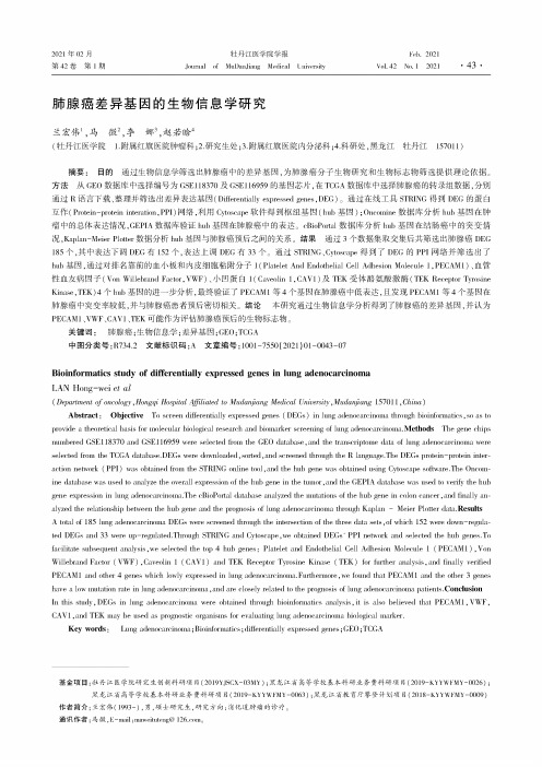
肺腺癌差异基因的生物信息学研究兰宏伟-马微2,李娜3,赵若啥4(牡丹江医学院1•附属红旗医院肿瘤科;2•研究生处;3•附属红旗医院内分泌科;4.科研处,黑龙江牡丹江157011)摘要:目的通过生物信息学筛选出肺腺癌中的差异基因,为肺腺癌分子生物研究和生物标志物筛选提供理论依据。
方法从GEO数据库中选择编号为GSE118370及GSE116959的基因芯片,在TCGA数据库中选择肺腺癌的转录组数据,分别通过R语言下载、整理并筛选出差异表达基因(Differentially expressed genes,DEG)。
通过在线工具STRING得到DEG的蛋白互作(Protein-protein interation,PPI)网络,利用Cytoscape软件得到枢纽基因(hub基因);Oncomine数据库分析hub基因在肿瘤中的总体表达情况,GEPIA数据库验证hub基因在肺腺癌中的表达。
cBioPortal数据库分析hub基因在结肠癌中的突变情况,Kaplan-Meier Plotter数据分析hub基因与肺腺癌预后之间的关系。
结果通过3个数据集取交集后共筛选出肺腺癌DEG 185个,其中表达下调DEG有152个,表达上调DEG有33个。
通过STRING、Cytoscape得到了DEG的PPI网络并筛选出了hub基因,通过对排名靠前的血小板和内皮细胞粘附分子l(Platelet And Endothelial Cell Adhesion Molecule1,PECAM1)、血管性血友病因子(Von Willebrand Factor,VWF)、小凹蛋白1(Caveolin1,CAV1)及TEK受体酪氨酸激酶(TEK Receptor Tyrosine Kinase,TEK)4个hub基因的进一步分析,最终验证了PECAM1等4个基因在肺腺癌中低表达,且发现PECAM1等4个基因在肺腺癌中突变率较低,并与肺腺癌患者预后密切相关。
诊断肌少症的肠道菌群及其应用[发明专利]
![诊断肌少症的肠道菌群及其应用[发明专利]](https://img.taocdn.com/s3/m/6a9d472d640e52ea551810a6f524ccbff021ca40.png)
(19)中华人民共和国国家知识产权局(12)发明专利申请(10)申请公布号 (43)申请公布日 (21)申请号 202010319012.6(22)申请日 2020.04.22(71)申请人 中国医学科学院北京协和医院地址 100730 北京市东城区王府井帅府园1号北京协和医院老年医学科(72)发明人 康琳 (74)专利代理机构 北京预立生科知识产权代理有限公司 11736代理人 高倩倩 孟祥斌(51)Int.Cl.C12Q 1/6883(2018.01)C12Q 1/689(2018.01)A61K 45/00(2006.01)A61P 21/00(2006.01)(54)发明名称诊断肌少症的肠道菌群及其应用(57)摘要本发明公开了诊断肌少症的肠道菌群及其应用,所述肠道菌群为Eisenbergiella。
本发明通过16S 核糖体核糖核酸测序首次发现了Eisenbergiella在肌少症中呈现显著性上调,进一步发现了使用Eisenbergiella作为检测变量区分肌少症和健康群体具有较高的特异性和敏感性,基于此,可以将Eisenbergiella应用于肌少症的无创诊断。
权利要求书1页 说明书8页 附图2页CN 111411150 A 2020.07.14C N 111411150A1.用于检测肠道菌群丰度的一种或多种试剂在制备用于预测肌少症的产品中的用途,其特征在于,所述肠道菌群至少包括肠道菌属Eisenbergiella。
2.根据权利要求1所述的用途,其特征在于,所述试剂包括与来自Eisenbergiella的靶核苷酸序列能够特异性杂交的一种或多种寡核苷酸。
3.根据权利要求1所述的用途,其特征在于,所述产品还包括从样本中分离核酸的试剂。
4.根据权利要求2所述的用途,其特征在于,所述寡核苷酸被可检测地标记。
5.根据权利要求2所述的用途,其特征在于,Eisenbergiella的靶核苷酸序列是16S核糖体核糖核酸基因的片段。
RNA_荧光适配体在生物传感与成像中的应用研究进展

480分析化学第52卷[81]QIAN S,CHEN Y,PENG C,WANG X,CHE Y,WANG T,WU J,XU J.Anal.Chim.Acta,2023,1239:340670.[82]XING G,SHANG Y,WANG X,LIN H,CHEN S,PU Q,LIN L.Biosens.Bioelectron.,2023,220:114885.[83]ZHUANG J,ZHAO Z,LIAN K,YIN L,WANG J,MAN S,LIU G,MA L.Biosens.Bioelectron.,2022,207:114167.[84]ZHOU J,CHEN P,WANG H,LIU H,LI Y,ZHANG Y,WU Y,PAEK C,SUN Z,LEI J,YIN L.Mol.Ther.,2022,30(1):244-255.Advances of CRISPR/Cas-based Biosensor in Detection ofFood-Borne PathogensZHANG Xiao-Yuan1,2,YAO Zhi-Hao2,HE Kai-Yu2,WANG Hong-Mei2,XU Xia-Hong2,WU Zu-Fang*1,WANG Liu*21(College of Food and Pharmacy,Ningbo University,Ningbo315000,China) 2(Institute of Agro-product Safety and Nutrition,Zhejiang Academy of Agricultural Sciences,Hangzhou310021,China)Abstract Rapid and accurate detection methods for food-borne pathogens are essential to ensure food safety and human health.One promising innovation in this area is the clustered regularly interspaced short palindromic repeats/CRISPR-associated systems(CRISPR/Cas)biosensor,which utilizes Cas protein and CRISPR RNA (crRNA)ribonucleo protein to specifically recognize target genes,and converts target signals into detectable physical and chemical signals.The CRISPR/Cas biosensor shows many advantages,such as high specificity, programmability,and ease of use,making it promising to pathogen detection.This paper introduced the principles and characteristics of CRISPR/Cas systems,along with the strategies for signal recognition,amplification,and output based on different CRISPR/Cas biosensors,and their respective applications in food-borne pathogen detection.Furthermore,the construction principles and challenges of multiple biosensors based on CRISPR/Cas were explored,as well as their potential for simultaneous detection of multiple pathogens.Finally,the challenges and future development trends of CRISPR/Cas-based biosensors in rapid pathogen detection were discussed, aiming to provide valuable reference and inspiration for biosensor designers and food safety practitioners. Keywords Food-borne pathogens;CRISPR/Cas;Biosensor;Review(Received2023-09-15;accepted2024-02-01) Supported by the National Natural Science Foundation of China(No.32172307)and the Basic Public Welfare Research Program of Zhejiang Province,China(No.LGN21C200007).第52卷分析化学(FENXI HUAXUE)评述与进展第4期2024年4月Chinese Journal of Analytical Chemistry481~491DOI:10.19756/j.issn.0253-3820.221448 RNA荧光适配体在生物传感与成像中的应用研究进展邱星晨#1,3范存霞#1,2白瑞1谷雨*1李长明*1郭春显*1(苏州科技大学材料科学与工程学院1,物理科学与技术学院2,环境科学与工程学院3,苏州215009)摘要RNA荧光适配体是近年来发展起来的一种简单且有效的RNA分子荧光标记工具,可与不发光或发光微弱的小分子荧光团结合,显著增强其发光性能。
抑郁症的动物模型课件

基础(jīchǔ)01级
陈瑶瑶
第一页,共二十页。
I am depressed. Me too.
第二页,共二十页。
Why we need animal models? The sorts of animal models.
The future development of animal
第七页,共二十页。
第八页,共二十页。
第九页,共二十页。
第十页,共二十页。
第十一页,共二十页。
5-HT1A受体敲除
抗抑郁表型
糖皮质激素受体基因损伤
5-HT1B受体敲除
DA-β-羟化酶基因敲除
α2A-肾上腺素能受体基 因敲除 α2C-肾上腺素能受体基 因敲除 α2C-肾上腺素能受体基 因过表达 NE转运子基因敲除
14.Willner P, Muscat R, Papp M. Chronic mild stress-induced anhedonia: a realistic animal model of depression. Neurosci
Biobehav Rev 1992;16:525–34.
15.许晶 , 李晓秋,慢性应激抑郁模型(móxíng)的建立及其评价, Chinese Journal of Behavioral Medical Science 12(1) ,2003
11.Barbara Vollmayr*, Fritz A. Henn,Stress models of depression,Clinical Neuroscience Research 3 (2003) 245–251 12.Katz RJ, Schmaltz K. Dopaminergic involvement in attention. A novel animal model. Prog Neuropsychopharmacol
一种细胞间通信的新的信号分子——Microvesicle运输的microRNA

一种细胞间通信的新的信号分子——Microvesicle运输的microRNA梁宏伟;陈熹;曾科;张辰宇【摘要】Microvesicle(MV)是机体内的细胞在正常和病理状态下都会分泌的直径在30~1000 nm之间的微小囊泡.细胞把特异的生物活性分子如蛋白质、mRNA 等包裹到MV中,这些生物活性分子会通过MV被运输到相应的受体细胞以调节受体细胞的生物功能.这种由MV介导的细胞间信息传递在一些生理和病理过程中扮演着十分重要的作用.近期研究表明MV中包含microRNA(miRNA),并且通过MV 的运输,这些miRNA会被运送入靶细胞以调控靶细胞相关基因的表达.本文就MV 运输的miRNA在细胞间通信的功能做一总结.【期刊名称】《分子诊断与治疗杂志》【年(卷),期】2010(002)006【总页数】7页(P417-423)【关键词】Microvesicle;MicroRNA;细胞间通信;外泌体【作者】梁宏伟;陈熹;曾科;张辰宇【作者单位】南京大学医药生物技术国家重点实验室,南京,江苏210093;南京大学医药生物技术国家重点实验室,南京,江苏210093;南京大学医药生物技术国家重点实验室,南京,江苏210093;南京大学医药生物技术国家重点实验室,南京,江苏210093【正文语种】中文【中图分类】R3Microvesicle(MV)是机体内细胞在正常和病理状态下都会分泌的直径在30~1000 nm之间的微小囊泡[1,2]。
多数研究者用MV来指代具有细胞间信号传递功能的囊胞[3,4]。
体内和体外的实验都证明MV可以由网织红细胞[5]、B细胞[6]、T细胞[7]、树突细胞[8]、肥大细胞[9]、上皮细胞[10,11]和肿瘤细胞[12,13]等多种细胞分泌。
细胞把特异的生物活性分子如蛋白质、mRNA等包裹到MV中,被运输到相应的受体细胞并调节受体细胞的生物功能。
这种由MV介导的细胞间信息传递在一些生理和病理过程中扮演着十分重要的作用[14~17]。
- 1、下载文档前请自行甄别文档内容的完整性,平台不提供额外的编辑、内容补充、找答案等附加服务。
- 2、"仅部分预览"的文档,不可在线预览部分如存在完整性等问题,可反馈申请退款(可完整预览的文档不适用该条件!)。
- 3、如文档侵犯您的权益,请联系客服反馈,我们会尽快为您处理(人工客服工作时间:9:00-18:30)。
INTERNATIONAL JOURNAL OF ONCOLOGY 42: 1875-1882, 2013Abstract. MicroRNAs (miRNAs) are a small class of non-coding RNAs that negatively regulate gene expression, and are considered as new therapeutic targets for treating cancer. In this study, we performed a gain-of-function screen using miRNA mimic library (319 miRNA species) to identify those affecting cell proliferation in human epithelial ovarian cancer cells (A2780). We discovered a number of miRNAs that increased or decreased the cell viability of A2780 cells. Pro-proliferative and anti-proliferative miRNAs include oncogenic miR-372 and miR-373, and tumor suppressive miR-124a, miR-7, miR-192 and miR-193a, respectively. We found that overexpression of miR-124a, miR-192, miR-193a and miR-193b inhibited BrdU incorporation in A2780 cells, indicating that these miRNAs affected the cell cycle. Overexpression of miR-193a and miR-193b induced an acti-vation of caspase 3/7, and resulted in apoptotic cell death in A2780 cells. A genome-wide gene expression analysis with miR-193a-transfected A2780 cells led to identification of ARHGAP19, CCND1, ERBB4, KRAS and MCL1 as potential miR-193a targets. We demonstrated that miR-193a decreased the amount of MCL1 protein by binding 3'UTR of its mRNA. Our study suggests the potential of miRNA screens to discover miRNAs as therapeutic tools to treat ovarian cancer. IntroductionMicroRNAs (miRNAs) are small non-coding RNAs of 20-22 nucleotides, and function to suppress the expression of target mRNAs by translation blockade and/or mRNA degrada-tion (1,2). They are involved in many biological processes including cell proliferation, differentiation and apoptosis, and their dysregulation can contribute to the pathological state including cancer (1,3,4). Several groups have documented miRNA expression profiling in ovarian cancers using miRNA microarray and massive parallel sequencing technology (5-10). miR-93, miR-141, miR-200 and miR-214 are frequently upregulated whereas miR-100, miR-143, miR-145 and let-7 are downregulated in ovarian carcinomas compared with normal counterparts (5-9). Abnormal miRNA expression is due to DNA copy number amplification and deletion, epigenetic modification and/or the dysregualtion of miRNA processing in cancer state (7,11,12). miR-214 upregulated in ovarian cancer can target PTEN tumor suppressor gene whereas down-regulated let-7 can target the RAS oncogene (8,13), suggesting that miRNAs may have a role as novel class of oncogenes or tumor suppressor genes in ovarian cancer (14).Based on these findings, the clinical potential of miRNAs as cancer biomarkers and/or therapeutic agents is widely recognized and accepted (15). A single miRNA can regulate multiple mRNA transcripts that cooperatively work in cellular differentiation and function (16-19). The use of miRNA mimics or anti-miRNAs may represent powerful therapeutic tools to accomplish regression and/or re-differentiation of cancer by effectively targeting tumor suppressive or oncogenic genes with less toxicity (15,20). Indeed, a number of pre-clinical trials of miRNAs are currently in progress (21). In this study, we performed a gain of function screen using miRNA mimics library containing 319 miRNAs to identify miRNAs that can affect cell proliferation in A2780 ovary cancer cells. We found several anti-proliferative miRNAs including miR-124, miR-192 and miR-193 in A2780, suggesting that the potential of miRNA screens for discovering miRNAs as therapeutic tools to treat ovarian cancer.Materials and methodsCell culture. Human ovarian cancer cell line A2780 was obtained from Dr T. Tsuruo (22), and human colorectal cancer cell line DLD-1 was obtained from the American Type Culture Collection (ATCC, Manassas, V A, USA). A2780 and DLD-1 were cultured in RPMI-1640 (Gibco, Life Technologies, Carlsbad, CA, USA) containing 50 IU/ml penicillin and 50 µg/ml streptomycin (Gibco, Life Technologies), supplemented with 5% (A2780) or 10% (DLD-1) fetal bovine serum (FBS, JRH Biosciences, Lenexa, KS, USA) at 37˚C in an atmosphere of 5% CO2.Gain-of-function microRNA screens identify miR-193a regulating proliferation and apoptosis in epithelial ovarian cancer cells HARUO NAKANO1,2, YOJI YAMADA1, TATSUYA MIYAZAWA1 and TETSUO YOSHIDA11Biologics Research Laboratories, Kyowa Hakko Kirin Co., Ltd., Machida-shi, Tokyo 194-8533, JapanReceived January 31, 2013; Accepted March 19, 2013DOI: 10.3892/ijo.2013.1896Correspondence to: Dr Tetsuo Yoshida, Biologics ResearchLaboratories, Kyowa Hakko Kirin Co., Ltd., 3-6-6, Asahi-machi,Machida-shi, Tokyo 194-8533, JapanE-mail: tetsuo.yoshida@kyowa-kirin.co.jpPresent address:2Laboratory of Environmental MolecularPhysiology, School of Life Sciences, Tokyo University of Pharmacyand Life Sciences, 1432-1, Horinouchi, Hachioji, Tokyo 192-0392,JapanKey words: microRNA, cell proliferation, apoptosis, miR-193,ovarian cancer cellNAKANO et al: MicroRNA MIMICS LIBRARY SCREENING ON CELL PROLIFERATION IN OVARIAN CANCER CELLS 1876miRNA library screening. A gain of function miRNA screen on cell viability was performed using A2780 as previously described (23). A2780 was seeded at 2,500 cells per well in 96-well plates the day before transfection. Synthetic miRNA mimic library (human Pre-miR™ miRNA precursor library-ver.1, Ambion, Applied Biosystems, Foster City, CA, USA) was screened using 50 nM in a duplicate. The library contained 319 miRNAs registered in miRBase ver. 7.1 (http://www.mirbase. org/). miRNA mimics were transfected using Lipofectamine 2000 (Life Technologies) according to the manufacturer's proto-cols. Pre-miR miRNA precursor molecule-negative control (13) (Ambion, Applied Biosystems) was used as a negative control for miRNA mimics. We confirmed transfection efficiency (>90%) using siControl TOX transfection control (50 nM, Dharmacon, Lafayette, CO, USA). After 3 days of transfection, the cell viability was measured using the Cell Titer-Glo Luminescent Cell Viability Assay (Promega, Madison, WI, USA) according to the manufacturer's instructions. Data were expressed as percentage of the negative control. Several miRNA hits were selected to assess reproducibility and dose-dependency (5, 25 and 50 nM).BrdU incorporation assay. miRNA (25 nM)-transfected cells in 96-well format were harvested for one day, and then were incubated with 10 µM of 5'-bromo-2-deoxy-uridine (BrdU) for 2-4 h. The cells were fixed with cold ethanol/HCl, and the incorporated BrdU was detected using BrdU labeling and detec-tion kit III (Roche Diagnostics GmbH, Mannheim, Germany) according to the manufacturer's instruction.Caspase 3/7 activation assay. miRNA (25 nM)-transfected cells in 96-well format were harvested for 2 days, and then used to measure caspase 3/7 activity using Caspase-Glo 3/7 assay (Promega) according to the manufacturer's instruction.RNA isolation and whole genome microarray. A2780 cells were transfected with miR-193a (Pre-miR miRNA precursor molecules, hsa-miR-193a-3p, Ambion, Applied Biosystems) or negative control miRNA (25 nM), and allowed to grow in the medium (RPMI-1640) for 10 h before RNA isolation. Total RNA was isolated using the RNeasy mini RNA isolation kit (Qiagen). The integrity of the RNA was verified using an Agilent 2100 Bioanalyzer (1.8-2.0: Agilent Technologies, Palo Alto, CA, USA). Transcriptome microarray analysis was carried out using the 44K Whole Human Genome Microarray chip (Agilent Technologies) according to the manufacturer's instructions. Scanning micro-array chips and processing data were done by Pharmafrontier Co., Ltd, Kyoto, Japan. Differentially expressed probe sets were identified with a fold change >1.5. Gene ontology (GO) pathway enrichment analysis was performed among genes differentially expressed after miR-193a transfection by SigTerm software (24). The downregulated genes with miR-193a transfection were compared with predicted miR-193a target genes searched by TargetScan (/). Over-representation of predicted miR-193a target genes within downregulated gene sets was assessed by SigTerm software.Western blot analysis. miRNA or siRNA (25 nM)-transfected A2780 cells were lysed in radio immunoprecipitation assay (RIPA) buffer [50 mM Tris-HCl (pH 8.0), 150 mM sodium chloride, 1% NP-40, 0.5% sodium deoxycholate, 0.1% sodium dodecyl sulfate] supplemented with 1% of a protease inhibitor cocktail stock solution (set III, Roche Diagnostics GmbH) after 1 or 2 days transfection. The following pre-designed siRNA was used as a positive control: MCL1 siRNA (Hs_MCL1_6 HP vali-dated siRNA, SI02781205, Qiagen GmbH, Hilden, Germany). Proteins (10 or 20 µg) were separated by SDS-PAGE. Upon electroblotting to polyvinylidene fluoride (PVDF) membrane (Immobilon-P, Millipore, Billerica, MA, USA), non-specific binding sites were blocked by incubation in TBST (Tris-buffered saline/0.05% Tween-20) containing 1% skim milk, and then incubated with rabbit polyclonal anti-MCL1 (1:200, S-19, Santa Cruz Biotechnology Inc., Santa Cruz, CA, USA), or mouse monoclonal anti-α-tubulin (1:2,500, clone DM1A, Sigma, St. Louis, MO, USA) in blocking solution. After washing with TBST, the membrane was incubated with HRP-conjugated rabbit anti-mouse IgG secondary antibody (P0161, Dako, Glostrup, Denmark) or HRP-conjugated swine anti-rabbit IgG secondary antibody (P0217, Dako). Signals were detected using enhanced chemiluminescence (ECL) or ECL-plus reagent (Amersham™ GE Healthcare UK Ltd., Buckinghamshire, UK).qRT-PCR. Total RNA was prepared from miRNA or siRNA (25 nM)-transfected cells 2 days after transfection using RNeasy mini kit (Qiagen), and then first strand cDNA was synthesized using SuperScript III (Life Technologies) according to the manufacturer's instruction. Real-time RT-PCR was performed using 7900 HT fast real-time PCR system (Applied Biosystems Inc., Foster City, CA, USA) with SYBR-Green as a reporter. The following primers were used for detection: MCL1 (222 bp) forward: TCTAAGTGCTGAC TGGCTACG, reverse: CCTGGCACAGCTATCAAAAG; GAPDH (137 bp) forward: ACTTTGTCAAGCTCATTTCCTG, reverse: CTCTCTTCCTCTTG GCTCTTG.Luciferase miRNA target reporter assay. 3'-untranslated regions (UTRs) of MCL1 gene (1546 bp), containing predicted binding sites of miR-193a, were amplified by PCR from A2780 cDNA, and inserted into the pGL3 control vector (Promega) by using Xba-I site immediately downstream from the stop codon of Firefly luciferase. The following primers were used: MCL1 3'-UTR forward: CGGCTAGCGAAAAGCAAGTGG CAAGAGG, reverse: CGGCTAGCAGGGAGGGTCACTCA GGTTT.Deletion of the first 3 nucleotides corresponding miR-193a seed-region complementary site was inserted in mutant constructs using KOD-plus-Mutagenesis kit (Toyobo, Osaka, Japan), according to the manufacturer's protocol. The following primers were used for generation of mutant constructs: MCL1-mutant-Primer 1: AGCCAGGCAAGTCATAGAATT GATT, MCL1-mutant-Primer 2: GGCCACTTTCCTGTTCT CAACAAGG.DLD-1 cells were cultured in 96-well formats and co-trans-fected with 100 ng of pGL3 Firefly luciferase reporter vector, 20 ng of pRL-TK Renilla luciferase control vector (Promega) and 25 nM miRNA or negative control miRNA using Lipofectamine 2000. Firefly and Renilla luciferase activities were measured consecutively using the Dual-Luciferase Reporter Assay System (Promega) 24 h after transfection. All the experiments were done in triplicate and repeated at least twice on different days.INTERNATIONAL JOURNAL OF ONCOLOGY 42: 1875-1882, 20131877ResultsEffects of miRNA mimic library transfection on cell proli-feration of A2780 cell line. To identify miRNAs that affect cell proliferation of ovarian cancer cells, we performed a gain of function screen using synthetic miRNA mimic library (319 miRNAs) for human epithelial ovary cancer cells (A2780). The library consists of miRNAs registered in early version of miRBase (ver. 7.1 in October, 2005, /), and many of them were expressed in ovarian normal and cancer tissues and cell lines (5). We detected cellular ATP to assess cell viability in miRNA (50 nM)-transfected cells 3 days after trans-fection. Frequency distribution indicated that broad ranges of miRNA mimic transfections affected the cell viability of A2780 (Fig. 1A). A total of 46 out of 319 miRNAs induced more than 50% changes in the cell viability of A2780 after 3 days transfec-tion. Table I shows top 10 miRNAs that increased or decreased the cell viability of A2780. They included known oncogenic miRNAs such as miR-372 (cell viability, 187%) and miR-373 (165%), and tumor suppressive miRNAs such as miR-124a (28.3%), miR-7 (37.1%), miR-192 (36.6%) and miR-193a (29.7%) in several different cancer types (18,25-27). The seed family miRNAs that have the same sequences in seed region (2nd to 8thnucleotide) of miRNAs showed similar effects on cell viabilityFigure 1. Gain of function screen of miRNA library in A2780 ovary cancer cell line. (A) A2780 cells were transfected with 319 miRNA mimics (50 nM) in duplicate, and their cell viability was measured 3 days after transfection. The graph shows the distribution of cell viability normalized by the miRNA mimic negative control. (B) Top 10 anti-proliferative miRNA mimics derived from 1st screen were transfected into A2780 at the final concentration of 5 nM (solid bar), 25 nM (open bar) and 50 nM (hatched bar). The cell viability was measured three days after miRNA mimic transfection. The data are normalized with the negative control and show average ± SD from three different experiments.Table I. Results of miRNA library screening. miRNAs that increased miRNAs that decreasedcell viabilitycell viability-----------------------------------------------------------------------------------------------------------------------miRNA Cell viability miRNA Cell viability (%) (%)miR-301 218 miR-124a 28.3miR-372 187 miR-517c 29.4miR-93 185 miR-193a 29.7miR-302b 181 miR-506 31.9miR-130a 173 miR-199a * 34.5miR-302d 172 miR-192 36.6miR-363 166 miR-7 37.1miR-373 165 miR-193b 37.7miR-9* 162 miR-432* 37.8miR-130b 162 miR-497 38.3Data represent the cell viability in miRNA mimics (50 nM)-transfected cells 3 days after transfection. Data were expressed as a percentage of the negative control in an average of duplicates. Top 10 miRNAs that increased or decreased the cell viability are listed.NAKANO et al : MicroRNA MIMICS LIBRARY SCREENING ON CELL PROLIFERATION IN OVARIAN CANCER CELLS1878in A2780 cells. For example, miR-93/miR-302/miR-372/mir-373 seed family miRNAs (miR-93, miR-302b, miR-302d, miR-372, miR-373) were pro-proliferative, while miR-193 seed family miRNAs (miR-193a, miR-193b) were anti-proliferative (Table I). miR-200/miR-141 seed family miRNAs that are upregulated in ovarian cancer (5,6,10) had a little effect on the cell viability in A2780 cells (the cell viability; 97.9, 113, 92.0 and 101% with miR-200a, miR-200b, miR-200c and miR-141 transfection, respectively). miR-100, miR-143 and miR-145 that are down-regulated miRNAs in ovarian cancer (5,6,8) induced a 15-30% decrease in the cell viability of A2780 (the cell viability; 84.1, 81.8 and 73.1 with miR-100, miR-143 and miR-145 transfec-tion, respectively). We are interested in miRNA mimics that decreased the cell viability of A2780 since these miRNA mimics themselves could have therapeutic potential to treat ovarian cancer. To further evaluate miRNA mimics on the inhibition of cell proliferation in A2780, we selected top 10 anti-proliferative miRNAs (miR-7, miR-124a, miR-192, miR-193a, miR-193b, miR-199a *, miR-432*, miR-497, miR-506 and miR-517c) from the first screen, and examined the cell viability in A2780 cells transfected with different concentrations of miRNAs (5, 25, 50 nM). We confirmed results of our first screening at 50 nM, and found that the transfection of miR-124a, miR-192, miR-193a and miR-193b induced a large decrease in the cell viability of A2780 even at 5 nM (Fig. 1B), indicating that these miRNAs had a profound anti-proliferative effect in A2780 cells. We examined whether miR-124a, miR-192, miR-193a and miR-193b affected DNA synthesis to inhibit cell proliferation in A2780 cells.One day after miRNA transfection, BrdU incorporation was examined to evaluate DNA synthesis in transfected cells. As shown in Fig. 2A, miR-124a, miR-192, miR-193a and miR-193b decreased an incorporation of BrdU compared with the negative control, indicating that these miRNAs induced the inhibition of DNA synthesis in A2780 cells. We next examined whether these miRNAs affected apoptotic pathway to inhibit cell proliferation in A2780 cells. We found that miR-193a and miR-193b but not miR-124a and miR-192 induced more than twofold increase in an activity of caspase 3/7, the effector of apoptotic pathway, in A2780 cells (Fig. 2B). The result indicated that miR-193a and miR-193b could induce the apoptotic cell death in A2780 cells. Actually, apoptotic cell debris was frequently observed in miR-193a-transfected A2780 cells (Fig. 2C, arrows).Transcriptome analysis to assess target genes regulated by miR-193a. We further characterized the anti-proliferative effect of miR-193a in A2780 cells. To examine target genes regulated by miR-193a, we performed genome wide gene expression anal-ysis using miR-193a (25 nM)-transfected cells compared with the negative control miRNA-transfected ones. We identified 518 genes that were downregulated more than 1.5-fold by miR-193a transfection after 10 h. To evaluate the potential functional significance of the genes downregulated after miR-193a trans -fection, we subjected the gene expression data to gene ontology (GO) pathway enrichment analysis. The 20 most significantly over-represented pathways listed in Table II include smallGTPase signaling and vesicular transport. We compared theseFigure 2. Effects of anti-proliferative miRNAs on DNA synthesis and caspase 3/7 activity. (A) Anti-proliferative miRNAs (miR-124a, miR-192, miR-193a and miR-193b) or negative control miRNA were transfected into A2780 at the final concentration of 25 nM. One day after transfection, the transfected cells were incubated with BrdU containing medium for 2 to 4 h, and then BrdU incorporation was measured. The data is normalized with the negative control and show average ± SD from three different experiments. (B) Anti-proliferative miRNAs (miR-124a, miR-192, miR-193a and miR-193b) or negative control miRNA were transfected into A2780 at the final concentration of 25 nM. Two days after transfection, caspase 3/7 activity in miRNA-transfected cells was measured. The data are normalized with the negative control and show average ± SD from three different experiments. (C) Cellular morphology of miR-193a (25 nM)-transfected A2780 cells. Arrows indicate apoptotic cell debris. Scale bar indicates 100 µm.INTERNATIONAL JOURNAL OF ONCOLOGY 42: 1875-1882, 20131879downregulated 518 genes with predicted miR-193a target genes (142 genes) obtained by TargetScan (Fig. 3). This resulted in the match of 34 candidate miR-193a target genes, and they were significantly over-represented in the downregulated gene sets by using the SigTerm software (24). Table III showed 34 candidate miR-193a target genes obtained by our transcriptome analysis.The candidate genes include ARHGAP19 (RhoGAP19), CCND1 (cyclin D1), ERBB4, KRAS and MCL1 that function in cell signaling, cell cycle and apoptotic pathway.MCL1 is a direct target gene of miR-193a in A2780 cells. From our results of transcriptome analysis, we focused on MCL1 gene as miR-193a targets, since MCL1 was an anti-apoptotic gene of BCL2 family (28), and therefore might contribute to miR-193a-induced cell death in A2780 cells. MCL1 3'UTR contains one potential target site of miR-193a and the site is conserved between human and mouse. To examine the regula-tion of miR-193a on MCL1 protein expression, we performed western blot analysis with miR-193a-transfected A2780 cells. Transfection of positive control MCL1 siRNA induced the decrease in endogenous MCL1 proteins in A2780 cellsTable II. Twenty most significantly enriched (P<0.05) gene ontology (GO) pathways among downregulated genes after miR-193a transfection into A2780 cells.Term P-valueSmall GTPase regulator activity 0.0022Ras guanyl-nucleotide exchange factor activity 0.0030Rho guanyl-nucleotide exchange factor activity 0.0052Regulation of Rho protein siganal transduction 0.0060Blood vessel development 0.0069Cytoplasmic vesicle part 0.0072Vasculature development 0.0080Post-Golgi vesicle-mediated transport 0.0080Protein localization 0.0149Phospholipid transporter activity 0.0155Regulation of small GTPase mediated 0.0156signal transductionGuanyl-nucleotide exchange factor activity 0.0168GTPase regulator activity 0.0181Macromolecule localization 0.0187Insulin receptor signaling pathway 0.0193Guanylate kinase activity 0.0196Early endosome 0.0213Neuron projection 0.0217Intracellular signaling cascade 0.0225One-carbon compound metabolic process0.0229Figure 3. Transcriptome analysis with miR-193a-transfected A2780. Venn dia-gram to illustrate the relationship between the downregulated genes (10 h after miR-193a transfection) and predicted target genes by TargetScan. Predicted miR-193a target genes within the downregulated gene sets were significantly enriched by SigTerm software (P=1.54E-17).Table III. Candidate miR-193a target genes downregulated in miR-193a-transfectants.Entrez gene ID Symbol Fold change23119 HIC2 -6.2010152 ABI2 -3.34595 CCND1 -3.3054756 IL17RD -3.2010238 WDR68 -3.153925 STMN1 -2.975324 PLAG1 -2.973845 KRAS -2.764076 CAPRIN1 -2.662066 ERBB4 -2.5857704 GBA2 -2.4584986 ARHGAP19 -2.2310620 ARID3B -2.177342 UBP1 -2.0927242 TNFRSF21 -2.074170 MCL1 -2.0056262 LRRC8A -1.9310160 FARP1 -1.9157472 CNOT6 -1.9123179 RGL1 -1.7923341 DNAJC16 -1.7988455 ANKRD13A -1.704215 MAP3K3 -1.67114991 ZNF618 -1.6623492 CBX7 -1.6423365 ARHGEF12 -1.6422883 CLSTN1 -1.619939 RBM8A -1.6054890 ALKBH5 -1.59115 ADCY9 -1.574189 DNAJB9 -1.511173 AP2M1 -1.519962 SLC23A2 -1.5123384 SPECC1L -1.50NAKANO et al : MicroRNA MIMICS LIBRARY SCREENING ON CELL PROLIFERATION IN OVARIAN CANCER CELLS1880(Fig. 4A). We demonstrated that overexpression of miR-193a decreased MCL1 proteins in A2780 cells (Fig. 4A). We next performed qRT-PCR with miR-193a-transfected cells to examine whether miR-193a affected MCL1 mRNA expres-sion. We found that miR-193a induced about 50% decrease in MCL1 mRNA expression in A2780 cells (Fig. 4B). These results indicated that miR-193a affected MCL1 expression at both protein and mRNA levels. To validate whether miR-193a can directly regulate the translation of MCL1 mRNAs, we constructed a luciferase reporter plasmid that inserted MCL1 3'UTR (around 1.5 kb) at the downstream of Firefly luciferase gene, and tested the luciferase activity. As shown in Fig. 4C, co-transfection of miR-193a and MCL1 3’UTR reporter vector induced around 40% reduction of the luciferase activity compared with co-transfection of the negative control miRNA and the reporter vector. The decrease of the luciferase activity was attenuated by using the mutant reporter vector deleting miR-193a seed region complementary sites in MCL13'UTR (Fig. 4C, MCL1-3'UTR-MU). These results indicated that MCL1 would be a direct target of miR-193a. We further examined whether the downregulation of endogenous MCL1 could induce apoptosis in A2780 cells. As shown in Fig. 4D, the transfection of MCL1 siRNA (25 nM) induced caspase 3/7 activation comparable with miR-193a transfection in A2780 cells (Fig. 4D), indicating that the downregulation of MCL1 by miR-193a could contribute to miR-193a-induced apoptosis in A2780 cells.DiscussionSeveral studies reveal that global miRNA expression is dysreg-ulated in ovarian cancer (5-10), and miRNAs may represent new targets for detection, diagnosis and therapy in ovarian cancer (14). However, functions of many miRNAs in ovarian cancer remain to be elucidated. In this study, we performeda gain-of-function screen using a miRNA mimic libraryFigure 4. miR-193a targets the anti-apoptotic factor MCL1. (A) Immunoblot analysis for detection of MCL1 with anti-MCL1 antibody. Cell lysate with miR-193a or MCL1 siRNA (25 nM)-transfected cells was collected two days after transfection. Total protein (10 µg) was loaded for analysis. The immunodetection of α-tubulin was used as loading control. The representative blots of three different experiments are shown. (B) Relative quantification of MCL1 expression by qRT-PCR in miR-193a or MCL1 siRNA (25 nM)-transfected cells two days after transfection. The data are normalized by GAPDH expression, and expressed as relative values against the negative control. Average ± SD from three different experiments are shown. (C) Reporter assay showing decreased luciferase activity in DLD-1 cells co-transfected with pGL3-MCL1-3’UTR (pGL3_MCL1_WT) and miR-193a (25 nM, open bar). Deletion of miR-193a seed region complementary site from the 3'UTR (pGL3_MCL1_MU) attenuated miR-193a-induced decrease of luciferase activity. Firefly /Renilla luciferase activity is expressed as relative values against the negative control. Average ± SD (n=3) from one representative experiment are shown. (D) Caspase 3/7 activity of miR-193a or MCL-1 siRNA (25 nM)-transfected cells was measured 2 days after transfection. The data show average ± SD from three different experiments.INTERNATIONAL JOURNAL OF ONCOLOGY 42: 1875-1882, 20131881(319 miRNA species) to identify those affecting cell prolif-eration in epithelial ovarian cancer cells (A2780). The library consists of miRNAs registered in early version of miRBase (ver. 7.1 in October, 2005, /), and many of them were expressed in ovarian normal and cancer tissues and cell lines (5). We discovered pro-proliferative miRNAs (miR-9*, miR-93, miR-130a, miR-130b, miR-301, miR-302b, miR-302d, miR-363, miR-372, miR-373), and anti-proliferative miRNAs (miR-7, miR-124a, miR-192, miR-193a, miR-193b, miR-199a*, miR-432*, miR-497, miR-506, miR-517c) in A2780 cells. By the same miRNA mimics library screening, we found that miR-93/miR-372/miR-373 and miR-124a were pro-prolif-erative and anti-proliferative, respectively, in DLD-1 colorectal cancer cells (23), suggesting consistent roles of these miRNAs on cell proliferation in ovary and colorectal cancer cells. The base-pairing between target mRNAs and the seed region (2nd to 8th nucleotides) of miRNA is important for miRNAs to func-tion to regulate their target genes (2). The seed family miRNAs induced similar cellular phenotypes on cell proliferation in this study (ex. pro-proliferative miR-93, miR-302b, miR-302d, miR-372, miR-373 and anti-proliferative miR-193a, miR-193b), supporting the importance of the seed region of miRNA on its function. Our miRNA hits did not always correspond to dysregulated miRNAs reported in ovarian cancer (5-10), but included pro-proliferative miR-93 that was upregulated in primary ovarian carcinomas (6), supporting an oncogenic role of this miRNA in ovarian cancer. Our miRNA hits also included tumor suppressive miR-7, miR-124a, miR-192 and miR-193a in several cancer types (18,25-27), suggesting that these miRNAs could be tumor suppressive in ovarian cancer. Among our miRNA hits, we further characterized miR-124a, miR-192, miR-193a and miR-193b that induced a large decrease in the cell viability of A2780 cells. miR-124a and miR-192 induced a decrease in BrdU incorporation, indicating that these miRNAs affected cell cycle resulting in inhibition of DNA synthesis in A2780 cells. Inhibitory effects of miR-124a and miR-192 on cell cycle gene pathway are reported in several cancer cell lines. miR-124a targets cyclin dependent kinase 6 (CDK6), and thereby inhibits the phosphorylation of retinoblastoma (Rb) in HCT116 cells (29). miR-192 is upregulated by genotoxic stress in HCT116, A549 and U2OS cell lines bearing wild-type p53, and induces the cell cycle arrest by enhancing CDKN1A/p21 expression (18,30).We showed that miR-193a and miR-193b inhibited BrdU incorporation and induced caspase 3/7 activation in A2780 cells, indicating that these miRNAs could affect cell cycle and apoptotic gene pathways. Our transcriptome analysis with miR-193a-transfected A2780 cells identified ARHGAP19, CCND1, ERBB4, KRAS, MCL1 as potential miR-193a target genes. We demonstrated that the translation of MCL1 proteins was suppressed by miR-193a, suggesting that anti-apoptotic MCL1 would be one of the target genes for miR-193a-induced cell death in A2780. Anti-proliferative and pro-apoptotic functions of miR-193 are reported in several cancer cell lines including MDA-MB-453 (breast cancer), Malme-3M, SKMEL-28, SKMEL-5 (melanoma), HO-1-N-1, HSC-2 (oral squamous cell carcinoma), 22Rv1 (prostate cancer), SK-Hep-1 (hepato-cellular carcinoma) and Kasumi-1 (acute myeloid leukemia) (26,31-36). Consistent with our results, CCND1, KRAS and MCL1 are identified as miR-193 target genes (26,32,33,37). miR-193a gene locus (chromosomal region 17q11.2) has CpG islands that are hyper-methylated in oral cancer (26) and acute myeloid leukemia (36) compared with normal tissues and cells. miR-193a is downregulated in epithelial ovary cancer compared with normal counterparts (10), but study is needed on whether the miR-193a gene locus is hyper-methylated in ovary cancer. Exogenous expression of a single miRNA mimic can coordinately regulate gene expression on cellular function, which encourages the therapeutic use of miRNA to direct cancer cell death and/or re-differentiation without undesirable side-effects (15,20). One of the challenges to the therapeutic use of miRNA is to predict precisely molecular consequences induced by modulating cellular miRNAs. Transcriptome analysis by microarray has been widely used for miRNA target identification at the transcription level. Protein-profiling techniques have been applied to miRNA-transfected cells for the identification of miRNA targets at the translational level (38-41).In summary, we performed a gain-of-function miRNA screen and discovered several miRNAs affecting cell prolif-eration and death in A2780 ovary cancer cells. Among them, we identified miR-193a as strong anti-proliferative miRNAs in A2780 cells. miR-193a induced the inhibition of DNA synthesis and apoptosis by targeting genes including ARHGAP19, CCND1, ERBB4, KRAS, MCL1, indicating a tumor suppres-sive role of this miRNA in epithelial ovarian cancer cells. Our study suggests the potential of miRNA screens to discover miRNAs as therapeutic tools to treat ovarian cancer. AcknowledgementsWe thank Ms. K. Hayama, Ms. I. Taki and Ms. M. Kamigaki for their technical assistance. This research was partly supported by a grant from the New Energy and Industrial Technology Development Organization (NEDO).References1. He L and Hannon GJ: MicroRNAs: small RNAs with a big role in gene regulation. Nat Rev Genet 5: 522-531, 2004.2. Fabian MR, Sonenberg N and Filipowicz W: Regulation of mRNA translation and stability by microRNAs. Annu Rev Biochem 79: 351-379, 2010.3. Calin GA and Croce CM: MicroRNA signatures in human cancers. Nat Rev Cancer 6: 857-866, 2006.4. Garzon R, Calin GA and Croce CM: MicroRNAs in cancer. Annu Rev Med 60: 167-179, 2009.5. Iorio MV, Visone R, Di Leva G, Donati V, Petrocca F, Casalini P, Taccioli C, Volinia S, Liu CG, Alder H, Calin GA, Menard S and Croce CM: MicroRNA signatures in human ovarian cancer. Cancer Res 67: 8699-8707, 2007.6. Nam EJ, Yoon H, Kim SW, Kim H, Kim YT, Kim JH, Kim JW and Kim S: MicroRNA expression profiles in serous ovarian carcinoma. Clin Cancer Res 14: 2690-2695, 2008.7. Zhang L, Volinia S, Bonome T, Calin GA, Greshock J, Yang N, Liu CG, Giannakakis A, Alexiou P, Hasegawa K, Johnstone CN, Megraw MS, Adams S, Lassus H, Huang J, Kaur S, Liang S, Sethupathy P, Leminen A, Simossis VA, Sandaltzopoulos R, Naomoto Y, Katsaros D, Gimotty PA, DeMichele A, Huang Q, Butzow R, Rustgi AK, Weber BL, Birrer MJ, Hatzigeorgiou AG, Croce CM and Coukos G: Genomic and epigenetic alterations deregulate microRNA expression in human epithelial ovarian cancer. Proc Natl Acad Sci USA 105: 7004-7009, 2008.8. Yang H, Kong W, He L, Zhao JJ, O'Donnell JD, Wang J, Wenham RM, Coppola D, Kruk PA, Nicosia SV and Cheng JQ: MicroRNA expression profiling in human ovarian cancer: miR-214 induces cell survival and cisplatin resistance by targeting PTEN. Cancer Res 68: 425-433, 2008.。
