天根Marker
RNA的提取
RNA的提取
一、试剂
1、depc处理水:在1000ml的去离子水中加入1ml depc ,充分摇匀,并用磁力搅拌
器搅拌2h,过夜,第二天高温灭菌(121℃、30min)。
2、0.1%depc水:在1000ml水中加入1ml depc ,用磁力搅拌器搅拌2h以上。
3、5xTBE缓冲液(1000ml)
Tris 54g
硼酸27.5g
EDTA-2Na 3.2g
加depc处理水至1000ml,搅拌至溶解
4、GenGree(荧光染料RNA专用)
5、Biowest Agarose
6、总RNA提取试剂盒
7、Marker
8、DEPC
二、耗材
1、枪头(10ul、200ul、1000ul):应用未开封过的枪头,应用0.1%depc水处理过的枪
头盒装。
2、EP管(1.5ml、200ul):用未开封的EP管,应用0.1%depc水处理过的容器装。
3、电泳槽:用0.1%depc水浸泡至过夜,后让其自然晾干。
4、制胶槽和梳子:用0.1%depc水浸泡至过夜,后让其自然晾干。
三、步骤
1、Rna的提取步骤为天根公司总RNA提取试剂盒
2、1%琼脂糖凝胶:
Biowest Agarose 0.2g
0.5xTBE 20ml
微波炉中加热至溶解,待其温度不烫手时,加入GenGree(荧光染料RNA专用),轻
轻摇匀倒入已放好梳子的制胶槽中,待其冷却备用。
3、将配好的胶放入电泳槽加入depc水至将其淹没,取Rna样品5ul加1ul 6 x loading bulf
(RNA双染料)混匀加入到胶孔中,取6ul Marker加入孔中,200V 10min。
小鼠干细胞marker基因-概述说明以及解释
小鼠干细胞marker基因-概述说明以及解释1.引言1.1 概述小鼠干细胞是一类特殊的细胞,具有自我更新能力和多向分化潜能。
它们在发育过程中起着关键作用,并且被广泛应用于生物医学研究领域。
为了更好地理解和利用小鼠干细胞,科学家们致力于寻找能够准确标记这些干细胞的特殊基因,即干细胞marker基因。
干细胞marker基因是指在干细胞中高表达的基因,其特异性表达可以用来鉴定和分离干细胞群体。
这些基因的研究对于小鼠干细胞的定位、纯化以及研究其分子机制等方面起到至关重要的作用。
通过研究已知的小鼠干细胞marker基因,我们可以更好地了解小鼠干细胞的特征和功能。
已知的marker基因包括Oct4、Sox2、Nanog等,它们在小鼠干细胞中表达较高,而在非干细胞中表达较低或不表达。
这些基因的发现,为我们准确鉴定和纯化小鼠干细胞提供了重要依据,并促进了对小鼠干细胞自我更新和分化调控机制的进一步研究。
尽管已知的小鼠干细胞marker基因已经有了一定的认识,但仍然有许多未知的marker基因等待我们去探索。
未来的研究方向是通过新的技术手段和策略,发现更多具有特异性表达的marker基因,从而全面揭示小鼠干细胞的特性和功能。
综上所述,小鼠干细胞marker基因研究具有重要的意义。
它为我们深入了解小鼠干细胞提供了基础,并为干细胞相关的疾病治疗和组织工程等领域的研究提供了指导和突破口。
未来的研究将进一步延伸我们对小鼠干细胞的认识,促进干细胞领域的科学发展和应用。
1.2文章结构文章结构部分的内容可以按照以下方式编写:文章结构:本篇文章分为引言、正文和结论三个部分。
引言部分将对小鼠干细胞marker基因进行概述,并介绍本文的文章结构和目的。
正文部分分为三个部分:小鼠干细胞的定义和特点、干细胞marker 基因的重要性以及已知的小鼠干细胞marker基因。
在小鼠干细胞的定义和特点部分,将介绍小鼠干细胞的来源、特点以及其在生物学研究中的重要性。
Ⅰ群禽腺病毒(4_型)Fiber-2_蛋白亚单位疫苗与全病毒灭活疫苗的免疫效果比较

·研究论文·Chinese Journal of Animal Infectious Diseases中国动物传染病学报摘 要:为了比较不同抗原含量的Ⅰ群禽腺病毒(4型)Fiber-2蛋白亚单位疫苗与全病毒灭活疫苗的免疫效力差异,本研究制备不同抗原含量的亚单位疫苗和全病毒灭活疫苗,采用超低免疫剂量20 μL/只免疫SPF 鸡,免疫21 d 后攻毒,通过死亡率、临床症状、大体剖检病变、免疫组化和泄殖腔拭子排毒等进行免疫效力比较。
试验结果表明,亚单位疫苗抗原含量AGP 效价不低于1∶8,全病毒灭活疫苗抗原含量为107.0 TCID 50/0.1 mL 时,可抵抗Ⅰ群禽腺病毒(4型)强毒株的攻击,提供100%的免疫攻毒保护,攻毒后3、5、7 d 泄殖腔拭子均未见排毒,且免疫组化均为阴性。
亚单位疫苗抗原含量AGP 效价为1∶4,全病毒灭活疫苗抗原含量为106.5 TCID 50/0.1 mL 时,在超低免疫剂量情况下,仍可提供至少80%的免疫攻毒保护,且攻毒后3、5、7 d 泄殖腔拭子排毒率仅为2/10~3/10,免疫组化阳性率仅为1/10。
但全病毒灭活疫苗抗原含量为106.0 TCID 50/0.1 mL 时,免疫攻毒保护效果不理想。
综合以上结果表明,Fiber-2蛋白具有良好的免疫原性,抗原含量AGP 效价不低于1∶4,与全病毒灭活疫苗抗原含量为106.5 TCID 50/0.1 mL 免疫效力相当,可用于疫苗研制开发。
关键词:禽腺病毒;Fiber-2蛋白;亚单位疫苗;全病毒灭活疫苗;免疫攻毒保护中图分类号:S859.797 文献标志码:A 文章编号:1674-6422(2023)03-0067-07Comparison of Immune Effect with Subunit Vaccine for Fiber-2 Protein Fowl Adenovirus Serotype 4 of Group I and Whole Virus Inactivated VaccineDU Dongying, HUO Huanyan, TIAN Hui, WANG Lulu, WANG Mengyue, HONG Sumei,GAO Xiaojing, TIAN Kegong(National Research Center for Veterinary Medicine, Luoyang 471000, China)收稿日期:2023-03-10基金项目:河洛创新人才(224100510007)作者简介:杜东颖,女,硕士,兽医师,主要从事禽病毒病疫苗研究;霍环艳,女,本科,主要从事禽病毒病疫苗研究通信作者:高晓静,E-mail:**********************.cn;田克恭,E-mail:**************Ⅰ群禽腺病毒(4型)Fiber-2蛋白亚单位疫苗与全病毒灭活疫苗的免疫效果比较杜东颖,霍环艳,田 辉,王璐璐,王孟月,洪素梅,高晓静,田克恭(国家兽用药品工程技术研究中心,洛阳471000)2023,31(3):67-73Abstract: In order to compare the difference of immune efficacy between Group I Avian adenovirus (type 4) Fibre-2 protein subunit vaccine and whole virus inactivated vaccine with different antigen content, this study prepared subunit vaccine and whole virus inactivated vaccine with different antigen content, immunized SPF chicken with ultra-low immune dose of 20 μL/bird, and challenged for 21 days after immunization. The immune efficacy was compared by mortality, clinical symptoms, gross biopsy, immunohistochemistry and cloacal swab detoxification. The results showed that when the subunit vaccine antigen content AGP titer was no less than 1∶8 and the whole virus inactivated vaccine antigen content was 107.0 TCID 50/0.1mL, it could resist the attack of Group I avian adenovirus (type 4) strong strain and provide 100% immune challenge protection. No detoxification was observed in cloacal swabs 3, 5, 7 days after challenge. The results of immunohistochemistry were negative. When the subunit vaccine antigen content AGP was 1∶4 and the whole virus inactivated vaccine antigen content was 106.5 TCID 50/0.1mL, it could still provide at least 80% immune challenge protection中国动物传染病学报· 68 ·2023年6月鸡肝炎-心包积液综合征(h e p a t i t i s-hydropericardium syndrome, HHS)是由血清4型禽腺病毒(Fowl adenovirus serotype 4, FAdV-4)引起的高致病性传染病[1],近几年来,该病在我国广泛流行,给我国养禽业造成了巨大的经济损失,目前已成为危害养禽业的主要疫病之一[2-4]。
天根 快速定点突变试剂盒说明书

KM101 (20 rxn)
30 μl 200 μl 20 μl 40 μl 80 μl 20×50 μl
储存条件
收到本产品后, 请立即将FDM competent cells置于-70℃条件下保存,试剂盒其他组分 置于-20℃条件下保存。感受态细胞可在-70℃条件下保存3个月,试剂盒其他组分可在-20℃ 条件下保存1年。
50 μl 体系 2 μl 4 μl 10 μl 1.5 μl
至50 μl
终浓度 0.2 ng/μl 400 nM
1× 0.075 U/μl
-
4、将实验组及对照组按照如下PCR反应程序进行PCR反应。
PCR反应程序: 注:下表中PCR程序按照对照组实验条件设置,客户可根据自身实验进行相应调整。
阶段 预变性
■ PCR、RT-PCR系列 ■ 核酸DNA、RNA分离纯化系列 ■ DNA分子量标准 ■ 克隆载体、感受态细胞 ■ 细胞生物学产品 ■ 蛋白分子量标准 ■ 蛋白质染色、检测及定量相关产品
版本号: KM131204
Order: 010-59822688 Toll-free: 800-990-6057 /400-810-6057 TIANGEN BIOTECH (BEIJING) CO., LTD
PCR反应
补充延伸
循环 1×
18×
1×
温度 95℃ 94℃ 55℃ 68℃ 68℃
时间 2 min 20 sec 10 sec 2.5 min 5 min
二、质粒模板的消化
1、按照下表所述配制酶切体系: 组成成分
PCR产物 Dpn I restriction enzyme (20 U/μl) Total Volume 2、充分混匀后,将上述酶切体系于37℃条件下消化1 h。
分子量Marker

12分子量Marker210211212214215216216216217218218218219219219DNA 分子量Marker 图预混合的即用型DNA Marker DNA Step Ladders DNA Ladders 常用DNA Marker 放射自显影Marker 超螺旋DNA Ladder 染色体DNA Marker 荧光DNA Marker RNA Markers 蛋白Marker 体外翻译Marker 基因组DNA 上样染料其它常见水生植物(亚马逊河之剑)Echinodorus paniculatus 的花粉。
放大倍数:1325×。
单个花粉直径为25µm ,这些植物生长在水中,或露出水面,但是花蕾只在水上开放。
DNA 分子量Marker 图分子量Marker核酸分子量Marker说明: Promega 提供用于传统及脉冲电场凝胶电泳(PFGE )所需的范围广泛的DNA 及RNA 分子量Marker (molecular weight marker )。
传统双链DNA 电泳Marker 包括ladders ,限制性内切消化的lambda, φX174和pGEM ® DNA 。
蓝色/橙色上样染料与大多数DNA Marker 一同提供。
同时提供未消化的DNA (如, Lambda, φX174,pBR322等) 以用于自制分子量Marker 。
提供的RNA Marker 的大小范围为281到6,583个碱基。
PFGE 大小Marker 包含Promega-Markers ® Lambda Ladders 。
PFGE Marker 大小范围大约为50kb 到1.0Mb 。
提供的PFGE Marker 为储存于50mM EDTA 缓冲液的橙色琼脂糖系列条带。
Promega 同时还提供蛋白Marker 及体外翻译Marker 。
BenchTop DNA MarkersAgarose, LE, Analytical Grade24Ethidium Bromide Solution, Molecular Grade 29Wizard ®SV Gel and PCR Clean-Up System 92– 1,353bp– 1,078– 872– 603– 310– 281/271– 234– 194– 118– 722% agarose3176T A 12_0ABenchTop φX174 DNA/HaeIII Markers Cat.# G7511 Load 5µl/lane.– 2,645bp– 1,605– 1,198– 6762% agarose– 517– 460– 396– 350– 222– 179– 126– 75/65 [51,36]3177T A 12_0ABenchTop pGEM ®DNA Markers Cat.# G7521 Load 5µl/lane.bp – 1,000– 750– 500– 300– 150– 502% agarose0094T A 05_3ABenchTop PCR MarkersCat.# G7531 Load 6µl/lane.bp– 250, 253– 500– 750– 1,000– 1,500– 2,000– 2,500– 3,000– 4,000– 5,000– 6,000– 8,000– 10,0000.7% agarose1409T A03_6ABenchTop 1kb DNA LadderCat.# G7541 Load 6µl/lane.bp – 1,500– 1,000– 900– 800– 700– 600– 500– 400– 300– 200– 1002% agarose0973T C 03_5ABenchTop 100bp DNA LadderCat.# G8291 Load 6µl/lane.预混和的即用型DNA Marker分子量Marker说明: BenchTop DNA Markers 可以方便地储存于室温(22-25℃),并可以在琼脂糖凝胶上直接上样。
高分子生物蛋白

蛋白分子量标准工作单位:蛋白质研究技术中心蛋白质制备与鉴定平台摘要蛋白分子量标准,顾名思义,其作用是用来指示蛋白质的分子量的大小。
非预染蛋白分子量标准是最简单,也是最准确的一种。
由于没有附带染料分子或者是标记分子,所示大小正好是蛋白原本的大小,是精确判断蛋白大小必须的,是生化实验室和生物物理实验室里的一种常规试剂,要求精确性和稳定性。
本成果基于蛋白质制备与鉴定平台的功能和强项,一方面参照目前市面上已有的蛋白Marker产品,调研蛋白条带蛋白信息,通过基因克隆或者直接购买原材料,准备相应蛋白。
另一方面根据自己平台的经验,选择一些性质稳定,表达量大的球型蛋白作为原材料,大大降低制作成本。
本平台通过采购以及规范化表达纯化的途径,成功研制出一批高精度、高稳定性、高适应性、低成本的非预染蛋白质分子量标准(10-116KDa),为平台、用户以及各实验室大大降低了在蛋白分子量标准方面的采购成本。
关键字蛋白分子量标准非预染蛋白分子量标准高稳定性低成本蛋白分子量标准分为未染蛋白分子量标准、预染蛋白分子量标准和修饰标记蛋白分子量标准三个级别。
未染色的蛋白分子量标准是最简单,也是最准确的一种。
由于没有附带染料分子或者是标记分子,所示大小正好是蛋白原本的大小,是精确判断蛋白大小必须的。
它又可分为低分子量、高分子量和宽分子量标准三个级别。
未染色的蛋白分子量标准属于比较常规的分子量标准,在生化实验室和生物物理实验室里,是最为常规的一类分子量标准。
预染蛋白分子量标准是一些纯化好的蛋白混合物,通过与染料共价耦联,在电泳过程中或者转膜时可以直接观察到。
预染蛋白分子量的出现方便了我们的实验,这种蛋白分子量标准可以帮助我们在电泳时、电泳后,以及转膜后监测电泳情况和估计迁移率。
预染蛋白标准可以分为两种:单色预染和多色预染。
预染蛋白分子量标准占据蛋白分子量标准的绝大部分市场。
特殊标记蛋白标准,如GST-, HA-, c-myc-, His6-以及生物素标记蛋白标准。
超低分子量1.2kdmarker
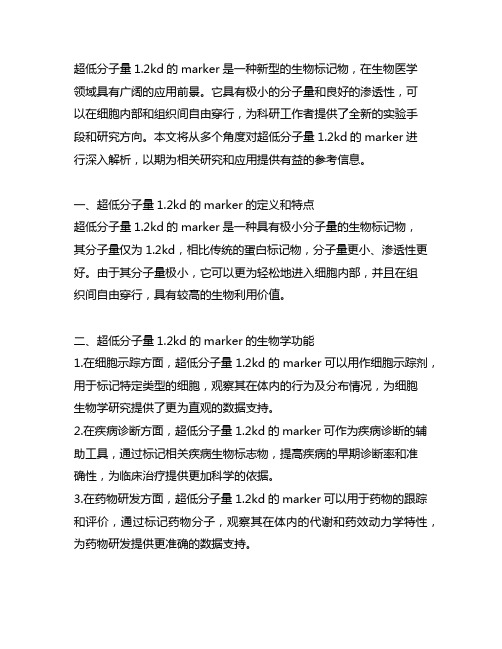
超低分子量1.2kd的marker是一种新型的生物标记物,在生物医学领域具有广阔的应用前景。
它具有极小的分子量和良好的渗透性,可以在细胞内部和组织间自由穿行,为科研工作者提供了全新的实验手段和研究方向。
本文将从多个角度对超低分子量1.2kd的marker进行深入解析,以期为相关研究和应用提供有益的参考信息。
一、超低分子量1.2kd的marker的定义和特点超低分子量1.2kd的marker是一种具有极小分子量的生物标记物,其分子量仅为1.2kd,相比传统的蛋白标记物,分子量更小、渗透性更好。
由于其分子量极小,它可以更为轻松地进入细胞内部,并且在组织间自由穿行,具有较高的生物利用价值。
二、超低分子量1.2kd的marker的生物学功能1.在细胞示踪方面,超低分子量1.2kd的marker可以用作细胞示踪剂,用于标记特定类型的细胞,观察其在体内的行为及分布情况,为细胞生物学研究提供了更为直观的数据支持。
2.在疾病诊断方面,超低分子量1.2kd的marker可作为疾病诊断的辅助工具,通过标记相关疾病生物标志物,提高疾病的早期诊断率和准确性,为临床治疗提供更加科学的依据。
3.在药物研发方面,超低分子量1.2kd的marker可以用于药物的跟踪和评价,通过标记药物分子,观察其在体内的代谢和药效动力学特性,为药物研发提供更准确的数据支持。
三、超低分子量1.2kd的marker在细胞内的应用1.细胞内示踪:超低分子量1.2kd的marker可用于标记细胞内的特定分子或细胞器,观察其运动轨迹和代谢动态,为细胞生物学研究提供更直观的数据。
2.细胞内信号转导:超低分子量1.2kd的marker可以用于标记细胞内的信号分子,研究其在信号传导途径中的作用和调控机制,为细胞信号转导研究提供更深入的认识。
四、超低分子量1.2kd的marker在动物体内的应用1.动物体内示踪:超低分子量1.2kd的marker可用于动物体内的示踪研究,通过体内注射等方式,标记特定类型的细胞或组织,观察其在动物体内的行为和分布情况,为相关研究提供数据支持。
琼脂糖凝胶电泳鉴定DNA
DNA的上样正确的DNA上样量是条带清晰的保证。
注意太多的DNA上样量可能导致DNA带型模糊,而太小的DNA上样量弱甚至缺失。
TIANGEN公司DNA分子量标准每次上样6ul即可得到清晰均匀的条带。
Marker的选择DNA电泳一定要使用DNA Marker或大小的正对照DNA来估计DNA片段大小。
Marker应该选择在目标片段大小附近ladderTIANGEN公司的DNA Marker条带清晰,亮度均匀,质量稳定,是您实验的首选。
需要注意的是Marker 的电泳同样也要符合DNA电泳的操作标准。
如果选择λDNA/HindIII或者λDNA/EcoRI的酶切Marker,需要预先65℃加热5min,冰上冷却后使用。
从而防止HindIII或EcoRI酶切造成凝胶的染色和观察实验室常用的核酸染色剂是溴化乙啶〔EB〕,染色效果好,操作方便,但是稳定性差,具有毒性。
而其他系列例如SYBR Green,GelRed,虽然毒性小,但价格昂贵。
TIANGEN公司的GeneGreen相比那么是性价比高的低毒替代染料,其灵敏度比传统EB染料高10倍以上。
注意观察凝胶时应根据染料不同使用适宜的光源和激发波长,如果激发波长不对,条带那么不易观察,出现条带模糊的现象。
实例实验:一、实验原理用于DNA别离,纯化和鉴定的凝胶电泳有二类,一类是琼脂糖凝胶电泳,适用于1kb和大于1kb以上DNA;另一类是聚丙烯酰胺凝胶电泳,适用小于1kb的DNA。
琼脂糖凝胶电泳是一种非常简便,快速别离、纯化和鉴定DNADNA在琼脂糖凝胶中迁移率和凝胶浓度、DNA 分子大小、DNAEB〕,EB在紫外钱照射下的发射荧光。
EB能插入DNA分子中形成荧光结合物,使发射的荧光增强几十倍。
荧光的强度正比于DNA的含量,如将浓度的标准样品作电泳对照,就可估计出待测样品的浓度。
二、试剂:1.电泳缓冲液:40mmol/L Tris-HCl(pH8.0)20mmol/L NaAC2mmol/L EDTA50倍电泳缓冲液,用时取5ml,重蒸水稀释至250ml。
天根说明书RNA
RP110413RNAsimple Total RNA KitCat. no. DP419Kit ContentsBuffer RZ should be stored protected from light at 2-8°C; others stored at room temperature (15-25°C).IntroductionRNAsimple Total RNA Kit uses a new RNA isolation technology based on Guanidine Thiocyanate / Phenyl method. It contains a unique buffer RZ that minimizes the contamination of genomic DNA and protein. RNAsimple Total RNA Kit can efficiently isolates high pure RNA from blood, cells, tissues and plant samples in one hour. The purified RNA is ready-to-use in downstream applications such as: RT-PCR and real-time RT-PCR, gene-chips assay, northern blot, dot blot, polyA screening, in vitro transcript, and molecular cloning.Important NoteFor isolation of bacterial RNA, RNAprep pure Kit (For Cell/Bacteria) should be used (Cat.no. DP430).Notes of preventing RNase contamination1.Change gloves regularly. Bacteria on the skin can result inRNase contamination.e RNase-free plastic and tips to avoid cross contamination.3.RNA can be protected in TRNzol. But RNA must be stored orprocessed in RNase-free plastic or glassware. To wipe off RNase, the glassware can be roasted at 150℃for 4 hours, while plastic can be dipped in 0.5 M NaOH for 10min, washed by RNase-free ddH2O thoroughly, and sterilized.e RNase-free ddH2O to confect solution.(Add DEPC intowater in clean glass container to a final concentration of 0.1% (v/v). Incubate overnight and autoclave for 15 min to remove any trace of DEPC.)ProtocolBuffer RD and Buffer RW are supplied as a concentrate. Before using for the first time, add ethanol (96–100%) as indicated on the bottle to obtain a working solution.1.Homogenizing samples.a.Plant (take leaves as an example): Place fresh leaves inliquid nitrogen and grind thoroughly with a mortar andpestle, or grind in Buffer RZ after cut leaves into pieces.This process is suggested to be finished within one minute.Use 1 ml Buffer RZ per 100 mg leaves.b.Tissues (take rat liver as an example): Add 1 ml Buffer RZfor per 30–50 mg of liver sample. Homogenize sampleusing a power homogenizer. Usually, the volume of tissue sample should not exceed 10% of the volume of Buffer RZ.c.Adherent Cells (do not use more than 1 × 10⁷ cells): Cellsgrown in a monolayer in cell-culture vessels can be either lysed directly in the vessel (up to 10 cm diameter) or trypsinized and collected as a cell pellet prior to lysis. (Cells grown in a monolayer in cell-culture flasks should always be trypsinized. )1)Method A: To lyse cells directly. Add 1 ml Buffer RZdirectly to the cells in the culture dish per 10 cm2ofculture dish surface area. Pipette the lysate up anddown several times. Note: the volume of Buffer RZshould be determined according to the surface areainstead of the number of cells. An insufficient volumecan result in DNA contamination of isolated RNA.2)Method B: To trypsinize and collect cells. Determinethe number of cells. Aspirate the medium, and washthe cells with PBS. Aspirate the PBS, and add 0.10–0.25% trypsin in PBS. After the cells detach from thedish or flask, add medium (containing serum toinactivate the trypsin), transfer the cells to an RNase-free glass or polypropylene centrifuge tube (notsupplied), and centrifuge at 300 x g for 5 min.Completely aspirate the supernatant.Note: Make sure that the supernatant has beencompletely removed. Residual medium could lead toincomplete lysis of cells and reduced yield of RNA. d.Suspension Cells: Harvest cells by centrifugation andremove culture medium. Add 1 ml of Buffer RZ per 5 × 10⁶- 107cells from animal, plant or yeast, or 1 × 10⁷ cells of bacterial. Do not wash cells before addition of Buffer RZ toavoid increased chance of mRNA degradation. Samplesfrom some yeast and bacteria maybe need to behomogenized by using a power homogenizer.e.Blood: Take fresh blood, and add three volumes of BufferRZ. Mix thoroughly. (Recommended amount: 0.75 mlBuffer RZ for 0.25 whole blood)2.Incubate homogenized samples at 15-30℃for 5 min, topermit complete dissociation of the nucleoprotein complex.3.Optional step: Centrifuge the sample at 12,000 rpm (~13,400 ×g)for 10 minutes at 4°C. Transfer the supernatant to a freshmicro-centrifuge tube.Note: When preparing samples with high content of fat, proteins, polysaccharides, or extracellular material (e.g., muscle, fat tissue, or tuberous plant material), an additional centrifugation may be required to remove insoluble material from the samples.RNA remains in the upper aqueous phase after centrifugation. However, when dealing with fat tissue, the upper phase is a lipid layer that should be discarded.Retain the clean homogenizing part for next step.4.Add 200 µl of chloroform per 1 ml Buffer RZ used forhomogenization. Cap the tube securely and vortex for 15 s.Incubate for 3 minutes at room temperature.Note: If vortexing is not applicable, shake tube vigorously by hand for 2 min.5.Centrifuge the sample for 10 min at 12,000 rpm (~13,400 × g)at 4°C. The mixture separates into a lower yellow phenol-chloroform phase, an interphase, and a colorless upper aqueous phase. RNA remains exclusively in the aqueous phase.Pipette the aqueous phase out into a new tube.6.Add the 0.5 volume ethanol (96%-100%) to the aqueous phase.Mix thoroughly (participate may appear in this step). Transferthe sample, including any precipitate that may have formed, to an RNase-free Spin Column CR3 placed in a 2 ml RNase-free Collection Tube. Close the lid gently, and centrifuge at 12,000 rpm (~13,400 × g) for 30 s at 4°C. Discard the flow-through.Note: If the sample is more than 700µl, transfer the sample to CR3 in two times and centrifuge separately.7.Add 500 μl Buffer R D to the RNase-free Spin Column CR3(Ensure ethanol has been added). Close the lid gently, and centrifuge at 12,000 rpm (~13,400 × g) for 30 s at 4°C. Discard the flow-through.8.Add 700 μl Buffer RW to the RNase-free Spin Column CR3(Ensure ethanol has been added). Close the lid gently, and centrifuge at 12,000 rpm (~13,400 × g) for 30 s at 4°C. Discard the flow-through.9.Add 500 μl Buffer RW to the RNase-free Spin Column CR3.Close the lid gently, and centrifuge at 12,000 rpm (~13,400 × g) for 30 s at 4°C. Discard the flow-through.10.Set the RNase-free Spin Column CR3 back to the CollectionTube. Centrifuge at 12,000 rpm (~13,400 × g) for 2 min at 4°C to dry the spin column membrane.Note: The long centrifugation dries the spin column membrane, ensuring that no ethanol is carried over during RNA elution. Residual ethanol may interfere with downstream reactions.11.Place the RNase-free Spin Column CR3 in a new 1.5 ml RNase-free Collection Tube (supplied). Add 30-100 μl RNase-free ddH2O directly to the spin column membrane. Close the lid gently, incubate at room temperature (15–25°C) for 2 min.Centrifuge at 12,000 rpm (~13,400 × g) for 2 min at 4°C to elute the RNA.Note: The volume of elution buffer should not be less than30 μl, or it may affect recovery efficiency. To obtain higherproductivity, add the solution gained from step 11 to the center of membrane again, let the columns stand for 1 min, and then centrifuge.Purified RNA should be stored at –70°C.Ordering InformationRNA IsolationRNAstore Reagent 20 ml100 ml DP408-01 DP408-02Reverse TranscriptionQuantscript RT Kit 20 μl × 25 rxns20 μl × 100 rxns KR103-03 KR103-04Real–Time PCRSuperReal PreMix (SYBR Green) 50 μl × 50 rxns50 μl × 200 rxns FP204-01 FP204-02Quant One Step qRT-PCR (SYBR Green) Kit 50 μl × 50 rxns FP303-01。
天根DP117-无内毒素质粒大提试剂盒说明书

For personal use only in study and research; not for commercial use产品简介本试剂盒采用独特的硅胶模吸附技术,高效专一地结合质粒DNA。
同时采用特殊的溶液P4和过滤器CS1,可有效的出去内毒素、蛋白质杂志;整个提取过程仅需1h,方便快捷。
使用本试剂盒提取的质粒DNA可适用于各种常规操作,包括酶切、PCR、测序、连接、转化和细胞转染多种细胞等试验。
推荐每次菌液使用量:高拷贝质粒推荐使用量为100ml,得率一般在500-1500μg左右;低拷贝质粒推荐使用量为200ml,得率一般在200-600μg左右。
注意事项:请务必在使用本试剂盒之前阅读此注意事项。
1.溶液P1在使用前先加入RNase A (将试剂盒中提供的RNase A全部加入),混匀,置于2-8℃保存。
2.第一次使用前应按照试剂瓶标签的说明先在漂洗液PW中加入无水乙醇。
3.使用前先检查平衡液BL,溶液P2和P4是否出现结晶或者沉淀,如有结晶或者沉淀现象,可在37℃水浴中加热几分钟,即可恢复澄清。
4.注意不要直接接触溶液P2和P4,使用后应立即盖紧盖子。
5.使用过滤器时请将推柄小心缓慢地从过滤管中抽出,避免滤膜因压力而松动。
6.提取的质粒量与细菌培养浓度,质粒拷贝数等因素有关。
如果所提质粒为低拷贝质粒或大于10kb的大质粒,应加大菌体使用量,同时按比例增加P1、P2、P4的用量;洗脱缓冲液推荐在65-70℃水浴中预热,可以适当延长吸附和洗脱时间,以提高提取效率。
7.实验前使用平衡液BL处理吸附柱,可以最大限度激活硅基质膜,提高得率。
8.用平衡液处理过的柱子最好立即使用,放置时间过长会影响使用效果。
质粒DNA 浓度及纯度检测得到的质粒DNA可用琼脂糖凝胶电泳和紫外分光光度计检测浓度与纯度。
OD260值为1相当于大约50μg/ml双链DNA。
纯化的质粒DNA OD260/OD280通常在1.8-2.0左右,可直接应用于细胞转染甚至动物体内实验等对DNA纯度要求很高的实验中。
几种miniprep提取质粒DNA方法的比较与改良
几种miniprep提取质粒DNA方法的比较与改良
牛蕾蕾0001)
摘要: 目的 比较并建立一种简单的、稳定的质粒DNA提取改良方法,探讨其在分子生物学实验研究中的应用价值。 方
质粒小提试剂盒),本试剂盒采用碱裂解法和离心 吸附柱提纯。详细步骤见其说明书。 1.3 DNA琼脂糖凝胶电泳各取上述4种方法制 备的质粒DNA溶液lO止上样,在l%的琼脂糖凝 胶上进行电泳,EB染色,以Marker l 000—10 000
万方数据
·282·
bp作为参照,紫外灯下检测电泳谱带。. 2结果
中图分类号:Q503
文献标识码:A
A comparative and improved study on plasmid DNA extraction NIU Lei—lei,ZHANG Geng-qian,YANG Si—yi,GUO Da-wei(Dept ofBioto#cal Evidence,School ofForensic Med如ine,Shanxi Medwd University,Taiyuan 030001,China) Abstract: Objective To explore a simple,improved and steady method for extraction of plasmid DNA and its value in molecular bio- logieal research. Methods Four routine DNA extractions of plasmid were compared by using agamse gel electrophoresis. Results The band using,11ANpmp Mini Plasmid Kit wns the brigIltest without RNA band.The brightness of the bands increased using boiling ly— sis method,alkaline lysis method,alkaline lysis method combined with phenol-cMomform extraction metllod in turns. Conclusion Ly- sis by髓ANprep Mini Plasmid Kit is simple and convenient for producing a high purity of ptasmid DNA.