消化道早癌的诊断
什么是消化道癌前病变、早期癌病理诊断?

什么是消化道癌前病变、早期癌病理诊断?发布时间:2023-04-23T12:18:03.165Z 来源:《医师在线》2022年12月24期作者:白晓英[导读]什么是消化道癌前病变、早期癌病理诊断?白晓英(大英县人民医院;四川遂宁629300)前言恶性肿瘤在当前生物医学研究中占据着关键地位,因为恶性肿瘤属于一种致死性、致残性非常明显的疾病,并且恶性肿瘤的发生率较高,我国恶性肿瘤患者患者数量较之于日本、西方发达国家更高。
在医学研究中,学者习惯根据肿瘤生长位置对肿瘤进行命名,但是这并不意味着不同恶性肿瘤之间的区别仅仅是生长位置的不同,因此针对不同类型恶性肿瘤的治疗需要专科医生负责。
消化系统是恶性肿瘤滋生的高发部位,相关研究显示我国的消化道恶性肿瘤患者人数占据世界前列。
针对恶性肿瘤的治疗在当前依旧推崇“早检查、早发现、早治疗”的原则,早期恶性肿瘤治疗后,患者预后良好,若能够察觉到癌前病变则更有利于预后改善,那么,消化道癌前病变具体包括哪些?涉及的早期癌变病理诊断包括哪些?一、消化道癌前病变1、癌前病变定义癌症令人们“谈癌色变”,很多患者在发现自己症状命名一旦和“癌”字沾边就以为自己已经患了癌症,而癌前病变在其中出现频率较高。
在病理学诊断中,无论何种部位的恶性肿瘤都必然会经历这几个阶段,即:癌前病变、原位癌、浸润癌,当病情发展到原位癌之后,则标志恶性肿瘤成型,此时在临床实践中才会考虑患者罹患xx癌,也就是说癌前病变阶段代表患者并不是癌症;但是癌前病变有相当可能性会演变为癌症,因此需要患者重视,而医生也会在此时特意嘱咐患者节制不良习惯。
2、消化道范畴结合解剖学、消化系统恶性肿瘤定义,消化道具体包括:口腔、咽喉、食管、胃部、结肠、直肠、肛管等部位。
3、消化道癌前病变较为典型、常见的消化道恶性肿瘤包括口腔癌、喉癌、食管癌、胃癌、直肠癌、结肠癌等。
可能演变为口腔癌且常见癌前病变为:口腔黏膜白斑,该情况下口腔黏膜癌变的可能性较低,而口腔红斑癌变可能性更高,此外,扁平苔藓、口腔黏膜下纤维化也是常见的口腔癌前病变,其中,口腔黏膜下纤维化和患者本人长期嚼槟榔存在直接的、关键的联系,这些口腔症状需要患者注意。
消化道早癌的诊断ppt课件

其他恶性肿瘤
消化道早癌还需要与其他恶性肿瘤进行鉴别诊断,如胃癌、结直肠癌等。
05
消化道早癌的预防与控制
预防措施
定期筛查
建议高危人群定期进行消化道内镜筛查,以 便早期发现病变。
健康饮食
保持均衡饮食,增加蔬菜、水果摄入,减少 高脂、高糖、高盐食物的摄入。
消化道早癌的诊断ppt课件
目录
• 消化道早癌概述 • 消化道早癌的诊断方法 • 消化道早癌的症状与体征 • 消化道早癌的诊断标准与流程 • 消化道早癌的预防与控制
01
消化道早癌概述
定义与特点
定义
消化道早癌是指发生在消化道黏 膜上皮的早期癌变,尚未突破黏 膜层及未发生淋巴结转移。
特点
早期症状不明显,与普通消化道 疾病症状相似,易被忽视。
监测与评估
定期对消化道早癌的防控工作进行监测和评估,及时发现问题并采取改进措施。
提高公众认知度
01
制作宣传资料
制作通俗易懂、图文并茂的宣 传资料,包括消化道早癌的预 防知识、诊疗流程等。
02
开展科普讲座
邀请专家学者开展消化道早癌 的科普讲座,向公众传播科学 知识和理念。
03
利用媒体渠道
利用电视、广播、报纸、网络 等媒体渠道,广泛宣传消化道 早癌的防控知识。
02
消化道早癌的诊断方法
内镜检查
放大内镜
能将消化道黏膜放大,观察微小 病变,提高诊断准确性。
超声内镜
结合内镜与超声技术,可观察消 化道管壁的层次结构。
01
无痛内镜
通过使用麻醉药物,使患者在检 查过程中处于睡眠状态,减少不 适感。
怎样发现消化道早癌
如何早期发现消化道肿瘤
消化道肿瘤在我国高发,其死亡率居于恶性肿瘤的首位。
患者大多数早期是没有任何症状的,一般出现症状后再检查几乎都是中晚期。
消化道肿瘤的早发现、早诊断、早治疗,无论对个人、对家庭、对社会都有非常重要的意义,早期消化道肿瘤治疗后5年生存率高达90%以上,晚期5年生存率只有30%左右。
目前,消化内镜检查(胃镜、肠镜)是消化道检查的首选方法。
一般来说,年龄在四十岁以上的中年人,有肿瘤家族史;对于前一次做胃镜有慢性萎缩性胃炎,中度不典型增生,胃出血、肠出血;胃、大肠有息肉,大便出现过黏液便、血便等,都需要重视胃肠镜检查。
在消化道肿瘤高发地区,更应该常规进行胃肠镜检查,以便对消化道早期肿瘤早发现、早治疗。
消化道早癌筛查“早知道”

消化道癌症是一种常见的恶性肿瘤,通常分为食管癌、胃癌、结直肠癌等类型。
这些癌症在早期时往往没有明显的症状,因此容易被忽视,直到疾病进展到晚期时才会出现明显的症状,如肿块、消瘦、便血等。
而在晚期时治疗难度较大,预后也不太乐观。
因此,对消化道早癌的筛查非常重要,可以在疾病发展到晚期前发现并及时治疗,提高治愈率和生存率。
本文将从消化道早癌筛查的定义、筛查对象、筛查方法、筛查意义等方面进行详细的介绍和解析。
一、消化道早癌筛查的定义消化道早癌筛查是一种针对消化道癌症进行的预防性医学检查方法,旨在早期发现消化道内的癌前病变和早期消化道癌症。
消化道癌症是指在食管、胃、小肠、结肠、直肠等消化系统器官中发生的恶性肿瘤,其中胃癌和结肠癌是最常见的类型。
早期消化道癌症指的是癌细胞仅限于黏膜和浆膜层,未侵犯肌层和粘膜下层的癌症。
早期消化道癌症通常没有明显的症状,但是可以通过特定的检查方法进行检测和诊断。
癌前病变是指消化道内的组织发生了不正常的细胞增生和形态变化,这些异常细胞可能会进一步发展成为癌症。
常见的癌前病变包括炎症、息肉、不典型增生等。
消化道早癌筛查旨在尽早发现和诊断消化道癌前病变和早期消化道癌,从而提高治愈率和生存率。
二、消化道早癌筛查的对象消化道早癌筛查的对象主要包括以下人群:1、年龄较大的人群:随着年龄的增长,身体的免疫功能逐渐下降,抵抗能力也会减弱,消化道癌的发病率也逐渐升高。
因此,年龄较大的人群是消化道早癌筛查的主要对象。
建议50岁以上的人每年进行一次消化道早癌筛查。
2、有消化道癌病史家族史的人群:如果一个人的直系亲属中有消化道癌的病史,那么他自己患上消化道癌的风险也会大大增加。
因此,有消化道癌病史家族史的人群需要更加重视消化道早癌筛查。
建议40岁以上的人进行消化道早癌筛查。
3、慢性胃炎、胃溃疡、食管炎等消化道疾病的患者:这些疾病的发生和消化道癌密切相关,同时也会导致消化道黏膜发生变化,容易发生消化道癌前病变和早期消化道癌。
关于消化道早癌,这些内容需要了解
8、小结:
消化道是人体中最忙、任劳无怨的器官,不管你给什么,都必须完全接受它。随着人们生活水平在不断上升,物质更加的丰富,消化道肿瘤发病的趋势却越来越明显,所以我们一定要注意自己的饮食习惯,减少不健康的食物,防止病从口入。
关于消化道早癌,这些内容需要了解
近几年来,我国消化系统的恶性肿瘤患者呈现中低龄化增长趋势,发病和患者死亡率也逐年快速上升,大多数肿瘤患者在被发现的时候都已经是中后期。以一名胃癌患者举例,如果能做到早发现和早诊5年,那么患者的生存率就可以达到95%以上,因此癌症的早期筛检无疑成为当务之急。
1、什么是早癌?
d.大便发生改变
患者在消化道肿瘤出现以后,可能会出现大便的改变,例如经常出现腹泻、便秘等较为"极端"的症状,同时其大便的颜色也会发生改变,会开始排出黑色或柏油状大便,甚至还可能会带有鲜血。
6、如何预防消化道早癌
a.避免病从口入
通常致癌的主要因素有物理、化学、遗传和生物性四大类。其中,遗传性肿瘤的发生率不超过20%。物理致癌的主要因素是放射性,约占人体肿瘤发病的5%-10%。致癌的生物性病因主要有热带恶性淋巴瘤和鼻咽癌,约占肿瘤发生的5%。约70%的肿瘤由化学致癌物引起,如我们熟知的腌制食品,亚硝酸盐含量较高的食品,含有黄曲霉毒素霉变食品,高温下分解的蛋氨酸和苯丙氨酸可产生致癌物质的油炸肉食品,这些常常会诱发食管癌、肝腺癌、胃瘤。
b.期才发现,这个原因是因为我们老百姓常常有以下几个误区:没有症状不就医,年轻不重视体检。为什么许多发达国家的消化道肿瘤治疗效果远远好于中国?为什么人家的5年生存率等等都比中国高?原因在于早发现,早诊断,早治疗。例如,早期的胃癌,完全能治愈。局部发展期,5年的生存率为15%至30%,而到晚期,5年的生存率为零。确诊胃癌的黄金标准有胃镜和活检。出现问题时,医生会建议病人做胃镜检查,绝对不能拖延时间。
消化道早癌的内镜诊断与治疗
消化道早癌的内镜诊断与治疗消化道早癌是指发生于消化道黏膜上的癌前病变和早期癌症,如果及时发现并治疗,患者的生存率和治愈率都较高。
内镜诊断和治疗是消化道早癌的主要手段之一,本文将对消化道早癌的内镜诊断和治疗进行科普介绍。
一、消化道早癌的分类和分布消化道早癌包括食管早癌、胃早癌和结直肠早癌,其中以胃早癌最为常见。
根据组织学类型,消化道早癌可分为腺癌、粘液癌、平滑肌瘤等。
根据病变形态,消化道早癌可分为隆起型、扁平型和凹陷型。
不同形态的早癌治疗方案也略有不同。
二、内镜诊断消化道早癌内镜检查是消化道早癌的首选诊断方法。
内镜检查可以明确病变的形态、大小、位置和深度,取活组织病理检查可以明确病变的组织学类型和分级。
内镜检查还可以同时进行活检、黏膜下层剥离术(ESD)等治疗操作,一次检查既可以明确病变,又可以进行治疗,具有较高的临床价值。
1.内镜检查前的准备内镜检查前,患者应进行一些准备工作。
首先是空腹检查,患者应在检查前6小时内禁食,并在检查前2小时内禁止饮水。
其次是肠道准备,如果是结肠镜检查,需要进行肠道准备,一般是采用口服泻剂或灌肠液进行清洗。
最后是心理准备,内镜检查可能会有一定的不适感,患者应做好心理准备。
2.内镜检查操作步骤内镜检查包括进口镜检查和出口镜检查,进口镜检查是通过口腔进入食管、胃、十二指肠进行检查,出口镜检查是通过肛门进入直肠和结肠进行检查。
下面以胃镜检查为例介绍内镜检查的操作步骤。
(1)患者取坐位或左侧卧位。
(2)给患者喉部局部麻醉,使用局部麻醉药物喷雾,减轻喉咙疼痛感。
(3)将内镜插入口腔,顺着食管进入胃部。
(4)检查胃黏膜表面,观察病变形态、大小、位置、边界和颜色等。
(5)如发现疑似病变,取活组织进行病理检查。
(6)根据病变的形态和位置,选择合适的治疗方法进行治疗。
三、内镜治疗消化道早癌内镜治疗是消化道早癌治疗的重要手段,包括黏膜下层剥离术(ESD)、内镜下黏膜切除术(EMR)和内镜下切除术(ESR)等。
消化道早癌诊治规范
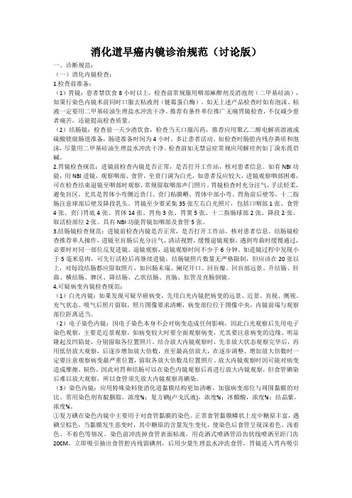
消化道早癌内镜诊治规范(讨论版)一、诊断规范:(一)消化内镜检查:1.检查前准备:(1)胃镜:患者禁饮食8小时以上,检查前常规服用咽部麻醉剂及消泡剂(二甲基硅油),如果行染色内镜术前同时口服去粘液剂(链霉蛋白酶)。
如无上述产品检查时如有泡沫、粘液一定要用二甲基硅油生理盐水冲洗干净。
推荐有条件单位推广无痛胃镜检查,不仅减少患者痛苦,还能提高检查质量。
(2)结肠镜:检查前一天少渣饮食,检查当天口服泻药,推荐应用聚乙二醇电解质溶液或硫酸镁做肠道准备,肠道准备时间为4小时,多让患者活动。
如检查时肠腔内残存粪质和泡沫,尽量用二甲基硅油生理盐水冲洗干净。
检查前如无禁忌症常规应用解痉剂如丁溴东莨菪碱。
2.胃镜检查规范:进镜前检查内镜是否正常,是否打开工作站,核对患者信息。
如有NBI功能,用NBI进镜,观察咽部、食管,至贲门调为白光,如患者反应较大,进镜观察咽部困难,可在检查结束退镜至咽部时观察,常规留取咽部声门照片。
胃镜检查时充分注气,手法轻柔,避免盲区,尤其是胃体小弯侧近贲门、贲门粘膜嵴、胃体中部小弯、胃角前后壁等,十二指肠注意球部后壁及降段乳头。
胃镜至少要采集35张左右白光照片,包括口咽部1张、食管4张、贲门胃底4张、胃体14张、胃角3张、胃窦5张、十二指肠球部2张、降段2张、取活检部位2张。
具有NBI功能胃镜加咽部及食管5张。
3.结肠镜检查规范:进镜前检查内镜是否正常,是否打开工作站,核对患者信息。
结肠镜检查推荐单人操作,进镜至盲肠后充分注气,清洁视野,缓慢退镜观察,遇到弯曲时缓慢通过,必要时对同一部位反复进镜、退镜观察,退镜观察时间不少于6分钟。
如进镜过程中发现小于5毫米息肉,可先行活检后再继续进镜。
结肠镜照片数量无严格限制,但应该在20张以上,对每段结肠都应留取照片,如回肠末端、阑尾开口、回盲瓣、回盲部远景、升结肠、肝曲、横结肠、脾区、降结肠、乙状结肠、直肠、肛管及直肠倒镜。
4.可疑病变内镜检查规范:(1)白光内镜:如果发现可疑早癌病变,先用白光内镜把病变的远景、近景、直视、侧视、充气状态、吸气后照片留取,照片图像要求清晰,病变部位位于图像中央,内镜前端与观察部位距离适当。
消化道早癌的内镜诊断 ppt课件
0.05%结晶紫(龙胆紫):吸收性,常用于侵 袭性病变染色。在病变表面滴数滳,然后再用 温水冲洗。最好用链霉蛋白酶。
5
ppt课件
6
ppt课件
Conventional white light imaging
பைடு நூலகம்
Indigo carmine chromoendoscopy
vessel caliber between IPCL-V3 (small white arrow) and VN (large white
arrow: T1b or deeper).
20
ppt课件
V: microvascular pattern • Subepithelial capillary (SEC) • Collecting venule (CV) • Pathological microvessels (MV)
marginal crypt epithelium;
(D)Atrophic gastritis.
collecting volume
Yao K.22Ann Gastroenterol. 2013;26(1):11-22. ppt课件
(A) C-WLI :erosion (B) M-NBI: a regular microvascular
消化道早癌的内镜诊断
1
ppt课件
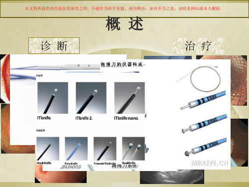
诊断
概述
治疗
2
ppt课件
发现早癌的内镜诊断技术
白光内镜检查。
染色内镜检查。
白光放大(ME)。 染色+放大。 ME+NBI (magnified
endoscopy)。 活检
消化道早癌的诊断培训课件
染料类型
被染对象
Lugol’s碘液 (碘+碘化钾) 磷状上皮内的糖原
染色原理
阳性颜色
临床应用
非角化上皮结合碘 深棕色
a) 正常食管磷状上皮着色。 b) 食管磷状细胞癌黏膜、Barrett食管黏膜、柱状上皮和食管
炎黏膜均不着色。
亚甲蓝
肠道上皮细胞,肠化上皮细 胞
吸收入上皮细胞内
蓝色
a) 食管和胃的肠化上皮、早期胃癌上皮和正常肠道上皮着色。 b) 十二指肠内化生的胃上皮不着色。
本文档所提供的信息仅供参考之用,不能作为科学依据,请勿模仿;如有不当之处,请联系网站或本人删除。
Pit pattern classification (1)
Kudo分型(pit pattern). 分为5型(Type I to type V):
Type I and II :良性,非肿瘤性。 type III to V:肿瘤性,其准确率达90%。 Type III:III-S and III-L
本文档所提供的信息仅供参考之用,不能作为科学依据,请勿模仿;如有不当之处,请联系网站或本人删除。
血管袢(CP,sano)分型(佐野分型) 本文档所提供的信息仅供参考之用,不能作为科学依据,请勿模仿;如有不当之处,请联系网站或本人删除。
CP分型分为I, II, III型,其中III型又分为A和B两亚型。NBI加放大能有效识别低级别 上皮内瘤变和高级别上皮内瘤变或浸润性癌。能有效预测病变的组织学类型。
消化道早癌的内镜诊断与治疗李强
消化道早癌的内镜诊断与治疗李强发布时间:2023-06-12T06:06:22.321Z 来源:《医师在线》2023年2月4期作者:李强[导读]消化道早癌的内镜诊断与治疗李强(四川省眉山市仁寿县中医医院;四川眉山620500)消化道早期癌症是指癌细胞侵袭黏膜和亚黏膜层的癌症,通常病变范围仅限于黏膜和亚黏膜层,没有浸润到肌层和淋巴结,是消化道癌症中最早期、最有机会治愈的阶段。
因为消化道癌症发展得比较缓慢,且病变范围有限,所以被称为早期癌症。
早期消化道癌症往往没有症状,很难被发现,如果不及时治疗,会逐渐扩散到深层组织,甚至转移至其他器官。
内镜诊断与治疗是早期消化道癌症的重要手段之一。
1、消化道早癌内镜诊断内镜检查是发现早期消化道癌症的主要手段之一。
内镜检查可以通过观察黏膜表面的变化,检查癌症的位置、大小、形态、颜色等信息,可以同时进行组织活检,以确定病变的良性或恶性。
1.1胃镜胃镜是检查胃部病变的主要方法。
医生会在患者服用轻度麻醉药物后,通过口腔将一根柔软的内镜插入胃内,观察胃壁的变化。
在胃镜检查中,医生还可以进行黏膜活检,以确定病变的性质。
1.2结肠镜结肠镜是检查结肠病变的主要方法。
医生会将一根柔软的内镜插入肛门,进入结肠,观察结肠壁的变化。
结肠镜也可以进行黏膜活检。
1.3食管食管是检查食管病变的主要方法。
医生会将一根柔软的内镜插入口腔,进入食管,观察食管壁的变化。
食管镜也可以进行黏膜活检。
除了内镜检查外,还可以进行X线造影、CT、MRI等影像学检查,以辅助诊断。
2、消化道早癌治疗消化道早癌的治疗方法包括内镜治疗、手术治疗、放疗和化疗等。
其中,内镜治疗是一种创伤小、恢复快、并发症少、疗效好的治疗方法,被广泛应用于早期消化道癌症的治疗。
2.1内镜治疗2.1.1EMREMR是一种比较简单的内镜治疗方法,适用于病变面积小于2cm的浅表性病变。
该方法通过内镜将注射针插入黏膜下层,注射生理盐水使黏膜隆起,然后用电切刀或剪切器将病变的黏膜切除,最后将切除的组织用夹子取出,进行组织学检查。
消化道早癌——这样诊断要慢一点

消化道早癌——这样诊断要慢一点上一回咱们聊了聊内镜切除标本的组织处理过程,这一回咱再聊聊相关的病理诊断报告格式和内容,还是那句老话各位看官各取所需、多提意见。
一、消化道早癌诊断报告须包括以下内容:1. 肿瘤解读:肉眼分型,组织学类型,分化程度,大小,癌前病变;2. 生物学行为描述:是否浸润/浸润层次/深度(如SM1、SM2,仅在垂直切缘阴性时测量深度),是否侵犯脉管(血管、淋巴管);3. 切缘情况:水平(侧切缘)和垂直切缘(基底)情况;4. 继发或伴发病变:有无溃疡形成或溃疡瘢痕,周围黏膜的其它病变。
二、大体检查文字描述规范和模板:文字描述内容应包括:1.标本送检部位、术式;2.标本描述:数量()块;大小 cm* cm* cm /最大径 cm— cm/蒂部长 cm,宽 cm;3.病变描述:肉眼分型0-Ⅰp(隆起型-带蒂)、0-Ⅰs(隆起型-广基)、0-Ⅱa(浅表隆起型)、0-Ⅱb(浅表平坦型)、0-Ⅱc(浅表凹陷型)、0-Ⅲ(凹陷型)、混合型(病变同时具有两种或以上肉眼分型,如0-Ⅱa Ⅱc);病变范围(cm* cm* cm),距标本侧边缘最近距离(cm);例如:送检贲门ESD切除黏膜组织一枚,大小3.5cm*3cm*0.2-0.3cm,未见明确食管黏膜。
黏膜面局部见一1.8cm*1.3cm的浅表隆起-凹陷型(0-Ⅱa Ⅱc)病变区,该区域距标本侧边缘最近距离0.2cm。
三、诊断报告规范和模板:1. 送检部位和术式;2. 病理诊断:肿瘤组织学类型(参考WHO肿瘤分类标准,不同组织学类型混合时应注明各区域所占的百分比)、分化程度、肿瘤大小(是否与巨检符合,如不符合需测量镜下病变最大径和累及组织条数目)。
各器官须评估的病理参数:如食管早癌应评估癌组织浸润方式(图1)、胃早癌评估是否伴发溃疡或存在溃疡瘢痕、大肠早癌评估Tumor Budding分级(即癌组织浸润前锋出芽状生长、由1-4个癌细胞组成的孤立细胞/细胞索/细胞簇,分级标准见图2)等。
上消化道早癌内镜检查与临床病理诊断对比分析
上消化道早癌内镜检查与临床病理诊断对比分析上消化道早癌是指在食管、胃、十二指肠等上消化道黏膜上发展的早期癌变或初期肿瘤,早期发现并及时治疗能够明显提高患者的存活率和生存质量。
内镜检查是目前早期癌症筛查和诊断的主要手段之一,临床病理诊断是早癌诊断的“金标准”。
对上消化道早癌内镜检查与临床病理诊断进行对比分析,对于优化早癌筛查、提高早癌诊断准确率具有重要的指导意义。
一、上消化道早癌内镜检查上消化道早癌内镜检查是通过内镜对食管、胃、十二指肠等部位进行检查,以观察和诊断患者是否存在早期癌变或癌症。
内镜检查可以直接观察黏膜表面的形态、颜色、纹理和管腺口等情况,还可以通过取活组织活检对病灶进行病理学检查,对癌变程度和病理类型进行评估,有助于早期癌症的诊断和鉴别诊断。
内镜检查的优点在于可以直接观察病变部位,可以及时发现和诊断早期癌变或肿瘤,对癌变黏膜进行活检可以提供临床病理诊断所需要的组织学资料。
内镜检查对于早期癌症的治疗也非常重要,可以通过内镜治疗技术对早期癌变进行治疗,避免了传统手术方式对患者的损伤和创伤。
二、临床病理诊断临床病理诊断是早期癌症诊断的“金标准”,通过对活检或手术标本进行镜下病理学分析和诊断,对病变的组织形态、细胞结构、细胞学特征以及免疫组织化学、分子病理等多方面的检测,以确定病变的性质,判断是否为早期癌变或癌症,并做出分期和分级,为临床治疗和预后评估提供重要依据。
临床病理诊断的准确性对早期癌症的诊断、治疗和预后具有重要的意义。
只有通过临床病理诊断,才能明确病变的性质和程度,为临床医生提供准确的诊断信息,从而制定合理的治疗方案,对病变进行有效的治疗,提高治疗效果和预后。
三、内镜检查与临床病理诊断的比较分析1. 一致性内镜检查和临床病理诊断应该是一致的,即内镜检查发现的癌变病灶应该在临床病理诊断中得到确认。
在实际情况中,内镜检查与临床病理诊断的一致性并不是百分之百的。
一些因素会导致内镜检查与临床病理诊断不一致,如内镜技术的操作者水平不同、检查中的病灶选择不当、活检取材不足或取材部位不对等问题都会影响内镜检查与临床病理诊断的一致性。
消化道早癌的诊断(课堂PPT)

2020/4/11
(A) C-WLI :erosion (B) M-NBI: a regular microvascular
pattern and a regular microsurface pattern with light blue crest. (C) chronic gastritis with intestinal metaplasia
SECN, RAC, CO, MCE, CV,
2020/4/11
subepithelial capillary network;
regular arrangement of collecting venules; (A, B) Normal gastric body mucosa.
crypt-opening;
2020/4/11
25
26
血管袢(CP,sano)分型(佐野分型)
CP分型分为I, II, III型,其中III型又分为A和B两亚型。NBI加放大能有效识别低级别上 皮内瘤变和高级别上皮内瘤变或浸润性癌。能有效预测病变的组织学类型。
2020/4/11
27
Modified 3-step strategy of NBI colonoscopy.
临床应用
非角化上皮结合碘 深棕色
a) 正常食管磷状上皮着色。 b) 食管磷状细胞癌黏膜、Barrett食管黏膜、柱状上皮和食
管炎黏膜均不着色。
亚甲蓝
肠道上皮细胞,肠化上皮 细胞
吸收入上皮细胞内
蓝色
甲苯胺蓝
胃或肠内的柱状上皮细胞 胞核差色
自由扩散入细胞
蓝色
a) 食管和胃的肠化上皮、早期胃癌上皮和正常肠道上皮着 色。
T4a: resectable tumor invading the pleura,
- 1、下载文档前请自行甄别文档内容的完整性,平台不提供额外的编辑、内容补充、找答案等附加服务。
- 2、"仅部分预览"的文档,不可在线预览部分如存在完整性等问题,可反馈申请退款(可完整预览的文档不适用该条件!)。
- 3、如文档侵犯您的权益,请联系客服反馈,我们会尽快为您处理(人工客服工作时间:9:00-18:30)。
invasive cancer
精品PPT
精品PPT
精品PPT
NBI imaging of a lesion of IPCL type III.
regional atrophic mucosa or low grade intraepithelial neoplasia
NBI imaging of a lesion of IPCL type IV high-grade intraepithelial neoplasia:Tis
arrow: T1b or deeper).
精品PPT
V: microvascular pattern • Subepithelial capillary (SEC) • Collecting venule (CV) • Pathological microvessels (MV)
S: microsurface pattern • Marginal crypt epithelium (MCE) • Crypt opening (CO) • Intervening part (IP) between crypts
精品PPT
Conventional white light imaging
Indigo carmine chromoendoscopy
精品PPT
Indigo carmine
精品PPT
白光内镜:7mm 扁平息肉样隆起
结晶紫:结构 消失,侵及黏 膜下层。
Indigo carmine
靛胭脂:中央凹陷
submucosa; PM, proper muscle; M1, cancer is limited epithelium; M2, cancer invades
LPM but does not reach MM; M3, cancer invasion reaches MM; SM, submucosally
变为深蓝或黑色 a) b)
泌酸的胃上皮变色,包括异位胃黏膜上皮。 胃癌上皮细胞不变色。
酚红
感染HP的胃上皮细胞
由 于 HP 周 边 有 “ 氨 云 ” , 局部呈碱性而便酚红 由黄变红 变色
诊断胃内HP的感染及其分布情况。
靛胭脂
细胞不着色
沉积于上皮表面的低 凹处,勾勒出病变形 蓝色 态。
全消化道黏膜均可使用。
精品PPT
A
B
C
D
MNBI, magnifying endoscopy with narrow-band imaging; LBC, light blue crest
SECN, RAC, CO, MCE, CV,
subepithelial capillary network;
regular arrangement of collecting venules; (A, B) Normal gastric body mucosa.
精品PPT
This pattern is called IPCL-V1. IPCL-V1 includes four major characteristic
morphological changes of IPCL: dilation, meandering, irregular caliber, and
黏膜均不着色。
亚甲蓝
肠道上皮细胞,肠化上皮细 胞
吸收入上皮细胞内
蓝色
a) 食管和胃的肠化上皮、早期胃癌上皮和正常肠道上皮着色。 b) 十二指肠内化生的胃上皮不着色。
甲苯胺蓝
胃或肠内的柱状上皮细胞胞 核差色
自由扩散入细胞
蓝色
食管磷状细胞癌上皮和Barret’s食管中的化生上皮着色
刚果红
胃内泌酸细胞
当pH<3.0时变色
figure variation. T1a.
精品PPT
This is typical image of intrapapillary capillary loop (IPCL)-V3. Cancer invasion depth was M3 (muscularis mucosae: T1a).
精品PPT
精品PPT
白光内镜发现早癌的前提
理想的消化内镜术前检查的准备:清理视 野,抵制蠕动。
严格的质量控制。 时刻准备发现早癌的警觉性。 特殊、小病品PPT
一、染色内镜
最常用的染料:
碘染色:食管黏膜染色。
0.1-0.4%靛胭脂:对比性染料,常用于腺瘤。 0.1-0.2%美蓝(亚甲蓝):吸收性,常用于腺瘤。 0.05%结晶紫(龙胆紫):吸收性,常用于侵
袭性病变染色。在病变表面滴数滳,然后再用 温水冲洗。最好用链霉蛋白酶。
精品PPT
表1 消化内镜下常用染料
染料类型
被染对象
Lugol’s碘液 (碘+碘化钾) 磷状上皮内的糖原
染色原理
阳性颜色
临床应用
非角化上皮结合碘 深棕色
a) 正常食管磷状上皮着色。 b) 食管磷状细胞癌黏膜、Barrett食管黏膜、柱状上皮和食管炎
消化道早癌的内镜诊断
精品PPT
诊断
概述
治疗
精品PPT
发现早癌的内镜诊断技术
白光内镜检查。
染色内镜检查。
白光放大(ME)。 染色+放大。 ME+NBI (magnified
endoscopy)。 活检
超声内镜。 共聚焦显微内镜。 自体荧光内镜
光学相干断层成像 术
细胞内镜 蓝激光成像
Large white arrows point to large tumor vessel (IPCL-VN). The striking
morphological feature is its extra-large diameter. Note the difference of
vessel caliber between IPCL-V3 (small white arrow) and VN (large white
精品PPT
二、特殊光谱及放大内镜
Narrow band imaging
C-WLI: 20-40倍 ME: 80-170倍
Magnifying endoscopy (ME)
精品PPT
精品PPT
精品PPT
精品PPT
EP, epithelium; LPM, lamina propria mucosae; MM, muscularis mucosae; SM,