子宫内膜异位症的诊治规范草案
医院子宫内膜异位症医疗技术操作规范与报告
文档序号:XXYY-ZWK-001文档编号:ZWK-20XX-001XXX医院子宫内膜异位症医疗技术报告表编制科室:知丁日期:年月日子宫内膜异位症医疗技术报告表子宫内膜异位症一、概述子宫内膜异位症(endometriosis)是指有活性的内膜细胞种植在子宫内膜以外的位置而形成的一种女性常见妇科疾病。
内膜细胞本该生长在子宫腔内,但由于子宫腔通过输卵管与盆腔相通,因此使得内膜细胞可经由输卵管进入盆腔异位生长。
目前对此病发病的机制有多种说法,其中被普遍认可的是子宫内膜种植学说。
本病多发生于生育年龄的女性,青春期前不发病,绝经后异位病灶可逐渐萎缩退化。
子宫内膜异位症的主要病理变化为异位内膜周期性出血及其周围组织纤维化,形成异位结节,痛经、慢性盆腔痛、月经异常和不孕是其主要症状。
病变可以波及所有的盆腔组织和器官,以卵巢、子宫直肠陷凹、宫骶韧带等部位最常见,也可发生于腹腔、胸腔、四肢等处。
二、病因1.种植学说经血逆流,内膜种植。
月经期,经血从宫口、阴道排出体外是顺流而下,但是有小部分经血或因其他原因夹杂着脱落的子宫内膜碎片,由输卵管道流入腹腔,种植在盆腔脏器的表层形成子宫内膜异位病灶。
2.化生内膜浆膜上皮,化生内膜。
人体在胚胎发育时期,卵巢表面上皮、腹膜、阴道直肠膈、脐部均由体腔上皮化生而来,这些组织在性腺激素、炎症、机械因素的刺激下能够转化,形成另一种组织,同样可以化生为子宫内膜。
3.良性转移血液、淋巴良性转移。
这是一种较为罕见的发病原因。
出现在肺部、脑膜、心包、四肢及其他远端的子宫内膜异位症,是通过血液循环或淋巴系统将子宫内膜碎屑转移停留在某脏器或组织上而发病。
4.医源性的内膜移植这是一种人为造成的使子宫内膜移植到某些部位,多见于剖宫产术,早期中期妊娠行刮宫术,分娩时行会阴侧切术,人工流产术等过程中。
5.免疫防御功能缺陷随经血逆流至腹腔的子宫内膜,如同一种异物,会激活身体内的免疫系统,动员出大量的免疫细胞及体液围歼消除,假如体内免疫功能缺陷,就会发展成为子宫内膜异位症。
子宫内膜异位症的诊断与治疗规范
(三)痛经与不孕的治疗 1.痛经的治疗:对合并盆腔结节或附件包块者,首选手 术治疗;无盆腔结节或附件包块者,首选药物治疗;对药物治 疗无效者可考虑手术治疗。 痛经的常用治疗药物包括:(1)一线用药:可选用非类 固醇类抗炎药或口服避孕药。口服避孕药可周期或连续用 药,有效者可继续应用,无效者改用二线用药。(2)二线用
1.口服避孕药:连续或周期用药,共6个月,可抑制排 卵;副作用较少,但可有消化道症状或肝功能异常等。
2.高效孕激素:醋酸甲羟孕酮(其他名称:安宫黄体酮) 20—30 mg/d,分2~3次口服,连用6个月。醋酸甲羟孕酮 可引起内膜组织蜕膜样改变,最终导致内膜萎缩,同时可负 反馈抑制下丘脑一垂体-卵巢轴。副作用主要是突破性出血、 乳房胀痛、体重增加、消化道症状以及肝功能异常等。
3.雄激素衍生物:用于治疗内异症的雄激素衍生物有: (1)达那唑:600~800 mg/d,分2~3次口服,共6个月。达 那唑可抑制月经中期黄体生成素(LH)峰,从而抑制排卵;还 可抑制参与类固醇合成的多种酶,并增加血液中游离睾酮的 水平。副作用主要是男性化表现,如毛发增多、情绪改变、声 音变粗;此外,还可能影响脂蛋白代谢、引发肝功能损害以及 体重增加等。(2)孕三烯酮:口服每次2.5 mg,2~3次/周, 共6个月。孕三烯酮可拮抗孕激素与雌激素,降低性激素结 合蛋白水平,以及升高血中游离睾酮水平。副作用主要是抗 雌激素及雄激素样作用,基本同达那唑,但较轻微。
生堡坦芒型盘壶!塑!至!旦筮丝鲞筮!翅g!也』Q!壁堕堡坚!!!!:!!出磐!塑呈!:!!!:丝:盟!:旦
·645·
子宫内膜异位症的诊治指南(2015年版)
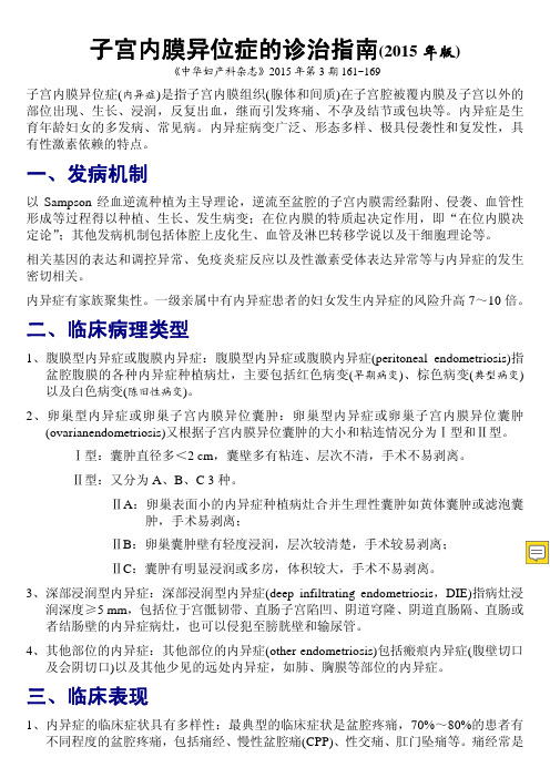
子宫内膜异位症的诊治指南(2015年版)《中华妇产科杂志》2015年第3期161~169子宫内膜异位症(内异症)是指子宫内膜组织(腺体和间质)在子宫腔被覆内膜及子宫以外的部位出现、生长、浸润,反复出血,继而引发疼痛、不孕及结节或包块等。
内异症是生育年龄妇女的多发病、常见病。
内异症病变广泛、形态多样、极具侵袭性和复发性,具有性激素依赖的特点。
一、发病机制以Sampson经血逆流种植为主导理论,逆流至盆腔的子宫内膜需经黏附、侵袭、血管性形成等过程得以种植、生长、发生病变;在位内膜的特质起决定作用,即“在位内膜决定论”;其他发病机制包括体腔上皮化生、血管及淋巴转移学说以及干细胞理论等。
相关基因的表达和调控异常、免疫炎症反应以及性激素受体表达异常等与内异症的发生密切相关。
内异症有家族聚集性。
一级亲属中有内异症患者的妇女发生内异症的风险升高7~10倍。
二、临床病理类型1、腹膜型内异症或腹膜内异症:腹膜型内异症或腹膜内异症(peritoneal endometriosis)指盆腔腹膜的各种内异症种植病灶,主要包括红色病变(早期病变)、棕色病变(典型病变)以及白色病变(陈旧性病变)。
2、卵巢型内异症或卵巢子宫内膜异位囊肿:卵巢型内异症或卵巢子宫内膜异位囊肿(ovarianendometriosis)又根据子宫内膜异位囊肿的大小和粘连情况分为Ⅰ型和Ⅱ型。
Ⅰ型:囊肿直径多<2 cm,囊壁多有粘连、层次不清,手术不易剥离。
Ⅱ型:又分为A、B、C 3种。
ⅡA:卵巢表面小的内异症种植病灶合并生理性囊肿如黄体囊肿或滤泡囊肿,手术易剥离;ⅡB:卵巢囊肿壁有轻度浸润,层次较清楚,手术较易剥离;ⅡC:囊肿有明显浸润或多房,体积较大,手术不易剥离。
3、深部浸润型内异症:深部浸润型内异症(deep infiltrating endometriosis,DIE)指病灶浸润深度≥5 mm,包括位于宫骶韧带、直肠子宫陷凹、阴道穹隆、阴道直肠隔、直肠或者结肠壁的内异症病灶,也可以侵犯至膀胱壁和输尿管。
子宫内膜异位症的诊断与治疗规范

子宫内膜异位症的诊断与治疗规范子宫内膜异位症(内异症)是指子宫内膜组织(腺体和间质)在子宫内膜以外的部位出现、生长、浸润、反复出血,可形成结节及包块,引起疼痛和不育等。
内异症是生育年龄妇女的多发病,发病率有明显上升趋势;其特点表现为(1)症状与体征及疾病的严重性不成比例,(2)病变广泛、形态多样,(3)极具浸润性,可形成广泛而严重的粘连,(4)具有激素依赖性,易于复发。
一、内异症的临床病理类型1.腹膜型内异症:腹膜型内异症(peritoneal endometriosis,PEM)是指发生在盆腹腔腹膜的各种内异症病灶,主要包括红色病变(早期病变)、蓝色病变(典型病变)及白色病变(陈旧病变)。
2.卵巢型内异症:卵巢型内异症(ovarian endometriosis,OEM)可形成囊肿,称为子宫内膜异位囊肿(内异症囊肿);根据囊肿大小和异位病灶浸润程度分为:I型:囊肿直径<2 cm,囊壁有粘连、解剖层次不清,手术不易剥离。
Ⅱ型:又分为3个亚型,ⅡA:内膜种植灶表浅,累及卵巢皮质,未达卵巢内异症囊肿壁。
常合并功能性囊肿,手术易剥离。
ⅡB:内膜种植灶已累及卵巢内异症囊肿壁,但与卵巢皮质的界限清楚,手术较易剥离。
II C:内膜种植灶穿透卵巢内异症囊肿壁并向周围扩展,囊肿壁与卵巢皮质粘连紧密,并伴有纤维化或多房腔。
囊肿与盆侧壁粘连,体积较大,手术不易剥离。
3.深部浸润型内异症:深部浸润型内异症(deep infiltrating endometriosis,DIE)是指病灶浸润深度I>5 mm,常见于宫骶韧带、子宫直肠陷凹、阴道穹隆、直肠阴道隔等。
其中直肠阴道隔包括两种情况,一种为假性阴道直肠隔内异症,即子宫直肠陷凹的粘连封闭,病灶位于粘连下方;另一种为真性直肠阴道隔内异症,即病灶位于腹膜外,在直肠阴道隔内,子宫直肠陷凹无明显解剖异常。
4.其他部位的内异症:其他部位的内异症(other endometriosis,OtEM)可累及消化、泌尿、呼吸系统,可形成瘢痕内异症及其他少见的远处内异症等。
子宫内膜异位症诊疗指南

1 2
长期随访和复发监测
建立完善的随访制度,对患者的病情进行长期跟 踪观察,及时发现并处理复发情况,提高患者的 生活质量。
心理支持和辅导
关注患者的心理健康,提供心理支持和辅导服务 ,帮助患者调整心态,积极面对疾病和治疗。
3
康复训练和生活指导
制定个性化的康复训练计划,指导患者进行合理 的运动锻炼和生活方式调整,促进身体康复,提 高生活质量。
疾病预防与日常管理
预防措施
01
02
03
定期体检
女性应定期进行妇科检查 ,以及时发现子宫内膜异 位症等妇科疾病。
避免高危因素
避免多次流产、剖宫产等 增加子宫内膜异位症风险 的因素。
注意个人卫生
保持外阴清洁,避免感染 ,减少炎症的发生。
饮食注意
均衡饮食
保持饮食均衡,摄入足够 的蛋白质、纤维、维生素 和矿物质。
药物治疗
激素治疗
通过调节患者内分泌水平,抑制 异位内膜的生长,常用药物包括 合成孕激素、促性腺激素释放激
素激动剂等。
非甾体抗炎药
用于缓解疼痛和减轻炎症反应,常 用药物包括布洛芬、吲哚美辛等。
抗凝治疗
部分患者可能出现血栓形成,需要 使用抗凝药物,如华法林等。
手术治疗
腹腔镜手术
开腹手术
通过腹腔镜进行异位内膜的切除或烧灼, 适用于轻度至中度子宫内膜异位症。
子宫内膜异位症诊疗指南
汇报人:XXX 2023-11-21
目录
• 子宫内膜异位症简介 • 诊断方法 • 治疗手段 • 疾病预防与日常管理 • 特殊人群的子宫内膜异位症诊疗 • 前沿研究与展望
01
子宫内膜异位症简介
定义与发病率
定义
子宫内膜异位症的诊断与治疗规范

子宫内膜异位症的诊断与治疗规范子宫内膜异位症(内异症)是指子宫内膜组织(腺体和间质)在子宫内膜以外的部位出现、生长、浸润、反复出血,可形成结节及包块,引起疼痛和不育等。
内异症是生育年龄妇女的多发病,发病率有明显上升趋势;其特点表现为(1)症状与体征及疾病的严重性不成比例,(2)病变广泛、形态多样,(3)极具浸润性,可形成广泛而严重的粘连,(4)具有激素依赖性,易于复发。
一、内异症的临床病理类型1.腹膜型内异症:腹膜型内异症(peritoneal endometriosis,PEM)是指发生在盆腹腔腹膜的各种内异症病灶,主要包括红色病变(早期病变)、蓝色病变(典型病变)及白色病变(陈旧病变)。
2.卵巢型内异症:卵巢型内异症(ovarian endometriosis,OEM)可形成囊肿,称为子宫内膜异位囊肿(内异症囊肿);根据囊肿大小和异位病灶浸润程度分为:I型:囊肿直径<2 cm,囊壁有粘连、解剖层次不清,手术不易剥离。
Ⅱ型:又分为3个亚型,ⅡA:内膜种植灶表浅,累及卵巢皮质,未达卵巢内异症囊肿壁。
常合并功能性囊肿,手术易剥离。
ⅡB:内膜种植灶已累及卵巢内异症囊肿壁,但与卵巢皮质的界限清楚,手术较易剥离。
II C:内膜种植灶穿透卵巢内异症囊肿壁并向周围扩展,囊肿壁与卵巢皮质粘连紧密,并伴有纤维化或多房腔。
囊肿与盆侧壁粘连,体积较大,手术不易剥离。
3.深部浸润型内异症:深部浸润型内异症(deep infiltrating endometriosis,DIE)是指病灶浸润深度I>5 mm,常见于宫骶韧带、子宫直肠陷凹、阴道穹隆、直肠阴道隔等。
其中直肠阴道隔包括两种情况,一种为假性阴道直肠隔内异症,即子宫直肠陷凹的粘连封闭,病灶位于粘连下方;另一种为真性直肠阴道隔内异症,即病灶位于腹膜外,在直肠阴道隔内,子宫直肠陷凹无明显解剖异常。
4.其他部位的内异症:其他部位的内异症(other endometriosis,OtEM)可累及消化、泌尿、呼吸系统,可形成瘢痕内异症及其他少见的远处内异症等。
子宫内膜异位症诊治规范

子宫内膜异位症诊断与治疗流程子宫内膜异位症(内异症endometriosis,EMT)是指子宫内膜组织(腺体和间质)在子宫腔被覆内膜及子宫以外的部位出现、生长、浸润,反复出血,继而引发疼痛、不孕及结节或包块等。
内异症是常见的妇产科问题之一,是一组综合征、慢性病,是生育年龄妇女的多发病、常见病。
内异症病变广泛、形态多样、极具侵袭性和复发性,具有性激素依赖的特点。
兼顾各方面的发展,2015年《中华妇产科杂志》发表了《子宫内膜异位症诊治指南》,推动了内异症的规范化诊治,特别是在疼痛、包块和不孕的处理上都有新意,也对手术、深部浸润型内异症(DIE)、恶变及术后管理都有深入的阐述。
本流程是基于2015年指南的基础上制订的子宫内膜异位症的诊疗规范。
一、发病机制以Sampson经血逆流种植为主导理论,逆流至盆腔的子宫内膜需经黏附、侵袭、血管性形成等过程得以种植、生长、发生病变;在位内膜的特质起决定作用,即“在位内膜决定论”;其他发病机制包括体腔上皮化生、血管及淋巴转移学说以及干细胞理论等。
相关基因的表达和调控异常、免疫炎症反应以及性激素受体表达异常等与内异症的发生密切相关。
内异症有家族聚集性。
一级亲属中有内异症患者的妇女发生内异症的风险升高7~10倍。
二、临床病理类型1. 腹膜型内异症或腹膜内异症:腹膜型内异症或腹膜内异症(peritoneal endometriosis)指盆腔腹膜的各种内异症种植病灶,主要包括红色病变(早期病变)、棕色病变(典型病变)以及白色病变(陈旧性病变)。
早期病变发展为典型病灶约需6-24个月,腹腔镜检查可以发现许多微小的腹膜内异症病灶。
2. 卵巢型内异症或卵巢子宫内膜异位囊肿:卵巢型内异症或卵巢子宫内膜异位囊肿(ovarianendometriosis)又根据子宫内膜异位囊肿的大小和粘连情况分为Ⅰ型和Ⅱ型。
Ⅰ型:囊肿直径多<2 cm,囊壁多有粘连、层次不清,手术不易剥离。
Ⅱ型:又分为A、B、C 3种。
子宫内膜异位诊断与治疗指南

子宫内膜异位诊断与治疗指南引言本指南旨在提供关于子宫内膜异位症的全面诊断与治疗建议。
子宫内膜异位症是一种常见的妇科疾病,患者常常在生殖器官周围出现异常的子宫内膜组织。
以下是诊断与治疗该症的指南。
诊断1. 患者病史- 询问患者相关症状,包括周期性腹痛、月经不规律等。
- 了解患者的月经史和生育史。
2. 临床体检- 对生育期患者进行检查,观察是否存在子宫内膜异位的体征。
- 进行盆腔腹部触诊,检查是否有异常病变存在。
3. 影像学检查- 建议使用超声检查来评估盆腔内异位灶和卵巢囊肿的存在。
- 对于无法确定诊断的病例,可以考虑进行MRI或其他影像学检查。
4. 手术诊断- 对于高度疑似子宫内膜异位症的患者,建议进行腹腔镜手术以明确诊断。
治疗1. 无症状的患者- 对于无症状的患者,观察观察期为6个月至1年。
- 定期随访,了解病情变化。
2. 症状轻微的患者- 非甾体抗炎药物(NSAIDs)用于缓解疼痛症状。
- 给予口服避孕药物,以抑制子宫内膜异位灶的生长。
- 长周期使用黄体酮可以减少月经次数和疼痛程度。
3. 症状严重的患者- 腹腔镜手术用于切除子宫内膜异位灶和囊肿,并改善症状。
- 激素治疗作为手术后的辅助治疗,以预防复发和控制病情。
4. 生育期患者- 考虑手术治疗来清除异位灶和囊肿,以增加生育机会。
- 手术后进行辅助治疗,例如促排卵药物、体外受精等。
结论本指南提供了子宫内膜异位症的诊断与治疗指南,以帮助医师更好地诊断和治疗该病。
请注意,针对每个患者的具体情况可能需要个体化的治疗策略,请在临床实践中综合考虑。
子宫内膜异位症诊治指南——中华医学会妇产科学分会子宫内膜异位症协作组 杨冬梓 -
-277例出现绝经,平均绝经年龄50岁(30-58) - 实际绝经年龄与模型预测相差0.5(±2.5)年左右 - 模型预测可信度为92%
AMH0.086ng/ml作为预测绝经的界值
Tehrani, F R, et al. J Clin Endocrinol Metab. 2013
2015版指南推荐临床分型和生育指数
1. 2. 3. 4. 5. 6. 7. 8. 充分考虑患者的病史、年龄、不孕年限、男方情况、能否承受IVF、卵巢及子宫情况 及症状决定决定治疗方式,有疼痛症状首选手术 如果因其他原因行腹腔镜手术,术中应尽量去除可见的内异症病灶 对轻中度内异症不孕患者,单纯腹腔镜手术能否提高妊娠目前尚无定论,故不作常 规推荐 年龄<35岁的Ⅰ-Ⅱ期内异症不孕患者,首选期待治疗或COH/IUI 年龄>35岁内异症不孕患者,可考虑更积极的手段,予COH/IUI或IVF助孕 对Ⅲ-Ⅳ期内异症相关不孕患者,保守手术治疗可能有益。 如行保守手术治疗,建议行囊肿剔除术,而非仅行囊肿穿刺引流和囊内壁电凝术 Ⅲ-Ⅳ期内异症不孕患者,如同时合并如下情况,则建议直接行IVF-ET助孕; ①保守手术后未妊娠或年龄较大者, ②输卵管功能受损并伴有男性因素不孕者, ③其他治疗失败者 建议在IVF前予GnRHa预处理3-6个月
优
点
Mohamed L:Int J Obstet Gynecol ,2011 online
指南引导的临床思维
如何决定是否手术? 术后的后续处理如何决策? 决定其整体治疗的最关键因素是哪些?
杨冬梓:内异症合并不孕症处理中的几个问题。 中华妇产科杂志,2012,48(1):1-3
பைடு நூலகம்
美国生殖医学协会的9条建议(2013 ASRM)
EM的诊断与治疗要求规范
子宫膜异位症的诊断和治疗规2012-07-12参与起草的专家协和医院郎景和冷金花朱兰郁琦大学人民医院魏丽惠崔恒大学第一医院周应芳中日友好医院卞美璐首都医科大学附属医院震宇复旦大学妇产科医院曹斌融天津妇幼保健所宝森大学附属第二医院冬梓华中科技大学同济医院顾美皎湘雅生殖专科医院肖红梅大学附属妇产科医院信美暨南大学附属第一医院罗新省妇幼保健院捷协和医院郎景和子宫膜异位症的诊断和治疗规协和医院郎景和编者按子宫膜异位症是中青年妇女的常见病、多发病,目前其发病率有明显上升的趋势,但诊断治疗仍有诸多问题有待解决。
为使子宫膜异位症的诊治规化,特邀请协和医院的郎景和教授等17位专家,经反复讨论、草拟方案,五易其稿后遂成此文,以期有助于指导临床医生。
子宫膜异位症(异症)是指子宫膜组织(腺体和间质)出现在子宫体以外的部位,其特点如下:生育年龄妇女的多发病,主要引起疼痛及不孕;发病率有明显上升趋势;症状与体征及疾病的严重性不成比例;病变广泛﹑形态多样;极具侵润性,可形成广泛、严重的粘连;激素依赖性,易复发。
一、异症的临床病理类型1.腹膜型子宫膜异位症(Peritoneal Endometriosis,PEM):是指盆腹腔腹膜的各种异症病灶,主要包括红色病变(早期病变)、蓝色病变(典型病变)及白色病变(旧病变)。
2. 卵巢型子宫膜异位症(Ovarian Endometriosis,OEM):可形成囊肿,称为子宫膜异位囊肿(习惯称“巧克力囊肿”)。
根据囊肿大小和异位病灶浸润程度分为: Ⅰ型:囊肿直径多小于2 cm,囊壁有粘连﹑层次不清,手术不易剥离。
Ⅱ型:又分为ABC三种。
ⅡA:膜种植灶表浅,累及卵巢皮质,未达囊肿壁,常合并功能性囊肿,手术易剥离。
ⅡB:异症的种植灶已累及巧克力囊肿壁,但与卵巢皮质的界限清楚,手术较易剥离。
ⅡC:异位种植灶穿透到囊肿壁并向周围扩展。
囊肿壁与卵巢皮质粘连紧密,并伴有纤维化或多房。
卵巢与盆侧壁粘连,体积较大,手术不易剥离。
子宫内膜异位症诊治指南

.
36
手术要点
对可疑肠道DIE,术前可进行肠镜检查,排除肠道肿瘤 对提示盆腔粘连的患者,应进行双肾超声检查除外肾盂输尿
管积水,必要时进行静脉肾盂造影(IVP) 充分暴露手术视野 手术应尽量切净病灶 未侵犯肠壁者,尽量切除病灶;
有肠壁浸润无狭窄,以病灶减灭为宜 肠壁病灶大,酌情行肠壁切除+肠壁 缝合、肠段切除+吻合;
.
37
特殊类型的DIE
输尿管DIE 较为少见
发病隐匿,早期诊断困难,症状与病变程度不平行
诊断根据内异症病史及影像学检查(首选超声),并除外 其他原因造成的输尿管梗阻
治疗以手术切除为主,术前、术后可辅助药物治疗;手术 以切除病灶、恢复解剖、尽量保留和改善肾功能为主要目 的,尽量切除盆腔其他部位内异症病灶以减少复发。
单用孕激素方案:醋酸炔诺酮 1.25~2.5 mg/d
连续应用替勃龙:1.种不同情况的治疗
痛经 不孕 DIE 内异症复发和未控 内异症恶变 绝经后内异症 青少年内异症 盆腔外内异症 子宫腺肌病 内异症患者的激素补充问题
.
30
痛经的治疗
一 线 药 物
.
15
内异症生育指数 EFI (Adamson和Pasia设计提出)
预测妊娠结局的前提是男方精液正常,女方卵巢储备功能良好且不合并子宫腺肌病。 缺点:仅能用于手术分期的患者,且无法预测内异症相关疼痛的情况,子宫畸形的问题没有包含在该系统内。
.
16
目的
治疗总则
减灭和消除病灶 减轻和消除疼痛 改善和促进生育 减少和避免复发
40%-50%患者合并不孕
17%-44%患者合并包块(子宫内膜异位囊肿)
子宫内膜异位症诊疗常规
子宫内膜异位症的诊疗常规【治疗原则】1.治疗应根据患者年龄、症状、病变部位和范围以及对生育要求等情况加以全面考虑。
2.病变轻微、无症状或症状轻微患者,一般可内数月随访一次。
3.经期有轻微疼痛者,可给予前列腺素合成酶抑制剂对症治疗。
4.希望生育的患者,轻度者先行药物治疗,重度患者行保留生育功能手术。
5.无生育要求的年轻重度患者采取保留卵巢功能手术,术后用性激素巩固治疗。
6.无生育要求的年长患者,可考虑行根治术。
【药物治疗】:抑制雌激素合成,是异位种植的子宫内膜萎缩或切断下丘脑-垂体-卵巢轴的刺激和出血周期。
但对较大的卵巢子宫内膜异位囊肿,特别是卵巢包块性质尚未明确者则不宜用性激素治疗。
(1)口服避孕药:适用于轻度内异症患者,采用低剂量高效孕激素和炔雌醇的复合片,使子宫内膜和异位内膜萎缩,缓解痛经和减少经量。
(2)孕激素:使子宫内膜脱落和萎缩,使治疗的首选药物。
(3)孕激素受体调节剂:米非司酮(4)孕三烯酮:抗孕激素、抗雌激素和抗性腺效应。
孕妇禁用。
(5)达那唑:抑制Gn-RH的分泌,直接抑制甾体激素的合成,增加雌二醇和孕激素的代谢,直接抑制和竞争子宫内膜的雌、孕激素受体。
适用于轻度和中度内义正痛经明显的患者,特别是对合并有自身免疫性疾病患者的治疗效果有帮助。
(6)GnRH-a:药物性卵巢切除。
【手术治疗】:适用于:①药物治疗后症状不缓解,局部病变加剧或生育能力仍未恢复者;②卵巢子宫内膜异位囊肿直径>5—6cm,特别是迫切希望生育者。
有保留生育功能手术、保留卵巢功能手术和根治性手术。
(1)保留生育功能手术:适用于年青有生育要求的患者,特别是采用药物治疗无效者。
术后复发率约40%。
(2)保留卵巢功能手术:适用于年龄在45岁以下且无生育要求的重症患者。
术后复发率约5%。
(3)根治性手术(去势手术):适用于45岁以上重症患者。
【青春期内异症】:1.有手术指征的轻度患者可清除病灶,术后连续用低剂量口服避孕药预防复发。
子宫内膜异位症的诊断与治疗规范

Norms on the Diagnosis and Treatment of Endometriosis Endometriosis Coordination Group, Gynecology & Obstetrics Society, Chinese Medical AssociationCommunication author:Lang Jinghe, Email: langjh@; Gynecology & Obstetrics Dept., Peking Union Medical College Hospital, Chinese Academy of Medical Sciences (100730)Editor’s note:Endometriosis is a common and frequent disease in the young and middle-aged women. At present, its incidence rate tends to rise evidently, but its diagnosis and treatment still have many problems for solving. In order to normalize the diagnosis/treatment of endometriosis, under the leadership of Prof. Lang Jinghe, the Endometriosis Coordination Group in the Gynecology & Obstetrics Society of Chinese Medical Association prepares this norm through the repeated discussion, protocol drafting and five revisions. This norm aims to better guide the work of clinical doctors.Endometriosis means the occurrence, growth, infiltration and recurrent hemorrhage of endometrial tissues (gland and mesenchyme) at the sites other than endometrium, which can form the nodes and mass and cause the pain and infertility. Endometriosis is frequent in the women of fertility age, and its incidence rate tends to rise evidently. It possesses the following characteristics: (1) Unmatched illness severity with the symptoms and signs; (2) Extensive polymorphic lesion; (3) Extremely infiltrating to form an extensive serious adhesion; and (4) Hormone-dependent, and highly recurrent.I. Clinical Pathological Type1. Peritoneal endometriosis (PEM): It means various endometriosis foci at the pelvic/abdominal peritoneum. Mainly including: Red lesion (early one), blue lesion (typical one) and white lesion (old one).2. Ovarian endometriosis (OEM): It can form a cyst, i.e. endometriotic cyst. According to the size of cyst and the infiltration degree of endometriosis foci, it can be classified into two types. Type I: Cyst diameter of <2cm; and adhesive cyst wall of unclear anatomical layer and difficult surgical stripping. There are three sub-types of Type II. IIA: Superficial endometrial planting foci, involving the ovary cortex but not reaching the OEM cyst wall; often with concomitant functional cyst of very easy surgical stripping; IIB: Endometrial planting foci involving the OEM cyst wall, but at a clear boundary with ovary cortex, and easy surgical stripping; and IIC: Endometrial planting foci penetrating the OEM cyst wall and spreading all around, close adhesion of cyst wall to ovary cortex, with concomitant fibrillation or multiple chambers, larger adhesion of cyst to pelvic wall, difficult surgical stripping.3. Deep infiltrating endometriosis (DIEM): It means a foci infiltration depth of ≥5mm. Including: uterosacral ligament, uterorectal pouch, vaginal fornix and rectovaginal septum. There are two types of rectovaginal septum endometriosis: pseudo one (adhesion and closure of uterorectal pouch, and a foci below this adhesion); and genuine one (foci outside the peritoneum and inside the rectovaginal septum; and uterorectal pouch of no evident anatomical abnormality).4. Other endometriosis (O t EM): It can involve the digestive, urinary and respiratory system. Including: scarry endometriosis and rare distal endometriosis.II. Clinical Manifestations, Gynecological Examination and Auxiliary Examination1. Clinical manifestations: (1) Pain: 70%~80% of endometriosis patients has a pelvic pain of various degrees and incomplete matching with the lesion degree. Including: dysmenorrhea (typically secondary one of progressive aggravation), non-menstrual abdominal pain, chronic pelvic pain (CPP), dyspareunia and dysuria, and acute abdominal pain (in the case of broken OEM cyst); (2) Infertility: About 50% of endometriosis patients has a concomitant infertility. (3) Menstrual disorder. (4) Pelvic mass. In the endometriosis at special sites, there are various symptoms, often with a concomitant cyclic change and following clinical manifestations of pelvic endometriosis. For example: (1) Digestive tract endometriosis: Frequent defecation (or constipation), hemafecia and dysuria; (2) Urinary tract endometriosis: Polyuria, dysuria, hematuria, waist, and even urinary obstruction or renal dysfunction; (3) Respiratory tract endometriosis: Menstrual hemoptysis and pneumothorax; (4) Scarry endometriosis: Nodes at the incision scar of abdominal wall, prolonged menstrual period and aggravated pain after some surgeries (e.g. Caesarean birth); and nodes at the incision or incision scar, prolonged menstrual period and aggravated pain after the perineum surgery.2. Gynecological examination: Typically uterus of retro position and poor mobility; uterosacral ligament, uterorectal pouch or rear fornix tenderness/nodes; and concomitant adnexa cyst and immobile mass.3. Auxiliary examination: (1) CA125: Often mild/moderate rise of serum CA125level; (2) Radiography: Ultrasonic B examination is mainly significant for the diagnosis of OEM cyst. In the typical OEM cyst, there is a mass of no echo and strong light spot at the adnexa site under the ultrasonic B examination. MRI is significant for the diagnosis and assessment of OEM cyst, extra-pelvic endometriosis and deep infiltration foci; and (3) Others: vein/pelvis radiography, cystoscopy and colonoscopy.III. Diagnosis1. Symptoms: Pain (e.g. dysmenorrhea, CPP and dyspareunia) and infertility.2. Gynecological/auxiliary examination: There is an endometriosis foci under both pelvic examination and radiography; and mild/moderate rise of serum CA125 level.3. Laparoscopy: At present, the endometriosis is universally diagnosed through the laparoscopy mainly according to the shape of foci, which can not completely verified by the pathological examination.IV. Clinical StagingAt present, the endometriosis is staged often through the r-AFS method revised by the American Fertility Society in 1985. It is scored mainly according to the size and depth of peritoneal/ovary lesion, the range and degree of ovary-oviduct adhesion, and the closure degree of uterorectal pouch.V. TreatmentThe treatment of endometriosis mainly aims to subside/eliminate the foci, relieve/eliminate the pain, improve/promote the fertility, and reduce/avoid the recurrence. Its treatment should mainly consider the following factors: age, fertility desire, symptom severity, lesion range, past treatmentand patient demand. The treatment measures should be normative and individualized. The pelvic pain, infertility and pelvic mass should be treated separately. Its treatment methods include: surgery, pharmacotherapy, interventional treatment and fertility-assisting treatment.(I) Surgery1. Objective: To excise the foci and resume the anatomy2. Classification: According to the technique, the surgery for endometriosis is classified as follows: (1) Conservative surgery: Retain the fertility function of patients, excise the macroscopic foci and OEM cyst as far as possible, and separate the pelvic adhesion. It is applicable for young patients or those of demanding to retain the fertility function. (2) Semi-radical surgery: Excise the uterus/foci, but retain the ovary. It is mainly applicable for the patients of no fertility desire but demanding to retain the endocrine function of ovary. (3) Radical surgery: Excise the whole uterus, both adnexa and all macroscopic foci. It is applicable for the patients of older age, no fertility desire, serious symptoms or multiple ineffective treatment. (4) Auxiliary surgery: Excise the uterine nerve and anterior uterosacral nerve. It is applicable for the patients with a pain at center-line site.3. Preparation: As the most important contents before the surgery, the severity of illness should be assessed accurately, and the patients or their family members should be fully communicated to obtain their understanding and informed consent. In addition, the risk of surgery should be assessed, and the possibility should be evaluated for the surgical injury (especially the injury of urinary system and intestinal tract) and for the replacement of laparoscopic surgery with laparotomy. The intestinal tract should be fully pre-treated for the DIEM (especially that with a lesion involving the rectovaginal site). In the patients with an evident deep infiltration foci beside the uterus, the ureter and kidney should be examined for abnormality before the surgery, and the assistance should be obtained from the Urinary Surgery and General Surgery if necessary.4. Essential points: The pelvic adhesion should be first separated, so as to resume the anatomy; the PEM foci should be excised or destroyed as far as possible, so as to subside and eliminate it; the smaller and superficial foci can burnt or vaporized; and the deep infiltration foci should be excised.(1) OEM cyst: The surrounding adhesion should be first separated, the dark brown viscous liquid should be sucked out of cyst, and the inner wall of cyst should be rinsed up, the fibrous tissue ring around the cyst breach should be excised, and the inner wall of cyst should be completely stripped, and the normal ovary tissues should be protected as far as possible. In the patients with concomitant infertility, both the uteroscopy and oviduct fluid infusion can be conducted. (2) DIEM: This endometriosis is more difficult to treat. In case of no lesion involvement into the rectal/colonic wall, the foci should be excised as far as possible. In case of intestinal wall infiltration but no intestinal tract stenosis, it was generally recommended not to excise the intestinal wall or segment but to subside and eliminate the foci. If the foci is large enough to cause an intestinal stenosis and even intestinal obstruction, the intestinal segment should be excised and anastomosed according to the actual conditions. (3) Vesical endometriosis: According to the size of foci, the foci or partial vesical wall should be excised. (4) Ureteric endometriosis: According to the state of lesion and the degree of ureteric obstruction, the adhesion should be loosened or the partial ureter should be excised and anastomosed. (5) Scarry endometriosis: It should be treated mainly through the surgery, because it is usually insensitive to the pharmacotherapy. If the endometriosis foci can not be completely excised or if the important organs/tissues may be injured,the gonadotropin releasing hormone agonist (GnRH-a) can be administered for 3~6 months before the surgery. At the time of adhesion separation, uterus excision, uterus blood vessel treatment and ligament treatment, special attention should be paid to the anatomic relation around the ureter, and the ureteral catheter should be laid as the guidance before the surgery if necessary. In addition, relevant drugs can be administered to prevent the adhesion after the surgery.(II) PharmacotherapyThe pharmacotherapy aims to inhibit the function of ovary, retard the progress of endometriosis, decrease the activity of endometriosis foci and reduce the formation of adhesion. Principles of pharmacotherapy: (1) Applicable for the patients of basically-definite diagnosis, and not recommended for long-term tentative treatment; (2) Of yet no standard protocol; (3) Basically same effectiveness but different adverse reactions of various protocols; (4) According to the demand and economic strength. The endometriosis can be treated with four categories of drugs with the following protocol, action mechanism and adverse reactions.1. Oral contraceptive: Administer continuously or cyclically for 6 months. It can inhibit the ovulation. Less adverse reactions: Digestive tract symptoms or liver dysfunction.2. High-performance progestin: Medroxyprogesterone Acetate, 20~30mg/d po, bid or tid, for 6 months. It can cause the decidual change of endometrium tissues and finally cause the atrophy of endometrium; and inhibit the hypothalamus-pituitary-ovary axis in negative feedback. Main adverse reactions: bloated pain rupture hemorrhage, breast swelling pain, body weight gain, digestive tract symptoms and liver dysfunction.3. Androgen derivatives: (1) Danazol: 600~800mg/d po, bid or tid, for 6 months. It can inhibit the peak of luteotropic hormone (LH) at middle menstrual period for ovulation inhibition, inhibit various enzymes participating in the synthesis of steroids, and improve the free testosterone level in blood. Main adverse reactions: Masculine signs (e.g. hypenrichosis, emotional change and voice coarseness), lipoprotein dysbolism, liver functional injury and body weight gain. (2) Ethylnorgestrienone: 2.5mg po, 2~3 times a week, for 6 months. It can antagonize the progestin and estrogen, decrease the level of sex hormone binding globulin, and improve the free testosterone level in blood. Main adverse reactions: Basically same but milder antagonism of estrogen and androgen to that of Danazol.4. GnRH-a: Inject subcutaneously or intramuscularly (according to the medicinal form), monthly administration for 3~6 months. It can down-regulate the function of pituitary to cause a transient medicinal desexuality and a low estrogen level in body. Main adverse reactions: Climacteric symptoms caused by the hypoestrinemia (e.g. hectic fever, vaginal dryness, hyposexuality, insomnia and bone substance loss (at long-term administration).The combined protocol of GnRH-a + Add-back is based on the theory of “Window dose of estrogen”. The sensitivity of estrogen varies with tissues. The estrogen level in body is maintained at 110~146pmol/L estradiol to not stimulate the growth of ectopic endometrium or cause the climacteric symptoms (or bone substance loss) and not affect the effectiveness but relieve the adverse reactions, so as to prolong the treatment time. Add-back protocol: (1) Combined estrogen-progestin protocol: Estrogen (trade name: Premarin) (0.3~0.625mg/d) + Medroxyprogesterone Acetate (2~4mg/d). (2) Tibolone (synonym: Livial): 1.25mg/d.The Add-back protocol at an individualized dose is recommended generally after the administration of GnRH-a for more than 3 months, or begins from the Month 2 of GnRH-aadministration according to the severity of symptoms. The estrogen level should be monitored if possible.(III) Auxiliary treatment of dysmenorrhea and infertility1. Treatment of dysmenorrhea: In the patients with concomitant pelvic nodes or adnexa mass, the surgery is preferred; and otherwise, the pharmacotherapy is preferred. In the patients ineffective to the pharmacotherapy, the surgery can be adopted.Common therapeutic drugs for dysmenorrhea: (1) First-line drugs: Non-steroid anti-inflammatory drugs or oral contraceptives. The oral contraceptives can be administered cyclically or continuously, continue in effective case, but is replaced with the second-line drugs in ineffective case. (2) Second-line drugs: Progestin, androgen derivatives and GnRH-a; preferably the combined protocol of GnRH-a + Add-back to effectively control the adverse reactions at long-term administration; and the surgery can be adopted in ineffective case. (3) Pre-surgical drugs: In the patients with a serious illness, unable complete excision or possible important organ injury, relevant drugs can be temporarily administered for 3 months to reduce the surgical difficulty. (4) Post-surgical drugs: In the patients with milder illness or complete excision, relevant drugs can temporarily not be administered according to the actual conditions. In the patients with serious pelvic lesion or unable complete foci excision, relevant drugs can be administered for 3~6 months according to the existence of pain or not.2. Treatment of infertility: In the infertile endometriosis patients without other infertility factors and ineffective to pure pharmacotherapy as shown by complete examination, the laparoscopy can be conducted to assess the type and stage of endometriosis. In the young patients with mild/moderate endometriosis, the natural pregnancy can be expected half a year after the surgery, and fertility guidance should be offered. In the high-risk patients (aged ≥35 years old, oviduct adhesion and low functional score, infertility duration ≥3 years; and especially primary infertility, moderate/serious endometriosis with concomitant pelvic adhesion, and incomplete foci excision), the fertility-assisting technique should be actively adopted. For infertility treatment, the conservative laparoscopic surgery should excise the foci as completely as possible, separate the adhesion, and resume the anatomy. While excising the OEM cyst, the normal ovary tissues should be protected, the oviduct fluid infusion test should be conducted to know the smoothness of oviduct, and the uteroscopy should be made to grasp the conditions of uterine cavity. The following fertility-assisting technique should be selected according to the actual conditions of patients. (1) controlled ovarian hyperstimulation and/or artificial fertilization: Mainly applicable for the infertility patients with mild/moderate endometriosis, masculine factor (mild oligospermia and asthenospermia), cervical factor and unknown cause. The artificial fertilization has a single-cycle pregnancy rate of about 15%. If the pregnancy fails after 3~4 treatment courses, the fertility-assisting technique should be adjusted. (2) In vitro fertilization-embryo transplantation (IVF-ET): Mainly applicable for the infertility patients with serious endometriosis, long illness duration, old age or ineffective to other therapeutic methods (e.g. natural pregnancy, induced ovulation, artificial fertilization and surgery). Before the IVF-ET, GnRH-a is preferably administered for 2~3 months, so as to better improve the success rate of fertility assistance. The duration pharmacotherapy should be adjusted according to the severity of endometriosis and the storage conditions of ovary.Fig.1: Diagnosis and treatment of endometriosisVI. Problems of Hormone TreatmentAfter the menopause or radical surgery, the endometriosis patients can receive the hormone treatment, so as to improve their quality of life. The hormone treatment should be individualized according to the symptoms. Even after the hysterectomy, the combined estrogen with progestin had better be administered in case of residual endometriosis foci. Without the residual endometriosis foci, the single estrogen can also be administered. When possible, the estradiol level should be monitored, so that the estrogen level is high enough to not cause the symptoms or recurrence of endometriosis and is low enough not to cause the loss of bone substance.VII. RecurrenceThe recurrence of endometriosis means the re-appearance of clinical symptoms and their restoration to the degree before the treatment (or their aggravation) or the re-appearance of endometriosis foci after the subsidence/disappearance of foci and the relief of symptoms throughthe surgery or normative pharmacotherapy. It should be treated basically through the original treatment measures of endometriosis, and in the following individualized ways. OEM cyst: surgery or ultrasonic puncture, with subsequent pharmacotherapy. Dysmenorrheal: surgery for its recurrence after the pharmacotherapy; first pharmacotherapy and then surgery at ineffective pharmacotherapy for its recurrence after the surgery; and radical surgery for the patients of old age, serious symptoms but no fertility desire. Infertility patients with concomitant OEM cyst: surgery or ultrasonic puncture, with subsequent first GnRH-a administration for 3 months and then IVF-ET. Infertility patient without concomitant OEM cyst: first GnRH-a administration for 3 months and then IVF-ET.VIII. CancerationThe canceration of endometriosis has an incidence rate of about 1%. Under the following conditions, the canceration should be alerted: (1) Cyst diameter >10cm, or evident cyst enlargement within a short time; (2) Recurrence after the menopause; (3) Change of pain pace or continuation of dysmenorrhea; (4) Solid or papillary structure of cyst (radiography); and abundant blood flow and low resistance index at the foci (color Doppler ultrasonic examination); (5) Evident rise of serum CA125 level (>200kU/L).Diagnostic criteria for atypical endometriosis: (1) Cancer tissues and endometriosis tissues coexist at the same foci; (2) Both above tissues are histologically correlated just as the endometrial mesenchyme and gland; or there is a remote hemorrhage; (3) Other primary tumors are excluded; or the cancer tissues appear at the endometriosis foci, but do not metastasize from other sites; (4) There is a morphological evidence for the transformation of endometriosis lesion to malignant lesion; or the benign endometriosis lesion is mutually infiltrated with the malignant tumor tissues. The atypical endometriosis is a histopathologically-diagnosed atypic or nucleus atypia change of ectopic endometrium glandular epithelium, which does not break through the basal membrane. It has the following histopathological signs: deep/light staining of ectopic endometrium glandular epithelium, with concomitant moderate/serious nucleus atypia, increase of nuclear/cytoplasm proportion, and dense cells of multiple layers or clustery protrusion. The atypical endometriosis is regarded as a precancerous lesion or boundary tumor state.The canceration of endometriosis occurs mainly at the ovary, but less at other sites (e.g. rectovaginal septum and belly/perineum incision). It is treated according to the treatment principles for oophoroma.IX. AdenomyosisIn the myometrium, there are endometrium gland and mesenchyme. Under the action of hormones, there will be a hemorrhage and hyperplasia of muscular fibers and connective tissues, to cause a diffuse/local lesion or endometrioma.1. Etiology: At present, there is yet unknown cause for adenomyosis, including: invasion of endometrium (primary cause), diffusion of blood/lymph vessel, metaplasia of epithelium, and influence of hormone.2. Clinical manifestations: (1) Dysmenorrhea: More than half of patients have a secondary dysmenorrhea and its progressive aggravation; (2) Menstrual disorder: Hypermenorrhea, prolonged menstrual period and irregular hemorrhage; (3) Infertility; (4) Uterus enlargement:Generally uniform spherical enlargement, and sometimes uneven protrusion and hard texture.3. Diagnosis: The preliminary diagnosis can be obtained according to the symptoms, pelvic examination and following auxiliary examinations: (1) Ultrasonic scanning: uterus enlargement, myometrium thickening, more evident posterior uterus wall, and forward shifting of endometrium equal or enhanced echo at the foci, intermittent punctual low echo between the foci, and no evident boundary between the foci and its periphery. (2) MRI: foci of unclear boundary and low signal intensity. T2 enhanced scanning: foci of high signal intensity, and widening bonding area between endometrium and myometrium (>12mm). (3) Serum CA125level: generally rise. (4) Pathological examination: golden diagnostic criteria.4. Treatment: (1) Temporization: Regularly observe the patients of no symptoms or no fertility desire. (2) Surgery: Main treatment method (including the radical surgery of uterus excision). In the young patients requiring to retain the fertility function, the foci can be excised and the uterus is wedge uterectomy; or the auxiliary surgery should be conducted (i.e. uterus nerve excision, anterior uterosacral nerve excision or uterus artery blockage). In the patients with concomitant hypermenorrhea and of no fertility desire, the endometrium can be excised. (3) Pharmacotherapy: Same as endometriosis. (4) Intervention treatment. (5) Fertility-assisting treatment: after the GnRH-a administration for 3~6 months in the infertility patients; and after the combined protocol of surgery + GnRH-a in the patients with a local lesion or adenomyosis.Members of Endometriosis Coordination Group:Peking Union Medical College Hospital, Medical University, Chinese Academy of Medical Sciences (Lang Jinghe, Y u Qi, Leng Jinhua, Zhu Lan); People’s Hospital of Peking University (Wei Lihi, Cui Heng); First Hospital of Peking University (Zhou Yingfang); Affiliated Beijing Chaoyang Hospital of Capital Medical University (Zhang Zhenyu); China-Japan Friendship Hospital (Bian Meilu); Affiliated Gynecology & Obstetrics Hospital of Medical College of Fudan University (Cao Binrong); Second Affiliated Hospital of Sun Yat-Sen University (Yang Dongzi); Affiliated Union Hospital of Huazhong University of Science and Technology (Gu Meijiao); First Affiliated Hospital of Medical College of Zhejiang University (Zhang Xinmei); Second Xiangya Hospital of Central South University (Xiao Hongmei); First Affiliated Hospital of Jinan University (Luo Xin); Maternity and Child Health Hospital for women & children of Fujian Province (Chen Jie)。
- 1、下载文档前请自行甄别文档内容的完整性,平台不提供额外的编辑、内容补充、找答案等附加服务。
- 2、"仅部分预览"的文档,不可在线预览部分如存在完整性等问题,可反馈申请退款(可完整预览的文档不适用该条件!)。
- 3、如文档侵犯您的权益,请联系客服反馈,我们会尽快为您处理(人工客服工作时间:9:00-18:30)。
子宫内膜异位症的诊治规范(草案)Guideline of Diagnosis and Treatment of Endometriosis (Draft)郎景和一﹑定义: 子宫内膜异位症(内异症)是指子宫内膜在子宫腔以外的部位出现﹑生长﹑侵润﹑反复出血,或者引发疼痛﹑不育及结节包块等。
二﹑内异症的临床病理类型:1.腹膜型或腹膜子宫内膜异位症(Peritoneal Endometriosis, PEM)2.卵巢型或卵巢子宫内膜异位症(Ovarian Endometriosis, OEM)3.阴道直肠隔型或者阴道直肠子宫内膜异位症(Recto-VaginalEndometriosis, RVEM4.其它型或其它部位的子宫内膜异位症(Other Endometriosis, OtEM):I(肠道)类,U(泌尿道)类﹑L(肺)类﹑S(瘢痕)类—A(腹壁)& P(会阴)类●腹膜型或腹膜子宫内膜异位症:指盆腔腹膜的各种内异症种植灶,主要包括红色病变(早期病变);棕色病变(典型病变)以及白色病变(陈旧病变)。
又根据侵润的程度分为表浅型及深部侵润型,后者表现子宫直肠窝的封闭。
●卵巢型或卵巢子宫内膜异位症(OEM): 又根据囊肿的大小以及囊壁的粘连以及侵润程度分成I型,II型。
I型囊肿多小于3cm, 囊壁多有粘连,手术不易剥离;II型囊肿又分为ABC三种。
IIA,卵巢囊壁无明显侵润但合并生理性囊肿如黄体囊肿或滤泡囊肿,手术易剥离;IIB囊壁有侵润,手术仍较易剥离;IIC囊肿明显侵润或多房,手术不易剥离。
●阴道直肠隔型或者阴道直肠隔内异症:病灶位于阴道直肠之间,在腹腔下阴道直肠窝无粘连或仅有轻度变形。
腹腔镜对其诊断意义有限。
三合诊检查更明显。
●其它部位的内异症:包括肠道﹑泌尿道﹑肺以及瘢痕内异症(腹壁切口及会阴切口),以及其它少见的内异症。
三﹑内异症的发病机制1.尚未完全明了,以Sampson经血逆流种植及体腔上皮化生学说为主导理论。
2.子宫内膜在宫腔外需经粘附﹑侵袭﹑血管性形成过程得以种植﹑生长﹑发生病变,在位内膜的特质起决定作用。
3.机体全身及局部免疫状态和功能,激素及细胞因子和酶亦起重要作用。
4.内异症有家族聚集性。
外界环境污染(如二恶英,Dioxin)有一定影响。
不同类型内异症其发病机制可能不同1. 腹膜型内异症:经血逆流种植。
2.卵巢型内异症:种植学说及卵巢间皮化生。
3.阴道直肠隔内异症:苗勒氏管残迹化生。
4.瘢痕内异症﹑腹膜外内异症:种植或血液淋巴转移。
5.远处内异症:血液淋巴转移或化生。
四﹑临床表现及辅助检查方法1.盆腔疼痛:70%-80%不同程度的盆腔疼痛,与病变程度不完全平行(1)痛经–多为继发性(2)非经期疼痛–慢性盆腔疼痛 (CPP)(3)性交痛及大便困难疼痛2.不育:40%~50% 的患者合并不育3.盆腔包块:17%~44%的患者合并盆腔包块(巧囊)4.特殊部位异位症(1)I (消化道)类–便血, 消化道症状(2)U (泌尿道)类-- 尿痛,尿血,泌尿系梗阻,肾功能障碍(3)L (肺)类-- 经期咯血,气胸(4)S (瘢痕)类--1)腹壁(A)- 通常于CS后结节,疼痛2)会阴(P)- 通常于侧切后结节,疼痛五﹑检查方法1.盆腔检查: 双侧宫骶韧带﹑子宫直肠窝或后穹隆触痛结节。
可同时有子宫后位﹑活动度差,附件囊性不活动的包块2.血CA125检查:CA125 (目前以>35u/ml为标准值)升高更多见于重度内异症﹑盆腔有明显严重反应﹑盆腔深部侵润﹑合并巧囊破裂或腺肌症者3.影像学检查:超声波--主要对巧囊的诊断有价值,典型的巧囊的超声波影像为无回声区内有密集光点;CT及MR--对卵巢巧囊﹑盆腔外内异症的诊断以及对深部病变的估价有意义。
六﹑诊断:1.腹腔镜检是诊断的准确方法。
诊断的依据主要基于腹腔镜下病灶的形态,70%左右可得到病理证实。
2.非手术诊断指标包括疼痛(痛经﹑CPP﹑性交痛)﹑不育﹑盆腔检查﹑超声波检查以及血清CA125检测5项,任何3项指标阳性都有很高的阳性预测值。
七﹑临床病理特点:1.生育年龄妇女的多发病,主要引起疼痛及不育2.症状与体征及疾病的严重性不成比例3.病变广泛﹑形态多样4.极富侵润性,形成广泛、严重的粘连5.激素依赖性,易于复发八﹑临床分期:目前常用的内异症分期方法是1985年Buttram 提出修订后的AFS 分期标准,即rAFS分期法。
这种分期主要根据腹膜﹑卵巢病变的大小及深浅,卵巢卵管粘连的范围以及粘连的厚薄,以及子宫直肠窝的封闭程度进行打分,共分为四期:I期(微小病变,minimal):1~5分,II期(轻度,mild): 6-15分,III期(中度,moderate): 16-40,IV期(重度,severe): >40分。
评分方法见表rAFS 评分表 1﹑腹膜病变1﹑腹膜病变表层 深层病灶<1cm12病灶1--3cm2 4病灶>1cm462﹑卵巢病变左卵巢表层 病灶<1cm 1 2病灶1--3cm 2 16 Ⅰ期() 病灶>1cm420右卵巢表层 Ⅱ期(轻度) 病灶<1cm 1 4病灶1--3cm 2 16 Ⅲ期(中度) 病灶>1cm4203﹑子宫直肠窝封闭Ⅳ期(重度)部分4 完全 4﹑附件粘连左卵巢表层 病灶<1/3 1 4 病灶1/3-2/3 2 8 病灶>2/3416右卵巢表层 病灶<1/3 1 4 病灶1/3-2/3 2 8病灶>2/3416左输卵管表层 深层病灶<1/3 1 4* 4病灶1/3-2/3 28*病灶>2/34 16表层 深层右输卵管病灶<1/3 1 4*病灶1/3-2/3 2 8*病灶>2/3 4 16*如果输卵管伞端完全粘连,计16分; 如果这名患者只残留一侧附件,其卵巢输卵管评分应乘2九﹑治疗(一)治疗的目的:内异症的治疗目的应是:减灭和消除病灶,减轻和消除疼痛,改善和促进生育,减少和避免复发。
(二)治疗的基本考虑:治疗时主要考虑的因素:1. 年龄; 2. 生育要求;3. 症状的严重性;4. 既往治疗史;5. 病变范围;6.病人的意愿。
治疗措施个体化。
对盆腔疼痛﹑不育以及盆腔包块的治疗要分别对待。
(三)治疗的方法:可分为手术治疗﹑药物治疗﹑介入治疗﹑中药治疗以及辅助治疗如辅助生育治疗等。
手术是第一选择,特别是腹腔镜手术应为首选。
十﹑手术治疗:●手术目的: 1. 切除病灶; 2. 恢复解剖。
分成保守性手术﹑半保守手术以及根治性手术。
●手术种类及选择原则4.保守性手术: 保留患者的生育功能, 手术尽量切除肉眼可见的病灶﹑剔除巧囊以及分离粘连。
适合年龄较轻﹑病情较轻或者需要保留生育功能者。
5.根治性手术: 切除全子宫及双附件以及所有肉眼可见的病灶。
适合年龄较大﹑无生育要求﹑症状重或者复发经保守手术或药物治疗无效者。
6.半保守手术: 切除子宫,但保留卵巢。
主要适合无生育要求﹑症状重或者复发经保守手术或药物治疗无效,但年龄较轻希望保留卵巢内分泌功能者。
7.辅助性手术:如宫骶韧带切除术(LUNA)以及骶前神经切除术(PSN),适合中线部纬的疼痛。
●手术前准备:1.充分的术前准备及评估2.充分的理解﹑认知和知情同意﹑手术的风险﹑手术损伤特别是泌尿系以及肠道损伤的可能性, 以及腹腔镜手术转开腹手术的可能。
3.深部侵润的内异症或者阴道直肠内异症,应做好充分的肠道准备。
4.必要时泌尿外科以及普通外科的协助。
手术实施的要点:1.首先分离盆腔粘连,以恢复解剖。
2.腹膜型内异症尽量切除或破坏,达到减灭的目的。
对较小以及较表浅的病灶,可进行烧灼或汽化,深部侵润的病灶或直径超过5mm的病灶,应进行切除。
3.卵巢巧囊首选囊肿剔除术,术中应先分离与周围的粘连,吸尽巧囊液并将囊内壁冲洗干净后,切除巧囊粘连处的纤维组织环并将囊内壁完整剥除。
创面以低功率的电凝或超声刀等能量器械止血。
一般不需要缝合止血。
手术时要注意组织的解剖层面,保护正常卵巢组织。
4.痛经特别是主要为中线部位疼痛者可同时进行盆腔神经切除术包括LUNA以及PSN。
5.合并不育者可同时进行宫腔镜检查以及输卵管通液术。
6.阴道直肠隔内异症。
即使有直肠壁侵润,一般不主张切除肠段, 以病灶切除为宜。
膀胱内异症根据病灶的大小施行病灶切除或部分膀胱壁切除。
输尿管内异症根据病变情况以及输尿管梗阻程度施行粘连松解或部分输尿管切除及吻合术。
7.其它部位内异症,手术治疗为主,药物多不敏感。
重要部位如病变侵及输尿管或者肛门括约肌, 手术难以切除干净或者有损伤重要组织可能时,术前可用药物如GnRH-a治疗3月。
8.分离粘连或切除子宫处理子宫血管以及韧带时,要注意输尿管解剖。
必要时术前输尿管内放置Double-J 作为指示。
9.术后可应用防粘连制剂。
十一﹑药物治疗(一)治疗的目的:抑制卵巢功能,阻止内异症的生长,减少内异症病灶的活性以及减少粘连的形成。
(二)选择原则:1 应用于基本确诊的病例,不主张长期“试验性治疗”;2 尚无标准化方案;3 各种方案疗效基本相同,但副作用不同,所以选择药物要考虑药物的副作用;4 患者的意愿以及经济能力。
(三)可供选择的药物:主要分为分为口服避孕药﹑高效孕激素﹑雄激素衍生物以及GnRH-a四大类。
(四)常用的药物治疗方案﹑作用机理以及副作用1)口服避孕药:●用法: 连续或周期用药, 共6 个月。
●作用机理:抑制排卵。
●副作用: 较少,偶有消化道症状或肝功能异常。
2) 安宫黄体酮(medroxyprogesterone, MPA):●用法:每天20-30mg, 分2-3次口服,连用6月。
●作用机理:合成高效孕激素, 引起内膜组织蜕膜样改变,最终导致萎缩,同时可负反馈抑制下丘脑-垂体-卵巢轴。
●副作用:主要是突破出血、乳房胀痛、体重增加、消化道症状以及肝功能异常。
3)丹那唑(Danazol):●用法: 每天600-800mg, 分次口服, 共6月。
●作用机理:是一种雄激素甾体衍生物,可抑制月经中期黄体生成素(LH)峰从而抑制排卵;还可抑制参与类固醇合成的多种酶并增加血液中游离睾酮的水平。
●副作用: 主要是男性化表现如毛发增多、情绪改变、声音变粗。
此外还可能影响脂蛋白代谢、肝功能损害以及体重增加等。
4) 孕三烯酮(Gastrinone):●用法: 2.5mg , 2-3 次/周, 共6月●作用机理:是合成的19-去甲睾酮衍生物-三烯炔诺酮,为一种抗孕激素的甾体激素。
主要作用机理为减少雌孕激素受体浓度、降低血中雌激素水平、降低性激素结合蛋白水平。
●副作用: 主要是抗雌激素及雄激素作用。
基本同丹那唑。
5) GnRH-a :●用法: 依不同的制剂有皮下注射或肌肉注射,每月一次,用3-6个月。