Spontaneous Bacterial Peritonitis - GeoCities自发性细菌性腹膜炎-地理
应用白细胞酯酶试纸诊断自发性细菌性腹膜炎

应用白细胞酯酶试纸诊断自发性细菌性腹膜炎陈晨;孔德润;许建明【摘要】自发性细菌性腹膜炎是肝硬化失代偿期的严重并发症之一,早期诊治对改善患者的预后尤为重要,但是现有的检验方法难以满足这一需要.近年来,国内外文献对白细胞酯酶试纸诊断自发性细菌性腹膜炎进行了研究.该文综述了白细胞酯酶试纸应用于腹水检验中的尚存问题及可能的解决方法.%Spontaneous bacterial peritonitis is one of the severe complications in patients with decompensated cirrhosis. Early diagnosis and treatment is particularly important in decreasing its mortality, but the existing tests are not quite satisfying. Recently many articles a-bout leucocyte esterase reagent strips were reported at home and abroad. In this article,the remaining problems of leucocyte esterase reagent strips in diagnosing spontaneous bacterial peritonitis and the possible solutions were reviewed.【期刊名称】《安徽医药》【年(卷),期】2012(016)008【总页数】3页(P1161-1163)【关键词】白细胞酯酶试纸;自发性细菌性腹膜炎;肝硬化【作者】陈晨;孔德润;许建明【作者单位】安徽医科大学第一附属医院消化内科,安徽,合肥,230022;安徽医科大学第一附属医院消化内科,安徽,合肥,230022;安徽医科大学第一附属医院消化内科,安徽,合肥,230022【正文语种】中文自发性细菌性腹膜炎(SBP)是肝硬化失代偿期的严重并发症之一,在肝硬化患者中的发生率为7% ~30%[1],其诊断的金标准为腹水多形核白细胞计数(PMN)≥250/mm3。
消化内科英文缩写

消化科罕见英文缩写之樊仲川亿创作GERD(Gastroesophageal reflux disease)胃食管反流病NERD(Non-erosive reflux disease)非糜烂性反流病RE(Reflux esophagitis)反流性食管炎IBD(Inflammatory bowel disease)炎症性肠病CD(Crohn’s disease)克罗恩病UC(Ulcerative colitis)溃疡性结肠炎IBS(Irritable bowel syndrome)肠易激综合征AIH(Autoimmune hepatitis)自身免疫性肝炎FD(Functional dyspepsia)功能性消化不良ALD(Alcoholic liver disease)酒精性肝病NASH(Non-alcoholic steatohepatitis)非酒精性脂肪性肝炎NAFLD(Non-alcoholic fatty liver disease)非酒精性脂肪性肝病DILI(Drug induced liver injury)药物性肝损伤VOD(Veno-occlusive disease)肝小静脉闭塞症SOS(Sinusoidal obstruction syndrome)肝窦阻塞综合征HCC(Hepatic cellular carcinoma)肝细胞肝癌HE(Hepatic encephalopathy)肝性脑病SBP(Spontaneous bacterial peritonitis) 自发性细菌性腹膜炎HRS(Hepatorenal syndrome)肝肾综合征MAP(Mild acute pancreatitis)急性轻症胰腺炎SAP(Severe acute pancreatitis)急性重症胰腺炎CP(Chronic pancreatitis)慢性胰腺炎AIP(Auto-immune pancreatitis)自身免疫性胰腺炎PC(Pancreatic adenocarcinoma)胰腺癌MALT(Mucosa-associated lymphoid Tissue)粘膜相关组织淋巴瘤SIRS(Systematic inflammatory response syndrome)全身炎症反应综合征MOF(Multiple organ failure)多器官功能衰竭EUS(Endoscopic ultrasonography)超声内镜EUS-FNA(Fine needle aspiration)超声引导下细针穿刺DBE/SBE(Double/Single balloon endoscopy)双/单气囊小肠镜CE(Capsule enodoscopy)胶囊内镜ERCP(Endoscopic retrograde cholangiopancreatography)内镜下逆行胰胆管造影术EST(Endoscopic sphincterotomy)内镜下乳头括约肌切开ERPD(Endoscopic retrograde pancreatic drainage)内镜下胰管支架引流术ENBD(Endoscopic nasobiliary drainage)内镜下鼻胆管引流EMBD(Endoscopic metal retractor biliary drainage)内镜下胆管金属支架引流术ERBD(Endoscopic retrograde biliary drainage)内镜下胆管支架引流术SOD(Sphincter of Oddidysfunction)Oddi括约肌功能障碍PTCD(Percutaneous transhepatic cholangial drainage)经皮肝穿刺胆道引流TACE(Transcatheter arterial chemoembolization )肝动脉化疗栓塞术消化科罕见英文缩写TAE(Transcatheter arterial embolization)肝动脉栓塞术TAI(Transcatheter arterial infusion) 肝动脉插管灌注化疗PSE(Partial splenic embolization)部分脾栓塞术TIPS(Transjugular intrahepatic portosystemic shunt)经颈静脉肝内门体分流术SMT(Submucosal tumor)粘膜下肿瘤EMR(Endoscopic mucosal resection)内镜下粘膜切除术ESD(Endoscopic submucosal dissection)内镜下粘膜下剥离术ESE(Endoscopic submucosal excavation)内镜粘膜下挖除术APC(Argon plasma coagulation)氩离子凝固术MBM (Multiband mucosectomy) 多环黏膜切除术(DT)LST(Lateral spreading tumor)侧向发育型肿瘤EVB(Esophageal variceal bleeding)食管静脉曲张破裂出血EVL(Endoscopic variceal ligation)内镜下曲张静脉套扎术EIS(Endoscopic injection sclerotherapy)内镜下曲张静脉硬化剂治疗术IPMN(Intraductal papillary mucinous neoplasms)胰腺导管内乳头状黏液性肿瘤FNH(Focal Nodular Hyperplasia)肝局灶性结节增生PBC(Primary biliary cirrhosis) 原发性胆汁性肝硬化PSC(Primary sclerosing cholangitis) 原发性硬化性胆管炎ICP(Intrahepatic cholestasis of pregnancy)妊娠期肝内胆汁淤积症BCS (Budd-Chiari syndrome) 布-加综合征。
【考研复试】大内科英语词汇

专业英语词汇:Influenza 流感Acute tracheobronchitis 急性气管-支气管炎Pneumonia 肺炎Community acquired pneumonia,CAP 社区获得性肺炎Hospital acquired pneumonia,HAP 医院获得性肺炎Ventilator associated pneumonia,V AP 呼吸机相关性肺炎Healthcare associated pneumonia,HCAP 卫生保健相关性肺炎Protected specimen brush ,PSB 防污染样本毛刷Bronchial alveolar lavage,BAL 支气管肺泡灌洗Percutaneous fine-needle aspiration,PFNA 经皮细针吸检Urinary antigen test 尿抗原试验Streptococcus pneumoniae 肺炎链球菌Pneumococcal pneumoniae 肺炎球菌Staphylococcal pneumonia 葡萄球菌肺炎Mycoplasmal pneumonia 肺炎支原体肺炎Mycoplasma pneumoniae 肺炎支原体Chlamydia pneumonia 肺炎衣原体肺炎Chlamydia pneumoniae 肺炎衣原体Viral pneumonia 病毒性肺炎Severe acute respiratory syndrome ,SARS严重急性呼吸综合征SARS-associated coronavirus,SARS-CoV SARS冠状病毒Pulmonary candidiasis 肺念珠菌病Pulmonary aspergillosis 肺曲霉病Invasive Pulmonary aspergillosis 侵袭性肺曲霉病Halo sign 晕轮征Crescent sign 新月体征Aspergilloma 曲霉肿Allergic bronchopulmonary aspergillosis ,ABPA 变应性支气管肺曲霉病Pulmonary cryptococcosis 肺隐球菌病Pneumocystis 肺孢子菌Pneumocystis carinii pneumonia ,PC 卡氏肺囊虫肺炎Lung abscess 肺脓肿Bronchiectasis 支气管扩张症Pulmonary tuberculosis 肺结核Purified protein derivative,PPD 纯蛋白衍化物Isoniazid,INH,H 异烟肼Rifampicin,RFP,R 利福平Rifapentine,RFT 利福喷丁Pyrazinatnide,PZA,Z 吡嗪酰胺Ethambutol,EMB,E 乙胺丁醇Streptomycin,SM,S 链霉素Multidrug resistant tuberculosis ,MDR-TB 耐多药结核病Extensive drug resistant or extreme drug resistant XDR-TB 超级耐多药结核病Chronic bronchitis 慢性支气管炎Aminophyllin 氨茶碱Chronic obstructive pulmonary disease COPDBronchial asthma 哮喘Bronchial provocation test ,BPT 支气管激发试验Bronchial dilation ,BDT支气管舒张试验Pulmonary thromboembolism,PTE 肺血栓栓塞症Pulmonary embolism ,PE 肺栓塞Deep venous thrombosis,DVT 深静脉血栓形成D-dimer D二聚体Pulmonary hypertension ,PH 肺动脉高压Cor pulmonale 肺源性心脏病Chronic pulmonary heart disease 慢性肺源性心脏病Allergic granulomatosis 过敏性肉芽肿病Interstitial lung disease,ILD 间质性肺疾病Diffuse parenchymal lung disease ,DPLD 弥慢性实质性肺疾病Idiopathic interstitial pneumonia,IIP 特发性间质性肺炎Idiopathic pulmonary fibrosis,IPF 特发性肺纤维化Pulmonary alveolar proteinosis,PAP 肺泡蛋白质沉积症Non-specific interstitial pneumonia,NSIP 非特异性间质性肺炎Chronic eosinophilic pneumonia 慢性嗜酸性粒细胞性肺炎Idiopathic pulmonary hemosiderosis 特发性肺含铁血黄素沉着症Extrinsic allergic alveolitis 外源性过敏性肺泡炎Sarcoidosis 结节病Granuloma-inciting factor 肉芽肿激发因子Fibroblasts growth factory ,FGF 成纤维细胞生长因子Parapneumonic diffusions 类肺炎性胸腔积液Pneumothorax 气胸Pleural bleb 胸膜下肺大疱Emphysematous bulla 肺大疱Primary bronchogenic carcinoma 原发性支气管癌Paraneoplastic syndrome 副癌综合症Hypertrophic pulmonaryosteoarthropathy 肥大性肺性骨关节病Transbronchial lung biopsy 经支气管镜肺活检Sleep apnea hypopnea syndrome 睡眠呼吸暂停综合症Polysomnography PSG 多导睡眠图Nasal-continuous positive airway pressure CPAP 经鼻持续气道内正压通气Bilevel positive airway pressure BiPAP 双水平气道内正压Uvulopalatopharyngoplasty UPPP 颚垂软腭咽成形术Hypoventilation 通气不足Diffusion abnormality 弥散障碍Ventilation-perfusion mismatch 通气/血流比例失调Functional shunt 功能性分流Dead space-like ventilation 死腔样通气Pulmonary encephalopathy肺性脑病Carbon didioxide narxosis CO2麻醉Dyspnea 呼吸困难CHEYNE-STOKES respiration潮式呼吸Arterial blood gas analysis 动脉血气分析Non-invasive positive pressure ventilation NIPPV无创正压通气Recruitment maneuver 肺复张法Sepsis 感染中毒症Hypovolemic shock低血容量性休克Cardiogenic shock 心源性休克Distributive shock 分布性休克Obstructive shock 梗阻性休克Cardiac dysfunction 心功能不全Angiotensin 2 血管紧张素Atrial natriuretic peptide,ANP and brain natriuretic peptide,BNP 心钠肽和脑钠肽Arginine vasopressin ,A VP 精氨酸加压素Endothelin 内皮素Cardiac arrhythmia 心律失常After depolarization 后除极Sinus node recovery time,SNRT 窦房结恢复时间Sinoatrial conduction time,SACT 窦房结传导时间Sinus tachycardia 窦性心动过速Sinus bradycardia 窦性心动过缓Sinus pause or sinus arrest 窦性停搏或窦性静止Sinoatrial block,SAB 窦房阻滞Sick sinus syndrome ,SSS,病态窦房结综合症Atrial premature beats 房性期前收缩Atrial flutter 心房扑动Atrial fibrillation 房颤Premature atrioventricular junctional beats 房室交界区性期前收缩A V junctional escape beats 房室交界区性异搏Paroxysmal supraventricular tachycardia ,PSVT 阵发性室上性心动过速Preexcitation syndrome 预激综合征Premature ventricular beats 室性期前收缩Ventricular tachycardia 室速Torsades de pointes 尖端扭转Ventricular flutter and ventricular fibrillation 心室扑动与颤动Atrioventricular block 房室传导阻滞Intraventricular block 室内传导阻滞Right bundle branch block ,RBBB 右束支阻滞Radiofrequency energy 射频电能Cardiac arrest 心脏骤停Cardiopulmonary resuscitation ,CPR 心肺复苏Congenital cardiovascular disease 先天性心血管病Atrial septal defect ,ASD 房间隔缺损Ventricular septal defect ,VSD室间隔缺损Patent ductus arteriosus ,PDA 动脉导管未闭Congenital bicuspid aortic valve 先天性二叶主动脉瓣Congenital coarctation of the aorta 先天性主动脉瓣缩窄Congenital pulmonary valve stenosis 先天性肺动脉瓣狭窄Congenital aortic sinus aneurysm 先天性主动脉窦动脉瘤Congenital tetralogy of fallot 先天性法洛四联症Insulin resistance 胰岛素抵抗Sodium nitroprusside 硝普钠Nitroglycerin 硝酸甘油Atherosclerosis 动脉粥样硬化Low density lipoprotein,LDL 低密度脂蛋白Coronary atherosclerotic heart disease 冠状动脉粥样硬化性心脏病Ischemic heart disease 缺血性心脏病Acute coronary syndrome ACS 急性冠脉综合征Stable angina pectoris 稳定型心绞痛Myocardial infarction,MI 心肌梗死Cardiac aneurysm 心室壁瘤Ischemic cardiomyopathy 缺血性心肌病Valvular heart disease 心脏瓣膜病Rheumatic heart disease 风湿性心脏病Mitral stenosis 二尖瓣狭窄Mitral incompetence 二尖瓣关闭不全Aortic stenosis 主动脉瓣狭窄Aortic incompetence 主动脉瓣关闭不全Tricuspid stenosis 三尖瓣狭窄Tricuspid incompetence 三尖瓣关闭不全Pulmonary stenosis 肺动脉瓣狭窄Infective endocarditis ,IE 感染性心内膜炎Dilated cardiomyopathy ,DCM 扩张型心肌病Hypertrophic cardiomyopathy,HCM 肥厚型心肌病Restrictive cardiomyopathy ,RCM 限制型心肌病Myocarditis 心肌炎Acute pericarditis急性心包炎Aortic dissection 主动脉夹层Intermittent claudication 间歇性跛行Cardiovascular neurosis 心脏神经症Helicopter pylori,H. pylori 幽门螺旋杆菌Gastroesophageal reflux disease 胃食管反流病Lower esophageal sphincter 食管下括约肌H2 receptor antagonist H2RA H2受体拮抗剂Proton pump inhibitor PPI质子泵抑制剂Gastritis 胃炎Gastropathy 胃病Acute erosive hemorrhagic gastritis 急性糜烂出血性胃炎Non-steroidal anti-inflammatory drug ,NSAID 非甾体抗炎药Atrophic gastritis萎缩性胃炎Acute purulent gastritis 急性化脓性胃炎Peptic ulcer 消化性溃疡Gastric ulcer GU 胃溃疡Duodenal ulcer DU十二指肠溃疡Gastric carcinoma 胃癌Endoscopic ultrasonography 超声内镜Intestinal tuberculosis 肠结核Tuberculous peritonitis 结核性腹膜炎Inflammatory bowel disease IBD 炎症性肠病Ulcerative colitis 溃疡性结肠炎Crohn’s diseaseCD克罗恩病Toxic megacolon 中毒性巨结肠Budesonine 布地奈德Cyclosorine 环孢素Colorectal carcinoma 直肠癌Functional gastrointestinal disorder 功能性胃肠病Functional dyspepsia 功能性消化不良Irritable syndrome 肠易激综合症Chronic diarrhea慢性腹泻Fatty liver disease 脂肪性肝病Alcoholic fatty liver酒精性脂肪肝Autoimmune hepatitis 自身免疫性肝病Primary biliary cirrhosis 原发性胆汁性肝硬化Hepatic cirrhosis 肝硬化Portal hyertension 门静脉高压Spontaneous bacterial peritonItis 自发性细菌性腹膜炎Hepatorenal syndrome HRS 肝肾综合症Hepatopulmonary syndrome HPS肝肺综合症Alpha fetoprotein,AFP 甲胎蛋白Porto-systemic encephalpathy,PSE 门体分流性脑病Glomerular basement membrane ,GBM 肾小球基底膜Acute golmerulonephritis 急性肾小球肾炎Rapidly progressive glomerulonephritis 急进性肾小球肾炎Chronic golmerulonephritis 慢性肾小球肾炎Asymptomatic hematuria and/or proteinuria 无症状性血尿或(和)蛋白尿(隐匿性肾小球肾炎Nephritic syndrome肾病综合征Minor golmerular abnormalities 轻微性肾小球病变Focal segmental lesions 局灶性节段性病变Focal glomerulonephritis 局灶性肾小球肾炎Diffuse golmerulonephritis 弥满性肾小球肾炎Membranous nephropathy 膜性肾病Mesangial proliferative golmerulonephritis 系膜增生性肾小球肾炎Endocapillary proliferative golmerulonephritis 毛细血管内增生性肾小球肾炎Mesangiocapillary glomerulonephritis 系膜毛细血管性肾小球肾炎Crescentic and necrotizing golmerulonephritis 新月体性和坏死性肾小球肾炎Sclerosing glomerulonephritis 硬化性肾小球肾炎IgA nephropathy IgA肾病Acute interstitial nephritis ,AIN 急性间质性肾炎Urinary tract infection ,UTI 尿路感染Intravenous pyelography ,IVP 静脉肾盂造影Renal artery stenosis 肾动脉狭窄Renal vascular hypertension 肾血管性高血压Hypertensive nephrosclerosis 高血压肾硬化症Benign arteriolar nephrosclerosis 良性小动脉性肾硬化症Malignant arteriolar nephrosclerosis 恶性小动脉性肾硬化症Acute renal failure ,ARF 急性肾衰竭Acute tubular necrosis ,ATN 急性肾小管坏死Uremic toxins 尿毒症毒素Stem cell 干细胞Totipotent 全能的Fertilized egg 受精卵Hematology血液病学Anemia 贫血Aplastic anemia 再生障碍性贫血Iron deficient anemia 缺铁性贫血Megaloblstic anemia MA 巨幼细胞贫血Hemolytic anemia 溶血性贫血Paroxysmal nocturnal hemoglobinuria 阵发性睡眠性血红蛋白尿Leukopenia 白细胞减少Neutropenia 中性粒细胞减少Agranulocytosis 粒细胞缺乏症Myelodysplastic syndroms骨髓增生异常综合症Acute lymphoblastic leukemia急性淋巴细胞性白血病Acute myetoid leukemia 急性白血病Lymphoma 淋巴瘤Hodgkin lymphoma霍奇金淋巴瘤Malignant lymphoma恶性淋巴瘤Multiple myeloma 多发性骨髓瘤Hypersplenism 脾功能亢进Allergic purpura过敏性紫癜Idiopathic thrombocytopenic purpura ITP特发性血小板减少性紫癜Hemolytic uremic syndrome 溶血尿毒综合症Hemophilia 血友病Dissenminated intravascular coagulation弥散性血管内凝血Thromboembolism 血栓栓塞Gigantism 巨人症Acromegaly 肢端肥大症Growth hormone deficiency dwarfism ,GHD 生长激素缺乏性侏儒症Pituitary dwarfism 垂体性侏儒症Diabetes insipidus ,DI 尿崩症Syndrome of inappropriate antidiuretic hormone secretion ,SIADH 抗利尿激素分泌失调综合症Simple Goiter 单纯性甲状腺肿Nontoxic goiter 非毒性甲状腺肿Endemic goiter 地方性甲状腺肿Iodine deficiency disorders ,IDD 碘缺乏病Thyrotoxicosis 甲状腺毒症Hyperthyroidism 甲亢Thyroid crisis 甲状腺危象Apathetic hyperthyroidism 淡漠型甲亢Hypothyroidism 甲状腺功能减退症Primary hypothyroidism 原发性甲减Subacute thyroiditis 亚急性甲状腺炎Autoimmune thyroiditis ,AIT 自身免疫甲状腺炎Hashimoto thyroiditis ,HT 桥本甲状腺炎Thyroid nodule 甲状腺结节Primary aldosteronism 原发性醛固酮增多症Chronic adrenocortical hypofunction 原发性慢性肾上腺皮质功能减退症Pheochromocytoma 嗜铬细胞瘤Hyperparathyroidism 甲旁亢Hypoparathyroidism 甲旁减Multiple endocrine neoplasia ,MEN 多发性内分泌腺瘤病Diabetes mellitus 糖尿病Metformin 二甲双胍Regular insulin 普通胰岛素Diabetic ketoacidosis ,DKA 糖尿病酮症酸中毒Hyperglycemic hyperosmolar status ,HHS 高血糖高渗状态Hypoglycemia 低血糖症Insulinoma 胰岛素瘤Dyslipidemia 血脂异常Chylomicron,CM 乳糜微粒Apoproein 载脂蛋白Very-low-density lipoprotein ,VLDL 极低密度脂蛋白Obesity 肥胖症Metabolic syndrome ,MS 代谢综合征Hyponatremia 低钠血症Water intoxication 水中毒Hypokalemia 低钾血症Potassium depletion 钾缺乏症Standard bicarbonate ,SB 标准碳酸氢盐Buffer base ,BB 缓冲碱Base excess ,BE 碱剩余Anion gap ,AG 阴离子间隙Hyperuricemia 高尿酸血症Gout 痛风Tophi 痛风石Osteoporosis ,OP 骨质疏松症Rheumatic diseases 风湿性疾病connective tissue disease ,CTD 结缔组织病rheumatoid factory,RF 类风湿因子nonsteroidal anti-inflammatory drugs,NSAID 非甾体抗炎药rheumatoid arthritis ,RA 类风湿关节炎glucocorticoid 糖皮质激素systemic lupus erythematosus ,SLE 系统性红斑狼疮lupus nephritis ,LN 狼疮肾炎ankylosing spondylitis ,AS 强直性脊柱炎vasculitides 血管炎takayasu arteritis,TA 大动脉炎idiopathic inflammatory myositis,IIM 特发性炎症性肌病systemic sclerosis ,SSC 系统性硬化病osteoarthritis ,OA 骨关节炎diseases of high altitude 高原病abstinence syndrome 戒断综合征。
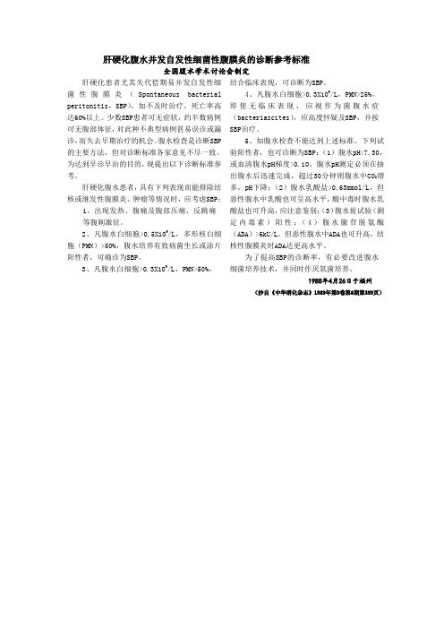
肝硬化腹水并发自发性细菌性腹膜炎的诊断参考标准

肝硬化腹水并发自发性细菌性腹膜炎的诊断参考标准全国腹水学术讨论会制定肝硬化患者尤其失代偿期易并发自发性细菌性腹膜炎(Spontaneous bacterial peritonitis,SBP),如不及时治疗,死亡率高达60%以上。
少数SBP患者可无症状,约半数病例可无腹部体征,对此种不典型病例甚易误诊或漏诊,而失去早期治疗的机会。
腹水检查是诊断SBP 的主要方法,但对诊断标准各家意见不尽一致。
为达到早诊早治的目的,现提出以下诊断标准参考。
肝硬化腹水患者,具有下列表现而能排除结核或继发性腹膜炎、肿瘤等情况时,应考虑SBP:1、出现发热、腹痛及腹部压痛、反跳痛等腹刺激征。
2、凡腹水白细胞>0.5X10⁹/L,多形核白细胞(PMN)>50%,腹水培养有致病菌生长或涂片阳性者,可确诊为SBP。
3、凡腹水白细胞>0.3X10⁹/L,PMN>50%,结合临床表现,可诊断为SBP。
4、凡腹水白细胞>0.3X10⁹/L,PMN>25%,即使无临床表现,应视作为菌腹水症(bacteriascites),应高度怀疑及SBP,并按SBP治疗。
5、如腹水检查不能达到上述标准,下列试验阳性者,也可诊断为SBP:(1)腹水pH<7.30,或血清腹水pH梯度>0.10,腹水pH测定必须在抽出腹水后迅速完成,超过30分钟则腹水中CO2增多,pH下降;(2)腹水乳酸盐>0.63mmol/L,但恶性腹水中乳酸也可呈高水平,酸中毒时腹水乳酸盐也可升高,应注意鉴别;(3)腹水鲎试验(测定内毒素)阳性;(4)腹水腺苷脱氨酶(ADA)>6kU/L。
但恶性腹水中ADA也可升高,结核性腹膜炎时ADA达更高水平。
为了提高SBP的诊断率,有必要改进腹水细菌培养技术,并同时作厌氧菌培养。
1988年4月26日于福州(抄自《中华消化杂志》1989年第9卷第6期第359页)。
60例慢性重型肝炎伴自发性细菌性腹膜炎患者的预见性干预护理

60例慢性重型肝炎伴自发性细菌性腹膜炎患者的预见性干预护理作者:江洁雅来源:《中国保健营养·中旬刊》2013年第03期【摘要】目的:通过对60例慢性重型乙型肝炎并发自发性细菌性腹膜炎(Spontaneous bacterial peritonitis,SBP)患者的临床预见性干预护理,初步探讨早期预见性干预护理对于防治慢性重型乙肝合并自发性腹膜炎的临床意义。
方法:60例重肝并发SBP患者分为治疗组(预见性干预护理组)和对照组(普通护理组),分别统计两组护理后有效率及并发症发生率,并进行分析和探讨。
结果:治疗组的肝性脑病和电解质紊乱发生率低于对照组(P【关键词】慢性重型乙型肝炎;自发性细菌性腹膜炎;预见性干预护理【中图分类号】R473 【文献编识码】A 【文章编号】1004-7484(2013)03-0016-02重型肝炎是机体在多种致病因子作用下,肝脏在短期内大量坏死所致的肝功能衰竭的一类综合征[1],病势凶险,病情发展快,并发症多,死亡率高。
由于重型病毒性肝炎病人伴有免疫功能下降,极易发生继发性细菌感染, Rolando等[2]认为约80%的重型肝炎病人可发生细菌或真菌感染,12%~28%的病人死亡原因与感染有关。
因此,如何在护理工作中减少感染发生机率成为挽救重型肝炎患者死亡率的重要途径。
本文针对60例患者经预见性干预护理后两组并发症的发生率及有效率的比较,探讨本护理方法对于重型肝炎患者的初步疗效性。
1 资料与方法1.1 病例资料收集我院感染科2009年9月至2010年8月住院重肝患者60例,病例诊断均符合2000年全国第十次病毒性肝炎及肝病学术会议(西安)重型肝炎诊断标准。
60例重肝患者随机分为治疗组(30例)和对照组(30例),分组标准详见后。
其中男性49例,女性11例,男、女比例为4.5:1。
平均年龄37.37±10.79岁。
早期22例,中期28例,晚期10例。
基础病为慢乙肝者39人,肝硬化者2人。
注射用哌拉西林钠他唑巴坦钠治疗肝硬化患者自发性细菌性腹膜炎的临床观察

注射用哌拉西林钠他唑巴坦钠治疗肝硬化患者自发性细菌性腹膜炎的临床观察【摘要】目的探讨注射用哌拉西林钠他唑巴坦钠在肝硬化患者自发性细菌性腹膜炎治疗中的应用效果。
方法选取2020年6月-2021年12月本院60例肝硬化合并自发性细菌性腹膜炎患者,随机分为对照组(头孢曲松治疗)与观察组(注射用哌拉西林钠他唑巴坦钠治疗),对比治疗效果。
结果治疗总有效率、TNF-α和IL-6方面,治疗后,观察组较对照组优(P<0.05)。
结论注射用哌拉西林钠他唑巴坦钠在肝硬化合并自发性细菌性腹膜炎治疗中效果显著,其有助于患者及早恢复健康,同时可降低炎性指标水平,值得采纳、推广。
【关键词】自发性细菌性腹膜炎;肝硬化;注射用哌拉西林钠他唑巴坦钠[Abstract] Objective To investigate the effect of piperacillin sulbactam in the treatment of spontaneous bacterial peritonitis in patients with liver cirrhosis. Methods 60 patients with cirrhosis complicated with spontaneous bacterial peritonitis in our hospital from June 2020 to December 2021 were randomly pided into control group (ceftriaxone) and observation group (piperacillin sulbactam). Results the total effective rate and TNF- α And IL-6, after treatment, the observation group was better than the control group (p<0.05). Conclusion piperacillin sulbactam is effective in the treatment of liver cirrhosis complicated with spontaneous bacterial peritonitis. It can help patients recover health as soon as possible and reduce the level of inflammatory indicators. It is worth adopting and popularizing.【 key words 】 spontaneous bacterial peritonitis; cirrhosis; Piperacillin and Sulbactam自发性细菌性腹膜炎为肝硬化患者常见,且病情相对严重的一种并发症,该病发生后,患者受损加重,具有较高的死亡率。
1例圣乔治教堂诺卡菌眼部感染病例及文献回顾

[收稿日期] 2020-03-22[作者简介] 刘艳芝(1990-),女(汉族),湖南省株洲市人,主管技师,主要从事临床微生物检验相关研究。
[通信作者] 李军 E mail:lijun198412@126.com犇犗犐:10.12138/犼.犻狊狊狀.1671-9638.20206002·论著·1例圣乔治教堂诺卡菌眼部感染病例及文献回顾刘艳芝1,李虹玲2,李艳明2,刘清霞2,晏 群2,邹明祥2,刘文恩2,李 军2(1.湖南省职业病防治院检验科,湖南长沙 410007;2.中南大学湘雅医院检验科,湖南长沙 410008)[摘 要] 圣乔治教堂诺卡菌隶属于诺卡菌属,诺卡菌广泛分布于土壤和水中,不属于人体正常菌群,主要通过呼吸道吸入和破损皮肤侵入人体。
诺卡菌病由诺卡菌感染所致,诺卡菌感染的常见部位以肺部和皮肤多见,眼部感染报道较少,圣乔治教堂诺卡菌所致眼部感染的报道更少。
因此,报道某院1例圣乔治教堂诺卡菌眼部感染的病例,并结合国内外文献进行复习,旨在提高临床对诺卡菌病的诊治水平。
[关 键 词] 圣乔治教堂诺卡菌;诺卡菌病;眼部感染[中图分类号] R772.2犈狔犲犻狀犳犲犮狋犻狅狀狑犻狋犺犖狅犽犪狉犱犻犪犮狔狉犻犪犮犻犵犲狅狉犵犻犮犪:狅狀犲犮犪狊犲狉犲狆狅狉狋犪狀犱犾犻狋犲狉犪 狋狌狉犲狉犲狏犻犲狑犔犐犝犢犪狀 狕犺犻1,犔犐犎狅狀犵 犾犻狀犵2,犔犐犢犪狀 犿犻狀犵2,犔犐犝犙犻狀犵 狓犻犪2,犢犃犖犙狌狀2,犣犗犝犕犻狀犵狓犻犪狀犵2,犔犐犝犠犲狀 犲狀2,犔犐犑狌狀2(1.犇犲狆犪狉狋犿犲狀狋狅犳犆犾犻狀犻犮犪犾犔犪犫狅狉犪狋狅狉狔,犎狌狀犪狀犘狉犲狏犲狀狋犻狅狀犪狀犱犜狉犲犪狋犿犲狀狋犐狀狊狋犻狋狌狋犲犳狅狉犗犮犮狌狆犪狋犻狅狀犪犾犇犻狊犲犪狊犲狊,犆犺犪狀犵狊犺犪410007,犆犺犻狀犪;2.犇犲狆犪狉狋犿犲狀狋狅犳犆犾犻狀犻犮犪犾犔犪犫狅狉犪狋狅狉狔,犡犻犪狀犵狔犪犎狅狊狆犻狋犪犾,犆犲狀狋狉犪犾犛狅狌狋犺犝狀犻狏犲狉狊犻狋狔,犆犺犪狀犵狊犺犪410008,犆犺犻狀犪)[犃犫狊狋狉犪犮狋] 犖狅犽犪狉犱犻犪犮狔狉犻犪犮犻犵犲狅狉犵犻犮犪(犖.犮狔狉犻犪犮犻犵犲狅狉犵犻犮犪)belongstothe犖狅犮犪狉犱犻犪狊狆狆.,犖狅犮犪狉犱犻犪distributeswidelyinsoilandwater,itdoesnotbelongtothenormalfloraofhumanbody,mainlyinvadeshumanbodythroughrespiratorytractinhalationanddamagedskin.Nocardiosisiscausedby犖狅犮犪狉犱犻犪infection,犖狅犮犪狉犱犻犪infectioniscommoninthelungandskin,therearefewreportsofeyeinfection,reportsofeyeinfectioncausedby犖.犮狔狉犻犪犮犻 犵犲狅狉犵犻犮犪isevenfewer.Thispaperreportsacaseofeyeinfectionwith犖.犮狔狉犻犪犮犻犵犲狅狉犵犻犮犪,andreviewsthelitera tureathomeandabroad,soastoimproveclinicaldiagnosisandtreatmentofnocardiosis.[犓犲狔狑狅狉犱狊] 犖狅犽犪狉犱犻犪犮狔狉犻犪犮犻犵犲狅狉犵犻犮犪;nocardiosis;eyeinfection 诺卡菌病(Nocardiosis)是由诺卡菌(犖狅犮犪狉犱犻犪)感染引起的一种急慢性化脓性疾病。
左氧氟沙星对肝硬变腹水并发自发性细菌性腹膜炎的临床预防分析(5.12细胞与分子

左氧氟沙星对肝硬变腹水并发自发性细菌性腹膜炎的临床预防分析贾贻红1王尉2 杨传雷3 杜以真1(1.济南市传染病医院,山东济南250021)(2.济南市第五人民医院药剂科,山东济南250021)(3.济南市卫生学校,山东济南250021)【摘要】目的观察左氧氟沙星预防肝硬化腹水并发自发性细菌性腹膜炎(SBP)的疗效。
方法对117例肝炎肝硬化并发SBP患者,随机分成两组,预防组59例,对照组58例,预防组在基础治疗的同时采用口服左氧氟沙星0.2g,每日2次,连续治疗12周;对照组只采用基础治疗。
观察两组腹腔感染情况。
结果预防组有效57例,无一例发生SBP,对照组有效33例,有17例发生SBP,占34.0%(17/50),两组间比较有显著性差异(P<0.05)。
结论左氧氟沙星对SBP的预防效果效果明显,在临床治疗上可以作为预防SBP的首选药物。
【关键】:左氧氟沙星;自发性腹膜炎;预防;肝硬变;Levofloxacin in prevention of Spontaneous Bacterial Peritonitis Secondary to Hepatitis-associated Cirrhosis[Abstract]: Objective To observe the clinical efficacy of levofloxacin for spontaneous bacterial peritonitis secondary to hepatitis-associated cirrhosis. Methods117 patients with spontaneous bacterial peritonitis secondary to hepatitis-associated cirrhosis were divided randomly into two groups, which included 59 patients in prophylactic group and 58 patients in control group. The baseline of prophylactic group was similar to that of control group. The patients in prophylactic group received levofloxacin 0.2g, twice daily, the period of therapy was 2 weeks. Results There was no SBP in therapy group. The rate of SBP was 34.0%%in control group. There was a significantly difference in effective rate between the prophylactic group and control group (P<0.05). Conclusions Levofloxacin has good effect in prevention of spontaneous bacterial peritonitis,and levofloxacin could be used as a first-choice antimicrobial for spontaneous bacterial peritonitis.[Keyword]: levofloxacin; spontaneous bacterial peritonitis; hepatitis-associated cirrhosis;肝硬化腹水并发自发性细菌性腹膜炎(SBP) 又称原发性腹膜炎,是肝硬化腹水患者常见的严重并发症,预后差,病死率高,因此,预防肝硬化腹水患者并发SBP,能改善患者预后。
肝硬化自发性腹膜炎的临床分析

中国医疗前沿China Healthcare InnovationFebruary ,2008V ol ,3No .42008年02月第3卷第4期肝硬化自发性腹膜炎的临床分析陈育霞(解放军第180医院肝病中心二区,福建泉州362000)[关键词]肝硬化自发性腹膜炎[中图分类号]R 575.2;R572.2[文献标识码]A作者简介陈育霞(6),女,医师,解放军第医院肝病中心二区。
自发性腹膜炎(Spontaneous bacte rial peritonitis,S BP )的发生率很高,在肝硬化腹水患者中达10%~30%,使肝脏功能受损加重,并使肝移植患者围手术期病死率增加,最严重的并发症是肝肾综合征,发生率约30%,SBP 的病死率很高,一次发生的病死率约20%,发生后1年的病死率高达70%,所以早期发现及时治疗有很重要的临床意义,由于临床表现多不典型,易造成漏诊,延误治疗。
现将我科2002~2007年间收治肝硬化腹水合并S BP170例分析如下。
1临床分析1.1一般资料男性70例,女性14例,年龄33~70岁,平均52岁。
均为肝硬化失代偿期,其中肝炎后肝硬化64例,酒精性肝硬化19例,血吸虫肝硬化1例。
所有病例甲胎球蛋白AFP 及癌胚抗原CEA 均在正常范围内。
1.2临床表现(1)发热,体温≥39℃12例(14.28%);38-39℃29例(34.52%);37-37.9℃26例(30.95%);体温正常17例(20.23%)。
(2)腹部表现:腹胀81例(96.42%),腹痛47例(55.95%),腹部压痛43例(51.19%);腹部反跳痛39例(46.43%),腹肌紧张26例(30.95%),移动性浊音76例(90.47%);(3)其他:出现血压下降休克者12例(14.28%),发生肝肾综合征死亡者19例(22.61%)。
1.3实验室检查外周血象:白细胞>10×109/L 34例(40.47%);腹水检查:外观浑浊35例,外观微浑40例,李凡他阳性73例,李凡他阴性11例。
sbp诊断标准 -回复

sbp诊断标准-回复SBP诊断标准:什么是SBP?Spontaneous bacterial peritonitis(自发性细菌性腹膜炎,简称SBP)是一种严重的腹膜感染,主要发生在肝硬化患者合并腹水的情况下。
这种疾病往往导致患者出现呼吸急促、发热、腹痛等症状。
及时的诊断和治疗对于患者的生存和预后至关重要。
一、什么是肝硬化?在深入理解SBP之前,有必要先了解肝硬化是什么。
肝硬化是一种常见的肝脏疾病,是指在慢性肝病发展的过程中,肝细胞逐渐受损并被瘢痕组织替代的情况。
肝硬化是许多肝疾病的最终阶段,可以由慢性病毒性肝炎、酒精滥用、脂肪肝、肝胆管梗阻等多种原因引起。
二、SBP的发病机制1. 腹水的形成:肝硬化患者会出现腹水的情况,这是由于肝功能衰竭导致血浆白蛋白水平下降,血管内血浆渗出到腹腔的结果。
2. 腹水的感染:腹水中的细菌可以通过消化道或血源性途径进入腹腔,导致腹水感染。
3. SBP的发生:当腹水中细菌数量超过一定阈值或者细菌的毒力增强时,SBP就会发生。
常见的致病菌包括肠道革兰氏阴性菌和肠球菌。
三、SBP的诊断标准为了明确SBP的诊断,医学界制定了一系列标准来指导临床判断。
以下是常用的SBP诊断标准:1. 腹水分析:腹水必须通过穿刺抽取,进行细菌培养和生化分析。
细菌培养是诊断SBP的关键步骤,阳性结果为SBP的确诊。
2. 中性粒细胞计数:腹水中的中性粒细胞计数可以反映腹水感染的程度。
SBP的诊断标准之一是腹水中中性粒细胞计数大于250个/mm³。
3. 腹痛和发热:SBP的典型症状包括腹痛和发热。
这些症状是SBP的常见表现,但并不能作为诊断的唯一依据。
四、SBP的监测和治疗对于肝硬化患者合并腹水的情况,医生需要密切监测患者的病情变化,并进行及时的治疗。
治疗SBP的关键是使用抗生素,目的是根除腹腔中的细菌感染。
常用的抗生素包括第三代头孢菌素(如头孢噻肟)或氟喹诺酮类(如诺氟沙星)。
抗生素治疗的疗程通常为5-7天。
肝性脑病的护理措施

肝性脑病的护理措施概述肝性脑病是一种由肝脏疾病引起的中枢神经系统功能障碍的护理病症。
该病症通常表现为认知障碍、行为改变和神经肌肉问题。
对于患有肝性脑病的患者,提供适当的护理和照料可以帮助缓解症状,并提高生活质量。
本文将介绍肝性脑病的护理措施,包括监测和评估、药物治疗、饮食管理、康复和心理支持等方面。
监测和评估在护理肝性脑病患者时,监测和评估病情的变化至关重要。
以下是一些监测和评估的关键要点:•意识状态:密切监测患者的意识状态,包括观察是否有混乱、嗜睡或昏迷的表现。
•神经症状:观察患者是否出现神经症状,例如震颤、肌肉僵硬或手部的构音困难。
•行为改变:注意观察患者是否有焦虑、易激惹、抑郁或其他行为改变。
•肝功能:定期检测患者的肝功能指标,包括肝功能酶、胆红素和凝血功能等指标。
•贫血情况:检查患者的血红蛋白水平,确保足够的氧气供应到大脑。
药物治疗药物治疗是控制肝性脑病症状的重要手段之一。
以下是常用的药物治疗方法:•乳果糖:乳果糖是常用的药物,可通过阻止肠道中的氨的吸收来减少血氨水平。
它还可以增加益生菌的生长,改善肠道菌群平衡。
•纳洛酮:纳洛酮是一种阿片受体拮抗剂,可用于治疗由便秘引起的高胆红素血症。
它可以减少氨的产生和吸收。
•抗生素:抗生素可以用于治疗肝性脑病的继发感染,如感染性胆道炎等。
常用的抗生素包括头孢呋辛、美罗培南等。
需要注意的是,药物治疗应在医生的指导下进行,以确保药物的合适用量和时机。
饮食管理饮食管理对于肝性脑病患者的康复和治疗非常重要。
以下是一些饮食管理的建议:•高蛋白饮食:患者需要摄入足够的蛋白质来帮助修复和保护肝脏组织。
但应避免高氨基酸含量的食物,如红肉和乳制品。
•限制盐摄入:减少患者对盐的摄入量,可以帮助减轻水肿和腹水等症状。
•补充维生素:适当补充维生素B群和C等维生素,有助于改善神经功能。
饮食管理的具体方案应根据患者的具体情况和医嘱进行调整。
康复和心理支持肝性脑病对患者的心理和身体都有很大的影响,因此提供康复和心理支持至关重要。
自发性细菌性腹膜炎

小结
• 早期诊断SBP对于提高治疗效果十分重要 • 推荐常规对肝硬化腹水患者腹腔穿刺及腹 水化验检查 • 足量抗生素治疗是提高治疗效果的关键 • 没有厌氧菌感染的临床或实验室依据,不 联用抗厌氧菌的药物 • 预防性抗生素治疗可降低SBP 复发风险 • SBP 发作后康复患者的长期生存率较差, 应考虑肝移植
欧洲SBP的管理-1
• 诊断SBP 后应立即开始经验性抗生素治疗 • SBP 最常见的病原体为革兰阴性需氧菌( 如大肠 杆菌) • 抗生素治疗的一线药物是三代头孢菌素。备选药 物包括阿莫西林/克拉维酸和喹诺酮类药物( 如环 丙沙星或氧氟沙星) • 在以下情况时,不使用喹诺酮类药物: 已使用该类药物预防SBP 的患者 喹诺酮类耐药菌的高流行地区 院内感染的SBP
欧洲无并发症腹水的管理-2
• 对于醛固酮拮抗剂无应答( 定义为每周体质 量下降< 2 kg) 的患者或出现高钾血症的患 者,应该逐步加用呋塞米( 从40 mg /d 开 始,每次增加40mg,最大剂量160 mg /d) • 第1 个月内应进行严密的临床和生化监测 • 对于腹水复发的患者,应给予醛固酮拮抗 剂+呋塞米的联合治疗,药物剂量应根据应 答的情况逐步增加
3 级
腹水大量或严重腹水,显著的腹部膨隆
治疗 无需治疗 限制钠的摄入和利尿剂
腹腔穿刺大量放液,随后限制钠的摄入和利尿剂
占国清,等.实用肝脏病杂志.2010,13(6):414-417
中国SBP的诊断标准-1
• 发热、腹痛及腹部压痛、反跳痛等腹膜刺 激征 • 腹水白细胞计数>0.5×10⁹/L,多形核 白细 胞(PMN)计数>50%,腹水培养有致病菌 生长或涂片阳性者,可确诊为SBP • 凡腹水白细胞计数>0.3×10⁹/L, PMN>50%,结合临床表现,可诊断为SBP • 凡腹水白细胞计数>0.3×10⁹/L, PMN>25%,既往无临床表现,应视作为菌 腹水症(Bacteriascites),应高度怀疑 SBP, 并按SBP治疗
肝硬化病人小肠细菌过度生长情况与肝功能、自发性腹膜炎的相关性

肝硬化病人小肠细菌过度生长情况与肝功能、自发性腹膜炎的相关性宋洁;张慧敏;王佳林【摘要】目的:探讨肝硬化病人小肠细菌过度生长与肝功能和、自发性腹膜炎之间的相关性。
方法:选取2016-01~2017-10期间我院收治的106例肝硬化病人作为研究组,对照组为此期间在我院进行体检人员106例。
研究组和对照组进行乳果糖氢呼气试验,对小肠细菌过度生长进行检测;并检测受试者肝功能,包括总胆汁酸(TBA)、白蛋白(ALB)、血清胆固醇(CHO);根据自发性腹膜炎诊断标准对每位受试者进行诊断。
通过ROC曲线曲线下面积分析肝硬化病人小肠细菌过度生长与肝功能、自发性腹膜炎之间的相关性。
结果:研究组检出小肠细菌过度生长63例(59.43%),对照组仅检出2例(1.89%);研究组检出自发性腹膜炎43例(40.57%),对照组未检出自发性腹膜炎;研究组平均TBA为10.6±1.8μmol/L,平均ALB为34.6±3.9 g/L,平均CHO为1.9±1.6 mmol/L。
研究组小肠过度生长阳性与肝功能减低、自发性腹膜炎阳性具有较强的相关性;对照组过度生长阳性与肝功能减低、自发性腹膜炎阳性均呈无相关性。
结论:肝硬化病人小肠过度生长阳性与肝功能减低、自发性腹膜炎阳性具有较强的相关性。
【期刊名称】《内蒙古医科大学学报》【年(卷),期】2018(040)002【总页数】5页(P147-151)【关键词】肝硬化;小肠细菌过度生长;肝功能;自发性腹膜炎;ROC曲线【作者】宋洁;张慧敏;王佳林【作者单位】[1]内蒙古自治区人民医院消化内科,内蒙古呼和浩特010017;;[1]内蒙古自治区人民医院消化内科,内蒙古呼和浩特010017;;[1]内蒙古自治区人民医院消化内科,内蒙古呼和浩特010017;【正文语种】中文【中图分类】R604肝硬化为临床常见病及多发病,主要以晚期病人发生门脉高压和肝功能损害等为其特征性临床表现,极大影响病人生活质量,严重者甚至威胁病人生命健康[1]。
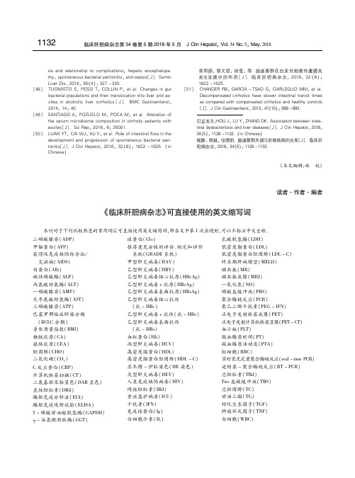
《临床肝胆病杂志》可直接使用的英文缩写词

乙型肝病毒e 抗原(HBeAg)
一氧化氮(NO)
一磷酸腺苷(AMP)
乙型肝炎病毒表面抗原(HBsAg) 磷酸盐缓冲液(PBS)
天冬氨酸转氨酶(AST)
乙型肝炎病毒核心抗体
聚合酶链反应(PCR)
三磷酸腺苷(ATP)
(抗- HBc)
聚乙二醇干扰素(PEG - ) IFN
巴塞罗那临床肝癌分期
乙型肝炎病毒e 抗体(抗- HBe) 正电子发射断层成像(PET)
(本文编辑:林 姣)
读者·作者·编者
《临床肝胆病杂志》可直接使用的英文缩写词
本刊对于下列比较熟悉的常用词汇可直接使用英文缩写词,即在文中第1 次出现时,可以不标注中文全称。
二磷酸腺苷(ADP)
球蛋白(Glo)
乳酸脱氢酶(LDH)
甲胎蛋白(AFP)
推荐意见分级的评估、制定和评价 低密度脂蛋白(LDL)
(BCLC 分期)
乙型肝炎病毒表面抗体
正电子发射计算机断层显像(PET -CT)
身体质量指数(BMI)
(抗- HBs)
血小板(PLT)
糖链抗原(CA)
血红蛋白(Hb)
凝血酶原时间(PT)
癌胚抗原(CEA)
丙型肝炎病毒(HCV)
凝血酶原活动度(PTA)
胆固醇(CHO)
高密度脂蛋白(HDL)
红细胞(RBC)
栾雨婷, 蔡文君, 徐莹, 等. 肠道菌群在自发性细菌性腹膜炎 发生发展 中 的 作 用 [ J] . 临 床 肝 胆 病 杂 志, 2016, 32 ( 8 ) : 1622 -1625. [51] CHANDER RB, GARCIA - TSAO G, CIARLEGLIO MM, et al. Decompensated cirrhotics have slower intestinal transit times as compared with compensated cirrhotics and healthy controls [J]. J Clin Gastroenterol, 2013, 47(10): 888 -893. 引证本文:HOU J, LU Y, ZHANG DK. Association between intes tinal dysbacteriosis and liver diseases[J]. J Clin Hepatol, 2018, 34(5): 1128 -1132. (in Chinese) 候静, 路越, 张德凯. 肠道菌群失调与肝脏疾病的关系[J]. 临床肝 胆病杂志, 2018, 34(5): 1128 -1132.
肝硬化腹腔感染的诊疗进展及挑战

!()*+!肝硬化腹腔感染的诊疗进展及挑战黎倍伶1,陈金军1,21南方医科大学南方医院肝病中心,广州510515;2南方医科大学南方医院增城分院肝病中心,广州510515摘要:感染是终末期肝病最常见并发症,其中,腹腔感染为最常见感染类型之一。
腹水培养阳性率低,腹腔感染的诊断主要基于腹水多核细胞计数,治疗以经验性抗菌素使用为主。
腹腔感染的诊断标准对临床实践指导意义有限,目前尚无可转化应用于临床实践的新型诊断标志物。
病原体诊断方面,宏基因组二代测序为潜在快速识别肝硬化腹腔感染病原体或复合感染的新方法。
治疗上,中国肝硬化腹水管理指南主要强调抗菌素的治疗,对联合输注人血白蛋白的剂量无明确推荐。
肝硬化腹腔感染的诊断和治疗上仍面临巨大的挑战,未来需要更多相关的研究,包括优化腹腔感染诊断、预防、治疗等,进一步回答相关的临床问题,从而更好指导临床实践。
关键词:肝硬化;腹腔内感染;诊断;治疗学中图分类号:R575.2 文献标志码:A 文章编号:1001-5256(2021)04-0757-04Diagnosisandtreatmentofabdominalinfectioninpatientswithlivercirrhosis:AdvancesandchallengesLIBeiling1,CHENJinjun1,2.(1.LiverDiseaseCenter,NanfangHospital,SouthernMedicalUniversity,Guangzhou510515,China;2.LiverDiseaseCenter,ZengchengBranchofNanfangHospital,SouthernMedicalUniversity,Guangzhou510515,China)Abstract:Infectionisthemostcommoncomplicationinpatientswithend-stageliverdisease,amongwhichabdominalinfectionisthemostcommontype.Thereisalowpositiverateofasciticfluidculture,andabdominalinfectionismainlydiagnosedbasedonmultinucleatedcellcountinascitesandismainlytreatedbyempiricalantimicrobialtherapy.Thediagnosticcriteriaforabdominalinfectionhavelimitedguidingsignificanceinclinicalpractice,andcurrentlytherearestillnonewdiagnosticmarkersthatcanbeusedinclinicalpractice.Forthepatho genicdiagnosisofabdominalinfection,metagenomicnext-generationsequencingisanewtechniqueforrapididentificationofpathogensofabdominalinfectionoroverlapinfectioninlivercirrhosis.Intermsoftreatment,Chineseguidelinesonthemanagementofascitesincirrhosisemphasizeantimicrobialtherapyandgivenoexplicitrecommendationforthedoseofhumanserumalbumininfusion.Therearestillgreatchal lengesinthediagnosisandtreatmentofabdominalinfectionincirrhoticpatients,andmorestudiesareneededinthefuturetoanswerrelevantquestionsandbetterguideclinicalpractice,includingtheoptimizationofthediagnosis,prevention,andtreatmentofabdominalinfection.Keywords:LiverCirrhosis;IntraabdominalInfections;Diagnosis;TherapeuticsDOI:10.3969/j.issn.1001-5256.2021.04.003收稿日期:2020-12-25;修回日期:2021-01-04基金项目:科技部病毒性肝炎及艾滋病等传染病重大专项(2018ZX10723203)作者简介:黎倍伶(1991—),女,医学博士,主要从事肝硬化感染相关研究通信作者:陈金军,chjj@smu.edu.cn 感染是终末期肝病最常见并发症,也是不良预后重要影响因素。
人血白蛋白处方点评标准

人血白蛋白处方点评标准(讨论稿)为规范人血白蛋白的临床使用,降低药品费用,改善人血白蛋白短缺现状,促进人血白蛋白的合理使用,开展人血白蛋白临床应用相关处方或医嘱点评工作具有重要意义。
为此,药剂科综合参考《处方管理办法》(卫生部令第53号)、《医院处方点评管理规范(试行)》(卫医管发〔2010〕28号)、美国大学医院联合会《人血白蛋白、非蛋白胶体及晶体溶液使用指南》、北京地区《血液制品处方点评指南》以及人血白蛋白说明书、相关循证医学依据等,结合我院临床实际,制定我院人血白蛋白临床使用评价标准初稿。
【点评标准】1.适应证不适宜;2.用法、用量不适宜;3.遴选的药品不适宜;4.药品剂型或给药途径不适宜;5.联合用药不适宜;6.重复给药;7.有配伍禁忌或者不良相互作用;8.其它用药不适宜情况;【点评细则】1.适应证不适宜:“诊断”栏未注有符合以下情况一项或一项以上适应证者判定为适应证不适宜。
➢严重失血、创伤和烧伤等引起的休克;纠正人血白蛋白作为补充血容量的首选药物的误区。
《美国医院联合会人血白蛋白、非蛋白胶体及晶体溶液使用指南》(简称UHC,下同)[1]中提到:对于出血性休克,晶体溶液可作为首选药物用于扩张血容量,成人患者输入4L晶体液后2h无效,可考虑非蛋白胶体液,当对非蛋白胶体液有禁忌时才考虑使用5%白蛋白。
目前的循证医学证据表明在外科病人中,对于病死率、并发症发生率的结局指标,不同种类的胶体液并未显示出明显差异。
➢脑水肿及大脑损伤所致的颅压升高;人血白蛋白可提高血浆胶体渗透压,将脑组织的水分转移到血管内而减轻脑水肿,降低颅内压。
对于蛛网膜下腔出血、缺血性中风和头部创伤引起的血管痉挛,应首选晶体溶液维持脑灌注压。
如果存在脑水肿的危险,应使用高浓度白蛋白(25%)胶体液维持脑灌注压【1】。
➢新生儿高胆红素血症;新生儿高胆红素血症为人血白蛋白的适应症,白蛋白能与血中胆红素结合,阻止胆红素通过血脑屏障,促进胆红素排泄。
亚胺培南与头孢哌酮舒巴坦治疗肝硬化自发性腹膜炎的疗效比较

亚胺培南与头孢哌酮舒巴坦治疗肝硬化自发性腹膜炎的疗效比较孙文锦【摘要】Objective To compare the curative effect of imipenem and cefoperazone sulbactam in treatment of spontaneous bacterial peritonitis of liver cirrhosis. Methods Seventy patients with spontaneous bacterial peritonitis of liver cirrhosis were divided into imipenem group (A group, 35 cases) and cefoperazone sulbactam group (B group, 35 cases). A group was treated with abdominal puncture combined with imipenem, and B group was treated with abdominal puncture combined with cefoperazone sulbactam. The cure time of two groups was 10 days. The treatment effects, the changes of clinical symptoms were observed after treatment. Results The total effective rate of A group (82. 9% ) was significantly better than that of B group(68. 6% ) ( P < 0. 05 ) . The remission time of clinical symptoms in A group after treatment was earlier than that of B group (P < 0. 05 ). Conclusion The treatment of imipenem for spontaneous bacterial peritonitis of liver cirrhosis has a good effect. It could alleviate the clinical symptoms and be worthy of clinical application.%目的比较亚胺培南与头孢哌酮舒巴坦治疗肝硬化自发性腹膜炎的临床疗效.方法 70例肝硬化自发性腹膜炎患者分成亚胺培南组(A组,35例)和头孢哌酮舒巴坦组(B组,35例),A组采用腹腔穿刺引流及亚胺培南治疗,B组采用腹腔穿刺引流及头孢哌酮舒巴坦治疗,两组疗程均为10 d.治疗结束后观察两组疗效及临床症状变化情况.结果 A组治疗总有效率(82.9%)明显优于B组(68.6%)(P<0.05);A组发热、腹胀、腹痛、腹部压痛和反跳痛等临床症状缓解时间明显早于B组(P<0.05).结论亚胺培南治疗肝硬化自发性腹膜炎疗效显著,不良反应少,可明显缓解临床症状,值得临床推广应用.【期刊名称】《胃肠病学和肝病学杂志》【年(卷),期】2012(021)003【总页数】2页(P240-241)【关键词】亚胺培南;头孢哌酮舒巴坦;肝硬化;自发性腹膜炎【作者】孙文锦【作者单位】湖北省鄂州市中心医院感染科,湖北鄂州436000【正文语种】中文【中图分类】R575.2自发性腹膜炎(SBP)是临床上肝硬化疾病常见的一种严重并发症,也是临床治疗的重点和难点之一。
- 1、下载文档前请自行甄别文档内容的完整性,平台不提供额外的编辑、内容补充、找答案等附加服务。
- 2、"仅部分预览"的文档,不可在线预览部分如存在完整性等问题,可反馈申请退款(可完整预览的文档不适用该条件!)。
- 3、如文档侵犯您的权益,请联系客服反馈,我们会尽快为您处理(人工客服工作时间:9:00-18:30)。
Abdominal pain and Tenderness 14/03/02 STEVENSIULearning Objectives 1Describe the anatomy and physiology of the peritoneum, including the innervations of the visceral and parietal layersThe peritoneum is a continuous, transparent serous membrane that lines the abdominopelvic cavity and invests the viscera. It consists of two continuous layers.1.Parietal peritoneum- lining the internal surface of the abdominopelvic wall.2.Visceral peritoneum – investing the visceraVisceral peritoneum supplied small type C pain fibers and can only response to chronic-aching suffering type of pain. These fibers can response to ischaemia, chemical stimuli, and spasm of a hollow viscus and over distention of a hollow viscusParietal peritoneum is supplied by the spinal nerves not sympathetic nerves→ sharp localized somatic painLearning objectives 2Discuss conditions predisposing to spontaneous bacterial peritonitis (SBP)SBP= The inflammation of the peritoneum without a clear event such as bowel perforation that would account for the entry of the pathogenic organism Liver Cirrhosis is the main predisposing factor. Other conditions leading to ascites e.g. Fulminant hepatic failure, Congestive heart failure, liver metastases can also predispose to SBP.Pathogenesis1.Seeding→ The exact pathogenic mechanism is unknown. Bacterial seeding is believed to involvehaematogenous spread of organisms in a patient in whom diseased liver and altered portal circulation result in a defect in the usual filtration.2.Growth--> The reduced levels of complement cascade and reduced opsonic and phagocyticproperties of neutrophils in advanced liver cirrhosis in an excellent culture medium provided by the ascitic fluids, promote the growth of the organisms.Symptoms1.Fever, Hypotension, Decreased or absent bowel sounds, Abdominal Pain and Abrupt onset of hepaticencephlopathy in patient with ascitesLearning Objective 3Outline intra- abdominal lesions that may be complicated by bacterial peritonitisBacterial peritonitis is due to entry of bacteria into the peritoneal cavity from a perforation in the GIT or from an external penetrating wound. E.Coli and Bacteroides are the most common causative agents. Common source of bacterial entry are:Bowel1.Appendicitis (By farthe most common)2.Diverticulitis3.Bowelstrangulations Chemical1.Ruptured pepticulcer2.Cholecystitis3.Acute pancreatitisOthers1.Acute salpingitis2.Abdominal trauma3.Peritoneal dialysisLearning Objectives 4Outline the causes of chemical peritonitisChemical peritonitis is due to the escape of bile, contents of the gastrointestinal tract, or pancreatic juice into the peritoneal cavity; the contents of the fluid causes chemical injury, shock, and peritoneal exudation prior to occurrence of any associated infection.Learning objective 5Compare the clinical features of localized and generalized peritonitis. In particular discuss the haemodynamic consequences of generalized peritonitisInvestigations1.Confirm the diagnosis of LP or GP2.Determine the cause of LP or GPHaemodynamic consequencesHypovolumic and septic Shock→1.The normal 7-8 L of fluids are not absorbed from the distal bowel and colon.2.High volume of inflammatory exudate.3.Decreased oral intake4.The peritoneum also provides a large surface area for the absorption of GN endotoxin, leading tosepticemia. High level of circulatory endotoxins can lead to widespread exudation and activation of the coagulation cascade, leading to DIC.5.The contraction of the circulation volume leads to hypotension and reflex tachycardia. Theperfusion to the kidneys is also reduced leading to oligouria.Learning Objective 6Discuss the pathogenesis of intra-abdominal abscesses. Outline the role of imaging in the management of such abscesses.Management1.Ultrasounds and abdominal X rays → elevation of the diaphragm with a fluid level below or aneffusion aboveLearning Objectives 7Outline the factors predisposing to the development of diverticular disease, emphasizing the role of diet. Describe the clinical presentation and complications of diverticular disease.Diverticular diseaseDefinition∙Acquired deformity where the mucosa and submucosa herniate through the underlying muscularis;95% of which occurs in the sigmond colon.Aetiology∙Affects 10% of American population, incidence increases from the age of 40.∙Functional aetiological factors → diet of highly refined foods and low fibre a/w modern affluent life∙Structural aetiological factors→ associated with connective tissue diseases such as Ehler-Danlos and Marfan’s sy ndromeClinical presentation1.Chronic constipation with diarrhoea and flatulence→ 80%2.Intermittent and unpredictable gripping lower abdominal painPathophysiology and Clinicopathological correlation1.Decreased dietary fibre means that the forward propulsion of the faeces more difficult at normaltransmural pressure more difficult. Reduced propulsion leads to constipation. Secondary bacterial liquecifaction of the retained faceal material leads to diarrhoea and flatulence.2.The subsequent increased peristaltic contractions, which causes the intermittent and unpredictablegripping lower abdominal pain. The pain may last for hours to days with sudden relief upon passing flatus and flatulence.3.The peristaltic contractions also cause an increased in intraluminal pressure and muscular hypertrophy.The raise pressure causes the herniation of mucosa through the weaker points of the muscular wall, which are located between the mesenteric and antimesenteric teniae.Complications1.Diverticular Bleeding →most common cause of painless lower GIT bleeding in the elderly. It iscaused by rupture and bleeding of the colonic intramural arteries.2.Diverticulitis→ stagnant faecal material causes inflammation of the wall of the diverticulum. Patientdevelops abdominal pain and fever. It may progress to abscess formation and perforation.Learning Objectives 8Discuss the differential diagnosis of right iliac fossa pain, describe the presentation and complications of acute appendicitis (AA).Pathophysiology and Clinical Presentation1.AA is initiated by the obstruction of the appendiceal lumen, most commonly by facolith.2.Continual secretion by the mucosal glands raises the intraluminal pressure.3.Raised pressure impedes mucosal blood flow and contributes to mucosal ischaemic damage4.Damaged mucosa predisposes to enteric bacterial invasion. The subsequent inflammation responsecauses further ischaemia in a feed forward manner.5.In early acute appendicitis, inflammatory mural exudate and the smooth muscle contractionsagainst an obstructive lumen stimulate the visceral afferent pain fibres. The visceral pain is referred to a T10 dermatome distribution, causing the epigastric pain. The pain is colicky due to the periodic peristaltic contraction of appendiceal smooth muscles.6.Reflex pyelospasm and inhibition of caecal peristalsis cause anorexia and constipationrespectively. Vomiting is rare due to anorexia.7.Release of inflammatory cytokines such as IL-1 and INF alpha causes systemic symptoms such asfever (<38 degrees), leucocytosis and raised coagulation factors.8.After a few hours, serosal fibrinosuppurative exudate stimulates the parietal peritoneum which hassomatic innervations, resulting in localized constant pain in the RIF and muscle guarding over the appendix.Complications1.Serosal fibrinosuppurative exudate is likely to cause adhesion to the greater omentum forming anappendiceal mass, which makes surgical removal difficult.2.At the stage, the appendix may become gangrenous and predisposes to rupture. Contained ruptureinto the appendiceal mass predisposes to periappendiceal abscess. Uncontained rupture into the peritoneum causes localized peritonitis and general peritonitis. Generalized peritonitis can lead to the fatal complication of GN septicaemia.3.Pyogenic microorganisms may enter the appendiceal vein, resulting in pyelophlebitis and liverabscesses.4.Residual abscesses in the paracolic gutter, subphrenic and pelvic regions may occur due to poordrainage of the fibrinosuppurative maternal5.Adhesion and bowel obstruction occurs months/ years after the appendicitis. Granulation tissuecan form between loops of bowel and appendix. Such adhesion can causes strangulations of bowel when another loop is trapped between adhered loop and appendix.。
