HE Staining
HE染色资料介绍

普通染色又称常规染色或HE(Haematoxylin and eosin)染色。
它是病理技术中最常用的一种方法,通过它可以做出病理诊断和发现寻求别的辅助方法,以达到准确,完整的病理诊断。
一、染色目的病理学的所有切片,都必须通过一种以上的染料,通过各种不同的方法,将切片中各种不同的物质,在不同染液的作用下,将其显示出来,使之在光学显微镜下,能够完全的观看各种结构。
例如,HE染色,好质量的切片可以清晰地显示出许多不同的结构,细胞核着蓝黑色,细胞浆着粉红色,软骨着蓝色等。
清晰的结构为诊断提供可靠的依据,因此,染色技术也是病理技术中的重要组成部分,必须不断地总结,方能提高。
如果染色不好,切片染色一团糟,红蓝不分,结构不清,层次不明,影响了镜下的观察,直接影响了临床诊断,染色结果的好坏直接关系到诊断的准确性。
二、染色的作用1.化学作用:所有的染色液中,可把它们分为两种类型,一种为酸性,另一种为碱性。
酸性染料中有染色作用的为阴离子,碱性染料中有染色作用的为阳离子,每个细胞也存在着两种物质,细胞核含的是酸性物质,细胞浆含的是碱性物质。
在染色时,细胞核中的酸性物质与苏木素染液中的阳离子发生作用,细胞浆中的碱性物质与伊红染液中的阴离子发生作用,由于反应的部位不同,结果着色有异。
2.物理作用:在染色过程中,染液中的色素微粒子浸入到被染组织的粒子间隙内,此时,因受分子的引力作用,色素微粒子被吸附而着色。
由于各种组织有不同的吸附能力和不同的吸附程度,因此就可显出来不同的颜色来。
一般来说,染色的学说还有许多,但说服力强的仅有上述两种。
但不管怎么样解释,实际上完成的每一种染色,都与上述两种学说分不开,它们的作用是相辅相成,同时存在的。
三、染色方法及步骤1.人工苏木素-伊红染色法(HE法)(1)切片浸入二甲苯中5-10min;(2)切片浸入二甲苯中5-10min;(3)100%酒精1min。
(4)100%酒精1min。
(5)95%酒精1min。
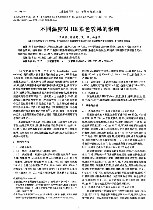
不同温度对HE染色效果的影响
醇 100 mL、硫酸铝钾 150 g、蒸馏水 2 000 mL、碘酸钠 1.2 g、冰 醋酸 120 mL、甘油 900 mE)、0.5% ~1.0%伊红染 色液 (95% 乙醇配制 )等。 1.1.3 试验仪器 本试验所 用的仪器主要有 精密 电子 天平 (Ate)、电热鼓风干燥箱 (101—5型)、包埋机(徕卡 YB一6LF)、 轮转式切片机(徕卡 RM2016)、摊片机(徕卡 1-111210)等。
1 材料与方法
1.1 材 料 与 仪 器 1.1.1 材料 草鱼 的肾、肝、肠组织切片。 1.1.2 试 剂配制 常 用于 组织学 切片 制作 的试剂 有 Bouin 氏液(苦味酸 75 mL、40% 甲醛 25 mL、冰醋酸 5 mL)、粘合剂 (鸡蛋清 )、清洗液 (重 铬酸钾 8O g、水 1 000 mL、粗制 浓硫 酸 120 mL)、乙醇 (分别 配制 了 70%、80%、90% 、95% 、100% 5 种浓度 )、1/2二 甲苯 (乙醇和二 甲苯按 1:l比例配制)、氨水 (1%)、酸水 (1% )、0.2%苏 木精染色液 (苏木 精 6 g、无水 乙
·-—
—
168- ——
江苏农业科学 2017年第 45卷第 22期
王庆容 ,徐 晓舒 ,夏 浪 ,等.不同温度 对 HE染色效果的影响[J].江苏农业科学,2017,45(22):168—170 doi:10.15889/j.isan.1002—1302.2017.22.046
不 同温度对 HE染 色效果 的影 响
收稿 日期 :2016—03—17 基金项 目:贵州省科技 厅农业 攻关项 目(编号 :黔科合 NZ[2013]3027
号 );贵州省教育 厅“125计划”重大科技专项 (编号 :黔教合重大专 项字 [2013]025号 );贵 州省科技 厅联 合基金 (编号 :黔 科合 J字 LKZS[2012]17号 )。 作者简介 :王庆容(1972一 ),女 ,贵州务川人 ,硕 士,教授 ,主要从事动 物分子生物学及疾病防治研 究。E—mail:610194977@qq.COITI。
石蜡切片制作和HE染色实验报告

石蜡切片制作和HE染色实验报告英文回答:Introduction:Paraffin sectioning and H&E staining are essential techniques in histopathology for examining tissue samples under a microscope. Paraffin sectioning involves embedding tissues in paraffin wax, cutting thin sections, and mounting them on glass slides. H&E staining is a common staining method that uses hematoxylin and eosin to differentiate cell components.Materials and Methods:Tissue samples。
Paraffin wax。
Microtome。
Glass slides。
Hematoxylin。
Eosin。
Ethanol。
Xylene。
Procedure:Paraffin Sectioning:1. Fix the tissue samples in formalin and process them through a series of graded ethanol solutions.2. Embed the tissues in paraffin wax and allow them to solidify.3. Use a microtome to cut thin sections (5-10 microns)from the paraffin block.4. Mount the sections on glass slides and allow them to dry.H&E Staining:1. Deparaffinize the sections by passing them through xylene and ethanol solutions.2. Stain the sections with hematoxylin and rinse with water.3. Differentiate the hematoxylin staining with hydrochloric acid and rinse with water.4. Counterstain the sections with eosin and rinse with water.5. Dehydrate the sections by passing them through graded ethanol solutions and clear them in xylene.6. Mount the sections with a coverslip and examine them under a microscope.Interpretation of Results:Hematoxylin stains the nuclei blue, while eosin stains the cytoplasm pink.The stained sections allow for the examination of tissue morphology, identification of cell types, and detection of pathological changes.Conclusion:Paraffin sectioning and H&E staining are fundamental techniques in histopathology that provide valuable information for the diagnosis and study of diseases.中文回答:石蜡切片制作和HE染色实验报告。
HE染色

结果的判断
实验结果
细胞核被苏木精染成鲜明的蓝色,软骨基质、钙盐颗粒呈深蓝色,粘液呈灰蓝色。细胞浆被伊红染成深浅不 同的粉红色至桃红色,胞浆内嗜酸性颗粒呈反光强的鲜红色。胶原纤维呈淡粉红色,弹力纤维呈亮粉红色,红血 球呈橘红色,蛋白性液体呈粉红色。
着色情况与组织或细胞的种类有关,也随其生活周期及病理变化而改变。例如,细胞在新生时期胞浆对伊红 着色较淡或轻度嗜碱,当其衰老时或发生退行性变则呈现嗜伊红浓染。胶原纤维在老化和出现透明变性时,伊红 着色由浅变深。
H-E染色评定标准:
(1)切片完整,厚度4-6微米,厚薄均匀,无皱褶无刀痕;
(2)染色核浆分明,红蓝适度,透明洁净,封裱美观。
谢谢观看
由于组织或细胞的不同成分对苏木精的亲和力不同及染色性质不一样。经苏木精染色后,细胞核及钙盐粘液 等呈蓝色,可用盐酸酒精分化和弱碱性溶液显蓝,如处理适宜,可使细胞核着清楚的深蓝色,胞浆等其它成分脱 色。再利用胞浆染料伊红染胞浆,使胞浆的各种不同成分又呈现出深浅不同的粉红色。故各种组织或细胞成分与
试剂与仪器
适用范围
组织学、胚胎学、生物学、病理学教学与科研。
原理
易于被碱性或酸性染料着色的性质称为嗜碱性( basophilia )和嗜酸性( acidophilia );而对碱性染料和 酸性染料亲和力都比较弱的现象称为中性(neutrophilia)。
构成组织内蛋白质的氨基酸的种类很多,它们有不同的等电点。在普通染色法中,染色液的酸碱度为pH6左 右,细胞内的酸性物质如细胞核的染色质、腺细胞和神经细胞内的粗面内质及透明软骨基质等均被碱性染料染色, 这些物质称为嗜碱性。而细胞质中的其它蛋白质如红细胞中的血红蛋白、嗜酸粒细胞的颗粒及胶原纤维和肌纤维 等被酸性染料染色,这些物质称为嗜酸型。如果改变染色液的酸碱度,pH值升高时,则原来被酸性染料染色的物 质可变为嗜碱性;pH值降低时,原来被碱性染料染色的物质则可变为嗜酸性。所以说染色液的pH值可以影响染色 的反应。
he染色原理和流程

he染色原理和流程English.Hematoxylin and Eosin (H&E) Staining Principle and Procedure.Hematoxylin and eosin (H&E) staining is a widely used histological staining technique that employs hematoxylin and eosin dyes to differentially stain the nuclei and cytoplasmic components of cells, respectively. This staining method provides a clear visualization of cellular structures, enabling the identification and characterization of tissues and cell types in tissue sections.Principle of H&E Staining.H&E staining relies on the interaction between the basic dye hematoxylin and acidic tissue components, primarily DNA and RNA. Hematoxylin stains these nucleicacids blue or purple, highlighting the nuclei of cells. In contrast, the acidic dye eosin binds to basic structures such as proteins, staining the cytoplasm and extracellular matrix in shades of pink or red. This differential staining allows for the clear distinction between cellular compartments and provides insights into the cellular composition and morphology.Procedure for H&E Staining.H&E staining involves a series of sequential steps:1. Tissue Preparation: Tissue samples are processed to create thin sections suitable for staining. This involves various steps such as fixation, embedding, sectioning, and mounting the sections on slides.2. Deparaffinization and Rehydration: If the tissue sections are paraffin-embedded, they undergo deparaffinization to remove the paraffin wax. Subsequently, the sections are rehydrated by passing them through a series of graded alcohol baths.3. Hematoxylin Staining: The tissue sections are immersed in hematoxylin solution, which stains the nuclei blue or purple. The staining time can be adjusted to achieve the desired intensity.4. Differentiation and Bluing: The hematoxylin staining is differentiated using acid alcohol or hydrochloric acid to remove excess dye and enhance the nuclear staining. Subsequently, the sections are treated with ammonia or sodium bicarbonate solution to "blue" the hematoxylin, resulting in a sharp nuclear staining.5. Eosin Counterstaining: The tissue sections are rinsed and counterstained with eosin solution, which imparts a red or pink color to the cytoplasm and extracellular components.6. Dehydration and Clearing: The stained sections are dehydrated by passing them through a series of graded alcohol baths. They are then cleared in a solvent such as xylene or toluene.7. Mounting: The cleared sections are mounted on slides using a resinous mounting medium for permanent preservation and visualization under a microscope.Applications of H&E Staining.H&E staining is a versatile technique with numerous applications in histology, including:Routine examination of tissues for identification of normal and abnormal structures.Diagnosis of various diseases, such as cancer, inflammation, and infections.Evaluation of tissue architecture and cell morphology.Research in cell biology, pathology, and developmental biology.中文回答:苏木精-伊红 (H&E) 染色原理和流程。
名词解释-问答
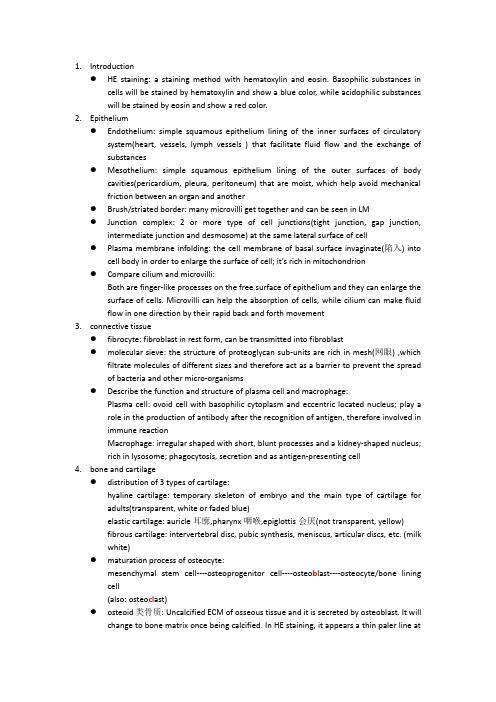
1.Introduction●HE staining: a staining method with hematoxylin and eosin. Basophilic substances incells will be stained by hematoxylin and show a blue color, while acidophilic substances will be stained by eosin and show a red color.2.Epithelium●Endothelium: simple squamous epithelium lining of the inner surfaces of circulatorysystem(heart, vessels, lymph vessels ) that facilitate fluid flow and the exchange of substances●Mesothelium:simple squamous epithelium lining of the outer surfaces of bodycavities(pericardium, pleura, peritoneum) that are moist, which help avoid mechanical friction between an organ and another●Brush/striated border:many microvilli get together and can be seen in LM●Junction complex: 2 or more type of cell junctions(tight junction, gap junction,intermediate junction and desmosome) at the same lateral surface of cell●Plasma membrane infolding: the cell membrane of basal surface invaginate(陷入) intocell body in order to enlarge the surface of cell; it’s rich in mitochondrion●Compare cilium and microvilli:Both are finger-like processes on the free surface of epithelium and they can enlarge the surface of cells. Microvilli can help the absorption of cells, while cilium can make fluid flow in one direction by their rapid back and forth movement3.connective tissue●fibrocyte: fibroblast in rest form, can be transmitted into fibroblast●molecular sieve: the structure of proteoglycan sub-units are rich in mesh(网眼) ,whichfiltrate molecules of different sizes and therefore act as a barrier to prevent the spread of bacteria and other micro-organisms●Describe the function and structure of plasma cell and macrophage:Plasma cell: ovoid cell with basophilic cytoplasm and eccentric located nucleus; play a role in the production of antibody after the recognition of antigen, therefore involved in immune reactionMacrophage: irregular shaped with short, blunt processes and a kidney-shaped nucleus;rich in lysosome; phagocytosis, secretion and as antigen-presenting cell4.bone and cartilage●distribution of 3 types of cartilage:hyaline cartilage: temporary skeleton of embryo and the main type of cartilage for adults(transparent, white or faded blue)elastic cartilage: auricle耳廓,pharynx咽喉,epiglottis会厌(not transparent, yellow)fibrous cartilage: intervertebral disc, pubic synthesis,meniscus, articular discs,etc. (milk white)●maturation process of osteocyte:mesenchymal stem cell----osteoprogenitor cell----osteo b last----osteocyte/bone lining cell(also: osteo c last)●osteoid类骨质: Uncalcified ECM of osseous tissue and it is secreted by osteoblast. It willchange to bone matrix once being calcified. In HE staining, it appears a thin paler line atthe surface of osseous tissue and below the osteoblasts.●osteons(Haversian systems)骨单位骨板: the major structure of long bone locatingbetween outer and inner circumferential laminae. They are concentric laminae surrounding central canal containing blood vessels, nerves and loose connective tissue.5.blood and haemopoiesis●neutrophil: neutrophilic granulocyte, the most numerous leukocyte in human blood;segmented nucleus with2-5 lobes; have azurophilic granules and specific granules; playa role in acute inflammation and defend body against the micro-organisms●who take part in allergy: eosinophil has enzymes that can dissolve histamine andleukotriene to inhibit allergy; basophil has leukotriene in its cytoplasm●reticulocyte: immature erythrocytes with ribosomes left in their cytoplasm, about onepercent of the total number of RBC;6.muscle tissue●Sarcomere: segment of the myofibril between two Z lines, consists of two halves of Iband and one A band. It’s the structural and functional unit of myofibril.●Sarcoplasmic reticulum: specialized SER, longitudinal aligned, enclose the myofibril,store and release calcium●Sarcoplasmic reticulum: specialized SER of muscle fiber that are longitudinal aligned andthey enclose the myofibril. Their function is to store and release calcium, especially during muscle contraction.●Terminal cisternae: expanded sarcoplasmic reticulum at the boundaries of A and I bands●T tubule: sarcolemma enfolds and penetrates into sarcoplasm●Triad: T tubule + two closely associated terminal cisternae; there’re two sets of triads inone sarcomere at the boundaries of A and I band; there function is to transmit impulses from sarcolemma into sarcoplasmic reticulum.●Intercalateddisk: junctional complexbetween cardiac muscle fibers, cross striationacross the cell under LM; at the level of Z line; it’s passage for information that contributes to the synchronization同步化of cardiac muscle fiber impulses.7.Nerve tissue●Compare dendrite with axon:There’s only one axon in most neurons but more dendrites; Dendrites are short and divided while axon is long and cylindrical. Dendrites are principal signal receptors and processing sites that●Blood-brain barrier: expanded foot plates of astrocytes that cover capillary endothelialcells, forming glia limitans that regulate the diffusion of substances between blood and the brain, which protect neurons from harmful substances in blood.●Ranvier node:a naked part of axon between adjacent Schwann cells that is not wrappedby neurolemma and mylinated sheath●Nissel body: strong basophilic clumps under LM; RER and free ribosomes that activelyproducing cytoskeletal protein, enzymes for neurotransmitters and peptide neuromodulators.。
石蜡切片制作和HE染色实验报告
石蜡切片制作和HE染色实验报告英文回答:Introduction。
Paraffin sectioning and HE staining are fundamental techniques used in histology to prepare tissue samples for microscopic examination. Paraffin sectioning involves embedding the tissue in paraffin wax, which allows for thin sections to be cut using a microtome. HE staining, also known as hematoxylin and eosin staining, is a common staining method used to differentiate between differentcell types and structures in the tissue. Together, these techniques provide a detailed view of the tissue's morphology and architecture.Materials and Methods。
Paraffin Sectioning。
1. Fix the tissue in formalin for 24 hours.2. Dehydrate the tissue by passing it through a seriesof graded alcohols (70%, 95%, 100%).3. Clear the tissue by passing it through xylene.4. Embed the tissue in paraffin wax.5. Cut thin sections (5-10 µm) using a microtome.HE Staining。
01-组织学绪论

(四) 组织化学和细胞化学技术
组织化学(histochemistry)和细胞化学(cytochemistry) 组织化学(histochemistry)和细胞化学(cytochemistry)技 术是应用化学反应原理检测组织和细胞的化学成分并进行 定位和定量的技术。组织细胞中的糖类、脂类、蛋白质、 定位和定量的技术。组织细胞中的糖类、脂类、蛋白质、 核酸、酶等均可与相应试剂反应, 核酸、酶等均可与相应试剂反应,最后形成有色反应终产 物或电子致密物,应用光镜或电镜进行观察。 物或电子致密物,应用光镜或电镜进行观察。 糖类物质常用过碘酸-Schiff反应(PAS反应)显示( 糖类物质常用过碘酸-Schiff反应(PAS反应)显示(图1-4)。 脂类常用苏丹染料、油红O 尼罗蓝等脂溶性染料染色。 脂类常用苏丹染料、油红O、尼罗蓝等脂溶性染料染色。
(六)原位杂交
原位杂交是一种在组织细胞原位进行的核酸分子杂交技术, 原位杂交是一种在组织细胞原位进行的核酸分子杂交技术, 敏感度高,特异性强,是当前分子生物学研究的重要手段。 敏感度高,特异性强,是当前分子生物学研究的重要手段。 原位杂交的原理是两条单核苷酸链通过碱基互补原则紧密 结合,形成稳定的杂交体。根据这一原理, 结合,形成稳定的杂交体。根据这一原理,用一条碱基序 列已知、经特定标记的核苷酸链为探针,与组织切片、 列已知、经特定标记的核苷酸链为探针,与组织切片、细 胞制备或染色体标本中的待检DNA或mRNA片段进行杂交, 胞制备或染色体标本中的待检DNA或mRNA片段进行杂交, 然后显示标记物, 然后显示标记物,从而获得待检核酸的分布和含量等信息 (图1-9) 。按照探针分子的性质,可将其分为cDNA探针、 按照探针分子的性质,可将其分为cDNA探针、 cRNA探针和寡核苷酸探针。 cRNA探针和寡核苷酸探针。
HE染色原理与试剂配制-梁英杰(1)
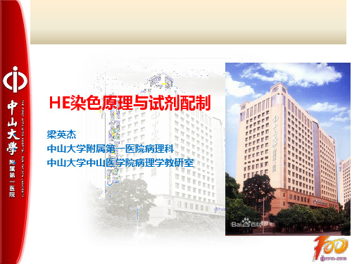
蓝色色淀(带正电荷) 细胞核核酸的磷酸根 (带负电荷)
极性结合
细胞核着色
10
苏木色精染色后的分化和蓝化
* 分化(differentiation): * 苏木色精染液为细胞核染料,与细胞核牢固结合而使细胞核着色。 * 组织切片经苏木色精染色后,细胞核染色过度会过染(深染),细 胞核以外的细胞质、胶原纤维等也附有少量苏木色精染料而着色。 * 用某些特定的溶液把过染的胞核和不应着色的组织成分脱色,从而
性环境中处于结合状态,呈蓝色,这时就称为色淀形成。
13
苏木色精染色后的分化和蓝化
* 常用返蓝液的配制: ① Scott促蓝液 碳酸氢钠 无水硫酸镁 蒸馏水 麝香草酚 ② 氢氧化铵水溶液 氢氧化铵 蒸馏水 ③ 碳酸锂水溶液 碳酸锂 蒸馏水
0.2g 1g 100ml 少量 0.3ml 100ml
1 g 100 ml
14
曙红的性质
* 曙红(eosin):又称伊红,为桃红色或粉红色的粉末。 * 曙红属于 ---人工合成染料(根据染料的来源) ---酸性染料(根据染料中所含助色团的性质) * 曙红分为:曙红Y,Y是带黄色之意,与蓝色的苏木精对比染色好。 曙红B,略带蓝色,与蓝色的苏木精对比染色不理想。 * HE染色选用曙红Y而不用曙红B。
15
曙红的种类
* 曙红是由荧光素衍生而来,主要有2种: 1. 水溶性曙红Y(eosin Y,water soluble), 也称曙红钠盐、 黄光曙红或四溴荧光素钠,易溶于水,微溶于乙醇
2. 醇溶性曙红Y(eosin Y,alcohol soluble),称四溴荧光素,
易溶于乙醇,不溶于水。
16
曙红染色液的配制
25
26
* 可根据需要选用不同的曙红液,较常用的是0.5%曙红水溶液。
病理HE切片常见的质量问题及处理方法
病理HE切片常见的质量问题及处理方法【摘要】目的探索病理HE切片常见的质量问题,并提出相应的预防和处理方法,提高HE切片质量。
方法回顾性地复习一定时期内本科所有的病理HE玻片,按照病理HE玻片中出现的质量问题进行总结、分析和分类,然后归纳。
结果病理HE切片常见的质量问题基本上都是由于病理技术员经验不足、操作不当、责任心不强所造成。
结论应加强从业病理技术员的系统而严格的工作培训,病理技术员要积极主动认真地工作,积累工作经验,端正工作态度,寻找出现质量问题的根源,提高HE切片的质量水平。
【Abstract】Objective To investigate the common quality problems on HE seetiong, and propose prevention and managements to improve the quality of HE stainings. Methods A retrospective review of all HE slides at a certain period of time were done to sum up, analysis and classify the quality problem in accordance with slides.Results The most common problems on quality are basically due to the inexperience of pathology technicians, improper operation,and poor responsibility. Conclusion Pathology technician practitioners should be trained strictly andsystemically. Theyshould take the initiative to work seriously, accumulate work experience, correct work attitude, look for the root causes of quality problems to improve the quality of HE sections.【Key words】HE sections;Quality;Management;Prevention; Retrospect苏木精(Hatmatoxylin)伊红(Eosin)染色法,简称HE染色法,是生物学和医学的细胞与组织学最广泛应用的染色方法[1]。
HE染色
一、定义苏木精—伊红染色法( hematoxylin-eosin staining ) ,简称HE染色法,石蜡切片技术里常用的染色法之一。
苏木精染液为碱性,主要使细胞核内的染色质与胞质内的核糖体着紫蓝色;伊红为酸性染料,主要使细胞质和细胞外基质中的成分着红色。
HE染色法是组织学、胚胎学、病理学教学与科研中最基本、使用最广泛的技术方法。
二、染色结果细胞核被苏木精染成鲜明的蓝色,软骨基质、钙盐颗粒呈深蓝色,粘液呈灰蓝色。
细胞浆被伊红染成深浅不同的粉红色至桃红色,胞浆内嗜酸性颗粒呈反光强的鲜红色。
胶原纤维呈淡粉红色,弹力纤维呈亮粉红色,红血球呈橘红色,蛋白性液体呈粉红色。
着色情况与组织或细胞的种类有关,也随其生活周期及病理变化而改变。
例如,很多细胞在新生时期胞浆对伊红着色较淡或轻度嗜碱,当其衰老时或发生退行性变则呈现嗜伊红浓染。
胶原纤维在老化和出现透明变性时,伊红着色由浅变深。
三、试剂配置A:0.5~1% 的伊红酒精溶液:称取伊红Y 0.5~1 g,加少量蒸馏水溶解后,再滴加冰醋酸直至浆糊状。
以滤纸过滤,将滤渣在烘箱中烤干后,以95%酒精(即工业酒精,如果不怕浪费,用无水乙醇配制也可)100毫升溶解。
B:苏木素染液配方:(配制3000 ml,可按比列减少)苏木精 6 g无水乙醇100 ml硫酸铝钾150 g蒸馏水2000 ml碘酸钠 1.2 g冰醋酸120 ml甘油900 ml配制方法:将苏木素溶于无水乙醇,再将硫酸铝钾溶于蒸馏水,溶解后将甘油倾入一起混合,最后加入冰醋酸和碘酸钠。
C:1%盐酸酒精分化液:将1毫升浓盐酸加入99毫升70%酒精中即可。
染色流程(1)二甲苯(Ⅰ)15min(2) 二甲苯(Ⅱ)15 min(3)二甲苯:无水乙醇=1:1 2min(4)100%乙醇(Ⅰ)5min(5)100%乙醇(Ⅱ)5 min(6)80%乙醇5min(7)蒸馏水5 min(8)苏木精液染色5 min(9) 流水稍洗去苏木精液1-3 s(10) 1%盐酸乙醇1-3 s(11) 稍水洗10-30 s(12)蒸馏水过洗1-2 s(13) 0.5%伊红液染色1-3 min(14) 蒸馏水稍洗1-2 s(15) 80%乙醇稍洗1-2 s(16) 95%乙醇(Ⅰ)2-3 s(17) 95%乙醇(Ⅱ)3-5 s(18) 无水乙醇5-10 min(19) 石炭酸二甲苯5-10 min(20) 二甲苯(Ⅰ)2 min(21) 二甲苯(Ⅱ)2 min(22) 二甲苯(Ⅲ)2 min(23) 中性树胶封固注:①第(11)、(12)步可省去,(13)步冲水时间需延长至20-30min。
病理技术HE染色在病理诊断中的应用价值
病理技术 HE染色在病理诊断中的应用价值【摘要】目的:研究病理技术HE染色在病理诊断中的应用价值。
方法:现选择2020年1月~2021年1月期间,随机抽取我院病理科400例石蜡切片开展研究。
根据电脑随机法将200例石蜡切片分为A组200例,采用常规HE染色,B 组200例,采用改进HE染色,对比两组染色诊断准确率。
结果:B组患者的染色诊断准确率高于A组(P<0.05)。
结论:通过将病理技术HE染色应用于病理诊断中,可有效提高染色准确率,进而可为临床诊断提供参考依据,具有较高的临床应用价值,值得推广和应用。
【关键词】病理技术;HE染色;病理诊断;应用价值Application value of pathological technique HE staining inpathological diagnosisShou XiaohuiHeilongjiang Qiqihar Medical College, Qiqihar 161006, China[Abstract] Objective: To study the application value of pathological technique HE staining in pathological diagnosis. Methods: from January 2020 to January 2021, 400 paraffin sections were randomly selected from the Department of pathology of our hospital. According to the computer random method, 200 paraffin sections were pided into 200 cases in group A and 200 cases in group B. the diagnostic accuracy of the two groups was compared. Results: the accuracy of staining diagnosis in group B was higher than that in group A (P < 0.05). Conclusion: the application of pathological technique HE staining in pathological diagnosis can effectively improve the accuracy ofstaining, and then provide reference basis for clinical diagnosis. It has high clinical application value and is worthy of popularizationand application.[Key words] pathological technique; He staining; Pathological diagnosis; Application value病理诊断主要是研究疾病的发生原因、发病机制、疾病发生过程中,机体的形态机构、功能代谢发生的变化、疾病的转归等[1],进而为疾病的诊疗、预防等提供理论支持和实践证据[2],其主要是通过对手术切下的组织,进行相关染色后,然后在显微镜下对其进行组织学检查[3],从而对疾病进行诊断,虽然在医学技术不断发展的情况下,各种影像学检查的广泛应用,且与病理学检查具有更简单快捷等特点[4],但是病理诊断,仍在临床诊断中具有不可替代的位置,且被誉为诊断金标准,是疾病最终的诊断方法,其因为此种检查诊断方法,主要通过观察其组织结构及细胞病变的特征[5],因此染色技术在病理诊断中具有一定重要性[6]。
组织病理切片以及HE染色
组织病理切片以及HE染色(一)实验原理:苏木素-伊红染色(hematoxylin-eosin staining,HE染色)能较好地显示组织结构和细胞形态,可用于观察、描述正常和病变组织的形态学,而且HE切片可较长时间保存,被称为常规染色方法。
(二)实验步骤:一、制作切片1、取材、固定:取小鼠肾脏组织适当大小,并且放入盛有固定液的小瓶中保存。
2、组织的脱水、透明、浸蜡与包埋组织固定后还需经过脱水、透明、浸蜡等过程才能制成蜡块,进行石蜡切片。
脱水是指用某些溶媒置换组织内水分的过程。
组织脱水后,必须经过一种既能与酒精相混合又能溶解石蜡的溶剂,通过这种溶剂的媒介作用,使石蜡浸入组织块。
在此过程中,因组织块中的水分被溶剂所取代,组织块变得透亮,因此称之为透明。
组织染色后也要进行透明。
最常用的透明剂为二甲苯,处理时间为30min左右。
组织经透明后在溶化的石蜡内浸渍的过程称浸蜡。
用其他浸透剂(如火棉胶、碳蜡和明胶等)渗入组织内部的过程称为浸透或透入。
为使石蜡充分渗入组织块中,常需经过3小时的浸渍,期间需更换3次石蜡。
一般用于浸蜡的石蜡熔点为52-56℃。
组织块经过浸蜡或浸透,用包埋剂(石蜡、火棉胶、碳蜡和树脂等)包起的过程称包埋,包埋后便制成含组织的块。
这种包埋块可使组织达到一定的硬度和韧度,有利于切成薄片。
石蜡包埋是病理日常工作中最常用的包埋方法。
用于包埋的石蜡熔点一般为60℃左右。
但在有些情况下,如某些酶染色时,则需采用低温石蜡包埋,以保存组织内酶的活性。
3、切片、制片石蜡切片常用的切片机有轮转式切片机和平推式切片机,以前者为多用。
切片厚度一般为3-5μm。
切片时先将组织片平摊于一块玻璃上,迅速滴加30%的酒精水溶液使组织片完全展开,再移入40℃恒温热水器中。
也可直接将组织切片移入40℃的恒温热水器中,待组织片完全展开后将其贴附于载玻片上,经56-60℃烤片30-60min后即可进行染色。
二、HE染色1. 二甲苯脱蜡2×10 min;2. 无水乙醇洗去二甲苯2×5min;3. 95%、80%乙醇各l0 min,自来水洗l min(不要让急水直接冲至载玻片上的组织),蒸馏水洗1min;4. 苏木素染色4 min,自来水洗2 min;5. 1%盐酸酒精分化20 s(镜下控制),自来水洗2 min;6. 1%稀氨水返蓝30s(镜下控制),自来水洗2分钟,蒸馏水洗1min;7. 伊红染色90s;8. 80%乙醇脱水10s,95%酒精10s,无水乙醇5min;9. 无水乙醇10min;10.二甲苯2×10 min;11.中性树胶或加拿大树胶封片。
HE染色技术
主要内容
一、HE染色原理 二、试剂 三、染色步骤 四、注意事项 五、几种常见的染色问题
一、HE染色的原理及分类
1、HE染色的原理
苏木精 — 伊红染色法 ( hematoxylin-eosin staining ) ,简称 HE染色法 ,是普通光学显微镜观察与鉴别细胞凋亡与细胞坏 死的一种染色方法,石蜡切片技术里常用的染色法之一 。苏木 精染液为碱性 ,主要使细胞核内的染色质与胞质内的核糖体着 紫蓝色 ;伊红为酸性染料 ,主要使细胞质和细胞外基质中的成 分着红色 。 HE染色是组织学、胚胎学、病理学教学与科研中最基本、使用 最广泛的技术方法.
速擦去,在滴上一滴树胶于组织片上,然后取干净的盖玻片,仔细 加在封固剂上,慢慢压平, 使盖片位置适中。切片封固后,放在温 箱中烤干,或平置凉干后装盒。
四、HE染色注意事项
• 为使切片制作更符合质量标准或最佳。除按步骤严格 进行外, 还应注意以下几点:
• <1>.用含升汞的固定液固定的组织块,在切片染色前, 应进行脱汞,具体方法:切片脱蜡,逐次进入70%酒精后, 入稀碘液(碘1克,碘化钾2 克,蒸馏水3000ml)内5~ 10min。蒸馏水稍洗,入5%硫代硫酸钠溶液内5min。 蒸馏水充分洗后进入染色。
HE染色注意事项
• <3>.如组织片含有大量黑色素时,影响染色和检查,可在脱蜡后, 浸水后用0.25%高锰酸钾胶2~4h ,然后,常水充分洗涤,或经1% 草酸液脱去切片上的黄褐色后,再充分水洗,后进入染色。
• <4>.在切片染色后,进行脱水时,当通过90%酒精后,应对切片进 行仔细擦试,此时,把切片中组织片周围的污染涂色或水分用纱 布(清洁)擦干净。当切片从 100%酒精出来时,也要认真仔细擦 去污物及组织片附近的水分。这样即可使组织脱水彻底,还可使 酒精浓度不至明显降低尽量保持酒精原浓度。
HE染色概念及定义
He染色介绍苏木精—伊红染色法 ( hematoxylin-eosin staining ) ,简称HE染色法,石蜡切片技术里常用的染色法之一。
苏木精染液为碱性,主要使细胞核内的染色质与胞质内的核糖体着紫蓝色;伊红为酸性染料,主要使细胞质和细胞外基质中的成分着红色。
染色分类易于被碱性或酸性染料着色的性质称为嗜碱性( basophilia )和嗜酸性( acidophilia ) 。
组织内的蛋白质构成蛋白质的氨基酸的种类很多,它们有不同的等电点。
在普通染色法中,染色液的酸碱度为pH6左右,细胞内的酸性物质如细胞核的染色质、腺细胞和神经细胞内的粗面内质网及透明软骨基质等均被碱性染料染色,这些物质称为嗜碱性。
而细胞质中的其它蛋白质如红细胞中的血红蛋白、嗜酸粒细胞的颗粒及胶原纤维和肌纤维等被酸性染料染色,这些物质称为嗜酸型。
如果改变染色液的酸碱度,pH值升高时,则原来被酸性染料染色的物质可变为嗜碱性;pH值降低时,原来被碱性染料染色的物质则可变为嗜酸性。
所以说染色液的pH值可以影响染色的反应。
脱氧核糖核酸(DNA)两条链上的磷酸基向外,带负电荷,呈酸性,很容易与带正电荷的苏木精碱性染料以离子键结合而被染色。
苏木精在碱性溶液中称蓝色,所以细胞核被染成蓝色。
伊红Y是一种化学合成的酸性染料,在水中离解成带负电荷的阴离子,与蛋白质的氨基正电荷的阳离子结合使胞浆染色,细胞浆、红细胞、肌肉、结缔组织、嗜伊红颗粒等被染成不同程度的红色或粉红色,与蓝色的细胞核形成鲜明对比。
伊红是细胞浆的良好染料。
由于组织或细胞的不同成分对苏木精的亲和力不同及染色性质不一样。
经苏木精染色后,细胞核及钙盐粘液等呈蓝色,可用盐酸酒精分化和弱碱性溶液显蓝,如处理适宜,可使细胞核着清楚的深蓝色,胞浆等其它成分脱色。
再利用胞浆染料伊红染胞浆,使胞浆的各种不同成分又呈现出深浅不同的粉红色。
故各种组织或细胞成分与病变的一般形态结构特点均可显示出来。
染色结果细胞核被苏木精染成鲜明的蓝色,软骨基质、钙盐颗粒呈深蓝色,粘液呈灰蓝色。
类脑he染色结果解读

HE 染色(Hematoxylin-Eosin staining)是病理学、组织学中最常用的一种染色方法,主要用于观察正常和病变组织的形态结构。
类脑组织的HE 染色结果解读也遵循常规HE 染色的基本原则,但可能更加关注神经元、胶质细胞以及血管等特殊结构的形态和分布特征。
分析类脑组织HE 染色结果时,以下是一些关键步骤和观察点:1.细胞核与胞浆:o细胞核:应被苏木精染成深蓝色或紫蓝色,通过观察核的大小、形状、数量及染色均匀度,可以评估细胞的活性状态和是否发生增殖、变异等情况。
o细胞浆:伊红染色后表现为不同程度的粉红色至桃红色,通过观察细胞浆内颗粒、空泡、包涵体等改变,可以反映细胞的功能状态和潜在的病理变化。
2.神经元和突触:o神经元细胞体的大小、形状和排列方式对诊断至关重要,如神经元萎缩、肿胀、坏死或者有无异型核都可能是神经系统疾病的指征。
o突触结构是否清晰可见,有无缺失或异常增生也能提供有关神经传递功能的信息。
3.胶质细胞:o星形胶质细胞、少突胶质细胞等在HE 染色下也有各自的特征,比如星形胶质细胞增生常伴随炎症反应或损伤修复过程,而少突胶质细胞可显示髓鞘形成状况。
4.血管结构:o血管壁的完整性、内皮细胞的形态以及周围是否有炎性细胞浸润都是评估血脑屏障功能和炎症状态的重要指标。
5.纤维结构:o在类脑组织中,白质区域的轴突、髓鞘以及灰质内的神经纤维网络可以通过HE 染色大致观察,尽管对于精细的纤维结构通常需要其他特殊染色法(如Luxol Fast Blue, Bielschowsky's silverstain 等)。
6.病理变化:o检查是否存在炎性细胞浸润、出血灶、脱髓鞘病灶、肿瘤细胞侵袭等病理现象。
因此,类脑HE 染色结果解读是一个综合性的过程,需要结合临床信息、实验目的和其他辅助检查手段进行专业分析,并由病理医生或相关研究人员根据标准图谱和实践经验做出准确判断。
心肌he染色解读
心肌he染色解读(中英文实用版)Title: Interpretation of Myocardial HE StainingTitle: 心肌HE染色的解读Introduction:HE staining is a commonly used histological technique for visualizing cells and tissues under a microscope.In the context of myocardial tissue, HE staining can provide valuable insights into the structure and function of cardiac cells.介绍:HE染色是一种常用于在显微镜下观察细胞和组织的历史技术。
在心肌组织的背景下,HE染色可以提供有关心脏细胞结构和功能的重要信息。
Procedure:The first step in HE staining is to fix the tissue sample using a chemical fixative such as formaldehyde.This fixative helps to preserve the cellular structure and prevent tissue degradation.After fixation, the sample is dehydrated by replacing the water content with alcohol.Dehydration is followed by clearing, where the alcohol is replaced with a clearing agent such as xylene.This step allows the tissue to be transparent and ready for embedding.步骤:HE染色的第一步是使用化学固定剂(如甲醛)固定组织样本。
- 1、下载文档前请自行甄别文档内容的完整性,平台不提供额外的编辑、内容补充、找答案等附加服务。
- 2、"仅部分预览"的文档,不可在线预览部分如存在完整性等问题,可反馈申请退款(可完整预览的文档不适用该条件!)。
- 3、如文档侵犯您的权益,请联系客服反馈,我们会尽快为您处理(人工客服工作时间:9:00-18:30)。
苏木素-伊红染色(HE)
【染色原理】
苏木素染液中带正电荷的蓝色色精与细胞核中带负电荷的脱氧核糖核苷通过
正、负电荷的极性吸附而守成染色;伊红是带负电荷的酸性染料,通过渗透或
弥散作用而使组织着色。
【组织固定与切片】
4%甲醛固定,石蜡切片厚度4~6μm。
【染色用具】
常规染色用具一套:染色皿或染色缸、刻字笔、标签、树脂胶、盖玻片、鸭嘴
镊、绸布或纱布。
滴管数支。
【试剂配制】
1. Harris苏木素液苏木素
2.5g,无水乙醇25ml,钾明矾50g,蒸馏水
500ml,氧化汞1.25g,乙酸20ml。
将苏木素溶于乙醇(加热)后加入预先已加热溶解明矾的蒸馏水中,煮沸
1~2min,改用小火加热,慢慢加入氧化汞(注意防止产生大量气泡和溶液溅
出)再煮沸2min后立即浸于冷水中,冷却后加入乙酸,过滤后使用。
2. 伊红液
(1) 水溶性伊红染液:伊红0.5g,蒸馏水100ml。
(2)醇溶性伊红染液:伊红0.5~1g,80%~95%乙醇100ml。
3. 分化液: 盐酸0.5~1ml,80%乙醇100ml。
4. 返蓝液: 氨水1ml,蒸馏水100ml。
5. 各级乙醇、二甲苯
【注意事项】
1. 石蜡切片在脱蜡前要烘烤使组织与切片粘贴。
烘烤温度50℃,时间30min 或30℃,24h。
2. 脱蜡要彻底,应定期更换脱蜡液。
3. 分化的时间要恰当掌握,可镜下观察着色情况。
4. 透明要充分。
【染色步骤】
1. 石蜡切片二甲苯Ⅰ脱蜡10~15min。
2. 二甲苯Ⅱ脱蜡10~15min。
3. 无水乙醇5min。
4. 95%乙醇5min。
5. 80%乙醇5min。
6. 自来水冲洗1min。
7. 蒸馏水洗1min。
8. 苏木素染液5~15min。
9. 自来水冲洗1~5min。
10. 化液分化数秒至数分钟。
11. 自来水冲洗1~3min。
12. 返蓝液返蓝30s~1min。
13. 自来水冲洗。
14. 伊红液5~10min。
15. 自来水冲洗。
16. 80%乙醇脱水30s。
17. 95%乙醇Ⅰ脱水1min。
18. 95%乙醇Ⅱ脱水1~3min。
19. 无水乙醇Ⅰ脱水2~5min。
20. 无水乙醇Ⅱ脱水2~5min。
21. 二甲苯Ⅰ透明2~5min。
22. 二甲苯Ⅱ透明2~5min。
23. 中性树胶封片。
【结果】
细胞核呈蓝色,软骨组织、钙盐呈深蓝色,黏液呈灰蓝色,细胞质呈粉红色,胶原纤维呈淡粉色,弹力纤维呈亮粉红色,红细胞呈桔红色。
