大鼠门静脉插管PortalVeinCannulation42707
门静脉减压对大鼠广泛肝切除术后肝功能的保护作用和肝再生的影响

【 bt c】 O jci T vla epoet e f c o dpes gm dr e eecsi otl rs r b en f p n A sr t a bet e oeaut t rt i f tf ers n oea lt es e r es e ym as l — v eh cv ee i tyh x v p ap u o se
门静 脉高压 可 以引起肝 损 伤导 致 肝衰 竭 。研 究
显示 门体分流 的方 法 可 以将 门静 脉压 力 降至 正 常范
围, 但是 过 度 降压 对 肝 再 生有 不 利 的影 响 。大 鼠
7 %肝 切除术 可 以引起 门静脉压 力 明显 迅速 升高 , 0 这
1 材 料与 方法 1 1 材料 . 雄性 S rg e a l pau —D w e y大 鼠 10只 。周 8
t e e tlt a ie a lr o pr v n e h lv rfiu e. l
【 e od 】 hpt t y le r ee tn prl yeesn le iu ;p n rr lao; c ete pa s K yw rs ea c m ;i re nri ; oa hprni ;i rn r sl iae gtn otod ; r o n eo v g ao t t o v j y e c t y i i r i zi
rtw r d ie t f r r p : uehptc m P x rop penat yla o (A )gop c et e ( C a ee i ddi o o o s pr ea t y( H )g u ,sle r r gt n S L r u ,ot od O T)gopad s v n u g u eo e i i r i ru n
积极溶栓治疗以挽救半影暗区的脑组织
积极溶栓治疗以挽救半影暗区的脑组织,改善脑循环。
③超过24h 溶栓效果欠佳。
④采用DSA 血管造影可清晰显示梗塞血管部位,程度和介入治疗后的变化,能直接验证溶栓治疗的效果。
[参考文献][1] 赵永生,杨海山,杨淑琴,等.尿激酶介入治疗急性脑血栓的应用研究[J ].介入放射学杂志,1998,(7)1:9.[2] 赵永生,邬英全,杨淑琴,等.急性脑梗塞DSA 监视下选择性脑动脉插管溶栓治疗的临床应用研究[J ].中风与神经疾病杂志,1997,(14)6:363.[3] 单鸿,邵国良,李少文,等.急性闭塞性脑血管病的腔内溶栓治疗[J ].中华放射学杂志,1996,30:591.[4] 尹琳胁理一郎,木村格.急性期或超急性期脑栓塞的超选择性动脉内溶栓疗法[J ].中华放射学杂志,1997,31(2):106.[5] 姜明辉,张显,等.实验不同时间缺血性脑水肿病理观察[J ].中风与神经疾病杂志,1993,10:1.[6] 刘卫平.脑水肿自由基病理机制研究进展[J ].国外医学神经病学神经外科分册,1994,2:3.大鼠种植性肝癌的活体影像学及病理对照刘 崎1,田建明,郝 强,王 莉,袁 敏,杨家和2,张智坚,陶文照3(1.上海长海医院放射科,上海 200433;2.东方肝胆外科医院;3.第二军医大学病理教研室)[基金项目]“九五”国家医学科技攻关项目(969070301)。
[作者简介]刘崎(1964—),女,博士,主治医师、讲师。
研究方向:影像医学及介入治疗。
[收稿日期]2000-08-13[摘 要] 目的 探讨大鼠Walker 2256种植性肝癌的活体影像学表现,并与病理对照,以便更好地应用这一经典的大鼠肝癌模型。
方法 种植瘤大鼠15只,10只行门静脉插管,5只行肝动脉插管。
所有动物均行MRI 平扫检查,CT平扫及动态增强扫描,DSA 血管造影;活体下缓慢注入超液化碘油,新鲜肝脏软X 线钼靶摄片并行病理检查。
大鼠解剖图谱中英文

Source #5
Rat Dissection
Source #5
Rat Dissection
MUSCLES
Iliacus:髂肌 Pectineus:耻骨肌 Adductor longus:长收肌 Gluteus medius:臀中肌 Vastus lateralis:股外侧肌 Rectus femoris:股直肌 Vastus medialis:股内侧肌 Adductor magnus:大收肌 Gracilis:股薄肌 Flexor digitorum superficialis: 指(趾)浅屈肌
Source #6
Rat Dissection
DIGESTIVE ORGANS
diaphragm:膈 Liver:肝脏 Small intestine:小肠 Spleen:脾脏 Caecum:tion
DIGESTIVE ORGANS
Caudal side of diaphragm:膈 肌尾侧面 Median lobe of liver:肝脏中叶 Ligamentum teres hepatis:肝 静脉索 Left lobe of liver:肝脏左叶 Stomach:胃 Greater omentum:大网膜 Small intestines:小肠
Source #5
Review and Quiz Yourself
Rat Dissection
THORACIC CAVITY
《2024年大鼠门静脉血栓模型构建和血栓动态观察》范文

《大鼠门静脉血栓模型构建和血栓动态观察》篇一一、引言门静脉血栓(Portal Vein Thrombosis,PVT)是一种常见的血管疾病,其发病机制和治疗方法一直是医学研究的热点。
为了更好地研究PVT的发病机制和治疗方法,建立可靠的动物模型是必要的。
本文旨在介绍大鼠门静脉血栓模型的构建方法和血栓动态观察,以期为PVT的研究提供可靠的实验依据。
二、材料与方法1. 实验材料实验所需材料包括:健康SD大鼠、手术器械、肝素、凝血酶、生理盐水等。
2. 模型构建(1)大鼠麻醉及准备:采用腹腔注射麻醉法,将大鼠麻醉后固定于手术台上。
(2)手术操作:在无菌条件下进行手术,暴露门静脉并注入凝血酶,以诱导血栓形成。
(3)术后护理:术后给予大鼠适当的护理,包括饮食、活动等方面的管理。
3. 血栓动态观察采用超声技术对大鼠门静脉血栓进行动态观察,记录血栓形成、发展及消退的过程。
三、实验结果1. 模型构建成功通过上述方法,成功构建了大鼠门静脉血栓模型。
术后大鼠生命体征稳定,无死亡病例。
2. 血栓动态观察结果超声结果显示,门静脉血栓形成过程中,血栓逐渐增大,并逐渐向肝内延伸。
随着时间推移,部分血栓逐渐溶解,门静脉再通。
整个过程中,超声技术可清晰显示血栓的形态、大小及位置。
四、讨论1. 模型构建的意义大鼠门静脉血栓模型的构建为PVT的研究提供了可靠的实验依据。
通过该模型,可以研究PVT的发病机制、治疗方法及预防措施等,为临床治疗提供理论支持。
2. 血栓动态观察的优点超声技术具有无创、无痛、可重复检查等优点,可以实时观察门静脉血栓的形成、发展及消退过程。
通过动态观察,可以更好地了解PVT的病程及治疗效果,为临床治疗提供更有价值的参考信息。
五、结论本文成功构建了大鼠门静脉血栓模型,并采用超声技术对血栓进行了动态观察。
实验结果表明,该模型稳定可靠,可为PVT 的研究提供有价值的实验依据。
同时,超声技术可实时观察门静脉血栓的形成、发展及消退过程,为临床治疗提供更有价值的参考信息。
大鼠经颈外静脉导丝引导插管测肺动脉压与传统方法的比较

大鼠经颈外静脉导丝引导插管测肺动脉压与传统方法的比较杨杰章;黄石安;陈灿;李波;高汉华;庞玲品【摘要】目的探讨大鼠经颈外静脉插管方法及测肺动脉压的最佳方法.方法将80只雄性SD大鼠按随机分组原则分成2组:经导丝引导插管测肺动脉压组(G组),传统方法插管测肺动脉压组(T组),每组均40只.记录插管操作一次成功率、多次调整成功率(n≤4次)、总成功率、一次插管时间、总插管时间、及一次测压时间、总测压时间及肺动脉高压大鼠肺动脉压力数值.结果 G组比T组插管操作一次成功率、多次调整成功率(n≤4次)、总成功率更高(P<0.05),G组比T组的一次插管时间、总插管时间以及一次测压时间、总测压时间要短(P<0.01),G组所测的肺动脉高压大鼠的肺动脉压力比T组所测的高(P<0.01).结论经导丝引导插管测肺动脉压法插管和测压具有成功率高、准确到达肺动脉、数据更准确、操作省时的优点.与用传统方法插管测肺动脉压组相比较,是一种更好的对大鼠进行颈外静脉插管和测肺动脉压的方法.【期刊名称】《中国比较医学杂志》【年(卷),期】2010(020)009【总页数】7页(P44-50)【关键词】静脉穿刺;插管术;颈外静脉;肺动脉压【作者】杨杰章;黄石安;陈灿;李波;高汉华;庞玲品【作者单位】广东医学院附属医院心血管内科,湛江,524000;广东医学院附属医院心血管内科,湛江,524000;广东医学院附属医院心血管内科,湛江,524000;广东医学院附属医院心血管内科,湛江,524000;广东医学院附属医院心血管内科,湛江,524000;广东医学院附属医院心血管内科,湛江,524000【正文语种】中文【中图分类】R332在心血管疾病研究中,肺动脉高压的研究很早就受到重视,而近年来更是越来越受关注。
动物实验作为其重要的研究媒介,早在1966年就有由Kydd等[1]复制成功与测压的报道。
本研究对已有的相关报道[2-9]的插管与测压方法进行比较总结发现它们的共同点:1)插管到静脉:对于体型较小的动物如大鼠,多用导管直接在已剪“V”形切口的颈外静脉上插管;对于体型较大的动物如犬类,多用经股静脉插管;2)对于肺动脉压的测量,多根据对实验动物解剖结构盲性进行直接插管至肺动脉测量的方法,偶有利用频谱多普勒超声心动图技术及Bernoulli方程定量估测心腔间压差或跨瓣压差,从而估测肺动脉压力的报道;3)对于测压导管是否到达动物的肺动脉的位置主要是依靠压力曲线波形和大小判断,间有解剖大鼠进行尸检确定[9]。
门静脉血栓动物模型的建立和应用

门静脉血栓动物模型的建立和应用刘壮1,2,陈纪宏1,3,祁兴顺1,许向波1,诸葛宇征41 北部战区总医院消化内科,沈阳 1108402 中国医科大学研究生院,沈阳 1101223 大连医科大学研究生院,辽宁大连 1160444 南京大学医学院附属鼓楼医院消化内科,南京 210008通信作者:祁兴顺,******************(ORCID: 0000-0002-9448-6739)摘要:门静脉血栓(PVT)是指在肝外门静脉主干和/或肝内门静脉分支内发生的血栓栓塞。
PVT是由多种因素共同作用的结果,但其发病机制尚未完全阐明。
建立动物模型是研究PVT病理生理机制的重要方法。
本文根据不同的动物种类,对现有PVT动物模型的建立方法、原理、优缺点和应用情况进行综述。
关键词:门静脉血栓;疾病模型,动物;发病机制基金项目:国家自然科学基金(8227034094)Establishment and application of animal models for portal vein thrombosisLIU Zhuang1,2,CHEN Jihong1,3,QI Xingshun1,XU Xiangbo1,ZHUGE Yuzheng4.(1. Department of Gastroenterology,General Hospital of Northern Theater Command, Shenyang 110840, China; 2. Graduate School of China Medical University, Shenyang 110122,China; 3. Graduate School, Dalian Medical University, Dalian, Liaoning 116044, China; 4. Department of Gastroenterology, Affiliated Drum Tower Hospital of Nanjing University Medical School, Nanjing 210008, China)Corresponding author: QI Xingshun,******************(ORCID: 0000-0002-9448-6739)Abstract:Portal vein thrombosis (PVT)refers to thromboembolism that occurs in the extrahepatic main portal vein and/or intrahepatic portal vein branches. PVT is the result of the combined effect of multiple factors, but its pathogenesis remains unclear. Animal models are an important method for exploring the pathophysiological mechanism of PVT. Based on the different species of animals,this article reviews the existing animal models of PVT in terms of modeling methods,principles,advantages and disadvantages, and application.Key words:Portal Vein Thrombosis; Disease Models, Animal; PathogenesisResearch funding:National Natural Science Foundation of China (8227034094)门静脉血栓(portal vein thrombosis,PVT)形成是指在肝外门静脉主干和/或肝内门静脉分支内发生的血栓栓塞[1]。
一种可恢复性门静脉高压症大鼠模型
内容摘要:作者:吴凤东窦剑任贵军赵鑫邸恩昌作者:吴凤东窦剑任贵军赵鑫邸恩昌a kind of recoverable portal hypertension model in rat1 材料与方法1.2 实验操作1.2.1 可复性肝前性门静脉高压症大鼠模型的建立 2%氯氨酮腹腔注射麻醉200mg/kg,上腹正中切口2cm进入腹腔,游离门静脉主干近左右支分叉处,用40丝线将一个20gauge钝头注射针头[1]及标志管(自制,外径1.5mm,用血管造影管作成梨形)与门静脉主干一同平行结扎一道,此时可见胃肠道明显淤血,抽出针头并摆好标志管位置防止其扭曲门静脉,此时可见胃肠道淤血减轻但仍有血管扩张,止血后缝合切口。
对照组开腹后只游离门静脉主干。
1.2.2 门静脉压力测定及造影一期手术3周后,实验组a大鼠原切口进入腹腔,找到回结肠静脉的一个分支,经此静脉插入-3f硬膜外导管至门静脉主干,以大鼠脊柱水平为零点,用八道生理记录仪测量门静脉压。
找到脾脏,向脾髓质内穿入-25gauge头皮针。
经3f硬膜外导管及25gauge头皮针分别同时注入36%泛影葡胺1ml,3s内注射完毕[1],摄x线片。
找到门静脉旁标志管,剪断、拔出,门静脉再次测压并注入造影剂摄片。
对照组大鼠同样方法做门静脉压力测定和造影。
实验组b大鼠进入腹腔后找到门静脉旁标志管,剪断、拔出,缝合切口,单笼饲养,待4周后测量门静脉压力。
1.3 标本采集与检测每组大鼠切取肝脏中叶组织,经he染色后,于普通光镜下观察。
各组大鼠分别从下腔静脉抽取全血3ml,取上清液送生化检测alt,ast,bil。
1.4 统计学处理方法采用单因素方差分析,应用spss10.0软件进行统计处理。
《2024年大鼠门静脉血栓模型构建及溶栓治疗》范文

《大鼠门静脉血栓模型构建及溶栓治疗》篇一一、引言门静脉血栓(Portal Vein Thrombosis,PVT)是一种临床常见的疾病,可导致门静脉血流受阻,引起肝损伤甚至危及生命。
由于人体实验的局限性,构建可靠的动物模型对门静脉血栓及溶栓治疗的研究显得尤为重要。
大鼠作为实验动物,在生理构造与人类具有一定的相似性,故其成为构建门静脉血栓模型的理想选择。
本文将重点探讨大鼠门静脉血栓模型的构建及溶栓治疗方法,为相关研究提供理论依据和实践指导。
二、大鼠门静脉血栓模型构建1. 实验材料与方法(1)实验动物:选用健康成年SD大鼠作为实验对象。
(2)模型构建:通过外科手术方法,将实验大鼠的肠系膜静脉部分结扎,以阻断血流并形成血栓。
(3)实验设备与试剂:包括手术器械、结扎线、生理盐水、肝素等。
2. 模型构建步骤(1)术前准备:对大鼠进行麻醉、备皮等术前处理。
(2)手术过程:暴露肠系膜静脉,用结扎线部分结扎,阻断血流。
(3)术后处理:缝合伤口,给予抗生素预防感染,观察大鼠生命体征。
3. 模型评估通过观察大鼠的生理指标、病理切片等手段,评估门静脉血栓模型的成功率及稳定性。
三、溶栓治疗方法1. 药物选择根据门静脉血栓的特点,选择合适的溶栓药物,如尿激酶、组织型纤溶酶原激活剂等。
2. 治疗方法将选定的溶栓药物通过适当途径(如静脉注射、腹腔注射等)给予大鼠,观察溶栓效果及大鼠的生理反应。
3. 治疗效果评估通过超声、CT等影像学检查,观察门静脉血栓的溶解情况,评估治疗效果。
同时,监测大鼠的生理指标,了解药物对大鼠的影响。
四、结果与讨论1. 模型构建结果通过上述方法,成功构建了大鼠门静脉血栓模型,模型具有较高的成功率及稳定性,为相关研究提供了可靠的实验基础。
2. 溶栓治疗效果选用合适的溶栓药物,通过适当途径给予大鼠,取得了较好的溶栓效果。
同时,药物对大鼠的生理影响较小,安全性较高。
3. 讨论与展望虽然大鼠门静脉血栓模型构建及溶栓治疗取得了一定成果,但仍存在一些问题和挑战。
大鼠门静脉插管simple method for chronic cannulation of the portal vein in intact unrestrained rats
rats
ALAIN BRAILLON AND MICHAEL J. BRODY Department of Pharmacology and the Cardiovascular Center, College of Medicine, University of Iowa, Iowa City, Iowa 52242
BRAILLON,ALAIN,ANDMICHAEL J. BRODY. Asimplemethod for chronic cannulation of the portal vein in intact unrestrained rats. Am. J. Physiol. 255 (Gastrointest. Liver Physiol. 18): Gl91-G193, 1988.-A simple new method is described for chronic cannulation of the portal vein in rats. A Silastic catheter (0.64 mm OD) with an internal metallic leader is directly inserted in the junction of the two small ileal veins. The catheter is advanced into the mesenteric vein to the portal vein. Then the leader is removed and the catheter tunneled out the back of the neck. This method allows for the measurement of portal vein pressure, direct injection of substances in the portal vein, or blood samples for at least 1 wk in intact unrestrained rats. Validation included the presence of elevated portal vein pressure in portal hypertensive rats compared with normal rats and the absence of clot development in the portal vein. This method should help provide a better understanding of hepatic physiology and pharmacology in normal and disease states. hepatic hepatic circulation; liver hemodynamics; sampling; portal injection portal hannulation is a necessary component of investigation of hepatic circulation, hepatic metabolism, and intestinal absorption. Measurement of portal vein pressure allows for the study of specific effects of vasoactive drugs on the hepatic circulation. Injection of drugs into the portal vein allows for evaluation of hepatic firstpass metabolism. Sampling of blood from the portal vein provides for analysis of intestinal absorption. Because portal vein cannulation in humans is an invasive procedure, experimental rat models have been developed (3) and are based on two methods: 1) the cannulation of the portal vein through the splenic vein, which requires splenectomy; and 2) the direct puncture of the portal vein, which is associated with a temporary interruption of hepatic portal perfusion and can be complicated by portal vein fibrosis. In this report we describe a new and simple method for chronic portal vein cannulation in intact rats by catheterization of a small ileal vein. The method was tested for its reliability in the measurement of portal vein pressure and for the potential appearance of portal vein thrombosis, a phenomenon not examined in reports of other techniques for catheterization of the portal vein (1, 3, 8). PORTAL METHODS
大鼠肝断面门静脉分支残端的静脉置管模型构建-病理学论文-基础医学论文-医学论文

大鼠肝断面门静脉分支残端的静脉置管模型构建-病理学论文-基础医学论文-医学论文——文章均为WORD文档,下载后可直接编辑使用亦可打印——前言由于大鼠肝脏各叶彼此分开,各叶肝蒂游离于肝外,肝叶比重相对固定,方便进行各种比例的肝部分切除。
肝部分切除模型主要应用于肝脏肿瘤、肝再生、急慢性肝衰竭以及肝移植等模型的研究。
理想的急性肝衰竭动物应具有①可逆性:肝衰竭有潜在的可逆性,适当的方法可以治疗并能长期存活;②可重复性:肝损伤模型的制备可以重复,并应具有相同的病死率和较相似的指标;③死于肝衰竭:肝损伤动物模型最终应死于肝衰竭;④治疗窗口期:由发生肝衰竭到应具有足够的时间,以进行适当的治疗;⑤大型动物;⑥对环境和实验人员危害最小。
随着干细胞治疗肝损伤及护肝药物研究的发展,对大鼠肝损伤模型也提出了很多新的要求。
本实验拟在肝部分切除术的基础上对模型鼠进行改进,并行细胞移植实验,对比分析新模型的优劣。
1 材料与方法1.1 材料1.1.1 实验动物F344 大鼠由上海Slack 实验动物中心提供,由第四军医大学动物中心实验动物房饲养。
1.1.2 材料动物手术台、手术显微镜(OPMI,德国)、高频电刀、无损伤血管夹、小儿硬膜外导管(内径约0 .7 mm),止血钳等常规手术器械、肝素钠、盐水、全自动生化分析仪(日立7170型,日本)。
第三代构建GFP 示踪的胎肝干细胞(本课题组三步分离法制备),20 % 胎牛血清(Hyclone,USA),胰蛋白酶(Gib-co 公司,USA),WilliamsE 培养基(Sigma 公司USA)。
1.2 方法1.2.1 模型制备⑴一次性硬膜外导管制备测孔,并用50 U/ml肝素钠注射液肝素化。
大鼠术前禁食12 小时、禁水 4 小时。
10%水合氯醛(3 ml/kg)腹腔内注射麻醉,将麻醉后固定于实验台上,备皮、消毒, ⑴60 只大鼠分为三组,A 组:对照组;B 组:经门静脉途径注射治疗组;C 组:经肝断面门静脉分支残端置管治疗组。
SD大鼠门腔静脉转位模型的建立

r t. e , -n 4 d y u vv l aewa 6 6 ( 6 3 ) 8 . 3 ( 5 3 )a d8 . 3 ( 5 3 ) r — a s 0n - 7a d 1 一a ss ria t s8 . 7 2 / 0 , 3 3 2 / 0 n 3 3 2 / 0 ,e r s e t ey,nt emo e. t1d ah a ewa 6 6 ( / 0 . n l s n Th o t c v 1 r n — p ci l i h d 1Toa e t sr t s1 . 5 3 ) Co cu i v o e p ra a a a s t
关 键 词 :门腔静脉转位术 ;门腔静脉半转位术;动物模型;大鼠
中 图 分 类 号 : 一3 R32
文 献 标 识 码 :A
文 章 编 号 : 00 2420)2 08 3 i0 —29(080 —01—0
M o i i d Po t c v lTr n p st0 n S Ra s d fe r a a a a s 0 ii n i D t
在 20 0 6年 1 1月 ̄2 0 0 7年 6月 的实 验 中 , 者 作
1 0号 缝线 , 微持 针器 , / 显 显微镊 , 微剪 ,- 线 , 显 80缝 血 管夹 ; 5mL、 1 mL注 射器等 。
1 3 门腔 静 脉 转 位 模 型 的 制 作 .
成 功地建立 了 S 大 鼠 门腔 静 脉转 位 模 型 , 较 好 D 能
AB T S RACT:Obe tv To e tbih t e p ra a a r n p st n mo e. t o s En o e d jc ie sa l h o tc v lta s o ii d 1 Meh d s o d t n
大鼠肝动脉插管并体外留置模型的改进论文
大鼠肝动脉插管并体外留置模型的改进论文大鼠肝动脉插管并体外留置模型的改进论文目的对体外留置肝靶向性给药途径的大鼠动脉插管模型进行改进,并探讨建立此模型的技术要点。
方法在10倍手术显微镜下,采用特制内径0.5 mm导管(由硬膜外麻醉导管加工而成)经胃十二指肠动脉剪口处插入肝动脉血管内,逆行到达肝固有动脉开口处后留置导管并固定。
结果 20只大鼠造模,成功者17只,成功率为85%。
结论改进型大鼠肝动脉插管并体外留置模型较以往造模方法简单,并提高了模型成功率,技术易于推广,可应用于抗肝肿瘤、肝衰竭等靶向性给药的研究。
〔Abstract〕Objective To improve the model of hepatic artery(HA) catheter and detain the catheter in vitro in rats,and approach the skill keys and value of establishing the model. Method Under the operation microscope(10×), a self-made catheter with 0.5mm ID, developed by peridural anesthetic catheter, was intubated into hepatic artery, through gastroduodenal artery, then the catheter was retainedand fixed in rats. Result 20 rats were used to establish the model, in which 17 rats model were successful, the ratio was 85%. Conclusion The improved model is much easily established and achievement ratio ishigher than others, which may be used to study on targetingadministration in curing liver tumor or hepatic failure.〔Keywords〕 Model; rats; HA catheterization; detaining in vitro; improvement肝固有动脉插管的动物模型建立早有报道,但原有的方法易造成肝脏损伤、死亡率高,技术难以掌握。
《2024年穿心莲内酯预防大鼠急性门静脉血栓形成的效果和机制》范文
《穿心莲内酯预防大鼠急性门静脉血栓形成的效果和机制》篇一一、引言随着人们生活水平的提高和饮食习惯的改变,血栓性疾病已成为严重威胁人类健康的主要疾病之一。
其中,门静脉血栓(Portal Vein Thrombosis, PVT)是一种常见的血栓性疾病,其发病率和死亡率均较高。
目前,对于PVT的治疗主要依赖于药物治疗和手术治疗,但治疗效果并不理想。
因此,寻找有效的预防和治疗PVT的药物成为当前研究的热点。
穿心莲内酯作为一种具有广泛药理作用的天然药物,其对于预防大鼠急性门静脉血栓形成的效果和机制成为本研究的主要探讨内容。
二、研究背景及目的穿心莲内酯作为一种传统中药,具有清热解毒、抗氧化的作用,并被证实具有一定的抗血栓作用。
然而,穿心莲内酯对于预防大鼠急性门静脉血栓形成的效果和机制尚未明确。
本研究旨在探讨穿心莲内酯对大鼠急性门静脉血栓形成的预防作用,并进一步探究其作用机制,为临床应用提供理论依据。
三、实验方法1. 实验动物与分组:选取健康大鼠,随机分为对照组、模型组、治疗组,每组10只。
2. 造模及给药:通过化学诱导法建立大鼠急性门静脉血栓模型,治疗组在造模前及造模过程中给予穿心莲内酯干预。
3. 检测指标:观察各组大鼠的生存情况、血液学指标、病理学改变等。
4. 实验方法:采用文献报道的方法进行实验操作,包括取材、检测等。
四、实验结果1. 生存情况:治疗组大鼠的生存率明显高于模型组,与对照组无显著差异。
2. 血液学指标:治疗组大鼠的血小板聚集率、凝血酶原时间等血液学指标较模型组有明显改善。
3. 病理学改变:治疗组大鼠的门静脉血栓形成程度较模型组明显减轻,病理学评分较低。
五、穿心莲内酯预防大鼠急性门静脉血栓形成的机制根据实验结果及文献报道,穿心莲内酯预防大鼠急性门静脉血栓形成的机制可能如下:1. 抗血小板聚集作用:穿心莲内酯能够抑制血小板聚集,降低血液黏度,从而减少血栓的形成。
2. 抗氧化作用:穿心莲内酯具有抗氧化作用,能够清除体内的自由基,减轻氧化应激反应对血管内皮细胞的损伤,从而减少血栓的形成。
大鼠门静脉高压形成过程中血浆内毒素水平与肠道iNOS的表达

大鼠门静脉高压形成过程中血浆内毒素水平与肠道iNOS的
表达
杨照新;常鹏环;姚茂忠;黄绵庆;符健
【期刊名称】《基础医学与临床》
【年(卷),期】2013(033)009
【总页数】2页(P1212-1213)
【作者】杨照新;常鹏环;姚茂忠;黄绵庆;符健
【作者单位】海南医学院药物安评中心,海南海口571101;山西医科大学护理学院,山西太原030001;海南医学院药物安评中心,海南海口571101;海南医学院药物安评中心,海南海口571101;海南医学院药物安评中心,海南海口571101
【正文语种】中文
【中图分类】R318.14
【相关文献】
1.内毒素血症大鼠膈肌iNOS活性和凋亡相关基因表达的变化 [J], 方迎艳;关宿东;郭晓磊;叶红伟;汪华学;高琴
2.大鼠肝硬化门静脉高压形成过程中内皮素A受体mRNA的表达 [J], 汤天平;周岩冰;陈桂明
3.辛伐他汀对内毒素所致脓毒症大鼠肾组织NF-κB、iNOS表达的影响 [J], 冼海涛;阮少川;蔡立华;曾晖;郑惊雷;邓见玲;姚华国
4.门静脉高压症大鼠肠道iNOS mRNA表达的实验研究 [J], 何美文;易辛;戴植本
5.雄附散抑制大鼠肺间质纤维化过程中肺组织iNOS的表达水平 [J], 杨永滨;王勇;马玉琛;董超;耿朝晖;肖锋
因版权原因,仅展示原文概要,查看原文内容请购买。
大鼠门静脉高压对血清内毒素及肠黏膜的影响分析
大鼠门静脉高压对血清内毒素及肠黏膜的影响分析李晓娟;张巧云;冯帆;姜棋予;柴燕涛;李润;侯俊;李瑞生【摘要】Objective To study the effects of simple portal hypertension on the endotoxin levels in serum and intestinal mucosa of rats. Methods A total of 16 rats were divided into the blank control group (4 rats) and the model groups (3-day group, 7-day group and 10-day group, 4 rats in each group). The rat model of partial portal vein ligation was established in the model groups, and samples of blood and jejunum, ileum and colon of the rats were taken on the 3rd, 7th and 10th days, respectively. Changes in the serum endotoxin levels were detected by ELISA. Histopathological changes of the intestinal tissues were examined by HE staining. Results The rat model of partial portal vein ligation was successfully established in all the model groups. The serum levels of endotoxin on the 3rd, 7th and 10th days in the model groups were not significantly different from that in the normal control group. Damages of different intestinal segments were not serious on the 3rd day after modeling. However, on the 7th day after modeling, there were some sowllen and damaged intestinal villi in the intestinal mucosa of each intestinal segment, and the connection between the epithelial cells and the lamina propria was broken, compared with that at 3 days after modeling. In addition, there were obvious damages in the intestinal mucosa and lamina propria on the 10th day, compared with that at 3 d and 7 d after modeling. Conclusions In the case of normal liver function, portal hypertension can cause intestinal mucosal damages withina short period of time, but the amount of endotoxin produced by intestine does not exceed the processing capacity of the liver and thus does not cause an increase in the serum endotoxin level.%目的研究大鼠单纯门脉高压对血清内毒素以及肠黏膜的影响.方法选取16只大鼠,分为空白对照组(4只)和模型组(3d组、7d组、10d组,每组4只),模型组构建门脉部分结扎模型,分别在3d、7d、10d采集血液及大鼠的空肠、回肠和结肠,采用ELISA法检测血清内毒素含量观察内毒素的变化情况,采集各组不同部位肠组织进行HE染色,进行病理组织学观察分析.结果模型组大鼠均成功构建了门静脉部分结扎模型.三个不同时间点血清内毒素的结果与正常对照组之间差异无显著性.成模3d后各肠段还未发生很大的损伤; 成模7d后各肠段较3d粘膜层的局部肠绒毛出现了部分破坏和肿胀,上皮细胞与固有层间连结出现了松散的状况; 成模10d较3d和7d的肠粘膜层、固有层均出现了明显的损伤.结论在肝功能正常的情况下,门脉高压短时间内可以造成肠黏膜的损伤,但肠道产生的内毒素不会超出肝脏的处理能力而出现血清内毒素的升高现象.【期刊名称】《中国比较医学杂志》【年(卷),期】2018(028)001【总页数】4页(P76-79)【关键词】门脉高压;内毒素;肠黏膜;大鼠【作者】李晓娟;张巧云;冯帆;姜棋予;柴燕涛;李润;侯俊;李瑞生【作者单位】解放军第302医院临床研究管理中心,北京 100039;山东省蓬莱市中医医院检验科,山东蓬莱 265600;解放军第302医院临床研究管理中心,北京100039;解放军第302医院临床研究管理中心,北京 100039;解放军第302医院临床研究管理中心,北京 100039;解放军第302医院临床研究管理中心,北京 100039;解放军第302医院临床研究管理中心,北京 100039;解放军第302医院临床研究管理中心,北京 100039【正文语种】中文【中图分类】R-33门脉高压和内毒素血症是肝硬化晚期最常见的并发症[1]。
大鼠和小鼠解剖图谱(照片版)
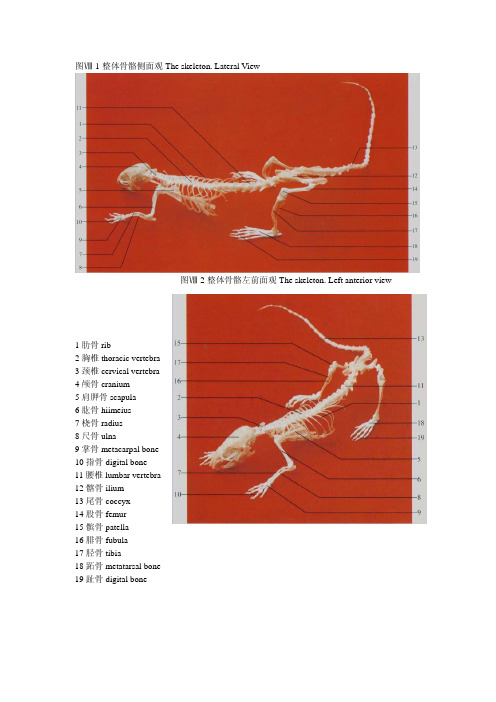
大鼠和小鼠解剖图谱生物秀—专心做生物!www.bbioo.com易生物-领先的生物医药商务平台www.ebioe.com生物秀论坛-学术交流,资源共享,互助社区www.bbioo.com/bbs/图Ⅷ-1整体骨骼侧面观The skeleton. Lateral View图Ⅷ-2整体骨骼左前面观The skeleton. Left anterior view1肋骨rib2胸椎thoracic vertebra3颈椎cervical vertebra4颅骨cranium5肩胛骨scapula6肱骨hiimeius7桡骨radius8尺骨ulna9掌骨metacarpal bone10指骨digital bone11腰椎lumbar vertebra12髂骨ilium13尾骨coccyx14股骨femur15髌骨patella16腓骨fubula17胫骨tibia18跖骨metatarsal bone19趾骨digital bone图Ⅷ-4头骨侧面The skull. Lateral aspect图Ⅷ-6下颌骨侧面The mandible. Lateral aspect1枕骨occipital bone2顶间骨interparietal bone3矢状缝sagittal suture4颧骨malar bone5上颌骨maxillary bone6前颌骨premaxillary bone7枕外嵴external occipital creat8顶骨parietal bone9额骨frontal bone10鼻骨nasal bone11鼻间缝internasal suture12前筛孔preethmoid pore13蝶腭孔sphenopalatine foramen 14门齿 incisor tooth15下颌骨mandible16视神经孔optic foramen17枕骨occipital bone18茎突styloid process19外耳道external acoustic meatus 20颞骨temporal bone21 腭裂patoschisis22臼齿molar tooth23腭骨palatine bone24翼孔pterygoid apertures25破裂孔foramen lacerum26枕大孔foramen magnum27腭后孔posterior palatine foramen 28鼻后孔posterior nasal apertures 29卵圆孔foramen ovale30鼓骨tympanic bone31舌下神经孔hypoglossal foramen 32下颌联合mandibular symphysis 33颏孔mental foramen34冠状突coronoid process35下颌支ramus of mandible36角状突process of horn37下颌孔mandibular foramen38翼肌窝pterygoid fossa39髁突condylar process图Ⅷ-7颈椎与胸椎背面The cervical 图Ⅷ-9前肢骨背面The bone of vertebra and thoracic vertebra.Dorsal aspect anterior 1imb.Dorsal aspect图Ⅷ-8前肢骨外侧面The bone of anteriorlimb. Lateral aspect1枢椎axis 2第4颈椎4th cervical vertebra3第6颈椎6th cervical vertebra 4第1胸椎1st thoracic vertebra5第4胸椎4th thoracic vertebra 6寰椎axis7第3颈椎3rd cervical vertebra 8第5颈椎5th cervical vertebra9第7颈椎7th cervical vertebra 10第3胸椎3rd thoracic vertebral1肱骨体shaft of humcrus 12桡骨radius13尺骨ulna 14尺骨茎突styloid process of ulna15掌骨metacarpal bone 16肱骨头head of hurnerusl7肘突cubital process 18腕骨carpal bone19指骨digital bone图Ⅷ-10股骨前面The femur. 图Ⅷ-11胫骨与腓骨前面The tibia Anterior aspect and fibula. Anterior aspect1大转子greater trochanter2股骨颈neck of femur3第三转子third trochanter4髌骨patella5股骨头femoral headG小转子lesser trochanter7股骨体shaft of femur8股骨内侧髁medial condyle of femur9外侧髁lateral condyle10腓骨fibula11外踝 lateral malleolus12跗骨tarsal bone13胫骨内侧髁medial condyle of tibia14径骨粗隆tibial tuberosity15胫骨tibiaI6内踝medial mallcolus17跖骨metatarsal bone18斜方肌trapezius muscle19臀浅肌glutoeus super(icialis muscle20股直肌rectus femoris muscle21腹外斜肌external oblique muscle of abdomen 22背阔肌latissimus dorsi muscle23肩斜方肌shoulder-trapezius muscle24大圆肌teres major muscle25肩三角肌shoulder-deltoid muscle26前锯肌serratus anterior muscle1锁骨提肌levator clavicle muscle 2肩三角肌shoulder-deltoid muscle 3肱三头肌triceps brachii muscle4臀浅肌gluteus suferficidlis muscle 5股直肌rectus femoris muscle6腓肠肌gastrocnemius muscle7夹肌splenius muscle8颈菱形肌rhomboideus cervicis muscle9胸菱形肌rhomboideus pectoralis muscle10背阔肌latissimus dorsi muscle11腹外斜肌external oblique muscle of abdomen 12股二头肌biceps femoris muscle13半腱肌semitendinosus muscle14二腹肌前腹anterior belly of digastric muscle 15二腹肌后腹posterior belly of digastric muscle 16胸骨舌骨肌sternohyoid muscle17肱二头肌biceps brachii muscle18腹直肌rectus abdominis muscle19股动、静脉femoral artery and femoral vein 20阴囊scrotum21咬肌masseter muscle22胸乳突肌sternomastoideus muscle23肩三角肌shoulder-deltoid muscle24桡侧腕伸肌extensor carpi radialis muscle25指总伸肌extensor digitorum communis muscle 26尺侧腕伸肌extensor carpi ulnaris muscle27胸浅肌pectoral superficialis muscle28前锯肌serratus anterior muscle29股内侧肌vastus medialis muscle30耻骨肌pectineus muscle31长收肌adductor longus muscle32股薄肌gracilis muscle图Ⅷ-15头、颈部侧面The head and neck. Lateral aspect上静脉supraorbitalveine 7颈外静脉external jugular vein 1颞浅动、静脉superficial temporal artery and vein 2眶3面横动、静脉transverse facial artery and vein 4下颌缘神经marginalmandibular nerv 5面后静脉posterior facial vein 6面前静脉anterior facial vein1颞浅动、静脉superficial temporal artery and vein 2眶上静脉supraorbitalvein3面横动、静脉transverse facial artery and vein 4下颌缘神经marginalmandibular nerve 5面后静脉posterior facial vein 6面前静脉anterior facial vein7颈外静脉external jugular vein 8硬腭褶fold of hard palate9臼齿molar tooth 10门齿incisor tooth11软腭soft palate 12舌根root of tongue13舌体body of tongue 14舌尖apex of tongue图Ⅷ-18脑与脊髓背面The brain and 图Ⅷ-19脑与脊髓腹面The brain and spinal cord.Dorsal aspect spinal cordVentral aspect1 喉口aperture of larynx 2舌根root of tongue 3舌尖apex of tongue 4硬腭褶fold of hard palate 5软腭soft palate 6会厌epiglottis 7臼齿molar tooth 8舌体body of tongue 9嗅球olfactory bulb 10中央纵裂central longitudinal fissue 1l 绒球flocculus 12颈膨大cervical enlargement 13腰膨大lumbar enlargement 14外侧纵沟lateral longitudinal sulcus15大脑cerebrum 16小脑(中央部)cerebellum (central part ) 17脊髓圆锥courts medullaris 18 脑垂体 pituitary gland 19脑桥pons 20延髓 myelencephalon 21嗅束olfactory tract 22视神经optic nerve 23三叉神经trigeminal nerve图Ⅷ-20磁共振冠状面定位像(1)The scout view of coronal images(1)图Ⅷ-21磁共振冠状面定位像(2)The scout view of coronal images(2)1眼球eyeball2嗅脑rhinencephalon3大脑皮层cerebral cortex4大脑镰cerebral faix5鼻旁窦paranasal sinus6纵裂longitudinal fissure冠状面T1加权像,颞颌关节线圈,SE序列,层厚3.0mm,无间隔,TR=500rns,TE=20ms。
- 1、下载文档前请自行甄别文档内容的完整性,平台不提供额外的编辑、内容补充、找答案等附加服务。
- 2、"仅部分预览"的文档,不可在线预览部分如存在完整性等问题,可反馈申请退款(可完整预览的文档不适用该条件!)。
- 3、如文档侵犯您的权益,请联系客服反馈,我们会尽快为您处理(人工客服工作时间:9:00-18:30)。
Surgery Model #: SU057
Portal Vein Cannulation Care and Use Document:
Anesthetic: Sodium Pentobarbital (IP); Analgesic: Buprenorphine (IP)Basic Surgical Procedure Description:
An anesthetized animal is surgically prepared and draped. A 2.5-cm ventral midline skin and abdominal muscle wall incision is made with a cranial terminus near the xiphoid process. The cecum is exteriorized and abdominal organs are covered and moistened with sterile gauze and saline. Using a dissecting scope, the ileocolic vein is observed and isolated with silk ligatures. A small incision is made in the portal vein and the cannula is inserted to the vicinity of the junction with the lineal vein. Cannula patency is determined at this time and once patency is established, the cannula is flushed with 200µL saline and filled with lock solution. The cannula is exteriorized through a hole in the abdominal wall to a point between the shoulder blades. Abdominal musculature is closed with absorbable suture, while the abdominal skin incision and sub scapular exteriorization site is closed with stainless steel wound clips.
Cannulas:
Portal vein cannula consists of a length of sterile silicone rubber with a 25mm PE 50 access port and 30-35mm polyurethane tippet. Fill volume of the cannula is 60µl. 23 gauge, blunted needles are required to access the port. Lock Solution:
Heparinized Dextrose (50 IU/ml): 1.0ml stock heparin (1000 IU/ml) + 19ml 50% dextrose solution (Henry Schein). Quality Control:
Patency is verified by the ability to withdraw a blood sample within 24 hours of shipment and is guaranteed upon animal receipt. To maintain animals over longer periods of time, cannulas need to be flushed twice a week (once every 3-4 days). Flush cannulas by following the sampling procedure below, minus the withdrawal of the whole blood sample.
Sampling:
Blood sample size can vary, but recommendations are in the range of 100-200µl. Sample size and frequency should be minimized to essential time points to maintain the health of the animal.
For blood withdrawal, gather the following materials: Syringe assemblies (1cc syringe attached to a 23G blunted needle), sterile saline and sterile fill solution.
1.)Place animal in a restrainer (small open topped boxes the size of a pipette container work well)
2.)Remove the pin from the cannula and set aside
3.)Insert an empty syringe assembly (SA) into the port
4.)Gently withdraw fill solution and blood (60µl)
5.)Attach a second SA and withdraw sample (syringe may contain anticoagulant)
6.)Slowly flush cannula with sterile saline ~200µl
7.)Add 60µl of sterile lock solution and replace pin
If blood fails to flow in step 4, remove the empty SA and replace with a SA containing sterile saline and gently flush the cannula. Continue as outlined above.
Housing:
Individually house animals to prevent cage mates from chewing on one another’s cannulas.
Notes:1. During animal manipulation (dosing / weighing), it is important not to place undo stress on the cannula. 2. Using needles larger than 23 gauge will stretch the port and make sampling difficult. Additionally, sampling by means of needles with bevels or rough edges will damage the port, again making sampling difficult. 3. Cannulas may become positional; changing the animal’s body position will often restore blood flow. 4. Cannula is low flow / low pressure.。
