CT评估肺动脉高压
肺动脉高压的影像学表现PPT

02
PAH可分为特发性、遗传性、药物或毒物相关性和疾病相关性
的类型。
PAH的症状可能包括疲劳、乏力、运动耐量下降、胸痛、晕厥
03
等。
影像学检查的重要性
影像学检查对于肺动脉高压的诊断、 评估和治疗具有重要意义。
常用的影像学检查方法包括超声心动 图、CT血管成像和磁共振成像等。
通过影像学检查,可以观察到肺动脉 和右心的结构和功能变化,评估疾病 的严重程度,监测治疗效果,以及发 现潜在的病因。
详细描述
这是由于肺动脉高压导致右心负 担加重,长期高负荷工作使得右 心逐渐增大。右心的增大也是诊 断肺动脉高压的重要依据之一。
05
肺动脉高压的影像学鉴别 诊断
与其他原因引起的肺动脉扩张鉴别
先天性心脏病
如房间隔缺损、室间隔缺损等, 可导致肺动脉扩张,但通常伴随 其他心血管异常征象,如心脏结
构畸形。
右心房和右心室扩大
01
02
03
心影增大
由于肺动脉高压,右心房 和右心室负担加重,导致 右心房和右心室扩大,在 X线片上表现为心影增大 。
心尖圆钝
右心房和右心室扩大时, 心尖部变得圆钝,有时呈 球形。
右心室流出道增宽
右心室流出道增宽是右心 室扩大的表现,X线片上 可见流出道内径增粗。
肺纹理减少或消失
慢性阻塞性肺疾病
COPD可引起肺动脉高压和肺动脉 扩张,但通常伴随肺部纹理增多、 紊乱等征象。
肺血栓栓塞
肺血栓栓塞可导致肺动脉扩张,但 通常伴随肺部异常病灶或胸腔积液 等征象。
与其他原因引起的右心增大鉴别
右心衰竭
右心衰竭可引起右心增大 ,同时伴有其他心血管异 常征象,如心脏结构畸形 、心脏杂音等。
肺动脉高压的规范化诊治(最全版)
肺动脉高压的规范化诊治(最全版)肺高血压(PH)是一组以肺动脉压力(pulmonary arterial pressure, PAP)增高为特征,伴或不伴有肺小动脉病变为特征的疾病,可引起右心功能衰竭甚至死亡。
目前可将肺高血压分为5大类。
肺动脉高压(PAH)属于其第一大类,是指肺动脉血压增高而肺静脉压力正常,主要原因是肺小动脉原发病变而导致的肺动脉阻力增加,表现为肺动脉压力升高而肺静脉压力在正常范围内。
其主要特征是肺动脉阻力进行性升高,最终导致患者因右心衰竭死亡。
2013年NICE会议将PH做了以下分类:PAH的诊断标准:在海平面状态下,右心导管检查测得肺动脉平均压(PAPm)≥25mmHg,同时肺毛细血管楔压(PAWP)<15mHg。
需要强调右心导管检查术仍是诊断PAH的金标准。
PAH的诊断包括1、病史,2、体格检查,3、辅助检查,4、鉴别诊断。
1.1症状:通常肺动脉高压早期通常无明显症状,最常见的临床表现为劳力性呼吸困难。
在美国国立卫生研究院(NIH)进行的PPH前瞻性、登记注册研究中,大约60%患者以劳力性呼吸困难为首发症状。
随着病程的进展,所有患者均可出现呼吸困难,其他常见症状有疲乏和活动耐量下降等。
严重肺动脉高压患者休息时也可出现呼吸困难。
大约40%肺动脉高压患者曾发生过心绞痛和晕厥。
由于肺动脉高压的症状没有特异性,因此以上症状仅提示肺动脉高压的诊断或排除其他疾病。
然而当呼吸困难无法用其他疾病解释时,应考虑到肺血管疾病的可能。
1.2既往史、个人史、家族遗传史可以为PH分类提供线索,有助于PAH的鉴别诊断。
2、体格检查:肺动脉高压的体征多与肺动脉压力增高,右心室扩大和右心衰竭有关,常见的有:紫绀;颈静脉充盈或怒张;肺动脉听诊区第二心音亢进,三尖瓣听诊区可闻及收缩期杂音,胸骨左缘出现抬举样搏动等。
3.1心电图:a、电轴右偏,b、右心室肥厚表现,c、I导联出现s波,d、肺型P波。
3.2胸片:主肺动脉扩张及肺门增大,可出现截断征象等,胸片同时可以发现原发性肺部疾病。
肺动脉高压

肺动脉高压的临床分类
临床上将 PH分为 5大类
分类
亚类
1.动脉性肺动脉高压(PAH) 1.1特发性肺动脉高压(IPAH)
1.2遗传性肺动脉高压(HPAH)
1.3药物和毒物相关肺动脉高压
1.4疾病相关的肺动脉高压
1.4.1 结缔组织病
1.4.2 HIV感染
1.4.3 门脉高压
1.4.4 先天性心脏病
4. 可溶性鸟苷酸环化酶(soluable guanylate cyclase,sGC)激动剂
5.前列环素类似物和前列环素受体激动剂
PAH的治疗
(四)靶向药物联合治疗和药物间相互作用
1.靶向药物联合治疗 2.药物相互作用:靶向药物联合治疗时需考虑到药物间的相互作用。
谢谢
5.2 系统性和代谢性疾病(如结节病、戈谢氏病、糖原储积症)
5.3复杂性先天性心脏病
5.4 其他(如纤维性纵隔炎)
临床表现
主要表现为进行 性右心功能不全的相关症状,常为劳累后诱发,表现为疲劳、呼 吸困难、胸闷、胸痛和晕厥,部分患者还可表现为干咳和运动诱发的恶心、呕吐。 晚期患 者静息状态下可有症状发作。 随着右心功能不全 的加重可出现踝部、下肢甚至腹部、全身水肿。
可直接评价右心室大小、 形态和功能,并可无创 评估血流量,包括心输 出量、每搏输出量和右 心室质量。
血液学检查
血液学检查主要用于 筛查PH的病因和评价 器官损害情况
PAH的治疗
(一)一般措施
1.体力活动和专业指导下的康复:PAH患者应在药物治疗的基础上、在专业指导下进行运动康复训练 2. 妊娠、避孕及绝经后激素治疗:随着靶向药物的广泛应用,妊娠 PAH患者死亡率有所下降,但仍在 5%~23%,且妊娠并发症多,因此,建议PAH患者避免怀孕。 3.择期手术:对PAH患者即使进行择期手术也会增加患者风险,接受择期手术者,硬膜外麻醉可能比全 身麻醉耐受性好。 4. 预防感染:PAH 患者容易合并肺部感染,而 肺部感染是加重心衰甚至导致死亡的重要原因之 一。 5.社会心理支持
肺动脉高压鉴别诊断

右室
房
左
左室
房
21
22
肺动脉增强CT
双肺多发性肺动脉狭窄、闭塞,肺动脉管壁增厚,未见 充盈缺损。
右心房室明显增大。 两肺灌注不均匀,右肺透过度增高。 主动脉弓降部和降主动脉管壁环形增厚,且管径粗细不
均匀,最窄处4.9mm,弓动脉分支近端显影好。 最后诊断: 多发大动脉炎累及肺动脉,重度肺动脉高压
诊断策略
特异性检查
体征, HRCT
肺静脉闭塞/肺毛细血
ANA
管瘤
结缔组织 疾病
药物毒 物
病史 HIV检查
体检,实验室检查
体
检 经胸超声 超 食道超声 声
心脏磁共 肝
振
功
实 验 室 检 血吸虫或第5类 查
慢性溶血
门脉高压性肺动脉 高压
HIV
先心病
特发性或可遗传性 PAH
BMPR-2,ALK1,
Endoglin(HHT), 家族史
胸壁、脊柱及隔肌等改变征象。 5.化验:血红蛋白和红细胞增多,血气:PaO2下降,PaCO2增加。 6.肺功能:明显通气(阻塞性、限制性或混合性)和/或弥散功能异常。 7.多导睡眠监测仪检查明确是否存在睡眠呼吸暂停综合征。
15
左向右分流性先天性心脏病相关性肺动脉高压特点
主要包括房间隔缺损、室间隔缺损、动脉导管未闭等。少见部位 的心房间隔缺损(上腔型、冠状静脉窦型等) 、部分肺静脉畸形引 流、无分流的动脉导管未闭等先心病超声心动图易漏诊,很容易被 误诊为特发性肺动脉高压。下列情况有助于考虑先心病诊断。 1.自幼体弱、易感冒、心脏杂音。 2.P2亢进伴固定性分裂,连续性杂音。 3.胸部X线平片:肺血增多。 4.食管超声心动图可有助于减少漏诊。 5.心血管CT或右心导管检查有助于明确诊断。
肺动脉高压的诊断与治疗指南解读
(5)第 4 大类 PH 定义为 CTEPH 及肺动脉阻塞引起的 PH。
(6)由于慢性溶血引起的 PH 病理特点与其他 PAH 不同,因此被列入了第 5 大类 。
(7)节段性 PH 被列入第 5 大类 PH中。
是
确诊为第二、三类PH
是
肺动脉高压不严重/无右心功能 障碍
治疗基础疾病
否 核素肺通气灌注扫描有无灌注缺
损
肺动脉高压诊治中心
肺动脉高压严重/右心功能障碍 转诊到肺动脉高压中心
是 考虑CTEPH:行CT肺血管造影/
右心导管和肺动脉造影
CTD 药物素物
HIV
否 PAH特异性检查
右心导管术: mPAP≥25mmHg,PAWP≤15mmHg,PVR≥3wu
1. 动脉型肺动脉高压
PAWP ≤ 15 mmHg
3. 肺部疾病相关肺动脉高压;4. 慢性
血栓栓塞性
肺动脉高压;5. 不明机制和(或)多
种因素所致
肺动脉高压
毛细血管后肺动脉高压 mPAP ≥ 25 mmHg
2. 左心疾病相关性肺动脉高压
PAWP >15 mmHg
5. 不明机制和(或)多种机制所致肺
动脉高压
舒张早期肺动脉反流速度 > 2.2 m/s
下腔静脉直径大于 2m1m,吸气塌陷率下降
( 深吸气时小于 50%, 平 静呼吸时小于 20%) 舒张末期和收缩末期右心房 面积变化大于 18 cm2
超声心动图评估肺动脉高压诊断可能 性分级
三尖瓣反流峰值速率
其他肺动脉高压
(m/s) 能性分级 ≤ 2.8 或未测量 低度
肺动脉高压的诊断与筛查

三、实验室检查
• 7、肺通气灌注扫描:肺动脉高压患者的肺通气灌 注扫描可以完全正常,也可在外周发现一些小的 非节段性缺损。由于肺动脉高压通气功能一般正 常,所以往往会呈现V/Q比例失调。肺通气灌注 扫描对于诊断慢性血栓栓塞性肺高压(CTEPH) 有比较重要的价值。 • 8、右心导管检查:右心导管检查不仅是确诊肺动 脉高压的金标准,也是指导确定科学治疗方案必 不可少的手段。对病情稳定、WHO肺动脉高压功 能分级Ⅰ~Ⅲ级、没有明确禁忌证的患者均应积 极开展标准的右心导管检查。
浓浓茅医情 健康伴君行
肺动脉高压的诊断与筛查
• 肺动脉高压的主要特征是肺动脉阻力进行 性升高,最终导致患者因右心衰竭死亡。 右心衰竭是所有类型肺动脉高压患者致残、 致死的共同途径,而肺动脉高压是右心衰 竭的最主要原因,其病因复杂、诊断治疗 棘手,致使该领域长期发展缓慢。然而, 右心衰竭的防治也是心肺医师无法回避, 且越来越重要的卫生保健问题。
一、病史
• 2、危险因素 • (1)既往史:先天性心脏病、结缔组织病、HIV感染史、 减肥药物治疗史、肝病及贫血等都是肺动脉高压病因分类 的重要线索,故需要全面采集患者的既往史,这样既有助 明确诊断分类,也有助于发现新的危险因素。 • (2)个人史:需要注意患者有无危险因素接触史,如印 刷厂和加油站工人接触油类物品、HIV感染、同性恋、吸 毒及染发剂等特殊接触史。 • (3)婚育史:女性要注意有无习惯性流产史,男性要注 意其母亲、姐妹等直系亲属有无习惯性流产等病史。 • (4)家族史:家族有无肺动脉高压患者至关重要,有无 其他家族遗传性病史对于发现新的危险因素、帮助诊断分 类亦具有重要意义。
放射医师如何帮助诊断肺动脉高压

精选ppt
18
图6 肺动脉高压的 血管标志,CTPA轴 位像显示肺动脉扩张 (29mm或以上), 肺动脉与升主动脉管 径比大于等于1。
精选ppt
19
心脏征象—在心电门控的CTPA,右心的代偿
和失代偿的表现包括:右室肥厚,室壁厚 度超过4mm;僵直或向左弯曲的室间隔; 右室扩张(轴位图像心室中心水平,右、 左心室直径比大于1);右室射血分数的减 少;下腔静脉和肝静脉的扩张;以及心包 积液(图7)(31-34)。
精选ppt
7
病理生理学和临床病程
正常成人肺血管床是低压,低阻系统,可 以容纳肺动脉压略有升高时增加的血流量。 肺动脉高压的患者,肺动脉压和血管阻力 的缓慢升高,导致右心室肥厚扩张(5–8)。 肺动脉高压的临床病程分为三个阶段:代 偿期,代偿失调期和失代偿期(图1)。
ቤተ መጻሕፍቲ ባይዱ
精选ppt
8
肺动脉高压导致右心室肥厚,其次是心室扩张和
动脉高压比超声心动图更准确,心室质量指数与
肺动脉平均压力具有良好的相关性(r= 0.81)。
但是,其他研究者(48)报告,心室质量指数和
平均肺动脉压具有较弱的相关性(r= 0.56);
因此,利用心室质量指数诊断肺动脉高血压需要
更多的研究。
精选ppt
27
相位对比成像
相位对比成像是一个磁共振成像序列,是测量 流速和血流变化。右心室心搏量可以通过容量 分析计算或使用相位对比成像测量肺动脉的流 量。若存在显著的三尖瓣返流,计算右心室心 搏量通过测量肺动脉流量比测量右心室容积更 可靠,因为后者往往高估了真正的收缩体积 (45)。
精选ppt
10
怀疑
肺动脉高压通常表现无症状或非特异 性症状,如呼吸困难,疲劳,和晕厥症 状,是由于在活动时无法增加所需要的 心输出量。
肺动脉高压的诊断和治疗进展
肺动脉高压的诊断和治疗进展肺动脉高压(PAH)归类于肺微血管疾患由于平滑肌细胞增殖、移行内膜增厚和血栓形成引起肺动脉高压。
其主要特征是肺血管阻力进行性升高最终导致患者右心衰竭而死亡。
肺动脉高压已逐渐成为被关注的一大类心血管疾病。
1定义与专用术语肺循环高压是指包括肺动脉高压、肺静脉高压和混合性肺动脉高压的总称。
整个肺循环任何系统或者局部病变而引起的肺循环血压增高均可称为肺循环高压。
肺动脉高压是指肺动脉血压增高而肺静脉压力正常主要原因是肺小动脉原发病变故需肺毛细血管嵌顿压( PCWP)正常才能诊断。
目前被划分为肺循环高压的第一大类。
特发性肺动脉高压是没有发现任何原因包括遗传、病毒、药物而发生的肺动脉高压。
另外需要重点突出肺循环血压增高的诊断标准:在海平面状态下静息时右心导管检查肺动脉收缩压>30mm Hg(1 mm Hg = 0.133 kPa) 和(或)肺动脉平均压>25mm Hg或者运动时肺动脉平均压>30 mm Hg。
此外诊断肺动脉高压尚需包括PCWP < 15 mm Hg。
需要强调上述标准为右心导管数据并非无创检查手段估测的数据。
2肺动脉高压的诊断分类1998年以前肺循环高压仅分为原发性肺动脉高压和继发性肺动脉高压两大类。
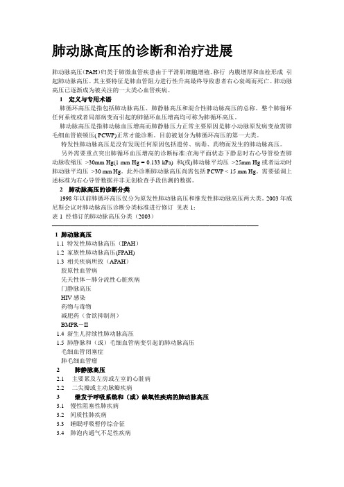
2003年威尼斯会议对肺动脉高压诊断分类标准进行修订见表1:表1 经修订的肺动脉高压分类(2003)——————————————————————————————————1 肺动脉高压1.1 特发性肺动脉高压(IPAH)1.2 家族性肺动脉高压(FPAH)1.3 相关疾病所致(APAH)胶原性血管病先天性体-肺分流性心脏疾病门静脉高压HIV感染药物与毒物减肥药(食欲抑制剂)BMPR-II1.4 新生儿持续性肺动脉高压1.5 肺静脉和(或)毛细血管病变引起的肺动脉高压毛细血管闭塞症肺毛细血管瘤2 肺静脉高压2.1 主要累及左房或左室的心脏病2.2 二尖瓣或主动脉瓣疾病3 继发于呼吸系统和(或)缺氧性疾病的肺动脉高压3.1 慢性阻塞性肺疾病3.2 间质性肺疾病3.3 睡眠呼吸暂停综合征3.4 肺泡内通气不足性疾病3.5 慢性高原病3.6 新生儿肺病3.7 肺泡-毛细血管发育异常4 慢性血栓或栓塞性肺动脉高压4.1近端肺动脉血栓栓塞4.2 远端肺动脉栓塞肺栓塞[血栓、肿瘤、寄生虫和(或)卵、外源性物质]4.3 原位血栓形成5 其他原因引起的肺动脉高压5.1 类肉瘤样病5.2 组织细胞增多症5.3 纤维素性纵隔炎5.4 淋巴结增大/肿瘤5.5 淋巴管肉瘤——————————————————————————————————3诊断因为肺动脉高压诊断的复杂性强烈建议患者到肺血管疾病专科中心就诊进行全面的诊断和功能评价。
肺血管疾病的影像学诊断

肺血管疾病的影像学诊断摘要:肺血管疾病是一类涉及肺循环的疾病,包括肺动脉高压、肺栓塞、肺血管炎等。
影像学检查是诊断肺血管疾病的关键手段,主要包括胸部X光片、CT肺动脉造影、磁共振成像等。
本文将对肺血管疾病的影像学诊断进行综述,以期为临床诊断提供参考。
一、引言肺血管疾病是指影响肺循环的疾病,其病因多种多样,包括遗传因素、炎症、肿瘤、感染等。
肺血管疾病的临床表现缺乏特异性,早期诊断困难,容易误诊和漏诊。
影像学检查在肺血管疾病的诊断中具有重要价值,可以直观地显示肺血管的病变情况,为临床诊断和治疗提供重要依据。
二、影像学检查方法1.胸部X光片:胸部X光片是筛查肺血管疾病的首选方法,可以初步观察肺血管的形态、分布和大小。
但对于肺血管疾病的确诊价值有限。
2.CT肺动脉造影(CTPA):CTPA是诊断肺血管疾病的重要手段,具有较高的空间分辨率和时间分辨率。
通过注射对比剂,可以清晰地显示肺动脉及其分支的充盈情况,对肺栓塞、肺动脉高压等疾病具有较高的诊断价值。
3.磁共振成像(MRI):MRI具有无辐射、多参数成像的优势,可以全面评估肺血管的病变。
MRI肺动脉造影(MRPA)可以显示肺动脉及其分支的充盈情况,对肺栓塞、肺动脉高压等疾病具有一定的诊断价值。
4.超声心动图:超声心动图可以评估右心功能,间接反映肺动脉高压的严重程度。
同时,超声心动图可以观察肺动脉血流速度,对肺栓塞等疾病具有一定的诊断价值。
5.放射性核素肺通气/灌注显像:放射性核素肺通气/灌注显像是评估肺血流分布的一种方法,对肺栓塞具有较高的诊断价值。
但该方法存在放射性损伤,不适用于孕妇和儿童。
三、影像学诊断要点1.肺动脉高压:CTPA和MRPA可以清晰地显示肺动脉及其分支的扩张、扭曲等改变,同时可以评估肺动脉压力。
超声心动图可以观察右心功能,间接反映肺动脉高压的严重程度。
2.肺栓塞:CTPA是诊断肺栓塞的首选方法,可以明确栓塞部位、范围和程度。
放射性核素肺通气/灌注显像可以评估肺血流分布,对肺栓塞具有较高的诊断价值。
2024版肺动脉高压的CT影像学表现以影识病

01定义02分类肺动脉高压(PAH)是一种由多种已知或未知原因引起的肺动脉压力异常升高的病理生理状态根据发病原因和病理生理机制,PAH可分为特发性、遗传性、药物或毒物诱导性、结缔组织病相关性、HIV相关性等类型定义与分类发病原因及危险因素发病原因PAH的发病原因多样,包括基因突变、自身免疫性疾病、药物使用、病毒感染等危险因素高龄、女性、家族史、结缔组织病、先天性心脏病等都是PAH的危险因素03PAH 患者常表现为呼吸困难、乏力、胸痛、晕厥等症状,严重者可出现右心衰竭临床表现PAH 的诊断需要结合患者的临床表现、体格检查、心电图、超声心动图等多种检查结果,其中CT 影像学检查在PAH 的诊断和评估中具有重要作用诊断方法CT 可清晰显示肺动脉主干及分支的扩张程度,是评估PAH 的重要指标之一肺动脉扩张肺血管纹理增多PAH患者肺部血管纹理增多、紊乱,呈“蜘蛛网”样改变右心室增大PAH患者右心室常增大,CT可准确测量右心室大小和形态肺水肿和间质性肺水肿部分PAH患者可出现肺水肿和间质性肺水肿,CT表现为磨玻璃样密度增高影和网格状影其他表现如肺动脉钙化、肺动脉瘤样扩张等也可在CT检查中发现01X射线源与探测器CT机通过X射线源发射X射线,穿透人体后被探测器接收。
02数据采集与处理探测器将接收到的X射线信号转换为数字信号,经过计算机处理后重建为图像。
03图像重建与显示通过特定的重建算法,将采集到的数据重建为横断面图像,并可进行三维重建和显示。
CT成像原理简介采用薄层扫描和高分辨率重建算法,能够清晰显示肺动脉及其分支的细节结构。
高分辨率CTCT 血管成像CT 心功能成像通过注射对比剂,使肺动脉显影,可评估肺动脉的形态、走行和管腔情况。
结合心电图门控技术,可评估心脏功能及肺动脉血流情况。
030201肺动脉高压相关CT 技术通常需要在检查前4-6小时禁食,以避免食物残渣对图像质量的影响。
检查前禁食患者应去除身上的金属物品,以避免对X 射线的干扰。
CT测量主肺动脉直径在肺动脉高压诊断中的作用研究
【摘要 】 目的 探 讨 CT主肺动脉直径 (dPA)和主肺动 脉与升主动脉直径 比值 (rPA)与肺 动脉 高压 的关系及其预测肺动脉高压 的价值 。方法 纳入 2010年 4月至 2013年 4月在 阜外 医院肺 血管病诊 治中心疑诊肺动脉高压 的患者 169例 ,所有患者住 院期 间接受肺动脉计算机 断层造 影成 像
(CTA),测量 dPA和升主动脉直径 ,计算 rPA,同时通过右心 导管检查测量肺动 脉平 均压 (mPAP)。 应用 Pearson相关分析方法分析 dPA 和 rPA 和 mPAP之 间的相关性 。绘制 ROC 曲线 ,评价 dPA 和 rPA 在 不同界值时预测肺动脉 高压 的敏 感度 和特 异度 。结果 142例 患者 经右心导管检查确诊为肺 动脉 高压,其余 27例证 实没有肺 动脉高压 ;肺 动脉 高 压患 者 dPA和 rPA分 别 为 (36.8±7.0)mm 和 1.41±0.29,较 肺 动脉 压 力 正常者 明显 增加 ( 0.000)。Pearson相关分析显示 dPA 与 mPAP 呈轻度 相关 ( 0.283,P=0.000),rPA与 mPAP呈轻 度相关 (r-0.478,P=-0。000)。以 dPA30InlTl为 界值 ,诊断肺动脉高压 的敏感度 9O.8%,特异度 66.7%;以 rPA 1.0为界值 ,诊断肺 动脉高压的敏感 度 94.3%,特异度 55.6%。结论 肺 动脉高压患者 dPA和 rPA与肺动脉平均压轻度相关 ;dPA>30 mill 和 rPA>1可 以有效地用于临床上协助预测肺动脉 高压 。
肺动脉高压的影像学表现

其他相关改变
上腔静脉扩张
由于右心房室扩大和压力升高,上腔静脉可能发生扩张。
胸膜腔积液
部分患者可能出现胸膜腔积液,这可能与右心房室的压力升高有关。
05
核医学检查表现
肺通气灌注显像
总结词
肺通气灌注显像可以显示肺部通气和血流灌注的情况,是诊断肺动脉高压的重要手段之一。
详细描述
通过吸入或注射示踪剂,利用核医学成像技术观察肺部通气和血流灌注的情况。在肺动脉高压患者中 ,通常会出现通气正常而血流灌注减少的情况,这有助于判断肺动脉高压的存在和严重程度。
总结词
正电子发射断层显像是一种高分辨率的 核医学成像技术,可以观察肺部组织的 代谢和血流情况,有助于诊断肺动脉高 压。
VS
详细描述
通过注射示踪剂,利用PET技术观察肺部 组织的代谢和血流情况。在肺动脉高压患 者中,通常会出现代谢减低和血流减少的 情况。PET成像还可以提供更准确的肺动 脉压力测量,有助于判断肺动脉高压的存 在和严重程度。
肺动脉高压的影像学表现
目录
• 肺动脉高压概述 • X线胸片表现 • 超声心动图表现 • CT与MRI表现 • 核医学检查表现 • 肺动脉高压影像学诊断流程与临
床应用
01
肺动脉高压概述
定义与分类
定义
肺动脉高压是指肺动脉压力升高,超 过一定阈值,导致右心负荷加重,最 终引起右心衰竭的一组疾病。
分类
01
CT和MRI可清晰显示肺动脉血管的形态,当肺动脉高压发生时,
血管壁受到压力,导致血管增粗。
肺动脉分支狭窄
02
由于肺动脉高压的影响,肺动脉分支可能发生狭窄或闭塞,影
响血液流动。
肺动脉血管钙化
03
随着病情发展,部分患者可能出现肺动脉血管钙化,这可能与
2021版中国肺动脉高压诊断与治疗指南要点(附原文下载)

2021版中国肺动脉⾼压诊断与治疗指南要点(附原⽂下载)肺动脉⾼压(PH)是指由多种异源性疾病(病因)和不同发病机制所致肺⾎管结构或功能改变,引起肺⾎管阻⼒和肺动脉压⼒升⾼的临床和病理⽣理综合征,继⽽发展成右⼼衰竭甚⾄死亡。
《中国肺动脉⾼压诊断与治疗指南(2021版)》的发布,旨在进⼀步规范我国肺动脉⾼压的诊断与治疗。
关于PH的诊断流程以及动脉性肺动脉⾼压(PAH)的治疗策略,指南主要涉及以下内容。
PH的诊断流程PH的诊断建议从疑诊(临床及超声⼼动图筛查)、确诊(⾎流动⼒学诊断)、求因(病因诊断)及功能评价(严重程度评估)四个⽅⾯进⾏。
这四个⽅⾯并⾮严格按照流程分步进⾏,临床操作过程中可能会有交叉,其中病因诊断贯穿于PH诊断的全过程。
诊断策略及流程见图2。
1疑诊通过病史、症状、体征以及⼼电图、X线胸⽚等疑诊PH的患者,进⾏超声⼼动图的筛查,以明确发⽣PH的可能性。
要重视PH的早期诊断,对存在PAH相关疾病和/或危险因素,如家族史、结缔组织病(CTD)、先天性⼼脏病(CHD)、HIV感染、门脉⾼压或能诱发PAH的药物或毒物摄⼊史者,应注意定期进⾏PH的筛查。
2确诊对于存在PAH相关疾病和/或危险因素的患者,如果超声⼼动图⾼度怀疑PH,需要做RHC进⾏诊断与鉴别诊断。
3求因对于左⼼疾病或肺部疾病患者,当合并重度PH和/或右⼼室功能不全时,应转诊到PH中⼼,进⼀步寻找导致PH的病因。
如果核素肺通⽓/灌注(V/Q)显像显⽰呈肺段分布、与通⽓不匹配的灌注缺损,需要考虑慢性⾎栓栓塞性PH(CTEPH)。
根据CT肺动脉造影(CTPA)、右⼼导管检查(RHC)和肺动脉造影进⾏最终诊断。
4 功能评价对于明确诊断为PAH患者,需要根据WHO功能分级、6分钟步⾏试验(6 minutes walkingtest,6MWT)及相关检查结果等进⾏严重程度评估,以利于制定治疗⽅案。
【推荐意见】推荐超声⼼动图作为疑诊PH患者⾸选的⽆创性检查(1C)。
肺动脉高压分类与诊断标准
肺动脉高压更新分类与诊断标准首都医科大学附属北京朝阳医院北京呼吸疾病研究所杨媛华肺动脉高压的诊断标准Mean PAP ≥ 25 mmHgPAWP ≤ 15 mmHgPVR > 3 Wood unitsPAP: pulmonary arterial pressure; PAWP: pulmonary artery wedge pressure; PVR: pulmonary vascular resistanceDefinition of PHDefinition of PAHMean PAP ≥ 25 mmHgHoeper MM, et al. J Am Coll Cardiol 2013; 62:D42-50.诊断标准血流动力学诊断标准在海平面、静息状态下,平均肺动脉压(mPAP)≥25mmHg右心导管检查为测定肺动脉压力的参比指标(“金标准”),是临床诊断肺动脉高压的确诊依据绝大多数多普勒超声心动图估测三尖瓣峰流速>3.4m/s 或肺动脉收缩压>50 mmHg的患者最终可确诊为PH基本概念•肺动脉高压(pulmonary hypertension, PH)是由已知或未知原因引起肺动脉内压力异常升高的疾病或病理生理综合征,存在肺循环障碍与右心高负荷,可导致右心衰竭甚至死亡。
肺动脉高压在临床常见,是严重危害人民健康的医疗保健问题。
动脉性肺动脉高压动脉性肺动脉高压(PAH)是指病变直接累及肺动脉并引起肺动脉结构和功能异常的肺动脉高压血流动力学诊断标准:mPAP≥25mmHg,PAWP≤15 mmHg,PVR>3WU特发性肺动脉高压(IPAH)是指原因不明的PAH,过去被称为原发性肺动脉高压(PPH)肺动脉高压分类的变迁原发性PHPAH左心疾病相关PH 肺动脉高压继发性PH 肺部疾病相关PH CTEPH其他未明机制1998年以前1998年以后肺动脉高压临床分类低氧或肺部慢性血栓动脉性肺动左心疾病其他未明肺动脉高压疾病相关PH栓塞性PH脉高压(PAH)相关性PH机制PH特发性PAH 遗传性 疾病相关性CTDHIV门脉高压 先心病 血吸虫1’PVOD or PCH 1’’新生儿收缩功能不全 舒张功能不全 瓣膜疾病 先天性/获得性左室流入/流出道梗阻慢阻肺 间质性肺病睡眠呼吸障碍肺泡低通气 长期居住高原环境发育性肺部疾病血液系统疾病骨髓增生异常 脾切除… … 系统性疾病结节病 平滑肌瘤病 … …代谢性疾病 其他肺动脉高压的新分类(2013年,尼斯)•动脉性肺动脉高压1.1 特发性肺动脉高压1.2 遗传性肺动脉高压1.2.1BMPR21.2.2ALK1,ENG,SMAD9,CAV1,1’肺静脉闭塞病、肺毛细血管瘤样增生症KCNK31.2.3unknown1.3 药物或毒素相关性肺动脉高压1.4 疾病相关性肺动脉高压1.4.1结缔组织疾病1.4.2HIV感染1.4.3门脉高压1.4.4先天性心脏病1.4.5血吸虫病1〞新生儿持续性肺动脉高压药物或毒素所致肺动脉高压的更新肯定相关(definite)可能相关(possible)阿米雷司可卡因芬氟拉明苯丙醇胺右芬氟拉明St. John’s wort有毒性的菜籽油化疗药物苯氟雷司干扰素a和b血清素再摄取抑制剂(SSRIs)苯丙胺类似药物很可能相关(likely)不可能相关(unlikely)苯丙胺类雌激素L-色氨酸口服避孕药甲基苯丙胺吸烟达沙替尼2015 ESC/ERS更新明确很可能有可能阿米雷司芬氟拉明右芬氟拉明安非他明达沙替尼左旋色氨酸可卡因苯丙醇胺圣约翰草毒菜籽油苯氟雷司选择性5-羟色胺再摄取抑制剂a 脱氧麻黄碱安非他明类似物干扰素α、β部分化疗药物如烷化剂(丝裂霉素C、环磷酰胺)ba母亲服药可增加新生儿患持续性肺动脉高压的风险b烷化剂可能是肺静脉闭塞性疾病的原因导致PAH的药物总结•过去5年,一些新的药物被证明可能导致PAH –苯氟雷司-PAH:2009年第1例报道–达沙替尼-PAH:2012年9例,法国注册登记研究–IFN-PAH:2013年,法国注册登记研究•启示–对PAH患者,需要仔细询问服用药物史–国内或国际注册登记研究有明显的优势–应强调向药物管理部门报告所用药物的副作用2015 ESC/ERS分类2015PVOD与PCH1’.肺静脉闭塞病与肺毛细血管瘤样增生症与PAH不同–显著的肺间质改变:听诊可有爆裂音,CT显示磨玻璃改变、小叶间隔增厚,DLCO降低;–治疗反应不同:对常用的肺动脉高压治疗药物无效,反而加重肺水肿;抗凝剂慎用与PAH无法截然分开–肺小动脉病理改变相似:内膜纤维化、中层肥厚等–临床特点相似:常难以鉴别,PVOD/PCH常被误诊为IPAH——Simonneau G, et al. J Am Coll Cardiol. 2009;54:s43-54PAH-CHD分类的更新PAH-CHD发病机制与疾病进程•扩张肺血管的物质减少(一氧化氮,前列环素)•缩血管物质增多(内皮素-1,血栓素A2)•肺血管平滑肌肥厚•肺血管丛状改变•内膜纤维化、血管闭塞肺动脉高压的新分类(2013年,尼斯)•左心疾病相关性 收缩功能不全舒张功能不全•肺部疾病和/或低氧相关性慢性阻塞性肺疾病间质性肺病瓣膜疾病先天性/获得性左室流入/流出道梗阻 睡眠呼吸障碍肺泡低通气综合征 长期居住高原环境 发育性肺部疾病先天性膈疝支气管肺发育不良如何判断左心疾病相关肺动脉高压舒张期压力梯度LHD-PH定义和分类术语PAWP (TPG )单纯毛细血管后PH>15mmHg<7mmHg 毛细血管后与前PH 共存>15mmHg≥7mmHgTPG=PAPd-PAWP慢性肺部疾病的PH:在1和3类间进行鉴别倾向于PAH的标准参数倾向于肺部疾病相关PH的标准正常或轻度损害:•FEV1 > 60% predicted (COPD)•FVC > 70% predicted (IPF)通气功能中到重度损害:•FEV1 < 60% predicted (COPD)•FVC < 70% predicted (IPF)无或仅重度气道或肺实质异常高分辨CT明显的气道或肺实质异常具有循环储备降低的特征•氧脉搏降低•CO/VO2环低•混合静脉血氧饱和度处于低限•运动时PaCO2不变或降低具有通气储备降低的特征•呼吸储备减低•氧脉搏正常•CO/VO2环正常•混合静脉血氧饱和度高于低限•运动时PaCO2升高CO: cardiac output; COPD: chronic obstructive pulmonary disease; FEV1: forced expiratory volume in 1 second; FVC: forced vital capacity; IPF: idiopathic pulmonary fibrosis; PaCO2: partial pressure ofcarbon dioxide in arterial blood; VO2: oxygen consumption慢性肺部疾病-PH的管理Underlying lung disease mPAP<25mmHg atrestmPAP≥25 mmHgand < 35 mmHg atrestmPAP≥35 mmHg at restCOPD with FEV1 ≥ 60% of predictedIPF with FVC≥ 70% of predicted No PHNo PAHtreatmentPH classificationuncertainNo data currentlysupport treatment withPH classification uncertain: discriminationbetween PAH (group 1) with concomitant lungdisease or PH caused by lung disease (group 3)Refer to a centre with expertise in both PH andchronic lung diseaseCT: absence of or only verymodest airway orparenchymal abnormalitiesrecommended PAH-approved drugsCOPD with FEV1 < 60% of predictedIPF with FVC <70% of predictedCombined pulmonary fibrosis and emphysema on CT No PHNo PAHtreatmentrecommendedPH-COPD, PH-IPF,PH-CPFENo data currentlysupport treatment withPAH-approved drugsSevere PH-COPD, severe PH-IPF, severe PH-CPFERefer to a centre with expertise in both PH andchronic lung disease for individualised patientcare because of poor prognosis; RCTs requiredCOPD: chronic obstructive pulmonary disease; CPFE: combined pulmonary fibrosis and emphysema;FEV1: forced expiratory volume in 1 second; FVC: forced vital capacity;IPF: idiopathic pulmonary fibrosis; mPAP: mean pulmonary arterial pressure; RCT: randomised controlled trial2015 ESC/ERS更新4. 慢性血栓栓塞性肺动脉高压和其他肺动脉阻塞性疾病4.1 慢性血栓栓塞性肺动脉高压4.2 其他肺动脉梗阻性疾病4.2.1 血管肉瘤4.2.2 其他血管内肿瘤4.2.3 动脉炎4.2.4 先天性肺动脉狭窄4.2.5 寄生虫病(包虫病/棘球蚴病)肺动脉高压的新分类(2013年,尼斯)其他未明或多种机制导致血液系统疾病:慢性溶血性贫血、骨髓增生异常、脾切除 系统性疾病结节病、肺组织细胞增多症X,肺平滑肌瘤病、神经纤维瘤、血管炎代谢性疾病糖原累积症、高雪病、甲状腺疾病其他:肿瘤阻塞、纤维纵隔炎、慢性肾功能衰竭透析治疗节段性PH慢性溶血性贫血•分类的变迁–第4类(依云)→第1类(威尼斯)→第5类(尼斯)•特征–解剖学:无丛样病变–血流动力学:•心输出量升高•mPAP中度升高(30-60mmHg)•PVR中度升高(<250 dyn.s.cm-5)–靶向治疗的反应:无效或加重小儿肺动脉高压分类•新生儿持续性肺动脉高压–具有特殊的解剖和生理特性,列入1’’,强调其特殊性•先天及获得性左心流入道和流出道梗阻,列入第2类–肺静脉狭窄、冠脉痉挛、高位二尖瓣环、伴有左室舒张末压升高的主动脉下狭窄,主动脉瓣狭窄,主动脉缩窄•发育性肺疾病,列入第3类–强调先天性膈疝和支气管肺发育不良,相对常见,在PH中对生存期及长期预后有重要作用–其他:表面活化蛋白缺陷和肺泡毛细血管功能障碍,相对少见•节段性肺动脉高压,列入第5类–肺动脉瓣闭锁伴室间隔缺损、主肺动脉狭窄、不同程度的肺动脉分支狭窄肺动脉高压的诊断诊断步骤–肺动脉高压的筛查•病史与查体、胸片、心电图、心脏超声病因诊断(明确类型及基础疾病或危险因素)–•血清学检查(包括免疫学检查、甲状腺功能、HIV、肝炎等)•V/Q扫描、肺功能、肺动脉造影、多导睡眠监测等–血流动力学诊断•右心导管检查(可结合肺动脉造影及急性血管反应试验)–严重程度评估•WHO功能分级、6MW D、心肺运动试验、血流动力学参数•血清生物标记物肺动脉高压的症状晕厥心绞痛胸痛劳力性常见症状声音嘶哑乏力咯血呼吸困难临床怀疑肺动脉高压•症状:没有特异性临床表现%)Jing ZC, et al. Chest 2007, 132(2): 373-379.中国PAH注册登记和生存率研究,收录了1999-2004年阜外心血管病医院住院的iPAH和FPAH患者72例, ,并按照WHO 功能分级将其分为两组( Ⅰ/Ⅱ 和Ⅲ/Ⅳ) ,收集临床及血流动力学资料,随访患者的生存状况临床症状发生率(肺动脉高压的症状致肺动脉高压的疾病症状–先天性心脏病:自幼心脏杂音易感冒差异性紫绀蹲踞现象等–结缔组织病:皮肤粘膜关节骨骼等异常–栓塞性疾病:静脉血栓的相关表现–呼吸系统疾病:职业史慢性咳、痰、喘病史临床怀疑肺动脉高压PAH 临床体征肺动脉高压的体征•颈静脉怒张•肝脏肿大•下肢浮肿•发绀•肢端发冷•胸前区抬举性搏动•肺动脉瓣第2心音亢进•三尖瓣收缩期反流性杂音•右心室第三心音晚期有心功能不全时还会出现:最重要杜军保, 主编. 肺动脉高压. 北京: 北京大学医学出版社2010, 120-122.肺动脉高压的体征重视致肺动脉高压疾病体征的检查–肺部听诊、睡眠呼吸异常等–先心病和瓣膜病心脏杂音听诊–肝掌、蜘蛛痣–杵状指(趾)、鼻出血等–皮肤、关节、粘膜、骨骼的变化应重视肺动脉高压的早期诊断法国注册研究资料显示从出现症状到确诊,平均27m75%以上患者诊断时为NYHA 3或4级1年生存率仅88.4%Humbert M, et al. Am J Respir Crit Care Med. 2006,173: 1023–30. Sitbon O, et al. J Am Coll Cardiol 2002; 40: 780–788. McLaughlin VV, et al. Circulation 2002; 106: 1477–1482.临床怀疑肺动脉高压结缔组织病服用减肥药HIV 感染溶血性贫血高危人群中华医学会心血管病学分会. 中华心血管病杂志2007, 35(11): 979-987.先天性心脏病肝硬化特发性PAH 及遗传性PAH 患者的亲属遗传性出血性毛细血管扩张症患者及亲属高危人群建议临床医师应积极对肺动脉高压高危人群定期进行超声心动图的筛查,必要时进行腺苷药物负荷超声心动图筛查,以便于早期发现其中的肺动脉高压患者并早进行干预治疗–心电图:反映:右心室肥大或负荷增加,右房扩大;对于重度PH,其敏感性55%,特异性70%;Ahearn GS, et al. Chest 2002;122:524–7.•胸片:肺动脉主干增宽,右心增大等征象;鉴别并存的其他或基础疾病超声心动诊断推荐等级证据水平不太可能是PH TRV ≤ 2.8 m/s, PA 收缩压≤ 36 mmHg 和无提示PH 的额外参数ⅠB TRV ≤ 2.8 m/s 超声对PH 诊断价值A :心室B :肺动脉C:下腔静脉及右心房??可能是PH ,PA 收缩压≤ 36 mmHg ,但有提示PH 的额外参数Ⅱa C TRV 2.9 –3.4 m/s,PA 收缩压37-50 mmHg ,伴或不伴提示PH 的额外参数Ⅱa C 很可能是PH TRV > 3.4 m/s, PA 收缩压> 50 mmHg ,伴或不伴提示PH 的额外参数ⅠB 运动后多普勒超声不推荐用于PH 筛查ⅢC 右室/左室内径比>1.0右室流出道加速时间<105msec,伴或不伴收缩中期切迹下腔静脉直径>21 mm 伴吸气相下腔静脉塌陷率减低(吸气时<50%或屏气时<20%)室间隔平直(收缩期或舒张期左室偏心指数>1.1)舒张早期肺动脉瓣返流峰速>2.2m/sec收缩末期右心房>18cm 2肺动脉直径>25mm依超声对疑似PH的管理肺动脉高压的诊断肺动脉高压的病因筛查–血液学及免疫学检查•血常规血沉CRP•动脉血气分析凝血功能•血清自身抗体甲状腺功能•肝肾功能肝炎标记物•HIV抗体–肺功能•除外肺病病变所致肺动脉高压•PAH患者一般呈轻度限制性通气功能障碍和弥散功能障碍肺动脉高压的诊断肺动脉高压的病因筛查–肺灌注/通气显像——诊断CTEPH的关键步骤•呈肺段分布的灌注缺损与通气显像不匹配,提示CTEPH•完全正常可除外CTEPH•亚段分布或“斑片状”灌注缺损或完全正常应考虑PAH•注意假阳性表现–肺动脉肉瘤、大动脉炎(肺型)、肺静脉闭塞病和血管外压等同样可见肺灌注/通气不匹配的现象,需要进一步鉴别肺动脉高压的诊断肺动脉高压的病因筛查–CT检查•HRCT对肺部疾病的诊断价值–间质性肺病、肺气肿,淋巴结疾病、胸膜阴影、胸腔积液–对肿瘤、纤维素性纵隔炎等引起的PH也有较高的诊断价值•CTPA对肺动脉高压的诊断价值–可观察到肺动脉内的栓子情况及病变程度–可提示PH的存在肺动脉高压的诊断肺动脉高压的病因筛查–核磁共振检查(MRI)•评价心肺循环的病理和功能改变,评价疾病严重程度–肺动脉造影•鉴别肺血管肿瘤、肺血管炎和肺动静脉畸形•CTEPH常规术前检查–多导睡眠呼吸监测•明确是否存在睡眠呼吸障碍–腹部超声•除外肝硬化和/或门静脉高压肺动脉高压的诊断PAH的血流动力学检查–检查目的•明确诊断和量化PH ,评价患者的严重程度,考虑使用PAH靶向药物治疗或评价药物治疗效果•先天性心脏病术前检查和评估•进行急性血管药物反应试验–右心导管检查需要监测的参数包括•心率、血压•RAP PAP(收缩压、舒张压、平均压)PCWP•CO(热稀释法或Fick法)•肺循环和体循环血管阻力•动脉和混合静脉血氧饱和度急性血管反应试验急性血管反应试验仅被推荐用于IPAH、HPAH、药物相关性PAH,其他类型PH患者则不推荐使用。
肺动脉高压的诊断标准

肺动脉高压得诊断标准肺动脉高压二维上可见肺动脉增宽,右心室增大等表现,但就是估测肺动脉得压力还就是诊断它得准确方法,关于测量肺动脉压(收缩压),方法如下:《一》正常情况下肺动脉压力得估测:ﻫ我们先弄清正常情况下如何估测肺动脉压。
我们知道,在没有右心室流出道梗阻或肺动脉狭窄时,ﻫ肺动脉压=右室收缩压,这时侯我们常规利用三尖瓣返流法估测肺动脉收缩压(PASP)。
我们计算得公式就是:RVSP=△P+SRAP(其中RVSP=右室收缩压; SRAP=收缩期右房压;△P=三尖瓣返流得最大压差)所以我们在测量出三尖瓣返流最大压差后,加上右心房收缩压即得出肺动脉收缩压。
右心房得正常压力就是5-7mmHg;当出现右房中度增大者为10mmHg;右房重度增大者为15 mmHg。
《二》当存在心室间分流时肺动脉压得估测:ﻫ{假如左右心室之间存在分流,如VSD,这时候左右心室得压力阶差△P=LVSP-RVSP(LVSP:左室收缩压;RVSP:右室收缩压)如果左心室流出道无梗阻出现,这时左室收缩压可用肱动脉收缩压(BASP)代替,这样PASP=BASP—△P,其中左右室之间得压力阶差△P=4V2,V为连续多普勒测得得收缩期室水平左向右最大分流速度。
ﻫ举例说明:如果测得一个VSD患者得室水平左向右分流得最大峰速为5m/s,肱动脉收缩压为120mmHg,则PASP=120-4×25=20mmHg,即肺动脉收缩压为20mmHg。
但就是当室间隔缺损合并重度肺动脉高压出现双向分流时,右室收缩压与左室收缩压几乎相等,甚至高于左室收缩压,应用分流速度间接估计肺动脉收缩压已无意义、ﻫ《三》存在大动脉水平分流如动脉导管未闭时ﻫH动脉导管两端得收缩压差△Ps=AOSP—PASP(AOSP:主动脉收缩压;PASP:肺动脉收缩压)。
在无左室流出道狭窄时,AOSP与肱动脉收缩压(BASP)相近,可替代主动压力,这样肺动脉收缩压PASP=BASP-△Ps。
CT测量肺动脉直径对COPD肺动脉高压的诊断价值
CT测量肺动脉直径对COPD肺动脉高压的诊断价值目的探讨胸部CT测量肺动脉直径在慢性阻塞性肺疾病(COPD)肺动脉高压(PH)诊断中的价值。
方法回顾性分析92例COPD患者。
所有患者接受胸部CT、肺功能及超声心动图检查。
通过CT测量肺动脉(PA)和升主动脉(A)直径。
计算PA及PA:A比值在诊断PH中的不同临界值。
结果92例COPD患者中,包括PH 43例,非PH 49例。
PH组、非PH组主肺动脉直径分别为(30.83±3.82)mm、(25.66±2.54)mm,肺动脉与升主动脉直径比分别为(0.99±0.12)、(0.84±0.10),两者差异有统计学意义(P<0.05)。
PA:A≥1的阴性预测值78%,阳性预测值为94%。
PA直径≥30mm的阴性预测值为77%,阳性预测值为64%。
PA和PA:A比值与肺动脉收缩压(PASP)之间呈显著正相关(r=0.73、0.54,P <0.01)。
结论PA及PA:A比值可作为筛查PH的指标。
标签:慢性阻塞性肺疾病;肺动脉高压;肺功能试验;超声心动图;CT慢性阻塞性肺疾病(chronic obstructive pulmonary disease,COPD)是全球范围内死亡率位居第五的疾病,至2030年预计将成为第三大死亡原因。
肺动脉高压(pulmonary artery hypertension,PH)是COPD的一种比较常见和重要的并发症,平均肺动脉压大于25mmHg即可诊断为PH。
PH的诊断金标准是右心导管检查。
不过,右心导管检查属于有创检查,需使用碘对比,存在一些潜在危险因素,如过敏反应、心率失常、血栓形成等,且检查费用较高。
超声心动图也常用于PH的临床诊断,属于无创性检查,但其准确性低于右心导管检查。
而胸部CT测量肺动脉直径(pulmonary artery diameter,PA)或肺动脉与升主动脉直径(ascendimg aorta diameter,A)的比值(PA:A)与右心导管肺动脉压有一定相关性,可作为PH的无创性评价指标。
- 1、下载文档前请自行甄别文档内容的完整性,平台不提供额外的编辑、内容补充、找答案等附加服务。
- 2、"仅部分预览"的文档,不可在线预览部分如存在完整性等问题,可反馈申请退款(可完整预览的文档不适用该条件!)。
- 3、如文档侵犯您的权益,请联系客服反馈,我们会尽快为您处理(人工客服工作时间:9:00-18:30)。
ห้องสมุดไป่ตู้
我院109例病人,取CT-PAD>29mm时判断肺源性心脏病, 其OR为16.48(95%CI:5.6-48.8,p<0.01)。 CT测定PAD可以作为肺心病独立诊断指标。
现纳入研究6篇,共395人。 采用QUADAS-2量表对各研究进行质量评估,评分 均大于7分,考虑纳入文献均为中高质量文献。 纳入各组间存在异质性( I2 =88.4%,Q检验p < 0.01)。由于纳入文献较少,暂无法对异质性进 行讨论,考虑异质性可能与PAH原发病、年龄、性 别构成以及各组研究设计等因素有关。 无发表偏移。
1.Pienn M, Kovacs G, Tscherner M, et al. Non-invasive determination of pulmonary hypertension with dynamic contrast-enhanced computed tomography: a pilot study.[J]. European Radiology, 2014, 24(3):668. 2. Revel M P, Faivre J B, Remy-Jardin M, et al. Pulmonary hypertension: ECG-gated 64-section CT angiographic evaluation of new functional parameters as diagnostic criteria[J]. Radiology, 2009, 250(2):558-566.
CT评估肺动脉高压
检索数据库:PubMed, Embase, Web of Science。 检索词与策略:“pulmonary hypertension”, “computer tomography”,“ right heart catherization” and “accuracy”;语种:英 文;其他无限制。 纳入标准:所有评估CT诊断肺动脉高压准确性的 原创性全文;所有患者需行右心导管检查;PAH的 诊断标准≥ 25mmHg。 排除标准:毛细血管前性PAH或心肺术后患者;无 法提取出所需数据。
Truong等人报道,706名健康成人,CT测定PAD的正常值为 男性≤29mm,女性≤27mm。
Truong Q A, Massaro J M, Rogers I S, et al. Reference values for normal pulmonary artery dimensions by noncontrast cardiac computed tomography: the Framingham Heart Study.[J]. Circulation Cardiovascular Imaging, 2012, 5(1):147-54.
Lange等人报道,以PAD≥29mm作为26例临界肺动脉高压的 诊断标准,其敏感度为77%,特异度为62%。
Lange T J, Dornia C, Stiefel J, et al. Increased pulmonary artery diameter on chest computed tomography can predict borderline pulmonary hypertension[J]. Pulmonary Circulation, 2013, 3(2):363-368.
Shen等
20
2134
部分
CT
PAD
rPA
0.88(95%CI, 0.56 0.84~0.92) (95%CI, 1,Shen Y, Wan C, Tian P, et al. CT-Base Pulmonary Artery Measurementin the Detection of Pulmonary Hypertension: A Meta-Analysis and 0.46~0.66) Systematic Review[J]. Medicine, 2014, 93(27):e256. Taleb等 全部 超声
研究
文献数
患者数
RHC金 标准
研究 对象
研究 指标
合并灵敏度
合并特异度
本组
6
395
全部
CT
PAD
0.79 (95%CI, 0.73 0.73~0.84) (95%CI, 0.66~0.79)
0.79 (95%CI, 0.83 0.72~0.84) (95%CI, 0.75~0.89) 0.74 (95%CI, 0.81 0.66~0.80) (95%CI, 0.74~0.76)
Corson等人报道,191例患者以rPA>1作为肺动脉高压的 诊断标准,其敏感度、特异度分别为89%、82 % 。
Corson N, Labby Z E, Straus C, et al. CT-based pulmonary artery measurements for the assessment of pulmonary hypertension[J]. Academic Radiology, 2014, 21(4):523-530.
CT目前也可以用于心功能及血流状态的评估。 Pienn等人报道,23例肺动脉高压患者的对比剂流速及加 速时间与对照组存在差异。 Reve等人报道,心电门控CT测定的45例PAH患者,其心脏 收缩期和舒张期时右肺动脉的横截面积会异常与常人 CT由于容积效应、伪影以及造影剂不均匀分布等多种因素, 导致CT下观察到的心脏结构多有误差。故相较于心脏MRI, CT很少在临床上用于心功能及血流状态评估。
Condliffe等人报道,48 例肺动脉高压患者以 RV/LV > 1作为肺动脉 高压的诊断标准,其敏 感度为80%,特异度为 89%。相比于rPA,RV/LV 能更好地预测生存率。
Condliffe R, Radon M, Hurdman J, et al. CT pulmonary angiography combined with echocardiography in suspected systemic sclerosisassociated pulmonary arterial hypertension[J]. Rheumatology, 2011, 50(8):1480.
Condliffe等人还报道了CT-rPA分别联合超声下三尖瓣返 流速度(TG)、RV/LV可以提高肺动脉高压筛查的准确率。
CT/echo composite index=0.27×TG+29.355×rPA - 9.031 CT composite index= 38.968×rPA + 8.589 ×RV/LV- 16.057
CT评估肺心病
目前外文文献已很少采用“肺心病”,但由于我 国肺心病的病人基数较大,目前肺心病这一词语 仍在临床上使用。
目前关于CT评估肺心病的文献较少。
1、MacNee W. Pathophysiology of cor pulmonale in chronic obstructive pulmonary disease[J]. Am J Respir Crit Care Med, 1994, 150: 833 -852. 2、2017 Global strategy for the diagnosis, management, and prevention of chronic obstructive pulmonary disease.
CT评估肺动脉高压
目前研究CT评估肺动脉高压(PAH),主要集中于肺动脉 内径(PAD)以及升主动脉与主肺动脉比值比(rPA)。 而越来越多的研究也证明其他CT指标如左、右心室内径比 值(RV/LV)、室壁厚度、室间隔厚度也具有评估价值。
1、Truong Q A, Massaro J M, Rogers I S, et al. Reference values for normal pulmonary artery dimensions by noncontrast cardiac computed tomography: the Framingham Heart Study.[J]. Circulation Cardiovascular Imaging, 2012, 5(1):147-54. 2、Corson N, Labby Z E, Straus C, et al. CT-based pulmonary artery measurements for the assessment of pulmonary hypertension[J]. Academic Radiology, 2014, 21(4):523-530.
Gao等人报道,63例COPD合并慢性肺源性心脏病 患者通过CT测定,其RV-EF以及RV随病情发生了 明显变化。
Gao Y, Du X, Liang L, et al. Evaluation of right ventricular function by 64-row CT in patients with chronic obstructive pulmonary disease and cor pulmonale[J]. European Journal of Radiology, 2012, 81(2):345-353.
2,Taleb M, Khuder S, Tinkel J, et al. The diagnostic accuracy of Doppler echocardiography in assessment of pulmonary artery s ystolic pressure: a meta-analysis[J]. Echocardiography, 2013, 30(3):258-265. 3.Shao F C. Diagnostic value of transthoracic Doppler echocardiography in pulmonary hypertension: a meta-analysis.[J]. American Journal of Hypertension, 2010, 23(12):1261-4.