股骨远端骨折手术图解
股骨远端骨折

3单髁骨折 ( AO之B型)
股骨远端骨折并发症
1 膝关节僵硬 ❖ 预防措施
早期活动,但是前提是必须采用有效可靠旳固定 方式,预防膝关节活动对于骨折端旳影响。提议应 用CPM机器
主被动相结合 间断与连续相结合 尤其是预防固定屈曲挛缩
CPM 机器
股骨远端骨折并发症
2 创伤性关节炎 ❖ 预防措施
治疗-手术
髁上及髁间骨折-动力髁螺钉
治疗-手术
❖ 髁Байду номын сангаас及髁间骨折-支持钢板
内易对因 翻出于为
现内缺 骨侧乏 折皮角 不质度 愈缺稳 合损定 及旳性 膝病, 关人所 节轻以
,
治疗-手术
治疗-手术
❖ 髁上及髁间骨 折-锁定钢板
治疗-手术
❖ 髁上及髁间骨折-逆行带锁髓内针固定
3单髁骨折( AO之B型)
治疗 ❖ 有移位 ❖ 牵引小夹板固定 ❖ 手术治疗
2 髁间骨折( AO之c型)
受伤机制 ❖ 高处坠落,首先
发生髁上骨折、 暴力继续作用, 造成两髁分离, 形成经典旳Y形和 T形骨折 ❖ 髌骨旳劈裂作用, 造成髁间骨折
2 髁间骨折( AO之c型)
❖ 症状与体征比髁上骨折更为严重
2 髁间骨折( AO之c型)
❖ 股骨髁上骨折旳分型 ❖ 股骨髁上骨折伸直型及屈曲型旳牵引方式 ❖ 股骨髁间骨折旳分型 ❖ 股骨髁上及髁间骨折旳常用治疗手段 ❖ 单髁骨折旳分型 ❖ 单髁骨折旳常用治疗手段
保持关节面旳平整 良好旳下肢力线
早期关节面旳模造,注意并非磨糙
还是提议CPM机
股骨远端骨折并发症
3 膝关节内外翻,大部分是内翻畸形 ❖ 预防措施
尽量选用具有角度稳定性旳内固定器械 对于非角度稳定性旳器械,一定要确保内侧 皮质旳连续性。 注意膝关节是存在着生理性外翻旳
手术讲解模板:股骨远端骨折切开复位术37页PPT

16、人民应该为法律而战斗,就像为 了城墙 而战斗 一样。 ——赫 拉克利 特 17、人类对于不公正的行为加以指责 ,并非 因为他 们愿意 做出这 种行为 ,而是 惟恐自 己会成 为这种 行为的 牺牲者 。—— 柏拉图 18、制定法律法令,就是为了不让强 者做什 么事都 横行霸 道。— —奥维 德 19、法律是社会的习惯和思想的结晶 。—— 托·伍·威尔逊 20、人们嘴上挂着的法律,其真实含 义是财 富。— —爱献 生
Thank you
பைடு நூலகம்
6、最大的骄傲于最大的自卑都表示心灵的最软弱无力。——斯宾诺莎 7、自知之明是最难得的知识。——西班牙 8、勇气通往天堂,怯懦通往地狱。——塞内加 9、有时候读书是一种巧妙地避开思考的方法。——赫尔普斯 10、阅读一切好书如同和过去最杰出的人谈话。——笛卡儿
股骨远端骨折演示-2022年学习材料

治疗-手术-。髁上及髁间骨-折-锁定钢板-ID:R1100682-image no:2-1D:R11006 2-姓名:二加松珍-KV-蛙名:=裤松珍-KV:-性别:下-性别:F-6:-年龄:-年:-CU电道U610 s0-1100683-1102682-00u-日期:20090616-时间:084329-计:08451分
损伤与分型一Hoffa骨折分型-Hoffa骨折分类-I型:骨折线位于股骨髁上后侧皮质切线;-Ⅱ型:股骨后髁 向分为4等份,折线位于由前向-后3等分线依次为a、b、c3个亚型;-Ⅱ型:折线位于股骨后髁由后上至前下方向
1髁上骨折A0之A型-受伤机制-~股骨髁上部位为坚硬的皮质骨冬轻微的屈曲位损伤,导致典型的股骨髁上骨-折损伤
3单髁骨折A0之B型-治疗-水-有明显移位的一般均需要手术切开复位-内固定
D:R1103185-image no:2-名:=张锋军-KV-别:M-MA-3单髁骨折-A0之B型-60 每01m288-Unicondylar-fracture-fixed with-lag screws-an -antiglide-plate-省霸:20090728-时间:154832-ID:R1103185-姓名 张锋军-牲别:M-88ZML0604-年龄:-SBLEOIL-省2090728-时间:154757
治疗-手术-髁上及髁间骨折-动力髁螺钉-00-三共种-ttitn-h-tH种-23
治疗-手术-髁上及髁间骨折-支持钢板-运药国L国-钟细-IBSSABOLEO-4t49-24
治疗-手术-姓名:MENG TUAN JIE-图像序号:1-编号:P222618-Lucyang Drth pedic Hospital-性别:M-Mag:1.00x-检查日期:20050908-检查时间:1012 2.700-易出现骨折不愈合及膝关节-对于内侧皮质缺损的病人容-由于缺之角度稳定性,因此-SUS华9RBE 172mm-222B1B-DIRECTVIEW3818-厂商:KODAK-4:4095-学可人g
股骨远端骨折的手术治疗

股骨远端骨折的手术治疗1. 引言1.1 疾病概述股骨远端骨折是一种常见的骨折类型,通常指的是股骨近端(髋关节处)或股骨干骨折。
这类骨折往往是由于高能量外伤(如车祸、摔倒)或骨质疏松等原因引起的。
股骨远端骨折不仅会导致严重的疼痛和功能障碍,还可能对患者的生活质量造成严重影响。
股骨远端骨折的治疗通常需要及时采取手术干预,以恢复骨折部位的正常结构和功能。
手术治疗能够有效稳定骨折部位,促进骨折的愈合,减少并发症的发生,尽快恢复患者的关节功能和活动能力。
对于股骨远端骨折患者来说,手术治疗具有重要的意义,可以帮助他们尽快康复,减轻疼痛,恢复正常生活。
在接受手术治疗之前,患者需要充分了解病情及手术相关信息,与医生密切沟通,做好充分的心理准备。
通过手术治疗,股骨远端骨折患者有望重获健康和活力。
1.2 手术治疗的重要性股骨远端骨折是一种常见的骨折类型,尤其在老年人和骨质疏松的患者中更为常见。
在面对股骨远端骨折的治疗选择时,手术治疗的重要性不容忽视。
手术治疗可以帮助恢复患者的髋关节功能,减少疼痛,提高生活质量。
手术治疗可以有效复位和固定骨折端,避免不良愈合和畸形愈合的发生。
通过外科手术的方式,医生可以精确地重新定位骨折部位,确保骨折端的正确对齐,从而有利于骨折愈合的过程。
这对于恢复髋关节的功能至关重要,避免出现步态异常、疼痛和功能受限等问题。
手术治疗还可以减少并发症的发生率。
相比于保守治疗,手术治疗可以更好地控制骨折部位的稳定性,减少术后感染、血栓形成等并发症的风险。
通过及时有效的手术干预,可以更好地保障患者的康复过程,降低治疗的风险。
2. 正文2.1 手术前的准备工作股骨远端骨折是一种严重的骨折类型,需要通过手术治疗来恢复骨折部位的功能和稳定性。
在进行手术治疗之前,患者需要进行一系列的准备工作,以确保手术顺利进行并降低手术风险。
患者需要进行全面的身体检查,包括血液检查、心电图、X光片等,以评估患者的身体状况和骨折的具体情况。
股骨骨折PPT课件
6 股骨转子间线 在大小转子间的前面。
分类方法多种。 (一)根据骨折后股骨矩的完整性 1 稳定性骨折 股骨矩的完整性未受到破坏。 2 不稳定性骨折
股骨矩不完整 (二)Evans分类法。 临床上比较常用。共分5型
Ⅰ型:骨折线由外上斜向下内,无移位,为简单骨折,稳 定性骨折
Ⅱ型:合并小转子骨折。股骨矩完整,稳定性骨折
完全移位
AO分类:
头上型, 轻度移
经颈型
头下型, 移位
与转子间骨折的鉴别
股骨颈骨折 股骨转子间骨折
外旋角度 45°~60°
90 °
局部肿胀 常无明显肿胀 肿胀明显
瘀斑
少见瘀斑
常见瘀斑
压痛点 腹股沟中点
大粗隆处
治疗
治疗方案选择取决于: ①.骨折部位 ②.骨折移位程度 ③.病人年龄
保守治疗:无明显移位的外展 “嵌插”型骨折或患者不能耐受手术,年龄大,
治疗方法
2.手术治疗
螺钉固定
关节置换
股骨头的剪切力骨折 合并股骨颈骨折时以 及头的主要骨折块证 明有缺血改变时,最 好的治疗是关节融合 或关节置换
1.部位:股骨干是指股 骨小转子下 2~5 cm 至股骨髁上2~4cm之 间的管状骨。
2.股骨粗线:后方有一 隆起的粗线,为肌肉 附着处。在切开复位 时,此骨嵴可作为骨 折复位的标志。
股骨骨折
主要内容:
(一)解剖概要 (二)分类与治疗 (三)术后康复
解剖概要:
上端朝向内上方,其末端膨大呈球形,叫股 骨头,与髋臼相关节
头的外下方较细的部分称股骨颈。颈与体的 夹角称颈干角,男性平均132°,女性平均 127°
颈体交界处的外侧,有一向上的隆起,叫做 大转子,其内下方较小的隆起叫做小转子
股骨远端骨折 (2)PPT讲稿

Muller分型
Muller依据骨折部位及程度将股骨远端分为
三
类
九
型
A型骨折:仅累及远端股骨干伴有不同程
度
粉
碎
骨
折
B型骨折:为髁部骨折。B1型:外髁矢
状
劈
裂
骨
折
பைடு நூலகம்
;
B2 型 : 内 髁 矢 状 劈 裂 骨 折 ;
B3 型 : 冠 状 面 骨 折
C型骨折:为髁间T形及Y形骨折。
C1 型 : 为 非 粉 碎 性 骨 折 ;
股骨髁周围有关节囊、韧带、肌肉及肌
诊断
•
股骨远端骨折膝上出现明显肿胀,股
骨髁增宽,可见畸形,做膝关节主动及被动
活动时,可听到骨擦音。偶可出现肢体远端
血管和神经损伤体征,X线片一般满足骨折
范围及移位,必要时拍斜位X线片,来明确
髌股关节构形和胫股关节面关系。应重视合
并损伤,当髁部骨折合并股骨和胫骨近端骨
股骨远端骨折 (2)课件
概述
股骨远端骨折是指股骨下
端9cm内的骨折,包括髁上 和髁间骨折。 其发生率占 所有股骨骨折的4%,由于 骨折部位骨结构的特点, 骨折后多为粉碎性,不稳 定骨折,难以牢固固定, 骨折接近膝关节,波及到 关节面,易影响膝关节活 动,是最难治的骨折之一。
解剖与解剖生理
股骨远端粗大呈“喇叭”状,主要由松质
手术技术——术中体位
手术技术——切口显露
手术技术——切口显露
手术技术——拉力螺钉固定
手术技术——动力髁内固定DCS
手术技术——髁接骨板固定
手术技术——粉碎性骨折的固定
手术技术——逆行髓内钉固定DFN
手术技术——外固定支架固定
股骨骨折

临床操作
骨牵引时的注意事项
随时注意调整牵引针方向、重量及肢体位置, 以防成角 畸形。
注意患肢远端的血液循环, 密切观察皮肤的颜色、温度, 足背动脉的搏动及足趾活动情况等; 预防腓总神经受压;
加强功能锻炼鼓励并指导患者主动活动踝关节及足趾屈
伸, 以促进血液循环, 利于机体修复, 促进骨折愈合。
骨 折 移 位 机 理
上1/3骨折 近折段 远折段 中1/3骨折 远折段 下1/3骨折 远折段 向外成角、缩短畸形 髂腰肌、臀中 屈曲、外旋和外展肌、臀小肌和髋 关节外旋诸肌 内收肌群 向上、向后、向内 按暴力的撞击方向而成角 内收肌 向外成角 向后倾倒 腓肠肌 向后倾倒、可压迫或刺激腘动静脉和坐 骨神经
股 骨 干 骨 折
定义
股骨干骨折:股骨小转子下
4cm之间的骨折。 2~5 cm至股骨髁上2~
股骨:是体内最长、最大的骨骼, 而且是下肢主要负
重骨, 如果治疗不当, 将引起下肢畸形及功能障碍。
其髓腔呈圆形,上、中1/3的内径大体一致,下1/3的
内径较膨大。
解剖
重要血管及神经解剖图
致伤原因及病理
临床表现、体征及辅助检查
1. 2.
一般有明显的受伤史。 专科体征:伤肢的剧痛、活动障碍、局部肿胀及压痛, 有异常活动,骨擦感及骨擦音;畸形(如患肢短缩、 远端肢体外旋)。
3. 4.
有并发症者伴相应症状及体征。 X-ray:股骨正侧位片、同侧髋关节及膝关节正侧片,
定骨折类型和伴随损伤。
治疗原则
谢谢!!能给骨折部位提 供轴向压力, 可以缩小骨折间隙, 促进骨折愈合。
普通髓内针内固定
优点:依靠弹性相嵌原理固定骨折, 能促进骨痂大
股骨远端骨折教学PPT课件
轴线约成3°夹角。
.
垂直轴 aMFA
机械轴
12
分型 解剖位置
• 髁上骨折-股骨中下 1/3向下从圆筒状的股 骨干到菱形的髁间区 之间部位的骨折
• 髁间骨折-又称双髁 骨折,属关节内骨折。
• 髁部骨折-单髁骨折
.
13
分型 AO分型
AO分型
•A型:关节外骨折
A1单纯性骨折
A2干骺部楔形骨折
A3干骺部复杂骨折
•B型:部分关节内骨折
B1 外髁、矢状
B2 内髁
B3 后髁(Hoffa骨折)
•C型:复杂关节内骨折
C1单纯关节、干骺部单 纯
C2单纯关节、干骺部 多碎片
C3关节多碎片
.
14
分型 Muller分型
A型骨折:关节外骨折
A1 简单两部分骨折
A2 干楔形骨折
A3 粉碎骨折 B型骨折:髁部骨折。
B1 外髁矢状劈裂骨折; B2 内髁矢状劈裂骨折; B3 冠状面骨折 C型骨折:为髁间骨折。 C1 非粉碎性T形及Y形; C2 远端粉碎骨折; C3 关节内粉碎骨折
.
11
解剖--冠状位
• 股骨和胫骨的解剖轴
解剖轴
与其骨干的中线一致。
• 股骨和胫骨的解剖轴 线构成一个外侧张开
股骨远 端外侧 角
的173°-175°角。
• 股骨的机械轴自股骨 头的中心到膝关节的 中心,与股骨干解剖 轴形成6°±1°成角 (aMFA)
• 下肢的机械轴自头端
外侧向尾端内侧略呈
斜行,与身体的垂直
• 1/3的年轻人伴有多发伤, 单纯股骨远端骨折只占1/5。
• 骨折往往存在明显软组织 损伤,几乎一半的关节内 骨折为开放性损伤。
股骨远端骨折

概述
• 股骨远端骨折是指股骨下端15cm内的骨折,包括髁上 和髁间骨折。 其发生率占所有股骨骨折的6%,由于骨 折部位骨结构的特点,骨折后多为粉碎性,不稳定骨折, 难以牢固固定,骨折接近膝关节,波及到关节面,易影 响膝关节活动,是最难治的骨折之一。
解剖与解剖生理
• 股骨远端粗大呈“喇叭”状,主要由松质骨组成,外侧髁比内侧 髁宽大,内侧髁较狭窄,其所属的位置较低。股骨两髁关节面于 前方联合,形成一矢状位线凹,即髌面,当膝伸直时,以容纳髌 骨。在股骨两髁间有一深凹,为髁间窝,膝交叉韧带经过其中间, 前交叉韧带附着于外髁内面后部,而后交叉韧带附着于股骨内髁 外面的前部。股骨髁解剖上的薄弱点在髁间窝。 股骨髁周围有关节囊、韧带、肌肉及肌腱附着,骨折块受这 些组织的牵拉不易复位,复位后难维持,股骨远端后方有动脉及 神经,严重骨折时,可造成其损伤。
髁间骨折( AO之c型)
•手术治疗--- 关节面复位更好,利于早期活动, 减少了膝关节僵硬及创伤性关节炎的机会。选 用可靠的固定方式,减少了骨折不愈合的机会
治疗-手术
髁上及髁间骨折--角钢板
治疗-手术
髁上及髁间骨折-动力髁螺钉
治疗-手术
• 髁上及髁间骨折-支持钢板
治疗-手术
• 髁上及髁间骨折-锁定钢板
治疗-手术
• 髁上及髁间骨折-逆行带锁髓内针固定
逆行带锁髓内针固定
• 通常情况下,顺行髓内钉治疗股骨近端 15 cm 以内的骨折效果不 理想,极易出现移位现象,且会因骨折端应力集中,引发锁钉断 裂。而相较于顺行髓内钉,逆行髓内钉的工作力臂明显较短,促 使固定的力学稳定性得到提升。逆行髓内钉能对手术时间进行控 制,有效减少出血量,不会给患者髋外展肌功能造成不利影响。 逆行髓内钉在伴有颅脑损伤患者中的应用,对其术后髋周围异位 骨化现象进行消除。同时相较于顺行髓内钉,逆行髓内钉不会导 致患者出现会阴部神经损伤,可经由一个切口处理伴有膝关节开 放性外伤的患者,能适用于孕妇、髋周皮肤条件差、股骨近端解 剖异常、肥胖等患者。
手术讲解模板:股骨远端骨折切开复位术

手术资料:股骨远端骨折切开复位术
概述:
胫骨延长术基础上发展了许多改进方法, 如经皮横穿胫骨上下端进针法,经皮骨钻 孔、闭合折断胫骨,腓骨截骨和胫腓下关 节融合术,防止踝关节外翻畸形等。 Anderson(1952)认为这种方法具有软组 织损伤轻、保留骨膜、促进局部骨组织生 长的优点。肢体延长术包括骨骼、肌肉、 神经及血管等组织的延
股骨远端骨折 切开复位术
手术资料:股骨远端骨折切开复位术
股骨远端骨折切开复 位术
科室:骨科 部位:股骨
手术资料:股骨远端骨折切开复位术
麻醉: 硬脊膜外阻滞麻醉或全身麻醉。
手术资料:股骨远端骨折切开复位术
概述:
股骨远端骨折切开复位术用于下肢骨折与 脱位的手术。肢体延长术只是矫正肢体不 等长的一种常用方法,而健侧短缩和骨骺 阻滞术也是实现肢体均衡的有效方法,然 而后者不易被普遍接受。因此,本章着重 介绍肢体延长术。 儿童由于各种原因, 如先天性胫骨假关节、先天性肢体短缩, 以及由于感染、外伤所致的肢体
术前准备: 常规术前检查。
手术资料:股骨远端骨折切开复位术
手术步骤: 1.切口与显露
手术资料:股骨远端骨折切开复位术
手术步骤:
自股骨下端外侧向下做一纵形切口,长 7~10cm,切开深筋膜、髂胫束和股外侧 肌,分离股外侧肌和股中间肌间隙,并向 两侧牵开,行骨膜下剥离显露股骨下端骨 折部位。但不要切开髌上囊。如果是股骨 内、外髁骨折,则须切开膝关节囊,显露 骨折部位(图12.42.3-3)。
手术资料:股骨远端骨折切开复位术
概述:
折上下两端各穿入两根克 氏针进行固定牵引,从而增强了牵引的拉 力,防止了钢针滑脱,并提高了骨延长的 效果,该作者又在1927年提出了胫骨延长 术。Bost(1956)采用斜形截骨和髓内针 固定。Westin(1967)在截骨缺损区,采 用骨膜包裹覆盖法达到延长的目的。目前 在Abbott提出
手术-股骨远端骨折手术治疗原则
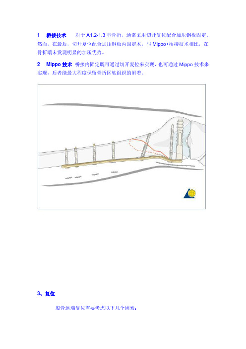
1 桥接技术对于A1.2-1.3型骨折,通常采用切开复位配合加压钢板固定。
然而,在最后,切开复位配合加压钢板内固定术,与Mippo+桥接技术相比,在骨折端未发现明显的加压优势。
2 Mippo技术桥接内固定既可通过切开复位来实现,也可通过Mippo技术来实现,后者能最大程度保留骨折区软组织的附着。
3、复位股骨远端复位需要考虑以下几个因素:∙varus/valgus 内翻/外翻∙flexion/extension 屈曲/伸直∙internal/external rotation 内旋/外旋∙translation 翻转∙lengthening/shortening 延长/缩短4、解剖特征股骨远端有其独特的解剖形态,从下图端视位来看,股骨远端外侧面是一个与垂线相交10°的斜面,而内侧为与垂线相交20-25°的斜面;股骨远端外侧髁(髌面)与内侧髁(髌面)连线有10°倾斜角。
这些解剖特征对于内固定来说是非常重要的,为避免穿入关节。
神经血管束:胫神经和腓总神经位于股骨远端的后面,由于其解剖特征,股骨远端骨折有3%病例伴有血管损伤,1%伴有神经损伤。
在股骨远端外侧面,没有重要神经血管束,在行手术显露过程中,膝上外侧动脉是可能需要结扎处理的血管。
在膝关节后方,有神经血管束存在,外伤医生需要注意。
5、生物力学恢复下肢生物力线是手术重要目的之一,正常人体下肢的力线,起自股骨头中心,通过胫骨平台中心,至踝关节中心。
此轴线可以通过用一根电线(电刀线)来估计,如下图,其体表测量方式为:起自髂前上嵴,通过髌骨正中(胫骨髁间嵴内侧),止于第1、2足趾之间。
在测量过程中确保股骨远端没有旋转。
下图为下肢力学图,其中,最上的虚线表示垂线(Vertical axis);中间线为生物力学(Mechanical axis);下线为解剖轴线(anatomical axis)。
6、内固定物选择LISS:适用于髁间骨折;髁钢板:适用于髁间骨折;95°角钢板:适用于髁上1.5-2cm骨折;95°动力髁钢板:适用于髁上2cm以上骨折;逆行髓内钉:适用于髁上6cm以上骨折,因为其远端需要有2个锁定螺钉固定。
- 1、下载文档前请自行甄别文档内容的完整性,平台不提供额外的编辑、内容补充、找答案等附加服务。
- 2、"仅部分预览"的文档,不可在线预览部分如存在完整性等问题,可反馈申请退款(可完整预览的文档不适用该条件!)。
- 3、如文档侵犯您的权益,请联系客服反馈,我们会尽快为您处理(人工客服工作时间:9:00-18:30)。
A 7-10cm lateral parapatellar incision is made which can be extended proximally into a formal lateral approach to the femur if necessary. The intercondylar reduction is performed through this limited arthrotomy.
After the plate is advanced subperiosteally up the femur, the distal fragment is manipulated such that the plating engages the lag screw.
After the plate is advanced subperiosteally up the femur, the distal fragment is manipulated such that the plating engages the lag screw.
These figures demonstrate the intracondylar extension of the fracture and a free intercondylar fragment (arrows).
FEMORAL AXIS
PLANNED SCREW POSITIONS When planning for the lag screw fixation, the surgeon must leave room for the lag screw of the fixed angle device or intramedullary nail.
AP VIEW
LATERAL VIEW
AP andting that the plate is against the femur and the metaphysis is generally reduced.
The lateral radiograph can be used in a similar fashion to a perfect circle technique used in nailing to place the proximal screws percutaneously.
AP view of a comminuted C3 intraarticular distal femur fracture.
Lateral view demonstrating the typical extension deformity caused by the pull of the origin of the gastrocnemius muscles from the posterior distal femur.
Note the anatomic reduction of the articular surface. At this point, the articular surface is reconstructed and the metaphyseal component of the fracture is still unfixed.
Periarticular or standard clamps are used to manipulate and reduce the fracture which is stabilized with the templated 6.5 mm lag screws with washers. The screws are placed anterior and posterior to the planned position of the fixed angle device.
After the lag screw is placed, the appropriate sized fixed angle plate is slid subperiosteally up the femur. Notice the bolster, which is supporting the fracture in a reduced position.
AP VIEW
LATERAL VIEW
AP VIEW
LATERAL VIEW
AP and lateral radiograph demonstrating the position of the lag screw as templated. Note that with traction, both the AP and lateral radiographs are manipulated such that the metaphysis is reduced.
95°
AP radiograph demonstrating the angle at which the guidewire for the fixed angle lag screw should be placed. It should be parallel with the distal femoral articular surface, which is at approximately 95o to 98o from the femoral shaft.
AP VIEW
LATERAL VIEW
This requires a bolster underneath the metaphyseal fracture and some flexion of the knee to correct the extension deformity of the quadriceps. The dotted line represents the axis of the femur where the plate will be placed.