局部解剖学-下肢前区双语
下肢局部解剖学
(2)闭孔神经
由闭孔进入股 内侧区分为前、 后两支,分别经 短收肌浅、深面 下行。分支支配 大腿肌内侧群和 闭孔外肌。
(五)局部记载 1.肌腔隙和血管腔隙
(1)肌腔隙 前界:腹股沟韧带 后外侧界:髂骨 内侧界:髂耻弓 内容:髂腰肌 股神经 股外侧皮神经。
(2)血管腔隙 前界:腹股沟韧带 后界:耻骨梳韧带 外侧界:髂耻弓 内侧界:腔隙韧带 内容:股鞘 股动脉 股静脉 生殖股神经股支 淋巴管。
(3)神经 腓深神经 腓浅神经
小腿中1/3横断面
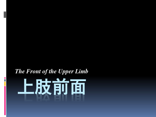
髋肌
前群 后群
前群 —— 股神经
大腿肌 内侧群 —— 闭孔神经
下肢肌
后群 —— 坐骨神经 前群 —— 腓深神经
小腿肌 外侧群 —— 腓浅神经
后群 —— 胫神经
足背肌 —— 腓深神经 足肌
足底肌 —— 足底内侧、外侧神经
第三章 下 肢
The lower limb
大连医科大学解剖学教研室 刘绍壮
第一节 股前区和内侧区
一、体表标志 1.髂前上棘 2.腹股沟 3.耻骨结节 4.股骨内、外侧髁
胫骨内、外侧髁 5.髌骨 6.胫骨粗隆 7.收肌结节
二、主要内容 (一)皮肤 (二)浅筋膜
1.浅动脉 ①腹壁浅动脉 ②旋髂浅动脉 ③阴部外动脉
2.股鞘:为腹横筋膜和髂筋膜向下延伸包裹股动脉、 股静脉上段所形成的筋膜鞘。分为3个腔,外侧 腔容纳股动脉,中间腔容纳股静脉,内侧腔称股 管,内有脂肪和腹股沟深淋巴结。
3.股管和股环 股管:股鞘的内侧腔称股管,内有腹股沟深淋
巴结。 股环:股管上口 前界:腹股沟韧带 后界:耻骨梳韧带 内侧界:腔隙韧带 外侧界:借纤维隔与股静脉相邻
(2)足背 1)足背内侧皮神经 2)足背中间皮神经 3)足背外侧皮神经 4)隐神经 5)腓深神经
下肢前面显微解剖学参考PPT

成骨-肌-皮复合瓣。 髂骨血供丰富为多源性,且血管比较恒定,可以
形成多种血管移植供体。
下肢前面显微解剖学
多源性 旋髂浅动脉:1978,Taylor报道 旋股外侧A升支 第3、第4腰A 旋髂深A 臀上A深上支及浅支 髂腰A髂骨支
缝匠肌瓣
股薄肌(皮)瓣
(一)形态 长带状肌,全长29cm, 肌腱长12cm。
(二)血供 多源性,股深A股薄肌支为其
主要血管,此外尚来自旋股内 侧A、股A、腘A、膝降A、第一 穿A及闭孔A。 (三)神经
闭孔N的股薄肌支
旋股内侧动脉 股薄肌支
闭孔神经 股薄肌支
股薄肌瓣
股薄肌肌(皮)瓣
Gracilis flap
下肢前面显微解剖学
1. 股前肌群
缝匠肌 sartorius: 起-来自前上棘 止-胫骨内侧面上部作用:屈髋屈膝,膝关节旋内 神经支配:股神经
股四头肌 quadriceps femoris : 股 直 肌:起—髂前下棘和髋臼上缘 股中间肌:起—股骨体前面 股内侧肌:起—股骨粗线内 侧唇 股外侧肌:起—股骨粗线外侧唇 止—胫骨粗隆 作用:伸膝;股直肌尚能屈髋关节 神经支配:股神经
(三)神经
腹股沟韧带下方3cm处由股 神经分出
股直肌支
股神经股直肌支
股直肌瓣
缝匠肌(皮)瓣
(一)形态 (二)血供
多源,呈节段性分布; 上部有股深A、旋股外侧A 或其降支)和股A 近侧段的分支。下部有恒定 的膝降A分支。此外还有 旋髂浅A和腘A分支。 (三)神经 股神经分支
股深动脉 隐神经 缝匠肌
隐神经
7lower limb(1)局部解剖英文课件下肢

• Kaplan点
– 仰卧,两腿并拢伸直,当两髂 前上棘处于同一平面时,由两 侧大转子尖经同侧髂前上棘作 一延长线。正常时两延长线相 交于脐或脐以上,相交点称 Kaplan点。 – 当髋关节脱位或股骨颈骨折时 ,此点偏移至脐下,并偏向健 侧。
颈干角Normal angle
of inclination 1250-1300
Coxa vara 髋内翻 (abnormally decreased angle of inclination, it occurs in fractures of the neck of the femur and slipping of the femoral epiphysis )
Coxa valga 髋外翻 (abnormally increased angle of inclination, in cases of congenital dislocation of the hip)
Superficial v. of lower limb
great saphenous v.
Small saphenous v.
Great saphenous vein
• beginning, ending, coursing
– – – – 在足背的内侧缘处起自足背静脉弓 向上行经内踝前方 沿小腿内侧与隐神经相伴行 经股骨内侧髁后方约2cm处,进入大腿内侧渐 斜向上前方 – 最后于耻骨结节外下方3~4cm处,穿过隐静 脉裂孔注入股静脉
1700
>1700
<1700
Normal alignment
Genu varum (bowleg) 膝内翻
Genu valgum 膝外翻
Surface projections
局部解剖学局解下肢
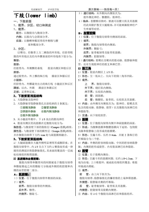
下肢(lower limb)一、下肢前面1、境界、分区、切口和剥皮1、境界:前方:以腹股沟与腹部分界。
内侧:以股沟与会阴部分界。
后面:以髂嵴和骶尾骨的外侧缘与腰部和骶部分界。
2、分区:以臀沟、经髌骨上方二横指的环形线、经胫骨粗隆的环形线以及经内外髁基部的环形线将下肢分为:臀部:股部:经股骨内、外侧髁的垂线股前内侧区和股后区膝部:通过股骨内、外上髁的纵行线膝前区和膝后区小腿部:经胫骨内、外髁最突出点的纵行线小腿前区和后区踝部:以内、外踝踝前区和踝后区足部:足背和足底。
2、下肢前面浅层结构1、大隐静脉及隐神经2、大隐静脉穿隐静脉裂孔以前收纳的5条属支:①旋髂浅静脉②腹壁浅静脉③阴部外静脉④股内侧浅静脉⑤股外侧浅静脉3、从小腿前外侧中、下1/3浅出的腓浅神经4、股前内侧区的浅筋膜在近腹股沟处分为:脂肪层:与腹前壁下部的脂肪层(Camper筋膜)相续。
膜性层:与腹前壁下部的膜性层(Scarpa筋膜)相续,并在腹股沟韧带下方约1cm处与阔筋膜相融合。
3、下肢前面深层结构1)大腿阔筋膜在大腿外侧明显增厚形成髂胫束;在腹股沟韧带中、内1/3交点下方约1横指处形成一卵圆形的薄弱区称隐静脉裂孔,其表面的覆盖的一层疏松结缔组织称筛筋膜。
2)肌腔隙和血管腔隙:腹股沟韧带和髋骨间的间隙被连于腹股沟韧带和髂耻隆起之间的髂耻弓分隔成外侧的肌腔隙和内侧的血管腔隙二部分。
A:肌腔隙:1)位置:位于腹股沟韧带外侧部的深面。
2)境界:前界:腹股沟韧带的外侧部;后外界:髂骨;内侧界:髂耻弓。
3)通行结构:从外侧向内侧依次为;股外侧皮神经、髂腰肌、股神经。
4)临床:患腰椎结核时,脓液可沿腰大肌及其筋膜经此间隙扩散至大腿根部,并可能刺激股神经产生疼痛等症状。
B:血管腔隙1)位置:位于腹股沟韧带内侧部的深面。
2)境界:前界:腹股沟韧带的内侧部;外侧界:髂耻弓;后界:耻骨梳韧带及耻骨肌筋膜;内侧界:腔隙韧带。
3)通行结构:股鞘以及鞘内的股动脉、股静脉和股管,还有生殖股神经的股支和淋巴管。
局部解剖学 下肢
后骨筋膜鞘:
完整ppt
20
(二)肌肉
1.髋肌—前群
髂腰肌
• 髂肌 • 腰大肌
腰小肌 阔筋膜张肌
完整ppt
21
2.大腿肌
前群
• 缝匠肌 • 股四头肌:
完整ppt
22
内侧群 • 耻骨肌 • 长收肌 • 短收肌 • 大收肌 • 股薄肌
收肌腱裂孔
完整ppt
23
(三)肌腔隙与血管腔隙
腹股沟韧带-----髂耻弓--------------耻骨梳韧带------------陷窝韧带---------
(腔隙韧带)
完整ppt
内面观
24
肌腔隙与血管腔隙
外侧:肌腔隙 内侧:血管腔隙
腹股沟韧带 髂耻弓
完整ppt
外面观
25
肌腔隙:
境界:
前界: 腹股沟韧带
内侧界: 髂耻弓 后界: 髂骨
肌腔隙通过的结构:
髂腰肌 股神经 股外侧皮神经
完整ppt
26
血管腔隙:
腹股沟韧带 髂耻弓
境界: 前界:腹股沟韧带 内侧界:腔隙韧带 后界:耻骨梳韧带 外侧界:髂耻弓
局部解剖学
Regional Anatomy
下肢
Lower limb
郭义威 人体解剖学教研室
guoyiwei@
完整ppt
1
下肢
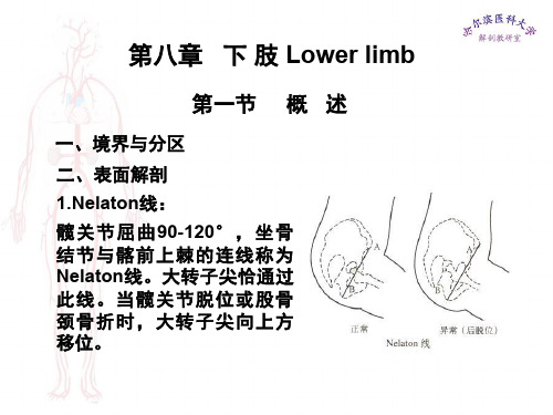
第一节 概 述
一、境界与分区 境界:与躯干相连。分臀、股、
膝、小腿、踝、足
分区:股前内侧区、臀区及股 后区、小腿前外侧区及足背区、 腘窝、小腿后区及足底区。
腹壁浅A
腹下部带蒂游离皮瓣移植常用的血管
完整ppt
11
2.大隐静脉
局部解剖学课件下肢-PPT文档资料57页
股深动脉
旋股外侧动脉
穿动脉
4)股静脉femoral vein:
5)腹股沟深淋巴结:
6)股神经 femoral nerve:
隐神经 saphenous nerve: 经股三角、收肌管,穿收肌腱 板,行于缝匠肌和股薄肌在膝 关节内侧穿深筋膜,伴大隐静 脉下行,分布于髌骨下方、小 腿内侧和足内侧缘。
的后壁、腘肌及其筋膜。
内容:由浅入深为 胫神经 腘静脉 腘动脉 外上界的腓总神经 血管周围有腘深淋巴结 脂肪
第五节 小腿部
一、小腿前外侧区
(一)浅层结构 大隐静脉:经内踝前方,沿小 腿内侧上行。 隐神经:伴行大隐静脉。 腓浅神经:于小腿外侧中、下 1/3交点处穿出深筋膜。
(二)深层结构
深筋膜在内侧与胫骨内侧面的骨膜相融合,在腓侧深筋 膜发出前、后肌间隔,附着于腓骨前、后缘,形成前、 后、外侧骨筋膜鞘。
2.腓肠神经:由腓肠内侧皮神 经与腓神经的交通支吻合而成。 分布于小腿后区下部和足背外 侧皮肤。
(二)深层结构 1.后骨筋膜鞘: 2.血管神经束: 胫后动脉—腓动脉 胫后静脉 胫神经
第六节 踝与足部
一、踝前区与足背
(一)浅层结构 足背静脉弓: 内侧合成大隐静脉 外侧合成小隐静脉 隐神经:足内侧缘 腓浅神经:足背中部 腓肠神经:足背外侧
5.收肌管adductor canal
前壁:缝匠肌、大收肌腱板 后壁:大收肌及长收肌 外侧壁:股内侧肌 上口与股三角尖相通 下口为收肌腱裂孔
从前向后内容:股神经的内侧 肌支和隐神经、股动脉、股静 脉以及淋巴管和疏松结缔组织。
6.股内侧区的血管和神经:闭孔血管神经束
二、股后区
(一)浅层结构:有股后 皮神经和小隐静脉等。
人体解剖学中英名词对照表
人体解剖学英汉名词对照表注:用星号(*)标注的名词为最常用的词汇,必须熟记掌握.一、总论(一)解剖学分类(Classification of Anatomy)anatomy 解剖学human anatomy,anthropotomy 人类解剖学systematic anatomy 系统解剖学regional (topographic)anatomy, topology 局部解剖学gross (macroscopic)anatomy 大体解剖学,巨视解剖学microscopic anatomy 显微解剖学macro-microscopic anatomy 巨—微解剖学sectional anatomy 断面解剖学pathological (morbid)anatomy 病理解剖学neuroanatomy 神经解剖学chemical neuroanatomy 化学解剖学developmental anatomy 发生(或发育)histologic anatomy 组织解剖学histology 组织学embryology 胚胎学anthropology 人类学(二)一般解剖学术语(General Anatomical Terms) dissection,dissect (v。
)解剖,解剖标本normality,normal (a.) 正常abnormality ,abnormal 异常variation 变异deformity,malformation 畸形specimen 标本model 模型(三)方位术语(Positional Terms)1。
anatomical position 解剖学姿势2。
方位术语(terms of direction)superior,upper 上inferior, lower 下anterior 前侧posterior 后侧ventral (dorsal)腹(背)侧medial (lateral) 内(外)侧intermediate 中间internal,interior, inner,inside 内external, exterior,outer,outside 外superficial 浅deep,profound 深proximal (distal)近(远)侧ulnar (radial)尺(桡)侧tibial (fibular)胫(腓)侧palmar 掌侧plantar 足底left 左right 右middle 中median 正中3。
【课件-局部解剖学】_02上下肢前面骨筋膜鞘
臂前鞘:
肱前群肌 肱动、静脉 正中神经 肌皮神经 尺神经上段
前臂前鞘:
前臂前群肌 桡动脉静脉 桡神经浅支 尺动脉静脉 尺神经 骨间前动静脉 正中神经
The Superficial Layers of the Lower Limb
下肢浅层
3.深筋膜及肌间隔 下肢的深筋膜比较发达,
坚韧。各部互相延续,包 绕肌肉、血管神经,分隔 肌群,形成肌间隔、骨筋 膜鞘等。
深筋膜及肌间隔 股前深筋膜最厚特称阔筋膜,外侧份形成髂胫束,
耻骨节结外下方3-4cm处形成卵圆形的薄弱区——卵 圆窝。
4. 下肢的骨筋膜鞘及其内容
股前鞘 大腿前群肌 股动、静脉 股神经
4. 下肢的骨筋膜鞘及其内容
股内侧鞘 内收肌群 闭孔动、静脉 闭孔神经
4. 下肢的骨筋膜鞘及其内容
小腿前鞘 前群肌 胫前动、静脉 腓深神经
小腿外侧鞘 外群肌 腓浅神经
【课件-局部解剖学】_03 上下肢前面
上肢前面
腋区
腋窝 axillary fossa
腋窝顶 锁骨中1/3部 第1肋外缘 肩胛骨上缘 上通颈根部
腋窝底 皮肤 浅筋膜 腋筋膜 朝向下外
腋窝四壁
内侧壁: 前锯肌 上位4个肋骨 肋间隙
外侧壁: 肱骨的结节间沟 肱二头肌长、短头 喙肱肌
前壁: 胸大肌、胸小肌 锁骨下肌 锁胸筋膜
出深筋膜
肘关节动脉网
桡侧副动脉与桡侧返动脉 中副动脉与骨间返动脉 尺侧上、下副动脉与尺侧 返动脉吻合(分前后支)
The Front of the Lower Limb
下肢前面
肌腔隙与血管腔隙
位于腹股沟韧带与髋骨之间,由髂耻弓 分隔成外侧的肌腔隙与内侧的血管腔隙。
肌腔隙:lacuna musculorum
内侧束及分支: 尺神经 胸内侧神经 臂内侧皮神经 前臂内侧皮神经 正中神经内侧头
臂丛 brachial plexus
后束及分支: 桡神经 腋神经 肩胛下神经 胸背神经
腋淋巴结 axillary lymph nodes
位于腋血管及其分支周围,分为五群
外侧淋巴结
腋静脉远端
胸肌淋巴结
胸外侧血管、胸长神经
外侧部容纳股动脉 中间部容纳股静脉 内侧部称al
为股鞘内侧份潜在性间隙, 股管的上口称股环:
前界:腹股沟韧带 后界:耻骨梳韧带 内界:腔隙韧带 外界:股静脉及纤维隔
管的下端为盲端,对向隐 静脉裂孔
当腹内压增高时,腹盆腔 脏器可经股环至股管而形成 股疝
收肌管 adductor canal
又称Hunter管
构成:
前内侧壁 缝匠肌
大收肌腱板
局部解剖学:下肢
Superficial epigastric v. Superficial circumflex iliac v.
External pudendal v. Superficial medial femoral v. Superficial lateral femoral v.
Great saphenous v.
Nerves of lower limb
Femoral n.: supplies anterior thigh muscles (quadriceps, sartorius and pectineus), hip and knee joint, and skin on anteromedial side of thigh, saphenous nerve is distributed to skin of medial side of leg and foot Obturator n.: enters thigh through obturator foramen; supplies medial group of muscles of thigh, obturator externus, and skin of medial side of thigh
Structures passing suprapiriform foramen
Superior gluteal n., a., v. from lateral to medial side
Structures passing infrapiriform foramen
Sciatic n., posterior femoral cutaneous n., inferior gluteal n., a.,v., internal pudendal v., a., and pudendal n. from lateral to medial side
- 1、下载文档前请自行甄别文档内容的完整性,平台不提供额外的编辑、内容补充、找答案等附加服务。
- 2、"仅部分预览"的文档,不可在线预览部分如存在完整性等问题,可反馈申请退款(可完整预览的文档不适用该条件!)。
- 3、如文档侵犯您的权益,请联系客服反馈,我们会尽快为您处理(人工客服工作时间:9:00-18:30)。
⑤ posterior cutaneous n. of thigh
⑥ cutaneous br. of obturator n.
⑧
⑦ medial sural cutaneous n.
⑦
⑧ lateral sural cutaneous n.
⑨
⑨ sural nerve
Cutaneous nerves
股骨内外侧上髁
Adductor tubercle 收肌结节 lateral and medial condyles of tibia 胫骨内外侧髁 Fibular head 腓骨头 tendon of biceps femoris 股二头肌腱 tendons of semitendinosus and semimembranosus
a line drawn from the anterior superior lilac spine to the ischial tuberosity, passing over or near the top of the greater trochanter.
The trochanter can be felt superior to this line in a person which dislocated hip joint or fractured femoral neck.
Course: passes upward directly in front of the medial malleolus, then ascends on the medial side of the leg, knee, and thigh to pass through the saphenous hiatus in deep fascia and join with the femoral vein.
normal
abnormal
Bryant line and bryant triangle
Superficial structure
1.Skin 2.Superficial fascia
Superficial arteries:
1. Superficial epigastric a.
腹壁浅动脉
③
① superior cluneal n.
② medial cluneal n
⑥
③ inferior cluneal n.
④ lateral femoral cutaneous n.
⑤ posterior cutaneous n. of thigh
⑥ cutaneous br. of obturator n.
⑥ femoral br. of genitofemoral n.
⑦ iliohypogastric n.
⑧ ilioinguinal n.
⑦ ⑧
② ④ ⑤
Superficial peroneal n.
Cutaneous nerves
⑦
⑥
③
⑧
股部皮神经有:
① anterior femoral cutaneous n.
⑤
⑦ iliohypogastric n.
⑧ ilioinguinal n.
Superficial peroneal n.
①
Cutaneous nerves
②
① superior cluneal n.
④ ③
② medial cluneal n ③ inferior cluneal n.
⑥
⑤
④ lateral femoral cutaneous n.
into the gluteal region (buttock), thigh, knee, leg, ankle and foot.
臀沟
2. Surface anatomy
(1) The surface landmarks Gluteal region and thigh iliac crest 髂嵴 anterior superior iliac spine 髂前上嵴 posterior superior iliac spine 髂后上嵴 tubercle of iliac crest 髂结节 ischial tuberosity 坐骨结节 greater trochanter 大转子 pubic symphysis 耻骨联合 pubic crest 耻骨嵴 pubic tubercle 耻骨结节 inguinal ligament 腹股沟韧带
⑦ medial sural cutaneous n. ⑧ lateral sural cutaneous n.
saphenous n.
⑨ sural nerve
④ ⑤
common peroneal n.
⑦ ⑧
Superficial inguinal lymph nodes
superior group
①
②
② medial femoral cutaneous n.
③ lateral femoral cutaneous n.
④
④ cutaneous br. of obturator n.
⑤ saphenous n.
⑥ femoral br. of genitofemoral n. Lateral sural cutaneous n.
Small saphenous vein
小隐静脉
Origin: from the lateral sides of a dorsal venous arch in the foot.
Course: passes behind the distal end of the fibula (lateral malleolus) and up the back of the leg to penetrate deep fascia and join the popliteal vein posterior to the knee.
Tributaries of the great saphenous vein
① Superficial iliac circumflex v. 旋髂浅静脉
② Superficial epigastric v. 腹壁浅静脉
③ External pudendal v. 阴部外静脉
④ Superficial medial femoral v. 股内侧浅静脉
inferior group
hip)
1700
>1700
<1700
Normal alignment
Genu varum (bowleg) 膝内翻
Genu valgum 膝外翻
The orientation of the pelvis in the standing position
In the standing position the plane of the linea terminalis is usually inclined at an angle of about 600 to the horizontal.
(it occurs in fractures of the neck of the femur and slipping of the femoral epiphysis )
greater than 1300: coxa valga 髋外翻
(in cases of congenital先 天性的dislocation of the
The projections of the blood vessels and nerves
1. Superior gluteal a.,v.,and n. 2. Inferior gluteal a.,v.,and n. 3. Sciatic n. 4. Femoral a. and v. 5. Popliteal a. 6. Anterior tibial a. 7. Posterior tibial a. 8. Dorsal artery of foot
2. Superficial iliac circumflex a.
旋髂浅动脉
3. Superficial external pudendal a.
阴部外浅动脉
②① ③
Superficial veins
great saphenous vein 大隐静脉
Origin: from the medial sides of a dorsal venous arch in the foot.
半腱和半膜肌腱
Leg, Ankle and Foot
anterior border of tibia 胫骨前缘 head of fibula 腓骨头 neck of fibula 腓骨颈 medial and lateral malleolus 内、外踝 calcaneal tuberosity 跟结节 tuberosity of navicular bone 舟骨结节 tuberosity of fifth metatarsal bone第五跖骨粗隆
normal abnormal
2. Kapian’s point (shoemaker’s sign)
The patient lies supine with stretched legs. Join a line from the apex of the greater trochanter to the anterior superior iliac spine. Normally, its prolonged line passes through or above the umbilicus. If the line passes below the umbilicus, it indicates the presence of superior displacement of the greater trochanter.