AQP1 抑制剂4
降糖药物GLP1A和DDP-4简介
常见的GLP-1RA
随着相关研究的不断深入,临床应用经验的不断积累,GLP-1RA 在国内外糖尿病 治疗指南中的地位逐渐提升。GLP-1RA 从2009年美国糖尿病学会(ADA)和欧洲糖尿
病研究学会(EASD)首次在指南中推荐纳入治疗流程,目前已成为继二甲双胍之后的
首选药物之一。 GLP-1RA 包括多肽和小分子化合物,在短短的几年中,多肽、血浆白蛋白重组肽、 生物表达重组肽等制剂先后问世,以下GLP-1RA 有些已经在临床应用中,有些仍在发 展临床试验过程中:①艾塞那肽注射液(商品名:百泌达,Byetta);②利拉鲁肽注 射液(商品名:诺和力,Victoza);③艾塞那肽长效制剂(商品名:Bydureon);④ 利西拉来Lixisenatide(商品名(Lyxumia);⑤ 阿必鲁肽(Albiglutide);⑥ 杜拉糖肽
验。
5.孕妇与哺乳期用药 妊娠期妇女使用安全性未知。动物实 验证实其可经乳汁分泌,故哺乳期不 推荐使用。
T1DM 患者在胰岛素基础上加用利拉
鲁肽可减少胰岛素剂量、降低体重、 减少低血糖发生率,但在HbA1c及血
糖变异性方面与安慰剂相比差异无统
计学意义。
二肽激酶抑制剂
胰高血糖素样肽-1(glucagon-like peptide 1),1964年,Elrick等发现 口服葡萄糖刺激胰岛素分泌的量明显大于静脉滴注相同剂量葡萄糖,这种现象 被称为“肠促胰素”效应。人体内主要有两种肠促胰素:葡萄糖依赖性促胰岛 素释放肽(glucose-dependent insulinotropic polypeptide, GIP)和GLP-1。因T2DM 患者的血GIP 水平正常或升高,对β细胞的促胰岛素分泌作用显著降低,且对α
症、免疫等相关:
浅析GLP-1受体激动剂与DPP-4抑制剂
0.3
0.2
0.2
20
0.1
0.1
60
120
时间(分)
0 180
0 0
口服葡萄糖 静脉注射葡萄糖
60
120
时间(分)
0 180
Adapted with permission from Nauck M et al. Diabetologia. 1986;29:46–52. Copyright © 1986 Springer-Verlag.
GLP-1具有多种重要生理作用
α
β
胰腺
肠
β 葡萄糖依赖性胰岛素分泌
β 胰岛素合成
α 葡萄糖依赖性胰高糖素 分泌
GLP-1
肝
葡萄糖生成
L细胞分泌 GLP-1 被 DPP-4 分解
Adapted from Baggio & Drucker. Gastroenterol 2019;132;2131–57
• 体内代谢完全
• 仅6%和5%的代谢产物经尿液和粪便排出 • 泌尿系或胃肠道内未见完整的利拉鲁肽
Malm-Erjefalt M. Drug metabolism and disposition 2019; 38(1944-53).
肾脏或肝脏受损患者体内利拉鲁肽药代动力学
•肝肾功能受损的患者利拉鲁肽药物暴露剂量未增加
UKPDS
9 8.5
8
常规治疗*
格列苯脲 二甲双胍 胰岛素
ADOPT
8 7.5
罗格列酮 二甲双胍
格列苯脲
7.5 7
7 推荐治疗达标目标
<7.0%†
6.5
6.5
6
6
6.2% – 正常值上限
水通道

调节神经兴奋性 星形胶质细胞上的 AQP4 依赖性水转运参与神经兴奋的调节。
参与脑水肿的发生及发展
可能通过对血管内水的快速转运而促进细胞毒性 脑水肿的发生、 发展; 促使缺血性中风后脑水肿的发生、 发展; 在血管源性脑水肿的发生发展过程中, 水进入脑 不依赖AQP4, 而水从脑中清除则与其密切相关。
早期,人们一直认为水是以单纯扩散透过细胞 膜的,但在某些细胞如红细胞、肾近曲小管 细胞对水的通透性很高,不能简单以单纯扩 散来解释,于是推测此类细胞膜上可能存在 某种功能性转运水的通道。
水通道蛋白的发现
Agre于1988年发现并成功分离出一种细胞膜蛋白并证 明这就是科学家孜孜以求的水通道蛋白(aquaporin, AQP)。 2000年,Agre公布了世界第一张水通道蛋白的高清晰 立体照片,照片揭示这种蛋白的特殊结构只允许水分 子通过。
其它眼病
泪腺疾病:AQP5. 参与泪液分泌,维持眼 表泪膜的完整和功能 —— 干眼症。 结膜病:AQP3. 参与泪膜的形成 —— 干 眼症。 角膜病:AQP3 、AQP5. 参与维持角膜的 脱水状态及角膜的透明度 —— 角膜的水 肿和变性。
其它眼病
晶状体病:AQP1 、AQP0. 参与维持晶状体的 脱水状态、 透明性 —— 白内障。 视网膜病:AQP1、AQP4,AQP4 主要存在于 Müller 细胞和星形胶质细胞,对维持视网膜 水平衡起关键作用 —— 视网膜脱离和黄斑水 肿的发病。 视神经病变:Na+-K+的平衡一旦失去平衡,可 能导致发生视乳头水肿(乃至脑水肿)等严重疾 病。
AQP4:脑组织中大量表达,主要分布于星形胶质细胞。
参与对中枢神经系统水、 电解质的调节 允许
水的快速转运,促进星形胶质细胞通过 Kir4.1 对钾 的摄取;在胶质细胞、 血、 脑脊液间进行水的调节, 维持脑的水盐平衡;感受渗透压变化,调节ADH分泌, 调节机体水平衡。
红细胞水通道蛋白1的研究进展

胞膜 水通 透的关 系。 在生理 环境 中, 网织红 细胞可 以
通 过 调节A 1 QP 的数 量来 适应环境 的变化 。 在细 胞成
膜A 1 QP 增加 来 适应 血浆 渗透 压 力的变化 , 表现 为 细 胞体 积的 膨胀和 缩小 。 根据 A 1‘ QP 使红 细 胞在 血 浆 H ‘  ̄
浓 度 的硝酸 银 浓 聚物 能延长 水 通 道 蛋白活性恢 复的 时 间, 并且 当硝酸 银一旦被去 除, QP 的活性仍然 会 A 1 恢 复。 在研 究 红细胞 上 尿素转运 体抑 制剂 时, 发现 其 并 未影  ̄ AQP 对水 的运 输效 率 , 明尿素转 运体 抑 N I 说
5A P 的抑制剂 Q 1
氯化 汞 ( CI 是 比较 明确的AQ 1 制 剂 , Hg : ) P抑 既 可 以阻滞水流 入细 胞内引起细胞 肿胀 , 能阻滞 细胞 也
内水流 出细 胞导致 细胞皱 缩【 。 1 用测定网织红细 胞的
电流 光闪射的方 法对 比了几种 已被报 道过 的抑制剂 ,
串联序 列是AQ 家 族成 员共 有的特征 性结 构 。 过 P 】通 电子显微 镜和 电子结晶学方法证实 了人红 细胞膜 上 的
AQ 1 P 是以 四聚体的形式 存在 ,  ̄AQ 1 分J i P 的三维结 构 :
胞容积 瞬间增高, 膜A P 密度也大大增高。 胞 Q 1 红细 胞
上大 量的A 1 QP 是适 应环境 变化而产生的红 细胞生 理
长 期镇静 的 高危病 人 , 比如 肾功 能障碍 、 严重 感染 及 毛 细血管渗漏 等病人 , 在选 择药物时 就应该考虑麻 醉 剂 对A 1 抑制 作用 , 免增加 细 胞水代 谢 障碍 的 QP 的 避
水通道蛋白
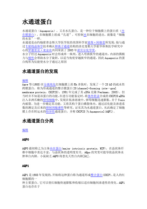
水通道蛋白水通道蛋白(Aquaporin),又名水孔蛋白,是一种位于细胞膜上的蛋白质(内在膜蛋白),在细胞膜上组成“孔道”,可控制水在细胞的进出,就像是“细胞的水泵”一样。
水通道是由约翰霍普金斯大学医学院的美国科学家彼得·阿格雷所发现,他与通过X射线晶体学技术确认钾离子通道结构的洛克斐勒大学霍华休斯医学研究中心的罗德里克·麦金农共同荣获了2003年诺贝尔化学奖。
水分子经过Aquaporin时会形成单一纵列,进入弯曲狭窄的通道内,内部的偶极力与极性会帮助水分子旋转,以适当角度穿越狭窄的通道,因此Aquaporin的蛋白构形为仅能使水分子通过之原因水通道蛋白的发现编辑Agre等(1988)在分离纯化红细胞膜上的Rh多肽时,发现了一个28 kD的疏水性跨膜蛋白,称为形成通道的整合膜蛋白28(channel-forming inte—gral membrane protein,CHIP28),1991年完成了其cDNA克隆(Verkman,2003)。
但当时并不知道该蛋白的功能,在进行功能鉴定时,将体外转录合成的CHIP28 mDNA 注入非洲爪蟾的卵母细胞中,发现在低渗溶液中,卵母细胞迅速膨胀,并于5 min 内破裂。
为进一步确定其功能,又将其构于蛋白磷脂体内,通过活化能及渗透系数的测定及后来的抑制剂敏感性等研究,证实其为水通道蛋白。
从此确定了细胞膜上存在转运水的特异性通道蛋白,并称CHIP28为Aquaporinl(AQPl)。
水通道蛋白分类编辑AQP0AQP0最初称之为主体内在蛋白(major intrinsic protein,MIP),在晶状体纤维中细胞中表达丰富,与晶状体的透明度有关.AQpo的突变可能导致晶状体水肿和白内障。
小鼠缺乏AQPO将患先天性白内障[61]。
AQP1AQP1是1988年发现的,开始将这种蛋白称为通道形成整合蛋白(CHIP),是人的红细胞膜的一种主要蛋白。
水通道蛋白在肾脏的表达及意义
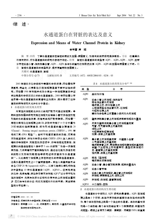
・综 述・水通道蛋白在肾脏的表达及意义Expression and Means of W ater Channel Protein in K idney常笑雪,黄 鹂摘 要:目的 了解水通道蛋白在肾脏的表达及生理、病理意义,为相关的临床研究提供帮助。
方法 收集国内外相关资料,对水通道蛋白的研究内容进行综述。
结果 肾脏水通道蛋白的类型有AQP1-AQP4、AQP6-AQP8,主要分布在近曲小管、细段和集合管,AQP1-AQP4参与水的重吸收和尿液浓缩.AQP6-AQP8的生理与病理意义不明。
结论 肾脏水通道蛋白的类型较多,提示有重要的生理意义。
关键词:水通道蛋白;肾脏中图分类号:Q73 文献标识码:B 文章编号:1672-688X(2004)03-0236-03 2003年诺贝尔化学奖授予美国科学家彼得、阿格雷和罗德里克、麦金农,以表彰他们在细胞膜通道方面开创性的研究。
阿格雷1988年发现并成功分离出一种细胞膜蛋白并证明这就是科学家孜孜以求的水通道蛋白。
2000年阿格雷公布世界第一张水通道蛋白的高清晰立体照片,照片揭示了这种蛋白的特殊结构只允许水分子通过。
1 水通道蛋白的发现及类型所有组织细胞都允许水以单纯扩散方式通过细胞膜。
早期的生物物理学研究发现红细胞及近端肾小管对渗透压改变引起的水的通透性很高,很难用单纯扩散来解释。
阿格雷[1]等在分离纯化红细胞膜上的Rh多肽时发现了一个分子量为28K D的疏水性跨膜蛋白,称为形成通道的整合膜蛋白28 (Channel-F orming integral membrane protein,CHIP28)。
1991年完成了其cDNA克隆[2]。
当时不知道该蛋白的功能,对其进行功能鉴定时,将体外转录合成的CHIP28cDNA注入非洲爪蟾的卵母细胞中,发现在低渗溶液中,卵母细胞迅速膨胀,细胞膜水的通透性增加8倍并于5min内破裂[3]为进一步确定其功能,又将其建构于蛋白磷脂体内,通过活化能及渗透系数的测定以及抑制剂敏感性等研究证实该蛋白为水通道蛋白[4]。
曲格列汀机制
曲格列汀是一种口服药物,属于二肽基肽酶-4(DPP-4)抑制剂,也被称为DPP-4抑制剂。
它的作用机制主要涉及调节胰高血糖素样肽-1(GLP-1)的代谢。
GLP-1是一种由肠道分泌的激素,在食物摄入后增加胰岛素的分泌并减少胃排空速度,从而帮助降低血糖水平。
然而,GLP-1在体内被DPP-4酶迅速降解,导致其活性持续时间较短。
曲格列汀通过抑制DPP-4酶的活性,延长GLP-1的半衰期,使其能够更长时间地发挥降低血糖的作用。
具体来说,曲格列汀与DPP-4酶结合,阻止其对GLP-1的降解,从而增加了GLP-1的稳定性和生物活性。
通过增加GLP-1的浓度,曲格列汀可促进胰岛素的分泌,并抑制胰高血糖素的分泌,从而协调血糖水平的调节。
此外,曲格列汀还可以降低胃排空速度和食欲,有助于控制体重。
总的来说,曲格列汀通过抑制DPP-4酶的作用,增加GLP-1的活性,从而改善胰岛素的分泌和血糖的调节,是一种常用的口服药物,用于治疗2型糖尿病。
1。
AQP1调节机制及其表达在胶质瘤血管新生中的作用

【 关键词 】 水通道蛋 白一 1 ; 调节 ; 血管新 【 文献标识码】 A 【 文章编号】 1 0 0 3 - - 6 3 5 0 ( 2 0 1 3 ) 0 9 —1 3 2 0 —O 3
血 管 生成 是 胶 质瘤 突 破 上皮 基 底膜 后 进 一步 生 长所必须 的, 对于高级别胶质瘤生长、 浸润和转移至 关 重要 。在 无血 管 期 , 胶 质瘤 生 长 缓慢 , 直 径 不会 超 过2 m l r l , 一旦进入血管生成期 , 胶质瘤就可在丰富血 供和营养的条件下 , 促进 自身的迅速生长和浸润 。在 血管新生过程 中, AQ P 1 发 挥 了极 其 重 要 的作 用 , 现 就其调节机制及在胶质瘤血管新生 中的作用新进展 综述 如下 : 1 AQP 1 的结构 和功 能 A QP 1 作 为参 与跨膜转 运水 的膜 通道蛋 白家族 成 员 之一 , 广 泛存 在 于各 种组 织 中 。研 究 发 现 AQ P 1 基 因定位于人染色体 7 p 一> p “ 处 】 , 其一级结构为 6 次 跨膜的单肽链, 含3 个胞外环( A 、 c 、 E ) 和2 个胞内环( B 、 D ) , 连接跨膜区的B 、 E 两攀环折返穿人脂质双分子层, 并且 分别 与邻 近 的跨 膜 区形成 半个 孔道 , 彼此 对称 分 布, 构成一 条狭窄 的分子通道 , 水分子则 排列成单一 纵 列, 以适当的角度通过。该通道具有高度的选择性 , 借 助一 个带 正 电的 区域 排斥 带正 电 的水合 质子 , 不允 许 其 他 离子 或分子 通过 的功 能 , 实 现选 择性 地通 过大 量 的水但 】 。 目前多 数学者 认为 A QP s 的三级结构 为 “ 沙 漏 模型” 。生理 情况 下 , AQ P 1 主要分 布于 脉络丛 及面 向 脑脊液 的顶 质膜 的微绒 毛表面 , 与 Na 一 一 A T P 酶定 位 相同, 参与脑脊液分泌, 并调节水和离子平衡 。在脊髓 中, AQP 1 主要 表达 痛 觉与 感 觉神 经纤 维 。近年 来 多 项 研究 显示 , A Q P l 作为 细胞膜 特异性 水转 运蛋 白 , 在促 进瘤周 水肿形成 、 肿瘤细胞迁 移 中发挥 重要作用 , 且认 为它在 实体肿 瘤血管新 生中扮 演重要 角色 。 2 AQ P 1 的调 节 ’ A Q P 1 的表达受到理化 因素、 激素 以及其他活性 蛋白分子等多种 因素的调控。从整体上讲 , A Q P 1 的 调节 大致 可 以分为 两种 : 一 种 是 通 过 改 变 细 胞 上 A Q P 1 浓 度来调节跨膜水流动 , 相对耗 时较长 , 故称
水通道蛋白相关疾病阅读材料

四.水通道蛋白相关疾病当水通道蛋白的调节出现紊乱的时候,则可能引起多种疾病。
(一)肾脏水通道蛋白和相关疾病研究表明,水通道蛋白基因突变将引起尿崩症(diabetesinsipidus,DI)。
尿崩症广义上讲是指多饮、低比重尿和低渗尿为特征的一组综合征。
目前报道的多数遗传性肾性尿崩症病例是以X连锁方式遗传的,由编码V2 受体的基因突变引起,另外的病例则是由于编码AQP2基因的突变引起,以常染色体显性或隐性方式遗传[11]。
(二)肺部水通道蛋白和相关疾病肺水通道蛋白的异常与肺疾病的关系已有诸多实验报道。
AQP可能参与肺水肿的发病机制。
在各种肺损伤中,存在着大量的水的异常跨膜转运及在肺组织中的异常聚集等情况,这些情况均可能与水通道蛋白有关。
在小鼠病毒性肺炎模型中,发现AQP1和AQP5在鼠肺中的表达降低,这说明肺水在肺间质中聚集的重要原因就是水通道蛋白的减少,导致水不能及时排出而出现水肿。
哮喘发作时,水分子运动在气道阻塞中起重要作用,特别在冷哮喘或运动哮喘时, 上皮黏膜下血管(含AQP1) 、气管及支气管(含AQP3 和AQP4) 的肿胀是形成气道阻塞的重要原因[1]。
从而说明了水通道蛋白和哮喘的发生也有密切关系。
(三)水通道蛋白及癌症水通道蛋白在肿瘤组织的表达及其与肿瘤细胞转移的关系可能将会是今后研究的热门。
多年研究表明,为满足快速增殖、分裂和侵袭转移的需要,肿瘤细胞内一系列酶的活性和表达会发生改变,细胞基本结构成分如蛋白质、脂类和核酸的合成加强。
癌细胞的所有生命活动都离不开水的微环境和参与,癌细胞比正常细胞更需要水分子的快速跨膜转运。
目前的研究表明,部分AQPs在肿瘤组织中表达明显增高或降低。
在脑胶质瘤中水通道蛋白的表达明显增多,脑胶质瘤多伴有脑水肿的发生。
经证实,AQP 1和AQP4在脑胶质瘤中的表达明显高于正常组织,且在星型细胞的表达量与恶性程度有直接关系[8]。
AQPs同时还可能促进肿瘤血管增生,增强肿瘤血管渗透性,在肿瘤的生长和扩散、侵袭和转移中有重要作用。
水通道蛋白1、3、4和5与急性肺损伤关系的研究进展

水通道蛋白1、3、4和5与急性肺损伤关系的研究进展刘笑玎;原铭贞;王司仪;刘相良;梁豪君;高歌;李波;董春玲【摘要】急性肺损伤(ALI)的显著病理生理学表现为非心源性肺水肿。
在多种因素诱导的肺损伤和其他非心源性肺水肿的发生进展中,水通道蛋白1、3、4和5(AQP1、3、4、5)发挥了重要作用,尤以 AQP1和 AQP5为主。
应用基因技术发现在 ALI中 AQP1、3、4和5在细胞迁移、黏蛋白产生等过程和核转录因子κB(NF-κB)和丝裂原活化蛋白激酶(MAPK)信号通路中均有参与。
多种因素能够干预 AQP1和 AQP5的表达进而加重或减轻肺水肿。
AQP1在 ALI的胸水形成中尚发挥一定作用。
本文主要从 ALI中 AQP1、3、4和5在蛋白质水平和基因水平上的参与、多种因素干预以及胸膜水通道蛋白几方面进行综合论述。
【期刊名称】《吉林大学学报(医学版)》【年(卷),期】2014(000)005【总页数】4页(P1119-1122)【关键词】水通道蛋白;急性肺损伤;肺水肿【作者】刘笑玎;原铭贞;王司仪;刘相良;梁豪君;高歌;李波;董春玲【作者单位】吉林大学基础医学院人体解剖学系,吉林长春 130021;吉林大学基础医学院人体解剖学系,吉林长春 130021;吉林大学基础医学院人体解剖学系,吉林长春 130021;吉林大学基础医学院人体解剖学系,吉林长春 130021;吉林大学基础医学院人体解剖学系,吉林长春 130021;吉林大学基础医学院人体解剖学系,吉林长春 130021;吉林大学基础医学院人体解剖学系,吉林长春 130021;吉林大学第二医院呼吸内科,吉林长春 130041【正文语种】中文【中图分类】R563急性肺损伤(acute lung injury,ALI)及其严重形式急性呼吸窘迫综合征(acute respiratory distress syndrome, ARDS)表现为与严重炎症和弥漫性肺泡损伤有关的急性呼吸衰竭[1]。
降糖药物glp1a和ddp4简介

降糖药物glp1a和ddp4简介汇报人:目录•介绍•glp1a和ddp4的药物特性•glp1a和ddp4的临床试验结果•glp1a和ddp4的适应症与副作用•glp1a和ddp4的研发前景与挑战•总结01介绍什么是glp1a和ddp4?•GLP-1(Glucagon-like peptide-1)是一种天然的肠促胰素,由肠道L细胞分泌,能够刺激胰岛素分泌并降低血糖。
然而,GLP-1在体内迅速被DPP-IV降解,因此需要使用类似GLP-1的药物,如GLP-1受体激动剂(glp1a)和DPP-IV抑制剂(ddp4)。
20世纪90年代,科学家们发现了GLP-1及其降糖作用。
2000年,第一个G L P -1受体激动剂(glp1a)和DPP-IV 抑制剂(ddp4)进入临床试验。
2005年,第一个glp1a(艾塞那肽)和ddp4(西格列汀)获得美国FDA批准上市。
glp1a和ddp4的发展历程通过与GLP-1受体结合,刺激胰岛素分泌并降低血糖。
此外,glp1a还能够抑制食欲和延缓胃排空,有助于减轻体重。
通过抑制DPP-IV酶活性,减少GLP-1降解,提高其血浆浓度和生物活性,间接发挥降糖作用。
同时,ddp4还可以改善β细胞功能和降低胰高血糖素水平,有助于控制血糖波动。
glp1a和ddp4的作用机制ddp4glp1a02glp1a和ddp4的药物特性GLP-1是由肠道L细胞分泌的一种激素,通过刺激胰岛素分泌、减少胰高血糖素分泌来降低血糖。
作用机制作用效果药物形式GLP-1可增强胰岛素的敏感性,减少肝糖输出,对胰岛β细胞具有保护作用。
目前市面上的GLP-1受体激动剂,如艾塞那肽、利拉鲁肽等。
030201DDP-4抑制剂是一种口服降糖药,通过抑制DDP-4酶的活性,减少GLP-1在体内的降解,使其降糖作用得以发挥。
作用机制DDP-4抑制剂可降低血糖、减轻体重、降低血脂,对心血管系统具有保护作用。
作用效果目前市面上的DDP-4抑制剂有西格列汀、沙格列汀等。
AQP1 抑制剂 2007 4
Int. J. Mol. Sci. 2007, 8, 229-240International Journal ofMolecular SciencesISSN 1422-0067© 2007 by MDPI/ijms/ Effects of Acetazolamide Combined with or without NaHCO3 on Suppressing Neoplasm Growth, Metastasis and Aquaporin-1 (AQP1)Protein ExpressionXue-Jun Li 2, Yang Xiang 1,*, Bing Ma 2 and Xiao-Qiang Qi 11 State Key Laboratory of Pharmaceutical Biotechnology,Life Science Institute, Nanjing University, Nanjing 210093, China2Department of Pharmacology, School of Basic Medical Sciences, Peking University, Beijing 100083, China* Author to whom correspondence should be addressed: Ph.D., M.D. Yang Xiang, E-mail:xiangy@. School of Life Science, Nanjing University, Nanjing, 210093, P.R.China. Tel/ Fax: +86-25-83685616Received: 16 December 2006 / Accepted: 5 March 2007 / Published: 13 March 2007______________________________________________________________________________Abstract: This study was made to explore the effects of acetazolamide on tumor growth,metastasis and the possible mechanisms. The mice bearing with Lewis lung carcinomaswere taken as the animal model. The effects of acetazolamide were compared with thecombination treatment of NaHCO3on tumor growth, metastasis and carbonic anhydraseactivity in lung and tumor tissues using imidazole-Tris technique. And also the possible roleof AQP1 in tumor tissues was investigated by Western blot and immuno-histochemicalanalysis. Results showed that acetazolamide alone could sharply reduce the number of lungmetastasis and primary tumor growth, and appeared in a dose-dependent manner.Acetazolamide significantly inhibited carbonic anhydrase activity in tumor tissue. After theaddition of NaHCO3, the suppression of acetazolamide on tumor growth, number ofmetastasis and carbonic anhydrase activity in primary tumor tissue could not be alteredsignificantly, but the inhibitory rate of metastasis in lung and carbonic anhydrase activity inlung tissue appeared to show a reversal trend in the dose dependency from the acetazolamidetreatment alone. The exactly mechanisms need to be clarified in future. Western blot andimmunohistochemical analysis demonstrated that AQP1 expression in the tumor tissue washigher than both tissue of normal and treated with acetazolamide treatment alone.Combination with NaHCO3 could not synergistically inhibit the expression of AQP1 withacetazolamide. The results suggested that the mechanism of acetazolamide on anti-tumorespecially on its anti-metastasis actions might partly involve either inhibiting the carbonicanhydrase activity or reducing AQP1 water channel protein expression, whatever if treatedwith or without NaHCO3.Key words: acetazolamide, aquaporin, carbonic anhydrase, tumor, metastasisAbbreviations: AQP1-aquaporin-1;CA-Carbonic anhydrase______________________________________________________________________________1. IntroductionTumor metastasis is the major characteristic of carcinoma and the direct cause of clinical death. Carbonic anhydrases (CA) are a family of zinc-binding metalloproteinases that catalyze reversible hydration of carbon dioxide, produce H+ and HCO3-, and induce pH decrease [1]. Some CA isozymes (CA IV, IX, XII) were prominently found to be expressed only in tumor cells [2]. The cell surface CA might play an important role in controlling the level of protons and bicarbonate in the immediate vicinity of tumor cells by sensing pH and tipping the proton balance across the cell membrane. It has been shown that acidic pH enhances invasive behavior of tumor cells [3, 4]. Acetazolamide is a kind of sulfanilamide served as a carbonic anhydrase inhibitor.Teicher et al. [5] reported that CA inhibitors, as part of a chemotherapy regimen enhances the chemotherapeutic drug effects and help the delay in tumor growth. Several new type CA inhibitors have acted as the effective compounds on suppressing tumor cell growth in vitro, such as on leukaemia, non-small cell lung cancer, melanoma, ovarian, renal, prostate, and breast cancer cell lines [6]. Although several mechanisms of action of CA inhibitors exist, it is believed that they could lead to the suppression in the acidification of tumor cell extra-environment after directly inhibiting tumor related CA isozymes. That seems to be contrary to the theory described above. Therefore, we postulated that there must be other pathways on suppressing tumor metastasis by CA inhibitors.Aquaporins, is a large family of membrane proteins that function as highly selective water channels [7]. Recently published articles have drawn attention to their role in physiology[21]and several human diseases involving rapid water transport and they have been identified as potential targets for therapeutic intervention. AQP1 as the first characterized water channel protein[8], was identified in erythrocyte membranes, renal proximal tubule, choroids plexus, eye, lung, vascular endothelium, hepatobiliary epithelium [9], and some tumor cells themselves [10]. Most tumors have shown to exhibit high vascular permeability and high interstitial fluid pressure [11,12], but the transport pathways for water within tumors remain unknown.In reviewing the previous published studies on AQP1 and carbonic anhydrase, especially carbonic anhydrase II and carbonic anhydrase IV, we found that they shared many common biological characteristics. For instance, they are all widely distributed in fluid transporting tissues including the kidney, lung, brain, eye, erythrocytes, pancreas and some glands and having the function of facilitating reabsorption and secretion of water [13]. AQP1 has the tendency to increase the response to estrin [14] and developmentally increase after birth [15]. In addition, AQP1 is capable of transporting CO2 [16],CO2 is a substrate of carbonic anhydrase in catalyzing the formation of HCO3-, which suggests a close relationship in biological characteristics between carbonic anhydrase and aquaporins.As a carbonic anhydrase inhibitor, acetazolamide mainly used for edematous diseases such as glaucoma, mountain sickness, congestive heart failure, drug-induced edema, and to correct metabolic alkalosis. Parkkila et al. [17]have shown that acetazolamide alone can inhibit the invasive potential of cancer cells in vitro, but the mechanism of action has not been clarified. Our laboratory have indicated that acetazolamide inhibited gene expression of AQP1 in rat kidney and testis, where we have established a AQP1expression system by using Xenopus oocytes to study the effect of acetazolamide on water transport function of AQP1,and confirmed that acetazolamide was a direct inhibitor of AQP1 [18-21]. Hence we have hypothesized that the reductive action of acetazolamide on AQP1gene expression and water transportation might be contributed to their effects on cancer growth and metastasis.In this study, we characterized and evaluated the expression of AQP1 in tumor tissues and its relation with carbonic anhydrase activity, and further explored the possible mechanisms of acetazolamide on tumor metastasis in vivo. It is well known that the most serious side effect of CA inhibitor is metabolic acidosis after a long period of administration. In order to correct the acidification of the extracellular milieu, animals were treated with acetazolamide that was combined with NaHCO3 for comparison.2. Methods2.1 Lewis-lung-carcinoma in vivo modelFemale C57BL/6 mice weighing 18-20 g were purchased from the Experimental Animal Center of Peking University (GradeⅡ, Certificate №11-00-0004). Lewis lung carcinoma cells were provided by the Chinese Medical Science Institute and were maintained in C57BL/6 mice by subcutaneous injections in the axillary region of 0.2 ml of homogenized tumor tissues [tumor tissue (g): 0.9% Sodium chloride (ml) = 1:3] that were prepared from donors who were similarly inoculated for experimental tumor transplantation purposes.2.2 Drug treatment and tissue collectionAcetazolamide was purchased from Sigma and given at a volume of 0.1 ml/mice. NaHCO330 mg/kg/day ig was applied. The animals were divided into nine groups of 10 mice each, control (C): normal animals; model (M): with tumor implantation; acetazolamide treatment (AH, AM, AL): corresponding to 80, 40, 20 mg/kg/day ig; acetazolamide combined with NaHCO3 (ABH, ABM, ABL): corresponding to acetazolamide 80, 40, 20 mg/kg/day and NaHCO3 30 mg/kg/day ig (the dosage of NaHCO3 used here was according to the dose clinically used in human and converted to mice dosage in order to neutralize the metabolic acidosis).Treatment was initiated the first day after tumor transplant for a period of 20 days. On day 21, animals of every group were killed to calculate the tumor weight ratio in comparison to the body and lung weights (g/g). Also the numbers of lung metastases were counted under microscopy as well.Primary tumor and lungs were then surgically resected and the tissue specimens were snap-frozen in liquid nitrogen for further analysis.2.3 Carbonic anhydrase activity assayThe tissue specimens of lung and tumor were sonicated in a lysis buffer containing 0.3 M sucrose. The activity of CA in tissue was analyzed according to an established procedure [22]. One enzyme unit (EU) of CA activity was defined as the amount of homogenate necessary to halve the time of the control. CA activity was generally calculated from the formula:CA (EU/mg prot) = [log(B/S)]/[(prot)log2]Where B and S are the times measured for paired boiled inactivated enzyme and active sample, respectively. (prot) is milligrams of protein used for the measurement.2.4 Western Blot analysisHomogenized tissue specimens were solubilized in a sample buffer and heated to 60°C for 15 min[23]. The total protein concentration was measured by Lowry’s method, using bovine serum albumin asa standard. Each sample containing 50 µg of protein was loaded on a 12% SDS-polyacrylamide gel and electroblotted onto nitrocellulose membranes. The SDS-PAGE gels were stained with Coomassie brilliant blue to confirm equivalence of the samples. The nitrocellulose membranes were blocked in blotting buffer containing 5% nonfat dry milk in Tris-buffered Saline (TBS, 50 mM pH 7.4), followed by incubation with anti-AQP1 antibody (rabbit anti-human IgG, a gift from Dr. Yang BX, University of California, San Francisco) diluted 1:1000 in blotting buffer at 4°C overnight. The membranes were washed three times for 5 min with TBS containing 0.05% Tween-20 (TBST) and incubated for 2 h with alkaline phosphatase-conjugated goat anti-rabbit IgG which was diluted in 1:5000 in TBS buffer. After washing three times for 5 min with TBST buffer, staining was developed with BCIP/NBT. Stained bands were scanned and the pixel intensity was quantified using Gel Doc 2000 Image system (Bio-Rad, USA) (n = 6-9 for each experiment).2.5 ImmunohistochemistryOn day 21st of treatment, mice were killed. Tissue specimens were then surgically resected into small blocks and transferred immediately into cold fixative solution (4% paraformaldehyde in PBS 0.1 M, pH 7.4). The tissue blocks were then rinsed in PBS, embedded in paraffin, and sectioned at 5 µm. The paraffin sections were deparaffinized with xylene and rehydrated in a gradient series of ethanol. Slides were then submerged in 3% hydrogen peroxide to quench any endogenous peroxidase activity, washed with distilled water, and heated in citrate buffer (10 mM, pH 6.0) in a microwave oven for 15 min, then cooled and washed with PBS. Non-immune serum for blocking was applied to eliminate nonspecific staining. Sections were incubated overnight at 4°C with rabbit anti-human AQP1antibody. The sections were washed with PBS and incubated with the secondary antibody of biotinylated goat anti-rabbit IgG for 40 min, rewashed with PBS and incubated with peroxidase-conjugated streptavidin for 40 min. The peroxidation activity was visualized by incubating the sections with a peroxidasesubstrate solution (DAB kit) after sufficient washing. The sections were counterstained with hematoxylin and mounted. Non-immune rabbit serum was used as a negative control.2.6 Statistical analysisData were expressed as a mean±SD. The comparison of differences in metastasis number and tumor weight indexes between treatment group and model group were evaluated with Mann-Whitney U-test. The comparisons of AQP1 expression level in the lungs and CA activity in tissues between treatment group and model group were done by one way analysis of variance (ANOVA). All statistical analyses were performed using SPSS version 10.0, p < 0.05 was considered to be statistically significant.3. Results3.1 Effect of acetazolamide with or without NaHCO3on primary tumor growth and formation of spontaneous lung metastasisThe murine Lewis lung carcinoma is known to produce spontaneous lung metastasis. To determine whether combination treatment with acetazolamide and NaHCO3could reduce the number of lung metastasis synergistically, we compared the effect of two medications. Data that was collected from the treatment with acetazolamide alone were previously reported in Life Sciences[24].We found that acetazolamide treatment alone could sharply reduce the number of lung metastasis and primary tumor growth. The tumor weight index were 19.9 ± 2.8, 20.2 ± 2.8, 19.5 ± 3.4 (p < 0.05) and the inhibition rate of metastasis were 62.0, 72.5, 77.7 % (p < 0.05) corresponding to the doses of 20, 40, 80 mg/kg/day, appeared in a dose-depend manner.After adding the NaHCO3, the suppression of acetazolamide on tumor growth and metastasis was almost the same as a single therapy, but the inhibition rate of lung metastasis appeared to show a reversal in the dose dependency from the acetazolamide treatment alone, which suggested the NaHCO3 might deprave the action of acetazolamide on metastasis especially in combination with higher dose of acetazolamide (Table1). The exact mechanisms need to be clarified.3.2 Effect of acetazolamide with or without NaHCO3 on carbonic anhydrase activityCarbonic anhydrase activity was measured by imidazole-Tris technique.The results that are displayed that acetazolamide treatment significantly inhibited CA activity in primary tumor tissue and lungs. Increased doses of acetazolamide showed a dose-dependent inhibitory effect. After adding NaHCO3, the result showed a trend to reverse the dose dependency of acetazolamide on CA activity in the lung. Whereas in primary tumor tissue, the NaHCO3 did not affect the action of acetazolamide dramatically, and appeared a dose dependent suppression and the tendency was same as that acetazolamide treated alone (Table 2).Table 1. Effect of acetazolamide treatment with NaHCO3 on experimental tumor growth and metastasis in mice (mean ± SD). Acetazolamide and NaHCO3 i.g. were used for 21days. C: normal, M: tumor model, ABL: low dose of acetazolamide at 20 mg/kg and NaHCO3 30 mg/kg, ABM: acetazolamide at medium dose of 40 mg/kg and NaHCO3 30 mg/kg, ABH: high dose of acetazolamide at 80 mg/kg with NaHCO3 30 mg/kg. a Tumor weight index means the ratio of tumor weight to body weight (g/g)×100. b Inhibition rate of lung metastases = Model-Treatment/Model-Control(lung wet weight)×100%. *p < 0.001, ** p < 0.01, *** p < 0.05 vs Model.Group Number Tumor weightindex a Number ofMetastasisLungs wetweight(mg)Inhibition rate oflung metastases %bC M 97 25.3±7.3 18.6±3.9*170.8±17.1*250.0±30.2 0ABL 8 18.6±3.7*** 11.0±2.6* 192.5±17.4* 72.8ABM 9 18.0±5.2*** 7.2±1.9* 199.7±23.6** 64.6ABH 6 19.8±6.5 7.2±2.3* 201.8±38.9*** 61.0Table 2. Effect of Acetazolamide or combination with NaHCO3 on total CA activity (mean±SD). C: normal, M: tumor model, B: NaHCO3, AL: acetazolamide 20 mg/kg, AM: acetazolamide 40 mg/kg, AH: acetazolamide 80 mg/kg,ABL: acetazolamide 20 mg/kg and NaHCO3 30 mg/kg, ABM: acetazolamide 40 mg/kg and NaHCO3 30 mg/kg, ABH: acetazolamide 80 mg/kg and NaHCO3 30 mg/kg. *p < 0.001, **p < 0.01, ***p < 0.05 vs Model.Number CA activity in lung(EU/mg prot)CA activity in primary tumor (EU/mg prot)C 9 1.98±0.39M 7 2.09±0.42 0.99±0.20B 9 1.68±0.25*** 0.78±0.20*** AL 8 1.16±0.33* 0.79±0.23 AM 7 0.95±0.27* 0.65±0.19** AH 8 0.71±0.25* 0.57±0.10* ABL 8 0.78±0.24* 0.89±0.08 ABM 9 1.31±0.26* 0.77±0.14*** ABH 6 1.98±0.38 0.65±0.16**3.3 AQP1 protein expressionWestern blot demonstrated that the AQP1 expression in the lungs bearing tumor was higher than that of normal mice tissue. Acetazolamide clearly inhibited the protein expression of AQP1 in the treated group in comparison to the tumor transplanted model group. The dose of 20 mg/kg is the most effective group. Adding NaHCO 3, there was a trend of decrease in the suppression action of acetazolamide (Figure 1) but not dramatically, which was identical with the changes of CA activity in the lung and the rate of metastasis in lungs.Figure 1.The AQP1 protein level in the lung specimen of each group. Arrow points to the 28 kD AQP1 protein. C: normal, M: tumor model, B: NaHCO 3, AL: acetazolamide 20 mg/kg, AM: acetazolamide 40 mg/kg, AH: acetazolamide 80 mg/kg, ABL: acetazolamide 20 mg/kg and NaHCO 3 30 mg/kg, ABM: acetazolamide 40 mg/kg and NaHCO 3 30 mg/kg, ABH: acetazolamide 80 mg/kg andNaHCO 3 30 mg/kg. n = 6-9, mean±SD, *p < 0.001, **p < 0.01 ***p < 0.05 vs Model.3.4 Localization and expression of AQP1 in tumor tissuesImmunohistochemical localization of AQP1 in mice tumor and lung was labeled in capillaries, postcapillary venules and bronchiolar basal membrane (Figure 2, positive staining was showed in dark color). Immunostaining showed strongly staining for AQP1 in mice bearing Lewis lung carcinoma (Figure 3. A), but presented a diminished staining in treated by acetazolamide at the medium dose (40mg/kg, Figure 3. B) which was consistent with the result of Western blotting. Combination with NaHCO 3 (30 mg/kg) could not dramatically inhibit the expression of AQP1 (Figure 3.D) , in comparison with mice bearing Lewis lung carcinoma (Figure 3. C).Marker C M B AL AM AH ABL ABM ABH MW 33kDFigure 2. Localization of AQP1 in mice lung. AQP1 positive staining were found in capillaries, postcapillary venules, bronchiolar basal membrane and alveolar epithelial cells as arrows pointed(Mgroup×100).Figure 3. Localization and expression of AQP1 protein in the lungs after treatment with or without NaHCO 3. The arrow indicate the expression of AQP1 in alveolar epithelial cells. A: M group, showedstrongly positive for AQP1 in mice bearing Lewis lung carcinoma (× 400); B: treated withacetazolamide showed a thin staining of AQP1 at the dose of 40 mg/kg (× 400), C: M group (× 100), D: treated with acetazolamide (40 mg/kg) and NaHCO 3 (30 mg/kg) which could not inhibit the expression of AQP1 dramatically in comparison with mice bearing Lewis lung carcinoma (× 100).4. DiscussionIt is reported that some new sulfonamide derivatives possessing potent carbonic anhydrase (CA) inhibitory properties are effective on repressing tumor cell growth and metastasis in vitro [6]. In the present study and in our previous study, we have demonstrated that acetazolamide could dramatically reduce the rate of lung metastasis and primary tumor growth, which appeared in a dose-dependent manner. Its inhibition rate of lung metastases was 77.7% (80 mg/kg/day i.g. for 21days). In addition, we observed that acetazolamide could significantly inhibit CA activity in tumor tissue and lung tissue. After adding NaHCO3, the suppression of acetazolamide on tumor growth, metastasis and CA activity could not be increased. Therefore, the suppressive effects of acetazolamide on some tumor-associated CA (I, II, IV, IV, IX, XII) isozymes activity may be one of the mechanisms in promoting its inhibitory effect on tumor metastasis.As we know, when acetazolamide was introduced as a chemotherapeutic agent, a notable side effect was detected which is metabolic acidosis. Furthermore extracellular pH in solid tumors is more acidic than that in adjacent normal tissue [25]. Acidic extracellular microenvironment is beneficial to tumor growth and metastasis [26]. Hence, the neutralizing the metabolic acidosis can assist the anti-tumor effect of acetazolamide. However our results did not show that adding NaHCO3 could synergize the effect of acetazolamide. This fact provides a compelling argument for a more thorough evaluation of metabolic acidosis as an underlying interfering factor or mechanism of action to CA inhibitors on its anti-tumor activity and leading the emergence of some other hypothesized mechanisms of acetazolamide.In this study, the results of Western blotting analysis indicated that AQP1 protein expression in the lung of mice bearing tumor was higher than that in normal mice tissue. Acetazolamide dramatically inhibited the expression of AQP1 and addiction of NaHCO3could not synergize the effect of acetazolamide. Immunohistochemical localization of AQP1in mice lung was labeled in capillaries, postcapillary venules and bronchiolar basal membrane. Immunostaining showed a strongly positive for AQP1in mice bearing Lewis lung carcinoma, but negative result in those animals treated with acetazolamide. This study suggests the possibility that tumors growth might have higher water permeability since that a general function of AQP1 is to increase membrane water permeability. We proposed that appropriate management of fluid in the vascular, interstitial, and cells would be in parallel to anti-tumor growth. Furthermore, the expression of AQP1 in tumors might reflect a role of this water channel in pathological processes including the development of effusions or edema due to changes in hydrostatic or tumor oncotic pressures. Ivanov et al. [27] reported that the high vascular permeability and high interstitial fluid pressure of most tumors might result from activation, as a consequence of the acidic tumor microenvironment, of trans-membrane aquaporins that are widely distributed in tumors. Therefore, the most straightforward explanation for our finding is that acetazolamide suppresses tumor metastases, at least partially by inhibiting the expression of AQP1. Our finding is consistent with the views of that transcript levels of AQP1 might serve as a new molecular prognostic marker in patients with renal cell carcinoma following nephrectomy [28], and might be a therapeutic target for human diseases in the coming future. The exact role of AQP1 in tumor growth and metastasis is being studied through gene transfection in vitro by our laboratory.On the basis of our data in vivo, the possible mechanisms remain to be explored in detail. We have carried out a comprehensive proteomic analysis by investigating the protein targets of acetazolamide on tumor growth and tumor metastases, which will help us to understand the molecular insight for its antagonistic effects on carcinogenesis and metastasis[24]. Angiogenesis, the formation of new blood vessels, is required for sustaining tumor growth in the face of an accumulating cell mass and providing access to the systemic circulation so that tumor microemboli can be transported to distant sites andestablished metastatic foci. In a previous study, our laboratory has demonstrated acetazolamide can repress the ability of the endothelial cell to participate in the angiogenic process, and the mechanism of acetazolamide on tumor metastases could be involved in reducing AQP1 water channel protein expression[29].In conclusion, acetazolamide has a strong anti-tumor and anti-metastasis effect on mice bearing Lewis-lung-carcinoma. The mechanism on its anti-tumor effects especially on the anti-metastasis actions might partly involve either inhibiting the carbonic anhydrase activity or reducing AQP1 water channel protein expression, whatever if treated with or without NaHCO3.AcknowledgementThis work was supported by grants from the National Natural Science Foundations of China (No. 30471991, 30572202, 30570731) and 973 Program of the Ministry of Science and Technology (No. 2004CB518902) and 985 and 211 Programs of Ministry of Education of China and the Natural Science Foundation of Jangsu Provincial (No. BK2004082).References1. Supuran, C. T.; Scozzafava, A. Carbonic anhydrase inhibitors and their therapeutic potential. Exp.Opin. Ther. Patents2000, 10, 575-600.2. Supuran, C. T.; Scozzafava, A. Carbonic anhydrase and matrix metalloproteinase inhibitors:sulfonylated amino acid hydroxamates with MMP inhibitory properties act as efficient inhibitors of CA isozymes I, II, IV, and N-hydroxysulfonamides inhibit both these zinc enzymes. J. Med. Chem.2000, 43, 3677-87.3. Stubbs, M.; McSheehy, P. M. J.; Griffiths, J. R.; Bashford, C. L. Causes and consequences oftumour acidity and implications for treatment. Mol. Med. Today2000, 6, 15-19.4. Martinez-Zaguilan, R.; Seftor, E. A.; Seftor, R. E.; Chu, Y. W.; Gillies, R. J.; Hendrix, M. J. AcidicpH enhances the invasive behavior of human melanoma cells. Clin. Exp. Metastasis1996, 14, 176-186.5. Teicher, B. A.; Liu, S. D.; Liu, J. T.; Holden, S. A.; Herman, T. S. A carbonic anhydrase inhibitoras a potential modulator of cancer therapies. Anticancer Res.1993, 13, 1549-1556.6. Supuran, C. T.; Scozzafava, A. Carbonic anhydrase inhibitors-Part 94. 1,3,4-Thiadiazole-2-sulfonamide derivatives as antitumor agents? Eur. J. Med. Chem.2000, 35, 867-874.7. Nielsen, S.; Smith, B. L.; Christensen, E. L.; Agre, P. Distribution of the aquaporin CHIP insecretory and resorptive epithelia and capillary endothelia. Proc. Natl. Acad. Sci. USA1993, 90, 7275-7279.8. Preston, G. M.; Carroll, T. P.; Guggino, W. B.; Agre, P. Appearance of water channels in Xenopusoocytes expressing red cell CHIP28 protein. Science1992, 256, 385-387.9. Hasegawa, H.; Lian, S. C.; Finkbeiner, W. E.; Verkman, A. S. Extrarenal tissue distribution ofCHIP28 water channels by in situ hybridization and antibody staining. Am. J. Physiol.1994, 266, C893-903.10. Endo, M.; Jain, R. K.; Witwer, B.; Brown, D. Water channel (aquaporin1) expression anddistribution in mammary carcinomas and gioblastomas. Microvasc. Res. 1999, 58, 89-98.11. Yuan, F.; Salehi, H. A.; Boucher, Y.; Vasthare, U. S.; Tuma, R. F.; Jain, R. K. Vascularpermeability and microcirculation of gliomas and mammary carcinomas transplanted in rat and mouse cranial windows. Cancer Res.1994, 54, 4564-4568.12. Hobbs, S. K.; Monsky, W. L.; Yuan, F.; Roberts, W. G.; Griffith, L.; Torchilin, V.P.; Jain, R. K.Regulation of transport pathways in tumor vessels: Role of tumor type and microenvironment.Proc. Natl. Acad. Sci. USA1998, 95, 4607-4612.13. Zhang, P.; Li, X. J. Biological research progress of carbonic anhydrase. Sheng Li Ke Xue Jin Zhan1997, 28, 359-361.14. Li, X. J.; Yu, H. M.; Koide, S. S. Regulation of water channel gene (AQP-CHIP) expression byestradiol and anordiol in rat uterus. Acta Pharmaceutica Sinica1997, 32, 586-592.15. Yamamoto, T.; Sasaki, S.; Fushimi, K.; Ishibashi, K.; Yaoita, E.; Kawasaki, K.; Fujinaka, H.;Marumo, F.; Kihara, I. Expression of AQP family in rat kidneys during development and maturation. Am. J. Physiol. 1997, 272, F198-204.16. Nakhoul, N. L.; Davis, B. A.; Romero, M. F.; Boron, W. F. Effect of expressing the water channelaquaporin-1 on the CO2 permeability of Xenopus oocytes. Am. J. Physiol.1998, 274 , C543-548. 17. Parkkila, S.; Rajaniemi, H.; Parkkila, A. K.; Kivela, J.; Waheed, A.; Pastorekova, S.; Pastorek, J.;Sly, W. S. Carbonic anhydrase inhibitor suppresses invasion of renal cancer cells in vitro. Proc.Natl. Acad. Sci. USA2000, 97, 2220-2224.18. Mu, S. M.; Yu, H. M.; Zou, W. Z.; Li, X. J. Localization of three water transport relevant proteinsin rat kidney and their changes in response to acetazolamide. Chinese Medical Journal 2001, 114, 47-47 (Netprint).19. Yu, H. M. ; Sun, B. M.; Bai, Q. ; Koide, S. S.; Li, X. J. Influence of acetazolamide on AQP1 geneexpression in testis and on sperm count/motility in epididymis of rats. Arch. Androl.2002 ,48, 281-294.20. Xiang, Y.; Ma, B.; Li, T.; Yu, H. M.; Li, X. J. Acetazolamide suppresses tumor metastases andprotein alteration of lung in mice bearing Lewis lung carcinoma. Acta Pharmacologica Sinica 2002, 23, 745-751.21. Ma B, Xiang Y, Mu SM, Li T, Yu HM, Li XJ. Effects of acetazolamide and anordiol on osmoticwater permeability in AQP1-cRNA injected Xenopus oocyte. Acta Pharmacologica Sinica2004, 25, 90-97.22. Brion, L. P.; Schwartz, J. H.; Zavilowitz, B. J.; Schwartz, G. J. Micro-method for the measurementof carbonic anhydrase activity in cellular homogenates. Anal. Biochem.1988, 175, 289-297.23. Lu, A.; Yu, H.; Chen, K.; Koide, S. S.; Li, X. Alteration in brain proteins following occlusion ofthe middle cerebral artery in rat. Life Sci.1999, 65, 493-500.24. Xiang, Y.; Ma, B.; Yu, H. M. ; Li, X. J. The protein profile of acetazolamide-treated sera in micebearing Lewis neoplasm. Life Sci.2004, 75, 1277-1285.25. Webb, S. D.; Sherratt, J. A.; Fish, R. G. Mathematical modelling of tumour acidity: regulation ofintracellular pH. J. Theor. Biol.1999, 196, 237-25026. Ivanov, S. V.; Kuzmin, I.; Wei, M. H.; Pack, S. Geil, L.; Johnson, B. E.; Stanbridge, E. J.; Lerman,M. I. Down-regulation of transmembrane carbonic anhydrases in renal cell carcinoma cell lines by wild-type von Hippel-Lindau transgenes. Proc. Natl. Acad. Sci. USA 1998, 95, 12596-12601.。
水通道蛋白研究进展

水通道蛋白研究进展水通道蛋白是一种专门负责水分子跨膜运输的蛋白,对于生物体的水分平衡和调节具有重要意义。
近年来,随着研究的深入,水通道蛋白的作用机制和应用领域逐渐引起人们的。
本文将概述水通道蛋白的基本概念、分类、功能,并重点介绍其研究进展。
水通道蛋白概述水通道蛋白是一种位于细胞膜上的运输蛋白,主要负责水分子在细胞膜上的跨膜运输。
水通道蛋白可根据其分布位置和功能不同分为不同类型,例如:AQP0、AQP1、AQP2等。
这些蛋白在细胞膜上形成水通道,帮助水分子快速、高效地通过细胞膜,从而维持细胞内外水平衡及细胞生长代谢。
水通道蛋白研究进展1、水通道蛋白的分子结构与功能关系水通道蛋白的分子结构由6个跨膜片段组成,形成一种特定的构象,从而有利于水分子通过。
不同的水通道蛋白具有不同的构象和功能,例如:AQP0主要分布于视网膜色素上皮细胞,参与调节眼部水分平衡;AQP1主要分布于肾脏、膀胱等器官,参与调节水平衡和尿生成;AQP2主要分布于肾小管和集合管,参与调节尿浓缩和稀释。
2、水通道蛋白的研究方法与技术目前,水通道蛋白的研究方法主要包括以下几种:基因克隆、表达与纯化;蛋白质结晶与结构解析;功能及动力学研究等。
这些方法分别从基因、蛋白质和功能等方面对水通道蛋白进行研究。
同时,随着生物技术的发展,如荧光标记、基因敲除等技术也为水通道蛋白研究提供了有力支持。
3、水通道蛋白的应用领域与展望水通道蛋白在生物学、医学等领域具有广泛的应用价值。
首先,水通道蛋白参与维持生物体内环境稳态,对治疗与预防水肿、脱水等疾病具有重要意义。
例如,AQP1在急性肾损伤和慢性肾功能衰竭等疾病中表达异常,成为治疗上述疾病的潜在靶点。
此外,水通道蛋白还与某些肿瘤细胞的生长和转移密切相关,因此有望为肿瘤治疗提供新思路。
其次,水通道蛋白在物质跨膜转运、药物研发等方面也具有潜在应用价值。
例如,通过研究AQP4在脑内的分布和作用机制,有助于理解脑内物质跨膜转运的规律,为药物研发提供新靶点。
细生PBL水通道

• 哮喘发作时,水分子运动在气道阻塞中起
重要作用,特别在冷哮喘或运动哮喘时, 上 皮黏膜下血管(含AQP1) 、气管及支气管 (含AQP3 和AQP4) 的肿胀是形成气道阻 塞的重要原因从而说明了水通道蛋白和哮 喘的发生也有密切关系。
抑制水通道功能的药物:四乙胺、锂制剂、
秋水仙碱、碳酸酐酶抑制剂、乙醇等。
增加水通道功能的药物:雌激素、抗雌激
素等。
水通道的临床应用前景
· AQPs可以作为预防和减轻脑水肿的靶点 · AQPs可以作为减轻或抑制卵巢癌腹水形成 · AQPs可以作为减少肿瘤转移、使之局限化、甚至降 级的靶点 · AQPs可以作为较早发现炎症、癌症的标志物
空间结构:四 聚 体
AQP1
一级结构:2个胞内襻,3个胞外襻
二级结构:40%α螺旋及60%β片层和转角
2个天冬氨酸-脯氨酸-丙氨酸
(NPA)折叠形成“沙漏模
型”
当水通道蛋白的调节出现紊乱的时候 可能会引起多种疾病
肾脏水通道蛋白和相关疾病
研究表明,水通道蛋白基因突变将引 起尿崩症(diabetesinsipidus,DI)。 尿崩症广义上讲是指多饮、低比重尿 和低渗尿为特征的一组综合征。目前 报道的多数遗传性肾性尿崩症病例是 以X连锁方式遗传的,由编码V2 受体 的基因突变引起,另外的病例则是由 于编码AQP2基因的突变引起,以常
是什么导致许多细胞对水的转运 很迅速?如果这种机制出现问题会带 来哪些疾病?
水通道蛋白的发现
水扩散通过人工膜的速率很低,人 们推测膜上有水通道。 2003年Agre与离子通道的研究者 MacKinnon同获诺贝尔化学奖。 目前在人类细胞中已发现的此类蛋白至 少有11种,被命名为水通道蛋白 Aquaporin,AQP)。
AQP1前列腺癌7

缺氧诱导上调水通道蛋白蛋白质在前列腺癌细胞中的p38依赖性关键词:水通道蛋白1、缺氧、p38、MAPK、PKC、钙离子、癌细胞摘要:目的与背景:AQP-1是一种介导渗透水转导通路的糖蛋白,它的表达被发现与一些肿瘤的肿瘤分期有关系。
然而,在肿瘤中水通道蛋白表达的调控机制目前尚未完全明了。
我们假设缺氧在水通道蛋白在肿瘤基因和晚期肿瘤的分期以及发展中发挥十分重要的作用。
方法:在PC-3M前列腺癌细胞中利用等张和血清低氧模型被用来调查AQP1表达式。
结果:在转录水平AQP1表达上调从而引起组织缺氧水平。
而且,磷酸化的丝氨酸蛋白激酶(MAPK)被细胞内低氧和二氧化钴引导的低氧所诱导。
P38MAPK的特殊抑制剂可以被AQP1表达所引导的集中缺氧区域所影响。
细胞外的钙离子和PKC被显示受p18MAPK通路活化的影响。
而且,AQP1在等密度培养基中取决于低的氧浓度。
在较高的细胞培养基中,一定浓度的分泌蛋白质可以直接诱导AQP1蛋白的表达。
结论:这些发现说明水通道蛋白1可以在转录水平被缺氧所引起,并且在PC-3M细胞中AQP1的调整取决于钙离子、PKC和p38MAPK,以及较低的氧浓度。
简介:水通道蛋白是细胞膜通道糖蛋白家族的一部分,一开始被证明通过一个细胞内外的渗透梯度介导的细胞膜转运蛋白。
家族第一成员AQP1在许多细胞类型中例如上皮细胞、内皮细胞以及大脑外的微脉管内皮细胞以及大量的肿瘤细胞被呈现。
最近显示,大量的研究集中在肿瘤细胞在调整水通道蛋白1表达上面和它对肿瘤基因的影响上面,肿瘤生长和进展方面。
根据临床研究,AQP1表达在肾细胞癌患者中被当做了一项新的肿瘤分子标记物。
在完全没有AQP-1的大鼠体内,我们发现了受损的肿瘤生长、减低的肿瘤生长速度以及大量的肿瘤坏死。
我们之前的研究已经说明一种碳脱甘酶抑制剂乙酰唑胺可以抑制AQP1蛋白表达和肿瘤组织的血管再生。
而且,在肿瘤区域内AQP-1过量的表达机制并没有被证明。
水通道蛋白的发现及应用讲稿
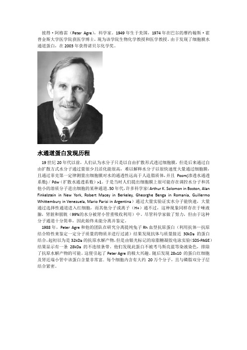
彼得·阿格雷(Peter Agre),科学家。
1949年生于美国,1974年在巴尔的摩约翰斯·霍普金斯大学医学院获医学博士,现为该学院生物化学教授和医学教授。
由于发现了细胞膜水通道蛋白,在2003年获得诺贝尔化学奖。
水通道蛋白发现历程19世纪20年代以前,人们认为水分子只是以自由扩散形式透过细胞膜,但是后来通过自由扩散方式水分子通过量很少且活化能很高,难以解释水分子以很快速度大量通过细胞膜,且通过菲克第一定律测量出细胞膜对水的通透性远高于人造脂质体,并且Posm(渗透水通透系数)/Pdw(扩散水通透系数)>1。
于是当时人们提出细胞膜上很可能存在调控水分子和其他小的溶质分子进出细胞的某种通道。
50年代,许多科学家(Arthur K. Solomon in Boston, Alan Finkelstein in New York, Robert Macey in Berkeley, Gheorghe Benga in Romania, Guillermo Whittembury in Venezuela, Mario Parisi in Argentina)通过大量实验证实水分子能快速,大量通过选择性通道进入红细胞,而其他分子或离子(H+)通不过,这种现象同样存在于唾液腺,肾脏和膀胱(99%的水分被肾小管重吸收利用)中。
尽管科学家做了努力,但由于这种分子通道十分简单,因此始终未能分离并鉴定。
1988年,Peter Agre和他的团队在研究分离提纯兔子Rh血型抗原蛋白(利用抗体—抗原结合特性来鉴定一定分子质量的物质并进行过滤)结果发现抗体与质量接近30kDa的蛋白结合,起初以为是32kDa的抗原水解产物,但是由银光标记的琼脂糖凝胶电泳实验(SDS-PAGE)结果显示有一条28kDa的不连续条带。
他们发现此蛋白不被考马斯亮蓝等染液染色,排除了抗原水解产物的可能。
这便引起了Peter Agre的极大兴趣。
视神经脊髓炎相关抗体
视神经脊髓炎相关抗体范欣;冯丽莎【摘要】视神经脊髓炎是主要累及视神经和脊髓的自身免疫性中枢神经系统疾病,水通道蛋白4是主要靶抗原,其特异性抗体NMO-IgG阳性患者以女性多见,临床症状较重,同时出现双侧视神经炎或视神经炎和脊髓炎,受累脊髓节段较长.而在NMO-IgG阴性患者血清中可以检出抗水通道蛋白1(AQP1)抗体和抗髓鞘少突胶质细胞糖蛋白(MOG)抗体,抗AQP1抗体阳性患者女性少见,长节段脊髓病变多见,视神经炎少见;抗MOG抗体阳性患者男性多见,视神经炎多见,尤其是双侧视神经同时受累,胸腰髓受累多见.【期刊名称】《中国现代神经疾病杂志》【年(卷),期】2016(016)010【总页数】5页(P660-664)【关键词】视神经脊髓炎;水通道蛋白质4;水通道蛋白质1;髓鞘;少突神经胶质;糖蛋白类;综述【作者】范欣;冯丽莎【作者单位】300120 天津市中医药研究院附属医院神经内科;300120 天津市中医药研究院附属医院神经内科【正文语种】中文视神经脊髓炎(NMO)是临床少见的中枢神经系统疾病,由Devic于1894年率先描述[1],主要影响视神经和脊髓,病程中可复发,复发时不完全缓解,可出现病残积累,导致失明和截瘫,因此,很长一段时间内被认为是多发性硬化(MS)的亚型。
近年来,随着临床、影像学、神经病理学研究的深入,特别是特异性抗体NMO⁃IgG(75%~90%视神经脊髓炎患者呈阳性)的发现[2⁃3],证实视神经脊髓炎是有别于多发性硬化的自身免疫性中枢神经系统疾病。
这种免疫反应的主要目标抗原是水通道蛋白4(AQP4)[3]。
此后,陆续在NMO⁃IgG阴性的视神经脊髓炎谱系疾病(NMOSDs)患者血清中发现抗水通道蛋白1(AQP1)抗体和抗髓鞘少突胶质细胞糖蛋白(MOG)抗体,其临床特征有别于NMO⁃IgG阳性患者,但发病机制尚不明确。
本文拟对近年新发现的视神经脊髓炎相关抗体进行综述,总结不同抗体在疾病发生与发展中的作用和临床意义,以为视神经脊髓炎谱系疾病的临床诊断与治疗提供帮助。
水通道蛋白-1及其在心脏中作用的研究进展

水通道蛋白-1及其在心脏中作用的研究进展闫玉梅;梅举;孙锟;丁芳宝【摘要】Aquaporin-1 (AQP1), which has a special molecular structure and is regulated by many factors, is the earliest found and most widely distributed aquaporin (AQP).In addition to transporting water, AQP1 may participate in a variety of gas transportation and be involved in cell metastasis process.In heart tissues, AQP1 is mainly expressed in erythrocytes,capillary endothelial cells and myocardial cells.AQP1 is located in cytoplasma membrane of myocardial cells, and may participate in the process of excitation-contraction coupling and transportation of water, which regulates water metabolism in various physiological and pathological process.Cardiac surgery with extracorporeal circulation can influence the expression and activity of AQPs, leading to cardiac edema.The investigations of AQPs have important guiding effects in clinical practice.%水通道蛋白-1(AQP1)是水通道蛋白(AQPs)家族中发现最早且分布最广泛的成员,具有特殊的分子结构,受多种因素的调节.除转运水分子外,AQP1还能转运多种气体分子并参与细胞游走过程.心脏组织中AQP1主要在红细胞、毛细血管内皮细胞以及心肌细胞中表达.心肌细胞内AQP1定位于细胞质膜,可能参加兴奋-收缩偶联过程以及水分子的转运,调节心脏的各种生理和病理过程的水代谢.体外循环心脏手术可以影响AQPs的表达及活性,导致术后心肌水肿.心脏中AQPs的研究对临床工作有重要的指导意义.【期刊名称】《上海交通大学学报(医学版)》【年(卷),期】2011(031)001【总页数】5页(P99-103)【关键词】水通道蛋白;心肌水肿;调节;体外循环【作者】闫玉梅;梅举;孙锟;丁芳宝【作者单位】上海交通大学医学院附属上海儿童医学中心心内科,上海,200127;上海交通大学医学院附属新华医院心胸外科,上海,200092;上海交通大学医学院附属上海儿童医学中心心内科,上海,200127;上海交通大学医学院附属新华医院心胸外科,上海,200092【正文语种】中文【中图分类】R654.2水是构成动植物体最主要的成分,在人体约占总体质量的60%,其代谢转运对生命活动非常重要。
最新AQP1抑制剂4
A Q P1抑制剂4关于乙酰唑胺与碳酸氢钠联合是否影响肿瘤的生长、转移及水通道蛋白的表达摘要:这项研究目的在于探索乙酰唑胺影响肿瘤的生长、转移以及可能的机制。
使用感染了Lewis肺癌的大鼠作为动物模型。
相比于联合使用碳酸氢钠是否对于肿瘤的生长转移以及碳酸脱苷酶的活动,使用咪唑影响肺癌的影响。
同时使用Western-blot技术和免疫组化技术研究水通道蛋白1在肿瘤组织中的可能发挥的作用。
结果显示单独使用乙酰唑胺可以大量降低肺癌的转移和原发肿瘤的生长,并且是一个独立影响发挥作用的因素。
乙酰唑胺可以极大的抑制碳酸脱氧苷酶在肿瘤组织中的活动。
在附加了碳酸氢钠之后,乙酰唑胺对肿瘤生长、转移的数量、以及原始肿瘤中碳酸酐酶发挥的作用并没有被改变,但是在肺组织中转移的抑制率以及在肺组织碳酸苷酶的活动似乎产生了一个明显的逆转,于单独使用乙酰唑胺相比。
明确的作用机制需要在未来被一步一步证明。
Wwstern-blot 技术和免疫组化技术同时显示水通道蛋白1在肿瘤组织中的表达明显高于正常组织和单独接受过乙酰唑胺治疗的肿瘤组织。
乙酰唑胺与碳酸氢钠结合使用并不能明显降低水通道蛋白的表达。
结果显示乙酰唑胺的抗肿瘤机制,其中尤其是对抗转移的作用可能一部分包含了抑制碳酸酐酶的活动或者降低了水通道蛋白中水通道蛋白的表达,无论联合碳酸氢钠与否。
简介:肿瘤转移是肿瘤的一大特点,并且是死亡的直接原因,碳酸托苷酶(CA)是锌结合金属蛋白的一个家族,这一家族可以促进二氧化碳可逆的水合作用,产生氢离子和碳酸氢根离子,并诱导ph值下降,一些碳酸托苷酶同工酶被显著的发现仅仅在肿瘤细胞中表达,这些CA同工酶通过细胞膜在邻近细胞中通过感应PH值和翻到质子和碳酸氢盐方面产生了一个重大的作用。
研究已经显示酸性PH可以增加肿瘤细胞的侵袭性。
乙酰唑胺是磺胺类的一种,被当做CA抑制剂。
T等报道CA抑制剂,作为化疗原则的一部分可以增加化疗药物的作用并且有助于延缓肿瘤进展,一些新型CA抑制剂,在试验中被当做抑制肿瘤细胞生长的有效成分,例如白血病、非小细胞肺癌、黑色素瘤、卵巢、肾、前列腺、乳腺细胞系列肿瘤。
最新型局部碳酸酐酶抑制剂派立明的临床前及临床研究
最新型局部碳酸酐酶抑制剂派立明的临床前及临床研究徐岩;庞广仁;陈祖基【期刊名称】《中华实验眼科杂志》【年(卷),期】2002(020)006【摘要】派立明是一种最新型的局部碳酸酐酶抑制剂.此药可选择性、高亲和力及明显地抑制碳酸酐酶同功酶Ⅱ的活性,有效地降低眼压.本品滴眼后可快速进入眼组织,在虹膜、睫状体、脉络膜、视网膜、晶状体和血液中有较长的半衰期(数天).虽然用派立明滴眼后,可在全血中测出药物浓度,提示该药可全身吸收,但主药和其代谢产物的血浆浓度非常低,在稳定状态下药物与红细胞内碳酸酐酶的结合达不到完全饱和.因此,不会出现全身酸中毒或其他与口服碳酸酐酶抑制剂有关的副作用.对兔眼滴用派立明还可增加视乳头血流量,而对全身酸碱平衡的影响极小.如这一作用在人眼被证实,将对有视神经病变的青光眼患者十分有益.1%派立明每日滴眼2次的降眼压效果最好,且患者的耐受性较多佐胺好,这可提高患者长期用药的依从性.滴眼后最常见的副作用是视物模糊(6%)及口苦、口酸等味觉异常(6%).总之,派立明的降眼压作用强,副作用小,滴眼舒适,患者耐受性好,是一种非常有价值的抗青光眼新药.【总页数】5页(P560-564)【作者】徐岩;庞广仁;陈祖基【作者单位】450003,郑州,河南省眼科研究所;450003,郑州,河南省眼科研究所;450003,郑州,河南省眼科研究所【正文语种】中文【中图分类】R988.1【相关文献】1.碳酸酐酶抑制剂的局部应用对大鼠角膜新生血管形成过程中水通道蛋白1表达的影响 [J], 张洁;李立2.碳酸酐酶抑制剂的局部应用对大鼠角膜新生血管形成过程中水通道蛋白1表达的影响 [J], 张洁;李立3.局部碳酸酐酶抑制剂对睫状体非色素上皮细胞AQP1表达的影响 [J], 李嘉文;李平华;张黎4.局部应用碳酸酐酶抑制剂治疗青光眼的前景 [J], 张延斌5.拉坦前列素、溴莫尼定、噻吗心安和局部碳酸酐酶抑制剂的混合物对青光眼及高眼压症患者24h眼压的影响 [J], NicolaOrzalesi,MD LucaRossetti,MD AndreaBottoli,MD ElenaFumagalli,MD PaoloFogagnolo,MD 张钰因版权原因,仅展示原文概要,查看原文内容请购买。
- 1、下载文档前请自行甄别文档内容的完整性,平台不提供额外的编辑、内容补充、找答案等附加服务。
- 2、"仅部分预览"的文档,不可在线预览部分如存在完整性等问题,可反馈申请退款(可完整预览的文档不适用该条件!)。
- 3、如文档侵犯您的权益,请联系客服反馈,我们会尽快为您处理(人工客服工作时间:9:00-18:30)。
关于乙酰唑胺与碳酸氢钠联合是否影响肿瘤的生长、转移及水通道蛋白的表达
摘要:这项研究目的在于探索乙酰唑胺影响肿瘤的生长、转移以及可能的机制。
使用感染了Lewis肺癌的大鼠作为动物模型。
相比于联合使用碳酸氢钠是否对于肿瘤的生长转移以及碳酸脱苷酶的活动,使用咪唑影响肺癌的影响。
同时使用Western-blot技术和免疫组化技术研究水通道蛋白1在肿瘤组织中的可能发挥的作用。
结果显示单独使用乙酰唑胺可以大量降低肺癌的转移和原发肿瘤的生长,并且是一个独立影响发挥作用的因素。
乙酰唑胺可以极大的抑制碳酸脱氧苷酶在肿瘤组织中的活动。
在附加了碳酸氢钠之后,乙酰唑胺对肿瘤生长、转移的数量、以及原始肿瘤中碳酸酐酶发挥的作用并没有被改变,但是在肺组织中转移的抑制率以及在肺组织碳酸苷酶的活动似乎产生了一个明显的逆转,于单独使用乙酰唑胺相比。
明确的作用机制需要在未来被一步一步证明。
Wwstern-blot技术和免疫组化技术同时显示水通道蛋白1在肿瘤组织中的表达明显高于正常组织和单独接受过乙酰唑胺治疗的肿瘤组织。
乙酰唑胺与碳酸氢钠结合使用并不能明显降低水通道蛋白的表达。
结果显示乙酰唑胺的抗肿瘤机制,其中尤其是对抗转移的作用可能一部分包含了抑制碳酸酐酶的活动或者降低了水通道蛋白中水通道蛋白的表达,无论联合碳酸氢钠与否。
简介:
肿瘤转移是肿瘤的一大特点,并且是死亡的直接原因,碳酸托苷酶(CA)是锌结合金属蛋白的一个家族,这一家族可以促进二氧化碳
可逆的水合作用,产生氢离子和碳酸氢根离子,并诱导ph值下降,一些碳酸托苷酶同工酶被显著的发现仅仅在肿瘤细胞中表达,这些CA同工酶通过细胞膜在邻近细胞中通过感应PH值和翻到质子和碳酸氢盐方面产生了一个重大的作用。
研究已经显示酸性PH可以增加肿瘤细胞的侵袭性。
乙酰唑胺是磺胺类的一种,被当做CA抑制剂。
T等报道CA抑制剂,作为化疗原则的一部分可以增加化疗药物的作用并且有助于延缓肿瘤进展,一些新型CA抑制剂,在试验中被当做抑制肿瘤细胞生长的有效成分,例如白血病、非小细胞肺癌、黑色素瘤、卵巢、肾、前列腺、乳腺细胞系列肿瘤。
尽管存在一些CA 抑制因子作用机制。
人们依旧认为在直接抑制肿瘤相关CA同工酶后,导致了酸化肿瘤细胞外环境的抑制作用,这一切似乎与之前描述的理论相矛盾。
因此,我们假设一定存在通过CA抑制因子抑制肿瘤转移的其他途径。
水通道蛋白,是一种作用于高选择性水通道膜蛋白的大家族,目前研究强调了他们在生物学上的作用,以及人类许多种疾病中,包含快速水通道的疾病,并且已经成为了介入治疗的目标靶向蛋白。
AQP1是第一种水特点通道蛋白,广泛存在于红细胞膜、肾输尿管、脉络膜、眼睛、肺、血管内皮、肝管上皮细胞以及其他肿瘤细胞。
许多肿瘤显示出极高的血管通透性和高的间隙渗透性,但是肿瘤中的水运输通路仍旧未知。
在之前已经出版的关于AQP1以及CA研究报道中,尤其是CA2和CA4,我们发现他们具有共同的生物学特点。
例如,它们均在液体通
道组织中广泛存在,包含肾脏、肺脏、大脑、眼睛、红细胞、胰腺以及一些腺体,它们均具有促进再吸收和分泌水的功能。
此外,AQP1具有转运CO2的能力,二氧化碳是催化形成碳酸氢根离子的CA的基质,即具有碳酸酐酶和水通道蛋白二者相互共有的生物学特点。
作为一种碳酸苷酶抑制剂,乙酰唑胺主要用来治疗一些水肿性质的疾病,例如青光眼、登山病、充血性心力衰竭、药物诱发的水肿、以及纠正新陈代谢产生的碱中毒。
P等已经显示单独使用乙酰唑胺可以抑制肿瘤细胞的潜在侵袭性作用,但是目前机制并没有被澄清。
我们的实验也已经显示乙酰唑胺可以抑制AQP1在小鼠肾脏和睾丸中基因表达作用,在其中我们已经使用卵母细胞建立了一个水通道蛋白表达系统用来研究乙酰唑胺对于AQP1对于水通道功能的作用。
并且已经证实乙酰唑胺是AQP1的一种直接抑制剂。
因此我们假设乙酰唑胺关于AQP1基因表达作用和水通道作用的减少也许可以促进他们关于肿瘤生长和转移的影响作用。
在此项研究中,我们进一步评估了在肿瘤组织中AQP1的表达和它与碳酸酐酶活动的联系,并且进一步探索了它在肿瘤转移中的可能机制。
众所周知,CA抑制剂最大的副作用就是新陈代谢引起的酸中毒,在长时间的使用之后。
为了纠正细胞外的酸化环境,动物模型被用来同时联合使用碳酸氢男使用纠正酸中毒。
方法:
2.1使用Lewis肺癌细胞用作实验模型
成年动物重18-20克雌性大鼠购于北京大学实验中心,Lewis肺
癌细胞有中国医学科学院提供,通过皮下注射均匀的将肿瘤组织注入体内,这些肿瘤组织由肺癌患者志愿者提供。
2.2药物治疗和组织收集。
2.3碳酸酐酶活动实验
2.4Western blot印记分析
2.5免于组织化学技术
2.6统计学分析
结果:
3.1 乙酰唑胺联合使用碳酸氢钠与否对于原发肿瘤生长以及自发形成肺转移的影响
3.2乙酰唑胺联合使用碳酸氢钠与否对于碳脱水酶活动的影响
3.2AQP1蛋白表达的影响
3.4AQP1在肿瘤组织中的定位和表达的影响
讨论:
目前已经报道一些新型的磺胺类衍生物包含强有力的CA抑制内容,这就在试管中显示了可以有效抑制肿瘤细胞的生长和转移。
在目前和之前的研究中,我们认为乙酰唑胺可以大量的减少肺部原发肿瘤生长和转移的几率,这似乎是一个剂量依赖型的方式。
在肺癌中抑制转移的几率是77.7%。
此外,我们注意到乙酰唑胺可以大量抑制肿瘤组织和肺组织中CA的活动率。
在增加了碳酸氢钠之后,乙酰唑胺关于肿瘤生长、转移和CA的活动并没有增加。
因此,乙酰唑胺关于许多肿瘤相关同工酶的抑制作用可能是这种机制中的一种,这类机制具
有抑制肿瘤转移的作用。
正如我们所知,当乙酰唑胺被用来当做化疗药物的一种,一个显著的副作用就体现了出来,那就是代谢性酸中毒,而且在实质性肿瘤细胞外的PH值是更加偏酸的比那些邻近的正常组织。
细胞外偏酸的细胞内环境更有利于肿瘤的生长和转移。
因此,中和代谢性酸中毒有助于抗肿瘤生长和转移的机制。
然而我们的研究结果并没有显示碳酸氢钠对乙酰唑胺的作用起到促进作用。
这个结果就迫使我们扩增了一个更加广泛的评价酸中毒的研究机制,从而执行CA抑制因子关于肿瘤的抗肿瘤活动,从而进一步导致了其他有关乙酰唑胺作用机制的出现。
在本实验中,蛋白免疫印迹分析结果显示AQP1蛋白在肺组织和感染肿瘤细胞的大鼠组织中表达远高于正常组织。
乙酰唑胺大量抑制水通道蛋白的作用并且沉溺于碳酸氢钠对于乙酰唑胺的增敏作用。
AQP1在大鼠肺组织中的免疫组化定位于毛细血管、毛细血管后静脉和细支气管基膜。
免疫标记显示了水通道蛋白在感染Lewis肺癌大鼠中的强阳性作用但是在接受乙酰唑胺的动物模型中结果为阴性。
这项研究显示肿瘤生长可能有更高的液体渗透性因为AQP1在肿瘤中的作用是增加细胞膜水通透性。